
Submitted:
16 November 2026
Posted:
18 November 2026
You are already at the latest version
Abstract
Human papillomavirus (HPV)-associated oropharyngeal squamous cell carcinoma (OPSCC) has become the dominant form of oropharyngeal cancer in many high-income countries and is now recognised as a biologically and clinically distinct entity with markedly better survival than its tobacco-related counterpart. Beyond the canonical inactivation of p53 and pRb by the viral oncoproteins E6 and E7, HPV profoundly remodels the host and viral epigenomes. HPV-positive tumours display a hypermethylated landscape, including a CpG island methylator-like phenotype that targets Polycomb repressive complex 2 genes, alongside altered histone-modifying activity, chromatin remodelling and deregulated non-coding RNA networks. Methylation of the viral genome itself, particularly of the upstream regulatory region and E2 binding sites, tracks with integration and oncogene expression. These changes shape the tumour immune microenvironment and treatment response. They also leave traces in the circulation: circulating tumour HPV DNA, methylated cell-free DNA and extracellular-vesicle microRNAs are emerging as tools for diagnosis, risk stratification and post-treatment surveillance. This review summarises current knowledge of HPV-driven epi-genetic reprogramming in OPSCC, evaluates the analytical and clinical validity of epigenetic and viral liquid-biopsy biomarkers, and discusses their translation into treatment de-escalation, epigenetic therapy, immunotherapy com-binations and nutri-epigenetic modulation.
Keywords:
human papillomavirus
; oropharyngeal squamous cell carcinoma
; DNA methylation
; histone modification
; non-coding RNA
; liquid biopsy
; circulating tumour HPV DNA
; epigenetic therapy
; biomarkers
1. Introduction
The epidemiology of oropharyngeal squamous cell carcinoma (OPSCC) has changed fundamentally over the past four decades. In a United States tumour series, the proportion of OPSCC testing HPV-positive rose from 16.3% in 1984–1989 to 71.7% in 2000–2004, while over the same period the population-level incidence of HPV-positive OPSCC increased by 225% and that of HPV-negative disease fell by 50% [1]. A worldwide systematic review of 9,541 OPSCC specimens from 23 countries confirmed a significant rise since the mid-1990s, with the HPV-positive fraction plateauing at around 65% in North America and approaching 50% in Europe [2]. In a more recent SEER-based analysis, HPV-positivity among OPSCC rose from 66.4% in 2010 to 79.3% in 2016, concentrated in tonsillar and base-of-tongue primaries [3].
Two distinctions should be kept in view when these figures are compared. The first is between the proportion of tumours that test HPV-positive and the incidence of HPV-positive disease in a population: the two move independently, and a rising proportion reflects both a genuine increase in HPV-driven tumours and the concurrent decline in tobacco-related disease [1]. The second is that HPV-positivity is not ascertained identically across studies. Detection of HPV DNA, p16 immunohistochemistry and demonstration of E6/E7 transcription define overlapping but non-identical case populations, and p16 in particular is a surrogate for HPV-driven disease rather than a genotype-specific measure of it. Proportions drawn from different periods, regions and assays are therefore not strictly interchangeable, and the series above should be read as a consistent direction of change rather than as a single continuous trend.
HPV-positive OPSCC is not simply a variant of tobacco-related head and neck squamous cell carcinoma (HNSCC). In the landmark RTOG 0129 analysis, patients with HPV-positive tumours had a 3-year overall survival of 82.4% versus 57.1% for HPV-negative tumours, and a 58% reduction in the risk of death after adjustment for clinical covariates [4]. The Cancer Genome Atlas (TCGA) later showed that HPV-positive HNSCCs carry few TP53 mutations, frequent PIK3CA alterations and a distinct copy-number profile, consolidating the view of a separate molecular entity [5].
These differences extend to the epigenome. Genome-wide methylation profiling has shown that HPV-positive HNSCCs segregate from HPV-negative tumours, that 87% of methylation-variable positions are hypermethylated in HPV-positive disease, and that a subgroup displays a candidate CpG island methylator phenotype (CIMP) affecting Polycomb repressive complex 2 (PRC2) targets such as cadherin genes [6]. Ectopic expression of E6 and E7 in HPV-negative cells partially reproduces this signature, identifying E6 as the main viral effector [6]. Methylation-based classification of 528 TCGA HNSCCs further resolved an HPV-positive subtype distinct from smoking-related and atypical subtypes [7]. Collectively, these findings support an active role for epigenetic reprogramming in HPV-associated carcinogenesis, although the functional contribution of individual epigenetic alterations remains incompletely defined.
The clinical relevance of this biology is now pressing. The favourable prognosis of HPV-positive OPSCC motivated treatment de-escalation, yet two pivotal phase 3 trials replacing cisplatin with cetuximab showed inferior tumour control and survival [8,9]. These results exposed the limits of HPV status as a sole stratifier and the need for biomarkers that capture biological heterogeneity within HPV-positive disease. At the same time, liquid biopsy has matured: in a prospective cohort of 115 patients, two consecutive positive plasma circulating tumour HPV DNA (ctHPVDNA) tests had a positive predictive value of 94%, and negative tests a negative predictive value of 100%, for recurrence after chemoradiotherapy [10]. Epigenetic marks, which are stable, tissue-specific and detectable in cell-free DNA, are natural candidates to complement viral DNA in such assays.
This review integrates these strands. We first outline HPV biology in the oropharynx (Section 2), then examine HPV-driven reprogramming of the host epigenome (Section 3) and of the viral genome itself (Section 4), and its consequences for the tumour immune microenvironment (Section 5). We next evaluate circulating viral and epigenetic biomarkers (Section 6) and their translation into prognostication, de-escalation, epigenetic and immune therapy, and diet-derived epigenetic modulation (Section 7). We close with the main unresolved questions and priorities for clinical implementation (Section 8).
2. HPV Biology and the Oncogenic Programme in the Oropharynx
2.1. Tissue Tropism and the Tonsillar Crypt Niche
HPV-driven head and neck carcinogenesis shows a marked anatomical predilection for the oropharynx, particularly the palatine and lingual tonsils, rather than an absolute restriction to these sites; HPV-driven tumours also arise, less commonly, elsewhere in the head and neck (Section 5.1). HPV-positive head and neck cancers arise in the tonsil crypts, deep invaginations at the tonsil surface that are lined by reticulated epithelium infiltrated by immune cells [11,12]. This architecture places keratinocytes in intimate contact with lymphoid tissue, and a genital-oral route of infection is considered the most likely mode of acquisition [11]. The natural history of HPV-driven oropharyngeal pathogenesis nevertheless remains poorly defined and appears to differ from that of cervical disease: it is not established whether the virus initiates a productive or an abortive infection in tonsil crypt keratinocytes, or whether infection progresses to carcinoma through a neoplastic phase as it does in the cervix [11]. The relevance of this niche is reflected in clinical data: in the SEER cohort, HPV positivity was significantly more frequent in tumours of the tonsil and base of tongue than in those of the posterior pharyngeal wall or soft palate [3].
2.2. E6 and E7: The Canonical Oncogenic Axis
High-risk HPV types transform cells through the constitutive expression of two oncoproteins. HPV16 dominates in the oropharynx to a greater extent than in the cervix: among HPV-positive tumours, HPV16 accounts for over 80% of OPSCC compared with approximately 60% of cervical cancers, whereas HPV18 accounts for under 2% of HPV-positive OPSCC against 10% of HPV-positive cervical cancers, and HPV33 is the second most prevalent high-risk type in OPSCC at 3.3% of HPV-positive cases [11]. E6 directs the ubiquitin-mediated proteasomal degradation of p53, abolishing the p53 transcriptional response to genotoxic and oncogenic stress, while E7 inactivates the retinoblastoma protein (pRB), releasing E2F-dependent S-phase entry. For E6, degradation rather than stoichiometric sequestration is the principal route of p53 inactivation. For E7, degradation of pRB contributes but is not the whole mechanism: E7 also binds pRB directly and displaces it from E2F, and it recruits histone-modifying and chromatin-remodelling activities (Section 3.3), so inactivation of the pRB pathway is multifactorial. Loss of pRB function de-represses CDKN2A, producing the p16INK4A overexpression that underpins the p16 immunohistochemistry used as a surrogate diagnostic marker in routine practice.
The genomic consequences of this axis are visible in large-scale sequencing data. HPV-positive HNSCCs carry few TP53 mutations, frequent PIK3CA alterations and a copy-number profile distinct from tobacco-related tumours [5]. Critically, however, E6 and E7 expression is necessary but not sufficient for malignant transformation, and additional secondary events, genetic and epigenetic, are required [13]. This insufficiency is the conceptual entry point for the epigenetic mechanisms considered in the remainder of this review, since several of the activities of E6 and E7 are exerted directly on the chromatin-modifying machinery rather than on cell-cycle regulators alone.
2.3. Integrated and Episomal Viral States
After infection, the HPV genome is maintained as a circular extrachromosomal episome, and integration into host chromosomes has traditionally been regarded as a critical step in progression to invasive cancer [14]. Whole-genome sequencing of 105 newly diagnosed HPV-positive OPSCCs has refined this view. HPV16 was present in 92 tumours (88%), and 874 virus–host breakpoints were identified in 81 tumours (77%); no breakpoints were detected in the remaining 24 tumours (23%), consistent with exclusively episomal virus [13]. HPV integration is therefore frequent but not obligatory in OPSCC. Integrated and episomal viral genomes may coexist within individual tumours, and the relative abundance of each physical state depends on tumour biology and on the analytical methods employed [11]. The detection of virus–host breakpoints in 77% of tumours establishes how often integration has occurred, not what proportion of the viral DNA within a tumour is integrated; these are different quantities and require different measurements. A third configuration has also been proposed in OPSCC: a circular, extrachromosomal HPV-human DNA hybrid episome in which the viral genome is present as a multimer, with some copies carrying viral deletions and host sequences [11].
Where integration does occur, its consequences are structural and far-reaching. Integrants recurrently flank or bridge focal regions of host genomic instability, including copy-number and structural variation, and in 95% of tumours with integration host gene transcription is disrupted through intragenic integrants, chimeric transcripts, outlier expression, gene breaking or de novo expression of non-coding or imprinted genes [13]. Five statistically significant integration hotspots were located near genes regulating epithelial stem cell maintenance [13]. Earlier TCGA analyses similarly showed enrichment of integration events in genic regions, with amplification of oncogenes and disruption of tumour suppressors [14].
Two caveats matter for the epigenetic argument developed below. First, disruption of the viral E2 gene, long used as an indirect indicator of integration, is a poor proxy for integration status as determined by sequencing [13]. Second, and more directly relevant here, tumours with integrated versus non-integrated HPV display different patterns of DNA methylation as well as differing human and viral gene expression [14]. The physical state of the viral genome and the epigenetic state of both genomes are thus interdependent, a relationship examined in Section 3 and Section 4. Notably, the same whole-genome study found no significant association between integration status and clinical outcome [13], which argues against using integration alone as a prognostic variable and strengthens the case for biomarkers that capture the downstream regulatory consequences instead.
3. Epigenetic Reprogramming of the Host Genome
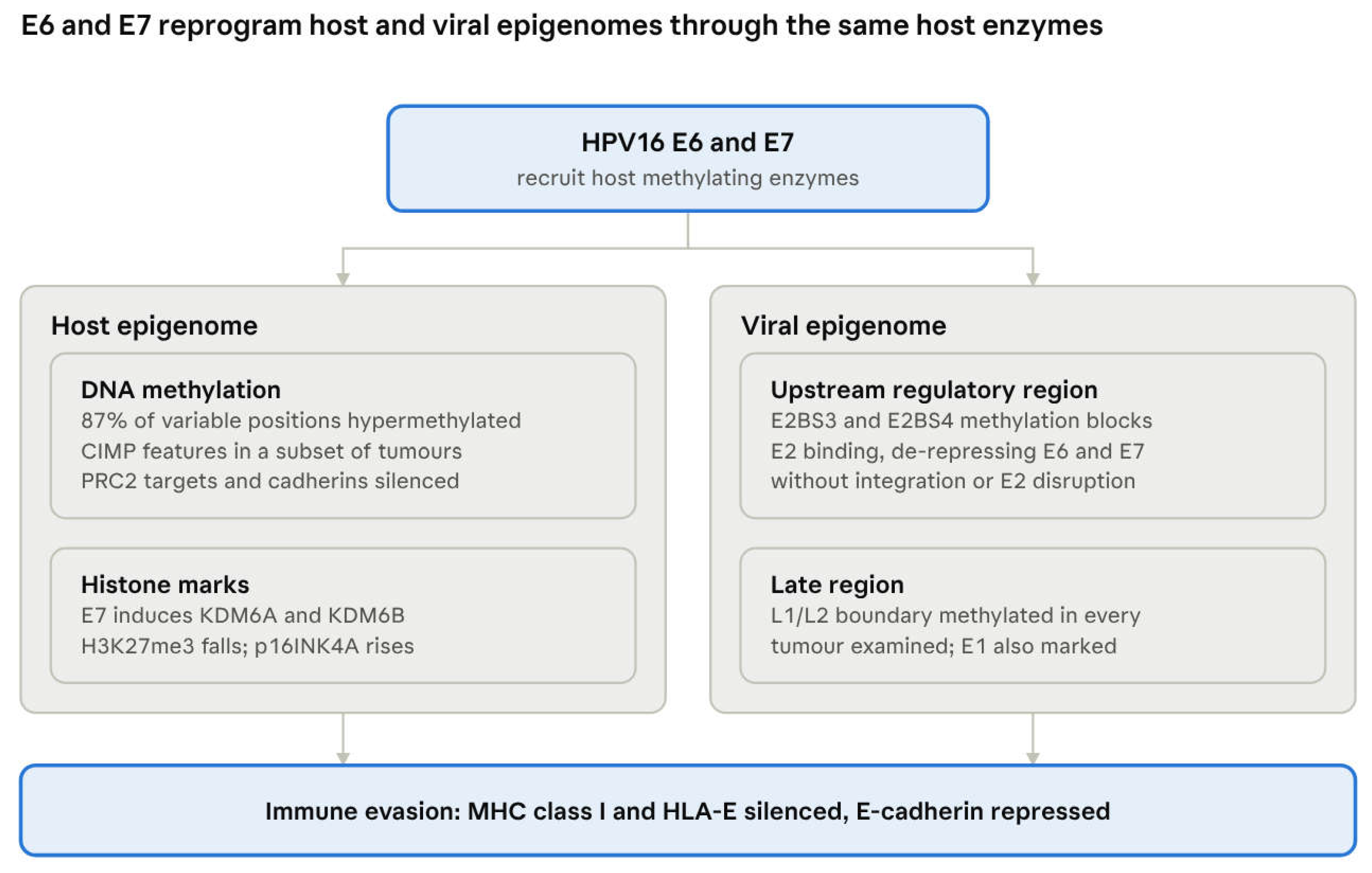
The virus encodes no methyltransferase of its own, so a single host enzymatic apparatus acts on both genomes: the DNMT activity that hypermethylates host promoters also methylates the viral regulatory region.
Figure 1.
HPV-driven epigenetic reprogramming of the host and viral genomes.
3.1. A Hypermethylated Landscape
The most consistently reproduced epigenetic feature of HPV-positive HNSCC is global promoter hypermethylation. Genome-wide profiling of laser-capture microdissected tumours using Infinium 450k arrays showed that samples segregate by HPV status, and that 87% of methylation-variable positions (MVPs) are hypermethylated in HPV-positive disease [6]. MVPs were significantly enriched at transcriptional start sites and grouping them into differentially methylated regions identified 43 hypermethylated promoter regions, including three cadherin genes that are Polycomb group targets [6]. Integration with independent expression data showed strong negative correlation between promoter methylation and transcript level, most markedly for the cadherin family [6].
Two observations give this signature mechanistic and taxonomic weight. First, a candidate CpG island methylator phenotype (CIMP) was identified in a subgroup of HPV-positive tumours, indicating that hypermethylation is not uniformly distributed across HPV-positive disease but concentrated in a distinct subset [6]. Second, meta-analysis against cervical and lung cancer methylation data confirmed the signature to be HPV-specific and tissue-independent [6], arguing that the virus, rather than the anatomical site, drives it.
Unsupervised methylation-based clustering of 528 TCGA HNSCC patients subsequently resolved five subtypes: one HPV-positive, two smoking-related and two etiologically atypical [7]. One atypical subtype was genomically stable but showed widespread CIMP-associated gene silencing, together with an antiviral expression profile linked to M1 macrophages and CD8+ T cell infiltration, CASP8 mutations and SOX2OT hypermethylation [7]. The existence of a CIMP-high subtype outside the HPV-positive cluster is important for biomarker design, since methylation signatures intended to report HPV-driven biology must be distinguished from CIMP arising by other routes.
3.2. Viral Oncoproteins as Direct Effectors of the Methylation Machinery
The hypermethylated phenotype is causally linked to viral oncogene expression rather than merely associated with it. Combinatorial ectopic expression of E6 and E7 in an HPV-negative HNSCC cell line partially phenocopied the hypermethylation signature observed in HPV-positive tumours, and established E6 as the principal viral effector gene in that system [6].
A parallel and more biochemically defined route operates through E7. HPV16 E7 associates with DNA methyltransferase 1 (DNMT1) both in vitro and in vivo, precipitates DNA methyltransferase activity from nuclear extracts, and stimulates DNMT1 enzymatic activity; the interaction is direct and maps to the zinc-finger CR3 region of E7 [15]. This gives the virus a mechanism for redirecting maintenance methylation, the process that restores methylation patterns on newly replicated DNA. A functional consequence has been demonstrated for E-cadherin: HPV16 E7 reduces E-cadherin expression through augmented DNMT1 activity, independently of pRB or AP-2α degradation, and pharmacological DNMT inhibition re-establishes E-cadherin levels [16]. Because Langerhans cells depend on E-cadherin to traffic through stratified epithelium, this single epigenetic event offers a candidate link between viral oncoprotein expression and impaired local immune surveillance [16], a theme developed, with its limitations, in Section 5.3. It also converges with the array data, in which cadherin promoters were among the most prominently hypermethylated and transcriptionally silenced loci [6].
3.3. Histone Modifications and Chromatin Remodelling
3.3.1. Polycomb, EZH2 and the H3K27me3 Paradox
HPV also remodels the histone landscape, and here E7 is the dominant effector. Repressive H3K27me3 marks, which are required for binding of Polycomb repressive complexes, are decreased in HPV16 E7-expressing cells and in HPV16-positive lesions [17]. The mechanism is transcriptional induction of the two H3K27-specific demethylases KDM6A (UTX) and KDM6B (JMJD3), which erase the mark written by the EZH2 subunit of PRC2; KDM6A- and KDM6B-responsive Homeobox genes are correspondingly expressed at higher levels, amounting to epigenetic reprogramming of the host epithelial cell [17]. These effects are independent of the ability of E7 to inhibit pRB and are reversed when E7 expression is silenced [17].
This pathway carries two translational implications. KDM6B induction accounts for the p16INK4A expression used diagnostically, which means the clinical surrogate for HPV status is itself the readout of a histone-demethylase event rather than a direct viral product. Furthermore, HPV E7-expressing cells become dependent on continued KDM6A and KDM6B expression for survival, creating a cellular vulnerability to small-molecule KDM6A/B inhibitors that does not exist in uninfected cells [17]. Together with the DNMT1 axis in Section 3.2, this establishes that the epigenetic machinery in HPV-positive OPSCC is not only deregulated but potentially druggable, a point taken up in Section 7.
The picture is complicated by evidence pointing the other way. EZH2, the catalytic subunit of PRC2 and the enzyme that writes H3K27me3, is overexpressed in HNSCC, where overexpression is associated with poor clinical prognosis and with aggressive HPV-positive phenotypes, and a global trend towards elevated H3K27me3 has been reported in the HPV-positive patient epigenome [18]. HPV contributes to this directly, since E7 interacts with the pathways responsible for EZH2 transcription and thereby upregulates it [18]. The pharmacological data are correspondingly mixed: of three EZH2 pathway inhibitors tested in two HPV-positive and two HPV-negative OPSCC cell lines, GSK-343 reduced H3K27me3 in all lines, DZNeP reduced it only in HPV-negative lines and EPZ-5687 produced no appreciable change, while only DZNeP showed antiproliferative activity and it decreased EGFR expression specifically in the HPV-positive lines [18].
Two literatures therefore coexist: one in which E7 drives H3K27me3 down through KDM6A and KDM6B, and one in which E7 drives EZH2 up and the mark with it. The most economical reconciliation is that they operate on different scales. KDM6-mediated demethylation is locus-directed, de-repressing a defined set of targets including CDKN2A and the Homeobox genes, whereas EZH2 overexpression raises the global mark and silences PRC2 target promoters elsewhere in the genome. Section 3.3.4 returns to this, because the resolution bears directly on how the two layers interact.
3.3.2. Histone Acetylation
Acetylation is the second arm of the histone programme, and, like methylation, it is engaged directly by the viral oncoproteins. The E7 proteins of HPV6, HPV16 and HPV18 interact with the pCAF acetyltransferase, binding its acetyltransferase domain and reducing its activity in vitro; mutation of a highly conserved leucine within the zinc-finger region of HPV16 E7 disrupts this binding and simultaneously impairs transformation and transcriptional activation [19]. That a single mutation abolishes both the interaction and the transforming capacity is the strongest available argument that the acetyltransferase contact is functionally required rather than incidental.
The therapeutic corollary has been pursued chiefly from the opposite direction. In vivo shRNA screening across HNSCC models, HPV-positive lines included, identified the histone acetyltransferases CREBBP and EP300 as determinants of radiation response: selected CREBBP mutations produce a hyperacetylated state with increased homologous recombination, and chemical inhibition of acetyltransferase function radiosensitises tumours carrying cognate mutations [20]. The effect is genotype-dependent rather than general, which is a caution against deploying acetylation-directed drugs based on HPV status alone.
3.3.3. Chromatin Accessibility and the Enhancer Landscape
The third level is the physical accessibility of chromatin, and the most striking finding here connects directly to viral integration (Section 2.3). The HPV genome carries a conserved CTCF binding site that is occupied by CTCF in HPV-positive cancer cell lines, so integration introduces a new CTCF site into the host genome. Changes in CTCF binding pattern and increases in chromatin accessibility occur exclusively within 100 kbp of integration sites, together with altered expression of neighbouring genes, and in TCGA HPV-positive tumours the genes upregulated at integration sites have significantly higher essentiality scores than randomly selected upregulated genes from the same tumours [21]. Integration of Epstein-Barr virus produced no comparable effect, which argues that the mechanism is specific to HPV rather than a general consequence of viral insertion [21]. Integration is therefore not only a genetic event that disrupts E2 but an architectural one that reorganises the chromatin around it.
At the level of regulatory elements, H3K27ac profiling of HPV-positive patient-derived xenografts, cell lines and normal oropharyngeal mucosa has mapped tumour-specific super-enhancer domains enriched for the transcription factors TP63, FOSL1 and JUND, with coordinated dysregulation of enhancer RNAs and messenger RNAs in their vicinity and upregulation of epithelial-mesenchymal transition and E2F target pathways; the bromodomain inhibitor JQ1 repressed these tumour-specific programmes [22]. Super-enhancer dependence is thus a candidate vulnerability in a tumour whose low mutational burden offers few conventional targets.
3.3.4. Crosstalk between DNA Methylation and Histone Modification
It is worth noting the apparent tension between the two arms of host reprogramming: HPV-positive tumours show widespread hypermethylation of PRC2 target promoters [6] while simultaneously showing reduced H3K27me3 and elevated H3K27 demethylase activity [17]. Reconciling these observations, most plausibly through a model in which DNA methylation substitutes for, and renders permanent, a silencing that was initially Polycomb-mediated, remains an open mechanistic question. A physical basis for that model exists, since EZH2 can influence CpG methylation through direct contact with DNA methyltransferases [18], providing a route by which a Polycomb-marked promoter is handed over to DNMT-mediated methylation and converted from a reversible to a heritable silent state. On that reading the global elevation of H3K27me3 reported in patient tumours (Section 3.3.1) and the hypermethylation of PRC2 targets are not contradictory but sequential, while the locus-specific loss of H3K27me3 driven by KDM6A and KDM6B affects a different and smaller set of genes. Testing this requires paired H3K27me3 and DNA methylation profiling on the same tumours, which has not been reported for HPV-positive OPSCC. Until it is, the field is describing two layers separately that it has reason to believe act as one.
3.4. Non-Coding RNAs
The third arm of host epigenetic reprogramming operates at the level of RNA. Unlike the methylation and histone changes discussed above, non-coding RNA deregulation in HPV-positive OPSCC has been characterised largely descriptively, and the literature is marked by limited reproducibility between cohorts. It is nonetheless the arm with the most direct route to a circulating biomarker, since microRNAs are stable in plasma and in extracellular vesicles (Section 6).
3.4.1. MicroRNAs
The causal dependence of the miRNA profile on viral oncogene expression was established in cell-line systems. In HPV16-positive SCCHN lines, miR-363, miR-33 and miR-497 were upregulated while miR-155, miR-181a, miR-181b, miR-29a, miR-218, miR-222, miR-221 and miR-142-5p were downregulated relative to both HPV-negative SCCHN lines and normal oral keratinocytes [23]. Expression of E6 in human foreskin keratinocytes was sufficient to alter miRNA levels, and siRNA knockdown of E6 in an HPV-positive line reduced miR-363, establishing E6 as an upstream determinant [23]. A functional consequence has since been defined: miR-363 is overexpressed in HPV16-positive relative to HPV-negative tumours, averaging 37.58 versus 11.17 reads per million in TCGA (a 3.36-fold increase), and it targets the 3'-UTR of myosin 1B (MYO1B), reducing cellular migration [24].
At the tissue level, comparative profiling of HPV-positive HNSCC, HPV-negative HNSCC and cervical squamous cell carcinoma showed that HPV-positive HNSCC has a distinct miRNA profile, and that it resembles cervical carcinoma more closely than it resembles HPV-negative HNSCC [25]. A set of core HPV-associated miRNAs was identified, of which the miR-15a/miR-16/miR-195/miR-497 family, miR-143/miR-145 and the miR-106-363 cluster appear most relevant to HPV pathogenesis [25]. This convergence across anatomical sites mirrors the tissue-independence of the methylation signature noted in Section 3.1, and strengthens the inference that the virus, not the epithelium, is the organising variable.
The largest dedicated OPC series profiled 365 miRNAs in 88 FFPE biopsies (56 p16-positive, 32 p16-negative) against seven normal tonsils [26]. Of the 224 miRNAs expressed in more than 80% of samples, 128 (57%) were differentially expressed between tumour and normal tissue, and upregulated miR-20b, miR-9 and miR-9* were significantly associated with HPV/p16 status [26]. Three candidate prognostic sets were identified, for overall survival (miR-107, miR-151, miR-492), disease-free survival and distant metastasis, and these retained significance after adjustment for p16 status [26]. That prognostic information survives adjustment for p16 is the key observation for clinical translation, since it implies miRNA profiles capture heterogeneity within HPV-positive disease that HPV status alone does not.
These findings must be read against a sobering methodological caveat. Whole-genome small-RNA sequencing of oral and oropharyngeal tumours, compared against the published literature, revealed extensive heterogeneity of miRNA deregulation between studies, with only a handful of miRNAs consistently affected and miR-9 emerging as the most likely genuinely HPV-related miRNA [27]. Differences in platform, specimen type, normalisation strategy and HPV ascertainment plausibly account for much of this divergence. No miRNA-based assay has yet reached the analytical validation required for clinical use in OPSCC.
3.4.2. Long Non-Coding RNAs
Long non-coding RNAs, transcripts exceeding 200 nucleotides without protein-coding capacity, are similarly HPV-responsive. Re-analysis of TCGA RNA-sequencing data from 426 HNSCC and 42 adjacent normal tissues identified 728 differentially expressed lncRNA transcripts in tumour versus normal tissue, and 140 transcripts differentially expressed between HPV-positive and HPV-negative tumours [28].
Several candidates have been examined individually. Expression of PRINS, CDKN2B-AS1 (ANRIL), TTTY14, TTTY15, MEG3 and H19 differs significantly by HPV status, and HPV-positive patients with high PRINS expression showed significantly better overall and disease-free survival together with a greater number of intratumoural immune cells and transcriptional changes associated with immune and antiviral responses [29]. More recently, EGOT has been proposed as a marker of HPV infection and a prognostic factor in HNSCC, with worse prognosis correlating with MIR31HG, TM4SF19-AS1 and LINC01123 [30].
Two limitations constrain this literature. Most lncRNA signatures are derived in silico from TCGA or GEO cohorts and have not been validated prospectively, and the mechanistic link between HPV oncoprotein expression and individual lncRNAs remains, with few exceptions, undefined. The association between PRINS and immune infiltration is of particular interest, however, because it connects the non-coding arm of epigenetic reprogramming to the tumour immune microenvironment considered in Section 5.
4. Epigenetic Regulation of the Viral Genome
4.1. The Viral Genome as a Substrate for Host Methyltransferases
The HPV genome encodes no DNA methyltransferase of its own, so methylation of viral DNA is necessarily carried out by host enzymes [31]. This single fact frames the entire section: the viral and host epigenomes are not two parallel systems but one machinery acting on two substrates, and the DNMT1 activation by E7 described in Section 3.2 is therefore as relevant to the viral genome as to the host's. Two opposing interpretations have been advanced for why viral methylation occurs at all. It may be directed by the virus, as a means of regulating differentiation-dependent and temporally ordered viral gene expression through cross-talk between viral proteins and DNMTs; alternatively, it may represent a host defence mechanism aimed at silencing viral gene expression once the virus has integrated [31]. The two are not mutually exclusive, and the evidence below supports elements of each.
Whole-genome methylation analysis of HPV-positive HNSCC by MeDIP-seq, with HPV16 identified in all cases, located the most pronounced viral methylation signal at the boundary of the L1 and L2 genes and within the E1 open reading frame [31]. Methylation of the L1/L2 boundary region was detected in every sample investigated and was validated by bisulfite sequencing at six CpG positions (5600, 5606, 5609, 5615, 5707 and 5724) in fresh-frozen tumours and in an independent panel of HPV-positive HNSCC cell lines [31]. The same study found that integration sites, while apparently random across the human genome, were significantly enriched within the viral E2, E4 and E5 region [31], which is the physical basis for the E2 disruption discussed in Section 2.3.
4.2. The Upstream Regulatory Region and the E2 Binding Sites
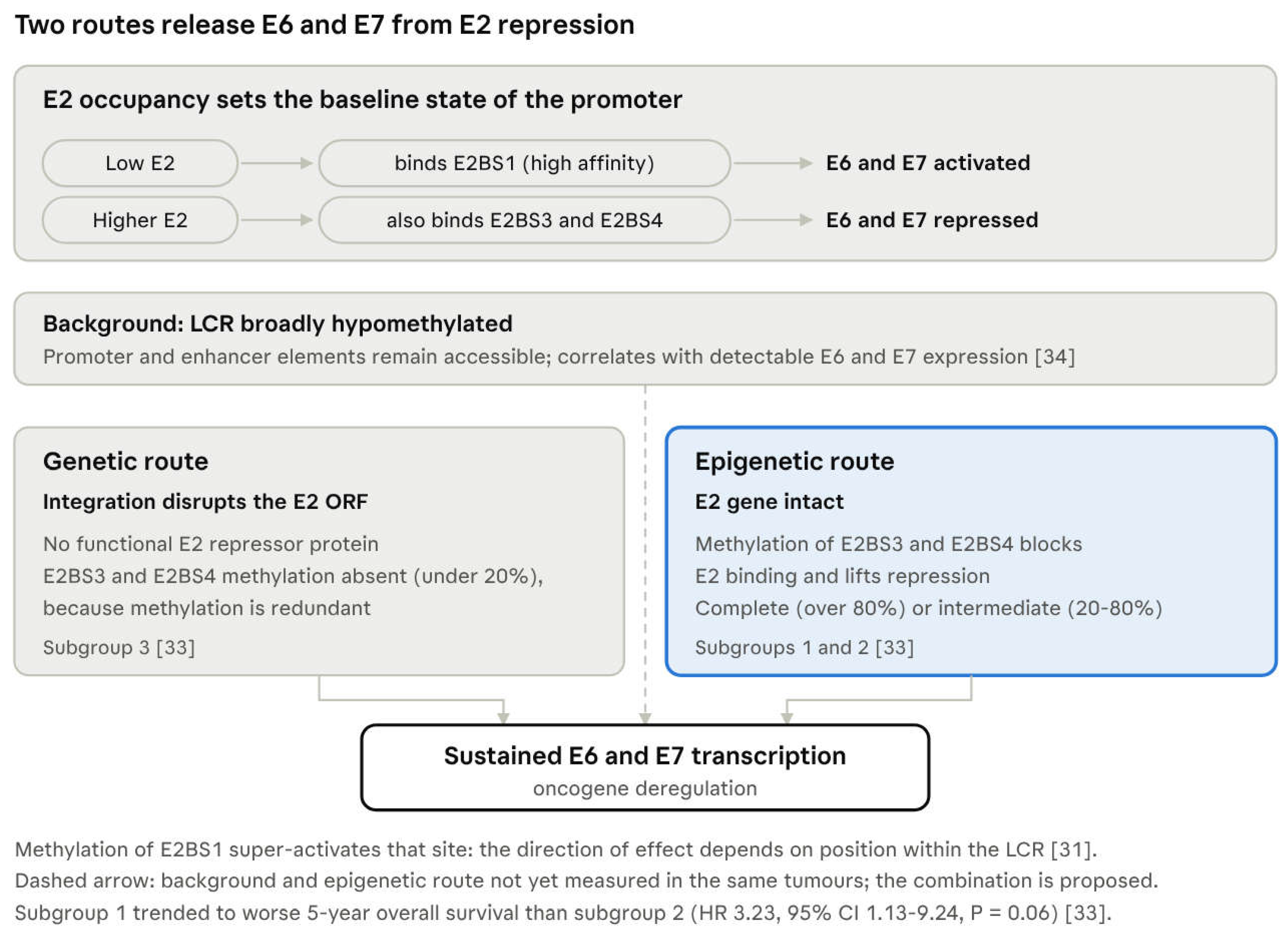
The clinically most consequential viral methylation occurs in the upstream regulatory region (URR, also termed the long control region, LCR). Four E2 binding sites, E2BS1 to E2BS4, lie within this region, and their occupancy sets the transcriptional output of E6 and E7. The arrangement is concentration-dependent: low levels of E2 bind the high-affinity E2BS1 and activate transcription, whereas higher levels additionally occupy the low-affinity E2BS3 and E2BS4 and repress it [32]. Loss of E2 function removes the repressive arm of this system, and because integration breakpoints cluster within the viral E2, E4 and E5 region (Section 4.1), integration and the consequent disruption of the E2 gene are the usual route by which that loss occurs.
Methylation of these sites supplies a second route to the same outcome, one that leaves the viral gene sequence intact, and its direction of effect depends on which site carries the mark. Methylation of E2BS1 super-activates that site, whereas methylation of E2BS3 and E2BS4 prevents E2 from binding and thereby lifts the repression it would otherwise impose on E6 and E7; methylation of distinct CpG dinucleotides within the E2 binding sites has been proposed as the initial trigger of HPV-mediated transformation [32]. The chain from viral persistence to oncogene deregulation is therefore branched rather than single: either integration disrupts the E2 gene and removes the repressor outright, or site-specific E2BS methylation disables the repressor while the E2 gene remains intact. Both routes converge on sustained E6 and E7 transcription, and the subgroups described below correspond to them.
Direct evidence for position dependence comes from oropharyngeal tumours themselves. Quantitative pyrosequencing of viral methylation in the LCR, performed in 81 OPSCC biopsies and restricted to patients with transcriptionally active HPV16 infection, found that E2BS1 carried a significantly higher mean methylation frequency than E2BS3 and E2BS4, reproducing a pattern described earlier in high-grade cervical precancer [33]. Methylation is therefore distributed unevenly across the regulatory region within a single tumour series, and it accumulates at the site that the mechanism above predicts should tolerate it. The same study reported that patients whose mean methylation frequency in the early promoter exceeded 10% showed only a tendency towards worse prognosis and separately identified HPV16 African variants in 10 of 51 HPV16-positive cases, the first such report outside the anogenital tract [33].
Quantitative bisulfite pyrosequencing of ten CpG dinucleotides spanning E2BS1 to E2BS4 in 57 HPV16-associated OPSCC resolved three subgroups based on E2BS3 and E2BS4 methylation [34]:
| Subgroup | E2BS3/E2BS4 methylation | Viral physical state | E2 gene |
| 1 | Complete (>80%) | Integrated | Intact |
| 2 | Intermediate (20–80%) | Predominantly episomal | Intact |
| 3 | Absent (<20%) | — | Disrupted |
The logic of this classification is reciprocal: where E2 is disrupted, methylation of its binding sites is redundant and absent; where E2 is intact, methylation supplies the derepression that gene disruption would otherwise provide. Patients with high methylation levels tended towards worse 5-year overall survival than those with intermediate methylation (hazard ratio 3.23; 95% CI 1.13–9.24; P = 0.06), although the authors noted that the prognostic significance requires further analysis [34]. The finding is hypothesis-generating rather than practice-changing, but it is one of the few epigenetic variables that stratifies within HPV-positive OPSCC rather than merely distinguishing it from HPV-negative disease.
A superficially conflicting picture emerges from bisulfite sequencing of all 110 CpG sites in the viral genome in advanced stage III/IV HPV16-associated head and neck cancers, which found the viral genome to be predominantly hypomethylated, particularly within the LCR, and showed that LCR hypomethylation correlated with detectable E6 and E7 expression [35]. Read against the site-resolved data above, the two observations are more plausibly complementary than contradictory. A broadly hypomethylated LCR leaves the promoter and enhancer elements that drive E6 and E7 accessible, while methylation confined to the repressive E2BS3 and E2BS4 removes the brake on that same transcription. Both states favour oncogene expression, and the sign of the methylation effect is a property of position within the LCR rather than of methylation as such [32]. This site-specific model is advanced here as the most economical reconciliation of the present data rather than as an established mechanism: no study has yet measured methylation across the whole viral genome and at site resolution within the E2 binding sites in the same tumours, whole-genome averages and site-specific values are not interchangeable measurements, and the cohorts concerned differ in stage, viral load and integration status. Testing the model requires that both be measured in a single series. The same study seen of direct translational relevance: the viral epigenetic alterations were detectable in serum and saliva, and correlated with the changes observed in the matched primary tumours [35].
Figure 2.
Two routes release E6 and E7 from E2 repression. E2 occupancy of the E2 binding sites sets the baseline transcriptional state. Oncogene deregulation follows either integration-associated disruption of the E2 gene or site-specific methylation of E2BS3 and E2BS4 with the E2 gene intact. The hypomethylated LCR background and the epigenetic route have not been measured in the same tumours; their combination is proposed rather than established.
Figure 2.
Two routes release E6 and E7 from E2 repression. E2 occupancy of the E2 binding sites sets the baseline transcriptional state. Oncogene deregulation follows either integration-associated disruption of the E2 gene or site-specific methylation of E2BS3 and E2BS4 with the E2 gene intact. The hypomethylated LCR background and the epigenetic route have not been measured in the same tumours; their combination is proposed rather than established.
One further source of variation bears on all of this. The upstream regulatory region is not only a substrate for methylation but also a site of sequence variation between HPV16 isolates, and that variation carries functional consequences of its own. A systematic review of HPV16 single-nucleotide polymorphisms in head and neck cancer notes that variants in the 5′ segment of the region fall within binding sites for AP-1, FOXA1 and C/EBPβ, each of which regulates HPV16 transcription, while variants in the 3′ segment, which contains the p97 promoter, may alter transcription of E6 and E7 [36]. Two distinct kinds of variation therefore converge on one control region. The practical consequence is that assays quantifying methylation at fixed CpG coordinates presuppose that those dinucleotides are present in every isolate examined, which sequence variation does not guarantee; the detection of African variants in the oropharyngeal series described above makes the point concrete rather than theoretical.
4.3. Methylation of the Late Region
Methylation of the late genes has attracted interest largely by analogy with cervical disease, where specific L1 positions track with progression. In a Greek HNSCC cohort, pyrosequencing of the HPV16 L1 gene and untranslated region revealed substantial methylation across six L1 sites and seven UTR sites, and methylation at two L1 CpG positions (7136 and 7145) was significantly associated with tumour histological grade (P < 0.01), with concurrent methylation observed across multiple sites [37]. Notably, HPV16 physical status, assessed by the E2/E6 ratio, correlated neither with viral genome methylation nor with tumour characteristics in that series [37].
That last observation deserves emphasis because it cuts against the model in Section 4.2, in which E2BS methylation and physical state are tightly coupled. The discrepancy may reflect the difference between the E2/E6 ratio and sequencing-based integration calling, an inference supported by the finding in Section 2.3 that E2 disruption is a poor proxy for integration [13]. Methods that infer viral physical state indirectly should therefore be interpreted with caution when they are used to anchor epigenetic correlations.
4.4. Synthesis
Three conclusions are reasonably secure. First, the viral genome is methylated by host machinery and is not epigenetically inert. Second, methylation is neither uniform across the genome nor uniform in its consequence: the L1/L2 boundary and E1 carry the strongest signal, while within the regulatory region the sign of the effect depends on which E2 binding site is modified, so that position rather than methylation itself determines whether oncogene transcription is favoured or restrained. Third, E2BS methylation provides a mechanism for oncogene deregulation that is independent of integration and of E2 disruption, and it therefore helps explain the substantial fraction of HPV-positive OPSCC that carries episomal virus with an intact E2 gene (Section 2.3). What remains unresolved is which viral CpGs should be measured, by what method, and whether the resulting values carry independent prognostic weight. These questions bear directly on the circulating-biomarker strategies considered in Section 6, since methylated viral DNA sequences are in principle detectable in plasma and saliva.
5. Epigenetic Regulation of the Tumour Immune Microenvironment
5.1. The Immune Contexture of HPV-Positive Disease
The survival advantage of HPV-positive OPSCC is not explained by tumour-cell biology alone, and the most informative comparison is between HPV-driven tumours at different anatomical sites. HPV-driven tumours account for 4.1% of non-oropharyngeal HNSCC, and regardless of anatomic site these tumours share highly similar gene expression and DNA methylation profiles, non-keratinizing basaloid histopathological features, and a lack of TP53 or CDKN2A alterations [38]. Improved overall survival, however, was largely restricted to HPV-driven OPSCC, which carried increased levels of tumour-infiltrating lymphocytes compared with HPV-driven non-OPSCC [38]. The molecular programme of the virus is therefore portable throughout the head and neck while the prognostic benefit is not. These observations suggest that the favourable prognosis of HPV-driven OPSCC cannot be attributed solely to viral oncogene expression or tumour-intrinsic molecular features. Instead, the distinctive immune microenvironment of the oropharynx may contribute substantially to treatment responsiveness and survival, although the relative contribution of immune, anatomical and clinical factors remains to be established. Consistent with an immune contribution, tumour-infiltrating lymphocyte density stratifies outcome within HPV-positive disease: in 270 consecutively treated patients from three centres, 3-year survival was 96% in HPV-positive tumours with high lymphocyte infiltration against 59% in those with low infiltration, and the survival of the latter group did not differ from that of HPV-negative patients (hazard ratio 1.01) [39]. That observation has been proposed as a means of identifying patients likely to respond poorly to treatment [11]. The shared methylation profile across sites also reinforces the tissue-independence of the HPV methylation signature noted in Section 3.1.
The immune system additionally shapes the HPV-positive genome itself. APOBEC3B cytosine deaminase activity is induced in virally infected cells, and APOBEC signature mutations occur at high frequency in cervical carcinoma, over 99% of which is caused by HPV [40]. Comparison of HPV-positive and HPV-negative HNSCC exomes from TCGA showed that HPV-negative disease carries a smoking-associated mutational signature whereas HPV-positive disease is enriched for the APOBEC signature [40]. APOBEC activity was further shown to generate the helical domain hotspot mutations in PIK3CA across multiple cancers [40], which supplies a mechanism for the frequent PIK3CA alterations characteristic of HPV-positive HNSCC (Section 2.2).
A distinction should be drawn at this point. Those findings establish that an APOBEC mutational signature is present in HPV-positive disease and that APOBEC activity accounts for driver mutations; they do not establish that APOBEC is the dominant source of mutation in these tumours. The further proposal that reduced exposure to exogenous carcinogens creates a selective pressure under which APOBEC becomes the principal mutagenic force [40] is an interpretation of the same signature data rather than an independent demonstration, since separating dominance from co-occurrence would require apportioning mutational burden between competing processes rather than establishing that a signature is present. With that qualification, the broader point stands: an innate antiviral defence is simultaneously a mutagenic force, and the enzyme family that restricts the virus contributes to the driver mutations on which the resulting tumour depends.
That the methylome and the immune contexture are coupled is evident from methylation-based subtyping. In the five-subtype classification of TCGA HNSCC discussed in Section 3.1, the genomically stable CIMP-Atypical subtype was characterised by an antiviral gene expression profile associated with pro-inflammatory M1 macrophages and CD8+ T cell infiltration [7]. That this subtype is HPV-negative is instructive: widespread CpG island methylation and an antiviral immune phenotype can co-occur independently of the virus, which means an immune phenotype cannot be read off HPV status alone.
5.2. E7-Directed Methylation as an Immune Evasion Programme
The most direct evidence that E7 directs host methylation to genes governing immune recognition comes from parallel transcriptome and methylome analysis of normal immortalised keratinocytes carrying episomal HPV16 or HPV18, compared with the parental line and with an HPV16 line lacking E7 [41]. Expression of MHC class I genes was downregulated in HPV-positive keratinocytes in an E7-dependent manner, and methylome analysis localised hypermethylation to a distal CpG island near the HLA-E gene in HPV16-containing cells relative to both the parental and the E7-deficient lines [41]. That CpG island functions as an active promoter element and is strongly repressed by methylation; correspondingly, HLA-E protein at the cell surface was downregulated by high-risk HPV16 and HPV18 E7, and demethylation at the island restored HLA-E protein expression [41].
Two features of this finding carry weight. First, low-risk HPV6 and HPV11 E7 did not produce the effect [41], which ties the mechanism specifically to oncogenic types rather than to HPV infection in general. Second, E7-dependent epigenetic repression of HLA-E may alter immune recognition by natural killer and CD8+ T cells. The functional consequences are likely to depend on the receptor repertoire and the cellular context, however, since HLA-E engages the inhibitory CD94/NKG2A and CD94/NKG2B receptors as well as the activating CD94/NKG2C receptor [42], and direct evidence linking this mechanism to immune escape in primary HPV-positive OPSCC remains necessary. The same study situates the observation within a broader programme of E7-directed methylation: E7-dependent promoter hypermethylation had previously been shown to downregulate the chemokine CXCL14 and thereby suppress antitumour immune responses [41]. Taken together, these results indicate that part of the hypermethylated landscape described in Section 3.1 falls on genes governing immune recognition, which makes altered immunity a functional consequence of that landscape rather than an incidental feature of it.
5.3. Epigenetic Barriers to Immune Cell Trafficking
A complementary mechanism operates on cell migration rather than antigen presentation. As described in Section 3.2, HPV16 E7 represses E-cadherin through augmented DNMT1 activity, and DNMT inhibition restores E-cadherin levels [16]. Langerhans cells depend on E-cadherin to move through stratified epithelium, and E-cadherin silencing has therefore been proposed to reduce the density of antigen-presenting cells within infected epithelium and to contribute to the prolonged viral persistence that precedes malignant transformation [16]. The experimental evidence establishes the repression of E-cadherin rather than its immunological consequence, however. Whether E7-mediated E-cadherin repression directly reduces Langerhans cell recruitment or retention in the tonsillar crypt microenvironment remains to be demonstrated. Subject to that caveat, antigen presentation and immune cell trafficking would be constrained by the same enzymatic axis, which is one reason DNMT inhibition is attractive as an immunological intervention rather than merely a cytotoxic one.
Non-coding RNAs add a further layer. HPV-positive patients with high expression of the lncRNA PRINS showed a greater number of intratumoural immune cells and transcriptional changes associated with immune and antiviral responses, alongside better overall and disease-free survival [29]. Whether PRINS is a driver of this phenotype or a marker of it is unresolved.
5.4. Therapeutic Corollary: Viral Mimicry and Its Limits
The immunological consequences of epigenetic silencing suggest that reversing it could restore immune recognition, and the mechanism by which this occurs has been characterised in other tumour types. DNA methyltransferase inhibitors upregulate immune signalling through the viral defence pathway: in ovarian cancer they trigger cytosolic sensing of double-stranded RNA with a type I interferon response and apoptosis, an effect accompanied by demethylation and expression of hypermethylated endogenous retroviruses and reduced by knockdown of the dsRNA sensors TLR3 and MAVS [43]. Clinically, high expression of viral defence genes in melanoma tracked with durable response to immune checkpoint therapy, and DNMT inhibitor treatment sensitised to anti-CTLA-4 therapy in a preclinical model [43].
Extrapolating this to HPV-positive OPSCC requires caution. These tumours already carry a globally hypermethylated genome (Section 3.1) together with a pre-existing antiviral context driven by a genuine virus, so the net effect of pharmacological demethylation is not predictable from the ovarian and melanoma data alone. A further distinction matters here. Induction of interferon signalling is not equivalent to more effective antitumour immunity, because prolonged interferon signalling can itself sustain an adaptive resistance programme: persistent signalling drives STAT1-related epigenomic change and upregulates ligands for multiple T cell inhibitory receptors, with both type I and type II interferons maintaining that state [44]. The informative endpoint for a demethylating agent in this setting is therefore the quality and durability of the resulting immune response, not the magnitude of interferon induction. A histone-directed precedent in head and neck cancer is similarly suggestive but not transferable: depletion of the methyltransferase SMYD3 upregulates type I interferon response and antigen presentation machinery genes in HNSCC cells, and SMYD3 overexpression is associated with decreased CD8 T cell infiltration and poor response to neoadjuvant pembrolizumab [45]. That work was conducted in HPV-negative disease, and whether an analogous axis operates in HPV-positive tumours remains to be established. The same caveat attaches to the myeloid compartment: single-cell and spatial transcriptomic profiling of HPV-negative HNSCC has identified IL1B+ tumour-associated macrophages arising within spatially co-localised inflammatory cancer-associated fibroblast niches, whose high infiltration correlates with poorer survival and higher recurrence risk through regulatory T cell recruitment and exhaustion [46]. Taken together with the SMYD3 and viral mimicry findings, this illustrates a structural gap: the immune targets currently being defined in head and neck cancer are largely being defined outside HPV-positive disease, even though it is HPV-positive disease whose prognosis is most clearly immune-determined (Section 5.1). These questions are taken up as therapeutic strategies in Section 7.
6. Circulating Viral and Epigenetic Biomarkers
Biomarker claims in this field are frequently made at one level of evidence and read at another, so the analytes below are judged against three distinct standards. Analytical validity asks whether an assay measures its analyte accurately and reproducibly. Clinical validity asks whether that measurement separates patients with the condition of interest from those without it, and is what sensitivity, specificity and predictive values report. Clinical utility asks the further and separate question of whether acting on the result improves outcome, and it can be established only by showing that management guided by the test does better than management without it. A test may have excellent clinical validity and no demonstrated utility. That is the present position of every analyte considered in this section, the most mature included.
6.1. Circulating Tumour HPV DNA: The Benchmark
Any circulating epigenetic marker proposed for HPV-positive OPSCC must be judged against circulating tumour HPV DNA, which is viral rather than epigenetic but is the only liquid-biopsy analyte in this disease that has reached routine clinical use. In a prospective biomarker trial of 115 patients treated with definitive chemoradiotherapy, no recurrence occurred among the 87 patients whose post-treatment ctHPVDNA results were negative at every time point, giving a negative predictive value of 100%; among 16 patients with two consecutive positive results, 15 developed biopsy-proven recurrence, a positive predictive value of 94%, with a median lead time over biopsy-proven recurrence of 3.9 months [10].
Real-world data have broadly confirmed this. In a retrospective series of 1,076 consecutive patients across 108 United States sites tested during routine post-treatment surveillance, circulating tumour tissue modified viral HPV DNA was positive in 80 patients (7.4%), with an overall positive predictive value of 95% and a point-in-time negative predictive value of 95% [47]. At the first positive surveillance test, 59 of the 80 positive patients (74%) were not known to have recurrent disease, so positivity was the first indication of recurrence in most cases, pre-dating detection by routine clinical and imaging examination [47].
These figures establish clinical validity; they do not establish clinical utility. No randomised trial has yet shown that managing patients according to ctHPVDNA results improves survival or reduces treatment morbidity, and a high predictive value is compatible with detecting recurrences that conventional follow-up would have found, and treated, only months later. Consensus recommendations from the California Head and Neck Consortium, developed by a hybrid Delphi process among 33 head and neck specialists drawn from academic, managed-care, county, veterans, community and private practice settings, take precisely this position. The panel reached strong consensus that ctHPVDNA is a valuable tool in the diagnostic and surveillance settings, but not for screening and not for modifying treatment in current clinical practice, and that randomised trials should be performed to optimise its use given the costs its adoption would add [48]. The distribution of agreement is itself informative: of 33 statements, 21 reached strong consensus and three reached consensuses, while four returned no consensus and five were rejected [48]. The bar for an epigenetic analyte is therefore set twice over. It must first match ctHPVDNA on clinical validity, and it will earn a place in surveillance only if it adds information that viral DNA does not: identifying the minority of HPV-positive tumours that shed little or no viral DNA, stratifying risk before treatment, or reporting on biology such as immune phenotype that viral copy number cannot.
6.2. Methylated Circulating DNA
The proof of principle for circulating epigenetic markers in this disease is old and underexploited. In the bisulfite-sequencing study of the HPV16 epigenome discussed in Section 4.2, the viral epigenetic alterations were detectable in serum and saliva and correlated with the changes observed in the matched primary tumours [35]. That observation, published in 2011, anticipated the ctHPVDNA literature by nearly a decade but has not been developed into a validated assay.
Host-genome methylation in cell-free DNA has advanced further in adjacent disease. A salivary liquid-biopsy approach for oral squamous cell carcinoma identified five genes with higher methylation in tumour than in matched non-tumour tissue that were analysable by high-resolution melting and were independent of tumour stage, aetiology and age; at least one of the five cfDNA methylation marks was detectable before surgery in 70% of cases, and complete surgical resection led to a significant disappearance of the salivary marks [49]. The relevance to OPSCC is indirect, since that cohort was oral cavity cancer and was not selected by HPV status, but it demonstrates the two properties a surveillance marker requires: detectability before intervention and clearance after it.
The gap is therefore specific. The host methylation signature of HPV-positive OPSCC is well characterised in tissue (Section 3.1) and the viral methylation landscape is mapped (Section 4), yet no methylated-cfDNA panel derived from either has been analytically validated in this tumour type.
6.3. Extracellular Vesicle MicroRNAs
Extracellular vesicles offer a third route, and the HPV-dependence of their microRNA cargo is reproducible in vitro. Small RNA sequencing of EVs from HPV-positive and HPV-negative OPSCC cell lines showed that unbiased hierarchical clustering separated samples by HPV status, with 14 miRNAs enriched in EVs from HPV-positive cells and 19 in those from HPV-negative cells; HPV-negative cells also produced more EVs [50]. Subsequent work extended the comparison across eight HNSCC lines and framed exo-miRNA explicitly as a candidate liquid biopsy for early detection [51].
The first steps into patient material are encouraging but preliminary. In serum-derived EVs, miR-21, let-7a and miR-181a were expressed 2.5- to 3-fold higher in HPV/p16-positive than in HPV/p16-negative HNSCC, and EV-miR-181a and miR-26a differed according to treatment modality [52]. Two cautions apply. Body fluids contain a heterogeneous mixture of vesicles of which only a fraction is tumour-derived, unlike cell-culture supernatants; and a biomarker claim resting solely on differential abundance in a body fluid, without functional validation linking the miRNA to a molecular target, warrants scepticism. The heterogeneity problem identified for tissue miRNA profiling in Section 3.4.1 applies here with greater force.
6.4. Pre-Diagnostic Serology
A distinct clinical question is whether HPV-driven cancer can be detected before it presents, and the evidence there comes from antibody rather than nucleic acid measurement. In a case-control study nested within nine prospective cohorts in Europe, North America and Australia, pre-diagnostic blood from 1,700 participants with incident HPV-related cancers, 788 of them oropharyngeal, was compared with 6,221 controls [53]. Among oropharyngeal cancers, seroprevalence rose as diagnosis approached for HPV16 E1, E2, E4, E6, E7 and L1 and for HPV18 E1, E2 and L1; HPV16-E6 seropositivity increased from 10.4% more than 20 years before diagnosis to 35.8% within 5 years of it, against 0.3% in controls [53]. Anal cancer showed a similar pattern, whereas no such trend was seen for cervical, penile or vaginal cancers [53].
Two features of this result bear on the present review. The lead time is of an order no epigenetic or viral DNA assay has approached, which places serology in the domain of risk stratification and early detection rather than post-treatment surveillance, and makes it complementary to rather than competitive with the analytes above. And the absence of a trend in cervical cancer alongside a strong one in the oropharynx is a further instance of the site-specificity met in Section 2.1 and Section 5.1, suggesting that the oropharyngeal lesion presents viral antigen to the immune system differently from the cervical one. Whether epigenetic marks behave comparably before diagnosis is unknown, and the cohorts assembled for this serological work are the obvious setting in which to establish it.
6.5. Comparative Status of the Analytes
| Analyte | Source | Strongest evidence in HPV-positive disease | Reported performance | Principal limitation | Highest level reached |
| Circulating tumour HPV DNA | Plasma | Prospective trial [10] and 1,076-patient real-world series [47] | NPV 100%, PPV 94% for two consecutive positives [10]; PPV 95%, point-in-time NPV 95% [47] | Viral, not epigenetic; uninformative where the tumour sheds little viral DNA | Clinical validity; utility not demonstrated |
| Methylated viral DNA | Serum, saliva | Correlation with matched primary tumours [35] | Not quantified | No validated assay; small cohorts; 2011 data undeveloped | Analytical feasibility only |
| Methylated host cfDNA | Saliva | Surgical monitoring in oral cavity cancer [49] | ≥1 of 5 marks detectable in 70% pre-surgery; marks clear after resection [49] | Not HPV-selected; oral cavity rather than oropharynx | Clinical validity in an adjacent disease |
| Extracellular vesicle miRNA | Cell supernatant, serum | HPV-dependent cargo in vitro [50,51]; serum differences by p16 [52] | miR-21, let-7a, miR-181a 2.5–3-fold higher in p16-positive [52] | Largely preclinical; heterogeneous vesicle origin in body fluids | Preclinical |
| Pre-diagnostic HPV serology | Serum, plasma | Case-control nested in nine prospective cohorts, 1,700 cases [53] | HPV16-E6 seropositivity 35.8% within 5 years of diagnosis vs 0.3% in controls | Not epigenetic; answers pre-diagnostic risk, not post-treatment surveillance | Clinical validity for pre-diagnostic risk |
The asymmetry is one of level rather than of promise. Circulating tumour HPV DNA has clinical validity demonstrated prospectively and confirmed in real-world practice, but no demonstrated clinical utility. Pre-diagnostic serology has clinical validity for risk, established in large prospective cohorts, but no intervention that a positive result would currently trigger. Methylated host cell-free DNA has clinical validity only in an adjacent disease and in saliva rather than plasma. Methylated viral DNA and extracellular vesicle microRNAs have not yet cleared analytical validation in patient material. No analyte in this section, including the one in routine use, has been shown to improve an outcome that matters to patients. The most realistic near-term role for epigenetic analytes is therefore complementary rather than competitive: combined with ctHPVDNA to resolve equivocal results or applied before treatment where their tissue counterparts already stratify risk. How that might inform treatment selection is considered in Section 7.
Figure 3.
Translational maturity of circulating analytes in HPV-positive OPSCC.
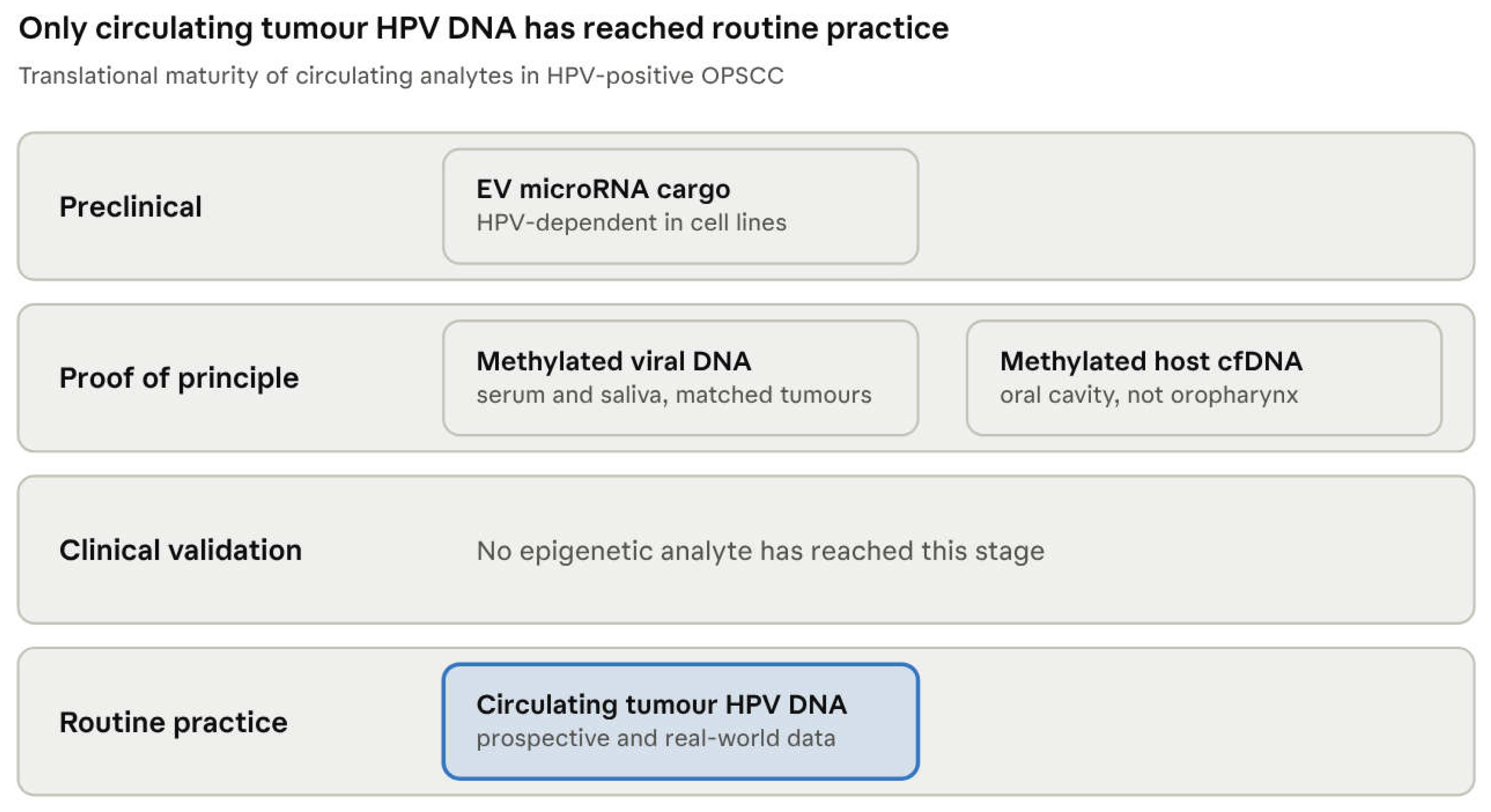
Nothing occupies the validation stage that separates proof of principle from clinical use, and closing that gap is the precondition for any epigenetic analyte entering surveillance.
7. Clinical Translation
7.1. Prognostication and Treatment De-escalation
The case for de-escalation rests on a sound premise and has so far failed on execution. Both pivotal phase 3 trials that replaced cisplatin with cetuximab produced inferior tumour control: in RTOG 1016, 5-year overall survival was 77.9% with cetuximab versus 84.6% with cisplatin, with higher locoregional failure [8]; De-ESCALaTE found no reduction in severe toxicity and significant detriment in tumour control, concluding that cisplatin and radiotherapy should remain standard even for low-risk patients [9]. The lesson is not that de-escalation is unachievable, but that HPV status is too coarse a stratifier to select patients for it.
Epigenetic variables are candidates to refine that selection precisely because they vary within HPV-positive disease. Three have supporting data. Methylation of the viral E2 binding sites resolves HPV16-positive OPSCC into three subgroups, with a trend towards worse 5-year overall survival in the high-methylation group (hazard ratio 3.23; 95% CI 1.13–9.24), although that comparison did not reach conventional significance and the authors cautioned that its prognostic value requires further analysis [34]. Three miRNA sets were associated with overall survival, disease-free survival and distant metastasis and retained significance after adjustment for p16 status [26]. The density of tumour-infiltrating lymphocytes differentiates HPV-driven tumours of the oropharynx from HPV-driven tumours elsewhere and tracks with the survival advantage [38], and within HPV-positive OPSCC low lymphocyte infiltration marks poorer prognosis [11]. None is validated for clinical use, but each reports biology that p16 immunohistochemistry cannot.
The nearer-term contribution is in dynamic monitoring rather than baseline selection. Circulating tumour HPV DNA identifies recurrence months before imaging [10,47], and a de-escalation strategy guided by post-treatment molecular response is mechanistically more attractive than one guided by pre-treatment risk category alone. Attractiveness is not evidence. The cetuximab trials were also built on a sound premise and a plausible mechanism, and both returned inferior tumour control; the reasoning that selected patients for them was not obviously weaker than the reasoning now being applied to molecular response. To let a circulating marker determine how much treatment a patient receives is a claim of clinical utility in the sense defined at the start of Section 6, and it is precisely the claim that the California Head and Neck Consortium declined to endorse outside clinical trials [48]. Response-adapted de-escalation is the most promising hypothesis this field has produced, and the appropriate conclusion is that it must be tested as one.
7.2. Demethylating Agents
The hypermethylated landscape described in Section 3.1 is itself a therapeutic target, and the most developed evidence concerns 5-azacytidine. In preclinical models and in a window clinical trial enrolling patients with HPV-positive HNSCC, 5-azacytidine induced growth inhibition and cell death, reduced the expression of HPV genes including E6 and E7, stabilised p53 and induced p53-dependent apoptosis [54]. It also repressed the expression and activity of matrix metalloproteinases, activated an interferon response in some HPV-positive cell lines, and inhibited the ability of HPV-positive xenografted tumours to invade mouse blood vessels [54].
The mechanism of selective toxicity has since been defined and is unusual. 5-azacytidine induces DNA double-strand breaks that occur preferentially at demethylated genomic DNA and depend on both active transcription and replication; blocking either prevents break formation or reduces the sensitivity of HPV-positive cells to the drug [55]. Sensitivity is therefore a consequence of the hypermethylated baseline rather than an incidental cytotoxic effect, which is a rational basis for targeting this tumour type specifically.
One mechanistic concern deserves explicit statement. If a broadly hypomethylated viral regulatory region is permissive for oncogene expression (Section 4.2), a systemic demethylating agent might be expected to derepress E6 and E7 rather than suppress them. The observed data point the other way: 5-azacytidine reduced the expression of HPV genes including E6 and E7 in the same models in which it demethylated the host genome [54]. The likely resolution is that the dominant effects of the drug are on host transcriptional programmes and on transcription-coupled DNA damage rather than on viral promoter accessibility. The question has not been addressed directly, however, and viral methylation and viral transcript levels would be worth measuring as endpoints in any future trial of this class of agent in HPV-positive disease.
The therapeutic logic is attractive on three counts: it reverses the epigenetic programme, it suppresses the viral oncogenes that sustain the malignant phenotype, and it restores p53 function that E6 had abolished. The evidence base, however, remains a small window trial, and the leap to a randomised efficacy study has not yet been made.
7.3. Combination with Immunotherapy
Demethylation has an immunological rationale in addition to a cytotoxic one. Interferon response activation was observed directly in HPV-positive cells treated with 5-azacytidine [54], consistent with the viral mimicry mechanism by which DNA methyltransferase inhibitors upregulate immune signalling through the viral defence pathway and sensitise to checkpoint blockade in other tumour types [43]. Combined with the evidence in Section 5.2 that E7-directed methylation silences MHC class I and HLA-E, this supports testing demethylating agents as immune-sensitising rather than cytotoxic agents in HPV-positive OPSCC, provided that such trials are read against immune endpoints that capture the durability of the response rather than interferon induction alone (Section 5.4). A histone-directed alternative exists in principle, since SMYD3 depletion upregulates interferon response and antigen presentation genes and SMYD3 overexpression tracks with poor response to neoadjuvant pembrolizumab, although that work was conducted in HPV-negative disease [45].
7.4. Nutritional Modulation of the Epigenome
Because DNA methylation depends on dietary methyl donors and is inhibited by several food-derived compounds, nutrition is a plausible and largely unexplored lever on the mechanisms described in this review. Two strands of evidence exist, and neither has been examined in oropharyngeal cancer.
The first concerns direct DNMT inhibition by dietary polyphenols. In HNSCC cell lines, epigallocatechin gallate reduced DNMT activity to 60% in SCC-1 and 80% in FaDu cells, downregulated DNMT3a and DNMT3b protein, and reactivated silenced tumour suppressors with decreased proliferation; dietary EGCG at 0.5% (w/w) in an AIN76A diet inhibited FaDu xenograft growth by 80% (P < 0.01), and the effect was associated with DNMT inhibition and tumour suppressor reactivation [56]. The mechanism is the same enzymatic axis that 5-azacytidine targets, reached by a different and far less toxic route, which is what makes the approach interesting for chemoprevention or for maintenance rather than for treatment of established disease.
The second concerns one-carbon metabolism and viral persistence, where the evidence is genuinely conflicting. Lower folate status was associated with high-risk HPV infection (P = 0.031) and with a diagnosis of cervical intraepithelial neoplasia or invasive cervical cancer (P < 0.05) in a cross-sectional study of 308 women [57]. Yet in a nested case-control study within the ARTISTIC cohort, a higher cervical cell folate concentration was an independent predictor of high-risk HPV infection at follow-up (P = 0.015), alongside promoter methylation of DAPK [58]. In a prospective cohort of young women, circulating vitamin B12 was inversely associated with HPV persistence (P for trend 0.037) while no association was found for dietary or circulating folate [59].
These results are not reconcilable as a simple dose-response relationship, and the discrepancy may reflect folate's dual role: deficiency compromises the methyl supply required for normal methylation, while repletion supports nucleotide synthesis and proliferation in cells already transformed. The direction of effect may therefore depend on the stage of disease at which the measurement is made. Three points follow for this review. First, no study has examined methyl-donor status in relation to oropharyngeal HPV persistence or OPSCC outcome, despite the oral cavity being directly exposed to dietary constituents. Second, the oropharyngeal niche differs from the cervical one in immune architecture (Section 2.1) and in immune contexture (Section 5.1), so cervical findings cannot be assumed to transfer. Third, any nutritional intervention study in this setting would need epigenetic endpoints, such as viral E2BS methylation or host methylation signatures, rather than viral clearance alone, if it is to test the mechanism rather than merely the outcome.
8. Challenges and Future Perspectives
8.1. Methodological Heterogeneity
The single most consistent finding of this review is inconsistency between studies. Whole-genome small-RNA sequencing compared against the published literature revealed extensive divergence in miRNA deregulation, with only a handful of miRNAs reproducible across studies [27]. Reports on the viral regulatory region appear to conflict, with site-specific hypermethylation of the E2 binding sites in one series [34] and global hypomethylation in another [35]; as argued in Section 4.2, this is better read as a difference in the resolution at which methylation was measured than as a true disagreement, although that reconciliation itself awaits direct testing. A genuine discrepancy does remain over whether the physical state of the virus correlates with methylation, which it did in one series [34] and did not in another [37]. Where a discrepancy is real, it is plausibly methodological: platform, specimen type, normalisation strategy, cohort stage, and how HPV status and integration were ascertained all differ. Since E2 disruption is a poor proxy for integration [13] and the E2/E6 ratio is an indirect measure, studies anchoring epigenetic correlations to indirectly inferred viral status are particularly vulnerable.
Three sources of heterogeneity deserve particular emphasis in HPV-positive OPSCC. The first is the cellular composition of the specimen. Bulk tumour methylation reflects a mixture of malignant epithelium, infiltrating lymphocytes and stroma, and because HPV-positive OPSCC is characteristically lymphocyte-rich (Section 5.1), differences attributed to the tumour cell may in part report differences in immune infiltrate. The second is the case definition discussed in Section 1: p16 positivity and transcriptionally active HPV infection are not equivalent, and cohorts assembled on the two criteria are not interchangeable [36]. The third is intratumoural heterogeneity of viral physical state, since integrated and episomal genomes may coexist within a single tumour (Section 2.3) and an assay reporting an average across them obscures which configuration is transcriptionally active.
Progress requires agreement on what is measured and how. For the viral genome this means specifying which CpG positions are informative, confirming that those positions are conserved across the variant lineages in circulation [36], and standardising the assay; for the host genome it means distinguishing HPV-driven methylation from the CIMP that arises independently of the virus [7].
8.2. Stratification Within HPV-Positive Disease
The failure of cetuximab substitution [8,9] established that HPV positivity alone cannot select patients for de-escalation, yet almost all epigenetic work in this field has been framed as distinguishing HPV-positive from HPV-negative disease. Although p16 immunohistochemistry is an established and widely accessible surrogate marker in OPSCC, it does not directly demonstrate transcriptionally active HPV infection [36]. The clinically useful question is nevertheless the other one: which HPV-positive patients differ from each other, and why. The variables with preliminary evidence on that question are E2BS methylation subgroups [34], p16-independent miRNA prognostic sets [26] and immune contexture [11,38]. These deserve prospective evaluation in that framing, and future epigenetic biomarkers should be assessed for incremental prognostic value beyond established HPV classification and clinical risk factors rather than for separation from HPV-negative disease.
8.3. From Association to Causation
Much of the non-coding RNA literature, and a growing proportion of the lncRNA literature, consists of signatures derived in silico from TCGA or GEO without prospective validation or mechanistic follow-up. Whether PRINS drives the immune phenotype it marks [29] is unknown, as is whether the EV microRNAs that distinguish HPV status in culture [50,51] retain that discrimination in the heterogeneous vesicle population of plasma [52]. The contrast with the E7–DNMT1–E-cadherin [16] and E7–KDM6A/B [17] axes, where causality was established by oncogene expression and knockdown, shows what the standard should be. Meeting it calls for specific experimental approaches: targeted epigenome editing to place or remove a mark at a defined locus and test its effect directly, perturbation of the writer, eraser or reader enzymes implicated, and functional confirmation in models that represent HPV-positive oropharyngeal disease rather than HPV-negative or cervical surrogates.
8.4. Research Priorities
- Define and analytically validate a methylated-cfDNA panel, viral or host, in HPV-positive OPSCC, and test whether it adds information to ctHPVDNA rather than duplicating it.
- Advance E2BS methylation along the evidence ladder before it enters a de-escalation trial: analytical validation of the assay, then independent prognostic validation in an external cohort, then demonstration of incremental value over established risk factors. Only once those steps are met does prospective evaluation as a stratification variable, with overall survival as the endpoint rather than a surrogate, become a reasonable proposition.
- Take 5-azacytidine beyond the window trial [54,55] into a randomised study with a clinical primary endpoint, measuring interferon response and antigen presentation as pharmacodynamic and exploratory endpoints rather than as co-primary ones, since the immune rationale [43,45] has still to be confirmed in this disease (Section 5.4).
- Determine whether the immune difference between oropharyngeal and non-oropharyngeal HPV-driven tumours [38] has an epigenetic basis, since this would identify what the oropharyngeal niche contributes beyond the viral programme.
9. Conclusions
HPV-associated OPSCC is defined as much by its epigenome as by its genome. The virus recruits host methylation and histone-modifying machinery through E6 and E7, producing a hypermethylated landscape with CIMP features in a subset [6,7], reduced H3K27me3 through induced demethylases [17], and a deregulated non-coding RNA network. Its own genome is methylated by the same host enzymes, and methylation of the E2 binding sites supplies a route to oncogene deregulation that is independent of integration and of E2 disruption [34]. These changes are not confined to the tumour cell: silencing of MHC class I and HLA-E [41] and of E-cadherin [16] places genes governing immune recognition and immune cell trafficking among the targets of that reprogramming [38]. The favourable prognosis of HPV-positive OPSCC likely reflects an interplay between viral oncogene-driven tumour biology, host epigenetic regulation and the distinctive immune microenvironment of the oropharynx. Dissecting these contributions will be essential for developing biologically informed prognostic models and safely translating epigenetic biomarkers into clinical practice.
Translation has advanced unevenly. Circulating tumour HPV DNA has prospective and real-world validation and detects recurrence months before imaging [10,47], while every epigenetic analyte remains at proof-of-principle or preclinical stage. Demethylating therapy has a mechanistic rationale specific to this tumour type and promising window-trial data [54,55] but no randomised evidence. Nutritional modulation of the epigenome is biologically plausible, supported in head and neck models for polyphenols [56], and entirely unstudied in the oropharyngeal setting.
The field's central task is now to move from describing how HPV reprograms the epigenome to using that knowledge where HPV status alone has proved insufficient: identifying which patients with HPV-positive disease can safely receive less treatment, and which cannot.
Author Contributions
Conceptualization, literature search, writing—original draft preparation, and writing—review and editing, S.P.D. The author has read and agreed to the published version of the manuscript.
Funding
This research received no external funding.
Institutional Review Board Statement
Not applicable.
Informed Consent Statement
Not applicable.
Data Availability Statement
No new data were created or analysed in this study. All data discussed are available in the cited publications.
Conflicts of Interest
The author declares no conflicts of interest.
Abbreviations
CIMP, CpG island methylator phenotype; ctHPVDNA, circulating tumour HPV DNA; HNSCC, head and neck squamous cell carcinoma; HPV, human papillomavirus; OPSCC, oropharyngeal squamous cell carcinoma; PRC2, Polycomb repressive complex 2; TCGA, The Cancer Genome Atlas.
References
- Chaturvedi, A.K.; Engels, E.A.; Pfeiffer, R.M.; Hernandez, B.Y.; Xiao, W.; Kim, E.; Jiang, B.; Goodman, M.T.; Sibug-Saber, M.; Cozen, W.; et al. Human papillomavirus and rising oropharyngeal cancer incidence in the United States. J. Clin. Oncol. 2011, 29, 4294–4301. [Google Scholar] [CrossRef] [PubMed]
- Stein, A.P.; Saha, S.; Kraninger, J.L.; Swick, A.D.; Yu, M.; Lambert, P.F.; Kimple, R.J. Prevalence of human papillomavirus in oropharyngeal cancer: a systematic review. Cancer J. 2015, 21, 138–146. [Google Scholar] [CrossRef] [PubMed]
- Kim, S.I.; Lee, J.W.; Eun, Y.-G.; Lee, Y.C. A SEER-based analysis of trends in HPV-associated oropharyngeal squamous cell carcinoma. Infect. Agents Cancer 2024, 19, 29. [Google Scholar] [CrossRef] [PubMed]
- Ang, K.K.; Harris, J.; Wheeler, R.; Weber, R.; Rosenthal, D.I.; Nguyen-Tân, P.F.; Westra, W.H.; Chung, C.H.; Jordan, R.C.; Lu, C.; et al. Human papillomavirus and survival of patients with oropharyngeal cancer. N. Engl. J. Med. 2010, 363, 24–35. [Google Scholar] [CrossRef] [PubMed]
- Cancer Genome Atlas Network. Comprehensive genomic characterization of head and neck squamous cell carcinomas. Nature 2015, 517, 576–582. [Google Scholar] [CrossRef] [PubMed]
- Lechner, M.; Fenton, T.; West, J.; Wilson, G.; Feber, A.; Henderson, S.; Thirlwell, C.; Dibra, H.K.; Jay, A.; Butcher, L.; et al. Identification and functional validation of HPV-mediated hypermethylation in head and neck squamous cell carcinoma. Genome Med. 2013, 5, 15. [Google Scholar] [CrossRef] [PubMed]
- Brennan, K.; Koenig, J.L.; Gentles, A.J.; Sunwoo, J.B.; Gevaert, O. Identification of an atypical etiological head and neck squamous carcinoma subtype featuring the CpG island methylator phenotype. EBioMedicine 2017, 17, 223–236. [Google Scholar] [CrossRef] [PubMed]
- Gillison, M.L.; Trotti, A.M.; Harris, J.; Eisbruch, A.; Harari, P.M.; Adelstein, D.J.; Sturgis, E.M.; Burtness, B.; Ridge, J.A.; Ringash, J.; et al. Radiotherapy plus cetuximab or cisplatin in human papillomavirus-positive oropharyngeal cancer (NRG Oncology RTOG 1016): a randomised, multicentre, non-inferiority trial. Lancet 2019, 393, 40–50. [Google Scholar] [CrossRef] [PubMed]
- Mehanna, H.; Robinson, M.; Hartley, A.; Kong, A.; Foran, B.; Fulton-Lieuw, T.; Dalby, M.; Mistry, P.; Sen, M.; O'Toole, L.; et al. Radiotherapy plus cisplatin or cetuximab in low-risk human papillomavirus-positive oropharyngeal cancer (De-ESCALaTE HPV): an open-label randomised controlled phase 3 trial. Lancet 2019, 393, 51–60. [Google Scholar] [CrossRef] [PubMed]
- Chera, B.S.; Kumar, S.; Shen, C.; Amdur, R.; Dagan, R.; Green, R.; Goldman, E.; Weiss, J.; Grilley-Olson, J.; Patel, S.; et al. Plasma circulating tumor HPV DNA for the surveillance of cancer recurrence in HPV-associated oropharyngeal cancer. J. Clin. Oncol. 2020, 38, 1050–1058. [Google Scholar] [CrossRef] [PubMed]
- Roberts, S.; Evans, D.; Mehanna, H.; Parish, J.L. Modelling human papillomavirus biology in oropharyngeal keratinocytes. Philos. Trans. R. Soc. B 2019, 374, 20180289. [Google Scholar] [CrossRef] [PubMed]
- Perry, M.E. The specialised structure of crypt epithelium in the human palatine tonsil and its functional significance. J. Anat. 1994, 185, 111–127. [Google Scholar] [PubMed]
- Symer, D.E.; Akagi, K.; Geiger, H.M.; Song, Y.; Li, G.; Emde, A.-K.; Xiao, W.; Jiang, B.; Corvelo, A.; Toussaint, N.C.; et al. Diverse tumorigenic consequences of human papillomavirus integration in primary oropharyngeal cancers. Genome Res. 2022, 32, 55–70. [Google Scholar] [CrossRef] [PubMed]
- Parfenov, M.; Pedamallu, C.S.; Gehlenborg, N.; Freeman, S.S.; Danilova, L.; Bristow, C.A.; Lee, S.; Hadjipanayis, A.G.; Ivanova, E.V.; Wilkerson, M.D.; et al. Characterization of HPV and host genome interactions in primary head and neck cancers. Proc. Natl. Acad. Sci. USA 2014, 111, 15544–15549. [Google Scholar] [CrossRef] [PubMed]
- Burgers, W.A.; Blanchon, L.; Pradhan, S.; de Launoit, Y.; Kouzarides, T.; Fuks, F. Viral oncoproteins target the DNA methyltransferases. Oncogene 2007, 26, 1650–1655. [Google Scholar] [CrossRef] [PubMed]
- Laurson, J.; Khan, S.; Chung, R.; Cross, K.; Raj, K. Epigenetic repression of E-cadherin by human papillomavirus 16 E7 protein. Carcinogenesis 2010, 31(5), 918–926. [Google Scholar] [CrossRef] [PubMed]
- McLaughlin-Drubin, M.E.; Crum, C.P.; Munger, K. Human papillomavirus E7 oncoprotein induces KDM6A and KDM6B histone demethylase expression and causes epigenetic reprogramming. Proc. Natl. Acad. Sci. USA 2011, 108, 2130–2135. [Google Scholar] [CrossRef] [PubMed]
- Lindsay, C.D.; Kostiuk, M.A.; Harris, J.; O'Connell, D.A.; Seikaly, H.; Biron, V.L. Efficacy of EZH2 inhibitory drugs in human papillomavirus-positive and human papillomavirus-negative oropharyngeal squamous cell carcinomas. Clin. Epigenet. 2017, 9, 95. [Google Scholar] [CrossRef] [PubMed]
- Avvakumov, N.; Torchia, J.; Mymryk, J.S. Interaction of the HPV E7 proteins with the pCAF acetyltransferase. Oncogene 2003, 22, 3833–3841. [Google Scholar] [CrossRef] [PubMed]
- Kumar, M.; Molkentine, D.; Molkentine, J.; Bridges, K.; Xie, T.; Yang, L.; Hefner, A.; Gao, M.; Bahri, R.; Dhawan, A.; et al. Inhibition of histone acetyltransferase function radiosensitizes CREBBP/EP300 mutants via repression of homologous recombination, potentially targeting a gain of function. Nat. Commun. 2021, 12, 6340. [Google Scholar] [CrossRef] [PubMed]
- Karimzadeh, M.; Arlidge, C.; Rostami, A.; Lupien, M.; Bratman, S.V.; Hoffman, M.M. Human papillomavirus integration transforms chromatin to drive oncogenesis. Genome Biol. 2023, 24, 142. [Google Scholar] [CrossRef] [PubMed]
- Zamuner, F.T.; Chan, S.S.; Kessler, M.D.; Vorontsov, I.E.; Loginov, A.; Erbe, R.; Imada, E.; Xie, D.X.; Guo, T.; Fertig, E.J.; et al. Epigenetic profiling reveals key super-enhancer networks driving oncogenesis in HPV-positive HNSCC. iScience 2025, 28, 113911. [Google Scholar] [CrossRef] [PubMed]
- Wald, A.I.; Hoskins, E.E.; Wells, S.I.; Ferris, R.L.; Khan, S.A. Alteration of microRNA profiles in squamous cell carcinoma of the head and neck cell lines by human papillomavirus. Head Neck 2011, 33(4), 504–512. [Google Scholar] [CrossRef] [PubMed]
- Chapman, B.V.; Wald, A.I.; Akhtar, P.; Munko, A.C.; Xu, J.; Gibson, S.P.; Grandis, J.R.; Ferris, R.L.; Khan, S.A. MicroRNA-363 targets myosin 1B to reduce cellular migration in head and neck cancer. BMC Cancer 2015, 15, 861. [Google Scholar] [CrossRef] [PubMed]
- Lajer, C.B.; Garnæs, E.; Friis-Hansen, L.; Norrild, B.; Therkildsen, M.H.; Glud, M.; Rossing, M.; Lajer, H.; Svane, D.; Skotte, L.; et al. The role of miRNAs in human papilloma virus (HPV)-associated cancers: bridging between HPV-related head and neck cancer and cervical cancer. Br. J. Cancer 2012, 106, 1526–1534. [Google Scholar] [CrossRef] [PubMed]
- Hui, A.B.Y.; Lin, A.; Xu, W.; Waldron, L.; Perez-Ordonez, B.; Weinreb, I.; Shi, W.; Bruce, J.; Huang, S.H.; O'Sullivan, B.; et al. Potentially prognostic miRNAs in HPV-associated oropharyngeal carcinoma. Clin. Cancer Res. 2013, 19, 2154–2162. [Google Scholar] [CrossRef] [PubMed]
- Božinović, K.; Sabol, I.; Dediol, E.; Milutin Gašperov, N.; Manojlović, S.; Vojtechova, Z.; Tachezy, R.; Grce, M. Genome-wide miRNA profiling reinforces the importance of miR-9 in human papillomavirus associated oral and oropharyngeal head and neck cancer. Sci. Rep. 2019, 9(1), 2306. [Google Scholar] [CrossRef] [PubMed]
- Nohata, N.; Abba, M.C.; Gutkind, J.S. Unraveling the oral cancer lncRNAome: Identification of novel lncRNAs associated with malignant progression and HPV infection. Oral Oncol. 2016, 59, 58–66. [Google Scholar] [CrossRef] [PubMed]
- Kopczyńska, M.; Kolenda, T.; Guglas, K.; Sobocińska, J.; Teresiak, A.; Bliźniak, R.; Mackiewicz, A.; Mackiewicz, J.; Lamperska, K. PRINS lncRNA is a new biomarker candidate for HPV infection and prognosis of head and neck squamous cell carcinomas. Diagnostics 2020, 10, 762. [Google Scholar] [CrossRef] [PubMed]
- Kolenda, T.; Białas, P.; Guglas, K.; Stasiak, M.; Kozłowska-Masłoń, J.; Tylkowska, K.; Zapłata, A.; Poter, P.; Janiczek-Polewska, M.; Mantaj, P.; et al. lncRNA EGOT is the marker of HPV infection and a prognostic factor for HNSCC patients. Biomedicines 2025, 13, 798. [Google Scholar] [CrossRef] [PubMed]
- Wilson, G.A.; Lechner, M.; Köferle, A.; Carén, H.; Butcher, L.M.; Feber, A.; Fenton, T.; Jay, A.; Boshoff, C.; Beck, S. Integrated virus-host methylome analysis in head and neck squamous cell carcinoma. Epigenetics 2013, 8, 953–961. [Google Scholar] [CrossRef] [PubMed]
- von Knebel Doeberitz, M.; Prigge, E.-S. Role of DNA methylation in HPV associated lesions. Papillomavirus Res. 2019, 7, 180–183. [Google Scholar] [CrossRef] [PubMed]
- Barbieri, D.; Nebiaj, A.; Strammiello, R.; Agosti, R.; Sciascia, S.; Gallinella, G.; Landini, M.P.; Caliceti, U.; Venturoli, S. Detection of HPV16 African variants and quantitative analysis of viral DNA methylation in oropharyngeal squamous cell carcinomas. J. Clin. Virol. 2014, 60, 243–249. [Google Scholar] [CrossRef] [PubMed]
- Reuschenbach, M.; Huebbers, C.U.; Prigge, E.-S.; Lorenzo Bermejo, J.; Kalteis, M.S.; Preuss, S.F.; Seuthe, I.M.C.; Kolligs, J.; Speel, E.-J.M.; Olthof, N.; et al. Methylation status of HPV16 E2-binding sites classifies subtypes of HPV-associated oropharyngeal cancers. Cancer 2015, 121, 1966–1976. [Google Scholar] [CrossRef] [PubMed]
- Park, I.-S.; Chang, X.; Loyo, M.; Wu, G.; Chuang, A.; Kim, M.S.; Chae, Y.K.; Lyford-Pike, S.; Westra, W.H.; Saunders, J.R.; et al. Characterization of the methylation patterns in human papillomavirus type 16 viral DNA in head and neck cancers. Cancer Prev. Res. (Phila.) 2011, 4, 207–217. [Google Scholar] [CrossRef] [PubMed]
- Tran, N.H.; Nguyen, H.G.; Sais, D.; Lima, N.; Todd, A.; Nguyen, T.V.; Elliott, M.; Tran, N. Human papillomavirus 16 genomic variation in head and neck cancer: a systematic review with structural and functional annotation. BJC Rep. 2026. [Google Scholar] [CrossRef] [PubMed]
- Zygouras, I.; Leventakou, D.; Pouliakis, A.; Panagiotou, S.; Tsakogiannis, D.; Konstantopoulos, G.; Logotheti, E.; Samaras, M.; Kyriakopoulou, Z.; Beloukas, A.; et al. Human papillomavirus 16 DNA methylation patterns and investigation of integration status in head and neck cancer cases. Int. J. Mol. Sci. 2023, 24, 14593. [Google Scholar] [CrossRef] [PubMed]
- Chakravarthy, A.; Henderson, S.; Thirdborough, S.M.; Ottensmeier, C.H.; Su, X.; Lechner, M.; Feber, A.; Thomas, G.J.; Fenton, T.R. Human papillomavirus drives tumor development throughout the head and neck: improved prognosis is associated with an immune response largely restricted to the oropharynx. J. Clin. Oncol. 2016, 34, 4132–4141. [Google Scholar] [CrossRef] [PubMed]
- Ward, M.J.; Thirdborough, S.M.; Mellows, T.; Riley, C.; Harris, S.; Suchak, K.; Webb, A.; Hampton, C.; Patel, N.N.; Randall, C.J.; et al. Tumour-infiltrating lymphocytes predict for outcome in HPV-positive oropharyngeal cancer. Br. J. Cancer 2014, 110, 489–500. [Google Scholar] [CrossRef] [PubMed]
- Henderson, S.; Chakravarthy, A.; Su, X.; Boshoff, C.; Fenton, T.R. APOBEC-mediated cytosine deamination links PIK3CA helical domain mutations to human papillomavirus-driven tumor development. Cell Rep. 2014, 7, 1833–1841. [Google Scholar] [CrossRef] [PubMed]
- Cicchini, L.; Blumhagen, R.Z.; Westrich, J.A.; Myers, M.E.; Warren, C.J.; Siska, C.; Raben, D.; Kechris, K.J.; Pyeon, D. High-risk human papillomavirus E7 alters host DNA methylome and represses HLA-E expression in human keratinocytes. Sci. Rep. 2017, 7, 3633. [Google Scholar] [CrossRef] [PubMed]
- Braud, V.M.; Allan, D.S.J.; O'Callaghan, C.A.; Söderström, K.; D'Andrea, A.; Ogg, G.S.; Lazetic, S.; Young, N.T.; Bell, J.I.; Phillips, J.H.; et al. HLA-E binds to natural killer cell receptors CD94/NKG2A, B and C. Nature 1998, 391, 795–799. [Google Scholar] [CrossRef] [PubMed]
- Chiappinelli, K.B.; Strissel, P.L.; Desrichard, A.; Li, H.; Henke, C.; Akman, B.; Hein, A.; Rote, N.S.; Cope, L.M.; Snyder, A.; et al. Inhibiting DNA methylation causes an interferon response in cancer via dsRNA including endogenous retroviruses. Cell 2015, 162, 974–986. [Google Scholar] [CrossRef] [PubMed]
- Benci, J.L.; Xu, B.; Qiu, Y.; Wu, T.J.; Dada, H.; Twyman-Saint Victor, C.; Cucolo, L.; Lee, D.S.M.; Pauken, K.E.; Huang, A.C.; et al. Tumor interferon signaling regulates a multigenic resistance program to immune checkpoint blockade. Cell 2016, 167, 1540–1554.e12. [Google Scholar] [CrossRef] [PubMed]
- Nigam, N.; Bernard, B.; Sevilla, S.; Kim, S.; Dar, M.S.; Tsai, D.; Robbins, Y.; Burkitt, K.; Sievers, C.; Allen, C.; et al. SMYD3 represses tumor-intrinsic interferon response in HPV-negative squamous cell carcinoma of the head and neck. Cell Rep. 2023, 42, 112823. [Google Scholar] [CrossRef] [PubMed]
- Zhao, Z.; Yap, J.Y.Y.; Quah, H.S.; Wu, K.Z.; Kuthubudeen, F.F.; Biswas, S.K.; Iyer, N.G.; Fong, E.L.S. IL1B+ tumor-associated macrophages arise from inflammatory cancer-associated fibroblasts niches and are correlated with head and neck squamous cell carcinoma recurrence. npj Precis. Oncol. 2026. [Google Scholar] [CrossRef]
- Berger, B.M.; Hanna, G.J.; Posner, M.R.; Genden, E.M.; Lautersztain, J.; Naber, S.P.; Del Vecchio Fitz, C.; Kuperwasser, C. Detection of occult recurrence using circulating tumor tissue modified viral HPV DNA among patients treated for HPV-driven oropharyngeal carcinoma. Clin. Cancer Res. 2022, 28, 4292–4301. [Google Scholar] [CrossRef] [PubMed]
- Ho, A.S.; Moyers, J.T.; Attarian, S.; Banker, R.; Birkeland, A.; Chen, M.M.; Coffey, C.S.; Guo, T.; Haidar, Y.; Harris, J.P.; et al. Evolving use of circulating tumor DNA for HPV+ oropharyngeal carcinoma: Consensus recommendations from the California Head and Neck Consortium. JCO Oncol. Pract. 2025, OP2500450. [Google Scholar] [CrossRef] [PubMed]
- Saidak, Z.; Milly, M.; Louandre, C.; Colin, E.; Rusu, P.-M.; Paasche, A.; Dakpe, S.; Testelin, S.; Galmiche, A. Salivary cell-free DNA methylation analysis for oncological monitoring of surgical resection of oral squamous cell carcinoma. Front. Oral Health 2025, 6, 1614371. [Google Scholar] [CrossRef] [PubMed]
- Peacock, B.; Rigby, A.; Bradford, J.; Pink, R.; Hunter, K.; Lambert, D.; Hunt, S. Extracellular vesicle microRNA cargo is correlated with HPV status in oropharyngeal carcinoma. J. Oral Pathol. Med. 2018, 47, 954–963. [Google Scholar] [CrossRef] [PubMed]
- Galiveti, C.R.; Kuhnell, D.; Biesiada, J.; Zhang, X.; Kelsey, K.T.; Takiar, V.; Tang, A.L.; Wise-Draper, T.M.; Medvedovic, M.; Kasper, S.; et al. Small extravesicular microRNA in head and neck squamous cell carcinoma and its potential as a liquid biopsy for early detection. Head Neck 2023, 45, 212–224. [Google Scholar] [CrossRef] [PubMed]
- Apeltrath, C.; Simon, F.; Riders, A.; Rudack, C.; Oberste, M. Extracellular vesicle microRNAs as possible liquid biopsy markers in HNSCC. Cancers 2024, 16, 3793. [Google Scholar] [CrossRef] [PubMed]
- Onwuka, J.U.; Nobels, M.-P.; Kreimer, A.R.; Schroeder, L.; Ferreiro-Iglesias, A.; Agalliu, I.; Albanes, D.; Cai, Q.; D'Souza, G.; Freedman, N.D.; et al. Comparison of HPV antibodies measured prior to diagnosis of anal, cervical, oropharyngeal, penile and vaginal cancers in the HPV Cancer Cohort Consortium. Int. J. Cancer 2026, 1–15. [Google Scholar] [CrossRef] [PubMed]
- Biktasova, A.; Hajek, M.; Sewell, A.; Gary, C.; Bellinger, G.; Deshpande, H.A.; Bhatia, A.; Burtness, B.; Judson, B.; Mehra, S.; et al. Demethylation therapy as a targeted treatment for human papillomavirus-associated head and neck cancer. Clin. Cancer Res. 2017, 23, 7276–7287. [Google Scholar] [CrossRef] [PubMed]
- Hajek, M.; Biktasova, A.; Sewell, A.; Gary, C.; Cantalupo, P.; Anderson, K.S.; Yarbrough, W.G.; Issaeva, N. Global genome demethylation causes transcription-associated DNA double strand breaks in HPV-associated head and neck cancer cells. Cancers 2020, 13, 21. [Google Scholar] [CrossRef] [PubMed]
- Agarwal, A.; Kansal, V.; Farooqi, H.; Prasad, R.; Singh, V.K. Epigallocatechin gallate (EGCG), an active phenolic compound of green tea, inhibits tumor growth of head and neck cancer cells by targeting DNA hypermethylation. Biomedicines 2023, 11, 789. [Google Scholar] [CrossRef] [PubMed]
- Flatley, J.E.; McNeir, K.; Balasubramani, L.; Tidy, J.; Stuart, E.L.; Young, T.A.; Powers, H.J. Folate status and aberrant DNA methylation are associated with HPV infection and cervical pathogenesis. Cancer Epidemiol. Biomark. Prev. 2009, 18, 2782–2789. [Google Scholar] [CrossRef] [PubMed]
- Flatley, J.E.; Sargent, A.; Kitchener, H.C.; Russell, J.M.; Powers, H.J. Tumour suppressor gene methylation and cervical cell folate concentration are determinants of high-risk human papillomavirus persistence: a nested case control study. BMC Cancer 2014, 14, 803. [Google Scholar] [CrossRef] [PubMed]
- Sedjo, R.L.; Inserra, P.; Abrahamsen, M.; Harris, R.B.; Roe, D.J.; Baldwin, S.; Giuliano, A.R. Human papillomavirus persistence and nutrients involved in the methylation pathway among a cohort of young women. Cancer Epidemiol. Biomark. Prev. 2002, 11, 353–359. [Google Scholar]
Disclaimer/Publisher’s Note: The statements, opinions and data contained in all publications are solely those of the individual author(s) and contributor(s) and not of MDPI and/or the editor(s). MDPI and/or the editor(s) disclaim responsibility for any injury to people or property resulting from any ideas, methods, instructions or products referred to in the content. |
© 2026 by the author. Licensee MDPI, Basel, Switzerland. This article is an open access article distributed under the terms and conditions of the Creative Commons Attribution (CC BY) license (http://creativecommons.org/licenses/by/4.0/).
Copyright: This open access article is published under a Creative Commons CC BY 4.0 license, which permit the free download, distribution, and reuse, provided that the author and preprint are cited in any reuse.