
Submitted:
16 November 2026
Posted:
18 November 2026
You are already at the latest version
Abstract
Prostate cancer has high incidence worldwide and the development of novel therapies are urgently needed. Currently used androgen receptor signaling inhibitors (ARSI) targeting the full-length androgen receptor (ARFL) are initially effective, however most patients develop therapy resistance denominated as castration-resistant prostate cancer (CRPC). On the molecular level, the constitutively active AR splice variant ARV7 promotes CRPC progression and cannot be targeted by ARSI due to an absent ligand binding domain. However, due to its distinct 3' untranslated region (3′UTR) compared to ARFL, ARV7 may be differentially regulated by microRNAs (miRNAs), which could be used as therapeutic opportunity. Of note, single nucleotide polymorphisms (SNPs) within this region could influence miRNA binding. Using an in silico screening approach, we identified SNPs predicted to alter miRNA binding sites in the ARV7 3′UTR. Selected miRNAs were functionally evaluated in CRPC cells for their effects on ARV7 and ARFL expression, allele-specific binding, and cell proliferation. Among the candidates with predicted allele-specific ARV7 binding, hsa-miR-574-5p was selected for further analyses after confirmation of allele-specific effects in luciferase assays. Overexpression of hsa-miR-574-5p in the ARV7-expressing cell line 22Rv1 was able to reduce ARV7 mRNA and protein levels without affecting ARFL. Functionality of this miRNA was demonstrated by reduced proliferation of 22Rv1 upon induced expression of hsa-miR-574-5p. In conclusion, we identified hsa-miR-574-5p as a miRNA binding to the 3'UTR of ARV7. This finding may be useful for the development of therapies specifically targeting ARV7 driving CRPC.
Keywords:
prostate cancer
; endocrine therapy resistance
; androgen receptor
; splice variant
; micro RNA
1. Introduction
The proliferation as well as progression of prostate cancer (PCa) cells is mainly driven by male sex hormones, known as androgens, which are ligands of the androgen receptor (AR) [1]. For this reason, the standard first-line therapy for the treatment of metastatic PCa is androgen deprivation therapy (ADT) combined to either androgen receptor signaling inhibitors (ARSI), radiotherapy or chemotherapy as double or triple therapy [2]. ARSIs act by reducing either systemic testosterone levels or by preventing dihydrotestosterone binding to the ligand binding domain (LBD) of AR ultimately inactivating its transcriptional activity [3,4]. However, resistance to these therapies often arises within 2 or 3 years, leading to the development of castration-resistant prostate cancer (CRPC) [5,6].
One molecular mechanism for the development of CRPC is the formation of alternative AR splice variants, with ARV7 being the most frequently expressed AR variant in human tissue [7]. Devoid of the LBD, ARV7 has constitutive transcriptional activity in the absence of androgens [5,8]. While full length AR (ARFL) is encoded by exons 1-8, the ARV7 isoform is formed by the spliceosomal complex at the alternative 3‘ splice site within intron 3 of the AR gene and thus encompasses exons 1-3 and cryptic exon 3 [9,10]. As a consequence, ARV7 possesses a distinct 3′ untranslated region (3’UTR) compared to ARFL and a differential post-transcriptional regulation of both variants by the 3’UTR is assumed. Current treatment strategies in the castration-resistant setting aim to target ARV7 for a full blockade of the AR pathway in combination with approved ARFL-targeting therapies, i.e. ARSI. Thus, the 3´UTR of ARV7 has potential as a drug target to specifically inhibit ARV7.
Genetic polymorphisms such as short tandem repeats (STRs) and single nucleotide polymorphisms (SNPs) correlate with PCa predispositions [11,12]. Genome-wide association studies and fine mapping efforts have linked more than 100 common SNPs with an increased risk of developing PCa, some of them being localised within the AR gene [13]. Besides its effects on amino acid composition of proteins, SNPs in non-coding regions (intronic, promoter, UTRs) can also have a decisive influence on transcriptional expression of cancer-associated genes [14]. Previously, we found that SNP rs5918762 regulates ARV7 mRNA expression through increased binding of the splicing factor SRSF9 in the alternative allele [15]. Besides splice factors, microRNAs (miRNAs) may post-transcriptionally regulate mRNA by binding to complementary sequences in the 3’UTR [16]. Accordingly, SNPs present in the 3’UTR of ARV7 may potentially alter regulatory properties of miRNAs. Since the AR gene is X-linked, functional polymorphisms in males lead to immediate phenotypic consequences, reflecting their hemizygous status. Identification of miRNAs regulating the expression of cancer driver genes can result in novel anti-cancer therapies, denominated miRNA therapeutics [17].
Here, we set out to improve the understanding of ARV7 post-transcriptional regulation taking advantage of the influence of SNP-mediated differential binding to identify miRNAs binding to ARV7's 3’UTR. Interesting miRNAs were identified by assessing the differential binding energy caused by the two allelic states of SNPs present in the miRNA binding region and by profiling their expression in PCa repositories. Finally, the selected miRNAs were validated functionally by assessing ARV7 downregulation and PCa cell proliferation.
2. Results
2.1. Selection of Five miRNAs Predicted to Bind ARV7´s 3´UTR in an Allele-Specific Manner
SNP-mediated alterations in miRNA binding are known to influence expression of oncogenes in cancer [14]. To identify novel ARV7 3´UTR-binding miRNAs, we made use of SNP-mediated differences in miRNA binding energy (Figure 1A). SNPs were selected based on their capability to create or destroy miRNA seed regions in silico [18]. All SNPs enumerated by dbSNP with a minimum allele frequency (MAF) > 0.1% were profiled. In total, 139 SNP-miRNA combinations were predicted to have a difference in minimum free energy (ΔΔMFE) between the two allelic states (Supplementary Table 2) [19]. After applying a threshold of ΔΔMFE>1.5, 70 miRNA-SNP combinations remained. Subsequently, we aimed to prioritize these candidates by plotting miRNA expression levels, extracted from The Cancer Genome Atlas prostate adenocarcinoma (TCGA-PRAD) dataset against the predicted binding differences (Figure 1B) [20]. Screening of two available microarray-derived prostate cancer datasets from the Gene Expression Omnibus revealed substantial differences in expression levels compared to the TCGA dataset (Figure 1C&D). Thus, in order to further reduce the number of candidate miRNAs, we made five selections based on ΔΔMFE (Supplementary Figure 1) [19], significant changes in expression across disease stages, and biological relevance extracted from published literature. These included hsa-miR-133a, hsa-miR-133b, hsa-miR-140-5p hsa-miR-324-5, and hsa-miR-574-5p [21-25]. Of note, hsa-miR-133b and hsa-miR-133a-3p were predicted to bind to the same SNP locus, preferring the reference allele (Figure 1B). Furthermore, they have almost identical sequences differing only at the 3´end by one nucleotide (Supplementary Figure 1) [19]. Hsa-miR-140-5p and hsa-miR-574-5p are predicted to bind to SNPs rs5918762 and rs7065530, who have the highest dbGaP Allele Frequency Aggregator (ALFA) MAF of all ARV7 3´UTR SNPs of 22.841% and 12.416%, respectively [26]. For experimental validation, expression of the selected five miRNAs was assessed in the DIANA-miTED database for the available ARV7-expressing cell lines showing medium to low expression levels (Figure 1E) [27]. An overview and detailed binding sequence information of the five selected miRNA-SNP combinations on the ARV7 3´UTR can be found in Figure 1F and Supplementary Figure 1 [19], respectively. Of note, ARV9, a more recently discovered variant with currently unclear clinical relevance, is spliced from Exons 1-3, CE5 and CE3 and is thus also a possible target of the selected miRNAs [28].
2.2. hsa-miR-574-5p Preferentially Interacts With ARV7 3’UTR mRNA in rs7065530 Reference Allele
To validate the effects of SNP-mediated miRNA binding to ARV7´s 3´UTR, a Dual-Luciferase assay was conducted in HEK293-FT cells (Figure 2A). To this end, the coding sequence of luciferase under the control of a CMV promoter was cloned 5´ of the full 3´UTR of ARV7 and the latter was point-mutated according to alternative alleles of the selected SNPs. Interestingly, a change in the allelic state for rs140982926 from reference to alternative in the presence of non-targeting control (ntc) miRNA mimics was sufficient to decrease the Firefly/Renilla signal (Figure 2B, upper left panel). Co-transfection of either hsa-miR-133b or hsa-miR133a-3p did not reduce the Firefly/Renilla signal significantly, albeit a tendency towards a reduced signal in the preferred rs140982926 allele for hsa-miR-133a-3p was apparent. When analyzing the combination rs5918763 and hsa-miR-140-5p mimic, an increase in the signal compared to ntc mimic was observed for both reference (non-preferred) and alternative (preferred) alleles (Figure 2B, upper right panel). No significant effects were found for the combination rs147764148 and hsa-miR-324-5p mimic (Figure 2B, lower left panel). In the combination rs7065530 and hsa-miR-574-5p mimic, a change of the allelic state from reference to alternative led to a signal increase when co-transfected with ntc mimic (Figure 2B, lower right panel). Co-transfection of hsa-miR-574-5p mimic decreased the signal in both alleles, but with a higher amplitude when rs7065530 was in the alternative, preferred state. Taken together, changing the allelic status of rs140982926 and rs7065530 is sufficient to change the expression of the employed luciferase-ARV7 3´UTR system suggesting that factors other than the analyzed miRNAs may affect ARV7 mRNA stability. Moreover, from the five selected miRNAs, only hsa-miR574-5p was confirmed to have allele-specific effects.
2.3. hsa-miR-574-5p Overexpression Decreases ARV7 Expression in 22Rv1, Despite Non-Preferred rs7065530 Reference Allele
To address the consequences of miRNA overexpression for transcript and protein expression in living cells, mimics of the miRNAs of interest were transfected in two ARV7-positive PCa cell lines, i.e. 22Rv1 and DuCaP. As determined by Sanger sequencing, both cell lines had identical SNP status, i.e. for rs140982926, rs7065530, and rs147764148 in the reference alleles, and for rs5918762 in the alternative allele (Table 1). In 22Rv1 cells, hsa-miR-133b, hsa-miR-574-5p, and hsa-miR-133a-3p significantly reduced ARV7 mRNA expression, with hsa-miR-574-5p showing the strongest effect (Figure 3A). In contrast, hsa-miR-140-5p and hsa-miR-324-5p did not affect ARV7 mRNA levels. Of note, the tested miRNAs did not alter ARFL mRNA expression. On protein level, only hsa-miR-574-5p lowered ARV7 expression, whereas hsa-miR-324-5p showed a trend toward increased ARV7 protein levels (Figure 3B). In contrast to 22Rv1 cells, hsa-miR-574-5p had no effect on ARV7 mRNA expression in DuCaP, while hsa-miR-140-5p significantly increased ARV7 mRNA levels (Figure 3C). Intriguingly, hsa-miR-133a-3p transfection led to a significantly increased ARV7 protein expression, whereas hsa-miR-133b, hsa-miR-574-5p, hsa-miR-324-5p, as well as hsa-miR-133a-3p did not alter ARV7 protein expression (Figure 3D). Similar results of the tested miRNAs were also found for ARV9 on the mRNA level, whose 3´UTR encompasses CE5 followed by CE3 (Supplementary Figure 3) [19]. As an ARV9-specific antibody is not available, these findings could not be corroborated on the protein level. Thus, hsa-miR-574-5p was able to downregulate ARV7 on the mRNA and protein levels in 22Rv1, however not in DuCaP, although both cell lines possess the hsa-miR-574-5p non-preferred rs7065530 reference allele.
A potential explanation for the strong effect of hsa-miR-574-5p despite the presence of the non-preferred allele may be the existence of further miRNA-binding sites in the ARV7 3´UTR. For this reason, we interrogated the publicly available sequencing data of an AGO-PAR-CLIP experiment conducted in 22Rv1 (Figure 4A) [29]. Of the five selected miRNAs, only hsa-miR-374-5p and hsa-miR-574-5p were found to bind to ARV7 3´UTR within their in silico predicted loci. Indeed, for hsa-miR-574-5p a further, 5´ upstream locus was found suggesting that the observed downregulation of ARV7 mRNA may be mediated by both binding sites.
The observed discrepancy for the contrasting results of hsa-miR-574-5p between 22Rv1 and DuCaP may be a result of STR rs760106489 encompassing SNP rs7065530 (Figure 1E). Sanger sequencing of this STR revealed that DuCaP cells possess a stretch of 18 ACs in this STR (reference 17 ACs), while 22Rv1 is characterized by a 20 AC stretch (Table 1). To further evaluate whether STR length may influence miRNA–mRNA interaction strength independent of the SNP status, we performed an in silico RNA–RNA hybridization analysis using either the reference or alternative allele of rs7065530 and varying rs760106489 repeat lengths (Figure 4B). This analysis revealed that in the alternative rs7065530 allele, binding efficiency is not affected by increasing rs760106489 AC repeats as ΔMFE remained at -43.90 kcal/mol. In rs7065530 reference allele, a slightly more favorable predicted binding energy for the longer STR configuration was observed in 22Rv1 cells (20 AC repeats; ΔMFE −24.313 kcal/mol) compared to the shorter STR in DuCaP cells (18 AC repeats; ΔMFE −23.22 kcal/mol). This supports a model in which SNP rs7065530 in its minor, alternative allele with an ALFA-MAF of 14.526% promotes strong binding of hsa-miR-574-5p, while when rs7065530 is present in its reference allele, STR rs760106489 can modulate the binding through the number of AC repeats. However, experimental validation of this model requires genome editing of both SNP and STR, which is technically difficult given that both cell lines have genomic copy number variations in the AR gene [15].
2.4. hsa-miR-574-5p Decreases 22Rv1 Proliferation
Following evidence of direct binding to the ARV7 3’UTR (Figure 4A), we next investigated the effect of hsa-miR-574-5p and hsa-miR-324-5p on the proliferation of 22Rv1 cells. Although hsa-miR-324-5p failed to modulate the expression levels of ARV7 (Figure 3), it might modulate other oncogenic drivers and was hence included in our analysis. To corroborate the proliferative capacity of 22Rv1 over a timeframe of seven days we made use of a doxycycline (dox)-inducible shRNA system stably transduced into 22Rv1 [30]. The shRNA cassette was replaced by the 5p arm sequences of hsa-miR-574 and hsa-miR-324-5p and its reverse complement separated by a hairpin loop (denominated shmiR-574-5p and shmiR-324-5p). Previously published shRNAs against Luciferase (shLuc), ARV7 (shARV7) and ARFL (shARFL) served as controls [31]. Protein expression analyses by immunoblot after induction of shRNA expression by 1 µg/ml doxycycline revealed that shmiR-574-5p, as well as shARV7 and shARFL, significantly reduced expression levels of their respective targets, as expected (Supplementary Figure 2B) [19]. To assess the impact on proliferative capacity of 22Rv1, confluence measurement by live cell imaging was performed (Figure 5). Both shARV7 and shARFL reduced proliferation of 22Rv1 as confluence at day 7 (168 h) was reduced from 57.74% in the shLuc control to 23.59% and 26.16%, respectively. While shmiR-324-5p did not alter 22Rv1 proliferation, a significant decrease from 57.74% to 40.65% confluence was noted. Thus, hsa-miR-574-5p leads to decreased expression of ARV7 protein levels in 22Rv1 and is able to affect the proliferative capacity of this model.
3. Discussion
Here, we identified hsa-miR-574-5p as an ARV7-binding miRNA with indications that its binding efficiency might be regulated by both SNP rs7065530 and STR rs760106489. In a luciferase-based assay, we found a higher potency of hsa-miR-574-5p when rs7065530 was in its alternative T allele compared to the reference A allele. A clear limitation of our study, however, is the lack of experimental validation of these findings in ARV7-expressing cells due to a lack of models differing in the SNP/STR alleles and technical challenges to genome-edit existing models. In addition, while hsa-miR-574-5p reduced ARV7 expression and proliferation of 22Rv1 cells, it remains unclear whether the observed phenotypic effects are directly mediated through ARV7 downregulation or reflect additional targets of this miRNA. Rescue experiments using ARV7 cDNA lacking the 3′UTR would not conclusively distinguish between on-target and off-target effects, as ectopic ARV7 overexpression promotes proliferation in prostate cancer models [32]. Therefore, further validation of the dependence of these phenotypic effects on ARV7 is required, which we acknowledge as a limitation of this study.
Yet, the rs7065530 polymorphism with a global ALFA-MAF of 14.526% is relatively common in the population, suggesting that allele-specific regulation of ARV7 by hsa-miR-574-5p could have clinically relevant implications for a substantial subset of patients and thus would be worth further investigations. Of note, Black men, who are disproportionately affected by a racial disparity in prostate cancer, have higher SNP rs7065530 ALFA-MAFs (African: 34.67%; African American: 34.27%; African Others: 44.7%) [33]. Similarly, differences in the frequency of the STR 760,106,489 reference allele AC17 are documented (Global: 59.33%, African: 88.63%, African American: 88.38%, African Others: 94.6%). Mechanistically, the AC stretches of STR rs760106489 may influence local RNA secondary structure and thus miRNA accessibility. Also, SNPs outside canonical binding sites have been shown to alter miRNA binding by reshaping RNA structure, as reported for hsa-miR-195 and hsa-miR-135 targeting MRAS 3’UTR via rs9818870. [31] Thus, the role of polymorphisms in modifying miRNA binding to the transcripts of cancer-associated genes needs further investigation in general and could help to explain racial disparities in cancer initiation and progression.
In a prostate cancer background, the regulatory function of hsa-miR-574-5p has been insufficiently investigated so far. However, due to its dysregulated expression in other tumor entities, the role of hsa-miR-574-5p in cancer has attracted increasing attention. Huang et al. summarized evidence demonstrating altered hsa-miR-574-5p expression in a wide range of malignancies, including lung, colorectal, gastric, thyroid, breast and ovarian cancer [25]. Across these tumor types, hsa-miR-574-5p has been implicated in fundamental cancer-associated processes such as proliferation, apoptosis, migration, invasion, epithelial–mesenchymal transition (EMT), angiogenesis, and metastasis. Accordingly, in colorectal cancer, hsa-miR-574-5p was found to regulate MACC-1 expression, an important metastasis-related gene playing a suppressive role in liver metastasis [34]. Moreover, in vitro studies in triple-negative breast cancer cells have shown that hsa-miR-574-5p exerts tumor-suppressive effects by inhibiting cell proliferation, migration, and EMT through modulation of the SKIL/TAZ/CTGF signaling axis [35]. In contrast to its tumor-suppressive role in some tumor entities, elevated levels of hsa-miR-574-5p in hypoxic gastric cancer cells have been reported to exert oncogenic effects by enhancing angiogenesis via reinforcing phosphorylation of p44/42 MAPKs [36]. The role of hsa-miR-574-5p in lung cancer appears to be subtype-dependent with hsa-miR-574-5p predominantly promoting the progression of non-small cell lung carcinoma by inhibiting the protein tyrosine phosphatase receptor type U [37]. Conversely, in small cell lung carcinoma, the miRNA has been reported to suppress tumor growth and EMT through interactions with lncRNA-mediated regulatory networks [38,39].
Our observations regarding the regulation of oncogenic ARV7 in prostate cancer and evidence from other cancer entities underscore that hsa-miR-574-5p is a functionally versatile miRNA with context-dependent roles across cancer types but offering potential therapeutic cancer-specific opportunities [25]. Even though miRNA-based studies have demonstrated promising antitumor effects in various cancer types, their clinical application remains challenging. Major limitations include the rapid degradation of miRNA mimics and inhibitors by RNases in the bloodstream, as well as potential off-target effects resulting from uptake by non-target tissues and organs [40]. While chemical modifications of miRNA mimics to prevent degradation are currently being developed, more basic research is needed to identify miRNA mimics with a low systemic toxicity [41].
In conclusion, we identified in this study hsa-miR-574-5p as a novel ARV7 3´UTR-binding miRNA able to downregulate ARV7 expression and the proliferative capacity of 22Rv1. However, more research elaborating the allelic interaction of SNP and STR is needed, before hsa-miR-574-5p can be considered a reasonable drug target for a combination treatment with ARSI for advanced PCa.
4. Material and Methods
4.1. In Silico Prediction of Differentially Binding miRNAs
A pipeline was set-up to profile miRNAs for mathematical prediction in their capacity to create or destroy a seed region for miRNAs in the 3’ UTR of ARV7. miRNASNP-v3 and microSNiPer were used to screen germline variants for potentially differentially binding miRNAs [42,43]. The software RNAhybrid was used to determine the minimum free energy (ΔMFE) of the miRNA-mRNA hybrids in both reference (ΔMFEREF) and alternative allele (ΔMFEALT). (Supplementary Figure 1) [19,44] ΔΔMFE was calculated by substracting ΔMFEALT from ΔMFERE to determine the binding preference either to alternative or reference allele. A ΔΔMFE > 0 indicated a preferred binding to reference allele, whereas ΔΔMFE < 0 preferred binding to alternative allele. A minimum ΔΔMFE threshold of 1.5 kcal/mol was applied to further filter the miRNAs to reduce false-positive predictions. Publicly available datasets (TCGA PRAD, GSE21036, GSE60117, DIANA miTed) were consulted and analyzed for expression of candidate miRNAs to reduce the number of false positive predictions [20,27,30,45].
4.2. Cell Culture
22Rv1 (RRID: CVCL_1045) were purchased and cultured as recommended by ATCC in RPMI 1640 (Sigma Aldrich, Vienna, Austria) supplemented with 10 % (v/v) fetal bovine serum (FBS, Sigma Aldrich), 1% (v/v) Pen/Strep (10 000 units/mL Penicillin; 10 mg/mL Streptomycin (Sigma Aldrich), 1 % (v/v) GlutaMax (Sigma Aldrich), 1% (v/v) 1 M HEPES (Sigma Aldrich), 1.4% (v/v) 250 g/l D-glucose (Sigma Aldrich) and 1% (v/v) 100 mM Na-pyruvate (Sigma Aldrich). The DuCaP cell line, which was a kind gift from Prof. J. Schalken (Radboud UMC, Nijmegen, the Netherlands), was cultured in RMPI 1640 supplemented with 10 % (v/v) FBS, 1% (v/v) Pen/Strep, and 1 % (v/v) GlutaMax. HEK293-FT cells (CVCL_6911) were purchased and cultured as recommended by Thermo Fisher (Vienna, Austria) in DMEM, supplemented with 10% FBS, 1% (v/v) GlutaMax, and 1% (v/v) non-essential amino acids (Sigma Aldrich). Cell line authentication was performed prior to the start of this study on all cell lines by STR analysis; since DuCaP originates from the same patient, the STR profiles for VCaP were used. All experiments were performed with mycoplasma-free cell lines, which was controlled by routine testing for mycoplasma contaminations by a qPCR-based assay.
4.3. Cloning of Constructs for the Dual Luciferase Reporter Assay and the Doxycycline-Inducible shRNAs
To generate lentiviral-based doxycycline-inducible shRNA constructs, the 5p arms of hsa-miR-574 and hsa-miR-324 were cloned into pENTR-THT by HindIII/BglII restriction cloning, as previously published [30]. The same procedure was used for the generation of controls shARV7 and shARFL using published shRNA sequences [31]. Sequences of the inserted oligonucleotides are shown in Supplementary Table 1 [19]. After sequence verification, the tetracycline (doxycycline)-responsive shRNA expression cassette was shuttled to pHR-DEST-SFFV-eGFP by Gateway LR restriction cloning (Thermo Fisher Scientific, Vienna, Austria). All final plasmids were fully sequence-verified by Nanopore-Sequencing (Eurofins Genomics, Munich, Germany).
The pmirGLO Dual-Luciferase miRNA target expression vector was cloned by In-Fusion cloning using 5x In-Fusion® Snap Assembly Master Mix (Takara, Saint-Germain-en-Laye, France). To this end, the plasmid was linearized by restriction enzyme digestion (NheI/SalI) and fused with the 3’UTR ARV7 insert generated by PCR from the pcDNA3.1 ARV7 minigene For introducing the alternative alleles into the plasmid the QuickChange Lightning Site-Directed Mutagenesis Kit (Agilent Technologies, Vienna, Austria) was used.
4.4. Stable Transfection
22Rv1 cells stably expressing a reverse TET transactivator (rtTA) were infected with lentiviral particles containing the doxycycline-inducible shRNA constructs of interest [46]. Infection efficiency was controlled after treatment with 1 µg/ml doxycycline for 48h (co-inducing expression of the shRNA constructs and eGFP) by fluorescence microscopy on near-confluent wells (Supplementary Figure 2A) [19] indicating a high (>90%) infection efficiency. Polyclonal cell cultures were used to perform experiments in order to minimize the effects of transduction variability.
4.5. Dual Luciferase Assay: Transfection and Luciferase Activity Measurement
HEK293-FT cells (6,600 cells/well) were seeded in a 96-well plate and transfected after 6 h by calcium phosphate transfection with 15 ng of pmirGLO constructs containing the different Firefly luciferase-ARV7 3’UTR alleles and a Renilla luciferase cassette used for normalization. The following day, cells were transfected with the corresponding miRIDIAN miRNA mimics (Dharmacon Discovery Horizon, Cambridge, UK) (Supplementary Table 1) [19] using Lipofectamine™ RNAiMAX (Thermo Fisher) and incubated for 48 h. Firefly and Renilla luciferase activities were measured after cell lysis by a Cytation 5 plate reader (Agilent Biotek, Vienna, Austria) using the Dual-Glo luciferase assay system from Promega (Walldorf, Germany). Normalization was done by calculating the ratios of Firefly to Renilla luminescence intensities followed by an intra-replicate normalization calculation.
4.6. Quantitative PCR and Western Blotting
For quantitative PCR, total RNA was isolated using EXTRACTME Total RNA kit (BLIRT, Qiagen, Hilden, Germany). 500 ng RNA was reverse-transcribed using LunaScript RT SuperMix (New England Biolabs, Frankfurt am Main, Germany). cDNA was diluted 1:10 with nuclease free water and 1 µl was measured with the Luna Universal Probe qPCR Master Mix using gene-specific forward and reverse primers (400 nM) and TaqMan probes (200 nM) for ARV7, ARV9 and ARFL, as previously published [15]. Relative gene expression was calculated according to the ΔΔCt method and normalized to the geometric mean of three housekeeping genes (HPRT1, TBP, and HMBS) [47].
Western blot analysis was performed as previously published [48]. In brief, equal amounts of protein (10–20 µg) were denatured, separated by SDS-PAGE, and transferred to 0.4 µm nitrocellulose membranes. Membranes were blocked and incubated with primary antibodies (mouse anti-GAPDH 1:50.000 (Millipore, RRID:AB_2107445), rabbit anti-ARV7 1:2000 (RevMAb Biosciences, RRID:AB_2716436), rabbit anti-AR-FL 1:1000 (Cell Signaling Technology, RRID:AB_2060162)) overnight at 4°C, followed by incubation with appropriate secondary antibodies. Protein expression was detected by fluorescence or enhanced chemiluminescence, with GAPDH used as a loading control.
4.7. Sanger Sequencing of 22Rv1 and DuCaP
Genomic DNA from 22Rv1 and DuCaP was extracted using the Qiagen DNA mini kit (Qiagen, Hilden, Germany) according to manufacturer’s protocol. Then, 50 ng of genomic DNA was used for PCR amplification using Hot start Taq DNA Polymerase (New England Biolabs (NEB), Frankfurt am Main, Germany) with forward primer (5´-TTGCTTTTCGTGGTGTGCTG-3´) and reverse primer (5´-ACAGCATTAATTCTCTCAGTCTGT-3´). The thermal cycling program was set according to the manufacturer’s guidelines. Amplicons were cleaned using Monarch PCR & DNA Cleanup Kit (NEB) and sent for forward and reverse Sanger sequencing to Microsynth (Balgach, Switzerland) with the same primers. The resulting sequences were aligned by SnapGene v6.1.1.
4.8. Live-Cell Imaging
The panel of 22Rv1 dox-inducible shRNA constructs (shLuc, shARFL, shARV7, shmiR-574-5p, and shmiR-324-5p) were seeded in a 96-well plate at a quantity of 2,500 cells/well in the presence of 1 µg/mL doxycycline treatment. Live-cell imaging with confluency measurements was done with the Incucyte S3 system (Sartorius, Göttingen, Germany) by acquiring images every 6 hours for 7 days in bright-field mode. To assess whether changes in cell morphology or size could confound confluence-based measurements, all images were manually inspected. This analysis did not reveal any detectable differences in cell size. Attempts to quantify cell number using the Incucyte Cell-by-Cell algorithm were unreliable due to the small size and clustered growth of 22Rv1 cells.
4.9. Statistics
All experiments were performed at least in triplicate. Graphs and statistical analysis were performed using GraphPad Prism v9 and RStudio (version 1.4.1106). Depending on the experimental design Student's t-test, one-way/two-way ANOVA or Wilcoxon-Mann-Whitney test was used. Results are displayed as mean ± standard error of the mean (SEM). Statistically significant p-values are encoded as follows: *: p-value < 0.05, **: p-value < 0.01, ***: p-value < 0.001, ****: p-value < 0.0001.
Supplementary Materials
The supporting information can be downloaded at the website of this paper posted on Preprints.org.. Figure S1: In silico analysis of the binding of the 5 selected miRNA-SNP combinations; Figure S2: Validation of the effects of the doxycycline-inducible shRNAs.; Figure S3: Impact of miRNAs on the expression of ARV9 mRNA in PCa models.; Figure S4: Uncropped immunoblots; Table S1: Sequences cloned into pENTR-THT to generate the various shRNA/eGFP constructs.; Table S2: List of SNP-miRNA combinations in ARV7´s 3´UTR predicted to have a difference in minimum free energy (ΔΔMFE) between the two allelic states.
Author Contributions
Conceptualization, E.M., J.V.G., S.W., M.P., F.C., M.V.C. and F.R.S.; methodology, E.M., J.V.G., S.W., M.P., F.C., M.V.C. and F.R.S.; software, E.M., J.V.G., S.W., M.P., F.C., M.V.C. and F.R.S.; validation, E.M., J.V.G., S.W., M.P., F.C., M.V.C. and F.R.S.; formal analysis, E.M., J.V.G., S.W. and F.R.S.; investigation, E.M., J.V.G., S.W., M.P., F.C., M.V.C. and F.R.S.; resources, M.V.C. and F.R.S.; data curation, E.M., J.V.G., S.W., M.P., F.C., M.V.C. and F.R.S.; writing–original draft preparation, E.M., J.V.G. and F.R.S.; writing – review and editing, E.M., J.V.G., S.W., M.P., F.C., M.V.C. and F.R.S.; visualization, E.M., J.V.G. and F.R.S.; supervision, F.C., M.V.C. and F.R.S.; project administration, M.V.C. and F.R.S.; funding acquisition, M.V.C. and F.R.S. All authors have read and agreed to the published version of the manuscript.
Funding
This work has been supported by the Fondation Cancer Luxembourg (grant number FC/2020/01 to FRS and MVC).
Institutional Review Board Statement
Not applicable
Informed Consent Statement
Not applicable
Data Availability Statement
Publicly available datasets were analyzed in this study: TCGA-PRAD (https://portal.gdc.cancer.gov/projects/TCGA-PRAD), Gene Expression Omnibus accessions GSE21036 and GSE60117 (https://www.ncbi.nlm.nih.gov/geo/), DIANA-miTED, AGO-PAR-CLIP data from 22Rv1 cells as described in the original publication, and dbSNP/ALFA (https://www.ncbi.nlm.nih.gov/snp/). The data generated in this study are openly available in Zenodo at https://doi.org/10.5281/zenodo.21354827.
Acknowledgements
We would like to thank the team members of the labs in Innsbruck, Leuven, Darmstadt and Bonn for feedback and suggestions to advance this project.
Conflicts of Interest
The authors declare no conflict of interest
References
- Davey, R.A.; Grossmann, M. Androgen Receptor Structure, Function and Biology: From Bench to Bedside. Clin. Biochem Rev. 2016, 37, 3–15. [Google Scholar]
- Tilki, D.; van den Bergh, R.C.N.; Briers, E.; Van den Broeck, T.; Brunckhorst, O.; Darraugh, J.; Eberli, D.; De Meerleer, G.; De Santis, M.; Farolfi, A.; et al. EAU-EANM-ESTRO-ESUR-ISUP-SIOG Guidelines on Prostate Cancer. Part II-2024 Update: Treatment of Relapsing and Metastatic Prostate Cancer. Eur. Urol. 2024, 86, 164–182. [Google Scholar] [CrossRef]
- Deluce, J.E.; Cardenas, L.; Lalani, A.K.; Maleki Vareki, S.; Fernandes, R. Emerging Biomarker-Guided Therapies in Prostate Cancer. Curr. Oncol. 2022, 29, 5054–5076. [Google Scholar] [CrossRef]
- Teo, M.Y.; Rathkopf, D.E.; Kantoff, P. Treatment of Advanced Prostate Cancer. Annu Rev. Med. 2019, 70, 479–499. [Google Scholar] [CrossRef]
- Konda, P.; Viswanathan, S.R. How splicing confers treatment resistance in prostate cancer. Elife 2022, 11. [Google Scholar] [CrossRef]
- Zheng, Z.; Li, J.; Liu, Y.; Shi, Z.; Xuan, Z.; Yang, K.; Xu, C.; Bai, Y.; Fu, M.; Xiao, Q.; et al. The Crucial Role of AR-V7 in Enzalutamide-Resistance of Castration-Resistant Prostate Cancer. Cancers 2022, 14. [Google Scholar] [CrossRef]
- Yu, Z.; Chen, S.; Sowalsky, A.G.; Voznesensky, O.S.; Mostaghel, E.A.; Nelson, P.S.; Cai, C.; Balk, S.P. Rapid induction of androgen receptor splice variants by androgen deprivation in prostate cancer. Clin. Cancer Res. 2014, 20, 1590–1600. [Google Scholar] [CrossRef]
- Sun, S.; Sprenger, C.C.; Vessella, R.L.; Haugk, K.; Soriano, K.; Mostaghel, E.A.; Page, S.T.; Coleman, I.M.; Nguyen, H.M.; Sun, H.; et al. Castration resistance in human prostate cancer is conferred by a frequently occurring androgen receptor splice variant. J. Clin. Invest 2010, 120, 2715–2730. [Google Scholar] [CrossRef]
- Sobhani, N.; Neeli, P.K.; D'Angelo, A.; Pittacolo, M.; Sirico, M.; Galli, I.C.; Roviello, G.; Nesi, G. AR-V7 in Metastatic Prostate Cancer: A Strategy beyond Redemption. Int. J. Mol. Sci. 2021, 22. [Google Scholar] [CrossRef]
- Dehm, S.M.; Tindall, D.J. Alternatively spliced androgen receptor variants. Endocr. Relat. Cancer 2011, 18, R183–196. [Google Scholar] [CrossRef]
- Dell'atti, L.; Aguiari, G. The Role of Genetic Polymorphisms in the Diagnosis and Management of Prostate Cancer: An Update. Anticancer Res. 2023, 43, 317–322. [Google Scholar] [CrossRef]
- Riley, D.E.; Krieger, J.N. Short tandem repeat polymorphism linkage to the androgen receptor gene in prostate carcinoma. Cancer 2001, 92, 2603–2608. [Google Scholar] [CrossRef]
- Benafif, S.; Kote-Jarai, Z.; Eeles, R.A.; Consortium, P. A Review of Prostate Cancer Genome-Wide Association Studies (GWAS). Cancer Epidemiol. Biomark. Prev. 2018, 27, 845–857. [Google Scholar] [CrossRef]
- Chen, C.; Liu, Y.; Luo, M.; Yang, J.; Chen, Y.; Wang, R.; Zhou, J.; Zang, Y.; Diao, L.; Han, L. PancanQTLv2.0: a comprehensive resource for expression quantitative trait loci across human cancers. Nucleic Acids Res. 2024, 52, D1400–D1406. [Google Scholar] [CrossRef]
- Van Goubergen, J.; Peřina, M.; Handle, F.; Morales, E.; Kremer, A.; Schmidt, O.; Kristiansen, G.; Cronauer, M.V.; Santer, F.R. Targeting the CLK2/SRSF9 splicing axis in prostate cancer leads to decreased ARV7 expression. Mol. Oncol. 2025, 19, 496–518. [Google Scholar] [CrossRef]
- Lin, S.; Gregory, R.I. MicroRNA biogenesis pathways in cancer. Nat. Rev. Cancer 2015, 15, 321–333. [Google Scholar] [CrossRef]
- Brillante, S.; Volpe, M.; Indrieri, A. Advances in MicroRNA Therapeutics: From Preclinical to Clinical Studies. Hum. Gene Ther. 2024, 35, 628–648. [Google Scholar] [CrossRef]
- Lin, H.Y.; Huang, P.Y.; Cheng, C.H.; Tung, H.Y.; Fang, Z.; Berglund, A.E.; Chen, A.; French-Kwawu, J.; Harris, D.; Pow-Sang, J.; et al. KLK3 SNP-SNP interactions for prediction of prostate cancer aggressiveness. Sci. Rep. 2021, 11, 9264. [Google Scholar] [CrossRef]
- Morales, E.; Van Goubergen, J.; Walder, S.; Perina, M.; Claessens, F.; Cronauer, M.; Santer, F.R. Data from: hsa-miR-574-5p decreases expression of prostate cancer therapy resistance driver ARV7. [CrossRef]
- Network, C.G.A.R. The Molecular Taxonomy of Primary Prostate Cancer. Cell 2015, 163, 1011–1025. [Google Scholar] [CrossRef]
- Strand, S.H.; Schmidt, L.; Weiss, S.; Borre, M.; Kristensen, H.; Rasmussen, A.K.I.; Daugaard, T.F.; Kristensen, G.; Stroomberg, H.V.; Røder, M.A.; et al. Validation of the four-miRNA biomarker panel MiCaP for prediction of long-term prostate cancer outcome. Sci. Rep. 2020, 10, 10704. [Google Scholar] [CrossRef]
- Li, X.; Wan, X.; Chen, H.; Yang, S.; Liu, Y.; Mo, W.; Meng, D.; Du, W.; Huang, Y.; Wu, H.; et al. Identification of miR-133b and RB1CC1 as independent predictors for biochemical recurrence and potential therapeutic targets for prostate cancer. Clin. Cancer Res. 2014, 20, 2312–2325. [Google Scholar] [CrossRef]
- Pashirzad, M.; Kesharwani, P.; Sahebkar, A. The clinical prognostic significance of miR-140-5p expression in patients with cancer: A Meta and Bioinformatic analysis. Pathol. Res. Pract. 2024, 261, 155475. [Google Scholar] [CrossRef]
- Pashaei, E.; Ahmady, M.; Ozen, M.; Aydin, N. Meta-analysis of miRNA expression profiles for prostate cancer recurrence following radical prostatectomy. PLoS ONE 2017, 12, e0179543. [Google Scholar] [CrossRef]
- Huang, W.; Zhao, Y.; Xu, Z.; Wu, X.; Qiao, M.; Zhu, Z.; Zhao, Z. The Regulatory Mechanism of miR-574-5p Expression in Cancer. Biomolecules 2022, 13. [Google Scholar] [CrossRef]
- NIH. The ALFA dataset: New aggregated allele frequency from dbGaP and dbSNP now available. Available online: https://ncbiinsights.ncbi.nlm.nih.gov/2020/03/26/alfa/ (accessed on 26.06.2026).
- Kavakiotis, I.; Alexiou, A.; Tastsoglou, S.; Vlachos, I.S.; Hatzigeorgiou, A.G. DIANA-miTED: a microRNA tissue expression database. Nucleic Acids Res. 2022, 50, D1055–D1061. [Google Scholar] [CrossRef]
- Kohli, M.; Ho, Y.; Hillman, D.W.; Van Etten, J.L.; Henzler, C.; Yang, R.; Sperger, J.M.; Li, Y.; Tseng, E.; Hon, T.; et al. Androgen Receptor Variant AR-V9 Is Coexpressed with AR-V7 in Prostate Cancer Metastases and Predicts Abiraterone Resistance. Clin. Cancer Res. 2017, 23, 4704–4715. [Google Scholar] [CrossRef]
- Hamilton, M.P.; Rajapakshe, K.I.; Bader, D.A.; Cerne, J.Z.; Smith, E.A.; Coarfa, C.; Hartig, S.M.; McGuire, S.E. The Landscape of microRNA Targeting in Prostate Cancer Defined by AGO-PAR-CLIP. Neoplasia 2016, 18, 356–370. [Google Scholar] [CrossRef]
- Lefort, K.; Ostano, P.; Mello-Grand, M.; Calpini, V.; Scatolini, M.; Farsetti, A.; Dotto, G.P.; Chiorino, G. Dual tumor suppressing and promoting function of Notch1 signaling in human prostate cancer. Oncotarget 2016, 7, 48011–48026. [Google Scholar] [CrossRef]
- Sharp, A.; Coleman, I.; Yuan, W.; Sprenger, C.; Dolling, D.; Rodrigues, D.N.; Russo, J.W.; Figueiredo, I.; Bertan, C.; Seed, G.; et al. Androgen receptor splice variant-7 expression emerges with castration resistance in prostate cancer. J. Clin. Invest 2019, 129, 192–208. [Google Scholar] [CrossRef]
- He, Y.; Lu, J.; Ye, Z.; Hao, S.; Wang, L.; Kohli, M.; Tindall, D.J.; Li, B.; Zhu, R.; Huang, H. Androgen receptor splice variants bind to constitutively open chromatin and promote abiraterone-resistant growth of prostate cancer. Nucleic Acids Res. 2018, 46, 1895–1911. [Google Scholar] [CrossRef]
- Lillard, J.W.; Moses, K.A.; Mahal, B.A.; George, D.J. Racial disparities in Black men with prostate cancer: A literature review. Cancer 2022, 128, 3787–3795. [Google Scholar] [CrossRef]
- Cui, Z.; Tang, J.; Chen, J.; Wang, Z. Hsa-miR-574-5p negatively regulates MACC-1 expression to suppress colorectal cancer liver metastasis. Cancer Cell Int. 2014, 14, 47. [Google Scholar] [CrossRef]
- Zhang, K.J.; Hu, Y.; Luo, N.; Li, X.; Chen, F.Y.; Yuan, J.Q.; Guo, L. miR-574-5p attenuates proliferation, migration and EMT in triple-negative breast cancer cells by targeting BCL11A and SOX2 to inhibit the SKIL/TAZ/CTGF axis. Int. J. Oncol. 2020, 56, 1240–1251. [Google Scholar] [CrossRef]
- Zhang, S.; Zhang, R.; Xu, R.; Shang, J.; He, H.; Yang, Q. MicroRNA-574-5p in gastric cancer cells promotes angiogenesis by targeting protein tyrosine phosphatase non-receptor type 3 (PTPN3). Gene 2020, 733, 144383. [Google Scholar] [CrossRef]
- Zhou, R.; Zhou, X.; Yin, Z.; Guo, J.; Hu, T.; Jiang, S.; Liu, L.; Dong, X.; Zhang, S.; Wu, G. MicroRNA-574-5p promotes metastasis of non-small cell lung cancer by targeting PTPRU. Sci. Rep. 2016, 6, 35714. [Google Scholar] [CrossRef]
- Sun, Y.; Yi, Y.; Gan, S.; Ye, R.; Huang, C.; Li, M.; Huang, J.; Guo, Y. miR-574-5p mediates epithelial-mesenchymal transition in small cell lung cancer by targeting vimentin via a competitive endogenous RNA network. Oncol. Lett. 2021, 21, 459. [Google Scholar] [CrossRef]
- Sun, Y.; Zhou, Y.; Bai, Y.; Wang, Q.; Bao, J.; Luo, Y.; Guo, Y.; Guo, L. A long non-coding RNA HOTTIP expression is associated with disease progression and predicts outcome in small cell lung cancer patients. Mol. Cancer 2017, 16, 162. [Google Scholar] [CrossRef]
- Ho, P.T.B.; Clark, I.M.; Le, L.T.T. MicroRNA-Based Diagnosis and Therapy. Int. J. Mol. Sci. 2022, 23. [Google Scholar] [CrossRef]
- Garreau, M.; Weidner, J.; Hamilton, R.; Kolosionek, E.; Toki, N.; Stavenhagen, K.; Paris, C.; Bonetti, A.; Czechtizky, W.; Gnerlich, F.; et al. Chemical modification patterns for microRNA therapeutic mimics: a structure-activity relationship (SAR) case-study on miR-200c. Nucleic Acids Res. 2024, 52, 2792–2807. [Google Scholar] [CrossRef]
- Barenboim, M.; Zoltick, B.J.; Guo, Y.; Weinberger, D.R. MicroSNiPer: a web tool for prediction of SNP effects on putative microRNA targets. Hum. Mutat. 2010, 31, 1223–1232. [Google Scholar] [CrossRef]
- Liu, C.J.; Fu, X.; Xia, M.; Zhang, Q.; Gu, Z.; Guo, A.Y. miRNASNP-v3: a comprehensive database for SNPs and disease-related variations in miRNAs and miRNA targets. Nucleic Acids Res. 2021, 49, D1276–D1281. [Google Scholar] [CrossRef]
- Rehmsmeier, M.; Steffen, P.; Hochsmann, M.; Giegerich, R. Fast and effective prediction of microRNA/target duplexes. RNA 2004, 10, 1507–1517. [Google Scholar] [CrossRef]
- Taylor, B.S.; Schultz, N.; Hieronymus, H.; Gopalan, A.; Xiao, Y.; Carver, B.S.; Arora, V.K.; Kaushik, P.; Cerami, E.; Reva, B.; et al. Integrative genomic profiling of human prostate cancer. Cancer Cell 2010, 18, 11–22. [Google Scholar] [CrossRef]
- Ploner, C.; Rainer, J.; Niederegger, H.; Eduardoff, M.; Villunger, A.; Geley, S.; Kofler, R. The BCL2 rheostat in glucocorticoid-induced apoptosis of acute lymphoblastic leukemia. Leukemia 2008, 22, 370–377. [Google Scholar] [CrossRef]
- Livak, K.J.; Schmittgen, T.D. Analysis of relative gene expression data using real-time quantitative PCR and the 2(-Delta Delta C(T)) Method. Methods 2001, 25, 402–408. [Google Scholar] [CrossRef]
- Furlan, T.; Kirchmair, A.; Sampson, N.; Puhr, M.; Gruber, M.; Trajanoski, Z.; Santer, F.R.; Parson, W.; Handle, F.; Culig, Z. MYC-Mediated Ribosomal Gene Expression Sensitizes Enzalutamide-resistant Prostate Cancer Cells to EP300/CREBBP Inhibitors. Am. J. Pathol. 2021, 191, 1094–1107. [Google Scholar] [CrossRef]
Figure 1.
In silico analysis of possible disruptive and generative characteristics of SNPs in miRNA binding to ARV7 3’ UTR. A) Bioinformatical pipeline used to identify candidate miRNAs whose binding to the target is altered by polymorphisms. B) MiRNAs predicted by the pipeline to bind ARV7 3´UTR based on their expression levels in the TCGA-PRAD database versus the SNP-caused difference in minimum free energy (ΔΔMFE). FPKM, fragments per kilobase of transcript per million mapped reads. Red dashed line indicates ΔΔMFE threshold of an absolute value of 1.5 C-D) Expression analysis of identified miRNAs throughout different disease stages using publicly available databases, as indicated. Statistically significant differences were calculated by multiple Mann-Whitney test with p-value adjustment by Holm-Sidak. E) miRNA expression of the five selected miRNAs in ARV7-positive cell lines 22Rv1 and VCaP from the DIANA-miTED database. RPM, reads per million. F) Overview on the identified SNP-miRNA combinations and their locations within the 3´UTR of ARV7 and ARV9.
Figure 1.
In silico analysis of possible disruptive and generative characteristics of SNPs in miRNA binding to ARV7 3’ UTR. A) Bioinformatical pipeline used to identify candidate miRNAs whose binding to the target is altered by polymorphisms. B) MiRNAs predicted by the pipeline to bind ARV7 3´UTR based on their expression levels in the TCGA-PRAD database versus the SNP-caused difference in minimum free energy (ΔΔMFE). FPKM, fragments per kilobase of transcript per million mapped reads. Red dashed line indicates ΔΔMFE threshold of an absolute value of 1.5 C-D) Expression analysis of identified miRNAs throughout different disease stages using publicly available databases, as indicated. Statistically significant differences were calculated by multiple Mann-Whitney test with p-value adjustment by Holm-Sidak. E) miRNA expression of the five selected miRNAs in ARV7-positive cell lines 22Rv1 and VCaP from the DIANA-miTED database. RPM, reads per million. F) Overview on the identified SNP-miRNA combinations and their locations within the 3´UTR of ARV7 and ARV9.
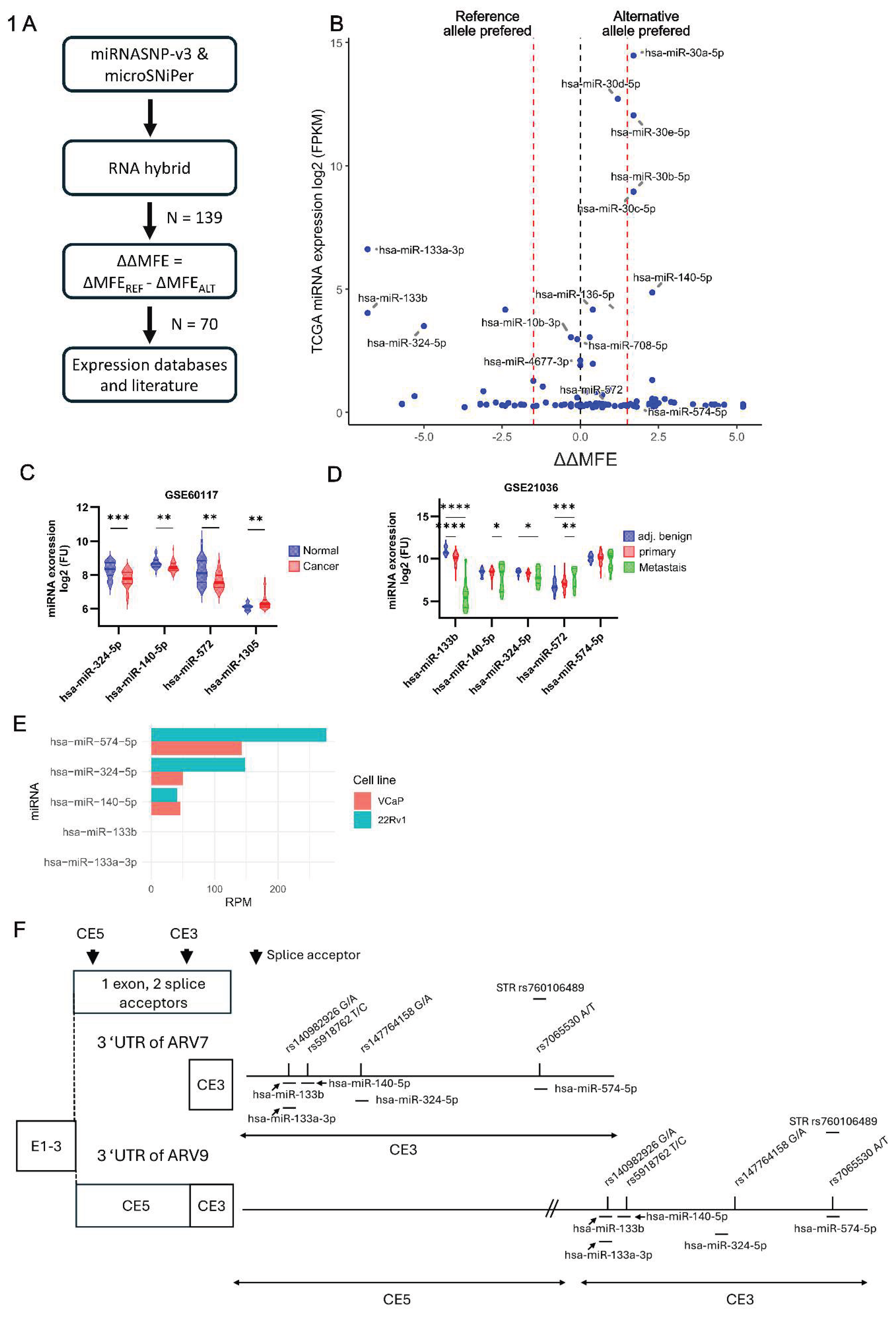
Figure 2.
Effects of SNPs and miRNAs on the expression of a luciferase/ARV7 3´UTR construct. A) Experimental design of the Dual-luciferase assay in HEK293-FT cells. HEK293-FT cells were co-transfected with miRNA mimics and a luciferase-ARV7 3´UTR expression pmirGLO-construct, which was point mutated to the respective alternative allele. B) Results of the Dual-Luciferase assay for the various SNP-miRNA combinations, as depicted. Normalization was done by Renilla luciferase co-expressed by pmirGLO. Statistically significant differences were calculated by two-way ANOVA with p-value adjustment by Tukey (mean ± SEM, n ≥ 3). Ntc, non-targeting control miRNA mimic.
Figure 2.
Effects of SNPs and miRNAs on the expression of a luciferase/ARV7 3´UTR construct. A) Experimental design of the Dual-luciferase assay in HEK293-FT cells. HEK293-FT cells were co-transfected with miRNA mimics and a luciferase-ARV7 3´UTR expression pmirGLO-construct, which was point mutated to the respective alternative allele. B) Results of the Dual-Luciferase assay for the various SNP-miRNA combinations, as depicted. Normalization was done by Renilla luciferase co-expressed by pmirGLO. Statistically significant differences were calculated by two-way ANOVA with p-value adjustment by Tukey (mean ± SEM, n ≥ 3). Ntc, non-targeting control miRNA mimic.
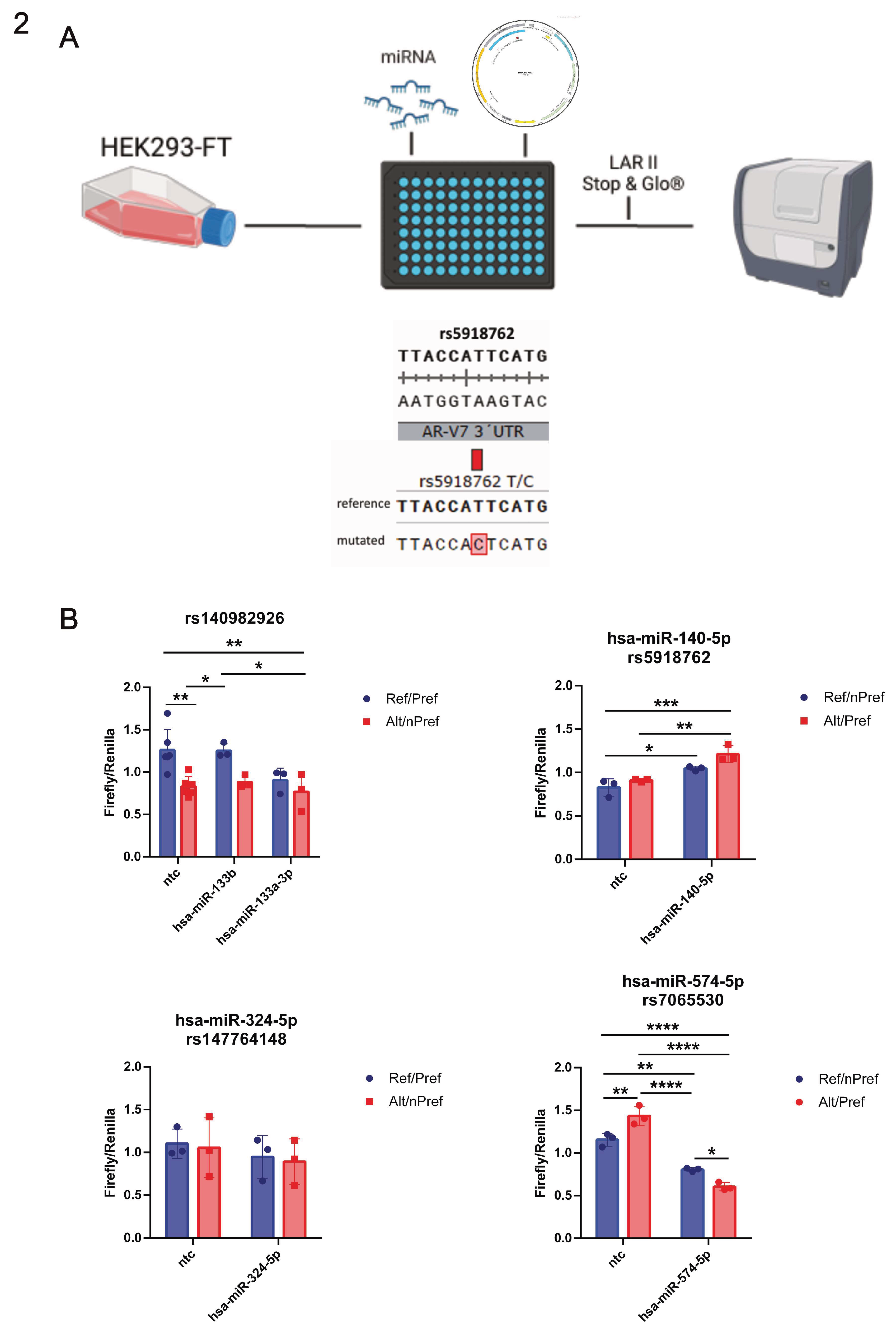
Figure 3.
Impact of miRNAs on the expression of ARV7 mRNA and protein in PCa models. The ARV7-positive cell lines 22Rv1 and DuCaP were transfected with the indicated miRNA mimics or non-targeting control miRNA mimic (ntc). A, C) Expression levels of ARFL and ARV7 mRNAs were quantified by qPCR and normalized to the geometric mean of three housekeeper genes in 22Rv1 (A) and DuCaP (C). B, D) Expression levels of ARFL and ARV7 proteins were quantified by immunoblotting in 22Rv1 (B) and DuCaP (D). GAPDH was used as a loading control. Bar plots indicate the relative expression of ARV7 or ARFL densitometrical analyses normalized to GAPDH of three independent experiments. A-D) Statistical testing was done by one-way ANOVA with p-value adjustment by Dunnett (mean ± SEM, n ≥ 3).
Figure 3.
Impact of miRNAs on the expression of ARV7 mRNA and protein in PCa models. The ARV7-positive cell lines 22Rv1 and DuCaP were transfected with the indicated miRNA mimics or non-targeting control miRNA mimic (ntc). A, C) Expression levels of ARFL and ARV7 mRNAs were quantified by qPCR and normalized to the geometric mean of three housekeeper genes in 22Rv1 (A) and DuCaP (C). B, D) Expression levels of ARFL and ARV7 proteins were quantified by immunoblotting in 22Rv1 (B) and DuCaP (D). GAPDH was used as a loading control. Bar plots indicate the relative expression of ARV7 or ARFL densitometrical analyses normalized to GAPDH of three independent experiments. A-D) Statistical testing was done by one-way ANOVA with p-value adjustment by Dunnett (mean ± SEM, n ≥ 3).
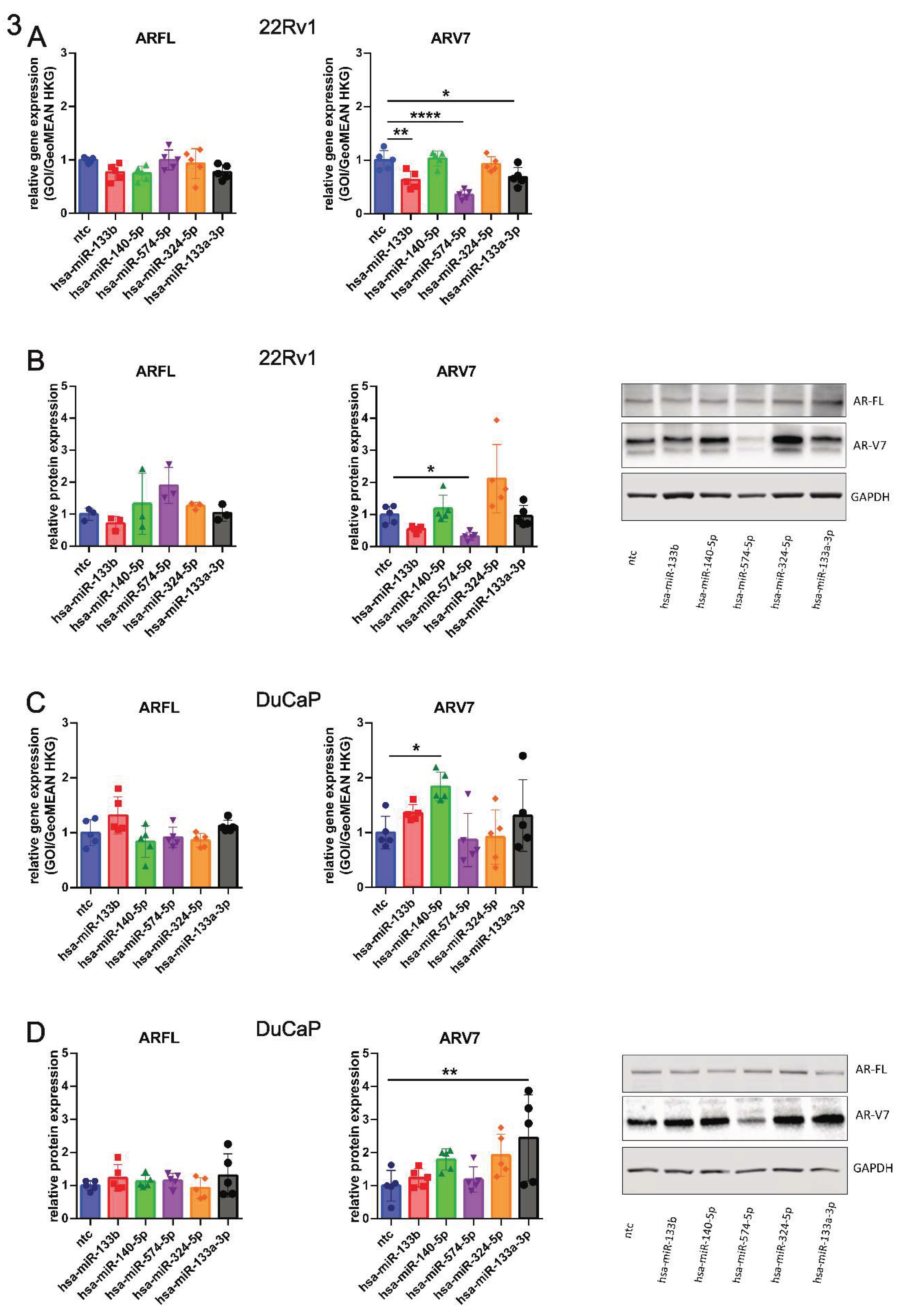
Figure 4.
Binding of hsa-miR-574-5p to the ARV7 3´UTR and potential influence of polymorphisms. A) A previously published database containing miRNA/mRNA binding results as assessed by Argonaute cross-linking immunoprecipitation (AGO ClIP-seq) in the ARV7-positive cell line 22Rv1 was interrogated for the five selected miRNAs from Figure 3 for binding to ARV7 3’ UTR. From the five selected miRNAs, only hsa-miR-574-5p and hsa-miR-324-5p were binding to ARV7´s 3´UTR. B) In silico prediction of the minimal free energy (MFE) of hsa-miR-574-5p for both alleles of SNP rs7065530 in relation to the number of AC repeats of STR rs760106489. Global ALFA allele frequencies in percentages are depicted for STR rs760106489.
Figure 4.
Binding of hsa-miR-574-5p to the ARV7 3´UTR and potential influence of polymorphisms. A) A previously published database containing miRNA/mRNA binding results as assessed by Argonaute cross-linking immunoprecipitation (AGO ClIP-seq) in the ARV7-positive cell line 22Rv1 was interrogated for the five selected miRNAs from Figure 3 for binding to ARV7 3’ UTR. From the five selected miRNAs, only hsa-miR-574-5p and hsa-miR-324-5p were binding to ARV7´s 3´UTR. B) In silico prediction of the minimal free energy (MFE) of hsa-miR-574-5p for both alleles of SNP rs7065530 in relation to the number of AC repeats of STR rs760106489. Global ALFA allele frequencies in percentages are depicted for STR rs760106489.
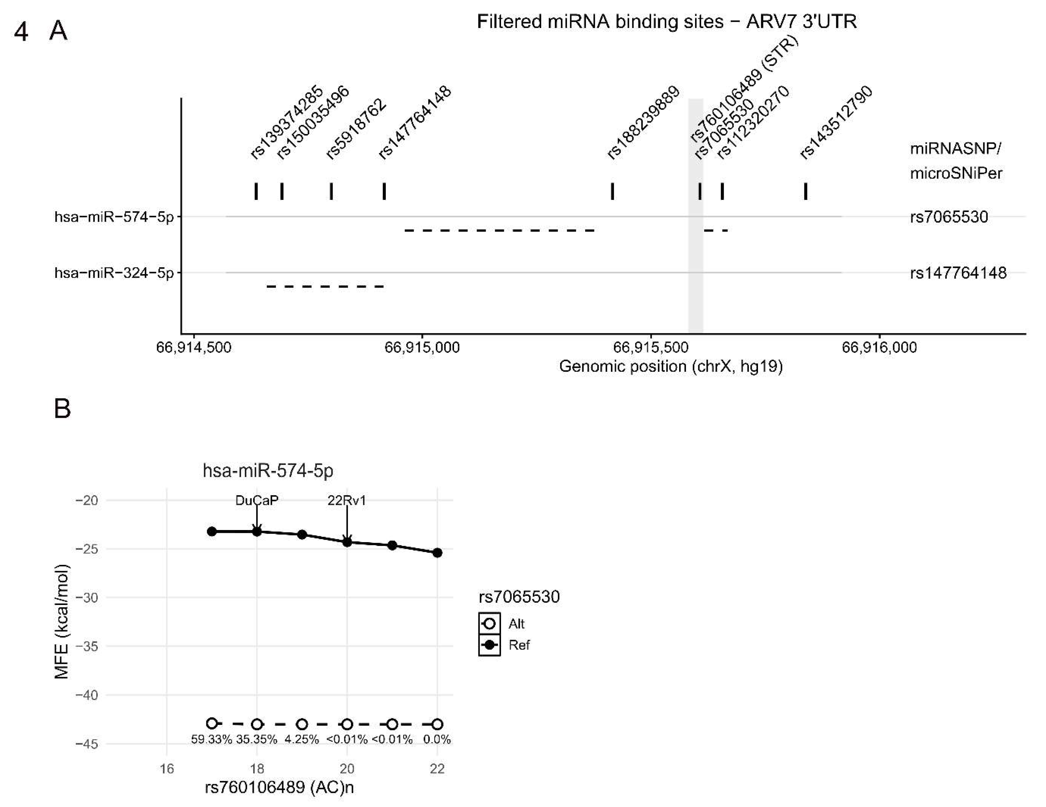
Figure 5.
Impact of hsa-miR-574-5p on proliferation of 22Rv1. 22Rv1 were stably transduced with a doxycycline-inducible shRNA system for the indicated miRNAs. Published shRNA sequences for luciferase (shLuc), ARV7 (shARV7), and ARFL (shARFL) were used as controls. Percentage of cell confluence was assessed in a live cell imaging system over 7 days after induction of the system with 1 µg/ml doxycycline. Statistical analysis of the final timepoint was done by two-way ANOVA with p-value adjustment by Dunnett (mean ± SEM, n = 3).
Figure 5.
Impact of hsa-miR-574-5p on proliferation of 22Rv1. 22Rv1 were stably transduced with a doxycycline-inducible shRNA system for the indicated miRNAs. Published shRNA sequences for luciferase (shLuc), ARV7 (shARV7), and ARFL (shARFL) were used as controls. Percentage of cell confluence was assessed in a live cell imaging system over 7 days after induction of the system with 1 µg/ml doxycycline. Statistical analysis of the final timepoint was done by two-way ANOVA with p-value adjustment by Dunnett (mean ± SEM, n = 3).
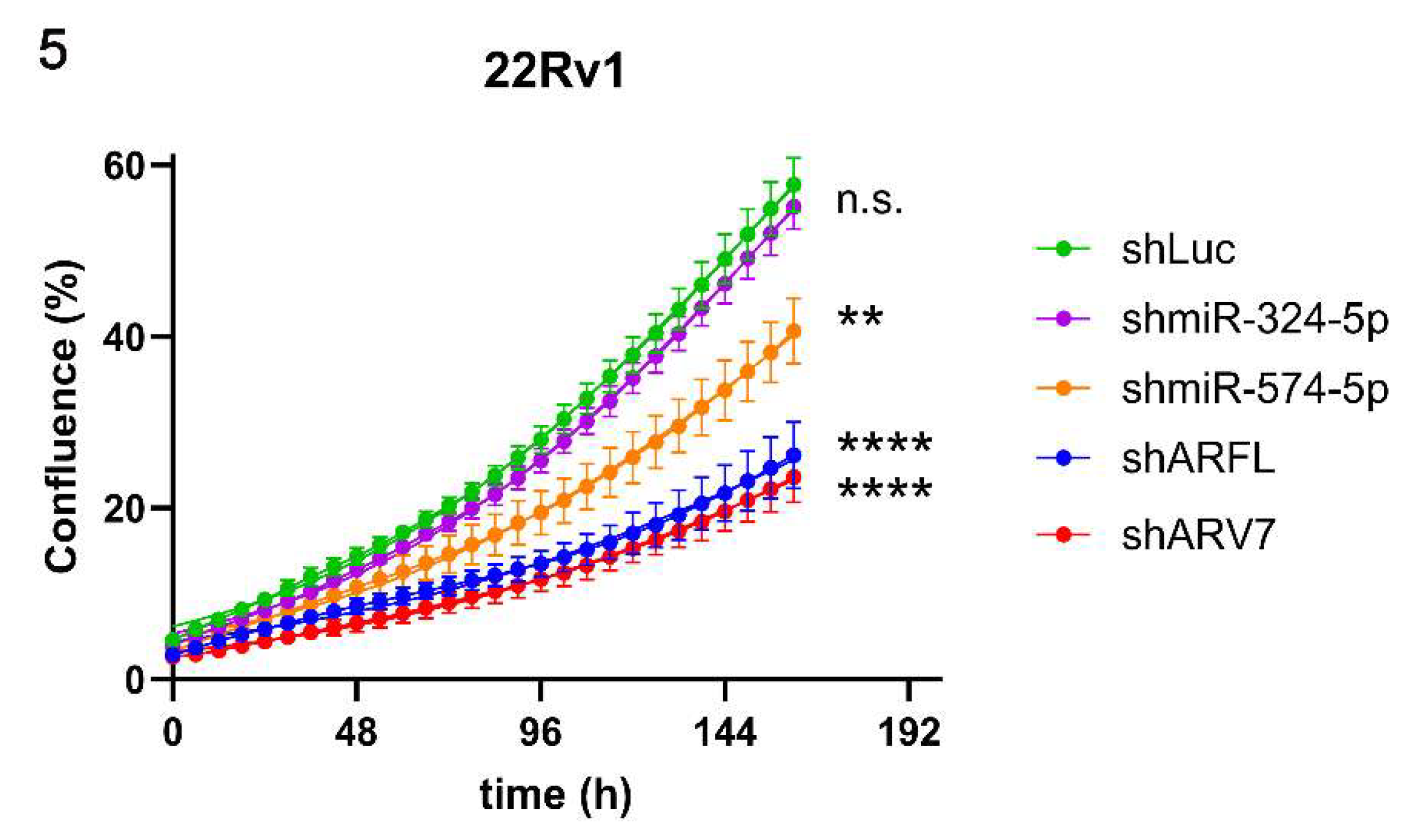

Table 1.
Selected miRNA-SNP combinations and their preferred binding allele. SNP/STR status of 22Rv1 and DuCaP cells was identified via Sanger sequencing.
Table 1.
Selected miRNA-SNP combinations and their preferred binding allele. SNP/STR status of 22Rv1 and DuCaP cells was identified via Sanger sequencing.
| miRNA | SNP | Reference allele | Alternative allele | Preferred allele | 22Rv1 status | DuCaP status |
|---|---|---|---|---|---|---|
| hsa-miR-133b | rs140982926 | G | A | reference | G | G |
| hsa-miR-133a-3p | rs140982926 | G | A | reference | G | G |
| hsa-miR-140-5p | rs5918762 | T | C | alternative | C | C |
| hsa-miR-574-5p | rs7065530 | A | T | alternative | A | A |
| hsa-miR-324-5p | rs147764148 | G | A | reference | G | G |
| STR | ||||||
| hsa-mir-574-5p | rs760106489 | (AC)17 | (AC)8-(AC)23 | reference | 20 | 18 |
Disclaimer/Publisher’s Note: The statements, opinions and data contained in all publications are solely those of the individual author(s) and contributor(s) and not of MDPI and/or the editor(s). MDPI and/or the editor(s) disclaim responsibility for any injury to people or property resulting from any ideas, methods, instructions or products referred to in the content. |
© 2026 by the authors. Licensee MDPI, Basel, Switzerland. This article is an open access article distributed under the terms and conditions of the Creative Commons Attribution (CC BY) license (http://creativecommons.org/licenses/by/4.0/).
Copyright: This open access article is published under a Creative Commons CC BY 4.0 license, which permit the free download, distribution, and reuse, provided that the author and preprint are cited in any reuse.