
Submitted:
15 November 2026
Posted:
17 November 2026
You are already at the latest version
Abstract
IgE-FcεRI interaction plays a central role in the pathogenesis of allergic diseases. Nevertheless, existing anti-IgE antibodies (such as Omalizumab) cannot disrupt the preformed IgE-FcεRI complex at therapeutic concentrations. Here we present a de novo design strategy for bipartite mini-protein inhibitors of the IgE-FcεRI interaction that engage both the Site 1 and Site 2 interfaces of FcεRIα on a single chain, joined by a flexible (GGGGS)n linker. Using the BindCraft framework, we designed Site 1 and Site 2 minibinders and measured their monovalent affinities by surface plasmon resonance (SPR); three Site 1 binders (KD = 3.90, 7.27, and 9.76 nM, respectively) and one Site 2 binder (KD = 2.25 nM) were selected for fusion. A seven-stage computational pipeline determined the fusion orientation, set the linker length, built, sampled, and geometrically validated clash-free (GGGGS)n linkers, resulting in three atomic models for the three Site 1 binder-(GGGGS)n-Site 2 binder fusions. Avidity was predicted with the effective molarity (EM) model that combines the Crothers–Metzger bivalent relation with a worm-like-chain end-density for the effective concentration, showing that the three fusions are predicted to bind FcεRIα with sub-picomolar effective dissociation constants (0.017, 0.009, and 0.003 pM), binding 2.8×10³–1.5×10⁴-fold tighter to FcεRI than IgE itself (KD = 48.4 pM). These predictions suggest that bipartite mini-protein blockers could rival and potentially displace IgE at FcεRIα, offering a mechanistically distinct alternative to current anti-IgE therapies, although experimental validation of the predicted avidity remains to be performed.
Keywords:
allergic diseases
; IgE-FcεRI complex
; de novo protein design
; bivalent inhibitor
; effective molarity model
1. Introduction
Allergic diseases—encompassing allergic rhinitis (AR), allergic Aspergillus sinusitis (AAS), atopic dermatitis (AD), food allergy (FA), and asthma—affect 10% to 30% of the global population and have been identified by WHO as one of the top three disease categories requiring prior management[1,2,3].
Allergic conditions are commonly a type-2 immune response to allergens, in which allergen-specific IgE is a central driver[4]. During allergic sensitization, allergen-specific IgE binds to the high-affinity IgE receptor (FcεRI) expressed on mast cells and basophils. In the effector phase, the ingested allergen cross-links the IgE-FcεRI complexes on the membrane[5] . This leads to degranulation and release of inflammatory mediators that cause the signs and symptoms of an allergic reaction.
The IgE epsilon (ε) heavy chain contains four constant domains (Cε1–Cε4), unlike IgG, which has three constant domains[7]. FcεRI is a member of the immunoglobulin superfamily. It is a tetramer consisting of four subunits: one α subunit (FcεRIα), one β subunit (FcεRIβ), and two γ subunits (FcεRIγ). The FcεRIα subunit has an extracellular region responsible for IgE binding, while the other three subunits are involved in signal transduction[8]. In the transmembrane (TM) regions of the αβγ2 tetramer, the single TM FcεRIα helix is positioned at the center. It packs against the FcεRIγ dimer to form a three-helix bundle, which then assembles with the FcεRIβ four-helix bundle[9]. Before IgE binding, FcεRI presents as a homodimer (αβγ2)2 on membranes. Cholesterol-like molecules embedded within the transmembrane domain may stabilize the dimeric assembly. Upon IgE binding, the dimeric FcεRI dissociates into two protomers, each of which binds to an IgE molecule[10].
IgE-FcεRI binding is among the strongest protein-protein interactions in nature, with a dissociation constant (KD) of 10-9 to 10-10 M. In crystal structures, the two Cε3 domains of IgE bind to two different sites of FcεRI, referred to as Site 1 and Site 2. The high affinity is attributed to the large number of aromatic receptor residues at the binding interface and the large buried surface area of the two binding partners[11].
Anti-IgE therapeutics have been developed for allergy treatment. DARPin E2_79, an ankyrin repeat protein, binds to IgE-Cε3 domains outside the FcεRIα binding site and facilitates dissociation of the IgE-FcεRI complex[12]. Omalizumab is an anti-IgE antibody approved to treat moderate-to-severe perennial allergic asthma, chronic spontaneous urticaria, chronic rhinosinusitis with nasal polyps, and food allergy[13]. It binds to IgE-Cε3 domains outside the FcεRIα binding site, and neutralizes free IgE in serum and reduces surface IgE levels on FcεRI-expressing cells[14]. Native Omalizumab shows negligible disruption of preformed IgE-FcεRI complexes at therapeutic concentrations. However, directed evolution can endow Omalizumab variants (e.g., C02) with potent disruptive activity, enabling rapid dissociation of receptor-bound IgE[15].
Herein, we designed mini-protein binders targeting FcεRIα Sites 1 and 2, connected by (GGGGS)n linkers to form bivalent inhibitors. Computational modeling based on effective molarity (EM)[16,17] predicts that these bivalent binders can achieve extremely high apparent avidity to FcεRIα. We therefore hypothesize that this ultra-high predicted avidity may enable them to actively disrupt preformed IgE-FcεRI complexes.
2. Materials and Methods
2.1. Design of FcεRI-Targeting Mini-Protein Binders
The Site 1 and Site 2 minibinders were designed using BindCraft[18]. The Cryo-EM structure of the IgE/FcεRI complex (PDB ID: 8YWA) was used as the initial template to design mini-protein binders that bind to the IgE-FcεRI interaction interface on the FcεRI side. The coordinates of residues 27–196 of the FcεRI α subunit extracellular region (ECRα) were extracted from the template structure and used as input for BindCraft; this structure was thereafter referred to as the “receptor”. The two interfaces on the extracellular domain of the FcεRI α subunit (ECDα), named Sites 1 and 2, bind to the ε domains in the two heavy chains of IgE. To design the Site 1 binders, the FcεRI ECDα residues 111, 112, 135, 136, 138, 141, 181, 182, 183 were used as the hotspot residues, and to design Site 2 binders, the FcεRI ECDα residues 142, 144, 146, 149, 150, 151, 154, 155, 156, 157, 179 were used. The binder length was set to [60, 150]. The receptor coordinates, hotspot residues, and the binder length were used as the input for BindCraft.
2.2. Affinity Measurement with Surface Plasmon Resonance (SPR)
The biotinylated minibinders were transferred into PBST buffer (1.8 mM KH2PO4, 10 mM Na2HPO4 (pH 7.4), 137 mM NaCl, 2.7 mM KCl, and 0.05% (v/v) Tween 20) and immobilized on an SA chip. Serially diluted FcεRI ECRα were then flowed over the chip in PBST buffer. Binding affinities were measured using a Biacore 8K (GE Healthcare) at 25℃ in the single-cycle mode. Binding kinetics were analyzed with Biacore Insight software (GE Healthcare) using a 1:1 Langmuir binding model. KD values of SPR experiments were obtained with Biacore 8K Evaluation Software (GE Healthcare), using a 1:1 binding model.
2.3. Design of Linkers Between Sites 1 and 2 Binders
The three mini-protein binders targeting Site 1 (s1_00005, s1_00008, s1_00010) and the Site 2 binder (s2_01161) were fused pairwise into three bipartite FcεRI binders, each joining a Site 1 binder to s2_01161 through a (GGGGS)n linker. The construction procedure for all three pairs is a 7-stage pipeline: Stage 1 establishes the fusion orientation (shorter of the two chemically allowed junctions, after Kabsch superposition[19]); Stage 2 sets the (GGGGS)n linker length n from the worm-like-chain force rule (F ≤ 10 pN at lp = 0.4 nm); Stage 3 assembles the construct as a single continuously numbered chain B in one frame, dropping any initiating methionine that becomes internal and leaving the linker as a reserved numbering gap; Stage 4 builds and samples 20 independent linker conformations along the shortest collision-free corridor (grid Dijkstra, 6 Å clearance, route-biased Monte Carlo); Stage 5 scores the samples with Modeller’s DOPE potential[20] and retains the top-ranked model; Stage 6 performs one restrained OpenMM[21] (AMBER14/GBn2) minimization, holding the receptor and both binders while the linker relaxes; Stage 7 applies the post-minimization geometric QC (stage7_qc_select.py) and accepts the model.
Fusion orientation (Stage 1). The receptor in each predicted Site 1 binder–receptor complex was superimposed onto the receptor in the Site 2 binder–receptor complex. Only two chemically distinct fusion topologies are possible, because a peptide junction can link a C-terminus only to an N-terminus: Orientation 1 joins the Site 1 binder C-terminus to the Site 2 binder N-terminus, and Orientation 2 joins the Site 2 binder C-terminus to the Site 1 binder N-terminus. We measured the CA–CA gap between the two junction termini for both orientations and adopted the shorter one. Where the Site 1 binder begins with an initiating methionine that is not part of the mature sequence, that residue becomes an internal, non-native residue in the fusion and is removed before the junction gap is measured. s1_00005 and s1_00008 initiate with Met and lose it; s1_00010 initiates with Ser and is retained unchanged. Removing the Met changes the junction gap, and therefore the required linker length, for the first two pairs.
Linker length (Stage 2). The number of (GGGGS) repeats n was set from the junction gap by a worm-like-chain (WLC) lower bound rather than a single contour-length division. The per-residue rise of a fully extended all-trans peptide is Δmax = 3.645 Å, calculated from the bond lengths (C–N 1.329 Å, N–Cα 1.458 Å, Cα–C 1.525 Å) and bond angles (Cα–C–N 116.2°, C–N–Cα 121.9°, N–Cα–C 111.2°) of the ideal backbone; with N = 5n residues the contour length is lc = N∙∆max. The entropic restoring force of a stretched linker is described by the Marko–Siggia relation (Eq. 1 and 2)[22] .
where kBT is the thermal energy, lp is the persistence length, and d is the gap extension (d ≤ lc). We require F ≤ 10 pN — roughly an order of magnitude below the ~20–150 pN forces needed to unfold protein domains mechanically[23,24] — so the linker tension cannot distort the binders. Substituting lc = N∙∆max,
F decreases monotonically with N, so this inequality is a lower bound on N. The unfolded-state persistence length reported in the literature spans lp ∈ [0.4, 1.0] nm[25,26] (doi:10.1146/annurev-biophys-062215-010915, doi:10.1515/hsz-2014-0132); the flexible end (lp = 0.4 nm) gives the largest restoring force and therefore the most stringent bound. The resulting lower bounds (N ≥ 28, 26, 22 for s1_00005, s1_00008, s1_00010 respectively) are continuous-residue counts. Because the (GGGGS)n linker contributes N = 5n residues, N must be a multiple of 5; rounding each bound up to the nearest multiple of 5 gives the operative lengths N = 30, 30, 25, corresponding to n = 6, 6, 5 for the three pairs. The F ≤ 10 pN threshold is a conservative design choice, not a measured quantity.
Construct assembly (Stage 3). For each pair, the fusion frame is built entirely by a custom NumPy/Biopython builder (fusion_geom.py together with stage1_orient.py, stage2_length.py, and stage3_align_frames.py). It (i) superposes the receptor onto the two binder structures by Kabsch superposition, (ii) measures both candidate junction gaps (orientations A and B) and selects the shorter one, (iii) drops a starting Met from the second binder when that residue is internal in the fusion and therefore not part of the mature sequence, and (iv) writes a single-chain fusion as a continuously numbered amino-acid sequence based on the orientation determined in (ii).
Linker build and sampling (Stage 4). The linker is built by a custom Stage-4 routine (stage4_sample_linker.py calling fusion_geom.build_routed). For each pair, it first computes the shortest collision-free path between the two junction Cα atoms on a 1.5 Å grid by Dijkstra search, enforcing a 6 Å clearance from all receptor atoms and from both binder bodies (the immediate junction neighbourhoods excepted); this path is the corridor the (GGGGS)n segment must follow. It then samples 20 independent linker conformations by a route-biased Monte Carlo: each sample grows the (GGGGS)n backbone from the two peptide junctions under a scoring function that combines (i) closure of the two C–N peptide junctions (target bond length 1.329 Å), (ii) a soft attraction of the linker Cα atoms to the route polyline, (iii) an explicit penalty for any linker atom entering a 6 Å shell around the receptor or either binder body, and (iv) a clash penalty against the receptor.
DOPE scoring and selection (Stage 5). For each pair, the 20 route-aware samples are scored with Modeller’s DOPE statistical potential (assess. DOPE — the only Modeller function used anywhere in the linker pipeline). DOPE ranks the samples, but only the DOPE-best sample is carried forward; the other 19 samples are discarded after ranking. DOPE ranking is only a coarse filter: a long, flexible linker gives a near-degenerate selection (the margin — the DOPE difference between the top and second models, read by the custom stage5_score_dope.py script, can be a few units).
Energy minimization (Stage 6). The DOPE-best sample is carried forward into a single restrained energy minimization performed with OpenMM (AMBER14 force field, GBn2 implicit solvent, no cutoffs), in which the receptor and both binder bodies are position-restrained (2,000 kJ·mol−1·nm−2) and only the linker moves. The minimized potential energy (reported by OpenMM) is not a discriminator.
Geometric quality control and final selection (Stage 7). The decisive step is the post-minimization geometric QC, performed by a custom script (stage7_qc_select.py, which reuses the geometry checks from qc_report.py): zero inter- and intra-chain clash pairs involving a linker residue, linker–receptor and linker–binder clearances ≥ 2.5 Å, junction peptide bonds within 0.05 Å of the ideal 1.329 Å, binder poses preserved to ≤ 0.3 Å Cα-RMSD against the input structures, and the burial check (n_cleft, the number of linker residues enclosed by the receptor, must be 0). The delivered structures are therefore (GGGGS)6 for s1_00005 and s1_00008 and (GGGGS)5 for s1_00010; at these lengths, both the DOPE ranking and all post-minimization QC gates are satisfied.
2.4. Prediction of Avidity of the Linker-Connected Bipartite Site 1 and 2 Binders to FcεRI by the EM Model
The three bipartite fusions each present two independent binding moieties on a single polypeptide chain — the Site-1 mini-protein binder (s1_00005, s1_00008, or s1_00010) and the shared Site-2 binder s2_01161 — tethered by a (GGGGS)n linker. Because the construct engages FcεRI at two sites simultaneously, its avidity cannot be read from a single-interface affinity predictor (such as PPB-Affinity), which returns one patch-level ΔG and contains no avidity term. The bivalent affinity was therefore computed from the two monovalent arm affinities together with the EM of the tethered second arm (here the Site-1 binder at the free end of the (GGGGS)n tether; s2_01161 supplies the fixed anchor at its C-terminus, chain-B residue 139).
Effective avidity model. For an asymmetric bivalent ligand with arm dissociation constants KD,A and KD,B, the effective dissociation constant follows the Crothers–Metzger bivalent-binding relation[27].
where KD,lo = min(KD,A, KD,B), and KD,hi = max(KD,A, KD,B). Two limits are informative: when Ceff≫ Kd,hi, the construct reaches full avidity:
When Ceff → 0, it collapses to the tighter monovalent arm (KD,eff → KD,lo).
EM from the linker worm-like chain (WLC). Suppose that the Site 2 binder is fixed; the EM Ceff of the tethered Site 1 binder (s1_00005, s1_00008, or s1_00010 — the moiety at the free end of the (GGGGS)n tether) at its native Site 1 position was obtained from the WLC end-density (Kratky & Porod, 1949; Kjaergaard et al., 2018) of the (GGGGS)n linker. The linker is a WLC with contour length lc = N·Δmax, with N the number of linker residues (Δmax = 3.645 Å per residue, the CA rise used in linker design) and persistence length lp (swept over 0.4–1.0 nm). Its free end is distributed around the fixed end, with the end-to-end displacement (r) of the tethered end from the anchor under thermal fluctuation of the flexible linker; its mean square ⟨r2⟩ is given by Eq. 5[28,29].
The local concentration of the free end at distance d — the junction gap measured directly from each delivered model — is the 3D Gaussian end-density (van Rosmalen, Krom & Merkx, 2017, Biochemistry 56, 6565, Eq. 6)[30].
Here n3D(r) is the number density (per Å3) of the free end at distance r (the junction gap d) from the anchor; the width is the mean-square end-to-end distance ⟨r2⟩ from Eq. 5, which converts to molarity as
where N_A is Avogadro’s constant (6.022 × 10²³ mol−1). The factor 1030 converts the end-density n3D(r) from Å−3 to m−3, and the factor 1000 in the denominator converts the resulting mol m−3 into mol L−1, so that C_eff is the EM of the tethered arm at its native binding position.
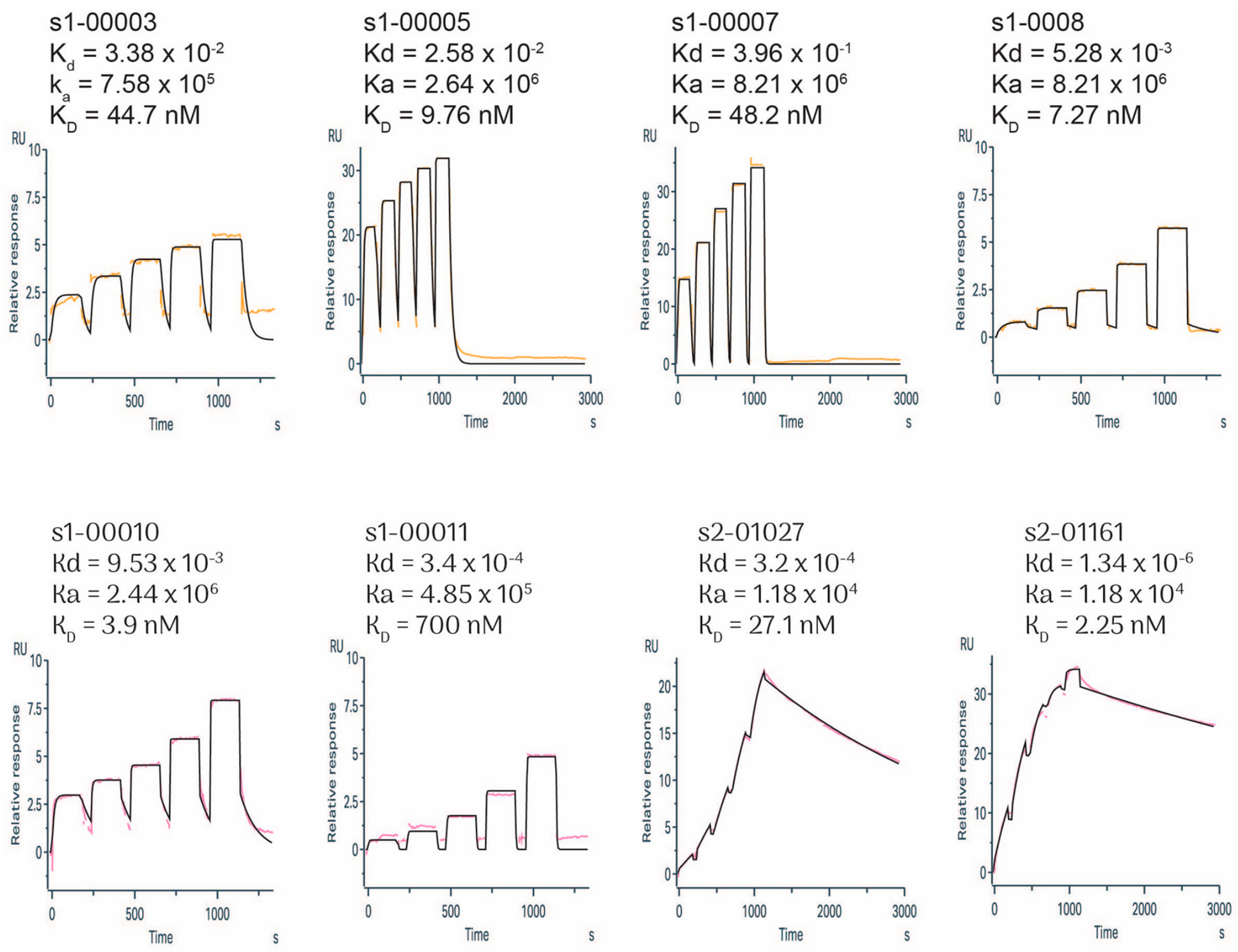
Inputs and references. For the avidity calculation, the monovalent arm affinities are the SPR-measured KD values from Figure 1 (s1_00005 9.76 nM, s1_00008 7.27 nM, s1_00010 3.90 nM, and the shared s2_01161 arm 2.25 nM). The fusions are compared with IgE using our own SPR-measured IgE KD of 48.4 pM. All values were computed by avidity_model.py across the lp sweep.
3. Results
3.1. In Silico Design of Mini-Protein Binders
Mini-protein binders targeting the two interfaces were designed. For Site 1, 10788 trajectories were generated by BindCraft, among which 3189 were accepted. 63780 protein sequences were generated for the accepted trajectories with ProteinMPNN implemented in BindCraft, and 127560 structural models were created. Finally, 843 designs passed the filtration. For Site 2, we generated 10971 trajectories, 100420 protein sequences, and 200840 structural models, and 1318 designs passed the filtration (Table 1). By visual inspection, we found that many designs expected to target Site 2 do not bind to this site but instead bind elsewhere. Therefore, we analyzed the predicted structures of the Site 2 design/target complexes and retained designs that contact at least 5 residues in Site 2 for further analysis. After this screening, only 192 Site 2-targeting designs remained. The top 200 predicted Site 1 structures and the 192 Site 2 structures in complex with the receptor, as output by BindCraft, were visualized in movies (Supplementary movies 1 and 2). The top 10 Sites 1 and 2 candidate binders ranked by i_pTM, as output by BindCraft, along with the top 5 Sites 1 and 2 candidate binders ranked by the affinity predicted by PPB-Affinity[31] (Table 2), were selected for experimental affinity measurement.
3.2. SPR Assay Revealed High-Affinity Binders to Both Sites 1 and 2
The affinities of the 30 Site 1 and Site 2 candidate binders for FcεRI ECRα (Table 2) were determined by SPR. Eight candidates showed positive binding, and 4 had a nanomolar affinity KD (< 10 nM). Among the four, s1_00005, s1_00008, and s1_00010 are Site 1 binders, and s2_01161 is a Site 2 binder. Notably, s2_01161, which has the highest affinity, showed slow association and slow dissociation dynamics, while the three Site 1 binders have fast association and fast dissociation dynamics (Figure 1).
Figure 1.
SPR sensorgram curves for the six Site 1 binders and two Site 2 binders.
3.3. The linker-Connected Bipartite Site 1 and 2 Binders Have Predicted Affinity to FcεRI Higher than IgE
Linkers (GGGGS)n connecting each of the three Site 1 binders (s1_00005, s1_00008, and s1_00010) to the Site 2 binder, s2_01161, all of which have nanomolar affinity, were designed as described in the Materials and Methods. The orientation of the linker is the same for all three pairs (s1_00005-s2_01161, s1_00008-s2_01161, and s1_00010-s2_01161): it runs from the s2_01161 C-terminus to the N-terminus of each Site 1 binder, so chain B is ordered s2_01161–linker–Site 1 binder in the N→C direction; the construct names below list the Site 1 binder first. The linkers for the three pairs are (GGGGS)6, (GGGGS)6, and (GGGGS)5. Finally, the structural models for the three bipartite binders were obtained: the s1_00005 fusion (s1_00005-(GGGGS)6-s2_01161), the s1_00008 fusion (s1_00008-(GGGGS)6-s2_01161), and the s1_00010 fusion (s1_00010-(GGGGS)5-s2_01161). The modeled structures of the three bipartite binders in complex with the FcεRI ECRα were illustrated in Figure 2.
Applying the EM model to the three delivered fusions predicts that all of them bind FcεRI with sub-picomolar avidity, far tighter than IgE itself. Using the SPR-measured monovalent KD as input, the effective KD (KD,eff) at the representative persistence length lp = 0.6 nm is 0.017 pM for the s1_00005 fusion, 0.009 pM for s1_00008, and 0.003 pM for s1_00010 (Table 3). With the SPR-measured IgE KD of 48.4 pM, the fusions remain 2.8×10³–1.5×10⁴-fold tighter.
4. Discussion
In this work, we combined computational design, experimental affinity measurement, and a physics-based linker pipeline to build bivalent mini-protein FcεRI binders. We obtained three geometrically rational bipartite binders with ultra-high predicted avidity to FcεRI. Their binding interfaces overlap with the two IgE-FcεRI binding sites on FcεRI, making them promising competitive inhibitors of the IgE interaction.
In terms of affinity, IgE-FcεRI is a tough target with a reported KD reaching 100 pM (we determined that the KD is 48.4 pM), making it difficult to design competitive inhibitors.The current AI-generated mini-protein binders usually achieve affinities of pM to nM[18,32], consistent with our results that the affinity of the monovalent binders we obtained did not exceed 1 nM, which is not high enough to compete with IgE. However, the two-site characteristics of the IgE-FcεRI binding interface provide special opportunities to design linker-connected bipartite inhibitors, as we did in this study. The power of the bivalent or multivalent minibinder design strategies has been demonstrated previously. The fusion structures of two or three monovalent SARS-CoV-2 RBD minibinders connected by flexible peptide linkers bound so tightly to the target that they exhibit virtually no measurable dissociation upon binding[33]. In this work, we predicted that the bipartite minibinders could achieve sub-picomolar apparent avidities under the position-only EM model, high enough to compete with IgE.
The central hypothesis of this study is that the ultra-high predicted avidity of the bipartite fusions may allow them to disrupt preformed IgE–FcεRI complexes. Because both moieties engage FcεRIα directly, a bivalent ligand with a sub-picomolar effective dissociation constant would, upon encountering an IgE-occupied receptor, bind the unoccupied Site and, through the high on-rate and low off-rate conferred by avidity, hold the receptor and displace the pre-bound IgE. This is mechanistically distinct from the epitope-competition route used by DARPin E2_79 and the C02 variant. Currently, this disruptive capability is a prediction based on the EM model and SPR-measured arm affinities; it has not yet been experimentally demonstrated.
The predicted avidity did not account for orientational entropy. The effective concentration calculated by Eqs. 5-6 only reflects position and can be denoted as Ceff, position; the rotational cost of registering the tethered paratope within a tolerance cone of half-angle θtol (interface-registration angular tolerance) is the solid-angle fraction[17,35],
with
where δ is translational docking tolerance (δ≈1 − 2 Å), and Riface is the interfacial contact radius (Riface ≈ 8 − 12 Å). As a result, the orientation-corrected effective concentration can be calculated as
In addition, lp can vary from 0.4 to 1.0 nm. At the most pessimistic joint values — lp = 0.4 nm (the most flexible chain, giving the smallest Ceff, position) and θ_tol = 5° (the largest penalty, ≈ 5×10²) — the three fusions give KD,eff ≈ 14.8 pM (s1_00005), 7.0 pM (s1_00008), and 2.2 pM (s1_00010), i.e., 3.3×, 6.9×, and 22× tighter than the measured IgE–FcεRI KD of 48.4 pM; even this most pessimistic joint estimate leaves all three competitive with IgE, although these absolute predicted dissociation constants remain conservative upper bounds.
These predictions now call for experimental validation. The most direct test is to measure the affinity of each fusion for FcεRI by SPR or biolayer interferometry and compare it with the modeled sub-picomolar avidity; a measured avidity within the predicted range (after accounting for the orientational penalty) would support the design principle. Competitive binding assays against IgE can establish whether the fusions displace pre-bound IgE from FcεRI, and basophil activation or degranulation assays can test whether they inhibit IgE-mediated signaling. Confirming the modeled linker flexibility — for example by FRET or solution NMR — would close the loop between the worm-like-chain model and the physical construct. Finally, the linker length and composition, as well as the choice of Site-1 binder, remain optimizable once experimental avidity data are available.
In summary, de novo-designed bipartite mini-protein binders linked by (GGGGS)n tethers are predicted to engage FcεRI with sub-picomolar avidity — orders of magnitude tighter than IgE itself — by presenting two nanomolar-affinity arms on a single polypeptide chain. If borne out experimentally, this avidity could enable a new class of FcεRIα-directed inhibitors capable of competing with and potentially displacing IgE at its own receptor, offering a mechanistic alternative to current anti-IgE antibodies.
Supplementary Materials
The following supporting information can be downloaded at the website of this paper posted on Preprints.org.
Code availability
The code for the seven-stage linker-construction pipeline is available at https://github.com/Sunyp-IM/Linker_builder.
References
- Wang, J. et al. Pathogenesis of allergic diseases and implications for therapeutic interventions. Sig Transduct Target Ther 8, 138 (2023).
- Maison, N. & Omony, J. Current challenges in allergic diseases and computational solutions towards personalized medicine. Front. Allergy 7, 1740694 (2026).
- Al-Ahmad, M. et al. Toward international collaboration in allergy care – WAO White Book on Allergy 2026 – Introduction. World Allergy Organization Journal 19, 101397 (2026).
- Caraballo, L. & Llinás-Caballero, K. Overview of allergic disease: Pathogenesis of allergic inflammation – WAO White Book on Allergy 2026 – 2.1. World Allergy Organization Journal 19, 101384 (2026).
- Parkkinen, T., Heiniluoto, H., Jänis, J., Takkinen, K. & Rouvinen, J. Structural Basis for Trivalent Cross-Linking of a Patient-Derived IgE Antibody by the Major Peanut Allergen Ara h 2.0201. Allergy 81, 2036–2046 (2026).
- Barshow, S. et al. The Immunobiology and Treatment of Food Allergy. Annual Review of Immunology 42, 401–425 (2024).
- Sutton, B. J. & Davies, A. M. Structure and dynamics of IgE–receptor interactions: Fcε RI and CD 23/Fcε RII. Immunological Reviews 268, 222–235 (2015).
- Kraft, S. & Kinet, J.-P. New developments in FcεRI regulation, function and inhibition. Nat Rev Immunol 7, 365–378 (2007).
- Deng, M., Du, S., Hou, H. & Xiao, J. Structural insights into the high-affinity IgE receptor FcεRI complex. Nature 633, 952–959 (2024).
- Chen, M., Su, Q. & Shi, Y. Molecular mechanism of IgE-mediated FcεRI activation. Nature 637, 453–460 (2025).
- Garman, S. C., Wurzburg, B. A., Tarchevskaya, S. S., Kinet, J.-P. & Jardetzky, T. S. Structure of the Fc fragment of human IgE bound to its high-affinity receptor FcεRIα. Nature 406, 259–266 (2000).
- Kim, B. et al. Accelerated disassembly of IgE–receptor complexes by a disruptive macromolecular inhibitor. Nature 491, 613–617 (2012).
- Casale, T. B. Omalizumab: The journey of the first anti-IgE approved for asthma and allergic disorders. Journal of Allergy and Clinical Immunology 155, 70–71 (2025).
- Pennington, L. F. et al. Structural basis of omalizumab therapy and omalizumab-mediated IgE exchange. Nat Commun 7, 11610 (2016).
- Pennington, L. F. et al. Directed evolution of and structural insights into antibody-mediated disruption of a stable receptor-ligand complex. Nat Commun 12, 7069 (2021).
- Thermodynamic Effective Molarities for Supramolecular Complexes. in Advances in Physical Organic Chemistry vol. 50 77–118 (Elsevier, 2016).
- Krishnamurthy, V. M., Semetey, V., Bracher, P. J., Shen, N. & Whitesides, G. M. Dependence of Effective Molarity on Linker Length for an Intramolecular Protein−Ligand System. J. Am. Chem. Soc. 129, 1312–1320 (2007).
- Pacesa, M. et al. One-shot design of functional protein binders with BindCraft. Nature (2025). [CrossRef]
- Lawrence, J., Bernal, J. & Witzgall, C. A Purely Algebraic Justification of the Kabsch-Umeyama Algorithm. J. RES. NATL. INST. STAN. 124, 124028 (2019).
- Shen, M. & Sali, A. Statistical potential for assessment and prediction of protein structures. Protein Science 15, 2507–2524 (2006).
- Eastman, P. et al. OpenMM 8: Molecular Dynamics Simulation with Machine Learning Potentials. J. Phys. Chem. B 128, 109–116 (2024).
- Marko, J. F. & Siggia, E. D. Stretching DNA. Macromolecules 28, 8759–8770 (1995).
- Rief, M., Gautel, M., Oesterhelt, F., Fernandez, J. M. & Gaub, H. E. Reversible Unfolding of Individual Titin Immunoglobulin Domains by AFM. Science 276, 1109–1112 (1997).
- Kellermayer, M. S. Z., Smith, S. B., Granzier, H. L. & Bustamante, C. Folding-Unfolding Transitions in Single Titin Molecules Characterized with Laser Tweezers. Science 276, 1112–1116 (1997).
- Schuler, B., Soranno, A., Hofmann, H. & Nettels, D. Single-Molecule FRET Spectroscopy and the Polymer Physics of Unfolded and Intrinsically Disordered Proteins. Annu. Rev. Biophys. 45, 207–231 (2016).
- Hofmann, H. Single-molecule spectroscopy of unfolded proteins and chaperonin action. Biological Chemistry 395, 689–698 (2014).
- Sørensen, C. S., Jendroszek, A. & Kjaergaard, M. Linker Dependence of Avidity in Multivalent Interactions Between Disordered Proteins. Journal of Molecular Biology 431, 4784–4795 (2019).
- Kratky, O. & Porod, G. Röntgenuntersuchung gelöster Fadenmoleküle. Recl. Trav. Chim. Pays-Bas 68, 1106–1122 (1949).
- Sørensen, C. S. & Kjaergaard, M. Disordered Protein Linkers: Predicting Effective Concentrations using Polymer Physics. Biophysical Journal 114, 368a–369a (2018).
- Van Rosmalen, M., Krom, M. & Merkx, M. Tuning the Flexibility of Glycine-Serine Linkers To Allow Rational Design of Multidomain Proteins. Biochemistry 56, 6565–6574 (2017).
- Liu, H. PPB-Affinity: Protein-Protein Binding Affinity dataset for AI-based protein drug discovery. Zenodo (2024). [CrossRef]
- Barykina, N. V. & Verkhusha, V. V. Intracellular protein binders for imaging, control and future therapeutics. Nat. Biomed. Eng (2026) . [CrossRef]
- Hunt, A. C. et al. Multivalent designed proteins neutralize SARS-CoV-2 variants of concern and confer protection against infection in mice. Sci. Transl. Med. 14, eabn1252 (2022).
- Ekaterina Shevchenko & Poso, A. Linking ATP and allosteric sites to achieve superadditive binding with bivalent EGFR kinase inhibitors. (2023) . [CrossRef]
- Zhou, H.-X. & Gilson, M. K. Theory of Free Energy and Entropy in Noncovalent Binding. Chem. Rev. 109, 4092–4107 (2009).
Figure 2.
The modeled structures of the three bipartite binders in complex with the FcεRI ECRα.
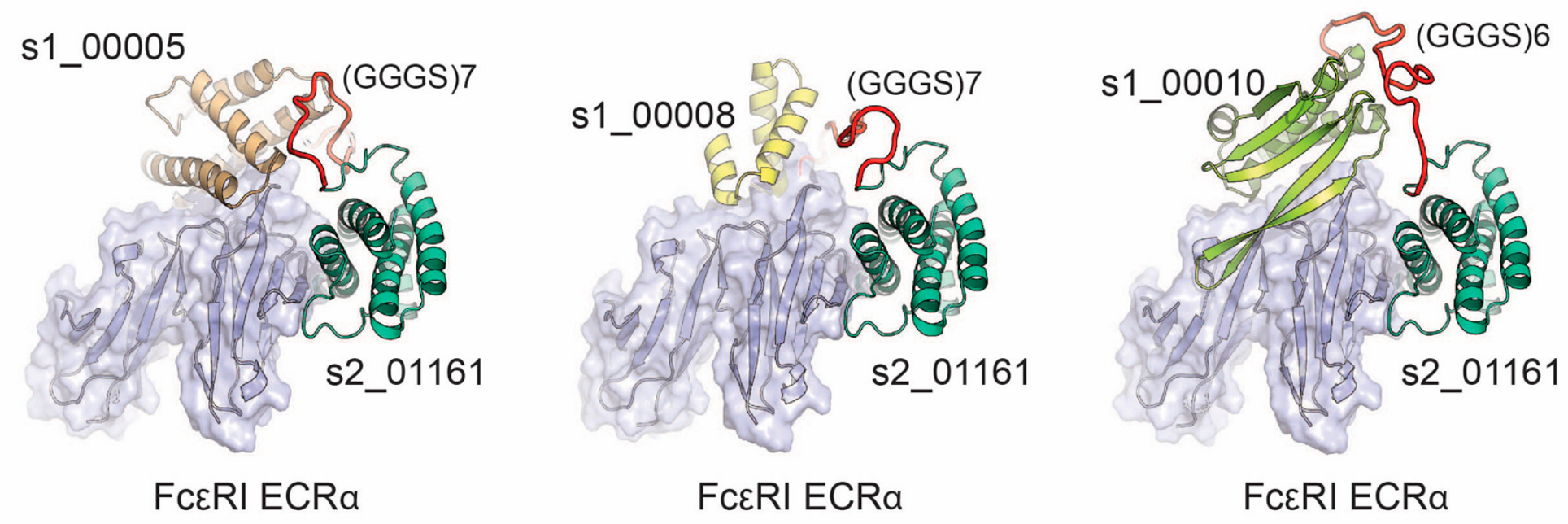

Table 1.
Trajectories, sequences, and models generated by BindCraft
| Site | Total runs | Total trajectories | Accepted trajectories | Rejected trajectories | mpnn sequences | models | Accepted designs |
| 1 | 4 | 10788 | 3189 | 7599 | 63780 | 127560 | 843 |
| 2 | 3 | 10971 | 5021 | 5850 | 100420 | 200840 | 1318 |
Table 2.
Sites 1 and 2 candidates with the highest i_pTM or predicted affinity
| No.* | Name | Average_i_pTM | Average_pLDDT | PPB-Affinity_dG_pred |
| 1 | s1_00001 | 0.88 | 0.95 | -10.54084949 |
| 2 | s1_00002 | 0.88 | 0.96 | -10.63850937 |
| 3 | s1_00003 | 0.86 | 0.96 | -8.600048161 |
| 4 | s1_00004 | 0.86 | 0.96 | -8.511462498 |
| 5 | s1_00005 | 0.86 | 0.95 | -10.40296783 |
| 6 | s1_00006 | 0.86 | 0.96 | -10.90649986 |
| 7 | s1_00007 | 0.86 | 0.95 | -11.11970177 |
| 8 | s1_00008 | 0.86 | 0.94 | -9.657225418 |
| 9 | s1_00009 | 0.86 | 0.94 | -9.624960899 |
| 10 | s1_00010 | 0.86 | 0.94 | -9.474826527 |
| 11 | s1_00231 | 0.8 | 0.94 | -11.8228611 |
| 12 | s1_00726 | 0.69 | 0.88 | -11.77914238 |
| 13 | s1_00044 | 0.84 | 0.95 | -11.65342064 |
| 14 | s1_00721 | 0.69 | 0.88 | -11.56102753 |
| 15 | s1_00054 | 0.83 | 0.95 | -11.55054512 |
| 16 | s2_00002 | 0.89 | 0.92 | -9.733365631 |
| 17 | s2_00004 | 0.89 | 0.94 | -10.38209515 |
| 18 | s2_00007 | 0.88 | 0.94 | -9.499232101 |
| 19 | s2_00011 | 0.88 | 0.91 | -10.04623337 |
| 20 | s2_00013 | 0.87 | 0.93 | -10.52284451 |
| 21 | s2_00015 | 0.86 | 0.91 | -9.027440643 |
| 22 | s2_00018 | 0.86 | 0.9 | -9.101892281 |
| 23 | s2_00027 | 0.86 | 0.92 | -10.69266663 |
| 24 | s2_00035 | 0.85 | 0.93 | -10.44034023 |
| 25 | s2_00037 | 0.85 | 0.96 | -9.915475845 |
| 26 | s2_01027 | 0.71 | 0.92 | -11.36397038 |
| 27 | s2_01119 | 0.7 | 0.92 | -11.30195313 |
| 28 | s2_01294 | 0.65 | 0.94 | -11.17784424 |
| 29 | s2_00407 | 0.79 | 0.94 | -10.89803715 |
| 30 | s2_01161 | 0.69 | 0.92 | -10.85950775 |
*No. 1-15 are Site 1 binders, and No. 16-30 are Site 2 binders. No. 1-10 and 16-25 are the top 10 Site 1 and 2 binders ranked by i_pTM, respectively. No. 11-15 and 26-30 are the top 5 Site 1 and 2 binders ranked by predicted affinity, respectively.
Table 3.
KD,eff of the bipartite binders calculated from the EM model
| Fusion | Junction gap (Å) | Ceff (mM) | KD,eff (pM) | Fold tighter than IgE |
| s1_00005 | 43.5 | 1.28 | 0.017 | ~2.8×10³ |
| s1_00008 | 40.4 | 1.74 | 0.009 | ~5.2×10³ |
| s1_00010 | 35.1 | 2.74 | 0.003 | ~1.5×10⁴ |
Disclaimer/Publisher’s Note: The statements, opinions and data contained in all publications are solely those of the individual author(s) and contributor(s) and not of MDPI and/or the editor(s). MDPI and/or the editor(s) disclaim responsibility for any injury to people or property resulting from any ideas, methods, instructions or products referred to in the content. |
© 2026 by the authors. Licensee MDPI, Basel, Switzerland. This article is an open access article distributed under the terms and conditions of the Creative Commons Attribution (CC BY) license (http://creativecommons.org/licenses/by/4.0/).
Copyright: This open access article is published under a Creative Commons CC BY 4.0 license, which permit the free download, distribution, and reuse, provided that the author and preprint are cited in any reuse.