

Submitted:
10 November 2026
Posted:
12 November 2026
You are already at the latest version
Abstract
The gastrointestinal tract is a dynamic regulatory interface in which the enteric nervous system (ENS), supported by an extensive enteric glial cell (EGC) network, interacts with the enteric immune system (EIS) and gut microbiota to maintain epithelial barrier integrity, neuroplasticity, motility, and tissue repair. In eubiosis, coordinated signaling across this tripartite axis supports intestinal homeostasis. Dysbiosis, however, can reduce short-chain fatty acid (SCFA) availability while increasing exposure to pathogen-associated molecular patterns (PAMPs), including lipopolysaccharide (LPS) and others. These changes may activate NF-κB and NLRP3 inflammasome signaling in enteric glial and immune cells, thereby promoting oxidative and nitrosative stress, neuronal hyperexcitability, apoptosis, barrier dysfunction, and local neuroinflammation, with possible consequences for gut–brain communication. This review examines the mechanisms through which dietary phytochemicals—including capsaicinoids, resveratrol, curcumin, catechins, quercetin, and sulforaphane—may modulate this ENS–EIS–microbiota axis. We focus on their actions at transient receptor potential channels, 5-hydroxytryptamine receptors, nicotinic acetylcholine receptors, SIRT1, and Nrf2-associated pathways, and their potential to limit inflammation, oxidative stress, and excitotoxic signaling. We also identify knowledge gaps and discuss future research and translational priorities, including human-relevant organoid models, spatial transcriptomics, and machine-learning-enabled multi-omics to define microbiota-derived metabolite–receptor interactions and guide personalized dietary strategies for enteric neuroglial health.
Keywords:
enteric neuroglia
; gut dysbiosis
; neuro-immune-microbiome axis
; dietary phytochemicals
; metabolites & metabiotics
1. Introduction
The gastrointestinal tract has evolved beyond a basic nutrient-absorbing structure into a complex regulatory hub where neuronal, immune and microbial entities converge to maintain systemic homeostasis (Furness et al., 2013). Central to this complex co-existance to systemic development is the enteric neuroglial cells (EGCs) regulatory network, which forms a dense syncytium throughout the gut wall and serve as essential mediators of intestinal barrier integrity, neuroplasticity, and mucosal defense (Veiga-Fernandes & Mucida, 2016). Far from acting merely as passive structural support, enteric neuroglia actively integrates chemical and mechanical cues from both the luminal environment and the host’s immune system. Co-evolutionary adaptations have further intertwined EGC functionality with the commensal microbiota—whose numerical abundance rivals host somatic cells—establishing a dynamic neuro-immune-microbiome interplay as a super-organ vital for gastrointestinal health and signaling (Yoo et al., 2020).
Maintaining the health and structural integrity of enteric neuroglia is increasingly recognized as a cornerstone of the function of overall digestive physiology and disease prevention. Disruption of glial homeostasis impairs mucosal repair mechanisms and triggers inflammatory cascades implicated in metabolic, gastrointestinal, and neurodegenerative pathologies (Balemba & Gulbransen, 2025; Q. Wang et al., 2021). Notably, dietary factors act as key extrinsic regulators of this network, directly influencing glial reactivity, neurotrophic factor secretion, and cellular survival (López-Gómez et al., 2021). In this review, we examine the molecular mechanisms governing enteric neuroglial health within the neuro-immune-microbiome framework, with a dedicated focus on how dietary phytochemicals and plant-derived bioactive molecules modulate glial responses and fortify gut functionality. Finally, we explore emerging therapeutic perspectives and outline future research directions aimed at targeting enteric neuroglia through targeted nutritional strategies.
2. Brain in the Gut
2.1. The Enteric Nervous System (ENS)
The enteric nervous system (ENS) constitutes an extensive, autonomous intrinsic neural network distributed along the entire length of the gastrointestinal (GI) tract. Often described as the body’s "second brain," the ENS contains hundreds of millions of neurons and a vast population of neuroglia—a structural and computational complexity that rivals the spinal cord. This intricate architecture enables the ENS to autonomously govern essential gastrointestinal functions, including peristaltic motility, local blood flow, fluid secretion, nutrient absorption, and mucosal barrier immune-functions, without requiring continuous instruction from the central nervous system (Furness, 2012; Sharkey & Mawe, 2023). Structurally, primary neuronal cell bodies and glia are organized into two major concentric ganglionic networks: the myenteric (Auerbach’s) plexus, situated between the outer longitudinal and inner circular smooth muscle layers to control motor patterns, and the submucosal (Meissner’s) plexus, embedded in the submucosa to regulate local secretion and vascular tone (Furness, 2012). Beyond these main ganglionic trunks, nerve projections extend into the mucosa, epithelium, and muscularis layers (Sharkey & Mawe, 2023). Enteric glial cells (EGCs) display distinct morphotypes—intraganglionic, interganglionic, mucosal extraganglionic, and intramuscular glia—that exhibit dynamic functional plasticity, actively sensing microenvironmental cues and maintaining epithelial barrier homeostasis (Boesmans et al., 2019; Seguella & Gulbransen, 2021).
At the molecular level, signal transmission within this autonomous network relies on a diverse chemical coding system encompassing a variety of distinct neurotransmitters and neuromodulators. Excitatory motor and intraneuronal pathways predominantly utilize small-molecule transmitters such as acetylcholine (ACh) and tachykinin neuropeptides (substance P and neurokinin A) to drive muscle contraction and secretion (Furness, 2012). Enteric circuits also integrate biogenic amines (serotonin/5-HT, GABA, dopamine); and enteric neuro-glial cells also express an array of ionotropic receptors and G-protein-coupled receptors (GPCRs), allowing them to execute reflex arcs and respond to local signals from smooth muscle, enteroendocrine cells, and mucosal capillaries independently of central brain inputs (Drokhlyansky et al., 2020).
2.2. The ENS-CNS Connection
Though the enteric nervous system (ENS) functions as an intrinsic, semi-autonomous neural processor, it remains anatomically and functionally integrated with the broader autonomic nervous system (ANS) and central nervous system (CNS) through the gut-brain axis (Spencer & Hu, 2020; Travagli & Anselmi, 2016). This integration is mediated by bidirectional sympathetic and parasympathetic extrinsic innervation pathways that establish direct connections with enteric ganglia and mucosal target tissues (Veiga-Fernandes & Mucida, 2016). Parasympathetic efferent fibers, originating primarily from the dorsal motor nucleus of the vagus (DMNV) in the brainstem, travel via the vagus nerve, or the pelvic splanchnic nerves in the distal colon, to synapse with cholinergic and peptidergic postganglionic neurons within the myenteric and submucosal plexuses, thereby promoting gastrointestinal motility and glandular secretion (Browning & Travagli, 2014; Travagli & Anselmi, 2016). In contrast, sympathetic preganglionic fibers arising from the thoracolumbar spinal cord relay through prevertebral ganglia, including the celiac and mesenteric ganglia, and give rise to noradrenergic postganglionic projections that enter the gut wall to inhibit smooth muscle contraction, constrict mucosal vasculature, and suppress enteric neurotransmission (Browning & Travagli, 2014; Spencer et al., 2020). Sensory information from the gastrointestinal tract is conveyed by vagal nodose ganglion and spinal dorsal root ganglion (DRG) afferent fibers, which innervate the intestinal mucosa, muscularis, and serosa before projecting centrally to the nucleus tractus solitarius (NTS) and spinal dorsal horn. These afferents provide continuous real-time monitoring of mechanical distension, luminal nutrient composition, and noxious inflammatory stimuli (Bellono et al., 2017). Figure 1 illustrates the anatomical organization and bidirectional communication pathways constituting the gut-brain axis.
Physiologically, this layered integration operates as a hierarchical autonomic reflex loop where the ENS computes routine local motor and secretory reflexes, while central networks in the hypothalamus, amygdala, and insular cortex modulate baseline enteric tone in response to metabolic, circadian, and emotional stressors (Bonaz et al., 2018; Kaelberer et al., 2018). Within the gut wall, the neuronal–glial–epithelial unit serves as the primary neuro-immune-microbial sensing interface for this reflex arc (Seguella & Gulbransen, 2021).
3. Gut Microbiota and the Enteric Microenvironment in Dysbiosis
The human gut houses a vast and intricate ecosystem teeming with thousands of microbial species. Shaped by an individual’s genetics and environment, this microbial community relies on rich diversity, metabolic adaptability, and complex microbe-to-microbe and microbe-to-host relationships to sustain an overall healthy environment for both the microbial community (gut microbiota) and the human host. The composition of gut microbiota is subjected to changes due to age, environmental and stress factors that may lead to instability to the balanced microbiota equilibria and may cause dysbiosis. Changes in gut microbiota is also associated with several disease conditions including diabetes, liver diseases and neurodegenerative diseases and diet or lifestyle patterns which lead to obesity (Das & Nair, 2019; Fukui, 2019; L. Wang et al., 2020). Though; the precise mechanism remains largely unknown; several intrinsic and extrinsic factors have been reported to impact gut microbiota composition profoundly. Host genetics, age and intestinal diseases are considered as the prominent intrinsic factors which have been associated with gut dysbiosis.
Importantly, variants of host genes encoding immune system-related proteins (e.g., Van Gogh-like protein 1, known as VANGL1 and Leucine-rich repeat and Ig domain-containing Nogo receptor-interacting protein 2 known as LINGO2) had been reported to modulate gut microbe populations (Goodrich et al., 2016; Kurilshikov et al., 2017). Alterations of healthy gut microbiota have been reported to be associated with older age of the human hosts resulted in significant reduction in short-chain fatty acid (SCFA) producing microbes thereby changing the Bacillota (Firmicutes) to Bacteroidota ratio and enrichment of Bacteroidetes and opportunistic bacteria in intestine (An et al., 2018; Favaron et al., 2023; Mariat et al., 2009; Salazar et al., 2017). Low-grade chronic inflammation, known as inflammaging and neuro-immune activation have been implicated with low SCFA production with possibility of secondary intestinal infection (Bosco & Noti, 2021; Favaron et al., 2023). Gut dysbiosis not only affects healthy gut environment; but also linked with several extra-intestinal diseases including colorectal cancer, Parkinson’s disease, and inflammatory bowel disease. Increased levels of potential pathogens, such as Bacteroides fragilis, Ruminococcus gnavus, E. coli, Clostridium bolteae, Ruminococcus torques and Clostridium hathewayi with significant reduction in beneficial species such as Roseburia intestinalis, Faecalibacterium prausnitzii, Eubacterium rectale, and Clostridium leptum have been observed for the patients with inflammatory bowel diseases (Qiu et al., 2022). Patients suffering from irritable bowel syndrome display similar reduction in health-associated microbial species such as Bifidobacterium and Lactobacillus species with substantial increase in pathogenic bacteria such as E. coli and Enterobacter species in their guts.
A few extrinsic factors also found to be involved with gut microbiota dysbiosis as well. These are different climate conditions related to specific geographical regions, several distinct lifestyle patters (unhealthy diet, excessive consumption of alcoholic beverages, smoking and lack of adequate sleep) and exposure to xenobiotic and environmental chemicals (Benedict et al., 2016; Bisanz et al., 2019; Brown et al., 2012; Day & Kumamoto, 2022; Garcia et al., 2022; He et al., 2018; Nolan-Kenney et al., 2020; Stewart et al., 2018; Sun et al., 2023; Z. Wang et al., 2021; Yatsunenko et al., 2012). Among these, diet has probably most dominant effect on shaping human gut microbiota. Long term consumption of high fat, high sugar, processed food and artificial sweeteners can significantly alter gut microbial composition which may lead to dysbiosis (Bisanz et al., 2019; Brown et al., 2012; Garcia et al., 2022). Increased Bacillota to Bacteroidota (Firmicutes to Bacteroidetes) ratio as well as a shift in the microbial composition has also been observed to be associated with high fat food consumption (Cheng et al., 2022). In many cases these food habits may cause irreversible change to healthy gut microbiota (Malesza et al., 2021; Velasquez, 2018). Smoking tobacco results in an increased relative abundance of Prevotella and simultaneous decreased Bacteroides in individuals in their guts (Stewart et al., 2018). Smoking also resulted in increased population of alphaproteobacteria and Erysipelotrichi-to-Catenibacterium lineage bacteria among the individuals (Nolan-Kenney et al., 2020). Alcohol consumption is linked with decreased beneficial species such as Faecalibacterium and Roseburia with an increase in opportunistic pathogens Streptococcus and Enterococcus (Day & Kumamoto, 2022). Sleep deprivation causes reduction in beneficial bacterial population (Akkermansia, Bacteroides, Bacteroidetes and Faecalibacterium) and increases bacterial population belonging to Firmicutes and Aeromonas which leads to an elevated Firmicutes-to-Bacteroidetes ratio (Sun et al., 2023). Xenobiotic agents, such as antimicrobial agents and environmental toxic chemicals can also lead to profound effect on gut microbiota. Among the various xenobiotic agents; the most distinct effect is observed with the use of antimicrobial agents causing altered gut microbial compositions, reduced microbial diversity, generation of antibiotic tolerance and reducing host resistance to colonization of pathogenic microbes (Lange et al., 2016). These changes may be short or long term; depending on antibiotic type, dose and duration of treatment, and may be partly due to the host genetics, age and immunity (Fassarella et al., 2021).
4. Enteric Immune System (EIS) Interference
4.1. The Enteric Immune System
The barrier immune system represents the largest site of immunological activity, occupying the mucosal surfaces of the intestinal, respiratory, and urogenital tracts, which collectively provide an internal surface area nearly 100-fold greater than the external body surface. Approximately 70% of all immune cells are dedicated to barrier protection, exceeding the immunocyte populations found within classical immune organs (Helander & Fändriks, 2014; Vijay-Kumar et al., 2014). Among barrier tissues, the gut is considered an evolutionarily ancient site where adaptive immunity first emerged. Continuous exposure to dietary antigens, ingested microorganisms, and digestion-derived molecules provided the selective pressure that drove the transition from generalized innate defenses to a highly specialized adaptive immune system (Graham & Xavier, 2023). This evolutionary process created a unique challenge: the intestinal immune system must tolerate a vast array of dietary and microbial inputs while remaining capable of mounting robust responses when tissue damage or pathogen invasion exceeds a critical threshold. Coevolution with the gut microbiota, the structural resilience of mucosal barriers, and close integration with an intricate neuronal network have collectively shaped the gut into a finely regulated interface of immune tolerance and host defense (Iliev et al., 2025; McKay et al., 2024; Miranda-Waldetario & Curotto de Lafaille, 2024; Okumura & Takeda, 2025). Therefore, understanding intestinal immune function requires consideration of these interconnected influences.
Within the intestine, immune cells are distributed both within and beyond organized lymphoid structures. They are embedded throughout the epithelial-mucosal layer, densely localized in the lamina propria, and positioned around microvascular networks in close proximity to neuronal branches extending into the lamina propria, epithelium, and muscularis externa (Vancamelbeke & Vermeire, 2017). Consequently, immune surveillance is not confined to isolated lymphoid follicles (ILFs) or gut-associated lymphoid tissues (GALTs), such as Peyer’s patches. Instead, immune cells continuously sample and interact with microbes and potential pathogens across both organized lymphoid structures and surrounding tissues (Lavelle et al., 2010; Rakoff-Nahoum et al., 2004). Antigen uptake occurs through specialized microfold (M) cells, while dendritic cells (DCs) coordinate interactions with B cells, T-cell subsets, and innate lymphoid cells (ILCs). Patrolling macrophages, DCs, plasma cells, effector and memory T cells, and naïve lymphocytes remain strategically positioned to sense and respond to local environmental and neuroimmune cues (Brandtzaeg et al., 2008; Cao et al., 2026; Mörbe et al., 2021; Mowat & Agace, 2014; Silva-Sanchez & Randall, 2020; Vivier, 2021).
The mucus layer, which is primarily formed by mucous and mucins secreted from the goblet cells, provides the primary protective layer against potential pathogens, where the paneth cells secret lysozymes, defensins (also known as antimicrobial protein or AMP), immunoregulatory proteins like IL-22 etc play important roles for maintaining host immunity, epithelial cell regeneration and mucus homeostasis (Keir et al., 2020; Maldonado-Contreras & McCormick, 2011; Pelaseyed et al., 2014). Along with AMP, secretory immunoglobin A (sIgA) in this mucous layer create the first protective layer, which can normally be penetrated by gut microbiota and fewer pathogens (Maldonado-Contreras & McCormick, 2011). At the core of this environment, innate lymphoid cells (ILCs) act as early, rapid-response sentinels that lack rearranged, antigen-specific receptors, relying instead on environmental sensors and cytokine signals for activation. Group 3 ILCs (ILC3s), which predominate in the intestinal lamina propria, express the orphan nuclear receptor RORγt along with environmental sensors such as the aryl hydrocarbon receptor (AHR) and short-chain fatty acid (SCFA) receptors like FFAR2 (Ryu et al., 2023). Activation of these pathways drives ILC3s to produce interleukin-22 (IL-22), which acts on epithelial cells to induce antimicrobial peptides (e.g., RegIIIγ) and reinforce mucosal repair (Ryu et al., 2023). Crucially, a subset of RORγt+ ILC3s expresses major histocompatibility complex class II (MHC-II) molecules without classic costimulatory machinery; these non-classical antigen-presenting cells load luminal commensal peptides onto MHC-II to engage CD4+ T cells, inducing cell death or suppressive responses that prevent aberrant systemic inflammation against benign microflora (M. Li et al., 2023; Ulezko Antonova et al., 2023). Furthermore, under conditions of acute inflammation or pathogen challenge, ILCs retain notable functional plasticity, as seen in the transdifferentiation of NCR+ ILC3s into interferon-gamma (IFN-γ)-secreting ILC1-like cells to rapidly recalibrate barrier defenses (Korchagina et al., 2023).
Complementing this innate architecture, adaptive T and B lymphocytes undergo molecular specialization to orchestrate mucosal tolerance and target antigen clearance. Regulatory T cells (Tregs), particularly those co-expressing Foxp3 and RORγt, accumulate within the colonic lamina propria under the influence of dietary fiber fermentation and microbial SCFA gradients, such as butyrate, which promote their expansion and suppressive programming (Grant et al., 2024; Howie et al., 2018). These mucosal Tregs maintain local unresponsiveness to dietary proteins and commensals by secreting IL-10 and TGF-β. Conversely, effector T cell populations provide targeted defense: tissue-resident CD4+ Th17 cells—driven by specific commensals such as segmented filamentous bacteria (SFB)—express CCR6 and integrin α4β7 to colonize the gut mucosa, where they deploy IL-17A and IL-22 to recruit neutrophils and fortify epithelial tight junctions (Wen et al., 2024). In parallel, tissue-resident memory CD8+ T cells (Trm) persist in mucosal sites to execute rapid local clearance upon secondary pathogen exposure (Shi et al., 2023). Therefore, the environment is crucially staged for all sensitivity communication channels to remain open to sense, adopt and react for. Figure 2 shows the enteric neuro-immune network anatomy.
4.2. Crosstalk Between ENS and EIS
The enteric nervous system (ENS) and the intestinal immune system form a deeply integrated, bidirectional neuroimmune network that maintains tissue homeostasis, coordinates motility, and defends against enteropathogens (Gabanyi et al., 2016; Ibiza et al., 2016; Muller et al., 2014; Talbot et al., 2020). Communication across this axis occurs via neuropeptides, neurotransmitters, neurotrophic factors, and cytokines acting on targeted receptors across neuronal, glial, and immune compartments. Recent structural, single-cell, and functional studies further delineate the mechanistic pathways operating among innate lymphoid cells (ILCs), muscularis macrophages, regulatory T cells (Tregs), mast cells, and enteric glia.
Crucially, the ENS as a second brain extends to its capacity as an integrative sensory interface that continuously monitors and responds to the luminal environment (Progatzky et al., 2021). Enteric neurons and glia express functional Pattern Recognition Receptors (PRRs)—notably Toll-like receptors (TLRs, such as TLR2 and TLR4) and NOD-like receptors—that directly bind microbe-associated molecular patterns (MAMPs) and tissue injury-induced danger-associated molecular patterns (DAMPs) (Burgueño et al., 2021; Progatzky et al., 2021). Furthermore, the ENS senses microbial metabolites via specialized GPCRs, including short-chain fatty acid (SCFA) receptors (FFAR2 and FFAR3) and the bile acid receptor TGR5, which modulate neuronal firing rates and GI transit speed in response to microbial metabolites (Soret et al., 2010; Vicentini et al., 2021). In addition, expression of cytokine receptors (for IL-1β, TNF-α, IL-6, and IL-18), chemokine receptors, purinergic receptors (P2X7), the receptor for advanced glycation end products (RAGE), and major histocompatibility complex class II (MHC-II) molecules on subsets of enteric glia enables the ENS to detect local inflammatory shifts and actively tune mucosal immunity (Gougeon et al., 2013; Progatzky et al., 2021). Table 1 summarizes some of the molecular interactions of this neuro-immune cross-talks within the Gut.
One of the most defined pathways involves group 2 innate lymphoid cells (ILC2s), which respond to helminth invasion and tissue injury by driving eosinophil recruitment, goblet cell hyperplasia, and parasite expulsion. Enteric neurons directly activate ILC2s via neuromedin U (NMU) binding to the neuromedin U receptor 1 (NMUR1), inducing rapid type 2 cytokine release (Cardoso et al., 2017; Klose et al., 2017). Calcitonin gene-related peptide (CGRP) acts as a functional counterweight to this axis. Signalling through its canonical complex—calcitonin receptor-like receptor (CALCRL) and receptor activity-modifying protein 1 (RAMP1)—CGRP constrains ILC2 proliferation and IL-13 production (Nagashima et al., 2019). Notably, CGRP is synthesized not only by sensory and enteric neurons but also by immune cell subsets, establishing both neuro-immune and immune-immune negative feedback loops (Xu et al., 2019).
Group 3 innate lymphoid cells (ILC3s) are similarly modulated by ENS signals. Enteric glial cells secrete neurotrophic factors (such as GDNF) that signal through RET receptors on ILC3s to stimulate IL-22 production, maintaining epithelial barrier integrity (Ibiza et al., 2016). Conversely, feeding-dependent vasoactive intestinal peptide (VIP) released by intrinsic enteric neurons acts through VIPR2 to suppress ILC3-derived IL-22 under steady-state metabolic conditions, coupling nutrient sensing directly to mucosal immunity (Talbot et al., 2020).
Muscularis macrophages (MMs) engage in reciprocal interactions with intrinsic enteric neurons to coordinate gut motility and tissue repair. MMs secrete bone morphogenetic protein 2 (BMP2) to regulate neuronal function and smooth muscle contraction, while enteric neurons produce colony-stimulating factor 1 (CSF1) to sustain MM population survival (Muller et al., 2014). During luminal infections, sympathetic norepinephrine signalling through macrophage β2-adrenergic receptors (ADRB2) induces a tissue-protective gene expression profile in MMs (Gabanyi et al., 2016). Cholinergic pathways further refine macrophage activation: acetylcholine signaling through α7 nicotinic and M2 muscarinic receptors attenuate pro-inflammatory mediator release in models of post-operative ileus (Kimura et al., 2019). Furthermore, ENS-derived transforming growth factor-beta (TGF-β) drives neurosupportive programming in MMs, reinforcing neuronal survival and network stability (Viola et al., 2023). Adaptive immune cells in the lamina propria are equally reliant on neuronal cues. Microbe-responsive regulatory T cells (Tregs) are directly shaped by enteric neuron-derived interleukin-6 (IL-6), which influences both their overall abundance and immunomodulatory phenotype (Yan et al., 2021).
Enteric and visceral sensory neurons continuously receive reciprocal input from mucosal effector cells. Mast cells situated near nerve terminals release histamine, proteases, and tryptase, activating proteinase-activated receptor 2 (PAR2) on sensory fibers. This signaling promotes visceral hypersensitivity, a hallmark of irritable bowel syndrome (IBS) (Buhner et al., 2009; C.-Q. Yang et al., 2012). Pro-inflammatory cytokines such as IL-1β and TNF-α can also exert neurotrophic and remodeling effects on enteric neurons during active inflammation (Gougeon et al., 2013; Ostertag et al., 2017). Closing this bidirectional circuit, adaptive immune mediators directly reprogram neuronal output. Effector cytokines IL-4 and IL-13 stimulate enteric sensory neurons to upregulate NMU and CGRP-β expression (Barilla et al., 2025). This feedforward loop amplifies neuroimmune signaling during chronic type 2 inflammatory responses and helminth clearance. Together, these pathways establish the enteric neuroimmune network as a dynamic, cell-type-specific system that balances mucosal defense, tissue repair, and organ performance. Figure 3 depicts how the molecular interactions modulate the enteric neuro-immune functions both direct and indirect ways.
5. ENS-EIS-Gut Microbiome Forms the Tripartite Interlocking Cascade
5.1. Gut Dysbiosis and ENS-EIS Modulation – Mostly an Open Question
During gut microbiota dysbiosis, distinct alterations in production level of specific metabolites like SCFAs, tryptophan metabolites, secondary bile acids and lipopolysaccharides (LPS), happen which differentially modulate CNS and ENS. Gut microbe derived LPS, which in normal condition balances gut motility with local immune tolerance via muscularis macrophages- bone Morphogenetic Protein 2 (BMP2) mediated enteric neuronal cell activation (Muller et al., 2014; Viola et al., 2023), when found in post dysbiosis high density range, also can activate Toll-like receptor 4 (TLR4) to trigger the release of pro-inflammatory cytokines, such as interleukin-1 (IL-1), interleukin-6 (IL-6), and tumor necrosis factor-α (TNF-α), that can trigger local enteric neuroinflammation (Gao et al., 2018). SCFA signalling through GPR43/free fatty acid receptor 2 (FFAR2) can modulate inflammatory responses, whereas butyrate-mediated histone deacetylase (HDAC) inhibition promotes regulatory T-cell (Treg) differentiation and Foxp3-associated transcriptional programmes (Furusawa et al., 2013; Maslowski et al., 2009). Therefore, a decrease in the population of SCFA producing gut microbiota during dysbiosis, will essentially be unable to elicit a controlled and natural immune response due to impaired immune regulation.
Bile acid metabolism provides another link between microbial activity and host physiology. Gut microorganisms transform primary bile acids into secondary bile acids, including deoxycholic acid (DCA) and lithocholic acid (LCA). These metabolites can influence host receptor signalling and cellular stress responses, but their effects vary with concentration, tissue, and physiological context (MahmoudianDehkordi et al., 2019; McMillin & DeMorrow, 2016). Therefore, dysbiosis generated imbalance can highly alter the cellular stress responses in both neuroglial, epithelial and immune cell populations in the gut mucosal tissue layer. Tryptophan metabolism provides an additional route of communication between microbial, immune, and neural systems where host enzymes metabolise tryptophan through the kynurenine pathway, which contributes to NAD+ biosynthesis and generates immunologically active metabolites. Meanwhile, intestinal microorganisms produce indole derivatives, including indole-3-propionic acid (IPA), whose biological effects depend on their chemical identity, concentration, and host context (Chyan et al., 1999; Kimmel et al., 2024; Roager & Licht, 2018; Rothhammer et al., 2016; Stone & Darlington, 2013; Tan & Guillemin, 2019). These pathways constitute plausible routes of microbiota-host communication with evident enteric neuroglial involvement, while also representing major targets of dysbiosis-induced perturbations. However, their specific contributions to enteric neuroglial function and their role in dysbiosis remain insufficiently understood and require further investigation.
Microbial products may also influence processes associated with neurodegeneration. Curli fibres, functional amyloids produced by certain bacterial strains, have been investigated for their capacity to promote host α-synuclein aggregation in experimental systems (Barnhart & Chapman, 2006; Cherny et al., 2005). Studies demonstrating cross-seeding between bacterial amyloids should be distinguished from those showing interactions between bacterial amyloids and host proteins. In addition, microorganisms can produce or modify neurotransmitter-related compounds, including γ-aminobutyric acid (GABA), that may influence intestinal physiology and gut–brain communication (Bravo et al., 2011; S. Li et al., 2025; Strandwitz et al., 2018). However, microbial production alone does not imply direct delivery to the CNS or direct modulation of central neurotransmission. Moreover, how gut microbiota-associated alterations and the resulting imbalance in the intestinal microenvironment affect enteric neuroglial signaling remains largely unresolved. Associations between microbial composition and altered serotonergic or catecholaminergic signalling have also been reported, although their directionality and underlying mechanisms require further investigation during gut dysbiosis (Lyte et al., 2020; Seo & Kwon, 2023).
Increasing evidence indicates that enteric neuroglial health is critically dependent on the local gut environment, which is shaped by the composition of the gut microbiota and the bioactive metabolites generated through their metabolic activities, as well as by the health status and reactive states of local immune components. It is also important to recognize that gut microbial composition is influenced by individual lifestyle patterns, adaptive responses, dietary habits, and overall physiological conditions. Conversely, neuroglial cells actively participate in this bidirectional network by modulating the gut milieu through the release of signaling molecules and metabolic products, while simultaneously responding to environmental cues through diverse sensory and receptor-mediated mechanisms. Table 2 is showing the impact of gut microbiota derived metabolites on neuroglial and immune cells.
5.2. ENS Functional Disruption Aided with EIS Reactivity in Dysbiosis
Dysbiosis can disturb enteric neuroimmune homeostasis through changes in metabolite availability and increased exposure to microbial-associated molecular patterns. Reduced SCFA availability may weaken FFAR2/FFAR3-associated signaling where these pathways are functionally expressed, while increased luminal abundance or mucosal translocation of pathogen-associated molecular patterns (PAMPs)—including LPS, lipoteichoic acid (LTA), peptidoglycans, and flagellin—can activate innate immune responses (Azimzadeh et al., 2026; Solanki et al., 2023; Suganya & Koo, 2020). TLR2- and TLR4-associated signaling can engage nuclear factor-κB (NF-κB), p38 mitogen-activated protein kinases (MAPKs), and inflammasome-related pathways in a cell- and context-dependent manner, promoting inflammatory mediator release (Kasarello et al., 2023; Kaur et al., 2020; H. Li et al., 2026; C. Yang et al., 2026; Yoo et al., 2020).
In enteric glial cells (EGCs), inflammatory activation may increase inducible nitric oxide synthase (iNOS) expression and S100B release. Nitric oxide generated by iNOS can react with reactive oxygen species (ROS) to form peroxynitrite, contributing to oxidative and nitrosative stress in myenteric and submucosal ganglia (Kasarello et al., 2023; Kaur et al., 2020; Stahlke et al., 2026). Reactive glia may also release extracellular ATP through connexin 43 (Cx43) hemichannels; ATP can then engage purinergic signaling in neighboring cells, with pannexin-1 (Panx1)-associated pathways proposed to contribute to altered neuronal calcium handling and excitability. The specific sequence linking these events to neuronal death, hypersensitivity, or dysmotility should be stated only to the extent demonstrated in the cited experimental models (Stahlke et al., 2026; C. Yang et al., 2026).
Enteric glia also participates in local immune recruitment and tissue maintenance. In some contexts, reactive glia express major histocompatibility complex class II (MHC-II) and release chemokines or growth factors, including CCL2 and macrophage colony-stimulating factor (M-CSF), that may influence monocyte recruitment and muscularis macrophage responses (Progatzky & Pachnis, 2022). Glial signaling can also contribute to epithelial repair, including through WNT-related support of intestinal stem-cell function. Sex-associated hormonal pathways, including estrogen receptor beta (ERβ) and progesterone receptor membrane component 1 (PGRMC1), may modify glial responses. Reports of preserved chromatin accessibility at Sox2 and PLP1 loci suggest potential for plasticity in selected glial populations, but this should not be taken as proof that dysbiosis clearance necessarily restores enteric neurogenesis or circuit architecture (Progatzky & Pachnis, 2022; Stahlke et al., 2026).
During inflammation, muscularis macrophages may shift from an anti-inflammatory M2 state towards a pro-inflammatory M1 state. EGC-derived M-CSF also contributes to macrophage differentiation (Jacobson et al., 2021; H. Li et al., 2026; Progatzky & Pachnis, 2022; H. Wang et al., 2022). Neural signals further fine-tune innate lymphoid cell (ILC2 and ILC3) activity via specialized neuropeptide and adrenergic pathways (e.g., NMU–NMUR1, CGRP–CALCRL/RAMP1, VIP–VIPR2, and GDNF–RET), as detailed in Section 4.2 and Table 1 (Jacobson et al., 2021; H. Li et al., 2026; H. Wang et al., 2022; C. Yang et al., 2026). Under dysbiotic conditions, this homeostatic neuro-immune dialogue shifts toward a pro-inflammatory axis, fundamentally altering barrier maintenance and mucosal defense.
Neuroimmune signaling can influence visceral sensation, mucus production, motility, and mucosal protection. Inflammatory mediators can sensitize TRPV1- and Nav1.8-expressing sensory neurons and contribute to visceral hypersensitivity; these nociceptors can also release calcitonin gene-related peptide (CGRP), which promotes goblet-cell Muc2 secretion. Acetylcholine acting through muscarinic receptors has been implicated in goblet-cell-associated antigen passages and mucosal immune tolerance (Jacobson et al., 2021; Kasarello et al., 2023; Macpherson et al., 2023). During chronic inflammation, altered excitatory cholinergic and inhibitory nitrergic/VIPergic motor signaling may contribute to dysmotility, altered secretion, and reduced epithelial protection (Chanpong et al., 2023; Iebba et al., 2016; H. Li et al., 2026). Changes in intestinal epithelial barrier function should be distinguished from changes in the gut-vascular barrier and blood–brain barrier, which have different cellular structures and mechanisms (Azimzadeh et al., 2026; Kociszewska et al., 2021; Macpherson et al., 2023).
Barrier dysfunction and altered mucosal signaling may increase systemic exposure to microbial products and inflammatory mediators, with consequences for gut–brain communication. Gut-derived signals can influence vagal afferent pathways, the nodose ganglion, and the nucleus tractus solitarius (NTS), and may affect hypothalamic–pituitary–adrenal (HPA) axis activity. Conversely, efferent vagal signaling can exert anti-inflammatory effects through acetylcholine and α7 nicotinic acetylcholine receptors (α7nAChRs) on immune populations, including muscularis macrophages (Jacobson et al., 2021; Kandpal et al., 2022; Kasarello et al., 2023; Merra et al., 2025). At the CNS level, circulating inflammatory mediators and altered microbial metabolites may influence microglial activation, astrocytic function, brain-derived neurotrophic factor (BDNF) signaling, glutamate clearance through EAAT2, and host tryptophan metabolism via indoleamine 2,3-dioxygenase (IDO) and the kynurenine pathway (Azimzadeh et al., 2026; Camberos-Barraza et al., 2024; Faysal et al., 2025). These pathways may contribute to altered neurotransmission and behavioral or cognitive symptoms in some gut–brain disorders, but the relative contributions of vagal, immune, endocrine, and circulating metabolic routes—and the causal links among them—require further investigation. Figure 4 summarizes the proposed connections between dysbiosis, enteric neuroimmune dysfunction, and altered CNS homeostasis.
6. Management of ENS Health
6.1. Approaches for Healthy Gut and ENS Physiology
Since gut dysbiosis disrupts the interconnected functions of the enteric nervous system (ENS), immune cells, neuroglia, and intestinal microbiota, effective management requires a combination of lifestyle practices, appropriate medications, and dietary interventions. Regular physical activities such as walking, exercise, yoga, and pranayama, together with mindfulness meditation, cyclic meditation, and gut-directed hypnotherapy, may improve intestinal motility, increase parasympathetic activity, and reduce stress-related digestive symptoms. These practices may also regulate the hypothalamic–pituitary–adrenal (HPA) axis, reducing excessive stress responses and visceral hypersensitivity (Bonaz, 2025; Hamal et al., 2025; Pathania et al., 2025). Similarly, transcutaneous auricular vagus nerve stimulation (taVNS) is an emerging non-invasive approach that stimulates vagal pathways and may strengthen cholinergic anti-inflammatory signaling through α7 nicotinic acetylcholine receptors (α7nAChR), helping control inflammation and intestinal dysfunction (Bonaz, 2025; Hou et al., 2022). Alongside these practices, medications target specific intestinal receptors and signaling pathways to manage constipation, diarrhea, and abdominal pain. For example, linaclotide and plecanatide activate guanylate cyclase-C (GC-C) to increase intestinal fluid secretion, while tenapanor inhibits the sodium transporter NHE3 to retain water in the intestine. Prucalopride stimulates 5-HT4 receptors to promote acetylcholine release and intestinal movement, whereas alosetron and ramosetron block 5-HT3 receptors to control diarrhea-related symptoms. Eluxadoline acts on intestinal opioid receptors to reduce diarrhea, while antispasmodics and selected gut–brain neuromodulators help relieve intestinal spasms and pain sensitivity (Agrawal et al., 2025; Fleming et al., 2020; Nezami & Srinivasan, 2010; Rueckert & Ganz, 2022).Thus, these approaches act through different but complementary mechanisms to regulate intestinal movement, inflammation, and gut–brain communication.
Beyond symptom-specific treatments, screening compounds with common therapeutic actions may identify agents capable of influencing several interconnected pathways simultaneously, including serotonin and acetylcholine signaling, oxidative stress, inflammation, and intestinal barrier protection (Agrawal et al., 2025). As elaborated already, beneficial microorganisms such as Bifidobacterium longum, B. infantis, Lactobacillus rhamnosus, Faecalibacterium prausnitzii, and Akkermansia muciniphila contribute to nutrient metabolism, immune regulation, mucus production, and neurotransmitter-related signaling, although their effects vary between species and strains (Giuffrè et al., 2020; Horn et al., 2022). Similarly, dietary phytochemicals can influence microbial composition, while intestinal bacteria convert these compounds into smaller, potentially more absorbable metabolites (Almuraee, 2026; F. Liu et al., 2025).
6.2. Dietary Phytochemicals and ENS Modulation
Several dietary phytochemicals, especially green tea catechins, turmeric curcumin, and chili pepper capsaicin are particularly interesting because their parent compounds and metabolites can influence multiple biological pathways. Green tea contains catechins such as epigallocatechin gallate (EGCG), epigallocatechin (EGC), epicatechin gallate (ECG), and epicatechin (EC), which undergo microbial breakdown into phenyl-γ-valerolactones and phenolic acids, including hydroxyphenyl-γ-valerolactones and hydroxyphenylacetic acids. These smaller metabolites may show improved absorption and contribute to antioxidant and anti-inflammatory effects, although their direct effects on enteric neurons remain under investigation (X. Chen et al., 2022; Fan et al., 2017; J. Yang et al., 2025). Similarly, curcumin from turmeric undergoes metabolic reduction and breakdown to produce derivatives such as tetrahydrocurcumin and ferulic-acid-related compounds, which may influence NF-κB-mediated inflammation, oxidative stress, and microbial balance (X. Chen et al., 2022; Enayati et al., 2023; Rudrapal et al., 2026). Capsaicin from chili peppers and its metabolites influence TRPV1-related sensory signaling associated with intestinal pain and inflammation, although their effects vary with exposure and may also cause irritation (Corral-Guerrero et al., 2025). Other dietary sources provide additional bioactive compounds and microbial derivatives, including menthol from peppermint, which relaxes intestinal smooth muscles; ellagitannins from pomegranates and berries, which gut microbes convert into urolithins A and B; chlorogenic acids from coffee, which contribute to the formation of smaller phenolic metabolites and hippuric acid; and quercetin and rutin from citrus fruits and other plant foods, which can be converted into absorbable phenolic derivatives. These compounds have shown potential anti-inflammatory, antioxidant, and intestinal barrier-supporting activities, although their effectiveness differs and requires further clinical validation (Agrawal et al., 2025; Diacova et al., 2026; Horn et al., 2022; Luesma et al., 2024; X. Wang et al., 2025). Overall, the combined use of lifestyle practices, targeted medications, beneficial microorganisms, dietary phytochemicals, and their microbial metabolites represent a promising integrative approach to supporting ENS function by addressing the closely connected activities of the intestinal microbiota, immune system, and nervous system.
Table 3 presents a comprehensive overview of major phytochemicals and their derivatives, highlighting their modes of action on the enteric nervous system (ENS), as well as their broader effects on gut physiology and function. This rapidly emerging field represents a major research hotspot, with the biological activities and therapeutic potential of many of these molecules remaining only partially characterized. Beyond the phytochemicals identified to date, numerous additional plant-derived compounds and their metabolites are likely capable of directly interacting with enteric neuronal receptors and modulating ENS activity, thereby influencing gastrointestinal homeostasis. However, the receptor targets, molecular mechanisms, and physiological consequences associated with many currently recognized neuroactive phytochemicals remain incompletely understood. Consequently, the discovery, mechanistic elucidation, and comprehensive functional characterization of these bioactive molecules constitute important priorities for future research and may provide new opportunities for the development of ENS-targeted therapeutic strategies.
6.3. Holistic Gut Environment Management for Neuroglial Health
Maintaining enteric neuro-glial health requires moving beyond single-target therapeutic strategies toward an integrated, systems-level approach. As outlined in preceding sections, the enteric nervous system (ENS), enteric immune system (EIS), and gut microbiota form a tightly coupled tripartite network (Furness et al., 2013; X. Wang et al., 2024, 2026). Pathological insults—such as microbial dysbiosis, chronic inflammation, or metabolic stress—disrupt this network simultaneously across both molecular and organ-system levels (Progatzky et al., 2021; Vicentini et al., 2021).
At the molecular level, dietary phytochemicals act through a dual framework of direct chemosensory modulation and intracellular stress-response activation (Geng et al., 2022; W. Wang et al., 2023). By engaging membrane ion channels (TRPV1, TRPA1), neurotransmitter receptors (5−HT3, nAChRs, GABAA), and intracellular sensors (SIRT1, Nrf2, AHR), these compounds prevent neuronal hyperexcitability and excitotoxic injury (Hinton & Johnston, 2024; Holzer, 2011; Hong et al., 2022). At the microbial level, the gut microbiota functions as both a target and an effector of phytochemical action (Akiba & Kaunitz, 2014; X. Wang et al., 2026). Dietary compounds can alter microbial composition and metabolic activity, while microbial biotransformation may generate metabolites with biological properties distinct from those of the parent compounds as already described (Almuraee, 2026; F. Liu et al., 2025).
At the systemic level, this tripartite interaction converges on the preservation and plastic capacity of the enteric neuroglial and epithelial barrier (Dora et al., 2021; Vicentini et al., 2021). Beneficiary dietary phytochemicals protect enteric neuroglial health by modulating a self-reinforcing circuit: direct host receptor modulation reduces immediate neuro-immune stress, microbial biotransformation amplifies protective metabolite generation, and the resulting eubiotic microenvironment maintains glial plasticity and neuronal survival (Cui et al., 2022; Progatzky et al., 2021; X. Wang et al., 2026).This holistic orchestration links dietary inputs to long-term neuroglial resilience and intestinal homeostatic stability (Ataei et al., 2026; Ben-Horin et al., 2024). Figure 5 has tried to represent a wholistic cascade effect of the mechanism under discussion.
However, most mechanistic evidence remains derived from cell culture and animal models, and receptor-specific effects are influenced by dose, formulation, diet, baseline microbiota, disease state, and host genetics. Future human studies should therefore integrate mucosal immunophenotyping, metagenomics, metabolomics, and ENS/glial biomarkers. This approach will clarify whether personalised phytochemical strategies can safely harness the enteric neuro–immune–microbial axis to improve gut motility, barrier function, metabolic health, and broader physiological resilience.
7. Future Directions
Although dietary phytochemicals and their microbial metabolites influence the enteric nervous system (ENS), mucosal immunity, and gut microbiota, translating these interactions into targeted therapies requires further mechanistic and translational investigation (Ataei et al., 2026; Geng et al., 2022). Both the scope and potential for future advancements are substantial, as the remarkable diversity of dietary phytochemicals, coupled with the complexity of receptor interactions and intracellular signalling networks, offers an expansive frontier for scientific exploration in this domain. As previously highlighted, the mechanisms underlying ENS dysfunction in the context of gut dysbiosis remain an open question. Therefore, understanding neuroglial health and developing strategies to re-establish homeostatic balance constitute promising frontiers for future investigation.
A major research priority is to characterize the microbial biotransformation of dietary phytochemicals and the biological activities of their resulting metabolites (Cui et al., 2022; Guo et al., 2026). Future studies should establish the receptor specificity and downstream signaling effects of these metabolites in defined enteric neuronal and glial populations, prioritizing candidate sensory channels, neurotransmitter receptors, and metabolite-responsive receptors. Single-cell and spatial transcriptomic approaches could identify relevant cellular subpopulations and candidate signaling pathways (Drokhlyansky et al., 2020; Morarach et al., 2021). However, receptor expression alone does not establish direct ligand binding or functional responsiveness; these interactions require targeted experimental validation.
Parallel investigations should examine how phytochemical metabolites influence mucosal immune signaling through pathways involving AHR, TLR4, SIRT1, and Keap1–Nrf2, while distinguishing receptor-mediated effects from broader intracellular regulatory mechanisms (Kang et al., 2025; Saha et al., 2020; Wu et al., 2021). Their effects on microbial community composition and intestinal barrier integrity also warrant investigation.
Species-specific differences in enteric neurobiology, neuroimmune interactions, and microbial metabolism limit the translational value of conventional animal models. Despite significant advances in organoid and microfluidics-assisted organ-on-a-chip platforms, faithfully recapitulating the complex tripartite interplay among the gut epithelium, enteric neuroimmune networks, and the gut microbiota continues to represent a major challenge and an important focus of future investigation (Ballerini et al., 2025; Valiei et al., 2023; Yokoi et al., 2025). Such human-relevant microphysiological systems are needed to integrate the polynomial measurements of neuronal activity, glial responses, cytokine dynamics, and epithelial barrier integrity for the functional evaluation of phytochemical metabolites on the ENS for a holistic and system level preliminary interpretation.
To map species-, age-, and individual-specific variations in phytochemical-mediated responses of the enteric nervous system (ENS) under both eubiotic and dysbiotic conditions, it is essential to decipher the intricate signalling networks operating at the transcriptomic and proteomic levels. Achieving this objective will require robust frameworks for data acquisition, integration, analysis, and predictive modelling. Given the multidimensional and highly complex nature of these datasets, the development and application of advanced artificial intelligence (AI) and machine-learning (ML) algorithms will be indispensable for extracting biologically meaningful insights and identifying novel mechanistic relationships. Although AI- and ML-driven approaches are increasingly being incorporated into gut microbiota research, their extensive implementation in studying phytochemical-microbiota-ENS interactions remains in its infancy, presenting a promising avenue for future investigation (D’Urso & Broccolo, 2024; Lee et al., 2025; Philip et al., 2026).
Integrating multi-omics profiling, human-relevant tissue models, and artificial intelligence (AI)-driven systems biology could help characterize spatial, temporal, and inter-individual variation in phytochemical responses and guide precision nutrition strategies (L. Chen et al., 2025; Drokhlyansky et al., 2020; Morarach et al., 2021). Ultimately, computational predictions with human-relevant models and multi-omics profiling may facilitate the development of personalized phytochemical interventions for gastrointestinal, metabolic, and inflammatory disorders. Establishing their clinical value will require rigorous evaluation of efficacy, safety, and individual responsiveness.
8. Conclusion
The management of enteric neuroglial health through dietary bioactives represents a fundamental paradigm shift from traditional single-target pharmacological interventions to a holistic, systems-level approach. Rather than acting as isolated or static chemical agents, bioactive plant components—including capsaicinoids, stilbenoids, diarylheptanoids, catechins, flavonols, isothiocyanates and others—function as dynamic physiological orchestrators that simultaneously engage the enteric nervous system, the mucosal immune network, and the gut microbiome. This tripartite circuit operates as an integrated sensory and homeostatic unit within the gastrointestinal tract, where a change in one compartment reverberates across the others. By coordinating host receptor dynamics across myenteric and submucosal neurons, dampening pro-inflammatory signaling pathways in mucosal immune populations, and serving as metabolic substrates for commensal bacteria, these bioactives help sustain the structural and functional resilience of enteric neurons and glia across changing environmental and physiological states. This multi-tiered mechanism of action addresses the root multifactorial nature of gut dysregulation, offering a level of therapeutic potency that single-molecule drugs rarely achieve in complex tissue environments.
The true therapeutic promise of this integrated approach lies in its capacity to preserve and restore balance across the entire intestinal ecosystem, thereby mitigating a broad spectrum of gastrointestinal, metabolic, and systemic lifestyle disorders. Protecting the ganglionic microenvironment from inflammatory breakdown, preserving epithelial barrier integrity, and supporting enteric glial cell plasticity provide a powerful framework for maintaining neurogenic motility, secretomotor balance, and localized tissue repair. As advanced human-relevant tissue environments—such as patient-derived microfluidic organoid-on-a-chip platforms—continue to bridge the gap between animal models and human neurobiology, researchers can evaluate these interactions with unprecedented physiological fidelity. Concurrently, leveraging artificial intelligence and machine learning platforms to integrate multi-omics datasets will allow us to decode inter-individual variations in microbial enzymatic biotransformation, localized receptor expression profiles, and disease-specific tissue shifts. This technological convergence will enable the transition from general nutritional guidance to precision, evidence-based phytotherapeutic formulations tailored to individual metabolic and neuroglial phenotypes. Ultimately, harnessing these plant-derived bioactives within a systems biology framework unlocks compelling new avenues for long-term neuroglial preservation, enhanced mucosal barrier resilience, and comprehensive gut health management.
Acknowledgments
The authors gratefully acknowledge the support and research facilities provided by their respective institutions. The authors also thank colleagues and collaborators for valuable scientific discussions that contributed to the development of this review.
AI Declaration
During the preparation of this review, the authors utilized Gemini (version 3.6 Flash, Google LLC) for the preliminary visual conceptualization and initial drafting of the scientific figures. The authors thoroughly reviewed, validated, and refined all visual elements, schematic pathways, and anatomical/cellular representations to ensure strict scientific accuracy and compliance with established neurobiological and immunological literature. The authors take full responsibility for the final integrity, content, and scientific accuracy of all presented figures.
Funding Information
NA.
Ethical Declaration
NA.
Author Contributions
Conceptualization, A.G., D.N.R. and A.B.; investigation, including literature search, screening and synthesis of the relevant evidence, A.S., A.S., S.P., A.P. and A.B.; writing—original draft preparation, A.S., A.S., S.P., A.P., A.B and A.G.; writing—review and editing, A.B., D.N.R., and A.G.; visualization, A.G. and D.N.R.; supervision, A.G.; project administration, A.G. All authors have read and agreed to the published version of the manuscript.
References
- Agrawal, H.; Agarwal, N.; Gupta, N. Enteric nervous system as a therapeutic target in gastrointestinal disorders. World Journal of Gastrointestinal Pharmacology and Therapeutics 2025, 16(4). [Google Scholar] [CrossRef]
- Akiba, Y.; Kaunitz, J. Duodenal Luminal Chemosensing; Acid, ATP, and Nutrients. Current Pharmaceutical Design 2014, 20(16), 2760–2765. [Google Scholar] [CrossRef]
- Almuraee, A.A. Nano-formulated herbal bioactives in animal-based functional foods: A nutritional approach to enhancing bioavailability and health benefits. Applied Food Research 2026, 6(1), 101587. [Google Scholar] [CrossRef]
- Aminullah, N.; Danish, F.; Zahir, A.; Azizi, M.N. Phytogenic as a natural resource for gut health management: A sustainable strategy for modern poultry production. Veterinary and Animal Science 2026, 33, 100804. [Google Scholar] [CrossRef]
- An, R.; Wilms, E.; Masclee, A.A.M.; Smidt, H.; Zoetendal, E.G.; Jonkers, D. Age-dependent changes in GI physiology and microbiota: Time to reconsider? Gut 2018, 67(12), 2213–2222. [Google Scholar] [CrossRef]
- Ataei, P.; Kalantari, H.; Bodnar, T.S.; Turner, R.J. The gut–brain connection: Microbes’ influence on mental health and psychological disorders. Frontiers in Microbiomes 2026, 4, 1701608. [Google Scholar] [CrossRef]
- Aydin, B.; Mamede, I.; Cardoso, J.; Deere, J.; Alvarez, Y.; Qiao, S.; Sharma, V.P.; Scavuzzo, M.A.; Donaldson, G.P.; Guo, C.-J.; et al. Gut bacteria-derived succinate induces enteric nervous system regeneration. Neuroscience 2024. [Google Scholar] [CrossRef]
- Azimzadeh, M.; Ababzadeh, S.; Gangi Kahaki, A.; Babaei, S.M.H.; Seyedebrahimi, R.; Eslami Farsani, M. The Gut-Brain Axis in Neurodegeneration: Mechanistic Links Between Dysbiosis and Neuropathology; (Online First); Iranian Journal of Pathology, 2026. [Google Scholar] [CrossRef]
- Balemba, O.B.; Gulbransen, B.D. A potential link between enteric glia and the pathophysiology of diet-induced obesity and related metabolic diseases. Acta Physiologica 2025, 241(1), e14258. [Google Scholar] [CrossRef]
- Ballerini, M.; Galiè, S.; Tyagi, P.; Catozzi, C.; Raji, H.; Nabinejad, A.; Macandog, A.D.G.; Cordiale, A.; Slivinschi, B.I.; Kugiejko, K.K.; et al. A gut-on-a-chip incorporating human faecal samples and peristalsis predicts responses to immune checkpoint inhibitors for melanoma. Nature Biomedical Engineering 2025, 9(6), 967–984. [Google Scholar] [CrossRef]
- Barilla, R.M.; Berard, C.; Sun, L.; Sandhu, S.; Zaghouani, S.; Iyer, K.S.; Altun, G.; Su, C.-W.; Deguine, J.; Singh, V.; et al. Type 2 cytokines act on enteric sensory neurons to regulate neuropeptide-driven host defense. Science 2025, 389(6757), 260–267. [Google Scholar] [CrossRef]
- Barnhart, M.M.; Chapman, M.R. Curli Biogenesis and Function. Annual Review of Microbiology 2006, 60(1), 131–147. [Google Scholar] [CrossRef]
- Bellono, N.W.; Bayrer, J.R.; Leitch, D.B.; Castro, J.; Zhang, C.; O’Donnell, T.A.; Brierley, S.M.; Ingraham, H.A.; Julius, D. Enterochromaffin Cells Are Gut Chemosensors that Couple to Sensory Neural Pathways. Cell 2017, 170(1), 185–198.e16. [Google Scholar] [CrossRef]
- Benedict, C.; Vogel, H.; Jonas, W.; Woting, A.; Blaut, M.; Schürmann, A.; Cedernaes, J. Gut microbiota and glucometabolic alterations in response to recurrent partial sleep deprivation in normal-weight young individuals. Molecular Metabolism 2016, 5(12), 1175–1186. [Google Scholar] [CrossRef]
- Ben-Horin, S.; Salomon, N.; Karampekos, G.; Viazis, N.; Lahat, A.; Ungar, B.; Eliakim, R.; Kuperstein, R.; Kriger-Sharabi, O.; Reiss-Mintz, H.; et al. Curcumin-QingDai Combination for Patients With Active Ulcerative Colitis: A Randomized, Double-Blinded, Placebo-Controlled Trial. Clinical Gastroenterology and Hepatology 2024, 22(2), 347–356.e6. [Google Scholar] [CrossRef]
- Bisanz, J.E.; Upadhyay, V.; Turnbaugh, J.A.; Ly, K.; Turnbaugh, P.J. Meta-Analysis Reveals Reproducible Gut Microbiome Alterations in Response to a High-Fat Diet. Cell Host & Microbe 2019, 26(2), 265–272.e4. [Google Scholar] [CrossRef]
- Boesmans, W.; Hao, M.M.; Fung, C.; Li, Z.; Van Den Haute, C.; Tack, J.; Pachnis, V.; Vanden Berghe, P. Structurally defined signaling in neuro-glia units in the enteric nervous system. Glia 2019, 67(6), 1167–1178. [Google Scholar] [CrossRef]
- Bonaz, B. Enteric neuropathy and the vagus nerve: Therapeutic implications. Neurogastroenterology & Motility 2025, 37(8), e14842. [Google Scholar] [CrossRef]
- Bonaz, B.; Bazin, T.; Pellissier, S. The Vagus Nerve at the Interface of the Microbiota-Gut-Brain Axis. Frontiers in Neuroscience 2018, 12, 49. [Google Scholar] [CrossRef]
- Bosco, N.; Noti, M. The aging gut microbiome and its impact on host immunity. Genes & Immunity 2021, 22(5–6), 289–303. [Google Scholar] [CrossRef]
- Brandtzaeg, P.; Kiyono, H.; Pabst, R.; Russell, M.W. Terminology: Nomenclature of mucosa-associated lymphoid tissue. Mucosal Immunology 2008, 1(1), 31–37. [Google Scholar] [CrossRef]
- Bravo, J.A.; Forsythe, P.; Chew, M.V.; Escaravage, E.; Savignac, H.M.; Dinan, T.G.; Bienenstock, J.; Cryan, J.F. Ingestion of Lactobacillus strain regulates emotional behavior and central GABA receptor expression in a mouse via the vagus nerve. Proceedings of the National Academy of Sciences 2011, 108(38), 16050–16055. [Google Scholar] [CrossRef]
- Brown, K.; DeCoffe, D.; Molcan, E.; Gibson, D.L. Diet-Induced Dysbiosis of the Intestinal Microbiota and the Effects on Immunity and Disease. Nutrients 2012, 4(8), 1095–1119. [Google Scholar] [CrossRef]
- Browning, K.N.; Travagli, R.A. Central Nervous System Control of Gastrointestinal Motility and Secretion and Modulation of Gastrointestinal Functions. In Comprehensive Physiology, 1st ed.; Terjung, R., Ed.; Wiley, 2014; pp. 1339–1368. [Google Scholar] [CrossRef]
- Buhner, S.; Li, Q.; Vignali, S.; Barbara, G.; De Giorgio, R.; Stanghellini, V.; Cremon, C.; Zeller, F.; Langer, R.; Daniel, H.; et al. Activation of Human Enteric Neurons by Supernatants of Colonic Biopsy Specimens From Patients With Irritable Bowel Syndrome. Gastroenterology 2009, 137(4), 1425–1434. [Google Scholar] [CrossRef]
- Burgueño, J.F.; Fritsch, J.; González, E.E.; Landau, K.S.; Santander, A.M.; Fernández, I.; Hazime, H.; Davies, J.M.; Santaolalla, R.; Phillips, M.C.; et al. Epithelial TLR4 Signaling Activates DUOX2 to Induce Microbiota-Driven Tumorigenesis. Gastroenterology 2021, 160(3), 797–808.e6. [Google Scholar] [CrossRef]
- Camberos-Barraza, J.; Guadrón-Llanos, A.M.; De La Herrán-Arita, A.K. The Gut Microbiome-Neuroglia Axis: Implications for Brain Health, Inflammation, and Disease. Neuroglia 2024, 5(3), 254–273. [Google Scholar] [CrossRef]
- Cao, W.H.J.; You, Y.; Wang, N.; Chaudhry, M.Z.; Yu, H.; Bell, P.T.; Noye, E.C.; Denman, R.; Lee, B.; Waddington, A.; et al. Peyer’s patch M cells organize an epithelial niche that sustains group 3 innate lymphoid cells and IL-22. Nature Immunology 2026, 27(9), 1829–1841. [Google Scholar] [CrossRef]
- Cardoso, V.; Chesné, J.; Ribeiro, H.; García-Cassani, B.; Carvalho, T.; Bouchery, T.; Shah, K.; Barbosa-Morais, N.L.; Harris, N.; Veiga-Fernandes, H. Neuronal regulation of type 2 innate lymphoid cells via neuromedin U. Nature 2017, 549(7671), 277–281. [Google Scholar] [CrossRef]
- Chanpong, A.; Borrelli, O.; Thapar, N. The Potential Role of Microorganisms on Enteric Nervous System Development and Disease. Biomolecules 2023, 13(3), 447. [Google Scholar] [CrossRef]
- Chen, H.-L.; Huang, J.-T.; Guo, J.-J.; Wang, L.-S. Enteric nervous system in exercise physiology: A microbiota-neural interface. Npj Metabolic Health and Disease 2026, 4(1), 12. [Google Scholar] [CrossRef]
- Chen, L.; Li, Z.; Fan, Y. Neurodevelopmental disorders and gut-brain interactions: Exploring the therapeutic potential of pycnogenol through microbial-metabolic-neural networks. Frontiers in Cellular and Infection Microbiology 2025, 15, 1601888. [Google Scholar] [CrossRef]
- Chen, X.; Pan, S.; Li, F.; Xu, X.; Xing, H. Plant-Derived Bioactive Compounds and Potential Health Benefits: Involvement of the Gut Microbiota and Its Metabolic Activity. Biomolecules 2022, 12(12), 1871. [Google Scholar] [CrossRef]
- Cheng, M.; Liu, H.; Han, M.; Li, S.C.; Bu, D.; Sun, S.; Hu, Z.; Yang, P.; Wang, R.; Liu, Y.; et al. Microbiome Resilience and Health Implications for People in Half-Year Travel. Frontiers in Immunology 2022, 13, 848994. [Google Scholar] [CrossRef]
- Cherny, I.; Rockah, L.; Levy-Nissenbaum, O.; Gophna, U.; Ron, E.Z.; Gazit, E. The Formation of Escherichia coli Curli Amyloid Fibrils is Mediated by Prion-like Peptide Repeats. Journal of Molecular Biology 2005, 352(2), 245–252. [Google Scholar] [CrossRef]
- Chyan, Y.-J.; Poeggeler, B.; Omar, R.A.; Chain, D.G.; Frangione, B.; Ghiso, J.; Pappolla, M.A. Potent Neuroprotective Properties against the Alzheimer β-Amyloid by an Endogenous Melatonin-related Indole Structure, Indole-3-propionic Acid. Journal of Biological Chemistry 1999, 274(31), 21937–21942. [Google Scholar] [CrossRef]
- Corral-Guerrero, I.A.; Martínez-Medina, A.E.; Alvarado-Mata, L.Y.; Chávez, A.C.F.; Muñoz-García, R.; Luévanos-Escareño, M.P.; Sosa-Martínez, J.D.; Castro-Alonso, M.J.; Nimmakayala, P.; Reddy, U.K.; et al. Capsaicin as a Microbiome Modulator: Metabolic Interactions and Implications for Host Health. Metabolites 2025, 15(6), 372. [Google Scholar] [CrossRef]
- Cox, M.A.; Jackson, J.; Stanton, M.; Rojas-Triana, A.; Bober, L.; Laverty, M.; Yang, X.; Zhu, F.; Liu, J.; Wang, S.; et al. Short-chain fatty acids act as antiinflammatory mediatorsby regulating prostaglandin E2 and cytokines. World Journal of Gastroenterology 2009, 15(44), 5549. [Google Scholar] [CrossRef]
- Cui, C.; Han, Y.; Li, H.; Yu, H.; Zhang, B.; Li, G. Curcumin-driven reprogramming of the gut microbiota and metabolome ameliorates motor deficits and neuroinflammation in a mouse model of Parkinson’s disease. Frontiers in Cellular and Infection Microbiology 2022, 12, 887407. [Google Scholar] [CrossRef]
- Das, B.; Nair, G.B. Homeostasis and dysbiosis of the gut microbiome in health and disease. Journal of Biosciences 2019, 44(5), 117. [Google Scholar] [CrossRef]
- Day, A.W.; Kumamoto, C.A. Gut Microbiome Dysbiosis in Alcoholism: Consequences for Health and Recovery. Frontiers in Cellular and Infection Microbiology 2022, 12, 840164. [Google Scholar] [CrossRef]
- De Vadder, F.; Grasset, E.; Mannerås Holm, L.; Karsenty, G.; Macpherson, A.J.; Olofsson, L.E.; Bäckhed, F. Gut microbiota regulates maturation of the adult enteric nervous system via enteric serotonin networks. Proceedings of the National Academy of Sciences 2018, 115(25), 6458–6463. [Google Scholar] [CrossRef]
- Diacova, T.; Heber, D.; Li, Z. Spices Beyond Antioxidants: From the Gut to the Brain. Nutrition Reviews 2026, 84 (Supplement_1), 52–69. [Google Scholar] [CrossRef]
- Dora, D.; Ferenczi, S.; Stavely, R.; Toth, V.E.; Varga, Z.V.; Kovacs, T.; Bodi, I.; Hotta, R.; Kovacs, K.J.; Goldstein, A.M.; et al. Evidence of a Myenteric Plexus Barrier and Its Macrophage-Dependent Degradation During Murine Colitis: Implications in Enteric Neuroinflammation. Cellular and Molecular Gastroenterology and Hepatology 2021, 12(5), 1617–1641. [Google Scholar] [CrossRef]
- Drokhlyansky, E.; Smillie, C.S.; Van Wittenberghe, N.; Ericsson, M.; Griffin, G.K.; Eraslan, G.; Dionne, D.; Cuoco, M.S.; Goder-Reiser, M.N.; Sharova, T.; et al. The Human and Mouse Enteric Nervous System at Single-Cell Resolution. Cell 2020, 182(6), 1606–1622.e23. [Google Scholar] [CrossRef]
- D’Urso, F.; Broccolo, F. Applications of Artificial Intelligence in Microbiome Analysis and Probiotic Interventions—An Overview and Perspective Based on the Current State of the Art. Applied Sciences 2024, 14(19), 8627. [Google Scholar] [CrossRef]
- Enayati, A.; Soghi, A.; Butler, A.E.; Rizzo, M.; Sahebkar, A. The Effect of Curcumin on the Gut-Brain Axis: Therapeutic Implications. Journal of Neurogastroenterology and Motility 2023, 29(4), 409–418. [Google Scholar] [CrossRef]
- Fan, F.-Y.; Sang, L.-X.; Jiang, M. Catechins and Their Therapeutic Benefits to Inflammatory Bowel Disease. Molecules 2017, 22(3), 484. [Google Scholar] [CrossRef]
- Fassarella, M.; Blaak, E.E.; Penders, J.; Nauta, A.; Smidt, H.; Zoetendal, E.G. Gut microbiome stability and resilience: Elucidating the response to perturbations in order to modulate gut health. Gut 2021, 70(3), 595–605. [Google Scholar] [CrossRef]
- Favaron, A.; McCoubrey, L.E.; Elbadawi, M.; Basit, A.W.; Orlu, M. The Ageing Microbiome, Pharmaceutical Considerations, and Therapeutic Opportunities. In Pharmaceutical Formulations for Older Patients; Orlu, M., Liu, F., Eds.; Springer International Publishing, 2023; Vol. 51, pp. 191–230. [Google Scholar] [CrossRef]
- Faysal, M.; Zehravi, M.; Sutradhar, B.; Al Amin, M.; Shanmugarajan, T.S.; Arjun, U.V.N.V.; Ethiraj, S.; Durairaj, A.; Dayalan, G.; Ahamad, S.K.; et al. The Microbiota-Gut-Brain Connection: A New Horizon in Neurological and Neuropsychiatric Disorders. CNS Neuroscience & Therapeutics 2025, 31(9), e70593. [Google Scholar] [CrossRef]
- Fleming, M.A.; Ehsan, L.; Moore, S.R.; Levin, D.E. The Enteric Nervous System and Its Emerging Role as a Therapeutic Target. Gastroenterology Research and Practice 2020, 2020, 1–13. [Google Scholar] [CrossRef]
- Fukui, H. Role of Gut Dysbiosis in Liver Diseases: What Have We Learned So Far? Diseases 2019, 7(4), 58. [Google Scholar] [CrossRef]
- Furness, J.B. The enteric nervous system and neurogastroenterology. Nature Reviews Gastroenterology & Hepatology 2012, 9(5), 286–294. [Google Scholar] [CrossRef]
- Furness, J.B.; Rivera, L.R.; Cho, H.-J.; Bravo, D.M.; Callaghan, B. The gut as a sensory organ. Nature Reviews Gastroenterology & Hepatology 2013, 10(12), 729–740. [Google Scholar] [CrossRef]
- Furusawa, Y.; Obata, Y.; Fukuda, S.; Endo, T.A.; Nakato, G.; Takahashi, D.; Nakanishi, Y.; Uetake, C.; Kato, K.; Kato, T.; et al. Commensal microbe-derived butyrate induces the differentiation of colonic regulatory T cells. Nature 2013, 504(7480), 446–450. [Google Scholar] [CrossRef]
- Gabanyi, I.; Muller, P.A.; Feighery, L.; Oliveira, T.Y.; Costa-Pinto, F.A.; Mucida, D. Neuro-immune Interactions Drive Tissue Programming in Intestinal Macrophages. Cell 2016, 164(3), 378–391. [Google Scholar] [CrossRef]
- Gao, X.; Cao, Q.; Cheng, Y.; Zhao, D.; Wang, Z.; Yang, H.; Wu, Q.; You, L.; Wang, Y.; Lin, Y.; et al. Chronic stress promotes colitis by disturbing the gut microbiota and triggering immune system response. Proceedings of the National Academy of Sciences 2018, 115(13). [Google Scholar] [CrossRef]
- Garcia, K.; Ferreira, G.; Reis, F.; Viana, S. Impact of Dietary Sugars on Gut Microbiota and Metabolic Health. Diabetology 2022, 3(4), 549–560. [Google Scholar] [CrossRef]
- Geng, Z.-H.; Zhu, Y.; Li, Q.-L.; Zhao, C.; Zhou, P.-H. Enteric Nervous System: The Bridge Between the Gut Microbiota and Neurological Disorders. Frontiers in Aging Neuroscience 2022, 14, 810483. [Google Scholar] [CrossRef]
- Giuffrè, M.; Moretti, R.; Campisciano, G.; Da Silveira, A.B.M.; Monda, V.M.; Comar, M.; Di Bella, S.; Antonello, R.M.; Luzzati, R.; Crocè, L.S. You Talking to Me? Says the Enteric Nervous System (ENS) to the Microbe. How Intestinal Microbes Interact with the ENS. Journal of Clinical Medicine 2020, 9(11), 3705. [Google Scholar] [CrossRef]
- Goodrich, J.K.; Davenport, E.R.; Beaumont, M.; Jackson, M.A.; Knight, R.; Ober, C.; Spector, T.D.; Bell, J.T.; Clark, A.G.; Ley, R.E. Genetic Determinants of the Gut Microbiome in UK Twins. Cell Host & Microbe 2016, 19(5), 731–743. [Google Scholar] [CrossRef]
- Gougeon, P.-Y.; Lourenssen, S.; Han, T.Y.; Nair, D.G.; Ropeleski, M.J.; Blennerhassett, M.G. The Pro-Inflammatory Cytokines IL-1β and TNFα Are Neurotrophic for Enteric Neurons. The Journal of Neuroscience 2013, 33(8), 3339–3351. [Google Scholar] [CrossRef]
- Graham, D.B.; Xavier, R.J. Conditioning of the immune system by the microbiome. Trends in Immunology 2023, 44(7), 499–511. [Google Scholar] [CrossRef]
- Grant, E.T.; Parrish, A.; Boudaud, M.; Hunewald, O.; Hirayama, A.; Ollert, M.; Fukuda, S.; Desai, M.S. Dietary fibers boost gut microbiota-produced B vitamin pool and alter host immune landscape. Microbiome 2024, 12(1), 179. [Google Scholar] [CrossRef]
- Hamal, A.; Shin, A.; Van Tilburg, M.A.L. Yoga, Meditation, Mindfulness, or Hypnotherapy for GI Disorders: Similar Mechanisms of Action? Neurogastroenterology & Motility 2025, 37(5), e15014. [Google Scholar] [CrossRef]
- Hamilton, A.M.; Blackmer-Raynolds, L.; Li, Y.; Kelly, S.D.; Kebede, N.; Williams, A.E.; Chang, J.; Garraway, S.M.; Srinivasan, S.; Sampson, T.R. Diet-microbiome interactions promote enteric nervous system resilience following spinal cord injury. Npj Biofilms and Microbiomes 2024, 10(1), 75. [Google Scholar] [CrossRef]
- He, Y.; Wu, W.; Zheng, H.-M.; Li, P.; McDonald, D.; Sheng, H.-F.; Chen, M.-X.; Chen, Z.-H.; Ji, G.-Y.; Zheng, Z.-D.-X.; et al. Regional variation limits applications of healthy gut microbiome reference ranges and disease models. Nature Medicine 2018, 24(10), 1532–1535. [Google Scholar] [CrossRef]
- He, Y.; Zhao, C.; Su, N.; Yang, W.; Yang, H.; Yuan, C.; Zhang, N.; Hu, X.; Fu, Y. Disturbances of the gut microbiota-derived tryptophan metabolites as key actors in vagotomy-induced mastitis in mice. Cell Reports 2024, 43(8), 114585. [Google Scholar] [CrossRef]
- Helander, H.F.; Fändriks, L. Surface area of the digestive tract – revisited. Scandinavian Journal of Gastroenterology 2014, 49(6), 681–689. [Google Scholar] [CrossRef]
- Hinton, T.; Johnston, G.A.R. GABA, epigallocatechin gallate, tea, and the gut-brain axis. Neurochemistry International 2024, 180, 105860. [Google Scholar] [CrossRef]
- Holzer, P. TRP Channels in the Digestive System. Current Pharmaceutical Biotechnology 2011, 12(1), 24–34. [Google Scholar] [CrossRef]
- Hong, M.; Cheng, L.; Liu, Y.; Wu, Z.; Zhang, P.; Zhang, X. Mechanisms Underlying the Interaction Between Chronic Neurological Disorders and Microbial Metabolites via Tea Polyphenols Therapeutics. Frontiers in Microbiology 2022, 13, 823902. [Google Scholar] [CrossRef]
- Horn, J.; Mayer, D.E.; Chen, S.; Mayer, E.A. Role of diet and its effects on the gut microbiome in the pathophysiology of mental disorders. Translational Psychiatry 2022, 12(1), 164. [Google Scholar] [CrossRef]
- Hou, K.; Wu, Z.-X.; Chen, X.-Y.; Wang, J.-Q.; Zhang, D.; Xiao, C.; Zhu, D.; Koya, J.B.; Wei, L.; Li, J.; et al. Microbiota in health and diseases. Signal Transduction and Targeted Therapy 2022, 7(1), 135. [Google Scholar] [CrossRef]
- Howie, D.; Ten Bokum, A.; Necula, A.S.; Cobbold, S.P.; Waldmann, H. The Role of Lipid Metabolism in T Lymphocyte Differentiation and Survival. Frontiers in Immunology 2018, 8, 1949. [Google Scholar] [CrossRef]
- Ibiza, S.; García-Cassani, B.; Ribeiro, H.; Carvalho, T.; Almeida, L.; Marques, R.; Misic, A.M.; Bartow-McKenney, C.; Larson, D.M.; Pavan, W.J.; et al. Glial-cell-derived neuroregulators control type 3 innate lymphoid cells and gut defence. Nature 2016, 535(7612), 440–443. [Google Scholar] [CrossRef]
- Iebba, V.; Totino, V.; Gagliardi, A.; Santangelo, F.; Cacciotti, F.; Trancassini, M.; Mancini, C.; Cicerone, C.; Corazziari, E.; Pantanella, F.; et al. Eubiosis and dysbiosis: The two sides of the microbiota. The New Microbiologica 2016, 39(1), 1–12. [Google Scholar]
- Iliev, I.D.; Blander, J.M.; Collins, N.; Guo, C.-J.; Longman, R.S.; Sonnenberg, G.F.; Zeng, M.Y.; Artis, D. Microbiota-mediated mechanisms of mucosal immunity across the lifespan. Nature Immunology 2025, 26(10), 1645–1659. [Google Scholar] [CrossRef]
- Jacobson, A.; Yang, D.; Vella, M.; Chiu, I.M. The intestinal neuro-immune axis: Crosstalk between neurons, immune cells, and microbes. Mucosal Immunology 2021, 14(3), 555–565. [Google Scholar] [CrossRef]
- Jiang, Z.-M.; Zeng, S.-L.; Huang, T.-Q.; Lin, Y.; Wang, F.-F.; Gao, X.-J.; Li, J.; Li, P.; Liu, E.-H. Sinomenine ameliorates rheumatoid arthritis by modulating tryptophan metabolism and activating aryl hydrocarbon receptor via gut microbiota regulation. Science Bulletin 2023, 68(14), 1540–1555. [Google Scholar] [CrossRef]
- Kaelberer, M.M.; Buchanan, K.L.; Klein, M.E.; Barth, B.B.; Montoya, M.M.; Shen, X.; Bohórquez, D.V. A gut-brain neural circuit for nutrient sensory transduction. Science 2018, 361(6408), eaat5236. [Google Scholar] [CrossRef]
- Kandpal, M.; Indari, O.; Baral, B.; Jakhmola, S.; Tiwari, D.; Bhandari, V.; Pandey, R.K.; Bala, K.; Sonawane, A.; Jha, H.C. Dysbiosis of Gut Microbiota from the Perspective of the Gut–Brain Axis: Role in the Provocation of Neurological Disorders. Metabolites 2022, 12(11), 1064. [Google Scholar] [CrossRef]
- Kang, H.; Chen, Z.; Wang, B.; Chen, Z. The AhR/IL-22 axis in chronic gut inflammation: Unraveling mechanisms and therapeutic prospects. Frontiers in Immunology 2025, 16, 1668173. [Google Scholar] [CrossRef]
- Kasarello, K.; Cudnoch-Jedrzejewska, A.; Czarzasta, K. Communication of gut microbiota and brain via immune and neuroendocrine signaling. Frontiers in Microbiology 2023, 14, 1118529. [Google Scholar] [CrossRef]
- Kaur, G.; Behl, T.; Bungau, S.; Kumar, A.; Uddin, M.S.; Mehta, V.; Zengin, G.; Mathew, B.; Shah, M.A.; Arora, S. Dysregulation of the Gut-Brain Axis, Dysbiosis and Influence of Numerous Factors on Gut Microbiota Associated Parkinson’s Disease. Current Neuropharmacology 2020, 19(2), 233–247. [Google Scholar] [CrossRef]
- Keir, M.E.; Yi, T.; Lu, T.T.; Ghilardi, N. The role of IL-22 in intestinal health and disease. Journal of Experimental Medicine 2020, 217(3), e20192195. [Google Scholar] [CrossRef]
- Kimmel, M.C.; Verosky, B.; Chen, H.J.; Davis, O.; Gur, T.L. The Maternal Microbiome as a Map to Understanding the Impact of Prenatal Stress on Offspring Psychiatric Health. Biological Psychiatry 2024, 95(4), 300–309. [Google Scholar] [CrossRef]
- Kimura, H.; Imura, Y.; Tomiyasu, H.; Mihara, T.; Kaji, N.; Ohno, K.; Unno, T.; Tanahashi, Y.; Jan, T.-R.; Tsubone, H.; et al. Neural anti-inflammatory action mediated by two types of acetylcholine receptors in the small intestine. Scientific Reports 2019, 9(1), 5887. [Google Scholar] [CrossRef]
- Klose, C.S.N.; Mahlakõiv, T.; Moeller, J.B.; Rankin, L.C.; Flamar, A.-L.; Kabata, H.; Monticelli, L.A.; Moriyama, S.; Putzel, G.G.; Rakhilin, N.; et al. The neuropeptide neuromedin U stimulates innate lymphoid cells and type 2 inflammation. Nature 2017, 549(7671), 282–286. [Google Scholar] [CrossRef]
- Kociszewska, D.; Chan, J.; Thorne, P.R.; Vlajkovic, S.M. The Link between Gut Dysbiosis Caused by a High-Fat Diet and Hearing Loss. International Journal of Molecular Sciences 2021, 22(24), 13177. [Google Scholar] [CrossRef]
- Korchagina, A.A.; Koroleva, E.; Tumanov, A.V. Innate Lymphoid Cell Plasticity in Mucosal Infections. Microorganisms 2023, 11(2), 461. [Google Scholar] [CrossRef]
- Kurilshikov, A.; Wijmenga, C.; Fu, J.; Zhernakova, A. Host Genetics and Gut Microbiome: Challenges and Perspectives. Trends in Immunology 2017, 38(9), 633–647. [Google Scholar] [CrossRef]
- Lange, K.; Buerger, M.; Stallmach, A.; Bruns, T. Effects of Antibiotics on Gut Microbiota. Digestive Diseases 2016, 34(3), 260–268. [Google Scholar] [CrossRef]
- Lavelle, E.C.; Murphy, C.; O’Neill, L.A.J.; Creagh, E.M. The role of TLRs, NLRs, and RLRs in mucosal innate immunity and homeostasis. Mucosal Immunology 2010, 3(1), 17–28. [Google Scholar] [CrossRef]
- Lee, J.-Y.; Kim, D.H.; Choi, J.-W.; Shong, M.; Lee, C.K. Utility of Machine Learning to Characterize Gut Microbiota Dysbiosis and Its Clinical Implications in Inflammatory Bowel Disease. Journal of Inflammation Research 2025, 18, 16857–16870. [Google Scholar] [CrossRef]
- Li, H.; Tan, H.; Liu, Z.; Pan, S.; Tan, S.; Zhu, Y.; Wang, Q.; Su, G.; Zhou, C.; Cao, Q.; et al. Succinic acid exacerbates experimental autoimmune uveitis by stimulating neutrophil extracellular traps formation via SUCNR1 receptor. British Journal of Ophthalmology 2023, 107(11), 1744–1749. [Google Scholar] [CrossRef]
- Li, H.; Ye, A.; Song, J.; Yang, Y.; Li, J. The gut microbiota-enteric nervous system axis: From bidirectional programming to precision therapeutics in digestive diseases. Frontiers in Cellular and Infection Microbiology 2026, 16, 1852596. [Google Scholar] [CrossRef]
- Li, M.; Wang, Z.; Jiang, W.; Lu, Y.; Zhang, J. The role of group 3 innate lymphoid cell in intestinal disease. Frontiers in Immunology 2023, 14, 1171826. [Google Scholar] [CrossRef]
- Li, S.; Li, Y.; Xue, C.; Zhang, Y.; Tong, T.; Ouyang, Z.; Liu, D.; Cai, J.; Sun, H. Progress in Research on the Mechanism of GABA in Improving Sleep. Foods 2025, 14(22), 3856. [Google Scholar] [CrossRef]
- Liu, F.; Lu, X.; Tang, M.; Chen, Y.; Zheng, X. Gut Microbiome and Metabolite Characteristics Associated With Different Clinical Stages in Non-Small Cell Lung Cancer Patients. Cancer Management and Research 2025, 17, 45–56. [Google Scholar] [CrossRef]
- Liu, S.; Cheng, L.; Liu, Y.; Zhan, S.; Wu, Z.; Zhang, X. Relationship between Dietary Polyphenols and Gut Microbiota: New Clues to Improve Cognitive Disorders, Mood Disorders and Circadian Rhythms. Foods 2023, 12(6), 1309. [Google Scholar] [CrossRef]
- López-Gómez, L.; Szymaszkiewicz, A.; Zielińska, M.; Abalo, R. Nutraceuticals and Enteric Glial Cells. Molecules 2021, 26(12), 3762. [Google Scholar] [CrossRef]
- Luesma, M.J.; López-Marco, L.; Monzón, M.; Santander, S. Enteric Nervous System and Its Relationship with Neurological Diseases. Journal of Clinical Medicine 2024, 13(18), 5579. [Google Scholar] [CrossRef]
- Lyte, J.M.; Gheorghe, C.E.; Goodson, M.S.; Kelley-Loughnane, N.; Dinan, T.G.; Cryan, J.F.; Clarke, G. Gut-brain axis serotonergic responses to acute stress exposure are microbiome-dependent. Neurogastroenterology & Motility 2020, 32(11), e13881. [Google Scholar] [CrossRef]
- Ma, N.; He, T.; Johnston, L.J.; Ma, X. Host–microbiome interactions: The aryl hydrocarbon receptor as a critical node in tryptophan metabolites to brain signaling. Gut Microbes 2020, 11(5), 1203–1219. [Google Scholar] [CrossRef]
- Ma, Z.; Akhtar, M.; Pan, H.; Liu, Q.; Chen, Y.; Zhou, X.; You, Y.; Shi, D.; Liu, H. Fecal microbiota transplantation improves chicken growth performance by balancing jejunal Th17/Treg cells. Microbiome 2023, 11(1), 137. [Google Scholar] [CrossRef]
- Macpherson, A.J.; Pachnis, V.; Prinz, M. Boundaries and integration between microbiota, the nervous system, and immunity. Immunity 2023, 56(8), 1712–1726. [Google Scholar] [CrossRef]
- MahmoudianDehkordi, S.; Arnold, M.; Nho, K.; Ahmad, S.; Jia, W.; Xie, G.; Louie, G.; Kueider-Paisley, A.; Moseley, M.A.; Thompson, J.W.; et al. for the Alzheimer’s Disease Neuroimaging Initiative and the Alzheimer Disease Metabolomics Consortium Altered bile acid profile associates with cognitive impairment in Alzheimer’s disease—An emerging role for gut microbiome. Alzheimer’s & Dementia 2019, 15(1), 76–92. [Google Scholar] [CrossRef]
- Maldonado-Contreras, A.L.; McCormick, B.A. Intestinal epithelial cells and their role in innate mucosal immunity. Cell and Tissue Research 2011, 343(1), 5–12. [Google Scholar] [CrossRef]
- Malesza, I.J.; Malesza, M.; Walkowiak, J.; Mussin, N.; Walkowiak, D.; Aringazina, R.; Bartkowiak-Wieczorek, J.; Mądry, E. High-Fat, Western-Style Diet, Systemic Inflammation, and Gut Microbiota: A Narrative Review. Cells 2021, 10(11), 3164. [Google Scholar] [CrossRef]
- Mariat, D.; Firmesse, O.; Levenez, F.; Guimarăes, V.; Sokol, H.; Doré, J.; Corthier, G.; Furet, J.-P. The Firmicutes/Bacteroidetes ratio of the human microbiota changes with age. BMC Microbiology 2009, 9(1), 123. [Google Scholar] [CrossRef]
- Maslowski, K.M.; Vieira, A.T.; Ng, A.; Kranich, J.; Sierro, F.; Di Yu, Y.; Schilter, H.C.; Rolph, M.S.; Mackay, F.; Artis, D.; et al. Regulation of inflammatory responses by gut microbiota and chemoattractant receptor GPR43. Nature 2009, 461(7268), 1282–1286. [Google Scholar] [CrossRef]
- McKay, D.M.; Defaye, M.; Rajeev, S.; MacNaughton, W.K.; Nasser, Y.; Sharkey, K.A. Neuroimmunophysiology of the gastrointestinal tract. American Journal of Physiology-Gastrointestinal and Liver Physiology 2024, 326(6), G712–G725. [Google Scholar] [CrossRef]
- McMillin, M.; DeMorrow, S. Effects of bile acids on neurological function and disease. The FASEB Journal 2016, 30(11), 3658–3668. [Google Scholar] [CrossRef]
- Merra, G.; Placa, G.L.; Covino, M.; Candelli, M.; Gasbarrini, A.; Franceschi, F. The Impact of Microbiota on Neurological Disorders: Mechanisms and Therapeutic Implications. OBM Neurobiology 2025, 09(01), 1–12. [Google Scholar] [CrossRef]
- Miranda-Waldetario, M.C.G.; Curotto de Lafaille, M.A. Oral tolerance to dietary antigens and Foxp3+ regulatory T cells. Immunological Reviews 2024, 326(1), 8–16. [Google Scholar] [CrossRef]
- Morarach, K.; Mikhailova, A.; Knoflach, V.; Memic, F.; Kumar, R.; Li, W.; Ernfors, P.; Marklund, U. Diversification of molecularly defined myenteric neuron classes revealed by single-cell RNA sequencing. Nature Neuroscience 2021, 24(1), 34–46. [Google Scholar] [CrossRef]
- Mörbe, U.M.; Jørgensen, P.B.; Fenton, T.M.; Von Burg, N.; Riis, L.B.; Spencer, J.; Agace, W.W. Human gut-associated lymphoid tissues (GALT); diversity, structure, and function. Mucosal Immunology 2021, 14(4), 793–802. [Google Scholar] [CrossRef]
- Mowat, A.M.; Agace, W.W. Regional specialization within the intestinal immune system. Nature Reviews Immunology 2014, 14(10), 667–685. [Google Scholar] [CrossRef]
- Muller, P.A.; Koscsó, B.; Rajani, G.M.; Stevanovic, K.; Berres, M.-L.; Hashimoto, D.; Mortha, A.; Leboeuf, M.; Li, X.-M.; Mucida, D.; et al. Crosstalk between Muscularis Macrophages and Enteric Neurons Regulates Gastrointestinal Motility. Cell 2014, 158(2), 300–313. [Google Scholar] [CrossRef]
- Nagashima, H.; Mahlakõiv, T.; Shih, H.-Y.; Davis, F.P.; Meylan, F.; Huang, Y.; Harrison, O.J.; Yao, C.; Mikami, Y.; Urban, J.F.; et al. Neuropeptide CGRP Limits Group 2 Innate Lymphoid Cell Responses and Constrains Type 2 Inflammation. Immunity 2019, 51(4), 682–695.e6. [Google Scholar] [CrossRef]
- Nezami, B.G.; Srinivasan, S. Enteric Nervous System in the Small Intestine: Pathophysiology and Clinical Implications. Current Gastroenterology Reports 2010, 12(5), 358–365. [Google Scholar] [CrossRef]
- Nolan-Kenney, R.; Wu, F.; Hu, J.; Yang, L.; Kelly, D.; Li, H.; Jasmine, F.; Kibriya, M.G.; Parvez, F.; Shaheen, I.; et al. The Association Between Smoking and Gut Microbiome in Bangladesh. Nicotine & Tobacco Research 2020, 22(8), 1339–1346. [Google Scholar] [CrossRef]
- Okumura, R.; Takeda, K. The role of the mucosal barrier system in maintaining gut symbiosis to prevent intestinal inflammation. Seminars in Immunopathology 2025, 47(1), 2. [Google Scholar] [CrossRef]
- Ostertag, D.; Annahazi, A.; Krueger, D.; Michel, K.; Demir, I.E.; Ceyhan, G.O.; Zeller, F.; Schemann, M. Tryptase potentiates enteric nerve activation by histamine and serotonin: Relevance for the effects of mucosal biopsy supernatants from irritable bowel syndrome patients. Neurogastroenterology & Motility 2017, 29(9), e13070. [Google Scholar] [CrossRef]
- Pathania, M.; Banjade, M.; Khapre, M.; Patnaik, I.; Kanchibhotla, D.; Dhar, M. Role of Meditation as Adjuvant Therapy on Gastrointestinal Quality of Life among Individuals with Functional Dyspepsia in an Outpatient Setting: A Randomized Pilot Trial. Journal of The Association of Physicians of India 2025, 73(3), 30–35. [Google Scholar] [CrossRef]
- Pelaseyed, T.; Bergström, J.H.; Gustafsson, J.K.; Ermund, A.; Birchenough, G.M.H.; Schütte, A.; Van Der Post, S.; Svensson, F.; Rodríguez-Piñeiro, A.M.; Nyström, E.E.L.; et al. The mucus and mucins of the goblet cells and enterocytes provide the first defense line of the gastrointestinal tract and interact with the immune system. Immunological Reviews 2014, 260(1), 8–20. [Google Scholar] [CrossRef]
- Philip, D.; Santos, D.; Mondal, S.; Alomar, H.; Gkoutos, G.; Acharjee, A. Generative AI-augmented transcriptomic and microbiome analysis across inflammatory and fibrotic disease states in Crohn’s disease. Frontiers in Artificial Intelligence 2026, 9, 1881820. [Google Scholar] [CrossRef]
- Poole, D.P.; Godfrey, C.; Cattaruzza, F.; Cottrell, G.S.; Kirkland, J.G.; Pelayo, J.C.; Bunnett, N.W.; Corvera, C.U. Expression and function of the bile acid receptor GpBAR1 (TGR5) in the murine enteric nervous system: GpBAR1 regulates intestinal motility. Neurogastroenterology & Motility 2010, 22(7), 814-e228. [Google Scholar] [CrossRef]
- Popov, J.; Bandura, J.; Markovic, F.; Borojevic, R.; Anipindi, V.C.; Pai, N.; Ratcliffe, E.M. Influence of bacterial components on the developmental programming of enteric neurons. Physiological Reports 2020, 8(21). [Google Scholar] [CrossRef]
- Progatzky, F.; Pachnis, V. The role of enteric glia in intestinal immunity. Current Opinion in Immunology 2022, 77, 102183. [Google Scholar] [CrossRef]
- Progatzky, F.; Shapiro, M.; Chng, S.H.; Garcia-Cassani, B.; Classon, C.H.; Sevgi, S.; Laddach, A.; Bon-Frauches, A.C.; Lasrado, R.; Rahim, M.; et al. Regulation of intestinal immunity and tissue repair by enteric glia. Nature 2021, 599(7883), 125–130. [Google Scholar] [CrossRef]
- Qiu, P.; Ishimoto, T.; Fu, L.; Zhang, J.; Zhang, Z.; Liu, Y. The Gut Microbiota in Inflammatory Bowel Disease. Frontiers in Cellular and Infection Microbiology 2022, 12, 733992. [Google Scholar] [CrossRef]
- Rakoff-Nahoum, S.; Paglino, J.; Eslami-Varzaneh, F.; Edberg, S.; Medzhitov, R. Recognition of Commensal Microflora by Toll-Like Receptors Is Required for Intestinal Homeostasis. Cell 2004, 118(2), 229–241. [Google Scholar] [CrossRef]
- Reigstad, C.S.; Salmonson, C.E.; Iii, J.F.R.; Szurszewski, J.H.; Linden, D.R.; Sonnenburg, J.L.; Farrugia, G.; Kashyap, P.C. Gut microbes promote colonic serotonin production through an effect of short-chain fatty acids on enterochromaffin cells. The FASEB Journal 2015, 29(4), 1395–1403. [Google Scholar] [CrossRef]
- Roager, H.M.; Licht, T.R. Microbial tryptophan catabolites in health and disease. Nature Communications 2018, 9(1), 3294. [Google Scholar] [CrossRef]
- Rosser, E.C.; Piper, C.J.M.; Matei, D.E.; Blair, P.A.; Rendeiro, A.F.; Orford, M.; Alber, D.G.; Krausgruber, T.; Catalan, D.; Klein, N.; et al. Microbiota-Derived Metabolites Suppress Arthritis by Amplifying Aryl-Hydrocarbon Receptor Activation in Regulatory B Cells. Cell Metabolism 2020, 31(4), 837–851.e10. [Google Scholar] [CrossRef]
- Rothhammer, V.; Mascanfroni, I.D.; Bunse, L.; Takenaka, M.C.; Kenison, J.E.; Mayo, L.; Chao, C.-C.; Patel, B.; Yan, R.; Blain, M.; et al. Type I interferons and microbial metabolites of tryptophan modulate astrocyte activity and central nervous system inflammation via the aryl hydrocarbon receptor. Nature Medicine 2016, 22(6), 586–597. [Google Scholar] [CrossRef]
- Rudrapal, M.; De Oliveira, A.M.; Singh, R.P. Dietary polyphenols maintain human health through modulation of gut microbiota. Frontiers in Pharmacology 2026, 16, 1710088. [Google Scholar] [CrossRef]
- Rueckert, H.; Ganz, J. How to Heal the Gut’s Brain: Regeneration of the Enteric Nervous System. International Journal of Molecular Sciences 2022, 23(9), 4799. [Google Scholar] [CrossRef]
- Ryu, S.; Lim, M.; Kim, J.; Kim, H.Y. Versatile roles of innate lymphoid cells at the mucosal barrier: From homeostasis to pathological inflammation. Experimental & Molecular Medicine 2023, 55(9), 1845–1857. [Google Scholar] [CrossRef]
- Saha, S.; Buttari, B.; Panieri, E.; Profumo, E.; Saso, L. An Overview of Nrf2 Signaling Pathway and Its Role in Inflammation. Molecules 2020, 25(22), 5474. [Google Scholar] [CrossRef]
- Salazar, N.; Valdés-Varela, L.; González, S.; Gueimonde, M.; De Los Reyes-Gavilán, C.G. Nutrition and the gut microbiome in the elderly. Gut Microbes 2017, 8(2), 82–97. [Google Scholar] [CrossRef]
- Seguella, L.; Gulbransen, B.D. Enteric glial biology, intercellular signalling and roles in gastrointestinal disease. Nature Reviews Gastroenterology & Hepatology 2021, 18(8), 571–587. [Google Scholar] [CrossRef]
- Seo, S.-K.; Kwon, B. Immune regulation through tryptophan metabolism. Experimental & Molecular Medicine 2023, 55(7), 1371–1379. [Google Scholar] [CrossRef]
- Sepahi, A.; Liu, Q.; Friesen, L.; Kim, C.H. Dietary fiber metabolites regulate innate lymphoid cell responses. Mucosal Immunology 2021, 14(2), 317–330. [Google Scholar] [CrossRef]
- Sharkey, K.A.; Mawe, G.M. The enteric nervous system. Physiological Reviews 2023, 103(2), 1487–1564. [Google Scholar] [CrossRef]
- Shi, F.; Zhang, S.; Zhang, N.; Yu, Y.; Sun, P.; Tang, X.; Liu, X.; Suo, X. Tissue-resident, memory CD8+ T cells are effective in clearing intestinal Eimeria falciformis reinfection in mice. Frontiers in Immunology 2023, 14, 1128637. [Google Scholar] [CrossRef]
- Silva-Sanchez, A.; Randall, T.D. Anatomical Uniqueness of the Mucosal Immune System (GALT, NALT, iBALT) for the Induction and Regulation of Mucosal Immunity and Tolerance. In Mucosal Vaccines; Elsevier, 2020; pp. 21–54. [Google Scholar] [CrossRef]
- Singh, B.; Mal, G.; Sharma, R.; Gopinath, D.; Jairath, G.; Rialch, A.; He, F.; Marotta, F. Biotransformation of phytochemicals: Way forward to gut microbial, nutraceuticals and herbal therapeutics advancements. Functional Food Science - Online ISSN: 2767-3146 2024, 4(3), 96–118. [Google Scholar] [CrossRef]
- Singh, N.; Gurav, A.; Sivaprakasam, S.; Brady, E.; Padia, R.; Shi, H.; Thangaraju, M.; Prasad, P.D.; Manicassamy, S.; Munn, D.H.; et al. Activation of Gpr109a, Receptor for Niacin and the Commensal Metabolite Butyrate, Suppresses Colonic Inflammation and Carcinogenesis. Immunity 2014, 40(1), 128–139. [Google Scholar] [CrossRef]
- Solanki, R.; Karande, A.; Ranganathan, P. Emerging role of gut microbiota dysbiosis in neuroinflammation and neurodegeneration. Frontiers in Neurology 2023, 14, 1149618. [Google Scholar] [CrossRef]
- Soret, R.; Chevalier, J.; De Coppet, P.; Poupeau, G.; Derkinderen, P.; Segain, J.P.; Neunlist, M. Short-Chain Fatty Acids Regulate the Enteric Neurons and Control Gastrointestinal Motility in Rats. Gastroenterology 2010, 138(5), 1772–1782.e4. [Google Scholar] [CrossRef]
- Spencer, N.J.; Hu, H. Enteric nervous system: Sensory transduction, neural circuits and gastrointestinal motility. Nature Reviews Gastroenterology & Hepatology 2020, 17(6), 338–351. [Google Scholar] [CrossRef]
- Spencer, N.J.; Kyloh, M.A.; Travis, L.; Dodds, K.N. Identification of spinal afferent nerve endings in the colonic mucosa and submucosa that communicate directly with the spinal cord: The gut–brain axis. Journal of Comparative Neurology 2020, 528(10), 1742–1753. [Google Scholar] [CrossRef]
- Stahlke, S.; Lappe, F.; Matschke, V.; Theiss, C. Enteric glial cells as regulators of the gut-brain axis: Hormonal signaling, stress responses and progenitor potential. Frontiers in Neuroscience 2026, 20, 1922802. [Google Scholar] [CrossRef]
- Stewart, C.J.; Auchtung, T.A.; Ajami, N.J.; Velasquez, K.; Smith, D.P.; De La Garza, R.; Salas, R.; Petrosino, J.F. Effects of tobacco smoke and electronic cigarette vapor exposure on the oral and gut microbiota in humans: A pilot study. PeerJ 2018, 6, e4693. [Google Scholar] [CrossRef]
- Stone, T.W.; Darlington, L.G. The kynurenine pathway as a therapeutic target in cognitive and neurodegenerative disorders. British Journal of Pharmacology 2013, 169(6), 1211–1227. [Google Scholar] [CrossRef]
- Strandwitz, P.; Kim, K.H.; Terekhova, D.; Liu, J.K.; Sharma, A.; Levering, J.; McDonald, D.; Dietrich, D.; Ramadhar, T.R.; Lekbua, A.; et al. GABA-modulating bacteria of the human gut microbiota. Nature Microbiology 2018, 4(3), 396–403. [Google Scholar] [CrossRef]
- Su, X.; Gao, Y.; Yang, R. Gut microbiota derived bile acid metabolites maintain the homeostasis of gut and systemic immunity. Frontiers in Immunology 2023, 14, 1127743. [Google Scholar] [CrossRef]
- Suganya, K.; Koo, B.-S. Gut–Brain Axis: Role of Gut Microbiota on Neurological Disorders and How Probiotics/Prebiotics Beneficially Modulate Microbial and Immune Pathways to Improve Brain Functions. International Journal of Molecular Sciences 2020, 21(20), 7551. [Google Scholar] [CrossRef]
- Sun, J.; Fang, D.; Wang, Z.; Liu, Y. Sleep Deprivation and Gut Microbiota Dysbiosis: Current Understandings and Implications. International Journal of Molecular Sciences 2023, 24(11), 9603. [Google Scholar] [CrossRef]
- Sun, J.; Zhang, Y.; Kong, Y.; Ye, T.; Yu, Q.; Kumaran Satyanarayanan, S.; Su, K.-P.; Liu, J. Microbiota-derived metabolite Indoles induced aryl hydrocarbon receptor activation and inhibited neuroinflammation in APP/PS1 mice. Brain, Behavior, and Immunity 2022, 106, 76–88. [Google Scholar] [CrossRef]
- Takada, R.; Watanabe, T.; Hara, A.; Sekai, I.; Kurimoto, M.; Otsuka, Y.; Masuta, Y.; Yoshikawa, T.; Kamata, K.; Minaga, K.; et al. NOD2 deficiency protects mice from the development of adoptive transfer colitis through the induction of regulatory T cells expressing forkhead box P3. Biochemical and Biophysical Research Communications 2021, 568, 55–61. [Google Scholar] [CrossRef]
- Talbot, J.; Hahn, P.; Kroehling, L.; Nguyen, H.; Li, D.; Littman, D.R. Feeding-dependent VIP neuron–ILC3 circuit regulates the intestinal barrier. Nature 2020, 579(7800), 575–580. [Google Scholar] [CrossRef]
- Tan, V.X.; Guillemin, G.J. Kynurenine Pathway Metabolites as Biomarkers for Amyotrophic Lateral Sclerosis. Frontiers in Neuroscience 2019, 13, 1013. [Google Scholar] [CrossRef]
- Tarasiuk-Zawadzka, A.; Fichna, J. Interaction between nutritional factors and the enteric nervous system in inflammatory bowel diseases. The Journal of Nutritional Biochemistry 2025, 144, 109959. [Google Scholar] [CrossRef]
- Travagli, R.A.; Anselmi, L. Vagal neurocircuitry and its influence on gastric motility. Nature Reviews Gastroenterology & Hepatology 2016, 13(7), 389–401. [Google Scholar] [CrossRef]
- Ulezko Antonova, A.; Fachi, J.L.; Gilfillan, S.; Colonna, M. The thin line between conventional dendritic cells (cDCs) and group 3 innate lymphoid cells (ILC3s) in the gut. International Immunology 2023, 35(3), 107–121. [Google Scholar] [CrossRef]
- Valiei, A.; Aminian-Dehkordi, J.; Mofrad, M.R.K. Gut-on-a-chip models for dissecting the gut microbiology and physiology. APL Bioengineering 2023, 7(1), 011502. [Google Scholar] [CrossRef]
- Vancamelbeke, M.; Vermeire, S. The intestinal barrier: A fundamental role in health and disease. Expert Review of Gastroenterology & Hepatology 2017, 11(9), 821–834. [Google Scholar] [CrossRef]
- Veiga-Fernandes, H.; Mucida, D. Neuro-Immune Interactions at Barrier Surfaces. Cell 2016, 165(4), 801–811. [Google Scholar] [CrossRef]
- Velasquez, M.T. Altered Gut Microbiota: A Link Between Diet and the Metabolic Syndrome. Metabolic Syndrome and Related Disorders 2018, 16(7), 321–328. [Google Scholar] [CrossRef]
- Vicentini, F.A.; Keenan, C.M.; Wallace, L.E.; Woods, C.; Cavin, J.-B.; Flockton, A.R.; Macklin, W.B.; Belkind-Gerson, J.; Hirota, S.A.; Sharkey, K.A. Intestinal microbiota shapes gut physiology and regulates enteric neurons and glia. Microbiome 2021, 9(1), 210. [Google Scholar] [CrossRef]
- Vijay-Kumar, M.; Chassaing, B.; Kumar, M.; Baker, M.; Singh, V. Mammalian gut immunity. Biomedical Journal 2014, 37(5), 246. [Google Scholar] [CrossRef]
- Viola, M.F.; Chavero-Pieres, M.; Modave, E.; Delfini, M.; Stakenborg, N.; Estévez, M.C.; Fabre, N.; Appeltans, I.; Martens, T.; Vandereyken, K.; et al. Dedicated macrophages organize and maintain the enteric nervous system. Nature 2023, 618(7966), 818–826. [Google Scholar] [CrossRef]
- Vivier, E. The discovery of innate lymphoid cells. Nature Reviews Immunology 2021, 21(10), 616. [Google Scholar] [CrossRef]
- Wang, H.; Foong, J.P.P.; Harris, N.L.; Bornstein, J.C. Enteric neuroimmune interactions coordinate intestinal responses in health and disease. Mucosal Immunology 2022, 15(1), 27–39. [Google Scholar] [CrossRef]
- Wang, L.; Alammar, N.; Singh, R.; Nanavati, J.; Song, Y.; Chaudhary, R.; Mullin, G.E. Gut Microbial Dysbiosis in the Irritable Bowel Syndrome: A Systematic Review and Meta-Analysis of Case-Control Studies. Journal of the Academy of Nutrition and Dietetics 2020, 120(4), 565–586. [Google Scholar] [CrossRef]
- Wang, Q.; Luo, Y.; Ray Chaudhuri, K.; Reynolds, R.; Tan, E.-K.; Pettersson, S. The role of gut dysbiosis in Parkinson’s disease: Mechanistic insights and therapeutic options. Brain 2021, 144(9), 2571–2593. [Google Scholar] [CrossRef]
- Wang, W.; Liu, P.; Zhang, Y.; Yan, L.; Zhu, M.X.; Wang, J.; Yu, Y. Expression and functions of transient receptor potential channels in liver diseases. Acta Pharmaceutica Sinica B 2023, 13(2), 445–459. [Google Scholar] [CrossRef]
- Wang, X.; Ding, C.; Li, H.-B. The crosstalk between enteric nervous system and immune system in intestinal development, homeostasis and diseases. Science China Life Sciences 2024, 67(1), 41–50. [Google Scholar] [CrossRef]
- Wang, X.; Piao, Y.; Xia, B.; Chu, W.; Yao, X.; Li, W. Diet, gut microbiota, and the gut-brain axis: Mechanistic interactions and therapeutic implications in neuropsychiatric disorders. Frontiers in Cellular and Infection Microbiology 2026, 16, 1834069. [Google Scholar] [CrossRef]
- Wang, X.; Zhang, W.; Wang, H.; Zhao, Y.; Wang, P.; Wang, R.; Sun, Y.; Ren, F.; Li, Y. Dietary Modulation of the Enteric Nervous System: From Molecular Mechanisms to Therapeutic Applications. Nutrients 2025, 17(22), 3519. [Google Scholar] [CrossRef]
- Wang, Z.; Chen, W.-H.; Li, S.-X.; He, Z.-M.; Zhu, W.-L.; Ji, Y.-B.; Wang, Z.; Zhu, X.-M.; Yuan, K.; Bao, Y.-P.; et al. Gut microbiota modulates the inflammatory response and cognitive impairment induced by sleep deprivation. Molecular Psychiatry 2021, 26(11), 6277–6292. [Google Scholar] [CrossRef]
- Wen, Y.; Wang, H.; Tian, D.; Wang, G. TH17 cell: A double-edged sword in the development of inflammatory bowel disease. Therapeutic Advances in Gastroenterology 2024, 17, 17562848241230896. [Google Scholar] [CrossRef]
- Wongkrasant, P.; Wallace, L.E.; Le, D.; MacNaughton, W.K.; Sharkey, K.A. Short-Chain Fatty Acids Activate Myenteric Neurons and Delay Colonic Motility via Free Fatty Acid Receptors 2 and 3 in the Mouse. Neurogastroenterology & Motility 2026, 38(4), e70299. [Google Scholar] [CrossRef]
- Wu, Z.; Huang, S.; Li, T.; Li, N.; Han, D.; Zhang, B.; Xu, Z.Z.; Zhang, S.; Pang, J.; Wang, S.; et al. Gut microbiota from green tea polyphenol-dosed mice improves intestinal epithelial homeostasis and ameliorates experimental colitis. Microbiome 2021, 9(1), 184. [Google Scholar] [CrossRef]
- Xu, H.; Ding, J.; Porter, C.B.M.; Wallrapp, A.; Tabaka, M.; Ma, S.; Fu, S.; Guo, X.; Riesenfeld, S.J.; Su, C.; et al. Transcriptional Atlas of Intestinal Immune Cells Reveals that Neuropeptide α-CGRP Modulates Group 2 Innate Lymphoid Cell Responses. Immunity 2019, 51(4), 696–708.e9. [Google Scholar] [CrossRef]
- Yan, Y.; Ramanan, D.; Rozenberg, M.; McGovern, K.; Rastelli, D.; Vijaykumar, B.; Yaghi, O.; Voisin, T.; Mosaheb, M.; Chiu, I.; et al. Interleukin-6 produced by enteric neurons regulates the number and phenotype of microbe-responsive regulatory T cells in the gut. Immunity 2021, 54(3), 499–513.e5. [Google Scholar] [CrossRef]
- Yang, C.; Lan, X.; Zhong, H.; Geng, J.; Wang, W. Enteric nervous system in microbiota-associated gut inflammation. Frontiers in Immunology 2026, 17, 1735727. [Google Scholar] [CrossRef]
- Yang, C.-Q.; Wei, Y.-Y.; Zhong, C.-J.; Duan, L.-P. Essential Role of Mast Cells in the Visceral Hyperalgesia Induced by T. spiralis Infection and Stress in Rats. Mediators of Inflammation 2012, 2012, 1–8. [Google Scholar] [CrossRef]
- Yang, J.; Chen, W.; Chen, J.; Xie, D.; Wang, Y.; Zhou, J. Tea Polyphenol Epigallocatechin Gallate and the Gut–Health Axis: Unraveling Structural Characteristics, Metabolic Pathways, and Systemic Benefits. Advances in Nutrition 2025, 16(12), 100545. [Google Scholar] [CrossRef]
- Yano, J.M.; Yu, K.; Donaldson, G.P.; Shastri, G.G.; Ann, P.; Ma, L.; Nagler, C.R.; Ismagilov, R.F.; Mazmanian, S.K.; Hsiao, E.Y. Indigenous Bacteria from the Gut Microbiota Regulate Host Serotonin Biosynthesis. Cell 2015, 161(2), 264–276. [Google Scholar] [CrossRef]
- Yatsunenko, T.; Rey, F.E.; Manary, M.J.; Trehan, I.; Dominguez-Bello, M.G.; Contreras, M.; Magris, M.; Hidalgo, G.; Baldassano, R.N.; Anokhin, A.P.; et al. Human gut microbiome viewed across age and geography. Nature 2012, 486(7402), 222–227. [Google Scholar] [CrossRef]
- Yokoi, F.; Deguchi, S.; Takayama, K. Advancing intestinal disease research using gut-on-a-chip. Regenerative Therapy 2025, 29, 541–550. [Google Scholar] [CrossRef]
- Yoo, J.; Groer, M.; Dutra, S.; Sarkar, A.; McSkimming, D. Gut Microbiota and Immune System Interactions. Microorganisms 2020, 8(10), 1587. [Google Scholar] [CrossRef]
- Yu, B.; Pei, C.; Peng, W.; Zheng, Y.; Fu, Y.; Wang, X.; Wang, W.; Wang, Z.; Chen, Y.; Wang, Q.; et al. Microbiota-derived butyrate alleviates asthma via inhibiting Tfh13-mediated IgE production. Signal Transduction and Targeted Therapy 2025, 10(1), 181. [Google Scholar] [CrossRef]
Figure 1.
This schematic illustrates the bidirectional "gut-brain axis" connecting the Central Nervous System (CNS) and the Enteric Nervous System (ENS) via autonomic pathways to regulate gastrointestinal physiology. At the top, central integration occurs in the brain (hypothalamus, amygdala, insula) and brainstem (DMNV, NTS), linked to the spinal cord. The Autonomic Nervous System (ANS) provides extrinsic connection: the parasympathetic pathway (blue) via the vagus nerve drives motor/secretory function and carries sensory feedback, while the sympathetic pathway (red) inhibits motility and controls vascular tone. Spinal afferents (purple) relay mechanical and noxious stimuli back to the spinal cord. At the bottom, the Intestinal Wall houses the ENS ("second brain"), where the myenteric and submucosal plexuses control peristalsis, blood flow, and secretion. Within the mucosa, the "Neuronal–Glial–Epithelial Unit" coordinates local sensing of microbiota, metabolites, and immune activity, while neuropod cells (EECs) provide fast sensory transmission, maintaining gut homeostasis.
Figure 1.
This schematic illustrates the bidirectional "gut-brain axis" connecting the Central Nervous System (CNS) and the Enteric Nervous System (ENS) via autonomic pathways to regulate gastrointestinal physiology. At the top, central integration occurs in the brain (hypothalamus, amygdala, insula) and brainstem (DMNV, NTS), linked to the spinal cord. The Autonomic Nervous System (ANS) provides extrinsic connection: the parasympathetic pathway (blue) via the vagus nerve drives motor/secretory function and carries sensory feedback, while the sympathetic pathway (red) inhibits motility and controls vascular tone. Spinal afferents (purple) relay mechanical and noxious stimuli back to the spinal cord. At the bottom, the Intestinal Wall houses the ENS ("second brain"), where the myenteric and submucosal plexuses control peristalsis, blood flow, and secretion. Within the mucosa, the "Neuronal–Glial–Epithelial Unit" coordinates local sensing of microbiota, metabolites, and immune activity, while neuropod cells (EECs) provide fast sensory transmission, maintaining gut homeostasis.
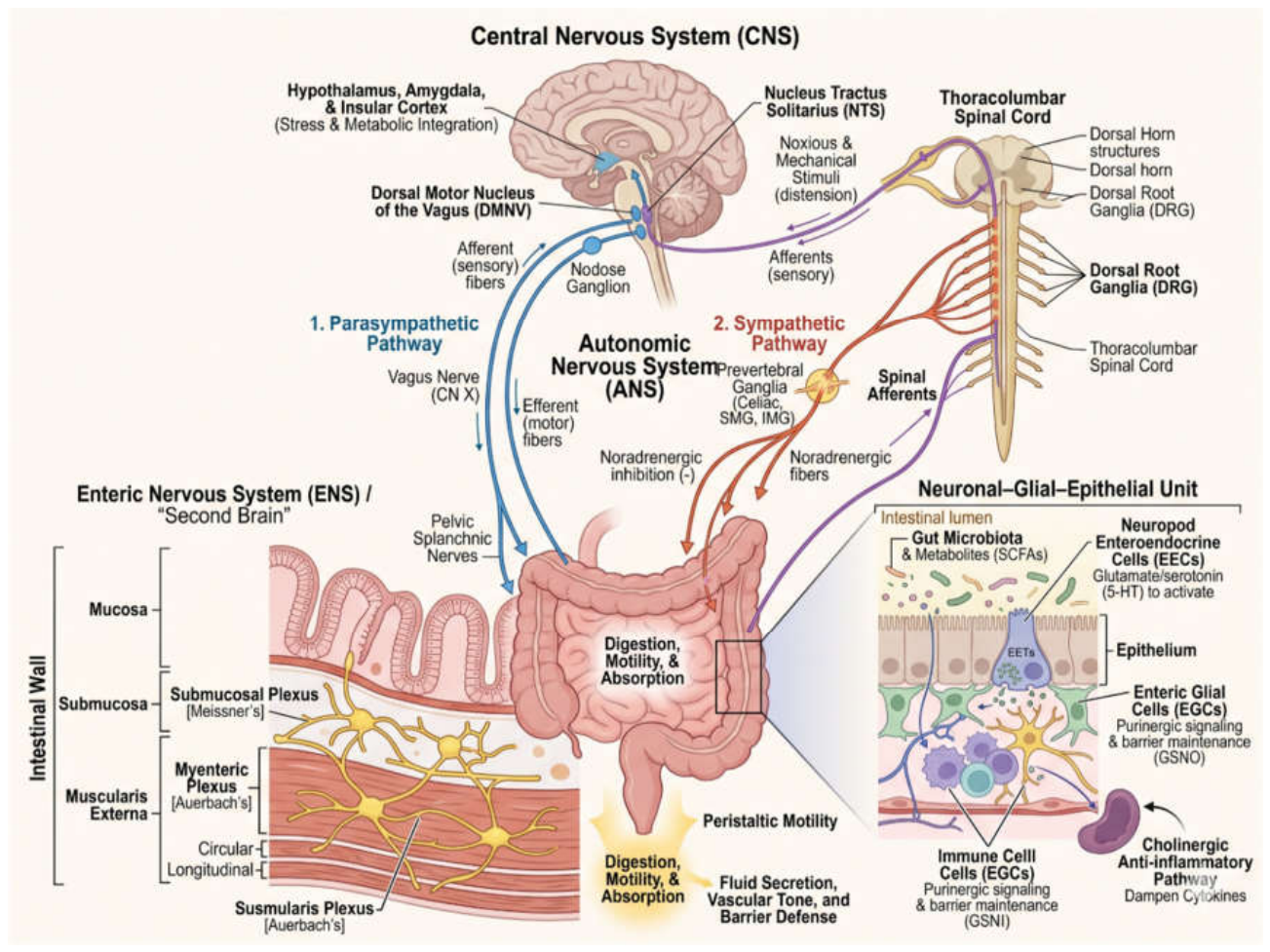
Figure 2.
This schematic illustrates the intestinal neuro-immune-microbiome interface. The epithelial layer features specialized enterocytes, mucin-secreting goblet cells, antimicrobial Paneth cells, and antigen-sampling M cells over lymphoid structures (Peyer’s patches and isolated lymphoid follicles). Within the lamina propria, a dense immune population—including dendritic cells, macrophages, plasma cells producing sIgA, ILC subsets (ILC1, ILC2, ILC3), and T cell lineages (Th17, Treg, CD8+ T cells)—co-localizes with blood capillaries. Beneath, the enteric nervous system’s submucosal and myenteric plexuses form an intricate mesh of neurons and enteric neuroglial cells (EGCs) mediating bidirectional signaling.
Figure 2.
This schematic illustrates the intestinal neuro-immune-microbiome interface. The epithelial layer features specialized enterocytes, mucin-secreting goblet cells, antimicrobial Paneth cells, and antigen-sampling M cells over lymphoid structures (Peyer’s patches and isolated lymphoid follicles). Within the lamina propria, a dense immune population—including dendritic cells, macrophages, plasma cells producing sIgA, ILC subsets (ILC1, ILC2, ILC3), and T cell lineages (Th17, Treg, CD8+ T cells)—co-localizes with blood capillaries. Beneath, the enteric nervous system’s submucosal and myenteric plexuses form an intricate mesh of neurons and enteric neuroglial cells (EGCs) mediating bidirectional signaling.
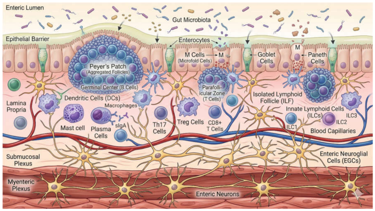
Figure 3.
This diagram illustrates how gut microbes and mucosal immune mediators regulate enteric neuronal depolarization through direct and indirect pathways. In the direct acute pathway, healthy symbionts and SCFA producers activate M2 macrophages and Tregs to suppress pro-inflammatory cytokines, maintaining neuronal homeostasis. Conversely, pathobionts inhibit Tregs, driving mast cells and immune cells to release histamine, proteases, and cytokines (TNF, IL-1β) that bind receptors like H1/H2, PAR2, TNFR1, and IL-1R1 on enteric neurons. This induces rapid depolarization, altered ion channel thresholds, and erratic peristalsis. Indirectly, eosinophils and activated glia release neurotrophin (NGF, BDNF) and glial signals (IL-1 β, IL-6, NO, ATP), causing nerve sprouting, TRPV1 upregulation, and long-term visceral hypersensitivity.
Figure 3.
This diagram illustrates how gut microbes and mucosal immune mediators regulate enteric neuronal depolarization through direct and indirect pathways. In the direct acute pathway, healthy symbionts and SCFA producers activate M2 macrophages and Tregs to suppress pro-inflammatory cytokines, maintaining neuronal homeostasis. Conversely, pathobionts inhibit Tregs, driving mast cells and immune cells to release histamine, proteases, and cytokines (TNF, IL-1β) that bind receptors like H1/H2, PAR2, TNFR1, and IL-1R1 on enteric neurons. This induces rapid depolarization, altered ion channel thresholds, and erratic peristalsis. Indirectly, eosinophils and activated glia release neurotrophin (NGF, BDNF) and glial signals (IL-1 β, IL-6, NO, ATP), causing nerve sprouting, TRPV1 upregulation, and long-term visceral hypersensitivity.
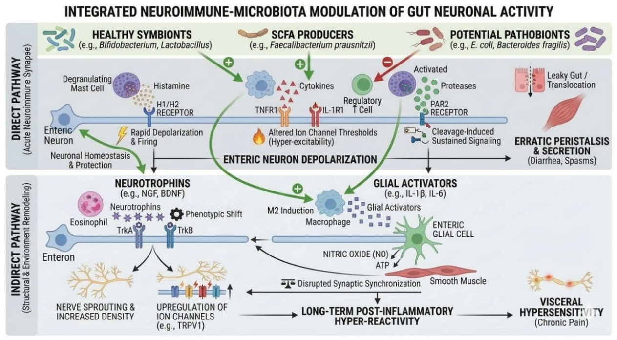
Figure 4.
This schematic illustrates the feed-forward pathological loop connecting gut microbiota, the enteric immune system (EIS), the enteric nervous system (ENS), and the central nervous system (CNS). Pathobiont expansion and SCFA depletion drive PAMP translocation (LPS, LTA), activating TLR2/4 on macrophages, enteric glia (EGCs), and neurons. Reactive EGCs upregulate iNOS, release S100B, and open Cx43 hemichannels to secrete ATP, which engages neuronal P2X7/Panx1 pathways to trigger Ca2+ overload, excitotoxicity, and neurodegeneration. Resulting ACh/VIP deficits impair mucus and AMP secretion, while mucosal nitrates promote facultative anaerobe growth. Systemically, ascending signals activate central microglia via TLR4, dampening BDNF and EAAT2 expression while upregulating IDO to shift tryptophan metabolism toward neurotoxic quinolinic acid, altering neurotransmission and central autonomic regulation.
Figure 4.
This schematic illustrates the feed-forward pathological loop connecting gut microbiota, the enteric immune system (EIS), the enteric nervous system (ENS), and the central nervous system (CNS). Pathobiont expansion and SCFA depletion drive PAMP translocation (LPS, LTA), activating TLR2/4 on macrophages, enteric glia (EGCs), and neurons. Reactive EGCs upregulate iNOS, release S100B, and open Cx43 hemichannels to secrete ATP, which engages neuronal P2X7/Panx1 pathways to trigger Ca2+ overload, excitotoxicity, and neurodegeneration. Resulting ACh/VIP deficits impair mucus and AMP secretion, while mucosal nitrates promote facultative anaerobe growth. Systemically, ascending signals activate central microglia via TLR4, dampening BDNF and EAAT2 expression while upregulating IDO to shift tryptophan metabolism toward neurotoxic quinolinic acid, altering neurotransmission and central autonomic regulation.
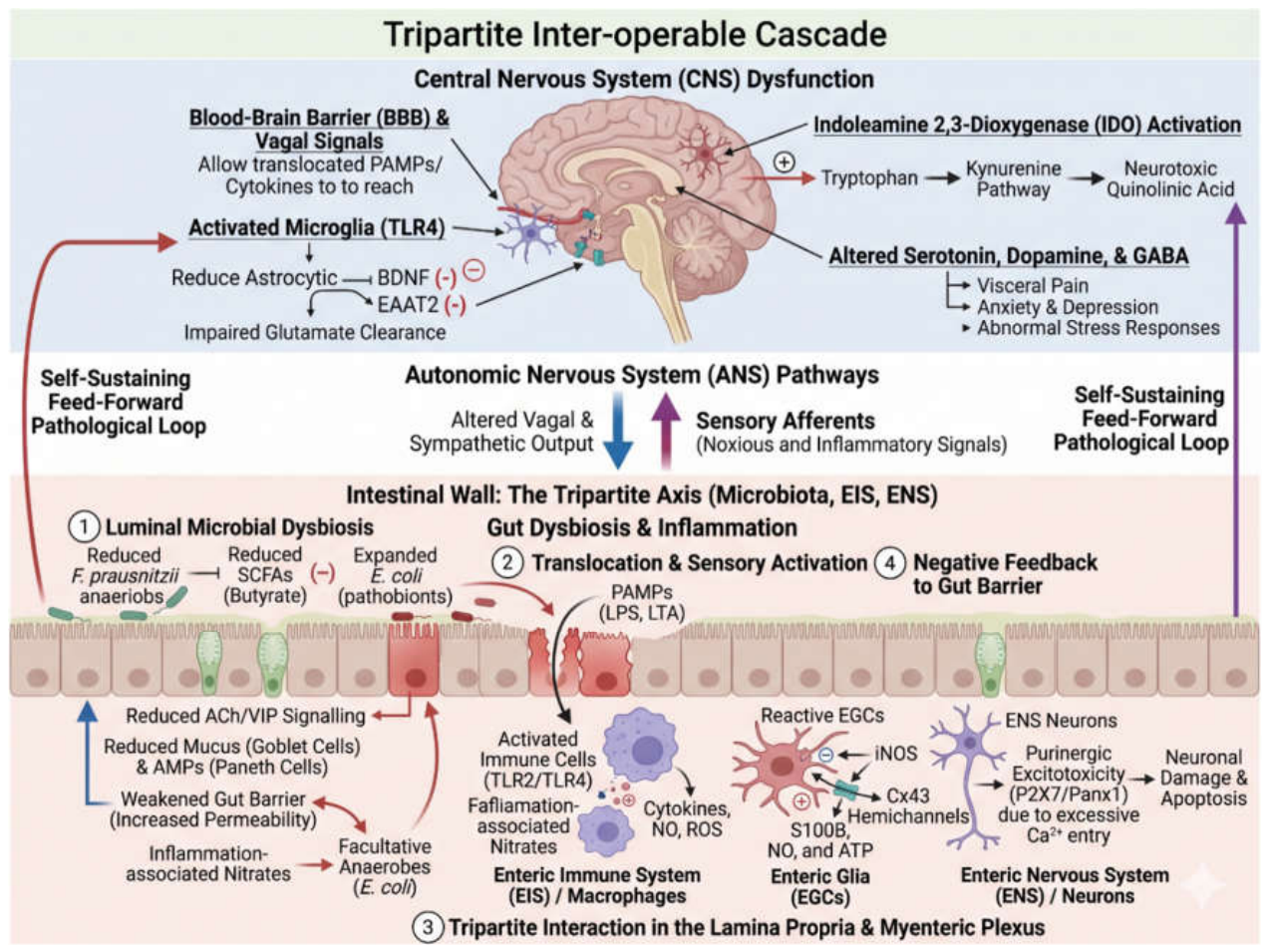
Figure 5.
The schematic illustrates the phytocurative regulation of the enteric neuroglial tripartite cascade. In the lumen, commensal bacterial biotransformation converts parent phytochemicals into active metabiotics while driving eubiotic shifts that expand SCFA- and secondary bile acid-producing taxa. These metabolites reinforce epithelial tight junctions, preventing endotoxin translocation. Below the barrier, the tripartite neuro-immune-microbial synapse coordinates crosstalk between the ENS and mucosal immune system. Phytochemicals directly target neuronal receptors (TRPs, 5-HT3R, nAChRs, FFAR3, TGR5) and immune pathways (TLR4, SIRT1, AHR, 67LR, FcεRI), resolving neuroinflammation, preserving nitrergic/cholinergic neurons, and promoting enteric glial neurogenesis and plasticity. It shows a holistic management concept of gut neuroglial health with dietary phytochemicals.
Figure 5.
The schematic illustrates the phytocurative regulation of the enteric neuroglial tripartite cascade. In the lumen, commensal bacterial biotransformation converts parent phytochemicals into active metabiotics while driving eubiotic shifts that expand SCFA- and secondary bile acid-producing taxa. These metabolites reinforce epithelial tight junctions, preventing endotoxin translocation. Below the barrier, the tripartite neuro-immune-microbial synapse coordinates crosstalk between the ENS and mucosal immune system. Phytochemicals directly target neuronal receptors (TRPs, 5-HT3R, nAChRs, FFAR3, TGR5) and immune pathways (TLR4, SIRT1, AHR, 67LR, FcεRI), resolving neuroinflammation, preserving nitrergic/cholinergic neurons, and promoting enteric glial neurogenesis and plasticity. It shows a holistic management concept of gut neuroglial health with dietary phytochemicals.
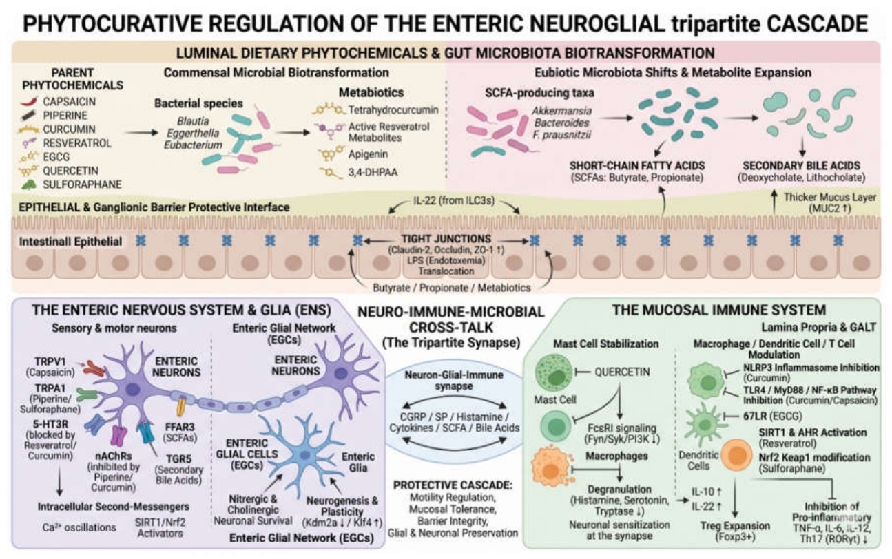

Table 1.
Some of the prominent ligand-receptor interactions and functional effects in ENS-EIS Crosstalk.
Table 1.
Some of the prominent ligand-receptor interactions and functional effects in ENS-EIS Crosstalk.
| Source Cell Type | Target Cell Type | Ligand-Receptor Pair | Direction of Regulation | Experimental Evidence | Ref | |
| Neuronal cell subtype | Enteric NMU-expressing neurons | Group 2 innate lymphoid cells (ILC2s) | NMU–NMUR1 | Promotes type 2 immune activation. | Neuronal localisation, receptor analysis and functional infection experiments. | (Cardoso et al., 2017; Klose et al., 2017) |
| Enteric neurons | Resident muscularis macrophages | TGF-β–TGF-β receptors | Supports neuron-associated macrophage programming and enteric nervous system maintenance. | Macrophage depletion, disruption of TGF-β signalling and assessment of neuronal density and intestinal transit. | (Viola et al., 2023) | |
| Enteric glia | Group 3 innate lymphoid cells (ILC3s) | GDNF-family ligands–RET | Supports IL-22-producing ILC3s and intestinal defence. | Genetic manipulation of RET signalling and analysis of intestinal immune and barrier outcomes. | (Ibiza et al., 2016) | |
| VIP-expressing enteric neurons | ILC3s | VIP–VIPR2 (VPAC2) | Suppresses ILC3-derived IL-22 in a feeding-dependent circuit. | Neuronal activation, receptor analysis and functional intestinal immune assays. | (Talbot et al., 2020) | |
| Enteric neurons | Colonic regulatory T cells (Tregs) | Neuron-derived IL-6–IL-6R signalling | Modifies Treg differentiation and subset composition. | Neuron-specific genetic manipulation, cell-culture experiments and analysis of colonised and germ-free mice. | (Yan et al., 2021) | |
| Sympathetic nerves | Muscularis macrophages | Norepinephrine–ADRB2 | Changes macrophage programming during intestinal infection. | Neuronal activity analysis, macrophage profiling and receptor-dependent experiments. | (Gabanyi et al., 2016) | |
| Enteric cholinergic pathway | Macrophages and neutrophils | ACh–α7nAChR and ACh–M2 muscarinic receptor signalling | Reduces selected inflammatory leukocyte recruitment responses. | Receptor-knockout mice, pharmacological agonists and postoperative ileus models. | (Kimura et al., 2019) | |
| Immune cell type | Muscularis macrophages | Enteric neurons | BMP2–BMPR | Regulates neuronal signalling and colonic peristalsis. | Macrophage depletion, recombinant BMP2 treatment and neuronal BMPR–SMAD pathway analysis in mice. | (Muller et al., 2014) |
| Type 2 immune response, including IL-4/IL-13-producing immune cells | NMU+/CGRPβ+ enteric sensory neurons | IL-4–IL-4Rα and IL-13–IL-4Rα/IL-13Rα1 | Increases neuronal NMU and CGRPβ expression, supporting anti-helminth immunity. | Cytokine stimulation, neuronal Il13ra1 deletion, neuropeptide-expression analysis and helminth-infection experiments. | (Barilla et al., 2025) | |
| Mast cells | Visceral sensory neurons | Histamine–histamine receptors; tryptase–PAR2 (F2RL1) | Sensitises sensory neurons and can contribute to visceral hypersensitivity. | Mast-cell activation and mediator studies support this interaction; the contribution of each receptor pathway varies by model. | (Buhner et al., 2009; Ostertag et al., 2017; C.-Q. Yang et al., 2012) | |
| CGRP-producing Group 2 innate lymphoid cells (ILC2s) | ILC2s | CGRP–CALCRL/RAMP1 | Restrains selected ILC2 responses and excessive type 2 inflammation. | Receptor-expression analysis, functional perturbation and experimental inflammation models. | (Nagashima et al., 2019; Xu et al., 2019) | |
| Inflammatory immune-cell populations | Enteric neurons | IL-1β–IL-1R; TNF-α–TNF receptors | Can promote neuronal survival and neurite outgrowth under experimental conditions. | Primary enteric-neuron culture and cytokine-exposure experiments. | (Gougeon et al., 2013) | |
Table 2.
Gut microbiota metabolites and their interaction with the neuroglial and immune receptors and resultant effects in the enteric system.
Table 2.
Gut microbiota metabolites and their interaction with the neuroglial and immune receptors and resultant effects in the enteric system.
|
Gut Microbiota Metabolites/ Byproducts |
Neuronal/Immune Cell Receptor | Effects | |
| Muramyl dipeptide (MDP) | Immune cell receptor | Nucleotide-binding oligomerization domain-containing protein 2 (NOD2) receptor | Inhibit the differentiation, expansion, and immunosuppressive function of regulatory T-cells (Takada et al., 2021) |
| Neuronal cell receptor | No significant receptor found | No direct action, but promote enteric neuronal (nitrergic and serotonergic neuron) precursor proliferation (Popov et al., 2020) | |
| Microbial lipopolysaccharides (LPS) | Immune cell receptor | Toll-like Receptor 4 (TLR4) on Muscularis macrophages (MMs) | Induce secretion of bone morphogenetic protein 2 (BMP2) (Muller et al., 2014) |
| Neuronal cell receptor | BMP receptor (BMPR) on enteric neurons | Induce secretion of colony stimulatory factor 1 (CSF1) (Muller et al., 2014) | |
| Short Chain Fatty Acids (SCFA) | Immune cell receptor | GPR43 | Induce production of prostaglandin E2 and inhibit constitutive monocyte chemotactic protein-1 (MCP-1) production, LPS-induced interleukin-10 (IL-10), tumor necrosis factor-α (TNF-α) and interferon-γ (IFN- γ) production (Cox et al., 2009) Induce Innate lymphoid cells (ILCs) proliferation (Sepahi et al., 2021) |
| Neuronal cell receptor | GPR41, GPR43 | Induce myenteric neurons and delay colonic motility (Wongkrasant et al., 2026) | |
| Butyrate (SCFA) | Immune cell receptor | Gpr109a | Induce differentiation of Treg and IL-10-producing CD4+ T cells, and suppress development of Th17 cells (N. Singh et al., 2014) |
| GPR43 | Supress p38 MAPK/NF-κB signaling in T follicular helper (specifically Tfh13) cells (Yu et al., 2025) | ||
| Neuronal cell receptor | 5-hydroxytryptamine receptor 4 or 5-HT4 (butyrate enhances TPH1 expression, required for 5-hydroxytryptamine production) | Modulate gastrointestinal (GI) motility, ENS maturation and other gastrointestinal functions (De Vadder et al., 2018; Reigstad et al., 2015; Yano et al., 2015) | |
| Tryptophan metabolites (Indole derivatives) | Immune cell receptor | Aryl hydrocarbon receptor (AhR) | Induce Treg differentiation and suppress Th1/Th17 responses (Jiang et al., 2023; Z. Ma et al., 2023) |
| Neuronal cell receptor | Aryl hydrocarbon receptor (AhR) | Induce bidirectional signaling between the gut microbiome and the brain (N. Ma et al., 2020) | |
| 5-Hydroxyindole-3-acetic acid (5-HIAA) | Immune cell receptor | Aryl hydrocarbon receptor (AhR) | Induce Breg function while inhibiting germinal center (GC) B cell and plasmablast differentiation (Rosser et al., 2020) |
| Neuronal cell receptor | Aryl hydrocarbon receptor (AhR) | Maintain intestinal homeostasis and prevent neuroinflammation (He et al., 2024; Sun et al., 2022) | |
| Succinate | Immune cell receptor | G-protein coupled receptor Sucnr1 (GPR91) | Induce neutrophil extracellular trap (NET) formation (H. Li et al., 2023) |
| Neuronal cell receptor | G-protein coupled receptor Sucnr1 (GPR91) | Induce enteric nervous system regeneration (Aydin et al., 2024) | |
| Secondary bile acids and derivatives | Immune cell receptor | Takeda G protein-coupled receptor 5 (TGR5) | Induce macrophage polarization from the M1 (pro-inflammatory phenotype) to the M2 (immune tolerogenic phenotype) (Su et al., 2023) |
| Neuronal cell receptor | Takeda G protein-coupled receptor 5 (TGR5) | Induce rapid and sustained inhibition of spontaneous phasic activity of isolated segments of ileum and colon by a neurogenic, cholinergic and nitrergic mechanism, and delayed gastrointestinal transit (Poole et al., 2010) | |
Table 3.
Positive and Negative Effects of Dietary Phytochemicals/Bioactive Compounds on the ENS–Immune–Microbiota Axis.
Table 3.
Positive and Negative Effects of Dietary Phytochemicals/Bioactive Compounds on the ENS–Immune–Microbiota Axis.
| Natural Product → Bioactive Derivatives | Nervous System (+/−) | Immune System (+/−) | Gut Microbiota (+/−) | References |
| 1. Curcumin (turmeric) → tetrahydrocurcumin, dihydro/hexahydrocurcumin, ferulic acid derivatives | (+) BDNF/TrkB & 5-HT signaling, neuroprotection, vagal modulation. (−) Poor parent bioavailability limits direct neuronal reach. |
(+) Inhibits TLR4/NF-κB; ↓ TNF-α, IL-1β, IL-6; ↑ IL-10. (−) High concentrations may induce cellular stress. |
(+) ↑ Bifidobacterium, Lactobacillus, and Roseburia. (−) High exposure may alter microbial diversity. |
(X. Chen et al., 2022; Enayati et al., 2023; Rudrapal et al., 2026; X. Wang et al., 2025) |
| 2. Tea catechins (EGCG, EGC, ECG, EC) → phenyl-γ-valerolactones, phenolic & hippuric acids | (+) 67LR & Nrf2/BDNF neuroprotection; remodels α-synuclein; modulates 5-HT. (−) Low parent bioavailability; oxidation-sensitive. |
(+) Inhibits TLR4/NF-κB and NLRP3; ↓ ROS and pro-inflammatory cytokines. (−) High-dose extracts risk pro-oxidant effects & hepatotoxicity. |
(+) Cleaved by Bacteroides/Lactobacillus; ↑ Bifidobacterium, Akkermansia & CsgA amyloids, SCFA producers. (−) Excess doses may inhibit commensals. |
(L. Chen et al., 2025; Rudrapal et al., 2026; B. Singh et al., 2024; J. Yang et al., 2025) |
| 3. Capsaicin (chili) → hydroxylated vanillyl derivatives | (+) Subsequent TRPV1 desensitization relieves visceral pain; modulates AhR. (−) Initial TRPV1 activation triggers burning & hypersensitivity. |
(+) Inhibits TLR4/NF-κB and reduces pro-inflammatory cytokines. (−) High exposure causes mucosal irritation and neurogenic inflammation. |
(+) ↑ Akkermansia, Faecalibacterium, and Lachnospiraceae; ↑ butyrate. (−) High doses may disrupt microbial balance and Muc2 mucus. |
(X. Chen et al., 2022; Corral-Guerrero et al., 2025; Diacova et al., 2026) |
| 4. Dietary fibers & resistant starches (oats, grains, legumes, bananas) → SCFAs (acetate, propionate, butyrate) | (+) FFAR2/FFAR3 signaling regulates ChAT, 5-HT, enteric motility & gut–brain axis. (−) Excess concentrations may dysregulate transit or firing. |
(+) FFAR2, GPR109A, and HDAC inhibition promote Treg activity and ↓ inflammation. (−) Effects vary by mucosal disease state. |
(+) Fuels colonocytes, tightens ZO-1/occludin, and sustains eubiosis. (−) Depleted during dysbiosis. |
(Geng et al., 2022; Hamilton et al., 2024; Horn et al., 2022; Tarasiuk-Zawadzka & Fichna, 2025; X. Wang et al., 2025) |
| 5. Peppermint oil → menthol | (+) TRPM8 activation (cross-desensitizing TRPV1/TRPA1) and Ca2+ channel blockade relieves spasms/pain. (−) Relaxes lower esophageal sphincter; may worsen reflux. |
(+) Reduces substance P-mediated neurogenic inflammation. (−) Direct clinical immune modulation remains insufficiently established. |
(+) Exhibits experimental antimicrobial activity against overgrowth. (−) Selective preservation of commensals remains unconfirmed. |
(Agrawal et al., 2025; Aminullah et al., 2026; Diacova et al., 2026) |
| 6. Ellagitannins (pomegranate, berries) → urolithin A/B | (+) Supports mitophagy and mitochondrial neuroprotection. (−) Direct clinical ENS trials remain limited. |
(+) Suppresses NF-κB/MAPK and cytokines; reinforces tight junctions via AhR/Nrf2. (−) Long-term clinical immune effects remain unclear. |
(+) Converted by Gordonibacter, Ellagibacter. (−) non-producer (’metabotype 0’) individuals generate little/no urolithin. |
(S. Liu et al., 2023; Rudrapal et al., 2026; B. Singh et al., 2024) |
| 7. Quercetin/rutin (citrus, fruits, vegetables) → phenylacetic acid derivatives (DOPAC) | (+) Protects myenteric neurons against oxidative injury. (−) High experimental concentrations may reduce ICC pacemaker density. |
(+) Inhibits NLRP3 inflammasome, NF-κB, and oxidative stress. (−) High concentrations may act as pro-oxidants. |
(+) Biotransformed by B. fragilis and F. prausnitzii into absorbable phenolic acids. (−) Excessive exposure may alter microbial richness. |
(X. Chen et al., 2022; Horn et al., 2022; Rudrapal et al., 2026; X. Wang et al., 2025) |
| 8. Resveratrol (grapes, peanuts) → dihydroresveratrol, lunularin | (+) Preserves HuC/D+ & nNOS+ myenteric neurons; inhibits S100+ reactive enterogliosis. (−) Rapid Phase II metabolism limits parent bioavailability. |
(+) Inhibits NF-κB, COX-2, lipid peroxidation, and inflammatory cytokines. (−) High concentrations may induce cellular stress. |
(+) Enriches Lactobacillus and Bifidobacterium; suppresses Enterococcus faecalis. (−) Efficacy depends on dose and baseline microbiota. |
(H.-L. Chen et al., 2026; Luesma et al., 2024; Rudrapal et al., 2026; X. Wang et al., 2025) |
| 9. Coffee chlorogenic acids → dihydrohydroxycinnamic acids, feruloylglycine, hippuric acid | (+) Antioxidant neuroprotection and gut–brain axis support. (−) Direct clinical ENS evidence remains limited. |
(+) Phenolic metabolites support antioxidant and anti-inflammatory pathways. (−) Clinical mucosal immune outcomes remain uncertain. |
(+) Colonic bacteria cleave chlorogenic acid into absorbable phenolic metabolites. (−) Bioconversion varies with host microbial composition. |
(Horn et al., 2022; S. Liu et al., 2023; X. Wang et al., 2025) |
| 10. Prebiotics & probiotics (inulin, guar gum; B. longum, B. infantis, L. rhamnosus, F. prausnitzii, A. muciniphila) → SCFAs, indoles, GABA, 5-HT | (+) Protects myenteric neurons, elevates BDNF/GABA/5-HT, and normalizes motility. (−) Neuro-modulatory benefits are highly strain- and condition-specific. |
(+) Reinforces Muc2/tight junctions (TLR2/4), activates AhR, and restrains LPS inflammation. (−) Live strains require caution in severely immunocompromised hosts. |
(+) Restores eubiosis, cross-feeds SCFA producers, and suppresses pathobionts (E. coli). (−) Engraftment and clinical responses vary across indiv iduals. |
(Agrawal et al., 2025; Geng et al., 2022; Giuffrè et al., 2020; Horn et al., 2022; X. Wang et al., 2025) |
| (+) Positive effect; (−) adverse effect or limitation; ↑ increase; ↓ decrease | ||||
Disclaimer/Publisher’s Note: The statements, opinions and data contained in all publications are solely those of the individual author(s) and contributor(s) and not of MDPI and/or the editor(s). MDPI and/or the editor(s) disclaim responsibility for any injury to people or property resulting from any ideas, methods, instructions or products referred to in the content. |
© 2026 by the authors. Licensee MDPI, Basel, Switzerland. This article is an open access article distributed under the terms and conditions of the Creative Commons Attribution (CC BY) license.
Copyright: This open access article is published under a Creative Commons CC BY 4.0 license, which permit the free download, distribution, and reuse, provided that the author and preprint are cited in any reuse.