

Submitted:
03 November 2026
Posted:
05 November 2026
You are already at the latest version
Abstract
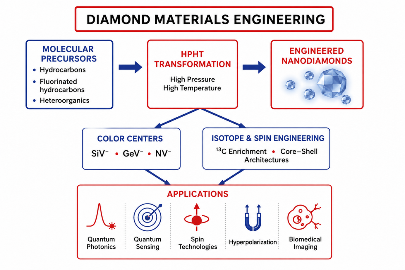
High-pressure high-temperature (HPHT) transformation of organic and heteroorganic systems has emerged as a versatile platform for the synthesis of diamond materials with tailored structural, optical, isotopic, spin, and biomedical functionalities. This work summarizes the results of our investigations on the synthesis and characterization of diamond materials produced via HPHT-induced transformations of hydrocarbon, fluorinated hydrocarbon, and heteroorganic systems. Particular attention is devoted to the mechanisms of diamond formation, the hydrogen–fluorine synergy responsible for stabilizing nanoscale diamond fractions, and the incorporation of optically active color centers during crystal growth. The formation and photophysical properties of nanodiamonds containing silicon-vacancy (SiV), germanium-vacancy (GeV), and nitrogen-vacancy (NV) centers are discussed together with size-dependent optical and quantum phenomena, spatial distributions of color centers, and light–matter interaction effects arising in nanoscale diamond particles. The review further examines isotopically engineered 13C-enriched diamonds and core–shell nanodiamond architectures designed for spin-based applications and hyperpolarization technologies. The capability of HPHT synthesis to independently control particle size, impurity incorporation, isotopic composition, and spin environment provides a versatile platform for engineering advanced diamond materials optimized for quantum photonics, nanoscale sensing, biomedical imaging, and spin-based technologies.
Keywords:
HPHT synthesis
; nanodiamonds
; impurity–vacancy color centers
; isotope engineering
; core–shell nanodiamonds
; hyperpolarization
; quantum sensing
1. Introduction
Diamond has emerged as one of the most versatile materials for advanced technologies owing to unique combination of exceptional mechanical, thermal, optical, electronic, and spin-related properties. In recent years, diamond has evolved far beyond its traditional role as a jewelry grade, super-hard tooling or structural material, and has become a key platform for quantum photonics [1,2,3,4], sensing [5,6], spin-based information processing [2,3,7], different biomedical applications [8,9,10,11,12], and hyperpolarization technologies [13,14,15,16,17,18]. All of this has stimulated the development of methods for producing diamond materials with precisely engineered structural, optical, isotopic, and spin characteristics.
Particular attention has been devoted to nanoscale diamond materials containing optically active color centers, including nitrogen-vacancy (NV), silicon-vacancy (SiV), germanium-vacancy (GeV), tin-vacancy (SnV), and lead-vacancy (PbV) defects. These atomic scale defects enable a broad range of functionalities, including stable single-photon emission, coherent spin manipulation, quantum sensing, nanoscale thermometry, and dynamic nuclear spin polarization (DNP). At the same time, the performance of these quantum systems depends not only on the nature of the color center but also on numerous characteristics of the host crystal, including particle size, crystal quality, impurity concentration, isotopic composition, surface chemistry, and the spatial distribution of optically and magnetically active defects. Consequently, the ability to simultaneously control several material’s parameters has become one of the major challenges in modern diamond materials science.
To date, various approaches for the fabrication of fluorescent and isotopically engineered diamond nanomaterials have been proposed [19]. They include mechanical fragmentation of bulk crystals containing color centers [20,21], ion implantation into preformed nanodiamonds [21,22], detonation synthesis [23], chemical vapor deposition (CVD) [24,25,26,27], and several variants of high-pressure high-temperature (HPHT) synthesis [28,29,30]. Each of these techniques possesses its own advantages but also inherent limitations with respect to crystal quality, particle-size control, impurity incorporation, scalability, and defect engineering. As a result, no single synthesis route currently provides complete and independent control over all parameters required for advanced quantum, optical, spin-based, and biomedical applications.
This paper examines the possibilities of producing various nanoscale diamonds for quantum-physical and biomedical applications based on transformations, induced by high pressure and high temperature in organic and heteroorganic growth systems that do not contain traditional metal catalysts, and identifies the characteristic features of diamond materials produced using this method. The goal of this work is to provide a comprehensive overview of diamond materials produced by HPHT transformation of organic and heteroorganic precursor systems and to establish the relationships between precursor chemistry, transformation mechanisms, crystal-growth processes, and the resulting structural, optical, isotopic, and spin-related properties of the synthesized materials. Particular attention is given to the mechanisms governing nanodiamond formation, hydrogen–fluorine synergistic effects, engineering of SiV, GeV, and NV color centers, isotopically enriched 13C diamond structures, core–shell nanodiamonds for hyperpolarization technologies, and the emerging applications of these materials in quantum photonics, quantum sensing, spin technologies, and biomedicine. Overall, the results reviewed here demonstrate that HPHT transformation of molecular organic and heteroorganic precursor systems should be regarded not merely as an alternative route for diamond synthesis but as a universal materials engineering platform that enables the rational design of multifunctional diamond materials for next-generation quantum, optical, spin-based, and biomedical technologies.
2. Materials and Methods
HPHT transformations of organic and heteroorganic precursors were investigated by using both pure compounds and multicomponent mixtures to evaluate the effects of precursor chemistry on the pressure and temperature conditions of the process, transformation mechanisms, and product composition. Transformations in pure hydrocarbon systems were studied using adamantane (C₁₀H₁₆) and a series of polycyclic aromatic hydrocarbons, including naphthalene (C₁₀H₈), anthracene (C₁₄H₁₀), pentacene (C₂₂H₁₄), perylene (C₂₀H₁₂), and coronene (C₂₄H₁₂). Pure perfluorocarbon systems were represented by graphite fluoride (CF₁.₁), octafluoronaphthalene (C₁₀F₈), and fluorinated carbon soot (CF₀.₉). Heteroorganic compounds, selected as dopant sources and added to the chosen HPHT precursor systems, included 1-fluoroadamantane (C₁₀H₁₅F), silicon triphenyl chloride (C₁₈H₁₅SiCl), germanium triphenyl chloride (C₁₈H₁₅GeCl), tetrakis(trimethylsilyl)silane (C₁₂H₃₆Si₅), tetraphenylgermanium (C₂₄H₂₀Ge), acridine (C₁₃H₉N), hexamethylenetetramine (C₆H₁₂N₄), and aminotetrazole (CH₃N₅).
HPHT experiments were performed using a toroid-type high-pressure apparatus [31]. Cylindrical pellets of the starting materials, measuring 4–5 mm in diameter and 3 mm in height, were prepared by cold pressing and placed in a graphite container that simultaneously served as a resistive heater within the high-pressure apparatus. The experimental procedure involved loading the apparatus to pressure up to 8.0 GPa at room temperature, followed by heating the sample to the onset temperature of diamond formation within the range of 900–1500 °C. The sample was subsequently maintained at the selected temperature and pressure for holding times ranging from 1 to 30 s.
Because the graphite container is permeable to gaseous transformation products, this reaction system can be regarded as thermodynamically open. Thermal transformations of organic compounds in such an open system may provide higher yields of solid products than transformations conducted in closed systems, in which mass exchange with the surrounding environment is restricted. The obtained products were quenched to room temperature under pressure and then, after unloading the apparatus, recovered from the reaction zone and analyzed by powder X-ray diffraction, Raman and fluorescence spectroscopy, atomic force microscopy (AFM), scanning electron microscopy (SEM), transmission (TEM) and high resolution transmission electron microscopy (HRTEM).
Characterization of a wide range of samples showed that the appropriate choice of the growth mixture composition and HPHT processing parameters resulted in the formation of diamond as the main transformation product. Minor amounts, up to approximately 1.5 wt%, of graphite and amorphous carbon could appear as by-products representing intermediate transformation products that had not been fully converted into diamond. Because of the radial temperature gradient within the reaction zone of the high-pressure apparatus, the synthesis products could contain mixtures of diamond particles belonging to different size fractions. The relative proportions of ultrananocrystalline, nanocrystalline, submicron, and micron-sized diamond fractions depended strongly on the composition of the growth system, the pressure–temperature conditions, and the duration of the thermobaric treatment. Increasing the proportion of fluorinated components in the growth mixture, conducting the synthesis at temperatures close to the threshold for diamond formation in the corresponding growth system, and limiting the isothermal holding time to suppress collective recrystallization of ultranano- and nanosized diamond particles increased the yield of the smallest diamond fractions.
When required, nondiamond impurities were removed from the synthesis products by boiling in a mixture of concentrated acids (HNO₃–HClO₄–H₂SO₄) or in a 40% hydrogen peroxide solution in sulfuric acid (H₂O₂–H₂SO₄). The diamond materials were separated into size fractions by dispersing the particles in aqueous media using a UP200Ht probe sonicator (Hielscher Ultrasonic Technology), followed by stepwise centrifugation at relative centrifugal forces of 1000 × g, 3000 × g, 5000 × g, and 10,000 × g. The smallest diamond fractions, which remained suspended after centrifugation at the highest relative centrifugal forces of 5000–10,000 × g, were recovered by evaporation of the corresponding dispersions.
3. HPHT Transformation of Organic and Heteroorganic Systems: From Precursor Chemistry to Functional Diamond Materials
3.1. Mechanistic Considerations of the HPHT Transformation of Organic and Heteroorganic Precursor Systems
The thermal transformations of organic compounds under high pressures represent a complex sequence of physicochemical processes. Their final stages are associated with the carbonization of the initial compounds, the progressive removal of hydrogen and other volatile transformation products from the reaction system, and the subsequent structural ordering of the resulting solid carbon residue [32,33,34]. From the point of view of the concept of physico-chemical evolution of matter [35], these transformations may be regarded as a condensation pathway to solid-state formation, in which solid carbon phases are produced by condensation from carbon-supersaturated vapors, melts, or vapor–fluid media. The evolution of the system proceeds through several successive stages, including the formation of primary carbon clusters, nucleation of metastable and stable carbon phases, growth of nanometer-sized nuclei, particle aggregation, and structural maturation [35]. Under conditions of substantial carbon supersaturation, several carbon allotropes may nucleate simultaneously, including graphene-like structures, spherical and polyhedral onion-like particles, carbon nanoribbons, various forms of amorphous carbon, and, ultimately, graphite and diamond. The relative thermodynamic stability of these phases under HPHT conditions governs their subsequent evolution through dissolution–reprecipitation and recrystallization processes.
An important feature of the HPHT transformation of organic systems is that the nanoscale state generally corresponds to an early stage of the structural evolution pathway. Continued particle growth and collective recrystallization favor the formation of larger and more structurally ordered crystallites. Consequently, the final particle size distribution of the diamond product is determined by the competition among diamond nucleation, crystal growth, particle aggregation, and recrystallization.
The chemical composition of the growth system and the permeability of the reaction-zone container to gaseous transformation products can also exert a substantial influence on both the transformation pathway and the resulting products.
The first demonstration of nanodiamond formation from pure hydrocarbon compounds at high static pressures of approximately 12 GPa and temperatures of approximately 2000 °C was reported for precursors enclosed in titanium capsules [36]. Under those synthesis conditions, the formation of micrometer-sized diamond crystals was not observed. The synthesis of submicron and micrometer-sized diamond fractions from organic precursors at substantially lower and technologically more accessible conditions of approximately 8.0 GPa and 1300 °C was subsequently demonstrated in the work [37]. In that study, graphite was used instead of titanium as the reaction-zone container because of its higher permeability to gaseous transformation products.
It is noteworthy that studies of various pure hydrocarbon precursors, including chemically distinct compounds such as naphthalene and adamantane, did not reveal a pronounced influence of the molecular structure of the starting material on either the pressure–temperature conditions required for the onset of diamond formation or the particle-size distribution of the resulting diamond products [37]. In these systems, micrometer-sized diamond crystals constituted the dominant product fraction.
However, the introduction of fluorinated and heteroorganic components into the growth system substantially modifies the transformation pathway. In particular, the simultaneous presence of hydrogen and fluorine produces a pronounced synergistic effect, lowering the temperature thresholds for carbonization, graphitization, and diamond formation while increasing the content of ultranano- and nanoscale diamond fractions in the synthesis products [28,38,39,40].
The incorporation of heteroatom-containing molecular precursors further expands the capabilities of this synthetic approach. Silicon-, germanium-, nitrogen-, and isotope-containing compounds can be introduced directly into the growth system, enabling the formation of diamond particles containing SiV, GeV, NV, and other optically active defects, as well as isotopically engineered 13C-enriched and core–shell diamond structures. Because the defects are incorporated during crystal nucleation and growth rather than introduced by post-synthesis ion implantation, implantation-induced lattice damage can be avoided. The resulting color centers can therefore exhibit favorable optical properties and high structural stability, although their specific photophysical characteristics remain dependent on the local strain, impurity concentration, surface chemistry, and size of the host diamond particles.
3.2. Diamond Formation in Pure Hydrocarbon Precursor Systems
Pure hydrocarbon precursor systems provide the simplest model for understanding the fundamental mechanisms of diamond formation during HPHT transformation. In the absence of heteroatoms or fluorinated additives, the evolution of the carbon system is governed primarily by the intrinsic thermodynamics and kinetics of carbonization, graphitization, and diamond crystallization. Therefore, these systems serve as an important reference for evaluating the effects of chemical modifications introduced in more complex precursor compositions.
Systematic investigations of the HPHT transformation of pure hydrocarbons, including naphthalene, anthracene, pentacene, perylene, coronene, and adamantane, were carried out at pressures of approximately 8 GPa and temperatures up to 1600 °C [34,41]. The experiments demonstrated that at temperatures above approximately 1300 °C the principal products of transformation are high-quality, ultrapure micrometer-sized diamond crystals. The accompanying by-products consist predominantly of highly crystalline graphite and carbon globules.
Representative SEM images of the products obtained from naphthalene at 8 GPa and 1300 °C are shown in Figure 1a,b. Large faceted diamond crystals coexist with graphite particles and spherical carbon globules, illustrating the multiphase nature of the HPHT transformation products.
High-resolution transmission electron microscopy revealed that the carbon globules are themselves heterogeneous nanostructures composed of several carbon phases (Figure 1c–e). They contain diamond nanoparticles with characteristic sizes of approximately 3–5 nm, spherical and polyhedral onion-like carbon particles, and amorphous carbon. These observations indicate that carbon supersaturation during the early stages of HPHT transformation leads to simultaneous nucleation of several metastable and stable carbon allotropes before the system evolves toward the final equilibrium products.
Quantitative analysis further showed that the nanodiamond fraction in the transformation products of pure hydrocarbons remains very limited. Diamond nanoparticles with sizes of approximately 3–5 nm typically constitute no more than 1–3 wt.% of the total product.
An important mechanistic conclusion follows from these observations. Although nanodiamond nuclei are formed during the earliest stages of the transformation process, they do not subsequently develop into the dominant micrometer-sized diamond crystals. Instead, the formation of large diamond crystals proceeds through a different pathway associated with the massive formation of microcrystalline graphite, which subsequently transforms into diamond under HPHT conditions. Thus, nanodiamond formation and the growth of micrometer-sized diamond crystals should be regarded as two parallel transformation pathways rather than successive stages of a single crystal growth process. Nanodiamond formation is restricted to the initial stages of carbon supersaturation, whereas continued thermobaric treatment favors graphitization, collective recrystallization, and the subsequent conversion of graphite into large diamond crystals. This mechanistic interpretation has important implications for the rational design of nanodiamond synthesis. If the objective is to maximize the yield of nanodiamonds, increasing the nucleation rate alone is insufficient. Instead, the competing processes responsible for graphitization and collective recrystallization must be suppressed before the system evolves toward the formation of large graphite particles and, ultimately, micrometer-sized diamonds. This conclusion provides the conceptual basis for the following sections, where fluorinated and heteroorganic precursor systems are shown to modify the transformation pathway, suppress collective recrystallization, and stabilize ultrananocrystalline and nanocrystalline diamond fractions.
3.3. Halogenated (Fluorinated) Hydrocarbon Systems
The results presented in the previous section demonstrate that HPHT transformation of pure hydrocarbon precursors predominantly yields micrometer-sized diamond crystals, whereas nanodiamond formation remains a minor parallel process. Consequently, increasing the yield of nanosized diamond fractions requires modification of the transformation pathway rather than simply enhancing diamond nucleation. One of the most effective approaches toward this objective was found to be the incorporation of fluorinated molecular precursors into the growth system.To investigate the influence of fluorine on diamond formation, systematic HPHT studies were performed using pure fluorocarbon compounds and homogeneous binary mixtures of fluorocarbon and hydrocarbon precursors. The investigated systems included hydrocarbon compounds (naphthalene, anthracene, pentacene, perylene, coronene, and adamantane), fluorocarbon compounds (octafluoronaphthalene, highly fluorinated graphite, and fluorinated carbon soot), as well as their binary mixtures under identical pressure–temperature conditions. A comparative analysis revealed that the simultaneous presence of hydrogen and fluorine fundamentally modifies the HPHT transformation process [38].
In contrast to pure hydrocarbons, binary hydrocarbon–fluorocarbon systems exhibit a substantial reduction in the temperature thresholds for all principal stages of thermal transformation, including carbonization, graphitization, and diamond formation. Furthermore, a pronounced change in the size-fraction composition of diamond products is observed in binary systems, resulting in the massive formation of ultrananocrystalline and nanocrystalline diamond particles along with the conventional micrometer-sized diamond fraction [39]. Figure 2 illustrates this change in the size fraction composition of diamond products. In contrast to the predominantly micrometer-sized crystals obtained from pure hydrocarbon systems, the binary hydrocarbon–fluorocarbon mixture produces a dense ensemble of nanosized diamond particles while retaining a fraction of micrometer-sized crystals. This change reflects the modified kinetics of nucleation, crystal growth, and recrystallization induced by the fluorine-containing precursor.
The most significant outcome of these studies was the discovery of a pronounced hydrogen–fluorine synergistic effect. Rather than merely lowering the temperature required for diamond synthesis, the simultaneous presence of hydrogen and fluorine redirects the evolution of the carbon system. Carbonization, graphitization, and diamond nucleation occur at lower temperatures, while the growth of large graphite particles and the collective recrystallization of nanosized diamonds are suppressed [38,40]. As a result, fluorinated precursor systems stabilize ultrananocrystalline and nanocrystalline diamond fractions that are produced only in minor quantities from pure hydrocarbons.
The discovery of this synergistic effect established a new strategy for engineering diamond materials. By appropriate adjustment of the precursor composition, pressure, temperature, and isothermal holding time, the diamond particle size distribution can be controlled over a broad range, from ultrananocrystalline and nanocrystalline particles to submicron and micrometer-sized diamonds [39,40,42]. As illustrated in Figure 3, variation of the precursor composition and HPHT processing parameters enables the preferential formation of different diamond size fractions. This degree of size control substantially expands the capabilities of HPHT synthesis and makes it possible to produce diamond materials optimized for different scientific and technological applications.
From the point of view of the synthesis of nanodiamonds, systems based on halogen derivatives of adamantane, and primarily 1-fluoroadamantane, can be distinguished into a special class of growth systems. The fact is that the HPHT transformation of pure 1-fluoroadamantane, in contrast to pure adamantane, can be accompanied by almost 100 % formation of nanoscale diamond fractions. [42,43]. This indicates a significant difference in the transformation mechanisms of these compounds. Moreover, varying the treatment parameters of 1-fluorodamantane allows for precise control of the average size of the resulting diamond nanoparticles [43,44]. This opens the way for obtaining homogeneous batches of diamond materials even in the single-digital nanometer range.
The proposed approach to the synthesis of nanoscale diamonds using halogenated growth systems has proven to be a highly promising base for developing methods to produce various functional diamond nanomaterials for quantum photonics, spin-based technologies, hyperpolarization, quantum sensing, and biomedical applications, including nanodiamonds with impurity-vacancy (SiV, GeV, and NV) color centers, as well as isotopically enriched 13C and core-shell structures.
3.4. Nanodiamonds with Impurity–Vacancy Color Centers
The hydrogen–fluorine synergistic effect described in the previous section enables not only an efficient route for controlling nanodiamond size but also creates favorable conditions for direct incorporation of optically active impurity–vacancy color centers during HPHT crystal growth. Among these defects, silicon-vacancy (SiV⁻) centers are of particular interest because of their inversion symmetry, narrow zero-phonon line (ZPL), reduced sensitivity to electric-field fluctuations, and excellent potential for quantum-photonic applications, including single-photon sources and quantum communication. Unlike conventional post-growth ion implantation, the present HPHT approach enables the formation of SiV centers directly during diamond crystallization, thereby avoiding implantation-induced lattice damage [28,45,46,47]. The characterization of the ultra-nanoscale fraction of these diamonds (Figure 4) was carried out in the works [48,49]. SiV-containing nanodiamonds were synthesized from homogeneous mixtures of naphthalene (C₁₀H₈), highly fluorinated graphite (CF₁.₁), and tetrakis(trimethylsilyl)silane (C₁₂H₃₆Si₅). Following HPHT synthesis, the products were chemically purified and separated into size fractions according to the procedure described in Section 2.
Transmission electron microscopy demonstrated that the isolated nanodiamond fraction possesses a median particle size of 7.5 ± 5 nm. More importantly, high-resolution TEM images reveal exceptionally well-ordered crystal lattices extending almost to the particle surface. In contrast to detonation nanodiamonds, no pronounced graphitic or amorphous sp²-rich surface layer is observed. The absence of a disordered surface shell indicates the high crystalline quality of the HPHT-grown nanoparticles and demonstrates that molecular HPHT synthesis is capable of producing structurally well-defined nanodiamonds even in the single-digit nanometer size range.
Raman spectroscopy provides independent confirmation of the excellent crystalline quality of the synthesized nanodiamonds. The Raman peak is shifted from the bulk diamond value of 1332.5 cm⁻¹ to approximately 1330 cm⁻¹ and exhibits moderate broadening. Since HRTEM observations reveal only a very low density of structural defects, the observed spectral changes are attributed primarily to phonon confinement associated with the small crystal dimensions rather than to crystal disorder [50,51,52].
The combination of high crystallinity, the absence of a defective sp²-rich surface layer, and the relatively narrow Raman linewidth demonstrates that HPHT molecular synthesis can produce nanodiamonds of substantially higher structural quality than is typically achieved by detonation-based methods.
The luminescence properties of nanodiamonds with SiV⁻ centers were investigated by room-temperature photoluminescence spectroscopy (Figure 5). A total of 44 spectra were recorded from different nanodiamonds, containing from one to ten SiV centers. Each spectrum was fitted using three Lorentzian functions, corresponding to the zero-phonon line and two phonon sidebands.
Statistical analysis yielded an average zero-phonon-line position of 738.06 ± 0.27 nm, while the average linewidth, taken as the full width at half maximum of the line intensity (FWHM), was 5.9 ± 0.8 nm. The obtained linewidth value is typical for room-temperature measurements because it is mainly determined by electron–phonon coupling in the excited state of the SiV center [53,54]. In contrast, the remarkably narrow distribution of ZPL positions provides direct evidence of the low internal strain present within the synthesized nanodiamond crystals [24].
Using the experimentally established dependence of the SiV zero-phonon-line energy on hydrostatic pressure [55], the upper limit of the internal lattice stress was estimated to be approximately 0.56 GPa. This relatively low value is particularly noteworthy for nanoparticles with characteristic dimensions below 10 nm and confirms that HPHT molecular synthesis can produce nanodiamonds possessing unusually low internal strain.
An additional important issue is the statistical incorporation of SiV centers into individual nanoparticles. Photon-correlation measurements demonstrated that the synthesized nanodiamonds may contain either single or multiple SiV centers. These results indicate that the probability of obtaining single-emitter nanodiamonds can be controlled through the composition of the precursor mixture. Accoding to our data, reducing the concentration of tetrakis(trimethylsilyl)silane by one order of magnitude increases the fraction of nanodiamonds containing a single SiV center by approximately 30%. This demonstrates that the concentration of quantum emitters can be directly adjusted during crystal growth by rational molecular design of the precursor system, eliminating the need for post-growth ion implantation.
Overall, these results demonstrate that molecular HPHT synthesis provides simultaneous control over particle size, crystal quality, internal strain, and color center incorporation within a single synthetic process. This capability distinguishes the present approach from conventional implantation-based methods and establishes HPHT transformation of molecular precursor systems as a promising platform for the fabrication of quantum-grade nanodiamonds for single-photon sources, quantum sensing, and integrated quantum photonics.
3.5. Distribution of SiV⁻ Centers and Size-Dependent Optical Effects
The spatial distribution of impurity–vacancy color centers within diamond nanoparticles is an important characteristic that can strongly influence their optical properties and practical performance. Different methods for producing fluorescent nanodiamonds involve fundamentally different mechanisms of defect formation, therefore the resulting color centers may exhibit different spatial distributions within the host crystals. Understanding these relationships is essential for the rational design of diamond-based quantum emitters, nanosensors, and bioimaging probes.
The spatial distribution of SiV⁻ centers in nanodiamonds, synthesized by HPHT transformation of metal-catalyst-free heteroorganic precursor systems, was investigated by using diamonds produced from a homogeneous mixture of naphthalene, highly fluorinated graphite, and tetrakis(trimethylsilyl)silane with a mass ratio of 3.90:1.00:1.75 [56]. The relatively high concentration of the silicon-containing precursor was intentionally selected to produce diamond particles containing multiple SiV⁻ centers.
The spatial distribution of the defects was investigated by correlating elastic light scattering and photoluminescence measurements performed on the same individual diamond particles. For particles substantially smaller than the excitation wavelength, the elastic scattering intensity, Rsc, is proportional to the square of the particle volume [57]. Consequently, the effective particle diameter can be estimated as
where Cs is a calibration factor determined by the optical configuration, detector efficiency, excitation wavelength, refractive index, and, to a lesser extent, particle shape. For the optical system used in this study, Cs was determined experimentally using commercially available spherical silica nanoparticles with a diameter of 100 nm.
Deff = CsRsc1/6,
Comparison of the elastic scattering and photoluminescence images (Figure 6) revealed a pronounced correlation between the two images. Approximately 65% of the particles were detected in both images, indicating that a large fraction of the synthesized diamonds contained optically active SiV⁻ centers. Nevertheless, even the high silicon concentration used in the growth mixture did not lead to the incorporation of SiV⁻ centers into every diamond particle.
A particularly important result was obtained from the dependence of the photoluminescence intensity on particle size. For SiV-containing diamond particles with effective diameters of approximately 50–160 nm, analyzed over an ensemble of about 300 particles, the photon emission rate was found to scale approximately as
RPL ∝ d5.8±0.2
If the SiV⁻ centers were homogeneously distributed throughout the diamond volume at a size-independent concentration, the total number of centers—and consequently the photoluminescence intensity—would be expected to scale approximately with particle volume, i.e., as d3. The experimentally observed dependence, which is considerably steeper than d3, thus provides evidence for a strongly nonuniform distribution of SiV⁻ centers. In particular, the average SiV⁻ concentration increases with increasing particle size, suggesting that the local concentration of these defects becomes higher toward the outer regions of the growing diamond particles. For nanodiamonds containing a single SiV⁻ center, this trend implies an enhanced probability of finding the emitter in a near-surface region.
A growth model proposed in Ref. [56] provides a qualitative explanation for this unusual spatial distribution. In contrast to NV centers, formation of SiV⁻ centers through a sequential mechanism involving substitutional incorporation of an isolated silicon atom followed by subsequent pairing with a mobile vacancy is energetically unfavorable. Instead, SiV⁻ formation was proposed to occur predominantly as a direct, effectively one-step process at the surface of the growing diamond crystal. Within this model, the probability of SiV⁻ formation is therefore related to the surface area of the growing particle, providing a natural explanation for the experimentally observed nonuniform defect distribution.
The ability to simultaneously control the size of the resulting nanodiamonds and the number of color centers they contain makes it possible to study the effect of the host diamond size on the photophysical properties of nanodiamonds with single and multiple optical centers. Such measurements were performed for nanodiamonds with characteristic sizes in the range of approximately 5–40 nm containing single and multiple SiV⁻ centers [58]. Figure 7 shows a TEM image and a histogram of the size distribution of the studied nanodiamond fraction, a typical confocal fluorescence image of diamond particles with SiV- centers and the photoluminescence spectrum of a single SiV- center with the ZPL energy of 1.736 eV, as well as histograms of the energies and widths (FWHM) of ZPLs of the photoluminescence spectra of SiV- centers of the studied group of diamonds. The presence of a single emitter in individual nanodiamond is routinely deduced from an autocorrelation measurement with g2(0) < 0.5. The gray color in the histograms shown in Figure 7d,e shows the data from all measurements, i.e. the characteristics of all studied nanodiamonds with both single and multiple SiV- centers. The blue and magenta colors represent data related to nanodiamonds with a single SiV- center, obtained before and after high-temperature annealing of these nanodiamonds at 425 ºC. Figure 7d shows an energy value of 1.681 eV, corresponding to the energy of the ZPL of a single SiV- center in a bulk unstressed diamond. The obtained data indicate that in the studied samples there is both red and blue shift in the positions of the ZPL of the SiV- center. The red-shift range reaches 180 meV, and the blue-shift reaches 80 meV. It should be noted that 98% of nanodiamonds with single SiV- centers show a blue shift in the position of the zero-phonon line.
Spectral shifts of impurity–vacancy centers in diamond are commonly associated with internal strain. Previous studies have shown that uniaxial stress can produce substantial red shifts, reaching hundreds of meV, whereas hydrostatic compression produces much smaller blue shifts, limited to approximately 10 meV over the pressure range up to 10 GPa [26,55,59,60]. In an attempt to explain the blue shift values exceeding 10 meV, the authors [26] suggested that these shifts are related to new types of optical defects in diamond associated with silicon. However, the analysis of the structure of the phonon sideband of the photoluminescence spectrum of single SiV- centers with a large blue shift was carried out using the nanodiamonds obtained in [58] showed that they belong to SiV- centers with a conventional structure. Therefore, it was assumed [58] that the large blue shift is explained by a giant quantum electrodynamic effect, namely the Lamb shift associated with the interaction of the emission center with local fluctuations of the vacuum field in the diamond structure near the surface. This interaction is sharply intensified when the radiating dipole approaches the dielectric interface, i.e. the surface of the diamond. For this reason, the effect becomes more pronounced on the small-sized (5-40 nm) nanodiamonds studied in the work. Results of model calculation of the value of the Lamb shift depending on the distance of the SiV⁻ center (defect) from the center of a spherical diamond particle with a diameter of 20 nm, performed in the paper [58], are shown in Figure 8.
According to this model, blue shifts exceeding approximately 10 meV are expected primarily for SiV⁻ centers located within about 1 nm of the diamond surface. This result is particularly interesting when considered together with the experimentally inferred nonuniform spatial distribution of SiV⁻ centers discussed above. The preferential formation of SiV⁻ defects in the outer regions of growing nanodiamonds provides a possible structural basis for the strong surface-related optical effects observed in the smallest particles. Nevertheless, the attribution of the entire large blue shift to a surface-induced Lamb shift should be regarded as a proposed interpretation rather than an experimentally unique identification of the mechanism. Additional contributions from local strain, surface termination, dielectric environment, and other nanoscale perturbations cannot be excluded. Further experimental studies designed to independently control the emitter–surface distance and the surrounding dielectric environment would therefore be valuable for establishing the relative contributions of these effects.
Studies of the size dependence of the photophysical properties of single SiV⁻ centers revealed a pronounced correlation between excited-state lifetime and ZPL linewidth, which has a dimensional nature. Figure 9a,b shows TEM images of two diamond particles with sizes about 900 and 25 nm with single SiV- centers, highlighted in red and blue, respectively. The lower panels of Figure 9c,d also show in red and blue the results of measuring the lifetime of excited states and fragments of photoluminescence spectra of single SiV⁻ centers located in large and small crystals.
These measurements demonstrate that the fluorescence decay rate of the SiV⁻ center hosted by the smaller diamond particle is substantially lower, while its ZPL is considerably narrower than that of the emitter hosted by the much larger particle. This behavior was attributed in Ref. [58] to a size-dependent modification of the phonon spectrum and, consequently, of electron–phonon interactions. Indeed, a decrease in the size of a diamond in the nanometer range leads to the disappearance of low-energy acoustic phonons in the phonon spectrum of small particles, since only vibrational modes with a wavelength smaller than the particle size can be present in it. Such a modification of the spectrum of phonon states reduces the probability of electron-phonon collisions, which leads to an increase in the time of thermal dephasing of the optical dipole in small particles.
Taken together, these results demonstrate that the host nanodiamond cannot be regarded merely as a passive matrix for the color center. At sufficiently small dimensions, the particle itself becomes an active component of the emitter system: its size, surface proximity, dielectric boundary, and modified phonon spectrum can influence the transition energy, excited state dynamics, and spectral linewidth of an embedded SiV⁻ center. Moreover, the relationship between optical transition energy and emitter–surface distance suggests that single-defect fluorescence spectroscopy may potentially provide information on the spatial localization of color centers within nanoscale diamond particles.
Detailed investigations of the HPHT-grown nanodiamonds with single SiV⁻ [46,47,58,61] and GeV⁻ [58,62] centers further demonstrated that their main photophysical characteristics (brightness, photoluminescence stability, and polarization contrast) are at the level of those reported for high-quality bulk diamond. These properties make the obtained nanodiamonds promising candidates for solid-state single-photon emitters, particularly for quantum photonic systems intended to operate at or near room temperature. Recent demonstrations of on-chip single-photon sources based on nanodiamonds containing individual GeV⁻ centers integrated with holographic metasurfaces illustrate the potential of this approach for controlling photon emission direction and polarization [63,64,65,66].
The same properties are relevant to biomedical applications. Fluorescent SiV- and GeV-containing nanodiamonds have been investigated as intracellular markers, platforms for drug delivery, and optical temperature sensors [67,68]. Their high Debye–Waller factors and correspondingly pronounced ZPL emission are advantageous for such applications. For optical thermometry in particular, nanodiamonds containing high concentrations of SiV⁻ or GeV⁻ centers can provide strong photoluminescence at reduced excitation powers and short acquisition times, thereby minimizing undesirable laser-induced heating.
Overall, HPHT transformation of molecular precursor systems provides access to high-purity diamond materials containing controlled concentrations of NV, SiV, and GeV centers over a broad range of particle sizes. At the same time, the method has so far not yielded comparable success for the formation of nanodiamonds containing SnV [69] and PbV centers [70]. This limitation was attributed to the increasing difficulty of incorporating the substantially larger Sn and Pb impurity atoms into the diamond lattice.
The results discussed in this section therefore reveal an important additional dimension of molecular HPHT engineering. Precursor chemistry controls not only a particular impurity–vacancy center incorporation into diamond, but also its statistical and spatial distribution within the growing particle. In turn, particle size and emitter position determine the local electromagnetic and vibrational environments experienced by the defect. Thus, precursor chemistry, defect localization, particle size, and photophysical response become interconnected parameters of a single materials engineering platform.
3.6. Isotopically Engineered 13C Nanodiamonds with NV⁻ Centers
The ability to control not only the impurity incorporation but also the isotopic composition of diamond represents an important extension of HPHT synthesis based on organic and heteroorganic precursor systems. Isotopically engineered diamond materials have attracted considerable interest because of their unique spin properties and potential applications in quantum technologies, dynamic nuclear spin polarization (DNP), hyperpolarization-assisted magnetic resonance imaging (MRI), and nanoscale sensing [5,13,14,15,16].
Natural diamond contains approximately 1.1% of the 13C isotope, whose nuclear spin (I = 1/2) interacts with electron-spin defects such as NV⁻ centers. From the perspective of quantum coherence, the 13C nuclear-spin bath represents an important source of magnetic fluctuations and decoherence. At the same time, however, these nuclear spins provide a reservoir into which electron-spin polarization can be transferred and subsequently stored. Consequently, the 13C concentration represents a key design parameter for diamond materials intended for spin-based and hyperpolarization applications.
The flexibility of molecular HPHT synthesis enables isotopically enriched carbon precursors to be introduced directly into the growth system. By varying the relative proportion of 13C-containing precursor molecules, diamond particles with controlled isotopic compositions can be synthesized over a broad concentration range while maintaining compatibility with the formation of optically active impurity–vacancy centers. This capability makes it possible to investigate a fundamental trade-off between nuclear-spin density, polarization transport, and quantum coherence. Increasing the 13C concentration increases the number of nuclear spins available for polarization transfer and strengthens the nuclear spin network, potentially facilitating spin diffusion. At the same time, stronger interactions within the nuclear-spin bath can accelerate decoherence and relaxation processes. Thus, optimization of 13C concentration requires a balance between efficient generation and transport of nuclear polarization and sufficiently long spin coherence and polarization-storage times.
An important property of NV⁻ centers in diamond is the possibility of efficient optical polarization of their electronic spin states at room temperature, with polarization degrees approaching ~92% under green light excitation [6,71,72]. The resulting nonequilibrium electron-spin polarization can subsequently be transferred to surrounding 13C nuclear spins through dynamic nuclear polarization. This combination makes 13C-enriched diamond particles containing NV⁻ centers particularly attractive for the development of hyperpolarized spin systems and, potentially, highly sensitive MRI contrast agents. Moreover, core–shell architectures combining NV-rich regions with strongly 13C-enriched diamond layers provide a possible route for transporting nuclear polarization from the NV centers toward the particle surface and, ultimately, to surrounding molecular species.
The possibility of synthesizing diamond materials containing both NV⁻ centers and controlled concentrations of 13C isotope was investigated in the work [17]. The growth systems consisted of homogeneous mixtures of naphthalene (12C₁₀H₈), 13C isotopically enriched benzoic acid, and ultrafine detonation nanodiamonds (DNDs) in different proportions. The DND particles served as diamond phase nuclei and simultaneously provided an additional source of nitrogen, thereby promoting the formation of nitrogen-related defects during subsequent HPHT growth. A series of diamond materials with particle sizes ranging from approximately 50 nm to 2.0 μm and 13C concentrations of 1.1% (natural abundance), 5%, 14%, 20%, and 30% was obtained. Electron paramagnetic resonance (EPR), nuclear magnetic resonance (NMR), optically detected magnetic resonance (ODMR), fluorescence microscopy, and correlative microscopy were used to determine the concentrations and spin characteristics of NV⁻ centers, substitutional nitrogen (P1 centers), and 13C nuclear spins. These measurements also provided access to spin-lattice relaxation and coherence properties of the corresponding electron- and nuclear-spin systems. The results indicated a homogeneous distribution of the 13C isotope throughout the diamond particles.
Correlative fluorescence and atomic force microscopy revealed an approximately cubic dependence of the number of NV⁻ centers on particle size (Figure 10). Such scaling is consistent with an approximately homogeneous volumetric distribution of NV⁻ centers within the diamond particles. This behavior is notably different from that observed for SiV⁻ centers discussed in Section 3.5. For SiV⁻-containing particles, the photoluminescence intensity exhibited a much steeper size dependence, approximately d5.8, indicating a strongly nonuniform spatial distribution of the defects. In contrast, the approximately d3 dependence observed for NV⁻ centers is consistent with a substantially more homogeneous distribution throughout the particle volume. This difference is also compatible with fundamentally different mechanisms of NV⁻ and SiV⁻ formation during diamond growth.
NMR and ODMR measurements further demonstrated that substantial 13C enrichment can be achieved while retaining useful spin properties of the NV⁻ centers. For example, despite the high nuclear spin density and a substantial concentration of P1 centers (~100 ppm), the sample containing 20% 13C exhibited an NV spin-coherence time of approximately T2 ≈ 1.2 μs, along with a spin-lattice relaxation time of T1 ≈ 2.2 ms. Although the T2 value is considerably shorter than that attainable in highly purified, isotopically depleted diamond, preservation of microsecond-scale coherence in the presence of both a dense 13C nuclear-spin bath and a substantial P1 concentration demonstrates that significant isotopic enrichment remains compatible with useful NV-spin functionality.
A particularly important consequence of increasing the 13C concentration was a pronounced reduction in the nuclear spin-lattice relaxation time. The 13C T1 decreased from approximately 186 s in natural-abundance diamond to approximately 18 s in the sample containing 30% 13C. This reduction was attributed to enhanced nuclear-spin diffusion within the increasingly dense 13C spin network. This observation reveals a fundamental dual role of isotopic enrichment. Increasing the 13C concentration creates a denser nuclear-spin network, which is advantageous for polarization transfer and spin diffusion. Simultaneously, however, the same dense spin network accelerates nuclear-spin relaxation and reduces the time over which the generated hyperpolarization can be stored. Thus, the requirements for efficient polarization transport and long-term polarization storage are intrinsically competing.
The experiments therefore demonstrate that molecular HPHT synthesis can produce diamond particles over a broad size range with controlled 13C content and substantial concentrations of NV⁻ centers. However, further improvement of these materials for DNP and hyperpolarization-assisted MRI requires optimization of the paramagnetic impurity content, particularly the concentration of P1 centers, together with control of other relaxation pathways.
For further reduction of the particle dimensions toward the nanometer range, surface-related relaxation is also expected to become increasingly important. Paramagnetic surface defects and dangling bonds can provide additional sources of magnetic noise and spin relaxation. Consequently, surface engineering and control of the distance between the active spin system and the particle surface become increasingly important as the dimensions of the diamond particles decrease. This consideration is particularly relevant for highly dispersed nanodiamonds intended for biomedical and nanoscale sensing applications.
Taken together, these results extend the concept of molecular HPHT engineering from control of optically active defects to control of the nuclear spin environment itself. The concentration of 13C nuclei can be treated as an additional materials design parameter, alongside particle size, impurity concentration, and color-center density. However, homogeneous isotopic enrichment inevitably couples competing requirements: (i) a dense ¹³C nuclear-spin network, which is desirable for efficient polarization transport; (ii) a relatively weak nuclear-spin environment, which favors preservation of NV coherence; and (iii) sufficiently long-lived nuclear polarization.
This fundamental conflict provides the physical motivation for moving beyond compositionally homogeneous particles toward spatially engineered diamond architectures. In particular, core–shell structures offer the possibility of assigning different functions to spatially separated regions of the same particle: an NV-containing region optimized for the generation of spin polarization and a 13C-enriched region optimized for its transport. Such spatial separation of spin functionalities represents the next stage in the development of isotopically engineered HPHT diamond materials.
3.7. Core–Shell Diamond Nanoparticles: Spatial Separation of Quantum Functionalities
The development of isotopically engineered diamond particles discussed in the previous section naturally leads to the concept of core–shell nanostructures specifically designed for hyperpolarization and spin-based applications. Such architectures provide an opportunity to combine efficient NV⁻-based generation of electron spin polarization with independent control over the spatial distribution of 13C nuclear spins within a single diamond particle. The central challenge in designing diamond-based hyperpolarization systems arises from competing requirements imposed by spin physics. Efficient dynamic nuclear polarization benefits from a high concentration of spin-active 13C nuclei, which provides a dense nuclear-spin reservoir and facilitates polarization transport through spin diffusion. At the same time, increasing the 13C concentration strengthens interactions within the nuclear-spin bath and can accelerate NV-spin decoherence. Consequently, homogeneous isotopic enrichment inevitably represents a compromise between efficient nuclear polarization transport and preservation of favorable NV-spin properties.
A possible solution to this conflict is provided by a core–shell architecture, in which the two functions are spatially separated. In this design, the core contains NV⁻ centers and retains the natural abundance of 13C isotope (~1.1%), providing a comparatively favorable environment for NV-spin coherence. In contrast, the surrounding shell is highly enriched with 13C and serves as a dense nuclear spin network for the accumulation and transport of polarization. Thus, the NV-rich core functions as an optically addressable source of spin polarization, whereas the 13C-enriched shell provides a pathway for nuclear spin diffusion toward the particle surface.
The core–shell diamond nanoparticles were prepared in two stages [18]. In the first stage, nanodiamonds with an average diameter of approximately 25 nm (ND25) and natural 13C abundance (~1.1%) were obtained by mechanical milling of submicron industrial diamond particles. These nanodiamonds, which subsequently served as the cores, were subjected to electron irradiation followed by high-temperature annealing to generate the required concentration of NV⁻ centers. In the second stage, the irradiated ND25 particles were subjected to HPHT overgrowth using highly 13C-enriched naphthalene (13C₁₀H₈) as the carbon source for formation of the outer diamond shell. Electron microscopy revealed a pronounced structural transformation during the overgrowth process. The starting milled nanodiamonds exhibited strongly damaged and irregular surfaces, whereas the resulting core–shell particles possessed substantially smoother surfaces and well-defined crystallographic facets. No clearly distinguishable interface between the original diamond core and the newly grown shell was observed in the electron microscopy images, indicating a high degree of structural continuity between the two diamond regions. The resulting core–shell particles had an average diameter of approximately 59 nm, with individual particle sizes ranging from approximately 30 to 100 nm. This size range is particularly attractive for further evaluation in biomedical and bioimaging applications.
Direct spectroscopic evidence for the formation of isotopically distinct diamond regions was obtained by Raman spectroscopy. As shown in Figure 11c, spectra recorded from five individual core–shell particles exhibit two characteristic diamond Raman components. The band at 1332 cm⁻¹ originates from the natural-isotopic-abundance diamond core, whereas the isotope-shifted band near 1284 cm⁻¹ is associated with the shell containing approximately 96% 13C. The observation of both Raman components within individual particles provides direct evidence for the coexistence of two isotopically distinct diamond regions. Quantitative analysis of the Raman intensities yielded an estimated core-to-shell volume ratio of approximately 2.3. Taking an average core diameter of approximately 25 nm, the analysis corresponds to a shell thickness of about 17 nm and a typical overall particle diameter of approximately 59 nm, in excellent agreement with the dimensions independently determined by electron microscopy. Further evidence for the designed spatial distribution of the spin-active components was obtained from optically detected magnetic resonance. The ODMR spectra exhibited only the characteristic two-line signature associated with NV⁻ centers residing in the natural-abundance diamond environment. No additional spectral features, expected for a significant population of NV⁻ centers embedded within the highly 13C-enriched shell, were observed. These measurements therefore indicate that the NV⁻ centers remained predominantly confined to the original core during the HPHT overgrowth process.
This result is particularly important because it demonstrates that HPHT overgrowth does not simply produce an isotopically heterogeneous particle but preserves the intended functional spatial separation: the NV⁻ centers responsible for generating electron-spin polarization remain localized within the natural-abundance core, whereas the surrounding 13C-rich shell provides a dense nuclear spin network capable of supporting polarization transport through spin diffusion.
The measured 13C nuclear spin-lattice relaxation time of approximately T1 ∼ 90 s further demonstrates that nuclear polarization can persist in the 13C-enriched region on a timescale sufficiently long to support polarization redistribution within the nuclear-spin network. Together with the high 13C concentration of the shell, this provides favorable conditions for spin-diffusion-mediated transport of polarization from the vicinity of the NV-containing core toward the outer regions of the particle.
Thus, the engineered core–shell architecture combines two functions that are difficult to optimize simultaneously within a compositionally homogeneous diamond particle. The core provides an optically addressable source of electron-spin polarization under conditions favorable for NV⁻ spin coherence, whereas the 13C-enriched shell acts as a nuclear-spin reservoir capable of accumulating and redistributing polarization through spin diffusion. In this sense, the architecture represents a spatial decoupling of polarization generation and polarization transport. More broadly, these results demonstrate an important extension of the molecular HPHT materials engineering platform. Precursor chemistry can be used not only to control particle size, defect incorporation, and overall isotopic composition, but also to create a spatially resolved isotopic and spin environments within individual diamond nanoparticles. The core–shell concept therefore introduces an additional design dimension in which different regions of the same crystalline particle are assigned distinct quantum functions. Such spatial separation of quantum functionalities represents a promising design strategy for diamond-based hyperpolarization technologies, MRI enhancement, quantum sensing, and spin-based biomedical applications. More generally, it illustrates how HPHT molecular synthesis can progress from compositional engineering toward the deliberate construction of functional quantum architectures at the nanoscale.
3.8. Quantum, Biomedical, and Sensing Applications of HPHT-Derived Diamond Nanomaterials
The ability to control particle size, crystal quality, impurity incorporation, isotopic composition, defect concentration and spatial distribution, and the local spin environment makes HPHT-derived diamond nanomaterials a versatile platform for a broad spectrum of emerging applications. In contrast to approaches in which these parameters are adjusted predominantly through post-growth processing, the transformation of organic and heteroorganic precursor systems enables many structural and functional characteristics to be engineered directly during diamond formation. The materials described in the preceding sections therefore provide a basis for application-oriented design spanning quantum photonics, nanoscale sensing, biomedical imaging, and spin-based technologies.
3.8.1. Quantum Photonic Applications
Among the most promising applications of HPHT-derived nanodiamonds is their use as nanoscale quantum-light sources. Group IV impurity–vacancy centers, particularly SiV⁻ and GeV⁻ defects, combine narrow and intense zero-phonon-line emission with high photostability and favorable optical properties, making them attractive candidates for solid-state single-photon sources and integrated quantum-photonic architectures. An important advantage of the molecular HPHT approach is the possibility of incorporating optically active impurities directly during crystal growth. For SiV⁻ and GeV⁻ centers, this provides an alternative to post-growth ion implantation, which can introduce lattice damage and strain and requires subsequent annealing. As demonstrated in the preceding sections, HPHT-grown nanodiamonds can simultaneously exhibit high crystallinity, relatively low internal strain, and controlled incorporation of single or multiple color centers. The nanoscale dimensions of the host crystal provide an additional functional advantage.
The results discussed in Section 3.5 demonstrate that a nanodiamond should not necessarily be regarded as a passive host for an embedded emitter. Reduction of particle dimensions can modify the local electromagnetic and vibrational environments of the defect, influencing its transition energy, excited state lifetime, and spectral linewidth. Consequently, engineering of the host nanoparticle itself becomes an additional degree of freedom for tailoring the photophysical properties of a quantum emitter.
The compatibility of fluorescent nanodiamonds with dielectric metasurfaces, photonic waveguides, optical cavities, plasmonic antennas, and other nanophotonic architectures further expands their potential. Recent demonstrations involving GeV-containing nanodiamonds integrated with engineered metasurfaces illustrate how the emission direction and polarization of single photons can be controlled using hybrid nanophotonic structures [65,66,73]. Thus, HPHT-grown nanodiamonds represent promising building blocks for compact quantum-photonic devices operating under experimentally accessible conditions.
3.8.2. Quantum Sensing and Nanoscale Metrology
Color centers in diamond provide atomically localized probes whose optical and spin properties respond to their surrounding environment. NV⁻, SiV⁻, and GeV⁻ centers can exhibit sensitivity to temperature, magnetic and electric fields, strain, pressure, and local changes in the dielectric environment, enabling nanoscale sensing with optical readout. The studies particularly discussed in this work clearly demonstrate that the optical response of SiV⁻ centers depends on particle dimensions, defect localization, and proximity to the diamond surface. The correlations between nanodiamond size, ZPL position, fluorescence lifetime, and spectral linewidth reveal that nanoscale confinement modifies both the electromagnetic and phononic environments experienced by the defect. These effects provide not only insight into fundamental nanoscale physics but also potential mechanisms for sensing local environmental perturbations.
Optical thermometry represents one particularly attractive application [67,68]. The well-defined ZPLs of SiV⁻ and GeV⁻ centers allow temperature-dependent spectral changes to be monitored optically. Nanodiamonds containing multiple emitters can provide sufficiently intense fluorescence for measurements at reduced excitation powers and short acquisition times, which is particularly advantageous in biological environments where laser-induced heating should be minimized.
More generally, the ability to engineer both the defect and its nanoscale diamond host creates opportunities for sensing strategies in which the response of the emitter can be optimized through particle size, defect position, isotopic composition, and surface environment rather than through the intrinsic properties of the color center alone.
3.8.3. Biomedical Imaging and Intracellular Diagnostics
Fluorescent nanodiamonds are attractive for biological applications because of their chemical stability, resistance to photobleaching, and the possibility of surface functionalization. In contrast to many conventional organic fluorophores, diamond color centers can provide stable fluorescence over extended observation periods, making fluorescent nanodiamonds promising probes for long-term imaging and intracellular tracking. Nanodiamonds containing multiple SiV⁻ or GeV⁻ centers can generate bright optical signals, while their pronounced zero-phonon-line emission facilitates spectral discrimination from cellular autofluorescence. Control over particle dimensions is particularly important in this context because size affects cellular uptake, intracellular transport, colloidal behavior, surface functionalization, and ultimately biological performance.
The core–shell particles discussed in Section 3.7, with characteristic diameters centered around approximately 59 nm and spanning roughly 30–100 nm, are therefore of particular interest for future biomedical evaluation. Their dimensions, combined with isotopic and spin functionality, make them candidates for multifunctional probes in which fluorescence imaging could potentially be combined with magnetic-resonance-based detection or spin-dependent sensing.
Surface functionalization provides another level of functionality. Attachment of peptides, antibodies, nucleic acids, or pharmaceutical molecules can potentially transform fluorescent nanodiamonds into multifunctional platforms combining imaging, molecular recognition, sensing, and delivery. However, the biological performance of such systems ultimately depends on surface chemistry, colloidal stability, biodistribution, cellular interactions, and toxicity, which require application-specific evaluation.
3.8.4. Hyperpolarization and Magnetic Resonance Applications
Isotopically engineered diamond materials containing NV⁻ centers extend the functionality of HPHT-derived materials from optical emission and sensing to manipulation of nuclear-spin polarization. Optical excitation can generate strongly polarized NV⁻ electron spins, and this nonequilibrium polarization can subsequently be transferred to surrounding 13C nuclei through dynamic nuclear polarization processes. The results discussed in Section 8 reveal an important materials-design constraint. Increasing the 13C concentration creates a denser nuclear-spin network and facilitates spin diffusion, but simultaneously accelerates nuclear-spin relaxation and can adversely affect NV-spin coherence. Homogeneous isotopic enrichment therefore cannot independently optimize polarization generation, transport, and storage.
The core–shell architecture described in Section 3.7 provides a conceptually different solution. By spatially separating an NV-rich, natural-isotopic-abundance core from a highly 13C-enriched shell, polarization generation and nuclear-spin transport are assigned to different regions of the same crystalline particle. The core functions as an optically addressable source of electron-spin polarization, whereas the shell provides a dense nuclear-spin network through which polarization can redistribute by spin diffusion. Such architectures are particularly interesting for DNP and hyperpolarization-assisted magnetic resonance because they introduce spatial organization of spin functionalities as an additional materials design parameter. Ultimately, efficient transfer of polarization from the diamond nuclear spin reservoir to external molecular species would be required for many biomedical MRI applications. Demonstration and optimization of this final polarization transfer step therefore remain important objectives for future research.
Taken together, these applications illustrate a common principle: the functionality of HPHT-derived diamond nanomaterials arises not from a single optimized parameter but from the coordinated engineering of crystal structure, particle dimensions, quantum defects, isotopic composition, spin environment, and spatial architecture. This multidimensional control constitutes one of the principal advantages of molecular HPHT synthesis.
4. Conclusions and Future Perspectives
The studies reviewed in this work demonstrate that HPHT transformation of organic and heteroorganic precursor systems has evolved from an alternative route for diamond formation into a versatile materials engineering strategy capable of controlling structural, optical, isotopic, and spin-related properties across multiple length scales. Molecular precursor chemistry provides an additional degree of freedom through which particle size, crystal quality, impurity incorporation, defect formation, isotopic composition, and spin environment can be deliberately modified within a unified synthetic framework. Studies of pure hydrocarbon precursor systems established an important mechanistic reference point. Under HPHT conditions, nanodiamond nuclei form during the early stages of carbon transformation but constitute only a minor fraction of the final products, whereas prolonged evolution favors graphitization, collective recrystallization, and formation of micrometer-sized diamond crystals. These observations indicate that nanodiamond formation and growth of micrometer-sized diamonds represent parallel rather than simply consecutive transformation pathways.
The introduction of fluorinated molecular components fundamentally changes this behavior. The hydrogen–fluorine synergistic effect lowers the characteristic temperatures of carbonization, graphitization, and diamond formation while suppressing processes responsible for collective recrystallization and growth of large particles. As a result, fluorinated precursor systems enable efficient stabilization of ultrananocrystalline and nanocrystalline diamond fractions and provide control over particle dimensions across a broad size range. Extension of this chemistry to heteroorganic precursors enabled direct incorporation of optically active impurity–vacancy centers during diamond growth. Highly crystalline nanodiamonds containing SiV⁻ and GeV⁻ centers can therefore be produced without relying on post-growth ion implantation for defect creation, while NV-containing and other spin-active diamond architectures can be generated through complementary synthesis and defect engineering strategies. This flexibility demonstrates that molecular precursor design can control not only the carbon framework but also the nature and concentration of functional atomic scale defects.
Detailed studies of SiV⁻-containing nanodiamonds revealed a further level of complexity: the spatial distribution of the defect itself is linked to the mechanism of crystal growth. The strongly super-volumetric dependence of SiV-related photoluminescence on particle size indicates a nonuniform defect distribution, consistent with preferential incorporation during surface growth. In contrast, NV⁻ centers exhibit an approximately volumetric size dependence, consistent with a substantially more homogeneous distribution. These observations demonstrate that different impurity–vacancy centers can follow fundamentally different incorporation pathways within the same general HPHT synthetic platform.
A particularly important outcome is the observation that reducing diamond dimensions can modify the photophysical properties of the embedded defect. Relationships between particle size, emitter position, ZPL energy, excited-state lifetime, spectral linewidth, and electron–phonon interactions demonstrate that nanodiamonds are not simply miniaturized bulk crystals. Instead, the nanoscale host forms part of the quantum system itself through its dielectric boundaries, surface proximity, strain state, and modified phonon spectrum.
Isotopic engineering introduces another independent level of materials control. HPHT transformation of isotopically defined molecular precursors enables the 13C nuclear spin concentration to be varied while retaining optically active NV⁻ centers. These studies reveal the fundamental trade-off between a dense nuclear spin network, which favors polarization transport, and the preservation of long spin coherence and relaxation times required for efficient quantum and hyperpolarization applications.
The development of isotopically structured core–shell nanodiamonds provides a strategy for overcoming this conflict through spatial separation of quantum functionalities. An NV-rich natural abundance core can be optimized for generation of electron spin polarization, whereas a highly 13C-enriched shell provides a nuclear spin reservoir and transport network. This architecture extends materials engineering from control of overall composition toward deliberate organization of different quantum functions within distinct nanoscale regions of a single crystalline particle. Collectively, these results establish a hierarchy of control extending from molecular precursor chemistry to nanoscale structure and ultimately to quantum functionality. Within this framework, precursor selection influences transformation chemistry; transformation chemistry determines particle size and crystal growth pathways; growth mechanisms govern impurity incorporation and defect localization; and particle dimensions, isotopic composition, and spatial architecture determine the electromagnetic, phononic, and spin environments experienced by the resulting quantum defects.
Several challenges nevertheless remain before the full potential of this approach can be realized. Deterministic incorporation of single color centers, suppression of residual paramagnetic impurities, improved control of defect depth relative to the surface, and reproducible synthesis of narrow particle-size distributions remain important objectives. For quantum photonic applications, further progress will require systematic characterization of spectral diffusion, optical coherence, and photon indistinguishability, including two-photon interference between independent HPHT-grown emitters. For spin-based applications, longer coherence times and quantitative characterization of polarization transport across isotopically engineered structures will be essential. For hyperpolarization and biomedical magnetic resonance, a particularly important future milestone will be the demonstration of efficient polarization transfer not only within the diamond particle but across the diamond–environment interface to external nuclear spins. Similarly, biomedical translation will require systematic studies of surface chemistry, colloidal stability, cellular uptake, biodistribution, clearance, and long-term biocompatibility.
Future developments are also expected to involve integration of HPHT-grown nanodiamonds with optical cavities, dielectric and plasmonic nanoantennas, waveguides, metasurfaces, and other photonic structures. Such hybrid architectures could combine the favorable intrinsic properties of diamond color centers with engineered photonic environments capable of enhancing emission rates, collection efficiency, directionality, and light–matter interaction. Recent demonstration of ultranarrow bright single-photon emitters in diamond exhibiting strong broadband phonon decoupling [74] further highlights the importance of controlling not only defect formation and spatial localization but also the local vibrational environment of quantum emitters. Such phonon-engineered or intrinsically phonon-decoupled centers may represent a particularly promising direction toward highly coherent room temperature single-photon sources.
Overall, HPHT transformation of organic and heteroorganic precursor systems provides a pathway from molecular design to functional quantum materials. Its distinctive capability lies not simply in producing diamond nanoparticles, but in engineering their composition, dimensions, defects, isotopic environment, and internal spatial architecture within a common synthetic framework. This combination of chemical flexibility and nanoscale structural control creates opportunities for designing next-generation diamond materials at the intersection of quantum photonics, spin science, nanoscale sensing, and biomedicine.
Author Contributions
Conceptualization, V.D., V.A. and V.K.; methodology, V.D., V.A. and V.K.; data curation, V.D. and V.A.; formal analysis, V.D. and V.A.; visualization, V.D. and V.A.; writing—original draft preparation, V.D., V.A. and V.K.; writing—review and editing, V.D., V.A. and V.K. All authors have read and agreed to the published version of the manuscript.
Funding
This work received no external funding.
Data Availability Statement
The original contributions presented in this study are included in the article. Further inquiries can be directed to the corresponding authors.
Conflicts of Interest
The authors declare no conflict of interest.
Abbreviations
The following abbreviations are used in this manuscript:
| HPHT | High-Pressure High-Temperature |
| CVD | Chemical Vapor Deposition |
| AFM | Atomic Force Microscopy |
| SEM | Scanning Electron Microscopy |
| TEM | Transmission Electron Microscopy |
| HRTEM | High Resolution Transmission Electron Microscopy |
| ZPL | Zero-Phonon Line |
| FWHM | Full Width at Half Maximum |
| DND | Detonation NanoDiamonds |
| DNP | Dynamic Nuclear spin Polarization |
| MRI | Magnetic Resonance Imaging |
| EPR | Electron Paramagnetic Resonance |
| NMR | Nuclear Magnetic Resonance |
| ODMR | Optically Detected Magnetic Resonance |
References
- Aharonovich, I.; Greentree, A. D.; Prawer, S. Diamond photonics. Nat. Photonics 2011, 5((7)), 397–405. [Google Scholar] [CrossRef]
- Ruf, M.; Wan, N. H.; Choi, H.; Englund, D.; Hanson, R. Quantum networks based on color centers in diamond. J. Appl. Phys. 2021, 130((7)). [Google Scholar] [CrossRef]
- Bradac, C.; Gao, W. B.; Forneris, J.; Trusheim, M. E.; Aharonovich, I. Quantum nanophotonics with group IV defects in diamond. Nat. Commun. 2019, 10, 5625. [Google Scholar] [CrossRef] [PubMed]
- Sahoo, S.; Davydov, V. A.; Agafonov, V. N.; Bogdanov, S. I. Hybrid quantum nanophotonic devices with color centers in nanodiamonds [Invited]. Opt. Mater. Express 2023, 13((1)), 191–217. [Google Scholar] [CrossRef]
- Rathi, A.; Oshimi, K.; Sasaki, K.; Kobayashi, K.; Shikano, Y.; Benson, O.; Schröder, T.; Chang, S. L. Y.; Fujiwara, M. Engineering Nanodiamonds for Quantum Sensing: Material Constraints at the Nanoscale. ACS Nano 2026, 20((24)), 17143–17162. [Google Scholar] [CrossRef] [PubMed]
- Barry, J. F.; Schloss, J. M.; Bauch, E.; Turner, M. J.; Hart, C. A.; Pham, L. M.; Walsworth, R. L. Sensitivity optimization for NV-diamond magnetometry. Rev. Mod. Phys. 2020, 92((1)), 015004. [Google Scholar] [CrossRef]
- Pezzagna, S.; Meijer, J. Quantum computer based on color centers in diamond. Appl. Phys. Rev. 2021, 8((1)). [Google Scholar] [CrossRef]
- Alkahtani, M. H.; Alghannam, F.; Jiang, L.; Almethen, A.; Rampersaud, A. A.; Brick, R.; Gomes, C. L.; Scully, M. O.; Hemmer, P. R. Fluorescent nanodiamonds: past, present, and future. Nanophotonics 2018, 7((8)), 1423–1453. [Google Scholar] [CrossRef]
- Simpson, D. A.; Thompson, A. J.; Kowarsky, M.; Zeeshan, N. F.; Barson, M. S. J.; Hall, L. T.; Yan, Y.; Kaufmann, S.; Johnson, B. C.; Ohshima, T.; Caruso, F.; Scholten, R. E.; Saint, R. B.; Murray, M. J.; Hollenberg, L. C. L. In vivo imaging and tracking of individual nanodiamonds in drosophila melanogaster embryos. Biomed. Opt. Express 2014, 5((4)), 1250–1261. [Google Scholar] [CrossRef] [PubMed]
- Sotoma, S.; Iimura, J.; Igarashi, R.; Hirosawa, K. M.; Ohnishi, H.; Mizukami, S.; Kikuchi, K.; Fujiwara, T. K.; Shirakawa, M.; Tochio, H. Selective Labeling of Proteins on Living Cell Membranes Using Fluorescent Nanodiamond Probes. Nanomaterials 2016, 6((4)), 56. [Google Scholar] [CrossRef] [PubMed]
- Wu, Y.; Weil, T. Nanodiamonds for Biological Applications. 2017, 2. [Google Scholar] [CrossRef]
- Kono, H.; Yukawa, H.; Hiromoto, T.; Igarashi, R.; Takakusagi, Y.; Adachi, M.; Baba, Y. Quantum Life Science: A Paradigm for Life Science Research. ACS Nano 2026, 20((1)), 5–13. [Google Scholar] [CrossRef] [PubMed]
- Henstra, A.; Wenckebach, W. T. Dynamic nuclear polarisation via the integrated solid effect I: theory. Mol. Phys. 2014, 112((13)), 1761–1772. [Google Scholar] [CrossRef]
- Lilly Thankamony, A. S.; Wittmann, J. J.; Kaushik, M.; Corzilius, B. Dynamic nuclear polarization for sensitivity enhancement in modern solid-state NMR. Prog. Nucl. Magn. Reson. Spectrosc. 2017, 102-103, 120–195. [Google Scholar] [CrossRef] [PubMed]
- Abrams, D.; Trusheim, M. E.; Englund, D. R.; Shattuck, M. D.; Meriles, C. A. Dynamic Nuclear Spin Polarization of Liquids and Gases in Contact with Nanostructured Diamond. Nano Lett. 2014, 14((5)), 2471–2478. [Google Scholar] [CrossRef] [PubMed]
- Chen, Q.; Schwarz, I.; Jelezko, F.; Retzker, A.; Plenio, M. B. Resonance-inclined optical nuclear spin polarization of liquids in diamond structures. Phys. Rev. B 2016, 93((6)), 060408. [Google Scholar] [CrossRef]
- Mindarava, Y.; Blinder, R.; Liu, Y.; Scheuer, J.; Lang, J.; Agafonov, V.; Davydov, V. A.; Laube, C.; Knolle, W.; Abel, B.; Naydenov, B.; Jelezko, F. Synthesis and coherent properties of 13C-enriched sub-micron diamond particles with nitrogen vacancy color centers. Carbon 2020, 165, 395–403. [Google Scholar] [CrossRef]
- Mindarava, Y.; Blinder, R.; Davydov, V. A.; Zaghrioui, M.; Agafonov, V. N.; Autret, C.; Balasubramanian, P.; Gonzalez Brouwer, R.; Jelezko, F. “Core–Shell” Diamond Nanoparticles with NV– Centers and a Highly Isotopically Enriched 13C Shell as a Promising Hyperpolarization Agent. J. Phys. Chem. C 2021, 125((50)), 27647–27653. [Google Scholar] [CrossRef]
- Shenderova, O. A.; Shames, A. I.; Nunn, N. A.; Torelli, M. D.; Vlasov, I.; Zaitsev, A. Review Article: Synthesis, properties, and applications of fluorescent diamond particles. J. Vac. Sci. Technol. B 2019, 37((3)). [Google Scholar] [CrossRef] [PubMed]
- Boudou, J. P.; Tisler, J.; Reuter, R.; Thorel, A.; Curmi, P. A.; Jelezko, F.; Wrachtrup, J. Fluorescent nanodiamonds derived from HPHT with a size of less than 10 nm. Diam. Relat. Mater. 2013, 37, 80–86. [Google Scholar] [CrossRef]
- Bao, Y.; Gulka, M.; Kumar, P.; Copak, J.; Balasubramanian, P.; Mindarava, Y.; Blinder, R.; Olney-Fraser, M.; Wen, H.; Spanielova, H.; Zeng, H. Z. J.; Whitefield, B.; Aharonovich, I.; Hruby, J.; Jelezko, F.; Belnap, J. D.; Chang, S. L. Y.; Cigler, P. Quantum-Grade Nanodiamonds from a Single-Step, Industrial-Scale Pressure and Temperature Process. Adv. Funct. Mater. 2026, 36((12)), e20907. [Google Scholar] [CrossRef]
- Sachero, S.; Waltrich, R.; Corte, E.; Fuhrmann, J.; Tchernij, S. D.; Jelezko, F.; Kubanek, A. Creation of negatively charged GeV and SnV centers in nanodiamonds via ion implantation. Carbon 2026, 247, 121054. [Google Scholar] [CrossRef]
- Makino, Y.; Mahiko, T.; Liu, M.; Tsurui, A.; Yoshikawa, T.; Nagamachi, S.; Tanaka, S.; Hokamoto, K.; Ashida, M.; Fujiwara, M.; Mizuochi, N.; Nishikawa, M. Straightforward synthesis of silicon vacancy (SiV) center-containing single-digit nanometer nanodiamonds via detonation process. Diam. Relat. Mater. 2021, 112. [Google Scholar] [CrossRef]
- Neu, E.; Steinmetz, D.; Riedrich-Moller, J.; Gsell, S.; Fischer, M.; Schreck, M.; Becher, C. Single photon emission from silicon-vacancy colour centres in chemical vapour deposition nano-diamonds on iridium. New J. Phys. 2011, 13. [Google Scholar] [CrossRef]
- Li, K.; Zhou, Y.; Rasmita, A.; Aharonovich, I.; Gao, W. B. Nonblinking Emitters with Nearly Lifetime-Limited Linewidths in CVD Nanodiamonds. Phys. Rev. Appl. 2016, 6((2)). [Google Scholar] [CrossRef]
- Lindner, S.; Bommer, A.; Muzha, A.; Krueger, A.; Gines, L.; Mandal, S.; Williams, O.; Londero, E.; Gali, A.; Becher, C. Strongly inhomogeneous distribution of spectral properties of silicon-vacancy color centers in nanodiamonds. New J. Phys. 2018, 20. [Google Scholar] [CrossRef]
- Bogdanov, K. V.; Kaliya, I. E.; Baranov, M. A.; Grudinkin, S. A.; Feoktistov, N. A.; Golubev, V. G.; Davydov, V. Y.; Smirnov, A. N.; Baranov, A. V. Multi-Frequency Light Sources Based on CVD Diamond Matrices with a Mix of SiV− and GeV− Color Centers and Tungsten Complexes. Materials 2022, Vol. 15, 8510. [Google Scholar] [CrossRef] [PubMed]
- Davydov, V. A.; Rakhmanina, A. V.; Lyapin, S. G.; Ilichev, I. D.; Boldyrev, K. N.; Shiryaev, A. A.; Agafonov, V. N. Production of nano- and microdiamonds with Si-V and N-V luminescent centers at high pressures in systems based on mixtures of hydrocarbon and fluorocarbon compounds. Jetp Lett.+ 2014, 99((10)), 585–589. [Google Scholar] [CrossRef]
- Alkahtani, M.; Lang, J.; Naydenov, B.; Jelezko, F.; Hemmer, P. Growth of High-Purity Low-Strain Fluorescent Nanodiamonds. Acs Photonics 2019, 6((5)), 1266–1271. [Google Scholar] [CrossRef]
- Ekimov, E. A.; Kondrin, M. V. High-pressure, high-temperature synthesis and doping of nanodiamonds. Semicond. Semimet. 2020, 103, 161–199. [Google Scholar] [CrossRef]
- Khvostantsev, L. G.; Vereshchagin, L. F.; Novikov, A. P. Device of Toroid Type for High Pressure Generation. High Temp.-High Press. 1977, 9, 637−639. [Google Scholar]
- Fitzer, E.; Mueller, K.; Schaefer, W. The chemistry of the pyrolytic conversion of organic compounds to carbon. Chem. Phys. Carbon 1971, 7((1)), 237–383. [Google Scholar]
- Fishbach, D. The kinetics and mechanism of graphitization. Chem. Phys. Carbon 1971, 7, 1–106. [Google Scholar]
- Davydov, V. A.; Rakhmanina, A. V.; Agafonov, V.; Narymbetov, B.; Boudou, J. P.; Szwarc, H. Conversion of polycyclic aromatic hydrocarbons to graphite and diamond at high pressures. Carbon 2004, 42((2)), 261–269. [Google Scholar] [CrossRef]
- Melikhov, I. V. Physico-Chemical Evolution of Solid State . In BINOM; Laboratory of Knowledge: Moscow, 2006. [Google Scholar]
- Wentorf, R. H. The Behavior of Some Carbonaceous Materials at Very High Pressures and High Temperatures. J. Phys. Chem. 1965, 69((9)), 3063–3069. [Google Scholar] [CrossRef]
- Yakovlev, E.; Voronov, O.; Rakhmanina, A. Diamond synthesis from hydrocarbons. Sverkhtverd Mater. 1984, 59((4)), 8–11. [Google Scholar]
- Davydov, V. A.; Rakhmanina, A. V.; Agafonov, V. N.; Khabashesku, V. N. Synergistic Effect of Fluorine and Hydrogen on Processes of Graphite and Diamond Formation from Fluorographite-Naphthalene Mixtures at High Pressures. J. Phys. Chem. C 2011, 115((43)), 21000–21008. [Google Scholar] [CrossRef]
- Davydov, V. A.; Rakhmanina, A. V.; Agafonov, V.; Khabashesku, V. N. On the nature of simultaneous formation of nano- and micron-size diamond fractions under pressure-temperature-induced transformations of binary mixtures of hydrocarbon and fluorocarbon compounds. Carbon 2015, 90, 231–233. [Google Scholar] [CrossRef]
- Davydov, V. A.; Agafonov, V.; Khabashesku, V. N. Comparative Study of Condensation Routes for Formation of Nano- and Microsized Carbon Forms in Hydrocarbon, Fluorocarbon, and Fluoro-Hydrocarbon Systems at High Pressures and Temperatures. J. Phys. Chem. C 2016, 120((51)), 29498–29509. [Google Scholar] [CrossRef]
- Davydov, V. A.; Rakhmanina, A. V.; Boudou, J. P.; Thorel, A.; Allouchi, H.; Agafonov, V. Nanosized carbon forms in the processes of pressure-temperature-induced transformations of hydrocarbons. Carbon 2006, 44((10)), 2015–2020. [Google Scholar] [CrossRef]
- Davydov, V. A.; Agafonov, V. N.; Plakhotnik, T.; Khabashesku, V. N. Insights on self-assembly of carbon in the processes of thermal transformations under high pressures. Funct. Diam. 2023, 3((1)), 2193212. [Google Scholar] [CrossRef]
- Ekimov, E. A.; Shiryaev, A. A.; Sidorov, V. A.; Grigoriev, Y. V.; Averin, A. A.; Kondrin, M. V. Synthesis and properties of nanodiamonds produced by HPHT carbonization of 1-fluoroadamantane. Diam. Relat. Mater. 2023, 136, 109907. [Google Scholar] [CrossRef]
- Dalmieda, J.; Tencio, D.; Khan, R.; Myers, C. J.; Shanahan, J. P.; Karube, Y.; Pagliero, D.; Wolcott, A.; Xu, K.; Ziffer, M. E.; Ghose, S. K.; Lee, S.-J.; Nordlund, D.; Schuck, P. J.; Billinge, S. J. L.; Ercius, P.; Meriles, C. A.; Walker, D.; Whitaker, M. L.; Braun, A.; Owen, J. S. High Pressure Synthesis of Ultrasmall Nanodiamonds with Nitrogen Vacancy Centers. Nano Lett. 2026, 26((27)), 8883–8892. [Google Scholar] [CrossRef] [PubMed]
- Jantzen, U.; Kurz, A. B.; Rudnicki, D. S.; Schafermeier, C.; Jahnke, K. D.; Andersen, U. L.; Davydov, V. A.; Agafonov, V. N.; Kubanek, A.; Rogers, L. J.; Jelezko, F. Nanodiamonds carrying silicon-vacancy quantum emitters with almost lifetime-limited linewidths. New J. Phys. 2016, 18. [Google Scholar] [CrossRef]
- Rogers, L. J.; Wang, O.; Liu, Y.; Antoniuk, L.; Osterkamp, C.; Davydov, V. A.; Agafonov, V. N.; Filipovski, A. B.; Jelezko, F.; Kubanek, A. Single Si-V- Centers in Low-Strain Nanodiamonds with Bulklike Spectral Properties and Nanomanipulation Capabilities. Phys. Rev. Appl. 2019, 11((2)), 024073. [Google Scholar] [CrossRef]
- Choi, S.; Leong, V.; Davydov, V. A.; Agafonov, V. N.; Cheong, M. W. O.; Kalashnikov, D. A.; Krivitsky, L. A. Varying temperature and silicon content in nanodiamond growth: effects on silicon-vacancy centres. Sci. Rep.-Uk 2018, 8. [Google Scholar] [CrossRef] [PubMed]
- Bolshedvorskii, S. V.; Zeleneev, A. I.; Vorobyov, V. V.; Soshenko, V. V.; Rubinas, O. R.; Zhulikov, L. A.; Pivovarov, P. A.; Sorokin, V. N.; Smolyaninov, A. N.; Kulikova, L. F.; Garanina, A. S.; Lyapin, S. G.; Agafonov, V. N.; Uzbekov, R. E.; Davydov, V. A.; Akimov, A. V. Single Silicon Vacancy Centers in 10 nm Diamonds for Quantum Information Applications. Acs Appl. Nano Mater. 2019, 2((8)), 4765–4772. [Google Scholar] [CrossRef]
- Zeleneev, A. I.; Bolshedvorskii, S. V.; Soshenko, V. V.; Rubinas, O. R.; Garanina, A. S.; Lyapin, S. G.; Agafonov, V. N.; Uzbekov, R. E.; Kudryavtsev, O. S.; Sorokin, V. N.; Smolyaninov, A. N.; Davydov, V. A.; Akimov, A. V. Nanodiamonds with SiV colour centres for quantum technologies. Quantum Electron+ 2020, 50((3)), 299–304. [Google Scholar] [CrossRef]
- Yoshikawa, M.; Mori, Y.; Maegawa, M.; Katagiri, G.; Ishida, H.; Ishitani, A. Raman scattering from diamond particles. Appl. Phys. Lett. 1993, 62, 3114–3116. [Google Scholar] [CrossRef]
- Stehlik, S.; Varga, M.; Ledinsky, M.; Jirasek, V.; Artemenko, A.; Kozak, H.; Ondic, L.; Skakalova, V.; Argentero, G.; Pennycook, T.; Meyer, J. C.; Fejfar, A.; Kromka, A.; Rezek, B. Size and Purity Control of HPHT Nanodiamonds down to 1 nm. J. Phys. Chem. C Nanomater Interfaces 2015, 119((49)), 27708–27720. [Google Scholar] [CrossRef] [PubMed]
- Ager, J. W.; Veirs, D. K.; Rosenblatt, G. M. Spatially resolved Raman studies of diamond films grown by chemical vapor deposition. Phys. Rev. B 1991, 43((8)), 6491–6499. [Google Scholar] [CrossRef] [PubMed]
- Jahnke, K. D.; Sipahigil, A.; Binder, J. M.; Doherty, M. W.; Metsch, M.; Rogers, L. J.; Manson, N. B.; Lukin, M. D.; Jelezko, F. Electron–phonon processes of the silicon-vacancy centre in diamond. New J. Phys. 2015, 17((4)), 043011. [Google Scholar] [CrossRef]
- Nguyen, C. T.; Evans, R. E.; Sipahigil, A.; Bhaskar, M. K.; Sukachev, D. D.; Agafonov, V. N.; Davydov, V. A.; Kulikova, L. F.; Jelezko, F.; Lukin, M. D. All-optical nanoscale thermometry with silicon-vacancy centers in diamond. Appl. Phys. Lett. 2018, 112, 20. [Google Scholar] [CrossRef]
- Lyapin, S. G.; Ilichev, I. D.; Novikov, A. P.; Davydov, V. A.; Agafonov, V. N. Study of optical properties of the NV and SiV centres in diamond at high pressures. Nanosyst. Phys. Chem. Math. 2018, 55–57. [Google Scholar] [CrossRef]
- Plakhotnik, T.; Duka, T.; Davydov, V. A.; Agafonov, V. N. Formation of SiV centers by doping in bottom-up grown HPHT nanodiamonds and its implication for optical nanosensing. Diam. Relat. Mater. 2023, 139, 110363. [Google Scholar] [CrossRef]
- Colpin, Y.; Swan, A.; Zvyagin, A. V.; Plakhotnik, T. Imaging and sizing of diamond nanoparticles. Opt. Lett. 2006, 31((5)), 625–627. [Google Scholar] [CrossRef] [PubMed]
- Bézard, M.; Babaze, A.; Mindarava, Y.; Blinder, R.; Davydov, V. A.; Agafonov, V.; Esteban, R.; Tamarat, P.; Aizpurua, J.; Jelezko, F.; Lounis, B. Giant Quantum Electrodynamic Effects on Single SiV Color Centers in Nanosized Diamonds. ACS Nano 2024, 18((8)), 6406–6412. [Google Scholar] [CrossRef] [PubMed]
- Meesala, S.; Sohn, Y.-I.; Pingault, B.; Shao, L.; Atikian, H. A.; Holzgrafe, J.; Gündoğan, M.; Stavrakas, C.; Sipahigil, A.; Chia, C.; Evans, R.; Burek, M. J.; Zhang, M.; Wu, L.; Pacheco, J. L.; Abraham, J.; Bielejec, E.; Lukin, M. D.; Atatüre, M.; Lončar, M. Strain engineering of the silicon-vacancy center in diamond. Phys. Rev. B 2018, 97((20)), 205444. [Google Scholar] [CrossRef]
- Vindolet, B.; Adam, M.-P.; Toraille, L.; Chipaux, M.; Hilberer, A.; Dupuy, G.; Razinkovas, L.; Alkauskas, A.; Thiering, G.; Gali, A.; De Feudis, M.; Ngandeu Ngambou, M. W.; Achard, J.; Tallaire, A.; Schmidt, M.; Becher, C.; Roch, J.-F. Optical properties of SiV and GeV color centers in nanodiamonds under hydrostatic pressures up to 180 GPa. Phys. Rev. B 2022, 106((21)), 214109. [Google Scholar] [CrossRef]
- Häußler, S.; Hartung, L.; Fehler, K. G.; Antoniuk, L.; Kulikova, L. F.; Davydov, V. A.; Agafonov, V. N.; Jelezko, F.; Kubanek, A. Preparing single SiV− center in nanodiamonds for external, optical coupling with access to all degrees of freedom. New J. Phys. 2019, 21. [Google Scholar] [CrossRef]
- Nahra, M.; Alshamaa, D.; Deturche, R.; Davydov, V.; Kulikova, L.; Agafonov, V.; Couteau, C. Single germanium vacancy centers in nanodiamonds with bulk-like spectral stability. AVS Quantum Sci. 2021, 3. [Google Scholar] [CrossRef]
- Siampour, H.; Kumar, S.; Davydov, V. A.; Kulikova, L. F.; Agafonov, V. N.; Bozhevolnyi, S. I. On-chip excitation of single germanium vacancies in nanodiamonds embedded in plasmonic waveguides. Light Sci. Appl. 2018, 7, 2047–7538 (Electronic)), 61. [Google Scholar] [CrossRef] [PubMed]
- Komisar, D.; Kumar, S.; Kan, Y.; Meng, C.; Kulikova, L. F.; Davydov, V. A.; Agafonov, V. N.; Bozhevolnyi, S. I. Multiple channelling single-photon emission with scattering holography designed metasurfaces. Nat. Commun. 2023, 14((1)), 6253. [Google Scholar] [CrossRef] [PubMed]
- Liu, X.; Kan, Y.; Kumar, S.; Kulikova, L. F.; Davydov, V.; Agafonov, V.; Ding, F.; Bozhevolnyi, S. I. Off-Normal Polarized Single-Photon Emission with Anisotropic Holography Metasurfaces. Nano Lett. 2024, 24((43)), 13867–13873. [Google Scholar] [CrossRef] [PubMed]
- Liu, X.; Kan, Y.; Kumar, S.; Kulikova, L. F.; Davydov, V. A.; Agafonov, V. N.; Ding, F.; Bozhevolnyi, S. I. Single-Photon Emission into Arbitrary Polarization States with Emitter Integrated Anisotropic Metasurfaces. Laser Photonics Rev. 2025, 19((23)), e01887. [Google Scholar] [CrossRef]
- Liu, W.; Alam, M. N. A.; Liu, Y.; Agafonov, V. N.; Qi, H.; Koynov, K.; Davydov, V. A.; Uzbekov, R.; Kaiser, U.; Lasser, T.; Jelezko, F.; Ermakova, A.; Weil, T. Silicon-Vacancy Nanodiamonds as High Performance Near-Infrared Emitters for Live-Cell Dual-Color Imaging and Thermometry. Nano Lett. 2022, 22((7)), 2881–2888. [Google Scholar] [CrossRef] [PubMed]
- Choi, S.; Agafonov, V. N.; Davydov, V. A.; Plakhotnik, T. Ultrasensitive All-Optical Thermometry Using Nanodiamonds with a High Concentration of Silicon-Vacancy Centers and Multiparametric Data Analysis. ACS Photonics 2019, 6((6)), 1387–1392. [Google Scholar] [CrossRef]
- Ekimov, E. A.; Lyapin, S. G.; Kondrin, M. V. Tin-vacancy color centers in micro- and polycrystalline diamonds synthesized at high pressures. Diam. Relat. Mater. 2018, 87, 223–227. [Google Scholar] [CrossRef]
- Trusheim, M. E.; Wan, N. H.; Chen, K. C.; Ciccarino, C. J.; Flick, J.; Sundararaman, R.; Malladi, G.; Bersin, E.; Walsh, M.; Lienhard, B.; Bakhru, H.; Narang, P.; Englund, D. Lead-related quantum emitters in diamond. Phys. Rev. B 2019, 99((7)). [Google Scholar] [CrossRef]
- Waldherr, G.; Beck, J.; Steiner, M.; Neumann, P.; Gali, A.; Frauenheim, T.; Jelezko, F.; Wrachtrup, J. Dark States of Single Nitrogen-Vacancy Centers in Diamond Unraveled by Single Shot NMR. Phys. Rev. Lett. 2011, 106((15)), 157601. [Google Scholar] [CrossRef] [PubMed]
- Doherty, M. W.; Manson, N. B.; Delaney, P.; Jelezko, F.; Wrachtrup, J.; Hollenberg, L. C. L. The nitrogen-vacancy colour centre in diamond. Phys. Rep. 2013, 528((1)), 1–45. [Google Scholar] [CrossRef]
- Liu, X.; Kan, Y.; Kumar, S.; Kulikova, L. F.; Davydov, V. A.; Agafonov, V. N.; Bozhevolnyi, S. I. Chip-integrated metasurface-enabled single-photon skyrmion sources. Nat. Commun. 2026, 17((1)), 8567. [Google Scholar] [CrossRef] [PubMed]
- Sahoo, S.; Udvarhelyi, P.; Li, J.; Kim, D.; Agafonov, V.; Davydov, V. A.; Lawrie, B.; Narang, P.; Bogdanov, S. I. Ultranarrow bright single-photon emitters in diamond with strong broadband phonon decoupling. Nat. Commun. 2026, 17((1)), 8070. [Google Scholar] [CrossRef] [PubMed]
Figure 1.
Representative SEM micrographs of carbon materials obtained after HPHT transformation of naphthalene at 8 GPa and 1300 °C: D—diamond, G—graphite, CG—carbon globule (a,b). HRTEM images of carbon nanostructures observed inside carbon globules: nanocrystalline diamond (c), spherical onion-like carbon (d), and faceted polyhedral carbon particles (e). Reprinted with permission from [34,41], copyright Elsevier.
Figure 1.
Representative SEM micrographs of carbon materials obtained after HPHT transformation of naphthalene at 8 GPa and 1300 °C: D—diamond, G—graphite, CG—carbon globule (a,b). HRTEM images of carbon nanostructures observed inside carbon globules: nanocrystalline diamond (c), spherical onion-like carbon (d), and faceted polyhedral carbon particles (e). Reprinted with permission from [34,41], copyright Elsevier.
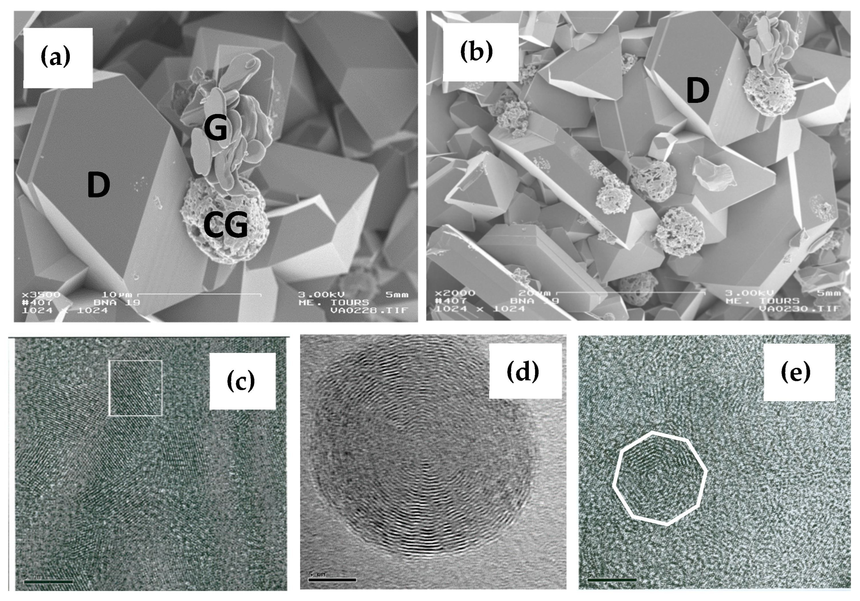
Figure 2.
Representative SEM micrographs of diamond particles synthesized from a binary mixture of naphthalene (C₁₀H₈) and highly fluorinated graphite (CF₁.₁) at 8 GPa and 1100 °C.
Figure 2.
Representative SEM micrographs of diamond particles synthesized from a binary mixture of naphthalene (C₁₀H₈) and highly fluorinated graphite (CF₁.₁) at 8 GPa and 1100 °C.
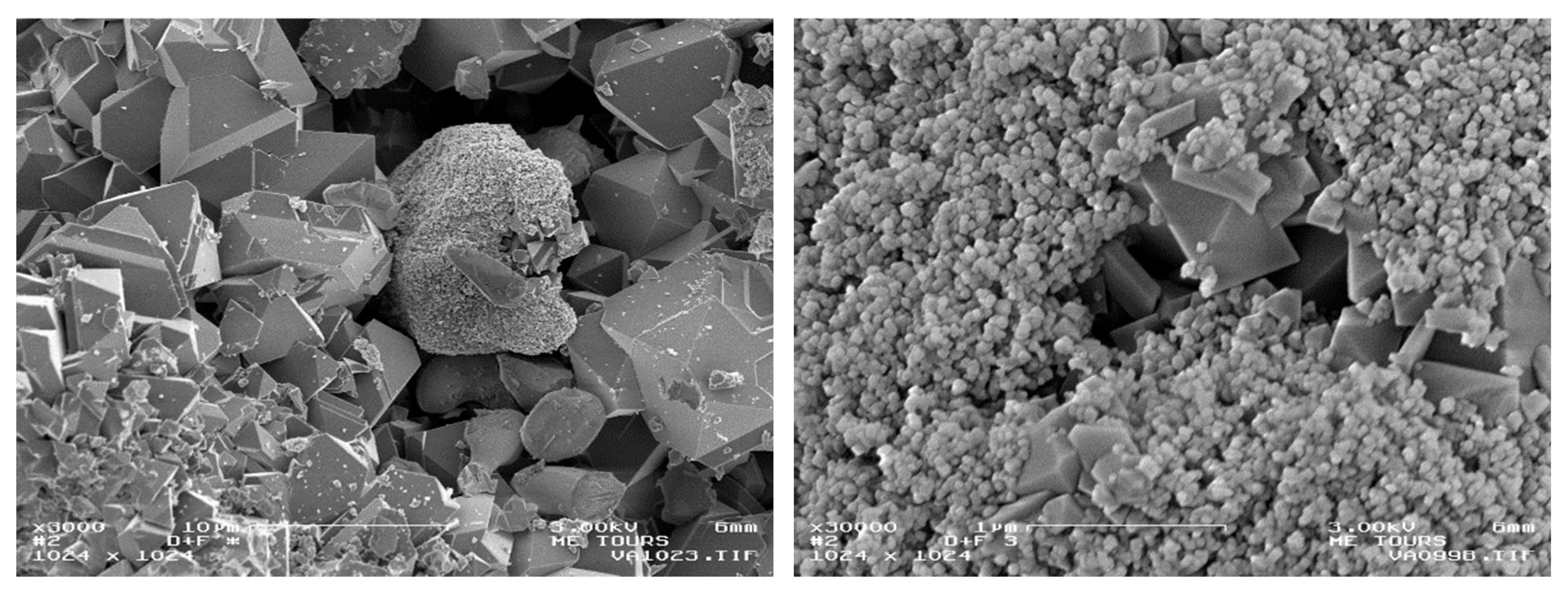
Figure 3.
Representative SEM micrographs of ultrananocrystalline (a), nanocrystalline (b), submicron (c), and micrometer-sized (d) diamond fractions obtained from growth systems based on mixtures of fluorocarbon and hydrocarbon precursors.
Figure 3.
Representative SEM micrographs of ultrananocrystalline (a), nanocrystalline (b), submicron (c), and micrometer-sized (d) diamond fractions obtained from growth systems based on mixtures of fluorocarbon and hydrocarbon precursors.
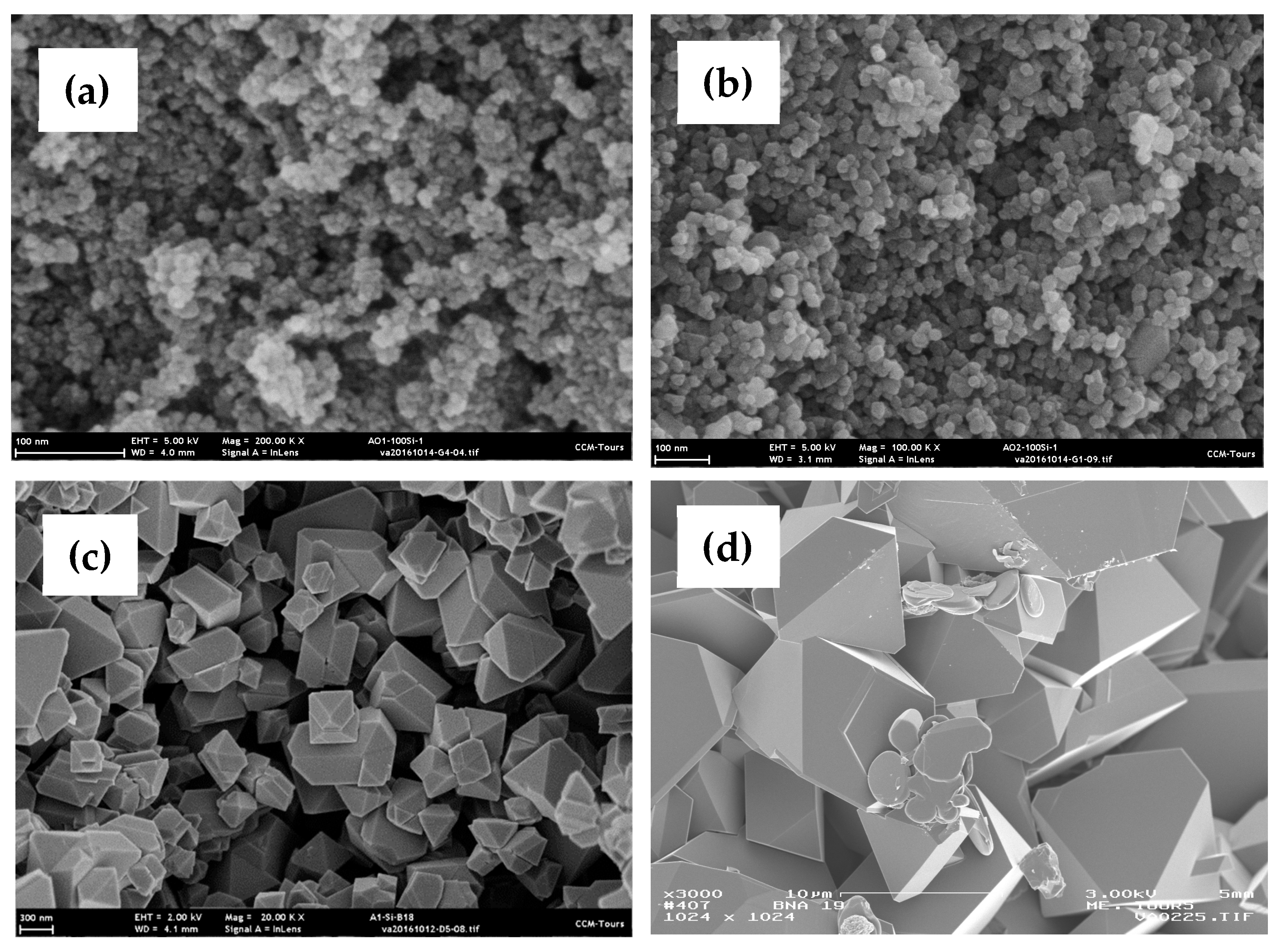
Figure 4.
Typical TEM image of the isolated nanodiamond fraction (a); high-resolution TEM (HRTEM) image of an individual nanodiamond particle (b); particle-size distribution histogram obtained from TEM analysis of 149 particles (c); Raman spectrum of the synthesized diamond material prior to chemical purification and size fractionation together with a Lorentzian fit (d). Reprinted with permission from [49], copyright IOP Science.
Figure 4.
Typical TEM image of the isolated nanodiamond fraction (a); high-resolution TEM (HRTEM) image of an individual nanodiamond particle (b); particle-size distribution histogram obtained from TEM analysis of 149 particles (c); Raman spectrum of the synthesized diamond material prior to chemical purification and size fractionation together with a Lorentzian fit (d). Reprinted with permission from [49], copyright IOP Science.
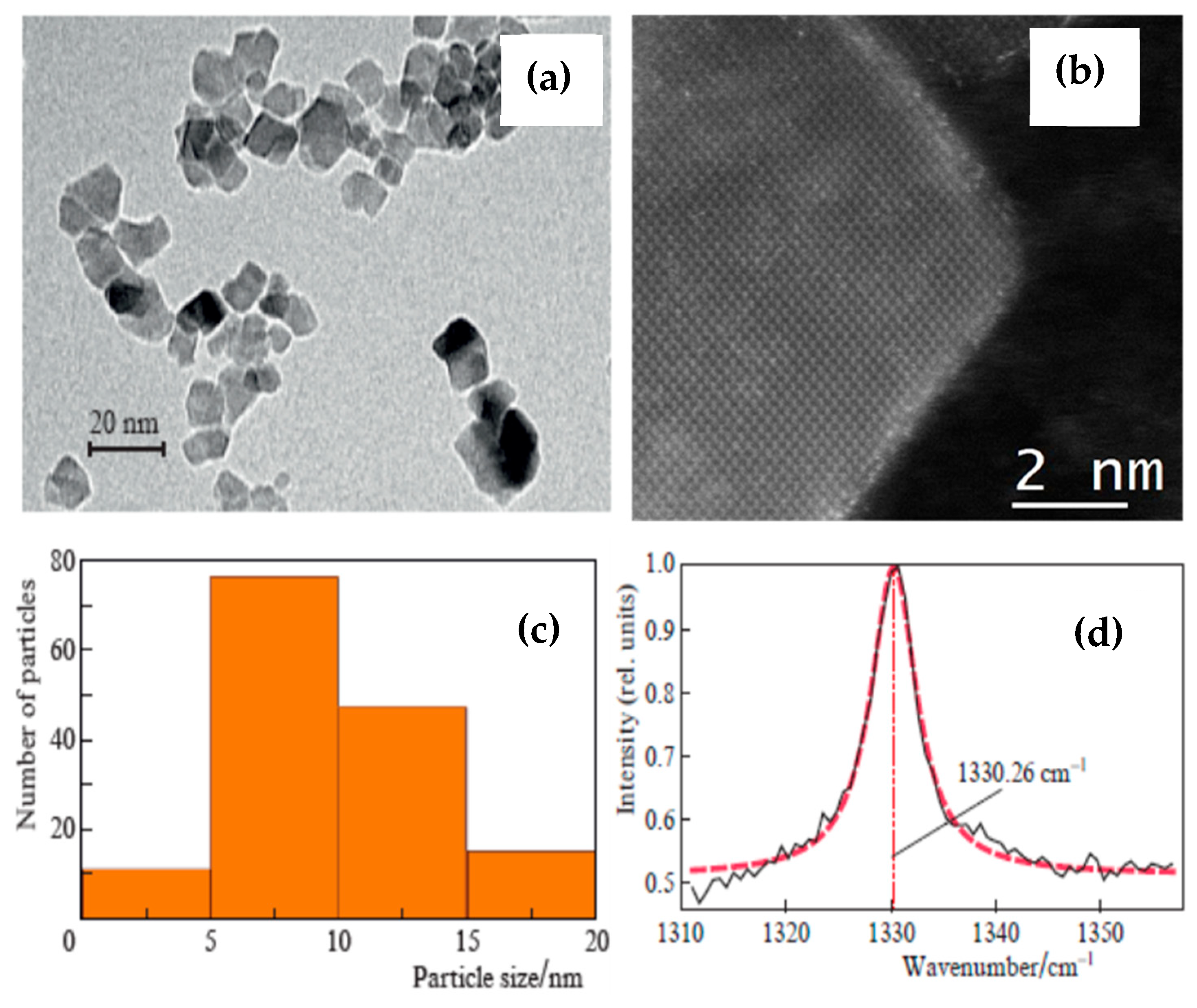
Figure 5.
Representative room-temperature photoluminescence spectra of individual SiV⁻ centers together with Lorentzian fits (a,b); statistical distributions of zero-phonon-line (ZPL) positions (c) and linewidths (FWHM) (d). Reprinted with permission from [49], copyright IOP Science.
Figure 5.
Representative room-temperature photoluminescence spectra of individual SiV⁻ centers together with Lorentzian fits (a,b); statistical distributions of zero-phonon-line (ZPL) positions (c) and linewidths (FWHM) (d). Reprinted with permission from [49], copyright IOP Science.
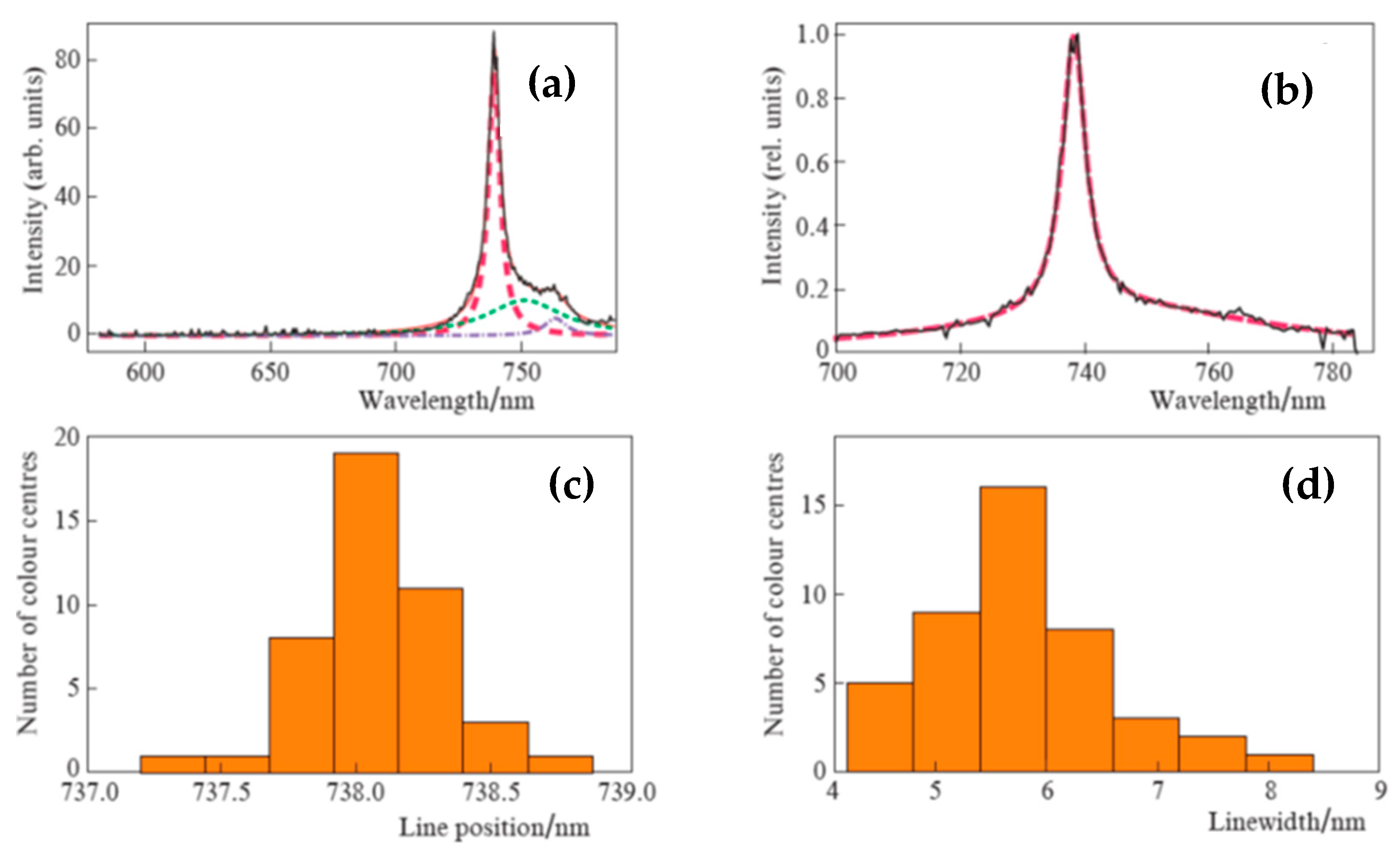
Figure 6.
(a) Elastic light-scattering image recorded at λ = 532 nm (upper panel) and photoluminescence image acquired near 738 nm (lower panel) for the same group of diamond particles. (b) Correlation between photon emission rate and particle size for diamonds synthesized from a fluorinated hydrocarbon growth system admixed with a high concentration of the silicon-containing dopant precursor. The experimentally measured photon emission rate scales approximately as d5.8, where d is the diamond particle diameter (blue line). The red line represents the dependence predicted by the model proposed in Ref. [56]. Reprinted with permission from [56], copyright Elsevier.
Figure 6.
(a) Elastic light-scattering image recorded at λ = 532 nm (upper panel) and photoluminescence image acquired near 738 nm (lower panel) for the same group of diamond particles. (b) Correlation between photon emission rate and particle size for diamonds synthesized from a fluorinated hydrocarbon growth system admixed with a high concentration of the silicon-containing dopant precursor. The experimentally measured photon emission rate scales approximately as d5.8, where d is the diamond particle diameter (blue line). The red line represents the dependence predicted by the model proposed in Ref. [56]. Reprinted with permission from [56], copyright Elsevier.
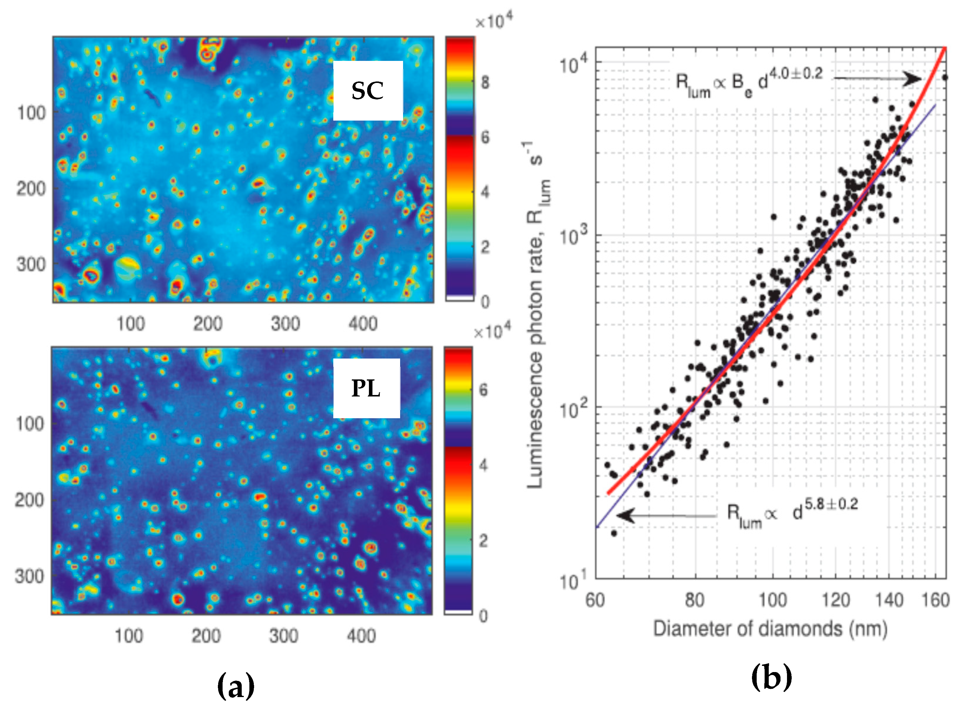
Figure 7.
(a) Size distribution of the studied nanodiamonds determined by TEM. The batch exhibits a broad size distribution with an average diameter of approximately 20 nm. Inset: representative high-resolution TEM image. (b) Typical confocal fluorescence image of nanodiamonds containing SiV⁻ centers. (c) Representative fluorescence spectrum of a single SiV⁻ center fitted with a Lorentzian profile, showing a sharp ZPL near 1.736 eV with a linewidth of approximately 2.2 meV. (d,e) Distributions of the ZPL energies and linewidths, respectively. Blue histograms correspond to single SiV⁻ centers, whereas gray histograms represent the complete dataset. The median ZPL energy of single SiV⁻ centers is blue-shifted by approximately 41 meV relative to the bulk-diamond SiV⁻ transition energy of 1.681 eV. Reprinted with permission from [58], copyright American Chemical Society.
Figure 7.
(a) Size distribution of the studied nanodiamonds determined by TEM. The batch exhibits a broad size distribution with an average diameter of approximately 20 nm. Inset: representative high-resolution TEM image. (b) Typical confocal fluorescence image of nanodiamonds containing SiV⁻ centers. (c) Representative fluorescence spectrum of a single SiV⁻ center fitted with a Lorentzian profile, showing a sharp ZPL near 1.736 eV with a linewidth of approximately 2.2 meV. (d,e) Distributions of the ZPL energies and linewidths, respectively. Blue histograms correspond to single SiV⁻ centers, whereas gray histograms represent the complete dataset. The median ZPL energy of single SiV⁻ centers is blue-shifted by approximately 41 meV relative to the bulk-diamond SiV⁻ transition energy of 1.681 eV. Reprinted with permission from [58], copyright American Chemical Society.
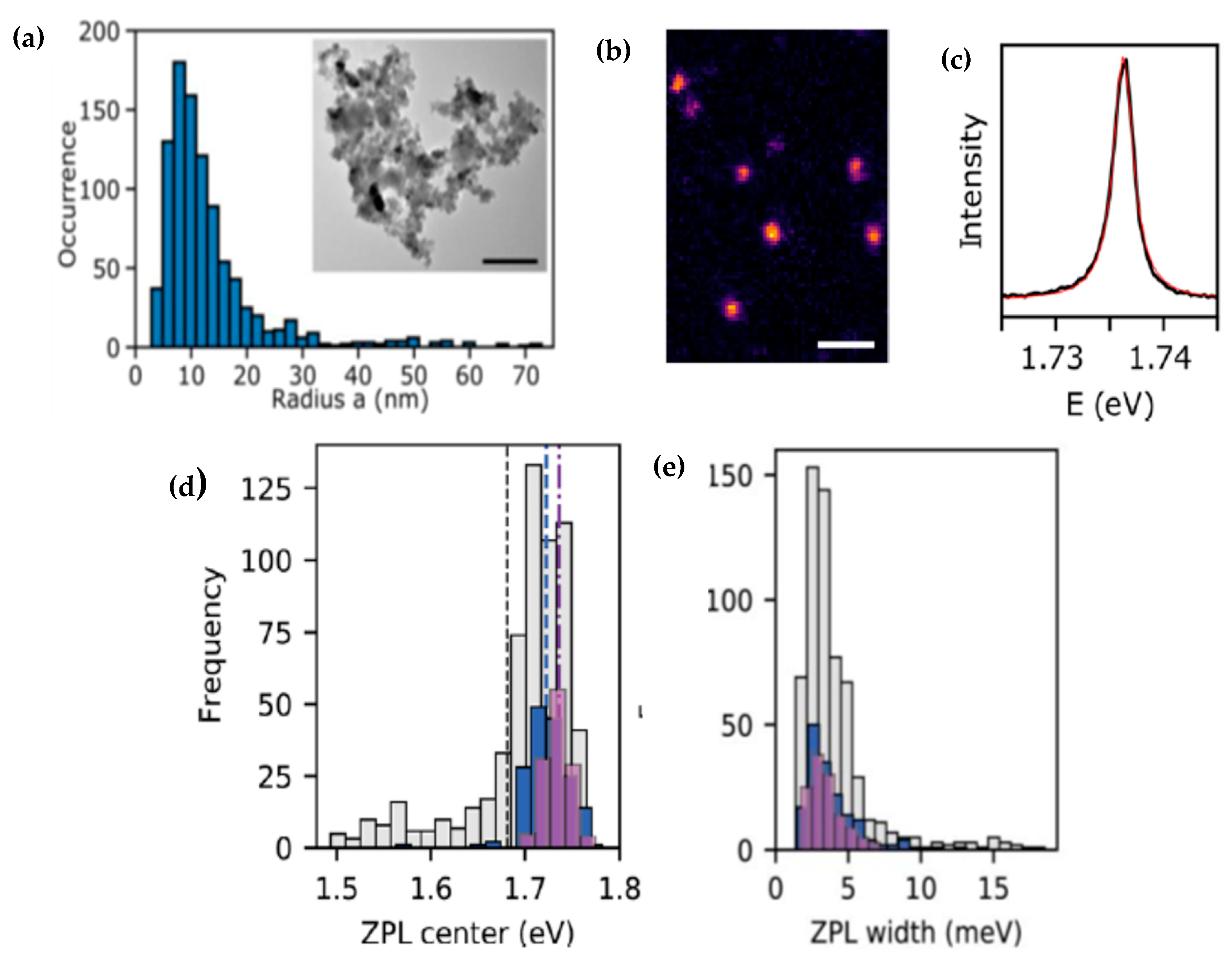
Figure 8.
Calculated energy shift ℏΔω0 of a single SiV⁻ center located inside a spherical diamond nanoparticle of radius a = 10 nm as a function of the distance of the emitter from the particle center. The diamond refractive index is characterized by nd = 2.45, and the surrounding medium has ne ≈ 1.24. The SiV⁻ transition dipole moment is oriented along θ = 45○. The bulk transition energy and radiative decay rate are ℏω0 = 1.68 eV and ℏγrad = 5.49 × 10⁻⁴ meV, respectively. Solid blue and dashed red lines represent the results of model calculations based on two different equations considered in the in work [58]. Reprinted with permission from [58], copyright American Chemical Society.
Figure 8.
Calculated energy shift ℏΔω0 of a single SiV⁻ center located inside a spherical diamond nanoparticle of radius a = 10 nm as a function of the distance of the emitter from the particle center. The diamond refractive index is characterized by nd = 2.45, and the surrounding medium has ne ≈ 1.24. The SiV⁻ transition dipole moment is oriented along θ = 45○. The bulk transition energy and radiative decay rate are ℏω0 = 1.68 eV and ℏγrad = 5.49 × 10⁻⁴ meV, respectively. Solid blue and dashed red lines represent the results of model calculations based on two different equations considered in the in work [58]. Reprinted with permission from [58], copyright American Chemical Society.
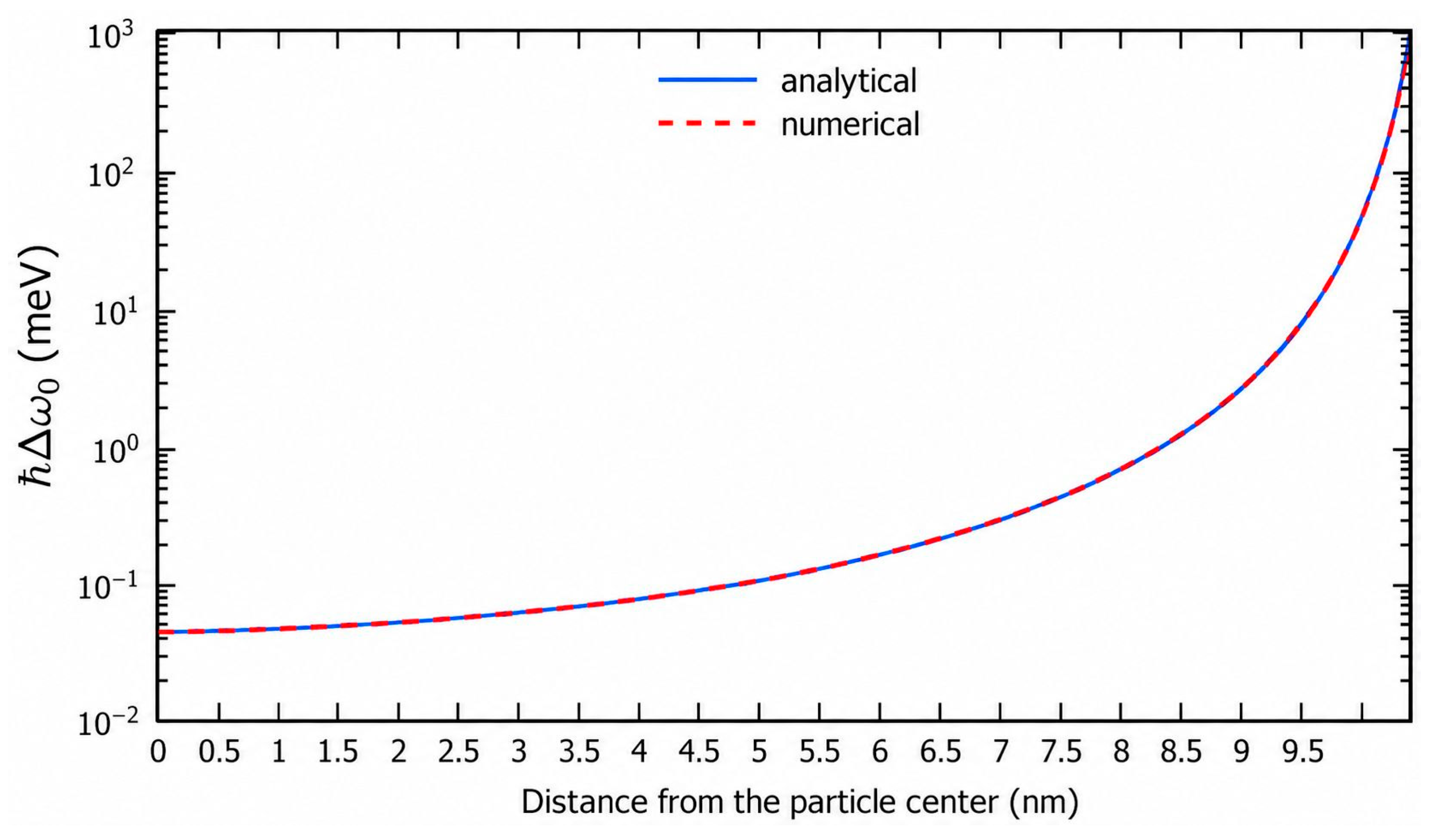
Figure 9.
High-resolution TEM images of a submicron-sized (~900 nm) diamond particle (a) and a nanosized (~25 nm) diamond particle (b), each hosting a single SiV⁻ center. The corresponding fluorescence decay curves (c) and photoluminescence spectra (d) are shown using the same color coding. The SiV⁻ center in the nanosized particle exhibits a fluorescence lifetime of approximately 2.5 ns and a ZPL linewidth (FWHM) of approximately 2.3 meV, whereas the SiV⁻ center in the ~900 nm particle exhibits a shorter lifetime of approximately 0.8 ns and a broader ZPL linewidth of approximately 5.7 meV. Reprinted with permission from [58], copyright American Chemical Society.
Figure 9.
High-resolution TEM images of a submicron-sized (~900 nm) diamond particle (a) and a nanosized (~25 nm) diamond particle (b), each hosting a single SiV⁻ center. The corresponding fluorescence decay curves (c) and photoluminescence spectra (d) are shown using the same color coding. The SiV⁻ center in the nanosized particle exhibits a fluorescence lifetime of approximately 2.5 ns and a ZPL linewidth (FWHM) of approximately 2.3 meV, whereas the SiV⁻ center in the ~900 nm particle exhibits a shorter lifetime of approximately 0.8 ns and a broader ZPL linewidth of approximately 5.7 meV. Reprinted with permission from [58], copyright American Chemical Society.
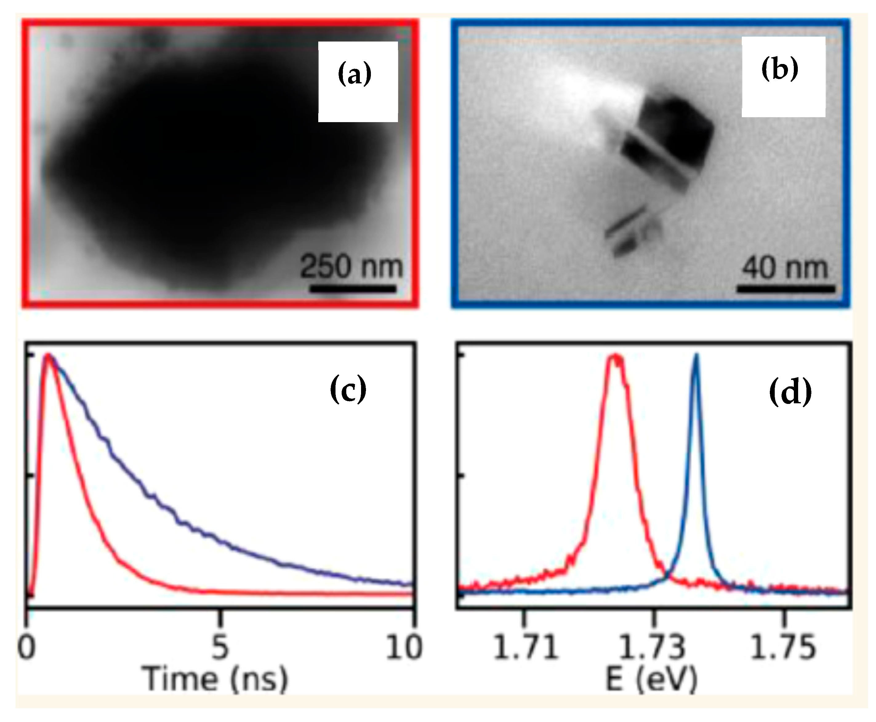
Figure 10.
(a) Confocal fluorescence image of individual diamond particles and (b) corresponding AFM topography image acquired from the same sample region using a correlative confocal microscopy–AFM setup. (c) Estimated number of NV⁻ centers in individual diamond particles as a function of particle height for 50 particles. The solid curve represents a fit to the experimental data using the cubic dependence y = ax3 [17]. Reprinted with permission from [17], copyright Elsevier.
Figure 10.
(a) Confocal fluorescence image of individual diamond particles and (b) corresponding AFM topography image acquired from the same sample region using a correlative confocal microscopy–AFM setup. (c) Estimated number of NV⁻ centers in individual diamond particles as a function of particle height for 50 particles. The solid curve represents a fit to the experimental data using the cubic dependence y = ax3 [17]. Reprinted with permission from [17], copyright Elsevier.
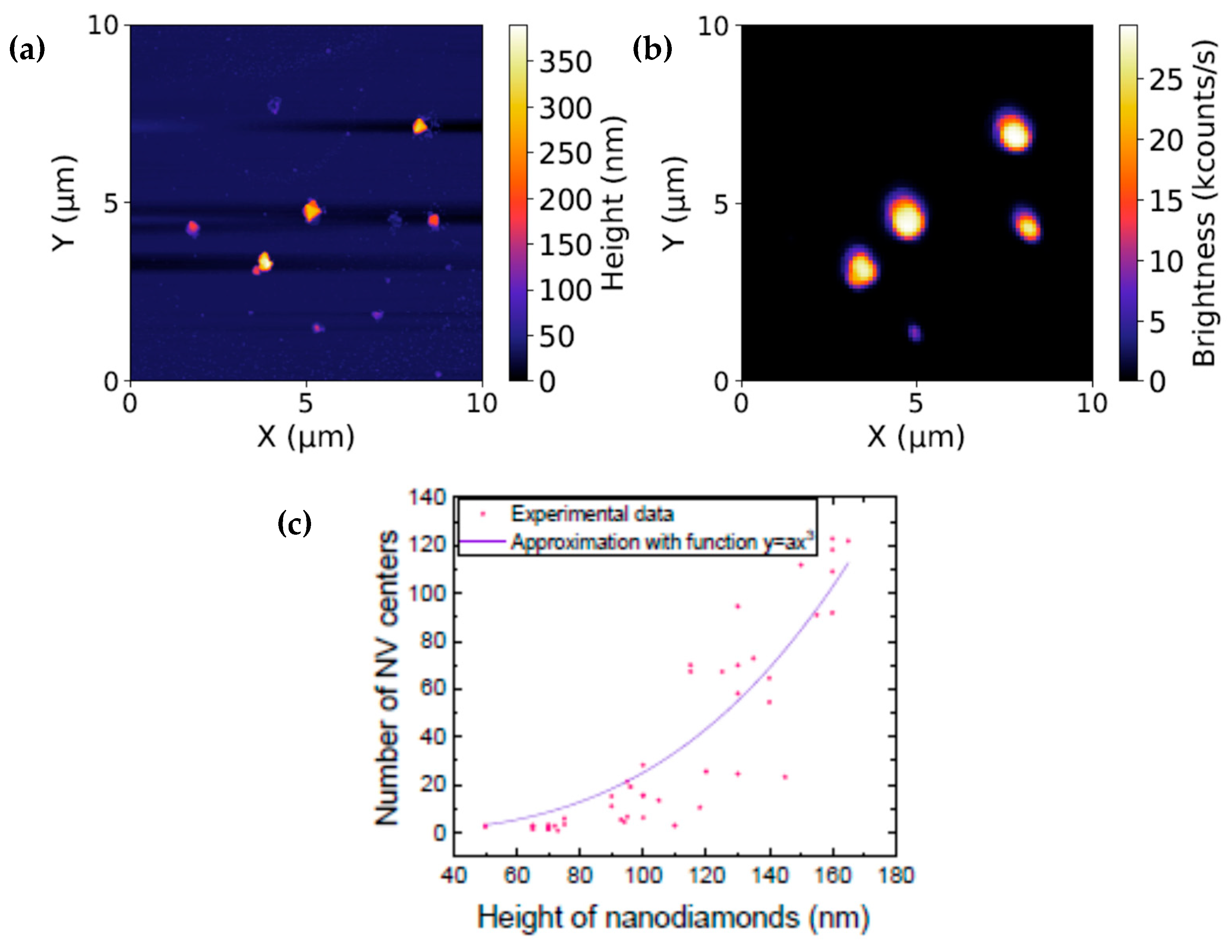
Figure 11.
Schematic representation of a core–shell diamond nanoparticle comprising a diamond core with natural isotopic abundance (~1.1% 13C) containing NV⁻ centers and a highly 13C-enriched diamond shell (a). TEM image of core–shell diamond nanoparticles obtained by HPHT overgrowth using ND25 nanodiamonds as seeds. Scale bar 100 nm (b). Raman spectra recorded from five individual core–shell particles (c). The peak at 1332 cm⁻¹ corresponds to the first-order diamond Raman band of the core with natural isotopic abundance, whereas the peak near 1284 cm⁻¹ is attributed to the shell enriched to approximately 96% 13C. Reprinted with permission from [18], copyright American Chemical Society.
Figure 11.
Schematic representation of a core–shell diamond nanoparticle comprising a diamond core with natural isotopic abundance (~1.1% 13C) containing NV⁻ centers and a highly 13C-enriched diamond shell (a). TEM image of core–shell diamond nanoparticles obtained by HPHT overgrowth using ND25 nanodiamonds as seeds. Scale bar 100 nm (b). Raman spectra recorded from five individual core–shell particles (c). The peak at 1332 cm⁻¹ corresponds to the first-order diamond Raman band of the core with natural isotopic abundance, whereas the peak near 1284 cm⁻¹ is attributed to the shell enriched to approximately 96% 13C. Reprinted with permission from [18], copyright American Chemical Society.
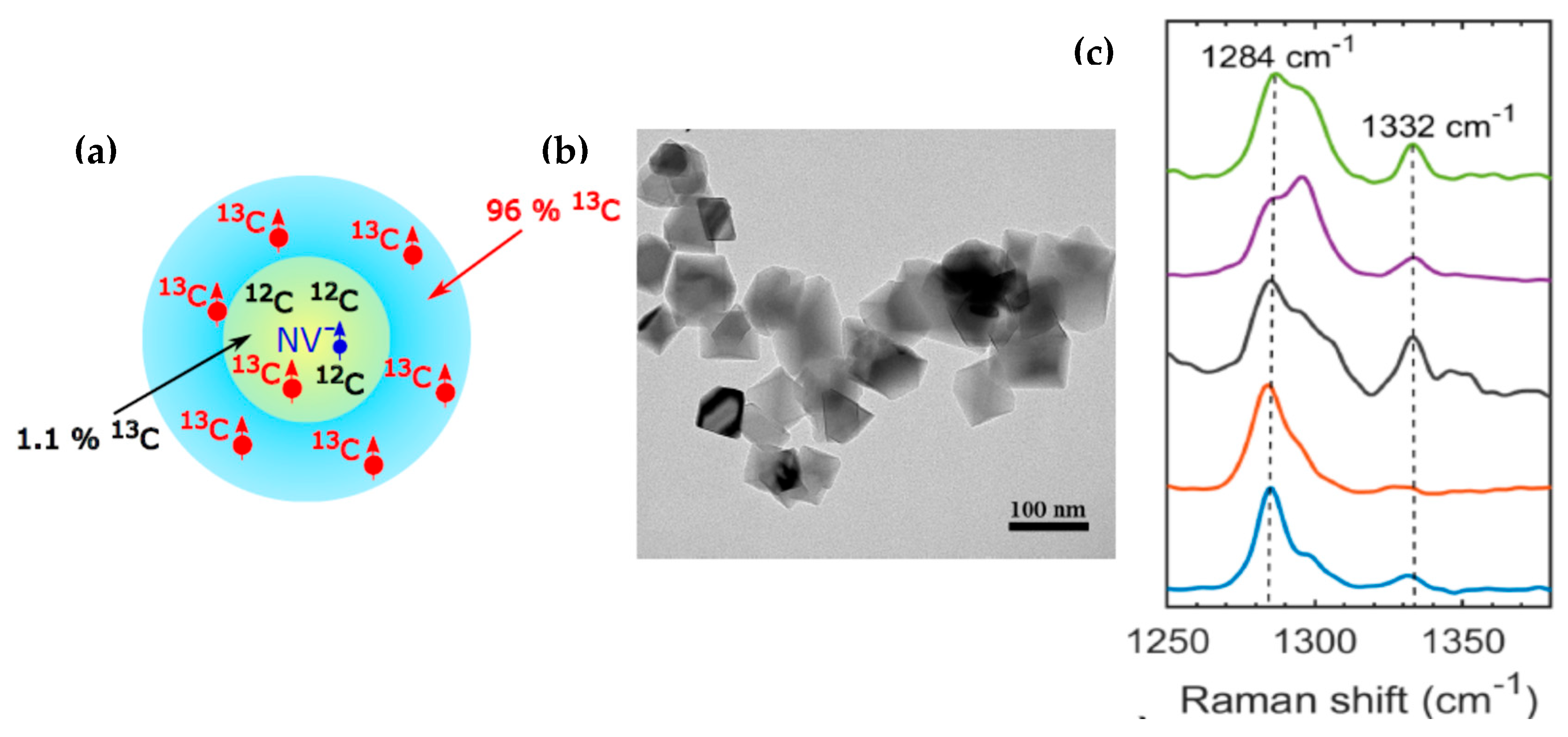
Disclaimer/Publisher’s Note: The statements, opinions and data contained in all publications are solely those of the individual author(s) and contributor(s) and not of MDPI and/or the editor(s). MDPI and/or the editor(s) disclaim responsibility for any injury to people or property resulting from any ideas, methods, instructions or products referred to in the content. |
© 2026 by the authors. Licensee MDPI, Basel, Switzerland. This article is an open access article distributed under the terms and conditions of the Creative Commons Attribution (CC BY) license (http://creativecommons.org/licenses/by/4.0/).
Copyright: This open access article is published under a Creative Commons CC BY 4.0 license, which permit the free download, distribution, and reuse, provided that the author and preprint are cited in any reuse.