

Submitted:
27 October 2026
Posted:
30 October 2026
You are already at the latest version
Abstract
This review asks whether female and male animals differ in neuroinflammatory measurements in Parkinson’s disease models and whether the findings establish different responses to disease induction. We searched APA PsycNET, PubMed, Scopus, Web of Science and SciELO, initially covering publications from January 2014 to December 31, 2024. Update searches ran from May 12 to May 31, 2025. Eligible studies used neurotoxin, pesticide or genetic models in rodents or nonhuman primates and included both sexes and control groups. We assessed risk of bias with the Systematic Review Centre for Laboratory Animal Experimentation (SYRCLE) tool. Findings were tabulated and compared descriptively, accounting for shared cohorts and distinguishing differences in measured values from interactions involving sex. Variation in methods and measurements, incomplete numerical data and uncertain independence prevented pooling. Eighteen publications represented 17 study units, all in rodents. No nonhuman primate studies were eligible. A total number of unique animals could not be established. Among mice that underwent sham surgery without dutasteride, striatal glial fibrillary acidic protein fluorescence increased after 1-methyl-4-phenyl-1,2,3,6-tetrahydropyridine (MPTP) in males; no increase was detected in females. An interaction between sex and MPTP exposure supported different responses. Microglial density and branching after rotenone varied with sex and genotype. Female mice overexpressing \(\alpha\)-synuclein had higher plasma interleukin 5 than males at 4 and 18 months. After a 6-hydroxydopamine lesion, male rats given a control oligonucleotide had more cells expressing inducible nitric oxide synthase per section in the substantia nigra pars compacta than females given the same treatment. These last two comparisons did not establish different responses to disease induction. Many findings relied on separate analyses within each sex. Unclear risk of bias, sparse replication and variation in assays and conditions limited interpretation. Some glial and inflammatory measurements differed between sexes under particular experimental conditions. The evidence does not establish generally greater neuroinflammation in either sex. Comparable independent experiments and direct assessment of cellular function are needed to clarify the biological significance of these differences.
Keywords:
Parkinson disease
; neuroinflammatory diseases
; sex factors
; disease models
; animal
; microglia
; astrocytes
; cytokines
; rodentia
1. Introduction
Parkinson’s disease (PD) is a progressive neurodegenerative disorder marked by degeneration of dopaminergic neurons in the substantia nigra (SN) [1]. Intracellular inclusions, known as Lewy bodies and Lewy neurites, containing aggregated -synuclein, are a hallmark of neurodegeneration in PD [1]. Neuroinflammatory responses in PD, are thought to involve microglia (the brain’s resident immune cells), as well as astrocytes, inflammatory signaling and interactions with other immune cell populations. In addition to brain changes, studies of people with PD have found alterations in circulating immune cells and in inflammatory mediators measured in the circulation [1]. These responses merit attention because experimental studies indicate that glial and immune activity can influence neuronal survival, with protective or harmful effects depending on the cells and conditions involved [1].
Studies outside of PD have identified differences between females and males in immune cell populations, their activation and the inflammatory mediators they produce [2]. Rodent studies and experiments with cultured glial cells have also reported differences in how astrocytes and microglia from females and males respond to injury and inflammatory stimuli [3]. Because the cells and signaling pathways involved in responses to pathology can differ between females and males, sex is a relevant biological variable when neuroinflammatory responses are studied in experimental PD. Whether the same differences occur in PD models must be examined in those models, rather than inferred from other diseases or cultured cells. Published studies in mouse models of PD have reported sex differences in selected astrocytic responses and circulating cytokine concentrations [4,5]. The broader question is how neuroinflammatory responses differ between females and males in experimental models of PD.
Experimental animal models allow researchers to compare neuroinflammatory measurements and responses between females and males under defined conditions. The exposure or genetic alteration used to model PD can be specified in advance [6]. When exposed and control groups include both sexes, female and male values can be compared after exposure. The differences between exposed animals and their respective controls can also be compared between sexes [7]. Tissue can then be collected from selected brain regions at particular times, so that glial and immune measurements are considered in relation to a known anatomical location and a defined point in the experiment [6]. These models also have clear limits. Rodent and nonhuman primate (NHP) models reproduce selected features of PD rather than the human disease in its entirety [6]. Even within those limits, they provide a setting in which female and male responses can be examined directly, and the available studies can then be considered together to determine what they establish about sex differences in neuroinflammatory measurements and responses.
Assessing these experiments together can make it clearer which parts of neuroinflammation show evidence of differences between females and males. Previous syntheses already provide useful evidence. Bourque et al. reviewed sex differences in immune processes related to PD, while Carrera Rengifo’s thesis included a systematic review of microglial responses in rodent models [8,9]. Both considered the conditions under which the experiments were performed, and their questions overlap with the present review. Glial and immune measurements describe different biological features. A difference found in one measure cannot therefore describe neuroinflammation as a whole [10,11]. How broadly a reported sex difference can be interpreted also depends on whether similar experiments support the same result. Studies that measure different features, or use different conditions, may be addressing different biological questions. Their findings should therefore not be treated automatically as evidence for the same conclusion. We therefore conducted a systematic review to examine how neuroinflammatory measurements and responses differ between females and males in rodent and nonhuman primate models of PD.
2. Results
2.1. Selection of Studies
Figure 1 illustrates the selection process used in this review. The database searches identified 6,256 records. After the removal of 926 duplicates, full texts were sought for all 5,330 remaining records. Twenty reports could not be retrieved, and 5,310 reports were assessed with reference to the full text. Of these, 5,293 were excluded for failing to meet the eligibility criteria during both the inclusion and exclusion phases. The majority of the exclusions occurred when reports either failed to include male and female subjects or did not differentiate the data by sex (n = 3,332). The second most common category of exclusion was related to not using an eligible PD model, such as a neurotoxin, pesticide, or genetic model (n = 840). Other records fell outside the predefined date range or were not in Spanish or English (n = 630). Additional records were excluded for using species other than rodents or NHPs (n = 253), and others were excluded for lack of measurements of an eligible neuroinflammatory outcome (n = 91). Finally, a smaller subset of records was excluded due to inadequate control groups (n = 86), ineligible study designs (n = 48), or because they were retracted or available only as preprints (n = 13). Ultimately, 17 studies met all criteria and were included in the systematic review.
2.2. Study and Sample Characteristics
We identified 17 study units based on 18 publications (See Table 1). All of these studies were performed using rodent models. While 15/17 studies were conducted with mice (88.2%), 2/17 studies were performed with Long-Evans rats (11.8%). None of the studies included data from NHPs. Among the studies using mouse models, 12/15 (80.0%) were based on the C57BL/6 background. Neurotoxin models of PD were the most common at 11/17 of the studies (64.7%). These studies included the administration of the neurotoxin 1-methyl-4-phenyl-1,2,3,6-tetrahydropyridine (MPTP) in 6/17 of the studies (35.3%). Other neurotoxins, including 6-hydroxydopamine (6-OHDA) and rotenone, were used in 3/17 of the studies (17.6%) each, with only a single study using both 6-OHDA and rotenone. The remaining 6/17 studies (35.3%) utilized genetically modified rodents to mimic PD. These studies involved alterations to hSNCA, LRRK2, Pink1, or Rasd2 genes. Notably, no study used an exact genetic model that was represented in more than one study. All 17 study units included both female and male animals. We treated the study units from Costa et al. (2020) and Sirabella et al. (2018) as part of a single experimental series because Costa et al. (2020) references data form their companion work displayed in Sirabella et al. (2018). Group sizes reported in the 17 studies ranged from 3 to 18 animals, and eleven of those ranges varied depending on the experimental groups. Age at baseline was not reported in 4/17 of the studies (23.5%). Importantly, one of the studies reported use of different ages for female and male animals. 5/17 studies (29.4%) identified in the current review utilized interventions that were associated with hormone manipulation. These studies included the administration of dutasteride in 2/17 (11.8%) and of 17-estradiol in 2/17 (11.8%). Gonadal surgery was used in 4/17 of the studies (23.5%), including gonadectomy in 2/17 (11.8%), ovariectomy in 1/17 (5.9%), and orchiectomy in 1/17 (5.9%). Regarding estrous cycle monitoring, 3/17 studies (17.6%) reported assessment of this aspect thorugh vaginal cytology in 2/17 (11.8%) and vaginal lavage in 1/17 (5.9%). Lipopolysaccharide exposure as an inflammatory manipulation was reported in 2/17 of the studies (11.8%).
2.3. Neuroinflammatory Outcomes Assessed
Assessment of neuroinflammation was distributed across nine outcome domains. Frequently, individual studies showed outcomes that corresponded to more than one domain (Table 2; Supplementary Table S1). The number of studies that reported the most frequent neuroinflammatory outcomes was as follows: microglial activation and phenotype were assessed in 13/17 studies (76.5%), while astrocyte reactivity was assessed in 11/17 studies (64.7%) and cytokine measurements in 9/17 studies (52.9%). Among the individual markers evaluated, ionized calcium-binding adaptor molecule 1 (Iba1) and glial fibrillary acidic protein (GFAP) were each assessed in 11/17 studies (64.7%). Within the cytokine domain, tumor necrosis factor (TNF-) was the most commonly assessed cytokine, appearing in 7/17 studies (41.2%). Other less frequent cytokines followed, such as interleukin-1 (IL-1; 6/17 studies, 35.3%), IL-6 (4/17 studies, 23.5%), interferon- (IFN-) and IL-10 (3/17 studies, 17.6%), and IL-4 (2/17 studies, 11.8%). Other domains were less common, such as oxidative/nitrosative stress and antioxidant defence (4/17 studies, 23.5%), inflammatory signalling and transcriptional pathways (3/17 studies, 17.6%), and immune cell composition and function (2/17 studies, 11.8%). Studies also assessed a variety of other outcome measures, including: complement system outcomes (1/17 studies, 5.9%); chemokine and leukocyte recruitment measures (2/17 studies, 11.8%); and mitochondrial or cellular bioenergetic function (1/17 studies, 5.9%). Supplementary Table S1 lists 60 grouped marker or outcome categories, of which 49 (81.7%) were represented in a single study. These are descriptive coverage categories, not independent effects. This showed substantial heterogeneity in the neuroinflammatory outcomes assessed across the included studies.
2.4. Risk of Bias in Studies
Unclear risk predominated in eight of the ten domains of the Systematic Review Centre for Laboratory Animal Experimentation (SYRCLE) tool (Figure 2). Sequence generation (Q1, whether animals were assigned to groups by chance) was judged unclear in all 17 studies (100.0%), followed by incomplete outcome data (Q8, whether missing results and animals left out of the analysis were properly accounted for) in 16/17 (94.1%). Allocation concealment (Q3, whether the next group assignment was kept hidden from those assigning animals) and caregiver and investigator blinding (Q5, whether those caring for animals or conducting the experiment were unaware of which treatment animals received) were each judged unclear in 15/17 studies (88.2%). Similarly, random outcome assessment (Q6, random selection of animals for measurement) was unclear in 14/17 (82.4%). For the domain of selective outcome reporting (Q9, whether the paper included results for everything the researchers had planned/expected to measure), there was the highest proportion of low-risk judgments (11/17, 64.7%). Outcome assessor blinding (Q7, whether those measuring the outcomes were unaware of which treatment each animal received) was divided between low risk (8/17, 47.1%) and unclear risk (9/17, 52.9%). Other potential sources of bias (Q10, problems not covered by the other questions) received equal proportions of low and unclear risk judgments (8/17 each, 47.1%). High risk judgments were recorded for allocation concealment in 2/17 studies (11.8%). Baseline characteristics (Q2, whether groups were similar at the start or initial differences were taken into account in the analysis), random housing (Q4, random placement of animals within the animal room), caregiver and investigator blinding, and other potential sources of bias each received one high risk judgment (1/17, 5.9%).
2.5. Reporting Completeness
The Animal Research: Reporting In Vivo Experiments (ARRIVE) Essential 10 is the minimum set of information needed in a manuscript. For this section of the ARRIVE guidelines, reporting was complete for several items as shown in Figure 3. "Study design" and "experimental procedures" were the most frequently fully reported, in 16/17 studies (94.1%). For the item "statistical methods," 15/17 studies (88.2%) reported this completely, as did the reporting of "experimental animal characteristics", which was complete in 14/17 studies (82.4%). Reporting criteria for "outcome measures" were satisfied by 13/17 studies (76.5%), while "results" reporting was complete in 12/17 articles (70.6%). In contrast, for "sample size", only 2/17 studies (11.8%) reported this item completely, including information on how the sample size was decided and any a priori calculation when done. "Inclusion and exclusion criteria" was also considered incomplete or unclear in 12/17 studies (70.6%). For this item, specific rules for data selection, reasons for exclusion, or the exact n for every analysis were not directly detailed in the text. Another important finding was for "randomisation" and "blinding", which ended up being the items with some of the largest reporting gaps among the Essential 10. "Randomisation" was rated as "Partially/Not clear" in nine studies (52.9%) and received a "No" in eight (47.1%). Comparably, "blinding" was rated as "No" in eight studies (47.1%), and it was partially or not clearly reported in seven (41.2%). As a result, only two studies (11.8%) provided complete information on blinding. Therefore, the main reporting gaps within the Essential 10 were related to sample size justification, inclusion and exclusion criteria, randomisation, and blinding.
The reporting in the ARRIVE Recommended Set (Figure 3) was complete for the "background" item in all 17 studies. This meant that the information provided an account of the underlying science for the experiments described and the reasons for choosing the particular animal models. In addition, the "objectives" were provided completely for 16/17 studies (94.1%), as were the corresponding ethical statements for 15/17 studies (88.2%). "Declaration of interests" was fully addressed in 13 of 17 studies (76.5%). Furthermore, 11/17 articles (64.7%) fully satisfied the "housing and husbandry" item by providing details on living conditions and environmental enrichment. Similarly, 11/17 studies (64.7%) showed complete information on "interpretation/scientific implications", including interpretation of the findings and discussion of potential biases or animal model limitations. Other items had lower reporting rates. For example, no study fully reported "animal care and monitoring". Twelve studies (70.6%) provided partial or unclear information, and 5/17 (29.4%) were rated as "No". In these studies, details on reduction of animal distress, adverse events, or humane endpoints were not completely reported. "Generalisability and translation" was also rated as "Partially/Not clear" in 11/17 studies (64.7%). The largest reporting gap was observed for "protocol registration", which was rated as "No" in 16 of 17 studies (94.1%). "Data access" showed a more divided pattern, with eight studies (47.1%) rated "Yes", eight (47.1%) "No", and one (5.9%) "Partially/Not clear".
2.6. Inter-Rater Agreement in Study Selection
Inter-rater agreement for study selection, in particular for the inclusion and exclusion of reports, is presented in Figure 4. For the primary inclusion of reports in the review, agreement between the four pairs of reviewers was assessed for 1,303 to 1,304 reports each. The observed agreement was between 91.8% and 97.2%. Corresponding Gwet’s agreement coefficient 1 (AC1) measures were between 0.904 and 0.971, with narrow 95% confidence intervals (CIs), as shown in Figure 4. The corresponding Cohen’s measures were much lower, between 0.132 and 0.423. The largest difference between the two measures was for Pair 2, with an observed agreement of 97.2% and AC1 of 0.971 (95% CI 0.962 to 0.981), whereas Cohen’s was 0.132 (95% CI to 0.276). Pair 1 also showed a large difference, with an observed agreement of 94.1%, AC1 of 0.936 (95% CI 0.921 to 0.951), whereas Cohen’s was 0.255 (95% CI 0.142 to 0.367). However, across the primary inclusion comparisons, the lower estimates occurred alongside consistently high observed agreement and AC1 values, consistent with the strongly imbalanced distribution of screening decisions. A separate set of 115 reports identified in the 2025 search update was assessed by the fifth pair. Agreement in this set was lower than in the primary inclusion screening, with an observed agreement of 68.7%, an AC1 of 0.540 (95% CI 0.380 to 0.699), and a of 0.152 (95% CI 0.029 to 0.275).
In contrast, agreement estimates for the exclusion criteria varied more across reviewer pairs, and the smaller number of reports assessed by each pair was reflected in wider 95% CIs. Pairs 1, 2, 3, and 5 each assessed 28 reports, while Pair 4 assessed 26. Observed agreement ranged from 71.4% to 85.7%. For Pair 1, observed agreement was 71.4%, with an AC1 of 0.596 (95% CI 0.285 to 0.906) and a of 0.059 (95% CI to 0.430). Pair 2 also had an observed agreement of 71.4%, with an AC1 of 0.493 (95% CI 0.142 to 0.844) and a of 0.360 (95% CI to 0.722). Pair 3 showed the highest observed agreement at 85.7%, with an AC1 of 0.785 (95% CI 0.558 to 1.000) and a of 0.579 (95% CI 0.198 to 0.960). For Pair 4, observed agreement was 76.9%, with an AC1 of 0.598 (95% CI 0.268 to 0.928) and a of 0.462 (95% CI 0.082 to 0.843), while Pair 5 had an observed agreement of 75.0%, an AC1 of 0.501 (95% CI 0.165 to 0.837), and a of 0.500 (95% CI 0.165 to 0.835). Therefore, compared with the primary inclusion screening, agreement estimates for the exclusion criteria varied more between reviewer pairs and were associated with wider confidence intervals. The highest agreement in the exclusion phase was observed for Pair 3, while the difference between the two chance-corrected measures was most pronounced during the primary inclusion screening.
The following sections present neuroinflammatory findings by biological domain, distinguishing statistical tests involving sex from results analysed separately in females and males.
2.7. Microglial Activation and Phenotype
Microglial findings varied across genetic backgrounds, ages, brain regions and assessment times (Figure 5). The evidence did not establish a consistent pattern of greater responses in either sex. Rocha et al. examined microglial density and branching after rotenone or vehicle treatment in female and male wild type (WT) mice and mice with IB kinase 2 (IKK2) deleted specifically from microglia [12]. In the SN, rotenone increased the density of cells expressing Iba1 in males with the deletion compared with vehicle controls of the same sex and genotype. No corresponding increase was detected in females with the same deletion. The regional analyses of cell density reported interactions between sex and rotenone exposure [12]. The branching analysis showed an interaction among sex, genotype and rotenone exposure. Relative to the corresponding vehicle groups, maximal branch length decreased in WT females and males. Branch counts decreased in WT males but increased in males lacking microglial IKK2 [12].
Two other genetic models were assessed at different ages, although their statistical comparisons addressed different questions. Costa et al. examined cluster of differentiation 11b (CD11b) immunoreactivity in female and male mice with Ras homolog enriched in striatum (Rhes) deficiency and WT controls at 3 and 12 months of age [15]. The regional analyses reported interactions among sex, genotype and age. At 3 months, males with Rhes deficiency had increased immunoreactivity in the caudate putamen (CPu) and substantia nigra pars compacta (SNpc) compared with WT males. At 12 months, comparisons with WT animals of the same sex showed increases in the female CPu and male SNpc [15]. Lamontagne-Proulx et al. assessed striatal Iba1 fluorescence intensity in female and male SNCA-OVX mice, a genetic model of human -synuclein overexpression, at 4 and 18 months of age. The overall sex effect across these ages was not significant, and the authors did not report a result for an interaction between sex and age [5].
Zhou et al. measured Iba1 protein in the SN of female and male mice given MPTP, with or without antibiotic pretreatment [24]. Within each sex, mice given antibiotics before MPTP had lower Iba1 protein than mice given MPTP alone. A direct comparison between females and males receiving both treatments found higher Iba1 protein in females. No interaction between sex and antibiotic treatment was reported, so the endpoint comparison did not establish whether antibiotics produced different changes in females and males [24].
Results from separate analyses within each sex varied by brain region, measurement and assessment time. In their rotenone study, Mitra et al. compared exposed female and male mice with vehicle controls. Flow cytometry measured the percentage of cells expressing Iba1, while immunoblotting measured Iba1 protein [16]. Both measurements increased in the SN of females and males. In the frontal cortex, both decreased in males. Females showed an increased percentage of cells expressing Iba1 in this region without a detected change in Iba1 protein [16]. In a separate MPTP experiment, Mitra et al. measured nigral Iba1 protein by immunoblotting and Iba1 labelling by flow cytometry [17]. Among mice without hormonal pretreatment, separate comparisons within each sex showed increases in both measurements relative to saline controls on days 1 and 7 in females and on day 3 in males. Increases were not detected in the other sex at the respective time points [17].
Changes detected in only one sex were not confined to males. Isenbrandt et al. examined striatal microglial density and branching in female and male mice given MPTP or saline [21]. Among mice that underwent sham surgery and received vehicle instead of dutasteride, males showed increased density and reduced branching complexity 6 days after MPTP compared with saline controls. Neither change was detected in females [21]. Somensi et al. assessed nigral Iba1 fluorescence in WT mice 30 days after 6-OHDA was injected into one side of the striatum. The measurement was expressed as the ratio of nigral fluorescence in the hemisphere on the side of the injection to that in the opposite hemisphere. Compared with vehicle controls, this ratio increased in females, but no increase was detected in males [26].
Siani et al. graded microglial activation in the SNpc from microglial morphology and density after injecting 6-OHDA into one side of the striatum. The opposite hemisphere served as the comparator [18]. Females and males with intact gonads showed activation on days 1 and 2 and resting stages on days 7 and 14. On day 2, phenotype labelling identified CD206 expression in females and TNF- expression in males. These were qualitative phenotype descriptions without a direct statistical test of the difference between sexes [18]. Wallings et al. examined microglial surface staining for major histocompatibility complex class II (MHC-II) in aged female and male mice carrying the Lrrk2 R1441C mutation, compared with aged WT C57BL/6 controls. Staining was measured by flow cytometry in microglia isolated from whole brain [22]. Females carrying the mutation had higher staining intensity than female controls; the genotype comparison in males did not detect a difference [22].
Microglial findings from two additional study units were retained in the evidence overview but did not allowed matched comparisons between sexes. The relevant female and male results from the companion publications by Costa et al. and Sirabella et al. could not be treated as observations obtained under equivalent experimental conditions [13,14]. In Zhao et al., the relevant measurements were assessed on day 3 in males and day 7 in females, and age comparability remained unresolved [25]. For findings analysed separately in females and males, a change detected in only one sex did not establish that their responses differed. The absence of a statistically detected difference did not establish equivalence.
2.8. Astrocyte Reactivity
Studies assessed astrocytes using different measurements and experimental models. Some experiments detected differences in GFAP responses through statistical interactions involving sex. Astrocyte findings are summarised in Figure 5. Isenbrandt et al. measured striatal GFAP fluorescence intensity after MPTP in female and male mice that underwent sham surgery or gonadectomy. Without dutasteride, GFAP increased relative to saline controls in males after sham surgery, with no detected increase in females. The analysis of mice that underwent sham surgery reported an interaction between sex and MPTP exposure. After gonadectomy, GFAP increased in both sexes, but the separate analysis detected no such interaction [4]. Zhou et al. measured GFAP protein in the SN after MPTP with or without antibiotic pretreatment. In both sexes, MPTP increased GFAP relative to saline controls, whereas antibiotic pretreatment reduced GFAP relative to MPTP alone. These comparisons within each sex did not establish whether the reductions differed between females and males [24].
Two weeks after rotenone exposure ended, Rocha et al. measured the area stained for GFAP. In the SNpc, staining increased in WT males relative to vehicle controls. In males with IKK2 deleted from microglia, staining decreased relative to controls of the same genotype. Neither change was detected in females. The nigral analysis reported an interaction among sex, genotype and rotenone exposure [12]. Striatal GFAP staining also decreased in males with the deletion, with no detected change in females. The striatal analysis reported interactions between sex and rotenone exposure and between sex and genotype. Male controls with the deletion had greater staining than WT controls in both regions [12].
Mitra et al. measured GFAP protein and used flow cytometry to quantify the proportion of cells labelled for GFAP one day after the final rotenone dose. Both measurements decreased in the hippocampus and SN of both sexes relative to vehicle controls. In the frontal cortex, decreases were detected in females but not males; the difference in response between sexes was not directly tested [16]. In the separate MPTP experiment without estradiol or tamoxifen treatment, the authors described increased nigral GFAP staining on days 1, 3 and 7 in females, but only on day 3 in males. The relative GFAP measure obtained by flow cytometry increased in both sexes on days 1 and 3. On day 7, it decreased in males, with no detected change in females. These responses were analysed separately rather than directly compared between sexes [17].
After striatal 6-OHDA administration, Siani et al. graded GFAP staining in the SNpc using cell appearance and density. Females and males with intact gonads had greater reactivity on the lesioned side than the opposite side at each assessment from day 1 to day 14. Estradiol treatment in ovariectomized females was associated with lower grades on days 2, 7 and 14 than ovariectomy alone. These descriptive grades were not accompanied by direct statistical comparisons of sex or treatment effects [18]. At day 30 after striatal 6-OHDA, Somensi et al. assessed the ratio of nigral GFAP fluorescence in the hemisphere on the side of the injection to that in the opposite hemisphere. Relative to vehicle controls, no change was detected in either sex, in WT mice or mice lacking Toll-like receptor 4 (TLR4) [26].
In the Rhes model, Costa et al. counted cells expressing GFAP at 3 and 12 months. At 3 months, males lacking Rhes had higher counts than females of the same genotype in the CPu and SNpc. Relative to WT males, Rhes deficiency increased counts in the CPu at 3 months and the SNpc at 12 months. These genotype differences were not detected in females. The nigral analysis reported an interaction between sex and age, but the endpoint comparisons did not establish whether the response to Rhes deficiency differed between sexes [15]. In SNCA-OVX mice, Lamontagne-Proulx et al. found higher striatal GFAP immunoreactivity at 18 than at 4 months in both sexes. No overall main effect of sex was detected across these ages, and no result for an interaction between age and sex was reported [5].
In their companion studies of mice expressing A53T -synuclein, Costa et al. and Sirabella et al. counted cells expressing GFAP. At 12 months, counts increased in the male striatum and SNpc relative to WT controls. No increase was detected in the female cohorts. The cohorts were not equivalent for a direct comparison between sexes [13,14]. Seven days after MPTP, Zhao et al. found increased striatal GFAP protein relative to saline controls in WT males, but not males lacking histone deacetylase 6 (HDAC6) or females of either genotype. The age of the female cohort was unclear, so comparability with males could not be confirmed. In the SNpc, the density of cells expressing GFAP was measured in males only and increased in both genotypes [25].
2.9. Cytokines
Cytokine data are detailed in Figure 5. Mitra et al. measured TNF- protein concentrations in brain tissue and plasma after rotenone exposure. Concentrations increased in the SN and frontal cortex of both sexes in comparison to vehicle controls, with no detected change in the hippocampus. The plasma increase was detected in males but not females. In the control groups, females had higher plasma and nigral concentrations than males; the responses to rotenone were not directly compared between sexes [16]. In separate MPTP experiment, Mitra et al. found increased nigral TNF- protein concentrations in both sexes on days 1, 3 and 5 in groups without estradiol or tamoxifen treatment. These comparisons with the corresponding control did not establish whether the increases differed between sexes [17].
After the two weeks of rotenone exposure ended, Rocha et al. measured cytokine transcripts in basal midbrain tissue. In WT mice, IFN- transcripts decreased in females and increased in males relative to vehicle controls. IL-1 transcripts increased in males; the lower mean in females was not statistically significant. The female and male responses were analysed separately, without directly testing the difference between them [12]. Among males exposed to rotenone in the same study, those lacking microglial IKK2 had lower IFN- and IL-1 transcript abundance than WT males [12].
Lee et al. measured IL-1 and TNF- transcripts in the SN two days after a unilateral 6-OHDA lesion. In rats given the control oligonucleotide, the ratios of transcript abundance on the lesioned side to the opposite side were descriptively higher in males than females; this comparison was not tested statistically. In the same experiment, an antisense oligonucleotide targeting Sry reduced both ratios in males relative to the control oligonucleotide, with no reduction detected in females. The separate treatment comparisons did not establish whether the responses differed between females and males [19].
Zhou et al. assessed cytokine proteins and transcripts in brain tissue from MPTP mice with and without antibiotic pretreatment. Relative to MPTP alone, antibiotic pretreatment reduced IL-1, IL-6 and TNF- protein concentrations and transcript abundance in both sexes [24]. After the combined treatment, females had higher IL-6 protein concentrations and higher IL-1, IL-6 and TNF- transcript abundance than males. The direct comparisons tested final values, not whether the reductions after antibiotics differed by sex. Corresponding female versus male comparisons were not established for IL-1 or TNF- protein [24].
In mice overexpressing human -synuclein, Lamontagne-Proulx et al. measured plasma cytokine concentrations at 4 and 18 months. Females had higher IL-5 concentrations than males at each age. IL-10 showed an overall sex effect, with higher concentrations in females across the two ages; differences between sexes were not established separately at each age. No significant overall sex effect was detected for IFN-, IL-4 or TNF- [5]. Between 4 and 18 months, IL-10 concentrations increased in both sexes. IL-1 was below detection at 4 months and detectable at 18 months, which prevented a statistical test of the age effect [5].
Wallings et al. compared plasma cytokine concentrations in mice carrying the R1441C Lrrk2 mutation with WT controls of the same sex and age. At 2-3 month, IL-10, IL-6 and TNF concentrations were higher in mutant females and males. At 18-21 months, IL-10 was lower in both sexes. A reduction in IL-6 was detected in aged females but not males, whereas no genotype difference in TNF was reported in either sex. These comparisons within each sex did not establish wether the genotype effects differed between females and males [22].
Two studies offered limited evidence for comparing sexes because cytokine results were pooled or reported without female measurements (Figure 5). Fang et al. measured serum and cerebrospinal fluid cytokines in mice expressing LRRK2 G2019S after intestinal inflammation induced by dextran sulfate sodium (DSS). The reported comparisons combined females and males and did not establish separate responses or a difference between sexes [23]. Zhao et al. measured striatal IL-1 and IL-6 transcripts three days after MPTP. Both increased in WT males relative to saline controls, with no increase detected in males lacking histone deacetylase 6. Measurements of these cytokines were not reported in females [25].
2.10. Oxidative Stress, Nitrosative Stress and Antioxidant Defense
Studies measured inducible nitric oxide synthase (iNOS), antioxidant enzyme activities and antioxidant protein abundance. Lee et al. measured iNOS in the SNpc two days after unilateral 6-OHDA lesion in rats. Among animals given the control sense oligonucleotide, males had more cells labelled for iNOS per section than females. This direct comparison of counts after the lesion did not test whether the response to the toxin differed by sex [19]. Pretreatment with an antisense oligonucleotide targeting the Y chromosome gene Sry reduced the number of cells labelled for iNOS per section in males relative to the control oligonucleotide. The ratio of iNOS transcript abundance on the lesioned side to the opposite side was also reduced in males. Neither effect was detected in females; treatment responses were not directly compared between sexes [19].
Amin et al. measured iNOS transcripts in brain tissue from mice given MPTP, with or without daily aspartame exposure for four months. No statistically significant change relative to saline controls was detected with MPTP alone in either sex. Under combined MPTP and aspartame exposure, transcript abundance increased in males relative to male saline controls, but no increase was detected in females. A direct comparison under the combined exposure showed higher final transcript abundance in males than females. No interaction test assessed wheter the response to MPTP or aspartame differed by sex [27].
After rotenone exposure, Mitra et al. measured glutathione S-transferase, catalase and superoxide dismutase activities in mouse brain tissue. At 24 hours after the final dose, activities increased in the frontal cortex, hhippocampus and SN of both sexes relative to their respective vehicle controls. The female and male treatment responses were not directly compared [16]. In the MPTP experiment reported by Amin et al., glutathione peroxidase activity in brain tissue was lower than in the respective saline controls in both sexes, with or without aspartame exposure. Within the MPTP groups without aspartame, males had higher final activity than females. The study did not test whether the reductions differed by sex [27]
Grigoruţă et al. measured antioxidant protein abundance in brain tissue from Pink1 knockout rats and WT controls. Among animals without imposed psychological stress, female knockout rats had lower catalase and superoxide dismutase 2 (SOD2) abundance in the prefrontal cortex than WT females. Striatal superoxide dismutase 1 (SOD1) and SOD2 abundance was also lower in knockout females. These genotype differences were not detected in males [20]. In knockout males, catalase abundance was lower in the midbrain and higher in the hippocampus than in WT males; neither regional genotype difference was detected in females. Hippocampal DJ-1 protein abundance was lower in knockout females and higher in knockout males than in their respective WT controls. The analyses compared genotypes within each sex but did not directly test whether the genotype effects differed between sexes [20]. Figure 5 summarises the measurements and comparison contexts.
2.11. Inflammatory Signaling and Transcriptive Pathways
Zhou et al. measured TLR4 and myeloid differentiation primary response 88 (MyD88) protein abundance in the SN of female and male mice. Tissue was collected on study day 10, after seven days of MPTP administration. TLR4 and MyD88 abundance was higher in the MPTP groups than in saline controls in both sexes. The ratio of phosphorylated to total p65, a subunit of nuclear factor kappa B (NF-B), also increased after MPTP in both sexes [24]. Compared with mice receiving MPTP alone, those given two weeks of antibiotic pretreatment followed by MPTP had lower TLR4 abundance in both sexes and lower MyD88 abundance in males. Females receiving antibiotics and MPTP had higher TLR4 and MyD88 abundance than males receiving the same treatment [24].
Mitra et al. measured cytosolic NF-B inducing kinase (NIK) and nuclear p52 and p65 in the SN after MPTP administration. Relative to saline controls, NIK abundance decreased in females on day 1, but increased in both sexes on days 3 and 7. Nuclear p65 abundance decreased in both sexes on day 3. On day 7, nuclear p65 and p52 abundance increased in females. No statistically significant increase in nuclear p65 was reported in males. The authors described an increase in nuclear p52 in males, but its statistical significance was unclear [17]. In the same study, NIK abundance increased in both sexes on day 7 after estradiol pretreatment followed by MPTP, compared with estradiol alone. After tamoxifen pretreatment followed by MPTP, NIK abundance decreased in females on day 3 relative to tamoxifen alone; no significant change was reported in males [17].
Rocha et al. examined RNA from basal midbrain tissue containing mainly the SN in WT mice and mice lacking IKK2 in microglia. Tissue was assessed after 14 days of rotenone exposure followed by 14 days without dosing. C-reactive protein (CRP) transcript abundance increased in males with the deletion relative to vehicle controls with the same deletion. No increase was detected in females [12]. In WT males, transcripts that differed between the rotenone and vehicle groups were assigned to noncanonical NF-B signaling through tumor necrosis factor receptor 2 [12].
These analyses compared treatment groups within each sex, except for the direct comparison of TLR4 and MyD88 abundance between females and males receiving antibiotics and MPTP. None of these comparisons established whether the response to treatment differed between sexes (Figure 5).
2.12. Immune Cell Composition and Marker Expression
Wallings et al. used flow cytometry to examine blood monocytes in female and male R1441C-Lrrk2 mice and C57BL/6 WT controls at 18 to 21 months of age. In females, the ratio of monocytes expressing lymphocyte antigen 6 complex, locus C (Ly6C) to those not expressing Ly6C was higher in mutant mice than in WT controls [22]. Programmed death-ligand 1 (PD-L1) staining intensity on Ly6C expressing blood monocytes was also higher in mutant females than in female WT controls. Neither measure differed by genotype in males [22].
The same animals were also assessed for monocytes expressing both Ly6C and MHC-II in immune cell isolates from whole brain. At 18 to 21 months, mutant females had fewer Ly6C and MHC-II positive monocytes than female WT controls, whereas no genotype difference was reported in males. The genotype effects observed in females and males were not directly compared statistically for either the blood or brain measurements [22].
Fang et al. examined brain lymphocyte proportions in mice expressing human LRRK2 G2019S after repeated DSS exposure or water alone. Cells were collected one week after the third DSS round. Specifically, B cells expressing B220 and T cells expressing CD4 or CD8 were quantified as proportions of cells with high CD45 expression [23]. Females and males were pooled for this analysis, and no differences in the measured lymphocyte proportions were detected between DSS treated and water treated mice. Sex specific results were therefore not available [23] (Figure 5).
2.13. Complement System
Rocha et al. measured complement component 3 (C3) fluorescence in astrocyte processes identified by GFAP and cell bodies identified by S100 in WT mice and mice lacking microglial IKK2. Brain sections were examined 14 days after the last rotenone or vehicle injection [12]. In WT mice, rotenone decreased C3 staining in astrocyte processes in the substantia nigra pars reticulata (SNpr) in females but increased it in males, compared with vehicle controls of the same sex. C3 staining in astrocyte cell bodies decreased in the SNpr and striatum in WT mice of both sexes relative to their vehicle controls [12]. In the SNpc, rotenone increased C3 staining in astrocyte processes in WT females but decreased it in females lacking microglial IKK2, compared with vehicle controls of the same sex and genotype [12]. Within each sex, mice lacking microglial IKK2 had lower C3 staining in astrocyte processes than WT mice in both nigral subdivisions under vehicle and rotenone conditions. These analyses did not directly test whether responses differed between sexes [12] (Figure 5).
In the same experiment, RNA was measured in basal midbrain tissue containing mainly the SN. Among males receiving vehicle, mean C4b transcript abundance in basal midbrain tissue containing mainly the SN was higher in mice lacking microglial IKK2 than in WT mice, but the comparison was not statistically significant () [12].
2.14. Chemokines and Leukocyte Recruitment
Rocha et al. measured transcripts for the chemokine receptors Ccr2 and Cxcr1 in basal midbrain tissue containing mainly the SN. Tissue was collected 14 days after the last rotenone or vehicle injection. In males lacking microglial IKK2, both transcripts were more abundant after rotenone than after vehicle. The authors reported no significant changes in females but did not provide statistics for individual female comparisons [12]. Analyses of groups of genes also identified expression patterns associated with chemotaxis, but the reference groups for these summaries were not fully specified [12].
Wallings et al. used flow cytometry to count monocytes expressing Ly6C among immune cells isolated from whole brain. They compared mice carrying the R1441C mutation in Lrrk2 with C57BL/6 WT controls at 18 to 21 months of age. Counts were higher in mutant females than in WT females; no genotype difference was reported in males [22]. Neither study directly tested whether responses differed between sexes [12,22] (Figure 5).
2.15. Mitochondrial Protein Abundance
Grigoruţă et al. measured translocase of outer mitochondrial membrane 20 (TOM20) protein abundance by Western blot in midbrain tissue from female and male Pink1 knockout and WT rats [20]. In the absence of imposed psychological stress, TOM20 abundance was lower in knockout males than in WT males. In females, the knockout mean was lower than the WT mean, but no statistical result was reported for this comparison [20]. Protein abundance was normalized to -tubulin and expressed relative to the WT group within each sex. The study did not directly compare the genotype effect between females and males [20] (Figure 5).
2.16. Sensitivity Checks
Of the 444 records in the main synthesis, 42 reported a statistical test involving sex. These came from seven of the 17 study units represented in the review, whose findings had been grouped into 16 cohort clusters. Keeping only these records left no evidence in four domains: immune cell composition and function, complement, chemokine and leukocyte recruitment, and mitochondrial and cellular bioenergetic function. For inflammatory signaling, direct comparisons came from a single study unit. Where no records remained, the available evidence could not answer the question about sex differences. It offered no basis for concluding that females and males were equivalent. The restrictions and omission scenarios are summarized in Supplementary Table S2.
The restriction to suitable comparisons retained 228 records from 15 study units. In the immune cell domain, only three monocyte records from Wallings et al. remained after results pooled across sexes were removed. The mitochondrial domain retained one comparison of midbrain TOM20 abundance between genotypes under unstressed conditions. Neither domain contained a direct statistical test involving sex. These findings allowed descriptions of the results reported separately for females and males, but could not establish that their responses differed.
Restriction to fully verified findings retained 307 records from all 17 study units, although their contributions to individual domains changed. All six Wallings plasma cytokine records were removed. Inflammatory signaling retained 13 records from two study units and lost its direct statistical comparisons between sexes. All five eligible TOM20 records remained. Removing findings added after the original extraction had a different effect: 12 microglial records, one cytokine record and three oxidative stress records were removed, leaving 428 records. All 17 study units and 16 cohort clusters remained represented. No signaling, immune cell or mitochondrial records were removed by this restriction.
Fifteen omission scenarios concerned the four domains revisited after the scope revision. Removing Zhou et al. left inflammatory signaling without direct statistical comparisons between sexes. Removing Wallings et al. left only immune cell findings pooled across sexes, while removal of the sole mitochondrial contribution left that domain without eligible evidence. The cytokine findings remaining after individual omissions did not support a general predominance in either sex and continued to vary by analyte, compartment and experimental condition. For the other 31 scenarios, the anticipated consequences of removing a cohort were retained from the earlier synthesis. These were not fresh reassessments of the remaining evidence.
3. Methods
This review is reported in accordance with the Preferred Reporting Items for Systematic Reviews and Meta-Analyses (PRISMA) 2020 statement [28]. The protocol was registered in PROSPERO (CRD42024600712). Subsequent amendments clarified measurement eligibility and documented changes to the review procedures and synthesis methods. The measurement scope was revised after data extraction and the initial synthesis; this decision was not prespecified. An update reflecting the procedures used in this review was submitted to PROSPERO on 8 October 2026.
3.1. Conceptual Framework
For this review, neuroinflammation was broadly defined as an immune response within the nervous system [29,30]. This complex physiological and pathophysiological reaction to injury and disease is reviewed here specifically in the contex of PD induction in rodent and NHP models. To operationalize the concept of neuroinflammation we assessed the sex changes in prespecified biomarkers such as microglial activation, reactive astrocyte markers, pro-inflammatory cytokines and chemokines, toll-like receptors, acute phase proteins, complement components, mitochondrial dysfunction, reactive oxygen species, T-cell activation markers, and memory T cells. We considered studies relevant if they reported these outcomes for both male and female animals across genetic, pesticide, or neurotoxin models of PD.
3.2. Eligibility Criteria
The inclusion and exclusion criteria are shown in Figure 6. We were interested in neuroinflammatory responses differentiated by sex after PD induction in rodent and NHP models. For the purpose of the analysis of neuroinflammatory responses, only studies in which the comparison was between males and females of the same species and that included a control group (either a control with no induction or a WT or non transgenic control for genetic models) were selected to be able to differentiate sex changes from baseline changes in neuroinflammation. NHPs and rodents were selected because they provide relevant neurodegenerative processes [31,32]. Additionally, disease induction encompassed only neurotoxin, pesticide, or genetic models; these methods represent environmental and familial forms of PD [32,33,34]. Models of parkinsonism caused by drugs, vascular parkinsonism, and other forms of parkinsonian disorders were excluded due to their distinct etiology and potential to create a confounded assessment of neuroinflammatory responses. During screening, reviewers checked that the reported male and female groups contained more than three animals. This minimum was not verified for every experimental group within each article.
3.3. Search Strategy
The authors developed the search strategy according to a modified framework. This version covers population, intervention/exposure, comparator, outcomes, and study design (Figure 6). APA PsycNET, PubMed, SciELO, Scopus, and Web of Science were searched for records published from 1 January 2014 to 31 May 2025. The first identification of records was conducted between 8 August and 13 September 2024, and a later updated search was done on 12 May 2025. The strategy consisted of nine concept blocks covering rodent and NHP species, biological sex, experimental models of PD, and neuroinflammatory outcomes as shown in Table A1 and Table A2. Synonyms within each block were combined using Boolean operators, and the blocks were adapted to the syntax and operational constraints of each database. Specifically, PubMed, Scopus, and APA PsycNET enabled the implementation of the complete strategy as a single, broad query. In contrast, the search capacities of Web of Science and SciELO required that the outcome component be divided into six separate queries, each combining the species, sex, and disease components, with the predefined outcomes divided into the six categories mentioned (Table A1 and Table A2). Exact search filters are provided in Table A3. All records were exported to Microsoft Excel for management, deduplication and screening. No supplementary searches of grey literature, reference lists, or citation indexes were undertaken.
3.4. Study Selection
Each report was assessed independently by at least two reviewers. Screening was done on full-text reports and divided into two sequential stages: inclusion and exclusion. Reports judged eligible by both reviewers or included after adjudication were moved from the inclusion stage to the exclusion stage. Reports not excluded by both reviewers, or reports that were retained by the third reviewer after adjudication, continued to data extraction. Reviewer pairings varied across reports, with separate assignments for the inclusion and exclusion stages. Disagreements were referred to a third reviewer, whose assignment also varied between stages.
Agreement was calculated separately for each reviewer pair in the inclusion and exclusion stages. To address potential evidence bias, we calculated inter-rater reliability for the inclusion as well as for the exclusion stage of screening [35,36]. Within the inclusion dataset, most decisions were strongly imbalanced toward exclusion. This imbalance sets off what is named the kappa paradox. This statistical phenomenon appears when Cohen’s Kappa artificially provides low values even when there is high concordance because it derives chance agreement from marginal distributions [37,38]. To address this, Gwet’s AC1 was used to provide a more stable chance agreement estimate [37]. While Gwet’s AC1 is better suited for skewed systematic review data, it assumes uniform disagreement probabilities and remains sensitive to base rate fluctuations [37,39,40,41,42]. Because specific pairs evaluated the records, we calculated pair coefficients instead of using Fleiss’ Kappa because the former allow for a structured and more accurate assessment of the screening process of this review [43,44]. For completeness, we report the results for Cohen’s Kappa, Gwet’s AC1, observed agreement as well as the corresponding confidence interval.
3.5. Data Items
Data extraction, source verification and synthesis were performed by one reviewer (V.M.S.). Data extraction covered five domains regarding study characteristics and design, animal characteristics, PD model and intervention details, control conditions, and reported outcomes. We extracted information about authorship, the institutions involved, the country where the study was conducted, the year and language of publication, the journal in which the study was published, whether ethical approval was mentioned and by whom it was funded, as well as any conflicts of interest. For animal characteristics, including species, strain or genetic background, sample sizes, age, body weight and oestrous cycle, were collected when reported. Details of how PD was induced were also collected including the model category, inducing agent or procedure, dose or concentration, route, frequency, duration, and timing relative to outcome assessment. Details of control conditions and additional interventions were also extracted including the purpose of the intervention and the principal administration parameters. For each outcome, we registered the marker or test, exposure condition, biological compartment, brain region, measurement method, assessment time point, data type, unit of measurement, numerical result, measure of variation, and sample size. Other behavioral, neuropathological, neurochemical, endocrine, reproductive and general physiological outcomes were also recorded. When available, results were recorded separately for females and males. Measurements reported for one sex or with sexes combined were identified as such. Subsequent checking against the articles and supplements corrected extracted information and identified eligible findings missing from the initial extraction. Corrections and additions were recorded separately, with the original entries preserved. Findings classified as fully verified had been confirmed as extracted or corrected against their sources. Partial verification indicated that some information remained unresolved. Such findings could contribute only to interpretations that did not depend on the unresolved information. Verification concerned the extraction and interpretation of a finding, not the methodological quality of the animal experiment.
3.6. Assessment of Study Quality
Risk of bias was assessed by four researchers using the SYRCLE Risk of Bias tool, which covers selection, performance, detection, attrition, reporting, and other bias [45]. Each article was independently assessed by at least two reviewers using the SYRCLE guidelines. Every signalling question was rated as Yes, No, or Unclear when the available information was insufficient. In cases of disagreement, the reviewer pairs further evaluated the articles and compared their judgments and comments. The reviewers then discussed each discrepant item to reach a consensus judgment for each of the official SYRCLE signalling questions. If no agreement could be reached after discussion of their respective positions, the entry was marked as ‘unresolved’ and sent to a third reviewer for adjudication. The resulting consensus or adjudicated judgments constituted the final judgments for the articles under review. Overall, an assessment of each of the 10 signalling questions is provided for each article. This was done to follow SYRCLE’s recommendation on implementation by avoiding a final summary score, as that would involve assigning difficult to justify “weights” to specific domains in the tool [45].
In addition to the SYRCLE criteria, the ARRIVE guidelines were used. The ARRIVE checklist is made up of 21 items that cover all the elements that should be reported in vivo experiments [46]. Each item on the checklist was scored as "yes" (3 points), "partially/not clear" (2 points) or "no" (1 point). Discrepancies in the evaluation were resolved through discussion between the original reviewer pair or by an independent third adjudicator who had not participated in the original ARRIVE assessment.The scores were intended to allow a more accurate interpretation of the results, with higher scores indicating higher scores indicating more complete reporting against the ARRIVE checklist.
3.7. Effect Measures
Reported group measurements, measures of variation, relative measurements calculated by the original investigators, and statistical results were retained according to the definitions and comparisons used in the source studies. The synthesis described the nature and direction of each finding together with its statistical support. We did not obtained numerical effect estimates for individual comparisons or converted results to a common numerical measure within outcome groups. Because of this, the synthesis did not support systematic comparison of effect magnitudes across findings. Each direction was tied to a specified condition and comparator and direction was recorded separately from statistical support. Approximate values that had previously been read from figures were not used to calculate effect estimates for the synthesis, and no additional digitisation was performed. When clearly identified, visual readings could still be used to determine the direction of a finding. During source checking and correction, the original extraction, the authors’ interpretation, and the review’s interpretation were kept separate. When the comparator or direction of a finding remained uncertain, interpretation of that finding was restricted accordingly.
3.8. Synthesis Methods
We assessed individual measurements within included publications for synthesis eligibility and then for suitability for specific comparisons between sexes. Findings were organized by biological domain using rules developed after extraction. After the initial synthesis, we revised the measurement scope to exclude subsequent experiments on cultured cells, including cultures without added stimulation, and functional experiments on living brain slices after collection. Measurements of collected blood and tissue and measurements of isolated cells without subsequent culture or functional experimentation, remained eligible under the revised measurement scope. Contextual findings were retained only if they met this scope. Eligibility of a measurement did not establish its suitability for a comparison between sexes.
We treated direct comparisons of female and male endpoint values, overall effects of sex, interactions involving sex, and experimental comparisons made separately within each sex as distinct types of evidence. Baseline differences were considered separately from responses to experimental manipulation. A difference between female and male endpoint values was not taken to show that the sexes differed in their response, and a statistically significant result in one sex but not the other was not interpreted as evidence of a sex difference in response. Likewise, failure to detect a difference was not taken to demonstrate equivalence. Findings based on pooled sexes, a single sex, or conditions that were not sufficiently comparable could still provide context, but they were not used to support the intended comparison between sexes. Increases and decreases were interpreted in relation to the specific construct being measured rather than being assigned a uniform meaning such as greater inflammation or greater protection.
Related findings were grouped into cohort clusters because a second paper, marker or time point did not necessarily represent a new animal experiment. Reports known to share animals were linked. When the available information did not establish that observations came from independent animals or samples, they remained in the same cluster. A contribution was considered separate only when different animals or samples were documented and there was, also, no overlap in controls or reused tissue. Different eligible measurements from the same cluster were still kept. For each domain, direct statistical tests involving sex were considered separately from analyses reported within females and males and from contextual findings. Marker, assay, compartment, brain region, time point, model, genotype, treatment, gonadal status and comparator were retained when they changed what could reasonably be compared. Findings addressing similar questions were considered together, while differences arising under other experimental conditions were left explicit. Several findings from one cluster could therefore contribute to the synthesis without being treated as separate replications or reduced to one direction. Conclusions for a domain were not based on the number of significant results or on whichever direction appeared most often.
Pooling was considered only after a feasibility check. Eight candidate subsets were examined, followed by a search for outcome and comparison combinations that recurred across studies. A subset was considered suitable for pooling only if the measurements and comparisons were sufficiently alike, numerical data and measures of precision were available and the contributing animal samples were independent. No subset satisfied all of these requirements. Consequently, no pooled estimate was calculated. We did not attempt further extraction solely for quantitative pooling unless a suitable subset could first be identified. The synthesis was based on structured tabulation and descriptive comparison. The feasibility check addressed whether pooling was supportable; it did not establish, for every individual comparison, whether a numerical effect estimate could still be calculated and reported on its own. Table 2 and summarise biological domain and marker coverage across study units. Figure 5 shows selected findings within these domains. For the figure, we chose a subset of findings and shortened their wording while keeping the measurement, the comparison supported by the source, and the experimental context needed to interpret it. Whether a finding appeared in the figure had no bearing on its eligibility for the synthesis.
3.9. Sensitivity Checks and Omission Scenario
After revising the measurement scope, we carried out four separate checks. Each started with the same eligible findings. The first kept only findings with a reported statistical test involving sex: a direct comparison of female and male values, an overall effect of sex or an interaction involving sex. Tests were retained whether or not they were statistically significant. The second check retained direct tests and experimental comparisons reported separately for females and males when the conditions were sufficiently comparable. This meant that it excluded findings that could provide context but could not answer the intended question about differences between sexes. The third check was intended to kept only findings whose extraction had been fully verified against the available sources. Corrected findings were retained if verification was complete; while partially verified findings were excluded. This examined dependence on findings with unresolved source details. The fourth check removed findings identified in the included articles after the initial extraction. It examined whether those additions affected the synthesis. Corrections to findings already present in the extraction were retained.
For each check, we identified the remaining findings and counted the study units and cohort clusters represented in each biological domain. We examined what the remaining comparisons allowed us to conclude in the four domains affected by the scope revision: cytokines, inflammatory signaling and transcriptional pathways, immune cell composition and function, and mitochondrial and cellular bioenergetic function. We recorded which findings were lost, which comparisons became more limited and which questions could no longer be answered. We did not reassess every finding in all domains. Interpretations in the other domains were retained from the earlier synthesis.
We separately examined dependence on individual cohort clusters. Within each of the four affected domains, we removed each contributing cluster in turn, including its eligible findings from linked reports. We then checked which sources and comparisons remained and what their loss meant for the interpretation. For the other domains, we retained the earlier judgments about what was expected to be lost if a cluster were removed. The remaining evidence was not reassessed after each of those omissions. These anticipated consequences were therefore not results of a repeated synthesis. No numerical cutoff was used to judge whether an interpretation was robust.
4. Discussion
4.1. Evidence for Sex Differences
This systematic review found differences between females and males in some glial responses and inflammatory measures reported in rodent models of PD. These findings do not support the conclusion that neuroinflammation is broadly greater in one sex than the other. The more relevant question is what these differences represent biologically. Specifically, what aspects of glial or immune biology do these measured differences represent, and how far do the measurements allow inferences about cellular function?
Evidence of a sex difference in the glial response to an MPTP exposure is provided by the MPTP experiment of Isenbrandt et al. In sham operated mice, given vehicle rather than dutasteride, striatal GFAP fluorescence intensity was higher six days after MPTP than after saline in males [4]. This increase was not detected in females. More importantly, the sex by MPTP interaction was significant, providing direct statistical support that the responses differed between females and males. The interpretation therefore does not depend on contrasting a significant result in males with a nonsignificant result in females [4]. The same experiment illustrates why control values, responses to MPTP, and values measured after exposure must be distinguished. Among saline controls, which included both sham operated and gonadectomized mice, GFAP fluorescence intensity was higher overall in females. A separate comparison restricted to sham operated mice exposed to MPTP found lower GFAP values in females than in males [4]. Because the reported control sex effect included both surgical groups, it should not be interpreted as a separate test of the baseline difference within sham operated mice. These findings instead show why a statement about higher GFAP measurements must specify the experimental conditions and comparison.
A separate rotenone experiment shows that the sex difference in glial response also depends on the experimental context. Rocha et al. compared female and male mice with intact microglial IKK2 or microglia specific deletion of IKK2. GFAP stained area in the substantia nigra pars compacta was measured two weeks after a 14 day course of rotenone or vehicle. The significant interaction among sex, genotype, and rotenone exposure shows that the difference between female and male responses to rotenone changed according to genotype [12]. This interaction supports a difference between female and male GFAP responses that depends on genotype. On its own, it does not establish which sex had the greater response within each genotype.
Sex differences in particular glial responses do not establish that one sex generally has higher measurements. Lamontagne-Proulx et al. detected no overall effect of sex on striatal GFAP or Iba1 fluorescence intensity in mice overexpressing human -synuclein across the groups examined at 4 and 18 months of age [5]. These findings and the response differences demonstrated in the toxin experiments support conclusions restricted to specific measurements and experimental comparisons; they do not support a general ranking of females and males.
4.2. Biological Interpretation of the Findings
GFAP is a protein of the astrocyte cytoskeleton. In their consensus statement, Escartin et al. explain that increased GFAP often accompanies the remodeling seen in reactive astrocytes. However, GFAP abundance does not provide a direct scale of the extent of reactivity, and not all reactive changes involve an increase in GFAP [10]. This distinction helps interpret the MPTP findings. The greater GFAP increase in males supports a sex difference in this measured astrocyte response. The absence of a detected increase in females does not establish that their astrocytes remained unchanged [4].
The biological interpretation also depends on how GFAP was measured. Isenbrandt et al. measured mean fluorescence intensity within the striatum, whereas Rocha et al. measured the proportion of tissue area stained for GFAP in the SNpc [4,12]. Neither measurement alone establishes astrocyte number or identifies which cellular functions changed. Escartin et al. also explain that reactive astrocytes may lose some normal functions while acquiring others, and that beneficial and detrimental effects can coexist [10]. The GFAP differences in these experiments therefore cannot, by themselves, establish that astrocytes were more damaging in one sex or more protective in the other.
The distinction is also relevant when GFAP decreases. In the rotenone experiment of Mitra et al., nigral GFAP protein abundance was lower than in vehicle controls 24 hours after the final dose. In the same experiment and brain region, TNF- concentration was higher than in vehicle controls. Both patterns were observed in separate comparisons of treated animals with controls within each sex [16]. Lower GFAP therefore coexisted with an increase in an inflammatory cytokine. The measurements captured different components of the tissue response, so the decrease in GFAP alone cannot be interpreted as reduced neuroinflammation.
A microglial change was reported in females even when no GFAP increase was detected. In the rotenone experiment of Rocha et al., WT females showed no detected change in GFAP stained area in the SNpc. However, the authors reported reduced maximal branch length in the sampled nigral microglia compared with vehicle controls. WT males also showed a reduction relative to their respective controls [12]. These findings describe a morphological response in both sexes. They illustrate why the absence of a detected GFAP increase in females should not be extended to an absence of other glial changes.
The functional meaning of the microglial morphology requires a separate interpretation. Paolicelli et al. explain that microglial shape does not reliably identify a particular function. For example, ramified microglia can perform phagocytosis, whereas rounded microglia can show impaired phagocytosis in some disease settings [11]. The reported branch changes in the rotenone experiment therefore provide evidence about microglial structure, but do not establish whether these cells released more inflammatory cytokines or changed their capacity to engulf material. Nor does the shared direction of the branch length findings establish that female and male responses were equal in magnitude.
The Rhes experiment showed that the consequences of a genetic alteration for a glial measurement differed with sex and age. Costa et al. found greater CD11b immunoreactivity in the CPu of males lacking Rhes than in WT males at 3 months of age. No corresponding genotype difference was detected in females. At 12 months, greater immunoreactivity relative to WT controls was detected in females lacking Rhes, but not in males. A significant interaction among sex, genotype and age supported this variation in the pattern of genotype effects [15]. This did not represent a reversal of which sex had higher measurements: males lacking Rhes had higher CD11b immunoreactivity than females lacking Rhes at both ages. Rather, the difference associated with Rhes deficiency, relative to WT animals of the same sex, varied between the ages examined. The experiment establishes this relationship without identifying the biological process responsible for it.
Another approach to explain sex differences in PD is to consider the possible contribution of genetic factors and gonadal hormones. Bourque et al. discuss ovarian hormones and the Y chromosome gene Sry as contributors to differences in dopaminergic vulnerability [8]. Their discussion of Sry draws on the experiment of Lee et al., which is also included in the present review. Its inflammatory measurements provide evidence relevant to this proposed explanation [19]. Two days after a unilateral 6-OHDA lesion, males treated with a control oligonucleotide had more iNOS positive cells per section in the SNpc than females given the same oligonucleotide. This number was significantly reduced in males pretreated with an antisense oligonucleotide targeting nigral Sry, whereas no corresponding reduction was detected in females treated with the same antisense oligonucleotide [19].
The intervention results support the candidacy of Sry for influencing higher iNOS measurements in males under these experimental conditions. However, the treatment effects were not directly compared between males and females, so these results do not establish that suppressing Sry removed the sex difference. In addition, the experiment did not distinguish a direct effect on inflammatory processes from an indirect effect through reduced neuronal injury. While the results of this study provide evidence for a specific genetic contribution to a model studying inflammatory processes, the data do not address by which cellular mechanism Sry exerts its effects, nor do they address the relationship between Sry and sex hormones [19].
4.3. Sex as a Biological Variable
Identifying a factor that contributes to inflammatory measurements does not establish whether it acts differently in females and males. Thus, it is necessary to use animals of both sexes and an experimental design that allows comparison between them. The comparison needed depends on whether the question concerns baseline measurements, values after exposure, or changes relative to controls. For studies determining whether an exposure or intervention affects glial or inflammatory measurements differently in females and males, the analysis must directly compare their responses [7]. These measurements must also be comparable in outcome, brain region, age, and time after exposure. Otherwise, studies could identify changes in females and males, but it would remain unclear whether sex modified the response or whether an apparent difference resulted from different baseline measurements or from measuring animals at different stages of the inflammatory response.
Interpreting comparisons in terms of biology requires information about the magnitude and precision of the estimated difference between females and males. Therefore, in addition to stating whether a particular test detected a difference (i.e., was significant or not), it is important to report the relevant numerical estimate and its confidence interval. These metrics help assess how large the difference could be and how precisely it has been estimated. Direct tests that do not detect a difference should also be reported with this information [47]. A nonsignificant result may still be compatible with differences of biological interest when the estimate is imprecise. It consequently provides information about the comparison tested but does not, by itself, establish equality. An unreported comparison cannot be assessed in the same way. By framing the question of interest, the associated data analysis, and subsequent reporting within the context of sex as an experimental variable, one can assess the role of sex in a particular glial or inflammatory response without assuming that every relevant measurement differs between females and males.
4.4. Absence of Eligible Non-Human Primate Evidence
The biological conclusions of this review concern the rodent models represented in the included studies. Nonhuman primate models were within the eligible population, but no study in these species met all review criteria. This identifies a gap in the evidence available for the present synthesis; it does not establish that relevant primate research is absent. Whether the particular differences in inflammatory measurements observed in the included rodent experiments also occur in nonhuman primates remains unanswered. The review therefore does not establish that these differences extend to nonhuman primates or that they are confined to rodents.
4.5. Contributions and Limitations
The contribution of this review is to clarify which findings support differences involving sex and how far they can be compared across experiments. The reported measurements, experimental conditions and comparisons were retained for each finding. Independent screening by at least two reviewers, with adjudication of disagreements, was carried out to reduce reliance on the eligibility judgments of a single reviewer. Data extraction, verification and synthesis relied on one reviewer’s assessment, with repeated checks against the original sources and documentation of corrections. Extraction errors and eligible findings missing from the initial extraction were identified and changes recorded for traceability. Reports sharing animals were linked together so that additional reports from those animals did not count as independent replication. This supports the interpretation and traceability of the resulting synthesis.
Confidence in the reproducibility of particular sex differences remains limited by the small amount of independent evidence addressing comparable questions. Differences in models, tissues, ages and assessment times restricted which findings could be treated as replication of the same response. For some findings, the available information did not allow us to fully assess the number of animals contributing to the analysis or how multiple measurements from individual animals were analysed. This limited our assessment of precision. The available information also left uncertainty about random allocation, allocation concealment and the handling of missing outcome data. This uncertainty does not establish that safeguards were absent. Neither verified extraction nor a high ARRIVE reporting score establishes low risk of bias.
The assessment of the studies did not result in a viable set of reports for pooling. Because of this, the synthesis remained descriptive. Additionally, no individual effect estimates were calculated for the synthesis under review, so no systematic comparison of effect magnitudes and their precision across comparable outcomes was undertaken. The pooling assessment did not establish whether estimates could be calculated for each individual comparison. Searches covered a defined publication period, and only English and Spanish reports were eligible. No supplementary searches of grey literature, reference lists or citation indexes were undertaken. Some reports could not be retrieved. Whether additional evidence would change the interpretation remains unknown.
Outcome groupings were developed after extraction. Measurement eligibility was revised during manuscript preparation, after extraction and the initial synthesis, to exclude cultured cell experiments and functional experiments on living brain slices after collection. This decision was not prespecified and narrowed the functional evidence available to the review. Following this, sensitivity checks reassessed interpretations in the four affected outcome domains. Interpretations in other domains were retained from the earlier synthesis. The synthesis enables readers to distinguish evidence for particular sex differences from observations that cannot establish them, while keeping the limits of each biological interpretation explicit.
4.6. Priorities for Future Research
A first question is whether a reported sex difference recurs when the same biological comparison is repeated in independent animals under comparable conditions. A second question also evaluates if a sex difference also occurs in another PD model, or under other somewhat different experimental circumstances. In other words, repeating the original comparison would test whether the finding recurs. As well as testing that comparison in a different model or under different experimental conditions would examine how broadly the finding applies [48].
As an example, Isenbrandt et al. reported a difference between female and male striatal GFAP responses to MPTP [4]. New independent experiments with comparable animals could test whether this response difference recurs, using the same measurement, in the same brain region and at the same time point. Another investigation could delve into whether the GFAP response also differs between sexes in another PD model. As a result, these studies would build on the original finding while answering two different questions: whether the reported difference recurs under comparable conditions and whether it also occurs in another PD model.
A third question is whether the reported differences in glial or inflammatory measurements are also seen in measures of cellular function. Researchers should identify which cellular activity they propose may differ between sexes and explain why existing evidence makes it relevant to the observed result. Researchers could test that explanation by assessing the original measurement alongside the proposed cellular activity in the same experimental groups. The study would examine a specific functional interpretation without assuming that a difference in a marker necessarily means that the cells function differently.
For instance, the striatal GFAP finding raises the question of whether MPTP also changes a particular astrocyte activity differently in females and males. Research on astrocyte associations to neuronal survival in male MPTP mice provides a biological context for developing this question [49]. Once the choice of cellular activity has been justified, measurements of that activity and GFAP can be compared between females and males in both the control groups and the groups treated with MPTP. This type of research would apply the recommendation by Escartin et al. for the characterization of astrocytes to combine molecular data with functional data [10]. The results could support or even contradict the proposed functional interpretation of the striatal GFAP finding.
When a functional sex difference is found, another relevant question that arises in the context of PD is how it relates to dopaminergic neuronal injury. Does the cellular activity contribute to injury, change in response to injury, or do both? Observing the functional difference alongside neuronal injury would not, by itself, distinguish these possibilities. Further research into this relationship could help determine whether the sex difference influences neuronal vulnerability, reflects how glial or immune cells respond to damage, or perhaps involves both.
5. Conclusions
This review found sex differences in some glial responses and inflammatory measurements in rodent models of PD. These included striatal GFAP responses to MPTP, CD11b immunoreactivity in the CPu of Rhes deficient mice, and iNOS positive cell counts in the SNpc after 6-OHDA. The findings concern specific measurements and experimental conditions. They do not establish generally greater neuroinflammation in either sex or equivalence between sexes for the outcomes examined. Repeating the same comparisons in independent experiments would test whether these differences recur. Their biological significance remains to be investigated by testing the proposed differences in cellular function and examining their relationship to neuronal vulnerability.
Supplementary Materials
The following supporting information can be downloaded at the website of this paper posted on Preprints.org.
Funding
This work was funded by the National Institutes of Health (NIH), grant MH113257, awarded to Álvaro Duque.
Competing interests
The authors declare that they have no competing interests.
List of abbreviations
6-OHDA, 6-hydroxydopamine; AC1, agreement coefficient 1; ARRIVE, Animal Research: Reporting In Vivo Experiments; C3, complement component 3; CD, cluster of differentiation; CI, confidence interval; CPu, caudate putamen; CRP, C-reactive protein; DSS, dextran sulfate sodium; GFAP, glial fibrillary acidic protein; HDAC6, histone deacetylase 6; Iba1, ionized calcium-binding adaptor molecule 1; IFN-, interferon-; IKK2, IB kinase 2; IL, interleukin; iNOS, inducible nitric oxide synthase; LRRK2, leucine-rich repeat kinase 2; Ly6C, lymphocyte antigen 6 complex, locus C; MHC-II, major histocompatibility complex class II; MPTP, 1-methyl-4-phenyl-1,2,3,6-tetrahydropyridine; MyD88, myeloid differentiation primary response 88; NF-B, nuclear factor B; NHP, nonhuman primate; NIK, NF-B-inducing kinase; PD, Parkinson’s disease; PD-L1, programmed death-ligand 1; PICOS, population, intervention/exposure, comparator, outcomes, and study design; PRISMA, Preferred Reporting Items for Systematic Reviews and Meta-Analyses; PROSPERO, International Prospective Register of Systematic Reviews; Rhes, Ras homolog enriched in striatum; SN, substantia nigra; SNpc, substantia nigra pars compacta; SNpr, substantia nigra pars reticulata; SOD1/SOD2, superoxide dismutase 1/2; SYRCLE, Systematic Review Centre for Laboratory Animal Experimentation; TLR4, Toll-like receptor 4; TNF, tumor necrosis factor; TNF-, tumor necrosis factor ; TOM20, translocase of outer mitochondrial membrane 20; WT, wild type.
Appendix A. Complete Search Strategies

Table A1.
Boolean concept sets for study identification.
| Set | Concept | Search expression |
|---|---|---|
| S1 | Animal species | ("Rodent" OR "Rodents" OR "Rat" OR "Rats" OR "Mouse" OR "Mice" OR "Guinea pig" OR "Hamster" OR "Gerbil" OR "Chinchilla" OR "Primate" OR "Non-Human Primate" OR "Chimpanzee" OR "Chimpanzees" OR "Bonobo" OR "Bonobos" OR "Macaque" OR "Macaques" OR "Macaca" OR "Rhesus macaque" OR "Macaca mulatta" OR "Japanese macaque" OR "Gorilla" OR "Orangutan" OR "Gibbon" OR "Baboon" OR "Capuchin" OR "Howler monkey" OR "Spider monkey" OR "Marmoset" OR "Common marmoset" OR "Lemur" OR "Tarsier") |
| S2 | Biological sex | ("Male" AND "Female") |
| S3 | Parkinson’s disease | ("Parkinson’s disease" OR "PD" OR "Parkinson disease" OR "Parkinson’s") |
| S4 | Neuroinflammation and microglia | ("neuroinflammation" OR "neuroinflammatory response" OR "microglia" OR "microglial cells" OR "microglial activation" OR "activated microglia") |
| S5 | Astrocytes and Toll-like receptors | ("astrocytes" OR "astroglial cells" OR "reactive astrocytes" OR "reactive astroglia" OR "activated astrocytes" OR "Toll-like Receptors" OR "Toll-like Receptor" OR "TLRs" OR "TLR") |
| S6 | Acute-phase proteins and complement system | ("Acute Phase Proteins" OR "Acute Phase Protein" OR "APPs" OR "APP" OR "Complement System Components" OR "Complement Proteins" OR "Complement Factors" OR "Complement Components") |
| S7 | Oxidative stress and mitochondrial dysfunction | ("Oxidative Stress" OR "Oxidative Damage" OR "Reactive Oxygen Species" OR "ROS" OR "mitochondrial dysfunction" OR "mitochondrial impairment" OR "mitochondrial damage" OR "mitochondrial defects" OR "mitochondria" OR "mitochondrion") |
| S8 | T cells and memory cells | ("T Cells" OR "T Lymphocytes" OR "T Cell Activation" OR "T Lymphocyte Activation" OR "Activated T Cells" OR "Activated T Lymphocytes" OR "Memory T Cells" OR "Memory T Lymphocytes" OR "T Memory Cells" OR "T Memory Lymphocytes") |
| S9 | Cytokines and chemokines | ("Cytokines" OR "Cytokine" OR "Inflammatory Cytokines" OR "Pro-inflammatory Cytokines" OR "chemokines" OR "chemokine" OR "chemotactic cytokines" OR "chemotactic cytokine") |
Table A1 shows the predefined concept sets used to construct the database search strategies. The sets cover the animal population, biological sex, PD, and six groups of neuroinflammatory outcomes. Terms within each set were combined using the Boolean operator OR, while the individual concept sets were subsequently combined using AND.
Table A2.
Application of predefined search sets by database
| Search approach | Query | Outcome set and theme | Combination of sets |
|---|---|---|---|
| Single broad search | Q1 | S4–S9: All sets of outcomes | S1 AND S2 AND S3 AND (S4 OR S5 OR S6 OR S7 OR S8 OR S9) |
| Separate thematic searches | Q2.1 | S4: Neuroinflammation and microglia | S1 AND S2 AND S3 AND S4 |
| Q2.2 | S5: Astrocytes and Toll-like receptors | S1 AND S2 AND S3 AND S5 | |
| Q2.3 | S6: Acute-phase proteins and complement system | S1 AND S2 AND S3 AND S6 | |
| Q2.4 | S7: Oxidative stress and mitochondrial dysfunction | S1 AND S2 AND S3 AND S7 | |
| Q2.5 | S8: T cells and memory cells | S1 AND S2 AND S3 AND S8 | |
| Q2.6 | S9: Cytokines and chemokines | S1 AND S2 AND S3 AND S9 |
Table A2 shows how the predefined concept sets presented in Table A1 were combined and applied across the databases used. PubMed, Scopus, and APA PsycNet allowed all outcome sets to be included in a single broad search. In Web of Science and SciELO, the outcome sets were searched separately because the complete combined query exceeded the practical search capacity of the database interfaces.
Table A3.
Publication date limits
| Publication date limits | |||
|---|---|---|---|
| Database | Original search | Update search | Search filters recorded |
| APA PsycNet | 01/01/2014–12/31/2024 | 01/01/2025–05/31/2025 | Any Field; methodology: Quantitative Study; exclusion of systematic reviews, reviews, and meta-analyses |
| PubMed | 01/01/2014–12/31/2024 | 01/01/2025–05/31/2025 | Abstract or full-text availability; exclusion of Clinical Trial, Meta-Analysis, Review, Systematic Review, and Books and Documents |
| Scopus | 01/01/2014–12/31/2024 | 01/01/2025–05/31/2025 | Document type: Article; languages: English or Spanish; Search within: Keywords |
| Web of Science | 01/01/2014–12/31/2024 | 01/01/2025–05/31/2025 | Document type: Article |
| SciELO | 01/01/2014–12/31/2024 | 01/01/2025–05/31/2025 | No additional search settings recorded |
Table A3 shows the filters and limits used. The dates presented are written in a month/day/year format. The first search covered records published between 1 January 2014 to 31 December 2024. A later updated search was subsequently done to identify records published from 1 January 2025 to 31 May 2025. In this second search the same search filters matched the original. The settings listed for each database reflect the total number of filters recorded for all reviewers. No additional filters were recorded for SciELO because no records were identified in the original search.
References
- Tansey, M.G.; Wallings, R.L.; Houser, M.C.; Herrick, M.K.; Keating, C.E.; Joers, V. Inflammation and immune dysfunction in Parkinson disease. Nat. Rev. Immunol. 2022, 22, 657–673. [Google Scholar] [CrossRef]
- Wilkinson, N.M.; Chen, H.C.; Lechner, M.G.; Su, M.A. Sex Differences in Immunity. Annu. Rev. Immunol. 2022, 40, 75–94. [Google Scholar] [CrossRef]
- Chowen, J.A.; Garcia-Segura, L.M. Role of glial cells in the generation of sex differences in neurodegenerative diseases and brain aging. Mech. Ageing Dev. 2021, 196, 111473. [Google Scholar] [CrossRef]
- Isenbrandt, A.; Morissette, M.; Bourque, M.; Lamontagne-Proulx, J.; Coulombe, K.; Soulet, D.; Di Paolo, T. Effect of sex and gonadectomy on brain MPTP toxicity and response to dutasteride treatment in mice. Neuropharmacology 2021, 201, 108784. [Google Scholar] [CrossRef]
- Lamontagne-Proulx, J.; Coulombe, K.; Morissette, M.; Rieux, M.; Calon, F.; Di Paolo, T.; Soulet, D. Sex and Age Differences in a Progressive Synucleinopathy Mouse Model. Biomolecules 2023, 13, 977. [Google Scholar] [CrossRef]
- Saponjic, J.; Mejías, R.; Nikolovski, N.; Dragic, M.; Canak, A.; Papoutsopoulou, S.; Gürsoy-Özdemir, Y.; Fladmark, K.E.; Ntavaroukas, P.; Bayar Muluk, N.; et al. Experimental Models to Study Immune Dysfunction in the Pathogenesis of Parkinson’s Disease. Int. J. Mol. Sci. 2024, 25, 4330. [Google Scholar] [CrossRef]
- Garcia-Sifuentes, Y.; Maney, D.L. Reporting and misreporting of sex differences in the biological sciences. eLife 2021, 10, e70817. [Google Scholar] [CrossRef]
- Bourque, M.; Morissette, M.; Soulet, D.; Di Paolo, T. Impact of sex on neuroimmune contributions to Parkinson’s disease. Brain Res. Bull. 2023, 199, 110668. [Google Scholar] [CrossRef]
- Carrera Rengifo, Y. Comparación de la respuesta neuroinflamatoria asociada a la microglía en modelos de roedores de la enfermedad de Parkinson en ambos sexos: una revisión sistemática. PhD thesis, 2025. [Google Scholar]
- Escartin, C.; Galea, E.; Lakatos, A.; O’Callaghan, J.P.; Petzold, G.C.; Serrano-Pozo, A.; Steinhäuser, C.; Volterra, A.; Carmignoto, G.; Agarwal, A.; et al. Reactive astrocyte nomenclature, definitions, and future directions. Nat. Neurosci. 2021, 24, 312–325. [Google Scholar] [CrossRef]
- Paolicelli, R.C.; Sierra, A.; Stevens, B.; Tremblay, M.E.; Aguzzi, A.; Ajami, B.; Amit, I.; Audinat, E.; Bechmann, I.; Bennett, M.; et al. Microglia states and nomenclature: A field at its crossroads. Neuron 2022, 110, 3458–3483. [Google Scholar] [CrossRef]
- Rocha, S.M.; Kirkley, K.S.; Chatterjee, D.; Aboellail, T.A.; Smeyne, R.J.; Tjalkens, R.B. Microglia-specific knock-out of NF-κB/IKK2 increases the accumulation of misfolded α-synuclein through the inhibition of p62/sequestosome-1-dependent autophagy in the rotenone model of Parkinson’s disease. Glia 2023, 71, 2154–2179. [Google Scholar] [CrossRef]
- Costa, G.; Sisalli, M.J.; Simola, N.; Della Notte, S.; Casu, M.A.; Serra, M.; Pinna, A.; Feliciello, A.; Annunziato, L.; Scorziello, A.; et al. Gender Differences in Neurodegeneration, Neuroinflammation and Na+-Ca2+ Exchangers in the Female A53T Transgenic Mouse Model of Parkinson’s Disease. Front. Aging Neurosci. 2020, 12, 118. [Google Scholar] [CrossRef]
- Sirabella, R.; Sisalli, M.J.; Costa, G.; Omura, K.; Ianniello, G.; Pinna, A.; Morelli, M.; Di Renzo, G.M.; Annunziato, L.; Scorziello, A. NCX1 and NCX3 as potential factors contributing to neurodegeneration and neuroinflammation in the A53T transgenic mouse model of Parkinson’s Disease. Cell Death Dis. 2018, 9, 725. [Google Scholar] [CrossRef]
- Costa, G.; Pinna, A.; Porceddu, P.F.; Casu, M.A.; Di Maio, A.; Napolitano, F.; Usiello, A.; Morelli, M. Rhes Counteracts Dopamine Neuron Degeneration and Neuroinflammation Depending on Gender and Age. Front. Aging Neurosci. 2018, 10, 163. [Google Scholar] [CrossRef]
- Mitra, S.; Chakrabarti, N.; Dutta, S.S.; Ray, S.; Bhattacharya, P.; Sinha, P.; Bhattacharyya, A. Gender-specific brain regional variation of neurons, endogenous estrogen, neuroinflammation and glial cells during rotenone-induced mouse model of Parkinson’s disease. Neuroscience 2015, 292, 46–70. [Google Scholar] [CrossRef]
- Mitra, S.; Ghosh, N.; Sinha, P.; Chakrabarti, N.; Bhattacharyya, A. Alteration of nuclear factor-kappaB pathway promote neuroinflammation depending on the functions of estrogen receptors in substantia nigra after 1-methyl-4-phenyl-1,2,3,6-tetrahydropyridine treatment. Neurosci. Lett. 2016, 616, 86–92. [Google Scholar] [CrossRef]
- Siani, F.; Greco, R.; Levandis, G.; Ghezzi, C.; Daviddi, F.; Demartini, C.; Vegeto, E.; Fuzzati-Armentero, M.T.; Blandini, F. Influence of Estrogen Modulation on Glia Activation in a Murine Model of Parkinson’s Disease. Front. Neurosci. 2017, 11, 306. [Google Scholar] [CrossRef]
- Lee, J.; Pinares-Garcia, P.; Loke, H.; Ham, S.; Vilain, E.; Harley, V.R. Sex-specific neuroprotection by inhibition of the Y-chromosome gene, SRY, in experimental Parkinson’s disease. Proc. Natl. Acad. Sci. USA 2019, 116, 16577–16582. [Google Scholar] [CrossRef]
- Grigoruţă, M.; Martínez-Martínez, A.; Dagda, R.Y.; Dagda, R.K. Psychological stress phenocopies brain mitochondrial dysfunction and motor deficits as observed in a Parkinsonian rat model. Mol. Neurobiol. 2020, 57, 1781–1798. [Google Scholar] [CrossRef]
- Isenbrandt, A.; Coulombe, K.; Morissette, M.; Bourque, M.; Lamontagne-Proulx, J.; Di Paolo, T.; Soulet, D. Three-Dimensional Analysis of Sex- and Gonadal Status- Dependent Microglial Activation in a Mouse Model of Parkinson’s Disease. Pharmaceuticals 2023, 16, 152. [Google Scholar] [CrossRef]
- Wallings, R.L.; McFarland, K.; Staley, H.A.; Neighbarger, N.; Schaake, S.; Brüggemann, N.; Zittel, S.; Usnich, T.; Klein, C.; Sammler, E.M.; et al. The R1441C-Lrrk2 mutation induces myeloid immune cell exhaustion in an age- and sex-dependent manner in mice. Sci. Transl. Med. 2024, 16, eadl1535. [Google Scholar] [CrossRef]
- Fang, P.; Yu, L.W.; Espey, H.; Agirman, G.; Kazmi, S.A.; Li, K.; Deng, Y.; Lee, J.; Hrncir, H.; Romero-Lopez, A.; et al. Sex-dependent interactions between prodromal intestinal inflammation and LRRK2 G2019S in mice promote endophenotypes of Parkinson’s disease. Commun. Biol. 2024, 7, 570. [Google Scholar] [CrossRef]
- Zhou, X.; Gao, Y.; Wei, J.; Luo, J.; Wang, Y.; Yue, M.; Wang, B.; Hong, D.; Chen, T.; Fang, X. The intestinal microbiota exerts a sex-specific influence on neuroinflammation in a Parkinson’s disease mouse model. Neurochem. Int. 2024, 173, 105661. [Google Scholar] [CrossRef]
- Zhao, J.; He, Y.; Duan, Y.; Ma, Y.; Dong, H.; Zhang, X.; Fang, R.; Zhang, Y.; Yu, M.; Huang, F. HDAC6 Deficiency Has Moderate Effects on Behaviors and Parkinson’s Disease Pathology in Mice. Int. J. Mol. Sci. 2023, 24, 9975. [Google Scholar] [CrossRef]
- Somensi, N.; Lopes, S.C.; Gasparotto, J.; Mayer Gonçalves, R.; Tiefensee-Ribeiro, C.; Oppermann Peixoto, D.; Ozorio Brum, P.; Pinho, C.M.; Agnes, J.P.; Santos, L.; et al. Role of toll-like receptor 4 and sex in 6-hydroxydopamine-induced behavioral impairments and neurodegeneration in mice. Neurochem. Int. 2021, 151, 105215. [Google Scholar] [CrossRef]
- Amin, S.N.; Hassan, S.S.; Rashed, L.A. Effects of chronic aspartame consumption on MPTP-induced Parkinsonism in male and female mice. Arch. Physiol. Biochem. 2018, 124, 292–299. [Google Scholar] [CrossRef]
- Page, M.J.; McKenzie, J.E.; Bossuyt, P.M.; Boutron, I.; Hoffmann, T.C.; Mulrow, C.D.; Shamseer, L.; Tetzlaff, J.M.; Akl, E.A.; Brennan, S.E.; et al. The PRISMA 2020 statement: an updated guideline for reporting systematic reviews. BMJ 2021, 372, n71. [Google Scholar] [CrossRef]
- Asslih, S.; Damri, O.; Agam, G. Neuroinflammation as a Common Denominator of Complex Diseases (Cancer, Diabetes Type 2, and Neuropsychiatric Disorders). Int. J. Mol. Sci. 2021, 22, 6138. [Google Scholar] [CrossRef]
- Castillo-Rangel, C.; Marin, G.; Hernández-Contreras, K.A.; Vichi-Ramírez, M.M.; Zarate-Calderon, C.; Torres-Pineda, O.; Diaz-Chiguer, D.L.; De la Mora González, D.; Gómez Apo, E.; Teco-Cortes, J.A.; et al. Neuroinflammation in Parkinson’s Disease: From Gene to Clinic: A Systematic Review. Int. J. Mol. Sci. 2023, 24, 5792. [Google Scholar] [CrossRef]
- Blesa, J.; Trigo-Damas, I.; Del Rey, N.L.G.; Obeso, J.A. The use of nonhuman primate models to understand processes in Parkinson’s disease. J. Neural Transm. 2018, 125, 325–335. [Google Scholar] [CrossRef]
- Airavaara, M.; Parkkinen, I.; Konovalova, J.; Albert, K.; Chmielarz, P.; Domanskyi, A. Back and to the Future: From Neurotoxin-Induced to Human Parkinson’s Disease Models. Curr. Protoc. Neurosci. 2020, 91, e88. Available online: https://currentprotocols.onlinelibrary.wiley.com/doi/pdf/10.1002/cpns.88. [CrossRef]
- Chia, S.J.; Tan, E.K.; Chao, Y.X. Historical Perspective: Models of Parkinson’s Disease. Int. J. Mol. Sci. 2020, 21, 2464. [Google Scholar] [CrossRef]
- Maegawa, H.; Niwa, H. Generation of Mitochondrial Toxin Rodent Models of Parkinson’s Disease Using 6-OHDA6-hydroxydoopamine (6-OHDA), MPTP1-methyl-4-phenyl-1,2,3,6-tetrahydropyridine (MPTP), and RotenoneRotenone. In Experimental Models of Parkinson’s Disease; Imai, Y., Ed.; Springer US: New York, NY, 2021; pp. 95–110. [Google Scholar] [CrossRef]
- Minozzi, S.; Cinquini, M.; Gianola, S.; Castellini, G.; Gerardi, C.; Banzi, R. Risk of bias in nonrandomized studies of interventions showed low inter-rater reliability and challenges in its application. J. Clin. Epidemiol. 2019, 112, 28–35. [Google Scholar] [CrossRef]
- Wang, Z.; Nayfeh, T.; Tetzlaff, J.; O’Blenis, P.; Murad, M.H. Error rates of human reviewers during abstract screening in systematic reviews. PLoS ONE 2020, 15, e0227742. [Google Scholar] [CrossRef]
- Cibulka, M.T.; Strube, M.J. The Conundrum of Kappa and Why Some Musculoskeletal Tests Appear Unreliable Despite High Agreement: A Comparison of Cohen Kappa and Gwet AC to Assess Observer Agreement When Using Nominal and Ordinal Data. Phys. Ther. 2021, 101, pzab150. [Google Scholar] [CrossRef]
- Feinstein, A.R.; Cicchetti, D.V. High agreement but low kappa: I. The problems of two paradoxes. J. Clin. Epidemiol. 1990, 43, 543–549. [Google Scholar] [CrossRef]
- Ohyama, T. Statistical inference of {Gwet’s AC1} coefficient for multiple raters and binary outcomes. Commun. Stat.-Theory Methods 2021, 50, 3564–3572. [Google Scholar] [CrossRef]
- Vanacore, A.; Pellegrino, M.S.; Ciardiello, A. Fair evaluation of classifier predictive performance based on binary confusion matrix. Comput. Stat. 2024, 39, 363–383. [Google Scholar] [CrossRef]
- Xu, M.; Li, Z.; Mou, K.; Shuaib, K.M. Homogeneity Test of the First-Order Agreement Coefficient in a Stratified Design. Entropy 2023, 25, 536. [Google Scholar] [CrossRef]
- Westover, A.M.; Westover, T.M.; Westover, M.B. Interrater Reliability Estimation via Maximum Likelihood for Gwet’s Chance Agreement Model. Open J. Stat. 2024, 14, 481–491. [Google Scholar] [CrossRef]
- McKenzie, K.A.; Mahnken, J.D. Simulating and estimating agreement in the presence of multiple raters and covariates. Stat. Med. 2023, 42, 1687–1698. [Google Scholar] [CrossRef]
- Hallgren, K.A. Computing Inter-Rater Reliability for Observational Data: An Overview and Tutorial. Tutor. Quant. Methods Psychol. 2012, 8, 23–34. [Google Scholar] [CrossRef]
- Hooijmans, C.R.; Rovers, M.M.; de Vries, R.B.M.; Leenaars, M.; Ritskes-Hoitinga, M.; Langendam, M.W. SYRCLE’s risk of bias tool for animal studies. BMC Med. Res. Methodol. 2014, 14, 43. [Google Scholar] [CrossRef]
- Percie du Sert, N.; Hurst, V.; Ahluwalia, A.; Alam, S.; Avey, M.T.; Baker, M.; Browne, W.J.; Clark, A.; Cuthill, I.C.; Dirnagl, U.; et al. The ARRIVE guidelines 2.0: Updated guidelines for reporting animal research. PLoS Biol. 2020, 18, e3000410. [Google Scholar] [CrossRef]
- Percie du Sert, N.; Ahluwalia, A.; Alam, S.; Avey, M.T.; Baker, M.; Browne, W.J.; Clark, A.; Cuthill, I.C.; Dirnagl, U.; Emerson, M.; et al. Reporting animal research: Explanation and elaboration for the ARRIVE guidelines 2.0. PLoS Biol. 2020, 18, e3000411. [Google Scholar] [CrossRef]
- National Academies of Sciences, Engineering, and Medicine. Understanding Reproducibility and Replicability. In Reproducibility and Replicability in Science; The National Academies Press: Washington, DC, 2019; pp. 39–54. [Google Scholar] [CrossRef]
- Dai, Y.; Bi, M.; Jiao, Q.; Du, X.; Yan, C.; Jiang, H. Astrocyte-derived apolipoprotein D is required for neuronal survival in Parkinson’s disease. npj Park. Dis. 2024, 10, 143. [Google Scholar] [CrossRef]
Figure 1.
Study selection. The Preferred Reporting Items for Systematic Reviews and Meta-Analyses (PRISMA) flowchart illustrates the study selection process. A total of 6,256 records were identified across five databases: APA PsycNET (n = 296), PubMed (n = 2,790), SciELO (n = 0), Scopus (n = 2,842), and Web of Science (n = 328). Before screening, 926 duplicate records were removed, including 918 removed by automation and 8 removed by a reviewer. We screened the remaining 5,330 records and sought them for retrieval, finding 11 reports unavailable. Of the 5,319 reports assessed against the inclusion criteria, 5,172 were excluded, which left 147 reports for review by exclusion criteria. Nine of these reports were not retrieved. Of the 138 reports assessed against the exclusion criteria, 121 were excluded, and 17 studies were included in the final review. Abbreviations: CBD, corticobasal degeneration; DLB, dementia with Lewy bodies; ExCr, exclusion criterion; InCr, inclusion criterion; MSA, multiple system atrophy; NHP, nonhuman primate; PD, Parkinson’s disease; PSP, progressive supranuclear palsy.
Figure 1.
Study selection. The Preferred Reporting Items for Systematic Reviews and Meta-Analyses (PRISMA) flowchart illustrates the study selection process. A total of 6,256 records were identified across five databases: APA PsycNET (n = 296), PubMed (n = 2,790), SciELO (n = 0), Scopus (n = 2,842), and Web of Science (n = 328). Before screening, 926 duplicate records were removed, including 918 removed by automation and 8 removed by a reviewer. We screened the remaining 5,330 records and sought them for retrieval, finding 11 reports unavailable. Of the 5,319 reports assessed against the inclusion criteria, 5,172 were excluded, which left 147 reports for review by exclusion criteria. Nine of these reports were not retrieved. Of the 138 reports assessed against the exclusion criteria, 121 were excluded, and 17 studies were included in the final review. Abbreviations: CBD, corticobasal degeneration; DLB, dementia with Lewy bodies; ExCr, exclusion criterion; InCr, inclusion criterion; MSA, multiple system atrophy; NHP, nonhuman primate; PD, Parkinson’s disease; PSP, progressive supranuclear palsy.
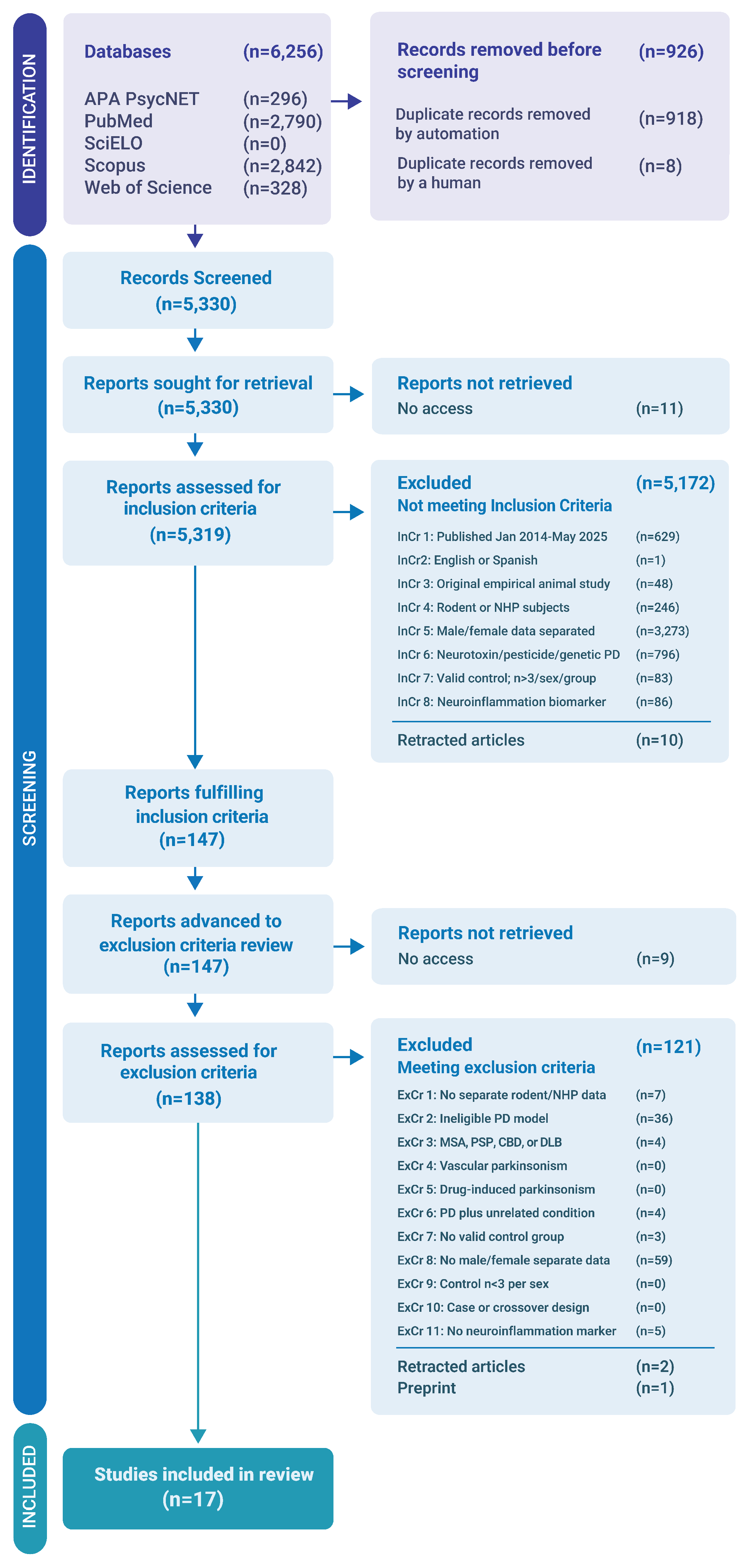
Figure 2.
Risk of bias assessment using SYRCLE. Systematic Review Centre for Laboratory Animal Experimentation (SYRCLE) risk of bias assessment for animal studies across included articles. The figure summarizes the final assessments for 17 included studies across 10 questions (Q1-Q10). These questions are related to: sequence generation (Q1), baseline characteristics (Q2), allocation concealment (Q3), random housing (Q4), caregiver and investigator blinding (Q5), random outcome assessment (Q6), outcome assessor blinding (Q7), incomplete outcome data (Q8), selective outcome reporting (Q9), and other potential sources of bias (Q10). Each horizontal bar shows the distribution of low, unclear and high risk of bias judgments for the corresponding question. Values shown within bar segments are reported as n (%). For each SYRCLE question, the full bar represents all 17 studies (100%).
Figure 2.
Risk of bias assessment using SYRCLE. Systematic Review Centre for Laboratory Animal Experimentation (SYRCLE) risk of bias assessment for animal studies across included articles. The figure summarizes the final assessments for 17 included studies across 10 questions (Q1-Q10). These questions are related to: sequence generation (Q1), baseline characteristics (Q2), allocation concealment (Q3), random housing (Q4), caregiver and investigator blinding (Q5), random outcome assessment (Q6), outcome assessor blinding (Q7), incomplete outcome data (Q8), selective outcome reporting (Q9), and other potential sources of bias (Q10). Each horizontal bar shows the distribution of low, unclear and high risk of bias judgments for the corresponding question. Values shown within bar segments are reported as n (%). For each SYRCLE question, the full bar represents all 17 studies (100%).
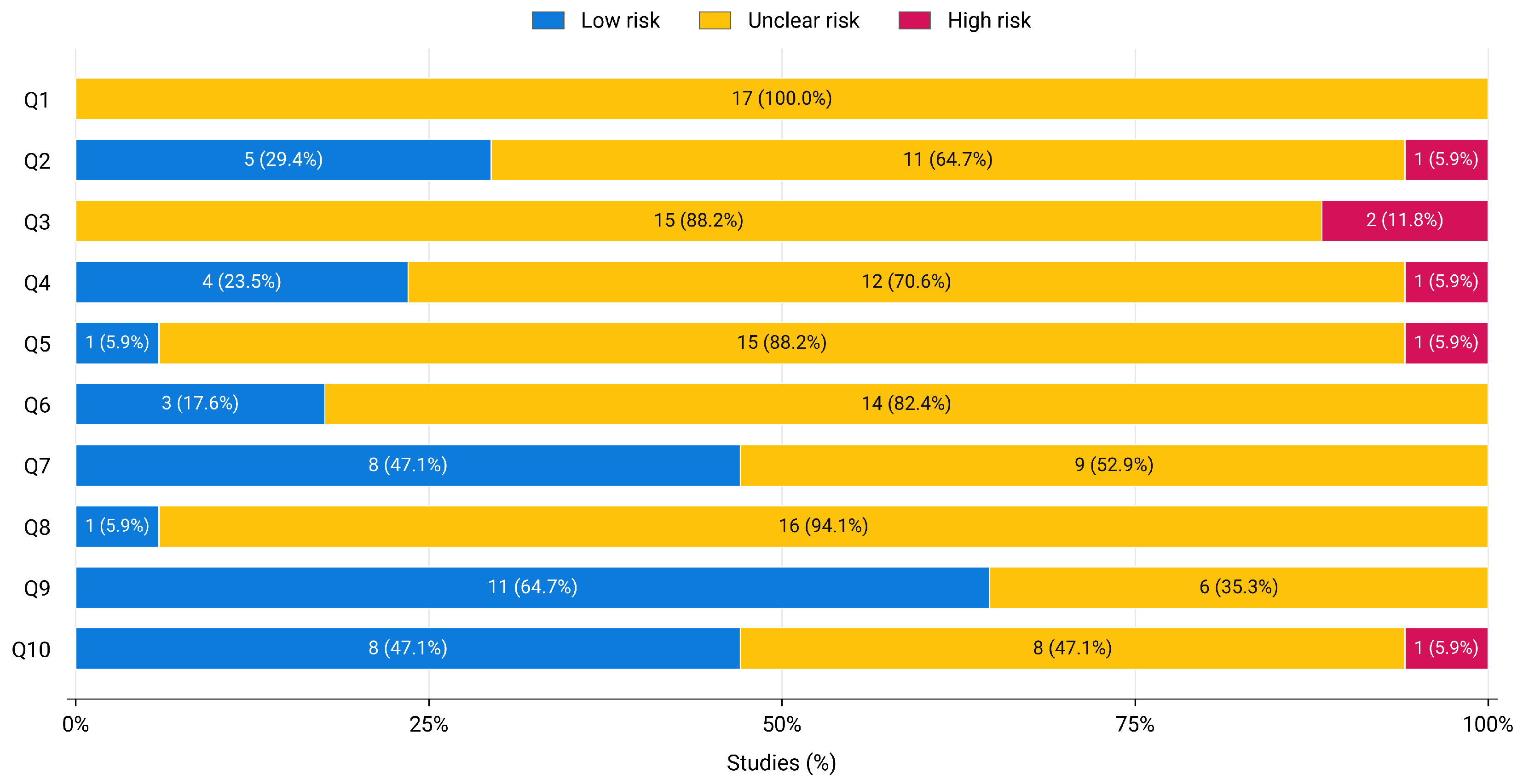
Figure 3.
Reporting assessment using ARRIVE. The graph shows the assessment of transparency and completeness of reporting of the included articles based on the Animal Research: Reporting In Vivo Experiments (ARRIVE) guidelines. Barchart (A) presents the ARRIVE Essential 10, which evaluate the presence of the minimum information necessary to assess the reliability of findings such as study design (1), sample size (2), inclusion and exclusion criteria (3), randomisation (4), blinding (5), outcome measures (6), statistical methods (7), experimental animals (8), experimental procedures (9) and results (10). Barchart (B) presents the Recommended Set, which analyses reporting of important study context such as abstract (11), background (12), objectives (13), ethical statement (14), housing and husbandry (15), animal care and monitoring (16), interpretation/scientific implications (17), generalisability/traslation (18), protocol registration (19), data access (20) and declaration of interest (21). Bar segments indicate judgments of Yes, Partially/Not clear, and No. Values shown within bar segments are reported as n (%). For each ARRIVE item, the full bar represents all 17 studies (100%). † denotes one study (5.9%).
Figure 3.
Reporting assessment using ARRIVE. The graph shows the assessment of transparency and completeness of reporting of the included articles based on the Animal Research: Reporting In Vivo Experiments (ARRIVE) guidelines. Barchart (A) presents the ARRIVE Essential 10, which evaluate the presence of the minimum information necessary to assess the reliability of findings such as study design (1), sample size (2), inclusion and exclusion criteria (3), randomisation (4), blinding (5), outcome measures (6), statistical methods (7), experimental animals (8), experimental procedures (9) and results (10). Barchart (B) presents the Recommended Set, which analyses reporting of important study context such as abstract (11), background (12), objectives (13), ethical statement (14), housing and husbandry (15), animal care and monitoring (16), interpretation/scientific implications (17), generalisability/traslation (18), protocol registration (19), data access (20) and declaration of interest (21). Bar segments indicate judgments of Yes, Partially/Not clear, and No. Values shown within bar segments are reported as n (%). For each ARRIVE item, the full bar represents all 17 studies (100%). † denotes one study (5.9%).
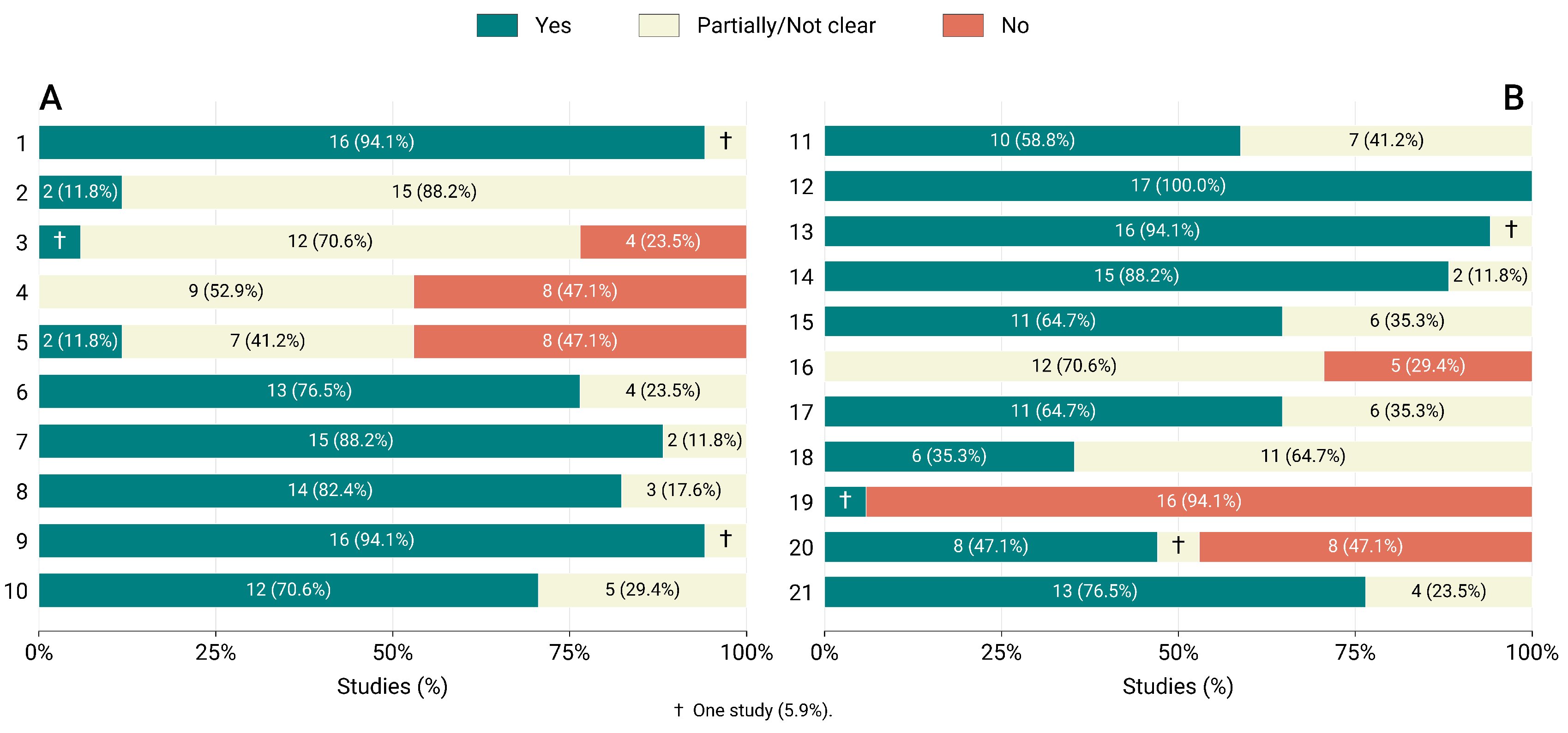
Figure 4.
Agreement between reviewers during study selection. The forest plots show pairwise agreement for the (A) inclusion and (B) exclusion phases of the review. Each row corresponds to one reviewer pair, with the number of records evaluated within parentheses. Gwet’s agreement coefficient 1 (AC1) is represented by green circles, while Cohen’s by open orange squares. Horizontal bars indicate 95% confidence intervals. Cohen’s and Gwet’s AC1 confidence intervals were calculated from multinomial delta method standard errors using a Student’s t critical value with degrees of freedom. The vertical dashed line shows an agreement coefficient of 0, corresponding to no agreement beyond that expected under the respective chance model. In graph A, the horizontal dashed separator distinguishes Pair 5, which represents an additional 115 record screening set, from the initial 5,215 record inclusion dataset. Observed agreement () represents the proportion of records for which both reviewers reached the same decision. The same coefficient scale is used in both forest plots to allow direct comparison in the two screening stages. Gwet’s agreement coefficient 1 (Gwet’s AC1); confidence interval (CI); Cohen’s kappa () and observed agreement ().
Figure 4.
Agreement between reviewers during study selection. The forest plots show pairwise agreement for the (A) inclusion and (B) exclusion phases of the review. Each row corresponds to one reviewer pair, with the number of records evaluated within parentheses. Gwet’s agreement coefficient 1 (AC1) is represented by green circles, while Cohen’s by open orange squares. Horizontal bars indicate 95% confidence intervals. Cohen’s and Gwet’s AC1 confidence intervals were calculated from multinomial delta method standard errors using a Student’s t critical value with degrees of freedom. The vertical dashed line shows an agreement coefficient of 0, corresponding to no agreement beyond that expected under the respective chance model. In graph A, the horizontal dashed separator distinguishes Pair 5, which represents an additional 115 record screening set, from the initial 5,215 record inclusion dataset. Observed agreement () represents the proportion of records for which both reviewers reached the same decision. The same coefficient scale is used in both forest plots to allow direct comparison in the two screening stages. Gwet’s agreement coefficient 1 (Gwet’s AC1); confidence interval (CI); Cohen’s kappa () and observed agreement ().
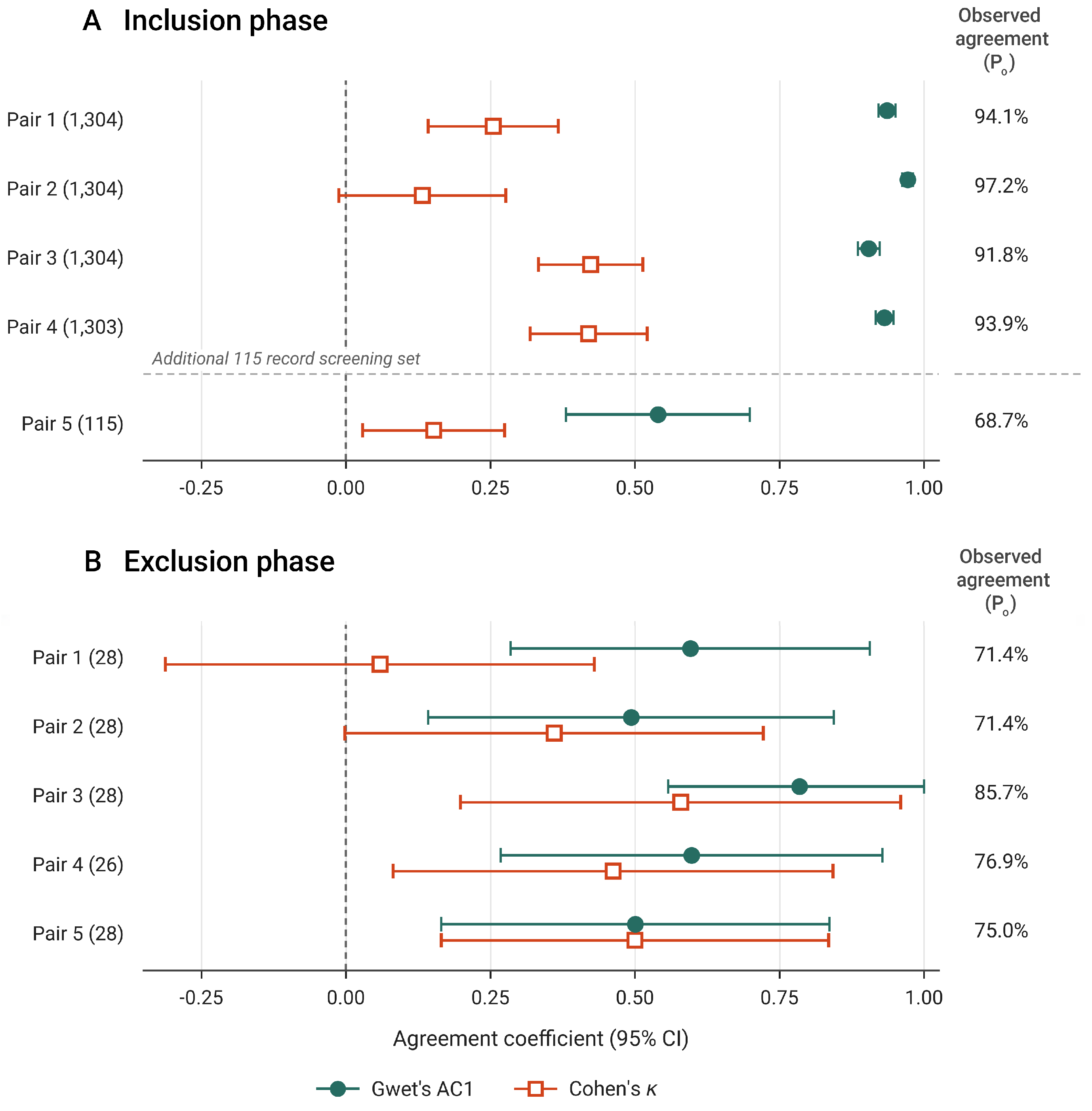
Figure 5.
Neuroinflammatory findings in female and male animals in experimental models of Parkinson’s disease. The figure groups the rodent findings into nine biological domains. Entries describe what was observed, under which experimental conditions, and in which articles. The numbers in parentheses show how many study units contributed to each domain, as listed in Table 2, including those with limited comparisons between sexes. A study unit can appear in several domains, and some publications draw on the same animal cohorts. Adding the counts would therefore count some evidence more than once; the totals do not represent independent replications. Lavender lines indicate patterns that varied with experimental conditions. Green lines indicate observed directions or differences in final values. Grey lines indicate no detected effect or limited comparability. An asterisk identifies a finding supported by a direct statistical test involving sex: a comparison of final values, an overall sex effect or an interaction. The asterisk does not denote statistical significance. Entries without an asterisk describe observations or limitations in comparing sexes. Abbreviations: C3, complement component 3; CRP, C-reactive protein; GFAP, glial fibrillary acidic protein; IL, interleukin; iNOS, inducible nitric oxide synthase; LRRK2, leucine rich repeat kinase 2; MPTP, 1-methyl-4-phenyl-1,2,3,6-tetrahydropyridine; PD, Parkinson’s disease; PINK1, PTEN induced kinase 1; Rhes, Ras homolog enriched in striatum; TNF, tumor necrosis factor; TOM20, translocase of outer mitochondrial membrane 20.
Figure 5.
Neuroinflammatory findings in female and male animals in experimental models of Parkinson’s disease. The figure groups the rodent findings into nine biological domains. Entries describe what was observed, under which experimental conditions, and in which articles. The numbers in parentheses show how many study units contributed to each domain, as listed in Table 2, including those with limited comparisons between sexes. A study unit can appear in several domains, and some publications draw on the same animal cohorts. Adding the counts would therefore count some evidence more than once; the totals do not represent independent replications. Lavender lines indicate patterns that varied with experimental conditions. Green lines indicate observed directions or differences in final values. Grey lines indicate no detected effect or limited comparability. An asterisk identifies a finding supported by a direct statistical test involving sex: a comparison of final values, an overall sex effect or an interaction. The asterisk does not denote statistical significance. Entries without an asterisk describe observations or limitations in comparing sexes. Abbreviations: C3, complement component 3; CRP, C-reactive protein; GFAP, glial fibrillary acidic protein; IL, interleukin; iNOS, inducible nitric oxide synthase; LRRK2, leucine rich repeat kinase 2; MPTP, 1-methyl-4-phenyl-1,2,3,6-tetrahydropyridine; PD, Parkinson’s disease; PINK1, PTEN induced kinase 1; Rhes, Ras homolog enriched in striatum; TNF, tumor necrosis factor; TOM20, translocase of outer mitochondrial membrane 20.
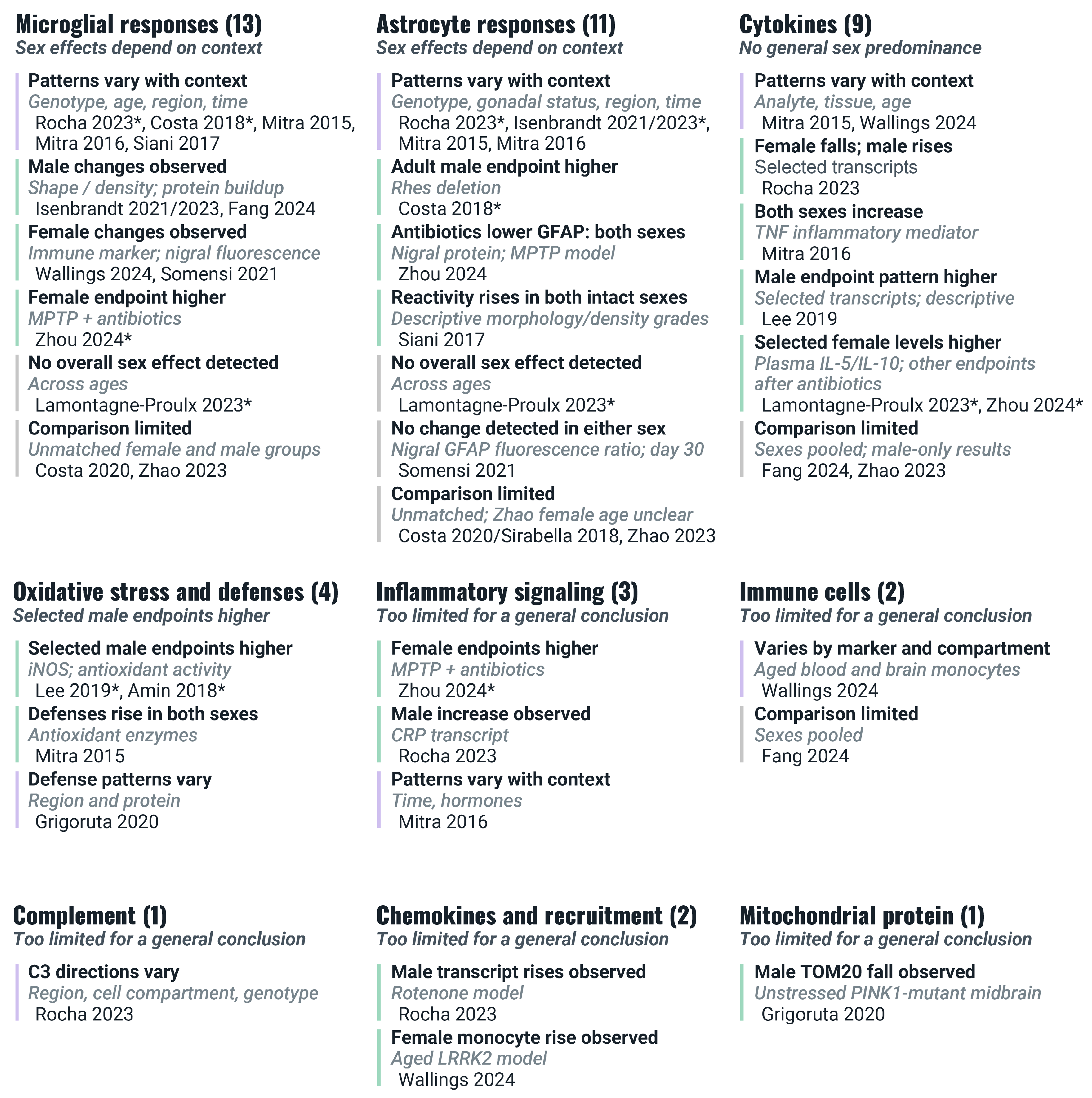
Figure 6.
Eligibility criteria according to the adapted PICOS framework. The graph illustrates the eligibility criteria for study inclusion and exclusion of this systematic review. These criteria were organised according to an adapted framework covering population, intervention/exposure, comparator, outcomes, and study design (PICOS). Abbreviations: CBD, corticobasal degeneration; Cre, Cre recombinase; DLB, dementia with Lewy bodies; MSA, multiple system atrophy; NHP, nonhuman primate; PD, Parkinson’s disease; PSP, progressive supranuclear palsy; n, sample size.
Figure 6.
Eligibility criteria according to the adapted PICOS framework. The graph illustrates the eligibility criteria for study inclusion and exclusion of this systematic review. These criteria were organised according to an adapted framework covering population, intervention/exposure, comparator, outcomes, and study design (PICOS). Abbreviations: CBD, corticobasal degeneration; Cre, Cre recombinase; DLB, dementia with Lewy bodies; MSA, multiple system atrophy; NHP, nonhuman primate; PD, Parkinson’s disease; PSP, progressive supranuclear palsy; n, sample size.
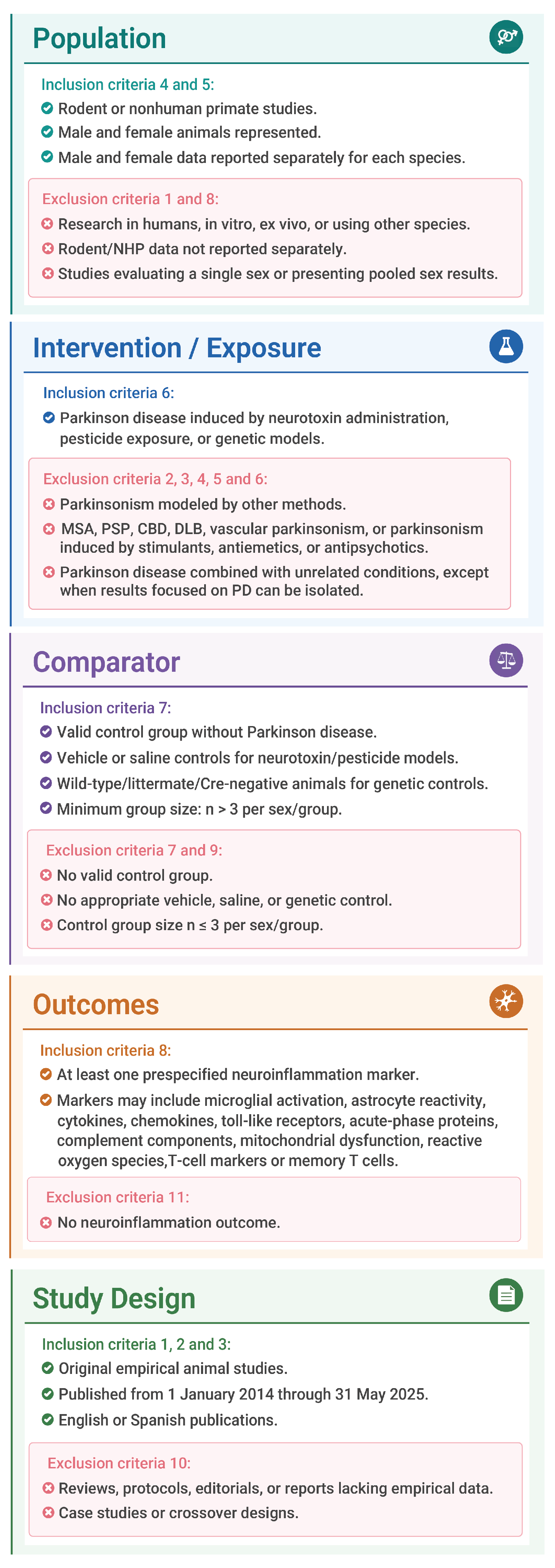
Table 1.
Study characteristics of included studies
| Study identification | Animal characteristics | PD model and interventions | Comparators and timing | ||||||
|---|---|---|---|---|---|---|---|---|---|
|
Study ID / citation |
Species strain |
Sex | Age |
n group |
PD model induction/protocol |
Genotype modifier |
Additional manipulation |
Comparator(s) |
Assessment timing |
|
ST01 Rocha et al., 2023[12] |
MouseC57BL/6J–C57BL/6N | F / M | NR | 3–10a | Rotenone | Ikbkb fl/fl; Cx3cr1-Cre+ | NR | Veh.; WT | 14 d post-inj. |
|
ST02b Isenbrandt et al., 2021[4] |
MouseC57BL/6N | F / M | 8 wk | 5–16a | MPTP | NR | Dutasteride; GDX | Veh.; sham | 6 d post-inj. |
|
ST03 Costa et al., 2020[13]; Sirabella et al., 2018[14] |
MouseC57BL/6 × C3H | F / M | 12 mo | 4–17; 8 | hSNCA A53T Tg | Hemizygous | NR | WT | Age 12 mo |
|
ST04b Costa et al., 2018[15] |
MouseC57BL/6J | F / M | 3 mo; 12 mo | 5–12 | Rasd2 KO | Rasd2 KO (-/-) | NR | WT | NR |
|
ST05 Mitra et al., 2015[16] |
MouseSwiss albino | F / M | 22–24 wk | 5–10a | Rotenone | NR | Vaginal cytol. | Veh. | 1 d post-inj. |
|
ST06 Mitra et al., 2016[17] |
MouseSwiss albino | F / M | NR | 5 | MPTP | NR | E2; tamoxifen; vaginal lavage | Veh. | 1, 3, and 7 d post-inj. |
|
ST07b Siani et al., 2017[18] |
MouseC57BL/6N | F / M | 7–10 wk | 7 | 6-OHDA | NR | E2; OVX + pellets; vaginal cytol. | Contralat. hemi.; placebo pellet | 1, 2, 7, 14 post-inj. |
|
ST08 Lee et al., 2019[19] |
RatLong-Evans | F / M | NR | 5–18 | 6-OHDA + Rotenone | Sry ASO | SO; ASO | Contralat. hemi. | 6-OHDA: 21 d; rotenone: d0, d7 |
|
ST09b Grigoruţă et al., 2020[20] |
RatLong-Evans | F / M | 9–11 wk | 4–10a | Pink1 KO | Pink1 KO (-/-) | Cat-urine stress | WT; untreated | NR |
|
ST10b Isenbrandt et al., 2023[21] |
MouseC57BL/6J | F / M | 8 wk | 4–16a | MPTP | NR | Dutasteride; GDX | Veh. | 6 d post-inj. |
|
ST11 Lamontagne-Proulx et al., 2023[5] |
MouseC57BL/6 | F / M | 4 mo | 6–10a | hSNCA Tg / mSnca KO | hSNCA Tg; mSnca KO (-/-) | LPS | WT; KO | NR |
|
ST12 Wallings et al., 2024[22] |
MouseC57BL/6 | F / M | 2–3 mo; 18–21 mo | 3–12a | Lrrk2 R1441C KI | Lrrk2 R1441C KI | PF-360; IFN- + LPS, ex vivo | Veh.; WT | NR |
|
ST13 Fang et al., 2024[23] |
MouseC57BL/6J | F / M | 10–13 wk | 4–18a | LRRK2 G2019S | hLRRK2 G2019S Tg; mLrrk2 G2019S KI | DSS; ORX | Veh.; sham; WT; XX/XY comparator | NR |
|
ST14b Zhou et al., 2024[24] |
MouseC57BL/6 | F / M | NR | 3–15a | MPTP | NR | Abx | Veh.; Abx only | 10 d post-inj. |
|
ST15 Zhao et al., 2023[25] |
MouseC57BL/6J | F / M | F 21–25 wk; M 10–15 wk | 3–8a | MPTP | Hdac6 KO (-/-) | NR | Veh.; WT | 7 d post-inj. |
|
ST16b Somensi et al., 2021[26] |
MouseC57BL/6 | F / M | 7–8 wk | 5–9a | 6-OHDA | Tlr4 KO (-/-) | NR | Veh. | 30 d post-inj. |
|
ST17b Amin et al., 2018[27] |
MouseNR | F / M | 10 wk | 6 | MPTP | NR | Aspartame | Veh. | NR |
Note: Each study unit was assigned a study ID (ST01–ST17) for consistent linkage across the main and supplementary tables. a Group size varied across experimental comparisons. b For studies in which the total sample size could be derived from the reported data, it was estimated by summing the sample sizes of separate experimental cohorts: ST02, N = 246; ST04, N = 59; ST07, N = 112; ST09, N = 58; ST10, N = 150; ST14, N = 110; ST16, N = 58; and ST17, N = 48. Characteristics are reported at the publication level; individual measurements were assessed separately for inclusion in the synthesis. Abbreviations: 6-OHDA, 6-hydroxydopamine; Abx, antibiotics; ASO, antisense oligonucleotide; Contralat. hemi., contralateral hemisphere; cytol., cytology; d, day(s); DSS, dextran sulfate sodium; E2, 17β-estradiol; F, female; GDX, gonadectomy; IFN-γ, interferon-γ; KI, knock-in; KO, knockout; LPS, lipopolysaccharide; M, male; mo, month(s); MPTP, 1-methyl-4-phenyl-1,2,3,6-tetrahydropyridine; NR, not reported; ORX, orchiectomy; OVX, ovariectomy; PD, Parkinson’s disease; post-inj., post-injection; SO, sense oligonucleotide; Tg, transgenic; Veh., vehicle; wk, week(s); WT, wild type. Prefixes h and m denote human and mouse, respectively.
Table 2.
Neuroinflammatory domains assessed across included studies
| Study ID | Microglia | Astrocytes | Cytokines | Oxidative stress | Inflammatory signaling | Immune cells | Complement | Chemokines | Bioenergetics |
|---|---|---|---|---|---|---|---|---|---|
| ST01 | ✓ | ✓ | ✓ | ✓ | ✓ | ✓ | |||
| ST02 | ✓ | ||||||||
| ST03 | ✓ | ✓ | |||||||
| ST04 | ✓ | ✓ | |||||||
| ST05 | ✓ | ✓ | ✓ | ✓ | |||||
| ST06 | ✓ | ✓ | ✓ | ✓ | |||||
| ST07 | ✓ | ✓ | |||||||
| ST08 | ✓ | ✓ | |||||||
| ST09 | ✓ | ✓ | |||||||
| ST10 | ✓ | ||||||||
| ST11 | ✓ | ✓ | ✓ | ||||||
| ST12 | ✓ | ✓ | ✓ | ✓ | |||||
| ST13 | ✓ | ✓ | ✓ | ||||||
| ST14 | ✓ | ✓ | ✓ | ✓ | |||||
| ST15 | ✓ | ✓ | ✓ | ||||||
| ST16 | ✓ | ✓ | |||||||
| ST17 | ✓ | ||||||||
| Studies, n | 13 | 11 | 9 | 4 | 3 | 2 | 1 | 2 | 1 |
| Studies, % | 76.5% | 64.7% | 52.9% | 23.5% | 17.6% | 11.8% | 5.9% | 11.8% | 5.9% |
Note: indicates that at least one outcome from the study was assigned to that domain in the revised neuroinflammatory synthesis. This indicates coverage, not the presence of a tested sex difference. Shortened column headings correspond to the full domains detailed in Supplementary Table S1. These domains are: Microglia (Microglial activation and phenotype), Astrocytes (Astrocyte reactivity), Cytokines, Oxidative stress (Oxidative/nitrosative stress and antioxidant defense), Inflammatory signaling (Inflammatory signaling and transcriptional pathways), Immune cells (Immune cell composition and function), Complement (Complement system), Chemokines (Chemokine and leukocyte recruitment), and Bioenergetics (Mitochondrial and cellular bioenergetic function). Note that the domains are not necessarily mutually exclusive or biologically equivalent. “Studies, n” and “Studies, %” indicate the number and percentage of the 17 included study units that were represented in each of the domains listed above. ST02 and ST10 remain separate study units in this coverage table; their overlapping animals are not treated as independent replications in the synthesis. Study IDs for the listed studies are provided in Table 1.
Disclaimer/Publisher’s Note: The statements, opinions and data contained in all publications are solely those of the individual author(s) and contributor(s) and not of MDPI and/or the editor(s). MDPI and/or the editor(s) disclaim responsibility for any injury to people or property resulting from any ideas, methods, instructions or products referred to in the content. |
© 2026 by the authors. Licensee MDPI, Basel, Switzerland. This article is an open access article distributed under the terms and conditions of the Creative Commons Attribution (CC BY) license.
Copyright: This open access article is published under a Creative Commons CC BY 4.0 license, which permit the free download, distribution, and reuse, provided that the author and preprint are cited in any reuse.