

Submitted:
29 October 2026
Posted:
30 October 2026
You are already at the latest version
Abstract
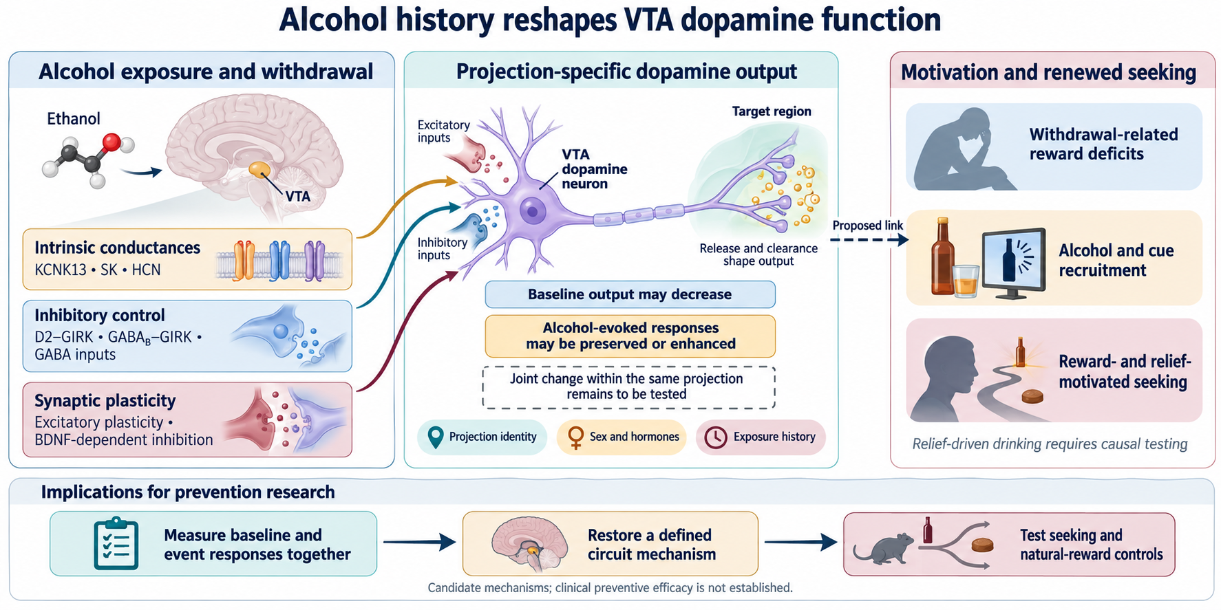
Alcohol use disorder involves persistent changes in motivation that a uniform increase or decrease in dopamine activity cannot explain. This narrative review examines how alcohol alters ventral tegmental dopamine circuits through membrane conductances, excitatory and inhibitory inputs, and synaptic plasticity. We propose that repeated exposure can reduce baseline dopamine output while preserving or enhancing responses to alcohol and its cues. Baseline output is defined as a prespecified measurement before an event; recruitment is the event-evoked increment above that baseline. Evidence for individual adaptations is distinguished from the unproven prediction that their joint change within a defined projection contributes to renewed seeking. Withdrawal-related reward deficits, stress recruitment, receptor-specific inhibitory signaling, and selective synaptic recovery provide complementary mechanisms. Projection identity, sex, hormonal state, and dopamine release and clearance at terminals help explain differences across studies. Regional rescue experiments strengthen causal inference, but most mechanisms have not been linked selectively to relief-driven drinking or recurrence. For prevention research, these findings identify candidate processes that may support progression to persistent use and vulnerability after cessation; they do not establish clinical preventive efficacy. Priorities include longitudinal measurements within defined circuits, independent tests of reward and relief motivation, and selective physiological rescue with appropriate behavioral controls. The practical contribution is a framework for evaluating when a dopamine adaptation is a cause of vulnerability, a compensatory response, or a marker of exposure.
Keywords:
alcohol use disorder
; ventral tegmental area
; dopamine
; withdrawal
; synaptic plasticity
; prevention
; recurrence
Abbreviations
AMPAR, α-amino-3-hydroxy-5-methyl-4-isoxazolepropionic acid receptor; AUD, alcohol use disorder; BDNF, brain-derived neurotrophic factor; BNST, bed nucleus of the stria terminalis; CRF, corticotropin-releasing factor; DA, dopamine; ERα, estrogen receptor α; fMRI, functional magnetic resonance imaging; FSCV, fast-scan cyclic voltammetry; GABA, γ-aminobutyric acid; GIRK, G protein-gated inwardly rectifying potassium channel; HCN, hyperpolarization-activated cyclic nucleotide-gated channel; Ih, hyperpolarization-activated current; LHb, lateral habenula; LTP, long-term potentiation; mPFC, medial prefrontal cortex; NAc, nucleus accumbens; NMDAR, N-methyl-D-aspartate receptor; PET, positron emission tomography; RMTg, rostromedial tegmental nucleus; SK, small-conductance calcium-activated potassium channel; TrkB, tropomyosin receptor kinase B; VTA, ventral tegmental area.
1. Introduction
Ethanol can excite ventral tegmental dopamine neurons despite its many inhibitory actions. However, reducing acute dopamine responsiveness through VTA Kcnk13 knockdown or GIRK3 deletion can increase binge-like drinking. These observations challenge a simple equation between dopamine activation and alcohol intake. The relevant question is how alcohol history changes the circuit response to reward, aversion, and renewed access. [1,2,3,4,5]
Ethanol self-infusion into the posterior VTA reinforces responding in selected rat preparations, and coadministered quinpirole reduces self-infusion. These experiments localize a reinforcing action but bypass oral pharmacokinetics and fail to isolate a single neuronal subtype. [6,7,8]
We organize the evidence around a change in the relationship between baseline dopamine function and responses to alcohol-related events. Intrinsic conductances set responsiveness, afferents determine recruitment, and plasticity influences later learning. Terminal release and clearance shape the signal reaching downstream targets. This distinction may help explain persistent alcohol motivation during withdrawal, when diminished reward function can coexist with strong responses to renewed alcohol or cues.
1.1. Scope and Approach
This focused narrative review integrates selected primary studies of ethanol regulation of VTA neurons with foundational addiction theories. Targeted searches supporting these revisions addressed withdrawal, inhibitory signaling, renewed drinking, and dopamine output, with additional consideration of sex, co-transmission, neuroimmune signaling, and human imaging. Selection was based on mechanistic relevance rather than an exhaustive systematic protocol. No formal risk-of-bias assessment or meta-analysis was performed. Physiological involvement, regional behavioral effects, cell specificity, and selective behavioral mediation are treated as different levels of evidence.
1.2. A Model Across Exposure States
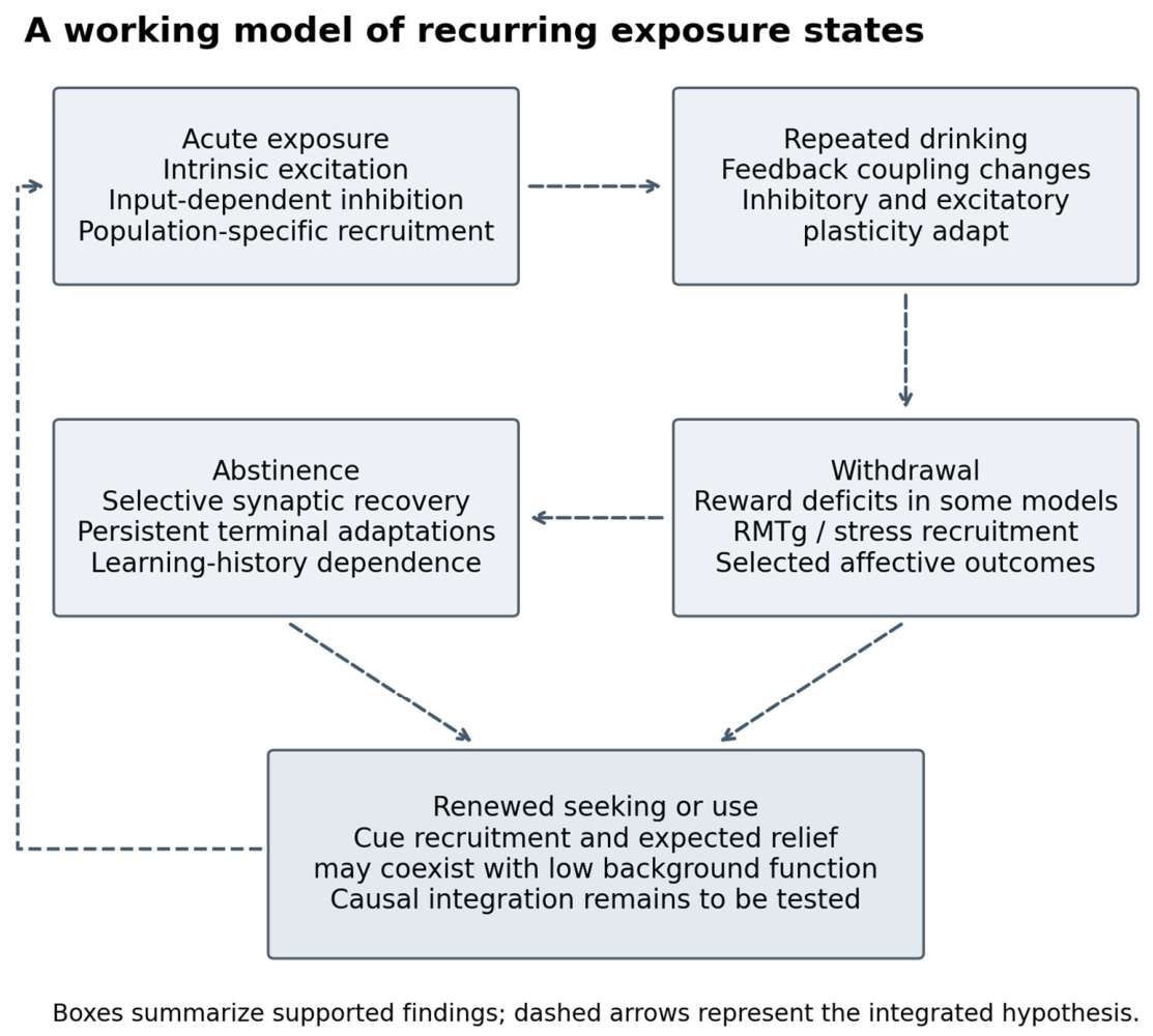
The framework distinguishes recurring exposure conditions rather than inevitable clinical stages. Acute alcohol recruits intrinsic and afferent mechanisms; repeated exposure changes feedback and plasticity. Withdrawal may combine reduced reward responsiveness with aversive signaling, while renewed use can be supported by anticipated reward or relief. Allostasis and incentive-sensitization theories offer complementary interpretations. Figure 1 summarizes the proposed transitions. [9,10,11]
The central prediction is that reduced baseline output and enhanced event-related recruitment will coexist within some of the same dopamine projections after repeated exposure. Selective restoration should then change a corresponding behavioral endpoint. Evidence from separate studies supports the plausibility of this prediction but does not yet demonstrate the complete mechanism within a single circuit.
1.3. Definitions and Causal Evidence
Baseline output, B, denotes a prespecified pre-event endpoint under matched conditions. Extracellular dopamine, mean firing among active cells, and the number of spontaneously active cells are distinct B measures. Event recruitment, E, denotes the increment above the pre-event baseline within a defined window. Cue-evoked dopamine, ethanol-evoked firing, and population calcium responses are likewise distinct E measures. The most direct output test pairs baseline and event-evoked dopamine in the same target; other measurements test component mechanisms.
A longitudinal test should ask whether B falls and E rises within a defined projection after exposure, and whether their joint change predicts later seeking better than either alone, recent intake, or task performance. Dividing E by B can exaggerate recruitment when only B falls. Absolute changes and compatible measurements should therefore be analyzed separately before constructing a combined predictor. Supplementary Figure S1 illustrates the hypothesis without assigning empirical values or a universal time course.
Figure 1.
Exposure states and renewed seeking. Boxes summarize observations in the cited preparations; dashed transitions are hypotheses. Recurring exposure states do not imply inevitable progression to AUD. A return toward baseline in one endpoint does not establish elimination of recurrence vulnerability. [9,11,65,66].
Figure 1.
Exposure states and renewed seeking. Boxes summarize observations in the cited preparations; dashed transitions are hypotheses. Recurring exposure states do not imply inevitable progression to AUD. A return toward baseline in one endpoint does not establish elimination of recurrence vulnerability. [9,11,65,66].
We use four descriptive causal-evidence levels: Level 1, an association or physiological effect without a behavioral intervention; Level 2, a regional or pathway manipulation affecting alcohol-related or affective behavior; Level 3, restoration or convergent perturbations supporting the mechanism; and Level 4, adult-onset, cell- and projection-specific mediation with performance controls. Level 4 would require selective prevention of physiological rescue to prevent the corresponding behavioral benefit. These are claim-specific descriptions, not formal quality ratings. Table 1 summarizes representative evidence; no mechanism is assigned Level 4.
1.4. Limitations
A narrative selection can favor positive findings and prominent mechanisms. Publication bias was not quantified. Age, strain, neuronal identification, sex, exposure schedule, and abstinence interval differ across studies; many findings remain male-only. Convergence across studies cannot establish simultaneous adaptations within a single circuit. The framework is therefore a set of testable relationships, not a pooled effect estimate or a universal disease trajectory.
2. Neuronal Identity and Sex
The VTA contains dopamine, GABA, and glutamate populations with diverse molecular and projection identities. A neuron’s anatomical position identifies where it was sampled, while its projection specifies a downstream target; neither alone predicts its response during drinking or withdrawal.
Broad action potentials, slow firing, dopamine sensitivity, and Ih helped identify putative dopamine neurons in foundational experiments, but no single electrophysiological feature is universal. Post hoc molecular validation or validated genetic targeting combined with projection labeling improves interpretation. [12]
DBA/2J and C57BL/6J dopamine neurons differ in ethanol responsiveness despite similar basal firing, illustrating that baseline activity is an inadequate proxy for ethanol sensitivity. [13]
Selected medial-VTA dopamine neurons show greater low-concentration ethanol sensitivity than lateral populations. Projection-defined work further identifies different intrinsic properties and stimulus-dependent plasticity in mesocortical and accumbal populations. Findings obtained with cocaine or aversion establish an experimental framework, not the same ethanol mechanism. Rostrocaudal location also cannot substitute for projection labeling. [14,15,16,17]
Mesoprefrontal and medial- or lateral-shell dopamine populations should be distinguished when assigning alcohol adaptations. Forebrain receptor manipulations implicate mPFC, accumbal shell, and ventral pallidum dopamine signaling in posterior-VTA ethanol reinforcement, but do not establish the firing response of labeled projections. Amygdala-projecting cells remain another unresolved population. Supplementary Table S1 separates established distinctions from alcohol-specific questions. [15,16,18]
2.1. Sex and Hormonal State
During quinine-adulterated drinking, male and female mice show different Fos recruitment patterns, with greater VTA dopamine cell recruitment in females. Fos indicates engagement, not behavioral mediation. [19]
Estradiol increases ethanol and dopamine sensitivity; subsequent work implicates ERα and mGluR1 signaling in enhanced ethanol excitation, with regional estrogen-receptor knockdown reducing binge-like intake in females. These findings support hormone-dependent regulation without assigning the same sex difference to every current or projected case. [20,21]
The principal KCNK13, GIRK3, and BDNF intervention studies used males, whereas GABA-neuron GIRK and inhibitory-plasticity studies included both sexes. Inclusion or a nonsignificant interaction does not establish equivalence. Supplementary Table S4 summarizes this evidence and the needed comparisons.
3. Intrinsic Conductances
Slice recordings established ethanol-associated depolarization and excitation that could persist during reduced synaptic transmission. Acutely dissociated neurons also remained responsive, supporting a cell-autonomous component while leaving intact-circuit contributions unresolved. [1,22,23]
Dissociation removes dendritic and network influences; persistence of excitation therefore establishes that intact afferents are not obligatory in that preparation, not that synaptic control is dispensable during behavior. [1]
3.1. M Current and HCN Signaling
Ethanol inhibits M-type potassium current, but excitation persists after maximal XE991 blockade. M-current suppression is a contributor rather than a complete explanation. Broad quinidine blockade and experiments with longer-chain alcohols constrain candidate mechanisms without establishing a unique ethanol-binding target. [24,25,26]
Ethanol modulation of Ih depends on competing conductances and preparation. HCN blockade can reveal delayed inhibition that barium prevents, whereas other experiments show enhanced Ih and attenuated firing responses during HCN blockade. Barium sensitivity does not identify a particular GIRK complex. Dynamic-clamp work connects Ih to rebound integration rather than universal necessity for excitation. [27,28,29]
3.2. KCNK13 and Drinking
VTA Kcnk13 knockdown reduces ethanol excitation and increases drinking-in-the-dark intake. This supports channel involvement and a regional behavioral contribution, but does not establish dopamine-cell exclusivity or direct binding. Later rat work found withdrawal-dependent expression and loss of the channel protein during slice preparation, suggesting that target availability is a potential source of variation. [4,30]
3.3. Firing Pattern and SK Currents
After intermittent exposure and seven days of withdrawal, dopamine neurons exhibit increased burst susceptibility, accompanied by reduced SK and altered Ih function. Acute SK blockade also potentiates ethanol excitation. These results distinguish firing pattern from mean rate and restraint of responsiveness from proof of direct ethanol gating. [31,32]
Conductance adaptations may independently change baseline activity, ethanol sensitivity, or the timing of responses. Their behavioral contribution requires measurement of dopamine output during the relevant event. Supplementary Table S2 compares the currents, exposure conditions, and limits of causal inference.
4. Afferent Control of Dopamine
Acute ethanol enhances AMPAR-mediated transmission in rat slices through dopamine-dependent facilitation, with paired-pulse and event-frequency changes supporting a presynaptic contribution. Dopamine depletion or D1 antagonism prevents facilitation, whereas agonism or transporter inhibition mimics aspects of it. Concentration dependence and action-potential blockade constrain the mechanism. [33]
A neuron–bouton preparation provides complementary evidence for retrograde dopamine facilitation, but does not identify the afferent source. Input-specific experiments are needed to determine where this feedback operates during voluntary drinking. [34]
4.1. Nicotinic and Serotonin Inputs
Cholinergic and serotonergic mechanisms provide additional routes to recruitment. α6-containing nicotinic receptors mediate selected low-dose effects through inhibition of VTA GABA neurons; anterior-VTA nicotinic involvement in accumbal dopamine and posterior-VTA 5-HT3 involvement in local ethanol reinforcement concern different endpoints. These pathways cannot be treated as one dopamine-cell mechanism. [35,36,37]
4.2. Stress-Related Afferents
Inhibition of VTA-projecting BNST CRF neurons reduces binge-like intake, whereas inhibition of local VTA CRF neurons does not under the tested conditions. Receptor manipulations implicate distinct contributions of CRF1 and CRF2. This establishes afferent involvement without resolving the downstream dopamine mechanism. [38]
4.3. Cortical and Habenular Pathways
Target-specific mPFC synapses provide an anatomical basis for different afferent control of VTA outputs, but local glutamatergic facilitation experiments do not identify their terminals as cortical. [33,39]
The LHb–RMTg route links negative outcomes to dopamine inhibition. Primate recordings establish a negative-reward pathway, and withdrawal studies implicate RMTg activity in selected affective outcomes. However, LHb inhibition does not prevent dependence-associated escalation or somatic withdrawal in all models, so RMTg recruitment cannot be equated with an obligatory LHb drive. [40,41,42,43]
The afferent evidence suggests that membrane responsiveness and recruitment by a particular event are separate intervention points. Identifying the input that changes after alcohol exposure is necessary before a regional receptor effect can be assigned to reward, stress, or relief motivation.
5. GABAergic Regulation
Ethanol reduces evoked inhibition and spontaneous IPSC frequency in the Xiao and Ye preparation, while miniature events in tetrodotoxin remain unchanged. An increased paired-pulse ratio supports a reduced activity-dependent inhibitory drive. Saturating μ-opioid agonism shifts the net ethanol effect toward facilitating the remaining inhibitory responses, demonstrating dependence on circuit state without identifying the endogenous peptide source. [2]
In vivo, acute ethanol may inhibit or increase GABA-cell firing depending on dose and paradigm; chronic exposure can increase baseline activity and produce tolerance to inhibition. These findings oppose a universal GABA-neuron response. [44,45,46]
Theile and colleagues find increased spontaneous and miniature IPSC frequencies during acute ethanol exposure, implicating action-potential-independent release, intracellular calcium stores, and 5-HT2C signaling. This mechanism can constrain dopamine-cell excitation despite intrinsic excitation. [3,47]
Blocking GABA inhibition increases ethanol-induced excitation in a subsequent preparation, whereas opioid blockade does not prevent the firing response. Inhibitory restraint and opioid-related disinhibition are therefore both supported under specific conditions. [48]
A direct anterior–posterior comparison in young rat slices finds opposite dopamine-cell responses with different GABAergic effects. This demonstrates anatomical weighting, but does not explain all discrepancies. Supplementary Table S3 compares the underlying observations. [17]
Terminal ATP receptors add another regulatory influence: in reduced preparations, ethanol shifts purinergic responses toward less inhibitory input. The in vivo ATP source and behavioral contribution remain unresolved. [49]
A useful conditional model separates firing-dependent drive from release at susceptible terminals. Disinhibition should dominate when the first component is large; facilitation can dominate when terminal effects are stronger. Matched input-specific measurements should test this prediction across dose, exposure history, neuronal identity, age, and sex. [2,3,17]
6. GIRK Signaling Across Cell Types
GIRK channels mediate slow inhibition downstream of dopamine D2 and GABAB receptors. Channel opening can hyperpolarize neurons at physiological potentials despite inward rectification. Receptor-coupled potassium inhibition must therefore be distinguished from fast GABAA transmission. [50,51]
Heterologous and cultured-neuron experiments support direct ethanol activation of GIRK channels, with mutagenesis and tagging implicating an alcohol pocket and phosphoinositide coupling. Direct gating differs from receptor-driven activation, and its contribution in native VTA assemblies remains to be quantified. Ethanol enhancement of native GABAB responses demonstrates an inhibitory action that can coexist with net excitation. [50,52,53,54,55]
6.1. GIRK3 and Binge Drinking
Constitutive GIRK3 deletion blunts ethanol-evoked VTA excitation and accumbal dopamine recruitment while increasing binge-like consumption. VTA-directed expression reverses elevated intake and reduces wild-type intake, strengthening regional causal inference. Continuous-access intake, metabolism, and tested intoxication measures are not comparably affected. [5]
Constitutive deletion is not equivalent to acute channel blockade: remaining assemblies, developmental compensation, and nondopamine cells may contribute. The finding supports a regional role for GIRK3 without proving that diminished dopamine reward causes compensatory drinking. [5]
6.2. Dopamine Autoreceptor Adaptation
Repeated ethanol injections strengthen and reduce desensitization of D2-mediated inhibition without equivalent enhancement of GABAB inhibition. This favors receptor regulation or coupling over generalized GIRK upregulation. [51]
6.3. GABA Neuron Adaptations
Three to seven days after four-week intermittent vapor exposure, male and female mice show weaker inhibitory control of VTA GABA neurons, with reduced presynaptic GABAB regulation and reduced postsynaptic GABAB–GIRK currents. A cell-specific manipulation mimicking the somatodendritic adaptation increases anxiety-related behavior. This supports affective relevance, not mediation of every withdrawal phenotype or alcohol seeking. [56]
Other GABA-cell studies identify increased excitatory input, resistance to muscimol inhibition, and impaired inhibitory long-term depression after chronic exposure. These are adaptations at inputs onto GABA neurons, not equivalent changes at their outputs. Stronger dopamine autoinhibition and weaker GABA-cell inhibition might converge on dopamine suppression, but this has not been demonstrated in a single circuit. Figure 2 separates the findings. [51,56,57,58,59]
7. Plasticity and Future Learning
Metaplasticity is an experience-dependent change in the capacity for later plasticity. Basal strength determines the present effect of an input, whereas altered induction thresholds change what the circuit can learn next. Both require independent behavioral tests before being assigned to maladaptive alcohol memory.
Single-exposure studies find enhanced basal GABA transmission, whereas acute ethanol can impair induction of inhibitory potentiation. Preserved downstream potentiation and opioid pharmacology argue against simple saturation in the latter preparation. Stronger existing inhibition can thus coexist with reduced capacity for further strengthening. [60,61]
Repeated exposure facilitates NMDAR potentiation through altered calcium signaling and PKA-related mechanisms. The signaling change persists for one week but not one month; enhanced cocaine conditioning is a correlate of altered learning, not proof of increased ethanol seeking. [62]
Intermittent voluntary drinking increases AMPAR/NMDAR ratio and miniature excitatory amplitude, supporting stronger existing excitation. This differs from easier induction of future NMDAR potentiation. Strain-dependent responses to a single exposure further oppose a universal shift toward stronger excitation. [63,64]
These excitatory findings may alter later cue processing, but the relevant memory process must be specified. Acquisition, expression, extinction, and motivation can change independently even when they share a synaptic mechanism. [62,63,64]
In male mice, chronic intermittent drinking reduces VTA BDNF and impairs inhibitory potentiation; both recover by seven days of withdrawal. BDNF restores potentiation ex vivo and reduces ethanol-related calcium responses and operant responding in vivo. These convergent results connect molecular signaling, physiology, and behavior. [65]
Regional BDNF acts on multiple cells and processes, and calcium photometry is not a spike count. Selectively preventing plasticity rescue while preserving other BDNF actions would establish whether restored potentiation mediates the behavioral benefit. Female and hormone-dependent responses remain unresolved. [65]
8. Withdrawal and Abstinence
Withdrawal denotes effects after cessation with the relevant signs specified; abstinence denotes time without ethanol. Protracted abstinence requires a stated interval beyond acute signs in that model. Recovery concerns an individual endpoint. Extinction involves nonreinforced learning, whereas reinstatement is renewed responding after extinction in the presence of a defined trigger. Drinking after passive abstinence should not automatically be called reinstatement or clinical relapse. Table 2 compares exposure timing and endpoint-specific recovery.
8.1. Baseline and Challenge Responses
An 18-day voluntary drinking regimen lowers spontaneous activity in dopamine neurons while enhancing responses to an ethanol challenge, accompanied by stronger GABA input in a subset of cells. Earlier repeated-injection and local posterior-VTA studies also demonstrate enhanced challenge responsiveness. Low baseline activity and strong ethanol responses can therefore characterize the same exposure condition. [66,67,68]
Enhanced responsiveness to renewed ethanol should be separated from increased baseline output and from amplified cue motivation. These endpoints have different implications for compensatory consumption, incentive sensitization, and intervention timing. [67,68]
Withdrawal studies find reduced firing and accumbal dopamine, fewer spontaneously active cells, or persistent reductions in population activity, depending on the preparation. These endpoints support dysfunction but are not interchangeable or part of a single obligatory trajectory. [69,70,71,72,73]
8.2. Negative Affect and Relief
Reduced reward sensitivity during withdrawal provides behavioral evidence beyond dopamine recordings, but does not assign the deficit exclusively to the VTA. The measured endpoint should remain explicit: firing, active-cell number, extracellular dopamine, and reward threshold answer different questions. [11,69,70,71]
In dependent male rats, accumbal dopamine declines during withdrawal and returns toward prewithdrawal levels during ethanol self-administration. This connects reduced dopamine and restoration within one exposure sequence, without isolating the VTA mechanism or proving that dopamine restoration causes relief-driven responding. [74]
RMTg inhibition improves selected depression- or anxiety-related outcomes after chronic drinking or vapor exposure, with different effects on somatic signs and reward thresholds. Even a shared inhibitory node need not control the whole withdrawal syndrome. [41,42]
Negative affect is an aversive state; negative reinforcement requires evidence that relief strengthens the behavior producing it. Dopamine recovery during drinking is consistent with that account but does not isolate the causal contingency. Restoring a specified circuit function before access, then testing withdrawal-associated seeking and natural-reward responding, would provide a stronger test. [74]
8.3. Persistence and Selective Recovery
Reduced dopamine population activity and terminal changes can persist beyond acute withdrawal, while a synaptic endpoint such as BDNF-dependent potentiation may recover earlier. Endpoint-specific recovery cannot establish elimination of recurrence vulnerability or uniformly low dopamine in all states. [65,72,73,75]
To distinguish recovery from latent susceptibility, baseline, cue, stress, and ethanol conditions should be sampled longitudinally. Passive abstinence must be distinguished from extinction, as the latter involves learning. [65,72,73,76]
8.4. Neuroimmune Signaling
Neuronal TLR4/MCP-1 and GABAA α2-associated signaling have been implicated in drinking and related molecular regulation. These studies do not establish a microglial TLR4 mechanism. An acute-ethanol macrophage study links peripheral manipulation to mesolimbic responses alongside microglial changes, but does not isolate a chronic immune-to-GIRK or immune-to-BDNF pathway. [77,78,79]
9. From Cell Activity to Output
Target dopamine depends on the timing of arriving spikes, release probability, presynaptic modulation, and clearance. Non-alcohol task recordings establish that accumbal dopamine dynamics can partly dissociate from somatic firing. This justifies direct output measurements without establishing the same mechanism in withdrawal. [80]
GCaMP photometry reports population calcium; dLight and GRABDA report extracellular dopamine-related fluorescence; FSCV resolves fast electrochemical changes and evoked release or uptake; microdialysis measures slower extracellular changes. Normalized fluorescence does not independently quantify tonic dopamine, and a larger percentage change from a lower baseline need not mean greater absolute release. [75,81,82]
In male rhesus macaques, altered accumbal dopamine-transporter and κ-opioid regulation persists after prolonged drinking and abstinence, while VTA transcript abundance does not simply predict terminal function. Alcohol reinstatement studies in corticostriatal circuits further show that abstinence and extinction histories change dopamine signals; these findings are informative comparisons, not proof of a VTA channel mechanism. [75,76]
Some dopamine neurons also release glutamate or GABA. These additional outputs mean that stimulation of dopamine-defined cells cannot automatically be interpreted as dopamine-only causation. Alcohol studies should distinguish fast co-transmission, dopamine release, projection identity, and exposure history. [83,84]
10. Addiction Models and Predictions
Allostatic and opponent-process accounts emphasize reward deficits and recruited stress responses, whereas incentive sensitization emphasizes cue-triggered wanting, distinct from liking. Withdrawal reward deficits, RMTg/CRF involvement, and enhanced ethanol challenge responses correspond to different components of these frameworks, but no single finding establishes a shared sequence. Table 3 maps their predictions and limits. [9,10,11,38,42,68]
A stronger alcohol-seeking link comes from VTA dopamine inhibition combined with intra-accumbal D1 agonist rescue. The result supports a contribution of downstream receptors to cue seeking without assigning all seeking to a single projection or equating sensitized firing with incentive sensitization. [85]
Metaplasticity could change later learning after ethanol has cleared, while extinction recruits new learning rather than passive recovery. A causal test must manipulate the relevant process during acquisition, expression, or extinction and measure its effect on a specified seeking outcome. [61,62,76]
The framework would require revision if low baseline output and enhanced recruitment occur in different projections, if terminal regulation explains the output change without altered somatic recruitment, or if physiological rescue leaves seeking intact. These alternatives distinguish population redistribution, terminal gain, compensation, and behavioral redundancy from the proposed within-projection mechanism.
11. Human Translation
Human PET studies report alcohol-related ventral-striatal dopamine release and blunted stimulant-evoked release in alcohol-dependent or detoxified groups. Different challenges and participant histories preclude interpreting these as a longitudinal transition or a specific VTA channel signature. [86,87,88]
Reward-task fMRI identifies altered responses in some detoxified cohorts but substantially preserved recruitment in others. Task phase, abstinence, sex, and sampling matter; BOLD is not dopamine release. Supplementary Table S5 summarizes the findings and reverse-translational tests. [89,90]
Human validation should pair a defined phenotype—reduced nondrug-reward response, cue motivation, or stress-related drinking—with a matched physiological readout. No current imaging measure in these studies is a validated biomarker of VTA KCNK13, GIRK, BDNF, or immune signaling. Longitudinal prediction beyond clinical severity and reproducible treatment-response associations are needed before stratification is justified.
12. Prevention and Future Research
Drinking opportunity, effort, conditioned approach, and aversion-resistant intake are distinct behavioral outcomes. Neither firing nor reduced consumption alone establishes reduced compulsivity or a reduced risk of recurrence. Intra-VTA quinpirole reduces sucrose- and ethanol-responding, illustrating the need for natural-reward and performance controls. [91]
Future intervention studies should prespecify whether the target is acute reinforcement, withdrawal-related relief, cue seeking, or learning. This choice determines when the manipulation is delivered, which projection is measured, and which comparison behaviors are needed.
12.1. Preventing Progression
For prevention of progression from alcohol use to persistent problematic drinking, the relevant mechanistic question is which exposure-induced changes precede increasing motivation or loss of behavioral flexibility. Findings from KCNK13 and GIRK3 indicate that weaker acute dopamine recruitment does not necessarily reduce intake. Prospective studies should therefore measure baseline output, event responses, and drinking before escalation, rather than assuming that suppressing alcohol excitation is protective. These are proposed prevention targets, not evidence that channel manipulation prevents AUD in people. [4,5]
12.2. Reducing Vulnerability After Cessation
After cessation, prevention of recurrence is a different objective from preventing initiation. Candidate mechanisms include withdrawal-associated reward deficits, stress-related afferent recruitment, altered inhibitory control, and persistent terminal regulation. Their importance may change as acute signs resolve and individual synaptic endpoints recover. An intervention should be tested during the state it is intended to modify; efficacy against cue seeking does not establish benefit against relief-driven drinking. [11,38,41,42,56,65,72,73,74,75,85]
A mechanistic rescue should first establish target engagement: restored receptor-evoked current for a GIRK intervention, restored inhibitory potentiation for BDNF–TrkB, or channel-dependent responsiveness for KCNK13. Target dopamine should then be measured during the relevant behavior. The strongest mediation test would prevent physiological rescue selectively and determine whether the behavioral benefit disappears. Natural-reward responding, motor function, sedation, and ethanol pharmacokinetics remain necessary controls when affected by the manipulation. [4,5,56,65,91]
12.3. Translational Priorities
The proposed framework complements behavioral and clinical prevention research by identifying processes to test, rather than supplying a new clinical intervention. Early human work should establish whether a reproducible reward, cue, or stress phenotype predicts outcomes beyond clinical measures. Sex-inclusive longitudinal animal studies can then test mechanisms under matched conditions. A biomarker of exposure is not automatically a marker of future vulnerability, and an imaging association is not evidence of channel engagement. This distinction is essential when translating VTA findings into strategies intended to reduce recurrence.
13. Conclusion
Alcohol history can alter the relationship between baseline dopamine function and recruitment by alcohol-related events through conductances, afferent control, plasticity, and terminal regulation. The strongest regional rescue findings identify candidate mechanisms, while selective behavioral mediation remains incomplete. For prevention research, the priority is to determine which adaptations contribute to persistent use or renewed seeking, and whether correcting them preserves natural reward and performance. Clinical preventive benefit cannot be inferred from normalization of an isolated physiological endpoint.
Supplementary Materials
The following supporting information can be downloaded at the website of this paper posted on Preprints.org.
Competing interests and editorial handling
One author serves as Editor-in-Chief of the Journal of Addiction & Prevention. Independent editorial handling and external peer review are requested. The Editor-in-Chief author will not participate in reviewer selection, peer-review management, or the editorial decision on this manuscript. All authors declare nonfinancial interests.
Funding and acknowledgments
This work receives no funding support.
Author Contributions
JHY contributions to conception, literature selection, drafting, critical revision, and final approval. WZ contributes to critical revision, and final approval.
References
- Brodie MS, Pesold C, Appel SB (1999) Ethanol directly excites dopaminergic ventral tegmental area reward neurons. Alcohol Clin Exp Res 23: 1848–1852. PubMed.
- Xiao C, Ye JH (2008) Ethanol dually modulates GABAergic synaptic transmission onto dopaminergic neurons in ventral tegmental area: role of mu-opioid receptors. Neuroscience 153: 240–248. PubMed. [CrossRef]
- Theile JW, Morikawa H, Gonzales RA, Morrisett RA (2008) Ethanol enhances GABAergic transmission onto dopamine neurons in the ventral tegmental area of the rat. Alcohol Clin Exp Res 32: 1040–1048. PubMed. [CrossRef]
- You C, Savarese A, Vandegrift BJ, He D, Pandey SC, et al. (2019) Ethanol acts on KCNK13 potassium channels in the ventral tegmental area to increase firing rate and modulate binge-like drinking. Neuropharmacology 144: 29–36. PubMed. [CrossRef]
- Herman MA, Sidhu H, Stouffer DG, Kreifeldt M, Le D, et al. (2015) GIRK3 gates activation of the mesolimbic dopaminergic pathway by ethanol. Proc Natl Acad Sci U S A 112: 7091–7096. PubMed. [CrossRef]
- Rodd-Henricks ZA, McKinzie DL, Crile RS, Murphy JM, McBride WJ (2000) Regional heterogeneity for the intracranial self-administration of ethanol within the ventral tegmental area of female Wistar rats. Psychopharmacology (Berl) 149: 217–224. PubMed. [CrossRef]
- Rodd ZA, Melendez RI, Bell RL, Kuc KA, Zhang Y, et al. (2004) Intracranial self-administration of ethanol within the ventral tegmental area of male Wistar rats: evidence for involvement of dopamine neurons. J Neurosci 24: 1050–1057. PubMed. [CrossRef]
- Rodd ZA, Bell RL, Melendez RI, Kuc KA, Lumeng L, et al. (2004) Comparison of intracranial self-administration of ethanol within the posterior ventral tegmental area between alcohol-preferring and Wistar rats. Alcohol Clin Exp Res 28: 1212–1219. PubMed. [CrossRef]
- Koob GF, Le Moal M (2001) Drug addiction, dysregulation of reward, and allostasis. Neuropsychopharmacology 24: 97–129. PubMed. [CrossRef]
- Robinson TE, Berridge KC (1993) The neural basis of drug craving: an incentive-sensitization theory of addiction. Brain Res Brain Res Rev 18: 247–291. PubMed. [CrossRef]
- Schulteis G, Markou A, Cole M, Koob GF (1995) Decreased brain reward produced by ethanol withdrawal. Proc Natl Acad Sci U S A 92: 5880–5884. PubMed. [CrossRef]
- Margolis EB, Lock H, Hjelmstad GO, Fields HL (2006) The ventral tegmental area revisited: is there an electrophysiological marker for dopaminergic neurons?. J Physiol 577: 907–924. PubMed. [CrossRef]
- Brodie MS, Appel SB (2000) Dopaminergic neurons in the ventral tegmental area of C57BL/6J and DBA/2J mice differ in sensitivity to ethanol excitation. Alcohol Clin Exp Res 24: 1120–1124. PubMed.
- Mrejeru A, Martí-Prats L, Avegno EM, Harrison NL, Sulzer D (2015) A subset of ventral tegmental area dopamine neurons responds to acute ethanol. Neuroscience 290: 649–658. PubMed. [CrossRef]
- Lammel S, Hetzel A, Häckel O, Jones I, Liss B, et al. (2008) Unique properties of mesoprefrontal neurons within a dual mesocorticolimbic dopamine system. Neuron 57: 760–773. PubMed. [CrossRef]
- Lammel S, Ion DI, Roeper J, Malenka RC (2011) Projection-specific modulation of dopamine neuron synapses by aversive and rewarding stimuli. Neuron 70: 855–862. PubMed. [CrossRef]
- Guan Y, Xiao C, Krnjevic K, Xie G, Zuo W, et al. (2012) GABAergic actions mediate opposite ethanol effects on dopaminergic neurons in the anterior and posterior ventral tegmental area. J Pharmacol Exp Ther 341: 33–42. PubMed. [CrossRef]
- Ding ZM, Ingraham CM, Rodd ZA, McBride WJ (2015) The reinforcing effects of ethanol within the posterior ventral tegmental area depend on dopamine neurotransmission to forebrain cortico-limbic systems. Addict Biol 20: 458–468. PubMed. [CrossRef]
- Arnold ME, Schank JR (2024) Sex differences in neuronal activation in the cortex and midbrain during quinine-adulterated alcohol intake. Alcohol Alcohol 59: agae031. PubMed. [CrossRef]
- Vandegrift BJ, You C, Satta R, Brodie MS, Lasek AW (2017) Estradiol increases the sensitivity of ventral tegmental area dopamine neurons to dopamine and ethanol. PLoS One 12: e0187698. PubMed. [CrossRef]
- Vandegrift BJ, Hilderbrand ER, Satta R, Tai R, He D, et al. (2020) Estrogen Receptor α Regulates Ethanol Excitation of Ventral Tegmental Area Neurons and Binge Drinking in Female Mice. J Neurosci 40: 5196–5207. PubMed. [CrossRef]
- Brodie MS, Shefner SA, Dunwiddie TV (1990) Ethanol increases the firing rate of dopamine neurons of the rat ventral tegmental area in vitro. Brain Res 508: 65–69. PubMed. [CrossRef]
- Brodie MS, Appel SB (1998) The effects of ethanol on dopaminergic neurons of the ventral tegmental area studied with intracellular recording in brain slices. Alcohol Clin Exp Res 22: 236–244. PubMed.
- Koyama S, Brodie MS, Appel SB (2007) Ethanol inhibition of m-current and ethanol-induced direct excitation of ventral tegmental area dopamine neurons. J Neurophysiol 97: 1977–1985. PubMed. [CrossRef]
- Appel SB, Liu Z, McElvain MA, Brodie MS (2003) Ethanol excitation of dopaminergic ventral tegmental area neurons is blocked by quinidine. J Pharmacol Exp Ther 306: 437–446. PubMed. [CrossRef]
- Appel SB, Wise L, McDaid J, Koyama S, McElvain MA, et al. (2006) The effects of long chain-length n-alcohols on the firing frequency of dopaminergic neurons of the ventral tegmental area. J Pharmacol Exp Ther 318: 1137–1145. PubMed. [CrossRef]
- McDaid J, McElvain MA, Brodie MS (2008) Ethanol effects on dopaminergic ventral tegmental area neurons during block of Ih: involvement of barium-sensitive potassium currents. J Neurophysiol 100: 1202–1210. PubMed. [CrossRef]
- Okamoto T, Harnett MT, Morikawa H (2006) Hyperpolarization-activated cation current (Ih) is an ethanol target in midbrain dopamine neurons of mice. J Neurophysiol 95: 619–626. PubMed. [CrossRef]
- Tateno T, Robinson HPC (2011) The mechanism of ethanol action on midbrain dopaminergic neuron firing: a dynamic-clamp study of the role of I(h) and GABAergic synaptic integration. J Neurophysiol 106: 1901–1922. PubMed. [CrossRef]
- You C, Vandegrift BJ, Brodie MS (2021) KCNK13 potassium channels in the ventral tegmental area of rats are important for excitation of ventral tegmental area neurons by ethanol. Alcohol Clin Exp Res 45: 1348–1358. PubMed. [CrossRef]
- Hopf FW, Martin M, Chen BT, Bowers MS, Mohamedi MM, et al. (2007) Withdrawal from intermittent ethanol exposure increases probability of burst firing in VTA neurons in vitro. J Neurophysiol 98: 2297–2310. PubMed. [CrossRef]
- Brodie MS, McElvain MA, Bunney EB, Appel SB (1999) Pharmacological reduction of small conductance calcium-activated potassium current (SK) potentiates the excitatory effect of ethanol on ventral tegmental area dopamine neurons. J Pharmacol Exp Ther 290: 325–333. PubMed.
- Xiao C, Shao XM, Olive MF, Griffin WC 3rd, Li KY, et al. (2009) Ethanol facilitates glutamatergic transmission to dopamine neurons in the ventral tegmental area. Neuropsychopharmacology 34: 307–318. PubMed. [CrossRef]
- Deng C, Li KY, Zhou C, Ye JH (2009) Ethanol enhances glutamate transmission by retrograde dopamine signaling in a postsynaptic neuron/synaptic bouton preparation from the ventral tegmental area. Neuropsychopharmacology 34: 1233–1244. PubMed. [CrossRef]
- Steffensen SC, Shin SI, Nelson AC, Pistorius SS, Williams SB, et al. (2018) α6 subunit-containing nicotinic receptors mediate low-dose ethanol effects on ventral tegmental area neurons and ethanol reward. Addict Biol 23: 1079–1093. PubMed. [CrossRef]
- Ericson M, Löf E, Stomberg R, Chau P, Söderpalm B (2008) Nicotinic acetylcholine receptors in the anterior, but not posterior, ventral tegmental area mediate ethanol-induced elevation of accumbal dopamine levels. J Pharmacol Exp Ther 326: 76–82. PubMed. [CrossRef]
- Rodd-Henricks ZA, McKinzie DL, Melendez RI, Berry N, Murphy JM, et al. (2003) Effects of serotonin-3 receptor antagonists on the intracranial self-administration of ethanol within the ventral tegmental area of Wistar rats. Psychopharmacology (Berl) 165: 252–259. PubMed. [CrossRef]
- Rinker JA, Marshall SA, Mazzone CM, Lowery-Gionta EG, Gulati V, et al. (2017) Extended Amygdala to Ventral Tegmental Area Corticotropin-Releasing Factor Circuit Controls Binge Ethanol Intake. Biol Psychiatry 81: 930–940. PubMed. [CrossRef]
- Carr DB, Sesack SR (2000) Projections from the rat prefrontal cortex to the ventral tegmental area: target specificity in the synaptic associations with mesoaccumbens and mesocortical neurons. J Neurosci 20: 3864–3873. PubMed. [CrossRef]
- Hong S, Jhou TC, Smith M, Saleem KS, Hikosaka O (2011) Negative reward signals from the lateral habenula to dopamine neurons are mediated by rostromedial tegmental nucleus in primates. J Neurosci 31: 11457–11471. PubMed. [CrossRef]
- Fu R, Zuo W, Shiwalkar N, Mei Q, Fan Q, et al. (2019) Alcohol withdrawal drives depressive behaviors by activating neurons in the rostromedial tegmental nucleus. Neuropsychopharmacology 44: 1464–1475. PubMed. [CrossRef]
- Glover EJ, Starr EM, Chao Y, Jhou TC, Chandler LJ (2019) Inhibition of the rostromedial tegmental nucleus reverses alcohol withdrawal-induced anxiety-like behavior. Neuropsychopharmacology 44: 1896–1905. PubMed. [CrossRef]
- Nentwig TB, Vaughan DT, Braunscheidel KM, Browning BD, Woodward JJ, et al. (2022) The lateral habenula is not required for ethanol dependence-induced escalation of drinking. Neuropsychopharmacology 47: 2123–2131. PubMed. [CrossRef]
- Gallegos RA, Lee RS, Criado JR, Henriksen SJ, Steffensen SC (1999) Adaptive responses of gamma-aminobutyric acid neurons in the ventral tegmental area to chronic ethanol. J Pharmacol Exp Ther 291: 1045–1053. PubMed.
- Stobbs SH, Ohran AJ, Lassen MB, Allison DW, Brown JE, et al. (2004) Ethanol suppression of ventral tegmental area GABA neuron electrical transmission involves N-methyl-D-aspartate receptors. J Pharmacol Exp Ther 311: 282–289. PubMed. [CrossRef]
- Steffensen SC, Walton CH, Hansen DM, Yorgason JT, Gallegos RA, et al. (2009) Contingent and non-contingent effects of low-dose ethanol on GABA neuron activity in the ventral tegmental area. Pharmacol Biochem Behav 92: 68–75. PubMed. [CrossRef]
- Theile JW, Morikawa H, Gonzales RA, Morrisett RA (2009) Role of 5-hydroxytryptamine2C receptors in Ca2+-dependent ethanol potentiation of GABA release onto ventral tegmental area dopamine neurons. J Pharmacol Exp Ther 329: 625–633. PubMed. [CrossRef]
- Theile JW, Morikawa H, Gonzales RA, Morrisett RA (2011) GABAergic transmission modulates ethanol excitation of ventral tegmental area dopamine neurons. Neuroscience 172: 94–103. PubMed. [CrossRef]
- Xiao C, Zhou C, Li K, Davies DL, Ye JH (2008) Purinergic type 2 receptors at GABAergic synapses on ventral tegmental area dopamine neurons are targets for ethanol action. J Pharmacol Exp Ther 327: 196–205. PubMed. [CrossRef]
- Federici M, Nisticò R, Giustizieri M, Bernardi G, Mercuri NB (2009) Ethanol enhances GABAB-mediated inhibitory postsynaptic transmission on rat midbrain dopaminergic neurons by facilitating GIRK currents. Eur J Neurosci 29: 1369–1377. PubMed. [CrossRef]
- Perra S, Clements MA, Bernier BE, Morikawa H (2011) In vivo ethanol experience increases D(2) autoinhibition in the ventral tegmental area. Neuropsychopharmacology 36: 993–1002. PubMed. [CrossRef]
- Kobayashi T, Ikeda K, Kojima H, Niki H, Yano R, et al. (1999) Ethanol opens G-protein-activated inwardly rectifying K+ channels. Nat Neurosci 2: 1091–1097. PubMed. [CrossRef]
- Lewohl JM, Wilson WR, Mayfield RD, Brozowski SJ, Morrisett RA, et al. (1999) G-protein-coupled inwardly rectifying potassium channels are targets of alcohol action. Nat Neurosci 2: 1084–1090. PubMed. [CrossRef]
- Aryal P, Dvir H, Choe S, Slesinger PA (2009) A discrete alcohol pocket involved in GIRK channel activation. Nat Neurosci 12: 988–995. PubMed. [CrossRef]
- Bodhinathan K, Slesinger PA (2013) Molecular mechanism underlying ethanol activation of G-protein-gated inwardly rectifying potassium channels. Proc Natl Acad Sci U S A 110: 18309–18314. PubMed. [CrossRef]
- Mitten EH, Souders A, Marron Fernandez de Velasco E, Aguado C, Luján R, et al. (2025) Chronic ethanol exposure in mice evokes pre- and postsynaptic deficits in GABAergic transmission in ventral tegmental area GABA neurons. Br J Pharmacol 182: 69–86. PubMed. [CrossRef]
- Williams SB, Yorgason JT, Nelson AC, Lewis N, Nufer TM, et al. (2018) Glutamate Transmission to Ventral Tegmental Area GABA Neurons Is Altered by Acute and Chronic Ethanol. Alcohol Clin Exp Res 42: 2186–2195. PubMed. [CrossRef]
- Nelson AC, Williams SB, Pistorius SS, Park HJ, Woodward TJ, et al. (2018) Ventral Tegmental Area GABA Neurons Are Resistant to GABA(A) Receptor-Mediated Inhibition During Ethanol Withdrawal. Front Neurosci 12: 131. PubMed. [CrossRef]
- Nufer TM, Wu BJ, Boyce Z, Steffensen SC, Edwards JG (2023) Ethanol blocks a novel form of iLTD, but not iLTP of inhibitory inputs to VTA GABA neurons. Neuropsychopharmacology 48: 1396–1408. PubMed. [CrossRef]
- Melis M, Camarini R, Ungless MA, Bonci A (2002) Long-lasting potentiation of GABAergic synapses in dopamine neurons after a single in vivo ethanol exposure. J Neurosci 22: 2074–2082. PubMed. [CrossRef]
- Guan YZ, Ye JH (2010) Ethanol blocks long-term potentiation of GABAergic synapses in the ventral tegmental area involving mu-opioid receptors. Neuropsychopharmacology 35: 1841–1849. PubMed. [CrossRef]
- Bernier BE, Whitaker LR, Morikawa H (2011) Previous ethanol experience enhances synaptic plasticity of NMDA receptors in the ventral tegmental area. J Neurosci 31: 5205–5212. PubMed. [CrossRef]
- Stuber GD, Hopf FW, Hahn J, Cho SL, Guillory A, et al. (2008) Voluntary ethanol intake enhances excitatory synaptic strength in the ventral tegmental area. Alcohol Clin Exp Res 32: 1714–1720. PubMed. [CrossRef]
- Wanat MJ, Sparta DR, Hopf FW, Bowers MS, Melis M, et al. (2009) Strain-specific synaptic modifications on ventral tegmental area dopamine neurons after ethanol exposure. Biol Psychiatry 65: 646–653. PubMed. [CrossRef]
- Xiong JW, Dou MY, Wang Y, Zeng T, Qi X, et al. (2026) BDNF restores impaired long-term potentiation of GABAergic synapses induced by chronic ethanol exposure in the VTA and attenuates reward-seeking behavior. Mol Psychiatry 31: 4205–4219. PubMed. [CrossRef]
- Ilari A, Curti L, Petrella M, Cannella N, Rocca AL, et al. (2022) Moderate ethanol drinking is sufficient to alter Ventral Tegmental Area dopamine neurons’ activity via functional and structural remodeling of GABAergic transmission. Neuropharmacology 203: 108883. PubMed. [CrossRef]
- Brodie MS (2002) Increased ethanol excitation of dopaminergic neurons of the ventral tegmental area after chronic ethanol treatment. Alcohol Clin Exp Res 26: 1024–1030. PubMed. [CrossRef]
- Ding ZM, Rodd ZA, Engleman EA, McBride WJ (2009) Sensitization of ventral tegmental area dopamine neurons to the stimulating effects of ethanol. Alcohol Clin Exp Res 33: 1571–1581. PubMed. [CrossRef]
- Diana M, Pistis M, Carboni S, Gessa GL, Rossetti ZL (1993) Profound decrement of mesolimbic dopaminergic neuronal activity during ethanol withdrawal syndrome in rats: electrophysiological and biochemical evidence. Proc Natl Acad Sci U S A 90: 7966–7969. PubMed. [CrossRef]
- Shen RY, Chiodo LA (1993) Acute withdrawal after repeated ethanol treatment reduces the number of spontaneously active dopaminergic neurons in the ventral tegmental area. Brain Res 622: 289–293. PubMed. [CrossRef]
- Shen RY (2003) Ethanol withdrawal reduces the number of spontaneously active ventral tegmental area dopamine neurons in conscious animals. J Pharmacol Exp Ther 307: 566–572. PubMed. [CrossRef]
- Diana M, Pistis M, Muntoni A, Gessa G (1996) Mesolimbic dopaminergic reduction outlasts ethanol withdrawal syndrome: evidence of protracted abstinence. Neuroscience 71: 411–415. PubMed. [CrossRef]
- Shen RY, Choong KC, Thompson AC (2007) Long-term reduction in ventral tegmental area dopamine neuron population activity following repeated stimulant or ethanol treatment. Biol Psychiatry 61: 93–100. PubMed. [CrossRef]
- Weiss F, Parsons LH, Schulteis G, Hyytiä P, Lorang MT, et al. (1996) Ethanol self-administration restores withdrawal-associated deficiencies in accumbal dopamine and 5-hydroxytryptamine release in dependent rats. J Neurosci 16: 3474–3485. PubMed. [CrossRef]
- Farahbakhsh ZZ, Holleran KM, Sens JP, Fordahl SC, Mauterer MI, et al. (2025) Synchrony between midbrain gene transcription and dopamine terminal regulation is modulated by chronic alcohol drinking. Nat Commun 16: 1944. PubMed. [CrossRef]
- Xie X, Chen R, Huang Y, Wang X, Wang J (2025) Abstinence and extinction drive opposing changes in striatal activity and dopamine signaling during alcohol relapse. Neuropharmacology 279: 110646. PubMed. [CrossRef]
- June HL, Liu J, Warnock KT, Bell KA, Balan I, et al. (2015) CRF-amplified neuronal TLR4/MCP-1 signaling regulates alcohol self-administration. Neuropsychopharmacology 40: 1549–1559. PubMed. [CrossRef]
- Balan I, Warnock KT, Puche A, Gondre-Lewis MC, June H, et al. (2018) The GABAA Receptor α2 Subunit Activates a Neuronal TLR4 Signal in the Ventral Tegmental Area that Regulates Alcohol and Nicotine Abuse. Brain Sci 8: 72. PubMed. [CrossRef]
- Nelson CA, Obray JD, Clarke TJ, Brundage JN, Folsom RJ, et al. (2025) Monocyte-Derived Macrophages Expressing Dopamine D2-Subtype Receptors Drive Alcohol Effects on Mesolimbic Neurons and Microglia. Biomedicines 13: 2327. PubMed. [CrossRef]
- Mohebi A, Pettibone JR, Hamid AA, Wong JMT, Vinson LT, et al. (2019) Dissociable dopamine dynamics for learning and motivation. Nature 570: 65–70. PubMed. [CrossRef]
- Patriarchi T, Cho JR, Merten K, Howe MW, Marley A, et al. (2018) Ultrafast neuronal imaging of dopamine dynamics with designed genetically encoded sensors. Science 360: eaat4422. PubMed. [CrossRef]
- Sun F, Zeng J, Jing M, Zhou J, Feng J, et al. (2018) A Genetically Encoded Fluorescent Sensor Enables Rapid and Specific Detection of Dopamine in Flies, Fish, and Mice. Cell 174: 481–496.e19. PubMed. [CrossRef]
- Stuber GD, Hnasko TS, Britt JP, Edwards RH, Bonci A (2010) Dopaminergic terminals in the nucleus accumbens but not the dorsal striatum co-release glutamate. J Neurosci 30: 8229–8233. PubMed. [CrossRef]
- Tritsch NX, Ding JB, Sabatini BL (2012) Dopaminergic neurons inhibit striatal output through non-canonical release of GABA. Nature 490: 262–266. PubMed. [CrossRef]
- Xiong J, Dou M, Li X, Zeng T, Liu Y, et al. (2026) VTA dopamine inputs activate accumbal D1 receptors to promote alcohol seeking. iScience 29: 116198. PubMed. [CrossRef]
- Boileau I, Assaad JM, Pihl RO, Benkelfat C, Leyton M, et al. (2003) Alcohol promotes dopamine release in the human nucleus accumbens. Synapse 49: 226–231. PubMed. [CrossRef]
- Martinez D, Gil R, Slifstein M, Hwang DR, Huang Y, et al. (2005) Alcohol dependence is associated with blunted dopamine transmission in the ventral striatum. Biol Psychiatry 58: 779–786. PubMed. [CrossRef]
- Volkow ND, Wang GJ, Telang F, Fowler JS, Logan J, et al. (2007) Profound decreases in dopamine release in striatum in detoxified alcoholics: possible orbitofrontal involvement. J Neurosci 27: 12700–12706. PubMed. [CrossRef]
- Wrase J, Schlagenhauf F, Kienast T, Wüstenberg T, Bermpohl F, et al. (2007) Dysfunction of reward processing correlates with alcohol craving in detoxified alcoholics. NeuroImage 35: 787–794. PubMed. [CrossRef]
- Bjork JM, Smith AR, Chen G, Hommer DW (2012) Mesolimbic recruitment by nondrug rewards in detoxified alcoholics: effort anticipation, reward anticipation, and reward delivery. Hum Brain Mapp 33: 2174–2188. PubMed. [CrossRef]
- Hodge CW, Haraguchi M, Erickson H, Samson HH (1993) Ventral tegmental microinjections of quinpirole decrease ethanol and sucrose-reinforced responding. Alcohol Clin Exp Res 17: 370–375. PubMed. [CrossRef]
Figure 2.
Circuit and GIRK mechanisms. Panel A separates cortical, BNST CRF, and LHb–RMTg influences without assigning a common postsynaptic target. Panel B contrasts dopamine-cell D2 inhibition with GABA-cell GABAB–GIRK adaptations found in different studies. Their combined effect on target dopamine is unproven. [38,39,51,56].
Figure 2.
Circuit and GIRK mechanisms. Panel A separates cortical, BNST CRF, and LHb–RMTg influences without assigning a common postsynaptic target. Panel B contrasts dopamine-cell D2 inhibition with GABA-cell GABAB–GIRK adaptations found in different studies. Their combined effect on target dopamine is unproven. [38,39,51,56].
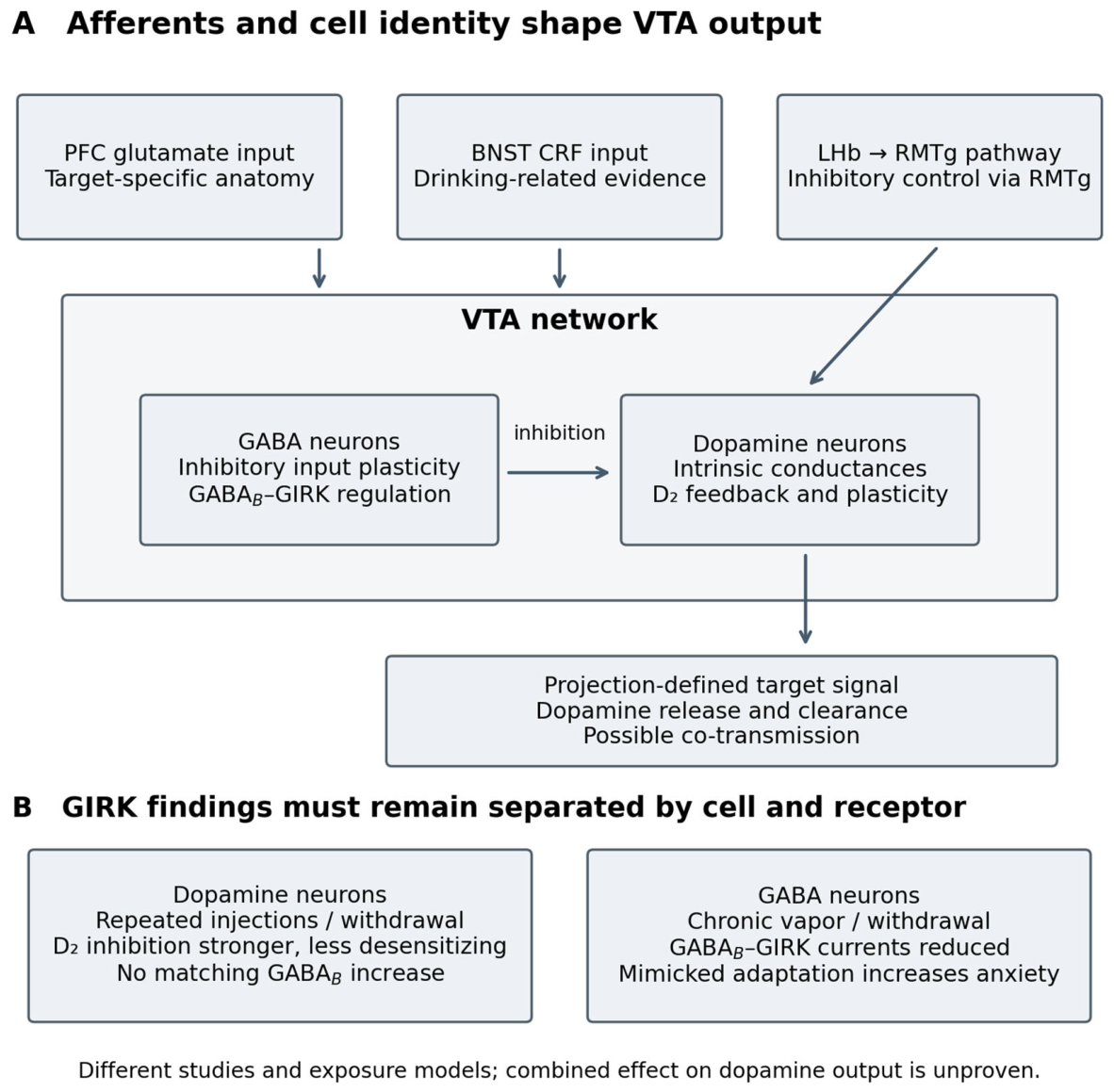

Table 1.
Causal evidence for VTA mechanisms.
| Mechanism | Manipulation and specificity | Direction and behavioral endpoint | Rescue or convergence | Evidence level and main confound |
| KCNK13 [4] | Male mice; VTA knockdown; regional, not DA-exclusive | Reduced excitation; greater limited-access intake | No cell-specific rescue in cited study | 2; regional attribution; direct binding unresolved |
| GIRK3 [5] | Male mice; constitutive deletion plus VTA expression | Blunted excitation and accumbal DA; greater binge-like intake | VTA expression reversed elevated intake | 3; developmental compensation and nondopamine cells |
| RMTg [41,42] | Rat studies; regional inhibition in different withdrawal models | Selected affective measures improved; not all withdrawal endpoints | Circuit intervention; not rescue of an identified VTA current | 2; regional effects and assay dependence |
| GABA-cell GIRK [56] | Mouse GABA-cell signaling; receptor/channel physiology and mimic | Loss of inhibition; mimic increased anxiety-related behavior | Phenocopy supports affective relevance; no complete seeking mediation | 2 for affect; not an alcohol-seeking causal assignment |
| BDNF–TrkB [65] | Male mice; ex vivo BDNF and regional in vivo intervention | Restored potentiation; reduced ethanol responses and operant seeking | Ex vivo rescue plus convergent receptor manipulations | 3; regional pleiotropy; LTP mediation incomplete |
| Estradiol and ERα [21] | Female mouse hormone/receptor experiments; VTA knockdown | Altered ethanol responsiveness; reduced binge-like intake after knockdown | Convergent endocrine and receptor evidence | 3 for pathway involvement; projection mediation unresolved |
Levels 1–4 are defined in Section 1.3 and describe specificity for the stated claim. They are not formal quality scores. No entry establishes Level 4 mediation.
Table 2.
Exposure timing and recovery.
| Study | Species, sex, and exposure | Time since cessation | Preparation, identity, and assay | Withdrawal verification | Persistence or recovery |
| Weiss et al. [74] | Male Wistar rats; 3–5 weeks liquid diet | 0–8 h; then 60-min self-administration | Accumbal microdialysis; operant ethanol | Dependence model; somatic score not the endpoint here | DA fell during withdrawal; restored during drinking |
| Xiong et al. [65] | Male mice; 21-day intermittent drinking | 0 h, 3 d, 7 d | VTA slices; putative DA physiology; BDNF assay | Plasticity deficit; not proof of somatic withdrawal | LTPGABA absent at 3 d; recovered by 7 d |
| Hopf et al. [31] | Male juvenile Sprague–Dawley rats; 5 d injections | 7 d | VTA slices; putative DA cells; NMDA-evoked bursting | Physiological adaptation; sign verification not assessed here | Altered burst susceptibility at that interval |
| Bernier et al. [62] | Mice; repeated injections; sex not audited here | 1 week versus 1 month | Slice NMDAR plasticity; later cocaine conditioning | No ethanol-seeking mediation test | Signaling change at 1 week; not 1 month |
| Mitten et al. [56] | Male and female mice; 4-week intermittent vapor | 3–7 d for slice recordings | GAD67 reporter cells; GABAB–GIRK physiology; anxiety-related assays | Established CIE protocol; affective mimic tested separately | Reduced GABA-cell inhibitory current; long-term recovery not established here |
Each row denotes a study and condition. Recovery refers to the measured endpoint. Not audited means that the detail was not systematically extracted in this synthesis, rather than absent from the source. DA, dopamine; d, days; h, hours; CIE, chronic intermittent ethanol exposure.
Table 3.
Addiction models and tests.
| Framework | Core prediction | Relevant support | Boundary or challenge | Critical test |
| Allostasis and opponent processes [9] | Exposure-induced reward deficit and recruited stress/relief motivation | Withdrawal reward deficits; RMTg affect and BNST CRF findings | Selective recovery and assay differences oppose a uniform persistent deficit | Rescue the aversive state before access; test withdrawal-associated drinking and natural reward |
| Incentive sensitization [10] | Stronger cue-triggered wanting, separable from liking | Enhanced ethanol challenge responses; DA/D1 contribution to cue seeking | Challenge sensitization is not cue motivation; reduced reward need not imply reduced wanting | Measure cue effort and hedonic responses separately; perturb the adapting projection |
| Maladaptive learning [62,76] | Exposure changes later encoding or extinction through altered plasticity | NMDAR metaplasticity; inhibitory potentiation deficits; learning-history effects | Recovery of plasticity and results with non-ethanol rewards limit alcohol-memory claims | Manipulate induction during acquisition versus retrieval or extinction, with output recordings |
The frameworks describe overlapping processes. Proposed tests distinguish mechanisms rather than requiring one theory to explain every state. NMDAR, N-methyl-D-aspartate receptor; BNST, bed nucleus of the stria terminalis; CRF, corticotropin-releasing factor.
Disclaimer/Publisher’s Note: The statements, opinions and data contained in all publications are solely those of the individual author(s) and contributor(s) and not of MDPI and/or the editor(s). MDPI and/or the editor(s) disclaim responsibility for any injury to people or property resulting from any ideas, methods, instructions or products referred to in the content. |
© 2026 by the authors. Licensee MDPI, Basel, Switzerland. This article is an open access article distributed under the terms and conditions of the Creative Commons Attribution (CC BY) license (http://creativecommons.org/licenses/by/4.0/).
Copyright: This open access article is published under a Creative Commons CC BY 4.0 license, which permit the free download, distribution, and reuse, provided that the author and preprint are cited in any reuse.