

Submitted:
26 October 2026
Posted:
28 October 2026
You are already at the latest version
Abstract
Tourniquet-induced ischemia-reperfusion injury (tourniquet/IR) causes oxidative stress, inflammation, neuromuscular junction (NMJ) dysfunction, and motor functional impair-ment. Although hyperbaric oxygen therapy (HBOT) exerts anti-inflammatory and antiox-idant effects, its impact on inflammatory and redox-associated pathways after tourni-quet/IR remains unclear. This study investigated the effects of HBOT on NMJ integrity, functional outcomes, skeletal muscle morphology, inflammatory and redox signaling in a mouse model of 3-hour unilateral hindlimb tourniquet followed by 1 or 2 weeks of tourni-quet release. Tourniquet/IR induced NMJ and skeletal muscle structural damage and func-tional impairment. Tourniquet/IR also increased protein expression of IL-1β, TNF-α, NOX2, p67-phox, and p47-phox, and decreased mitochondrial complex I-III expression, while tourniquet/IR did not change antioxidation demonstrated by Mn-SOD and Cu/Zn-SOD. HBOT improved NMJ and skeletal muscle function in the tourni-quet/IR-injured hindlimb, but it did not significantly affect tourniquet/IR-induced inflam-mation and oxidative stress. These findings demonstrate that HBOT mitigates tourni-quet/IR-induced skeletal muscle remodeling and neuromuscular dysfunction, and its ben-efits are not linked to the evaluated markers of inflammation or oxidative stress. A longer follow-up is needed to determine whether the early benefits of HBOT can translate into the sustained functional recovery of the NMJ and skeletal muscle, and to clarify the cellular and molecular mechanisms underlying HBOT-mediated functional recovery.
Keywords:
tourniquet
; ischemia-reperfusion injury
; hyperbaric oxygen therapy
; inflammation
; oxidative stress
; mitochondrial dysfunction
; neuromuscular junction
1. Introduction
Tourniquets are medical devices used to achieve hemostasis after life-threatening extremity hemorrhage on the battlefield or traumatic civilian accidents. Tourniquets apply circumferential pressure that exceeds arterial pressure to adequately occlude distal blood flow and prevent exsanguination [1,2,3,4]. Although rudimentary tourniquets have been used for centuries, French army surgeon Étienne Morel is credited with introducing the windlass tourniquet during the Siege of Besançon and the battle of Flanders, both in 1674 [3,5]. The modern screw tourniquet was developed by Jean Louis Petit in 1718 and was subsequently modified to become the standard method for controlling emergency extremity hemorrhage [3,5,6]. The widespread use of tourniquets in military trauma care significantly improved survival after severe extremity injury, which subsequently contributed to their adoption into civilian prehospital care [7,8,9]. Tourniquets are also commonly used during orthopedic and reconstructive surgeries to create a bloodless operative field [10,11,12]. Despite these benefits, unavoidably prolonged tourniquet use and their subsequent release can result in tourniquet-induced ischemia-reperfusion (tourniquet/IR) injury [13,14].
Prolonged tourniquet use leads to progressive tissue hypoxia, ATP depletion, and accumulation of metabolites (e.g., lactate, H+), within the ischemic limb [15,16,17]. Although restoring blood flow after ischemia is critical for tissue survival, reperfusion paradoxically triggers a cascade of pathological events characterized by excessive oxidative stress, inflammation, and mitochondrial dysfunction [17,18,19]. Tourniquet/IR contributes to skeletal muscle and peripheral nerve injury and impaired motor function, including the neuromuscular junction (NMJ), a specialized synapse to connect motor neurons and skeletal muscles. Prolonged tourniquet/IR also causes serious complications such as compartment syndrome, severe hyperkalemia and metabolic acidosis requiring renal replacement therapy, and in severe cases, limb amputation [15,20,21]. To study this phenomenon, we have extensively characterized tourniquet/IR using a validated mouse model of unilateral hindlimb tourniquet/IR that exhibits key features of ischemia-reperfusion injury, including limb ischemia, inflammation, oxidative stress, mitochondrial dysfunction, skeletal muscle injury, and NMJ dysfunction [14,18,19,22,23,24,25]. Because tourniquets are a vital part of emergency hemorrhage control and surgical care, there is a need for effective therapeutic strategies that mitigate the secondary tissue damage associated with tourniquet/IR.
Oxygen is essential for energy production in most cells. As a therapeutic strategy, hyperbaric oxygen therapy (HBOT) involves giving 100% oxygen at pressures greater than the normal atmospheric pressure, usually 2 – 3 atmospheres absolute (ATA). HBOT exhibits anti-inflammatory and antioxidant activity, and it has emerged as a potential therapeutic strategy to mitigate tourniquet/IR [26,27,28]. Although previous studies have shown that HBO treatment can reduce oxidative stress after prolonged tourniquet use and 48 hours of reperfusion, its long-term effects on skeletal muscle structure and function are unclear [18,22]. In addition, we have previously reported that neuromuscular recovery following tourniquet/IR is a dynamic process, with reinnervation and functional recovery occurring progressively over several weeks beginning from the 1-week and 2-weeks timepoints [29]. The present study investigated whether HBOT administered after tourniquet/IR improves skeletal muscle morphology and promotes NMJ recovery by reducing inflammation, oxidative stress, and mitochondrial dysfunction during the early regenerative phases of recovery (i.e., 1-wk and 2-wk tourniquet/IR).
2. Materials and Methods
All experiments followed the National Institute of Health’s Guide for the Care and Use of Laboratory Animals and received approval from the Institutional Animal Care and Use Committee (IACUC: 23-011-03-FC) at the University of Nebraska Medical Center. To achieve robust and unbiased data, all animals and tissue samples were blinded during experimentation (including the allocation) and data acquisition until statistical analysis.
2.1. General Procedures and Experimental Design
We only used male mice in the present study although we understood that sex-based differences in skeletal muscle fiber-type composition, capillary density and function could affect tourniquet/IR-induced hindlimb injuries. The fact is that if male and female mice were body weight-matched, the females are older than the males; if they are age-matched, the males are heavier than the females. In our present study, the difference of both age and body weight (i.e., limb dimension) can affect the stability and replication of the mouse model with hindlimb tourniquet/IR. For the sake of clarify and focus, therefore, only male mice were used in the present study.
Male C57BL/6 mice (10 – 12 weeks old, 25 – 30 g, Jackson Laboratory, Bar Harbor, ME) were housed in a temperature- and humidity-controlled animal facility on a 12-hour light-dark cycle and provided water and mouse chow ad libitum. Induction of hindlimb tourniquet/IR was performed with isoflurane anesthesia (2% inhalant), with post-procedure analgesia provided via slow-release buprenorphine (1 mg/kg, s.c.). Non-survival, terminal procedures were performed under anesthesia with urethane (800 mg/kg, i.p., Sigma Aldrich, St. Louis, MO) and α-chloralose (40 mg/kg, i.p., Sigma Aldrich, St. Louis, MO). Skeletal muscle structure and function were assessed by histological staining, rotarod testing, and in-situ sciatic nerve-evoked gastrocnemius contraction. NMJ integrity, including NMJ morphology and function, was assessed by immunofluorescent staining and motor end-plate potential (EPP) recording. After mice were euthanized by cervical dislocation at the end of in-vivo experiments, muscle samples were collected and protein expression was assessed by western blotting.
2.2. Mouse Model of Tourniquet-Induced Hindlimb Injury with HBO Treatment
A validated model of mouse unilateral hindlimb tourniquet/IR was used, as previously described [14,18,19,22,23]. Mice were randomly assigned to 5 groups: sham, 1-wk tourniquet/IR, 1-wk tourniquet/IR + HBOT, 2-wk tourniquet/IR, and 2-wk tourniquet/IR + HBOT. Under anesthesia with 2% inhalant isoflurane (EZ-Anesthesia; Euthanex Corp., Palmer, PA), a McGivney Hemorrhoidal Ligator was used to place an elastic orthodontic rubber band (ORB) at the left hip joint in the mice (Figure 1). The ORB was released after 3 hours of tourniquet-induced ischemia to begin reperfusion for 1-week (1-wk tourniquet/IR or 1-wk tourniquet/IR+HBOT) or 2-weeks (2-wk tourniquet/IR or 2-wk tourniquet/IR + HBOT). The sham mice were subjected to the same procedure, without the placement of an orthodontic rubber band. After tourniquet release, the mice assigned to the tourniquet/IR + HBOT groups received 100% oxygen with 2.5 ATA in a hyperbaric chamber (RSD B11, Reimers Systems Inc., Alexandria, VA) for 1 hour daily during reperfusion. The sham and tourniquet/IR alone mice received room air at normal atmospheric pressure (21% oxygen, 1 ATA) in the same hyperbaric chamber. A Laser Speckle Contrast Imaging (RFLSI-ZW, RWD Life Science Inc., Dover, DE, USA) was used to confirm hindlimb ischemia and subsequent reperfusion.
2.3. Rotarod Test
Mice were placed on an accelerating rotarod (76-0772 RotaRod, Harvard Apparatus, Holliston, MA). Mice underwent three trials per day at 30-minute intervals, with each trial lasting 5 minutes, for 3 consecutive days. The rotating speed increased progressively from 4 to 40 RPM. Each trial ended when the mouse fell from the rotarod or reached the 5-minute duration, whichever occurred first. Latency to fall was calculated as a measure of individual performance.
2.4. In-Situ Detection of Gastrocnemius Muscle Contractile Force
Gastrocnemius muscle contractile force was measured as described in our previous studies [15,24]. Under anaesthetized condition (800 mg/kg urethane, 40 mg/kg α-chloralose), the mouse was placed in a prone position on a heating pad at 37 °C. The left foot was fastened to a plank, and the left ankle was flexed at 90o. The middle and distal ends of the gastrocnemius muscle were exposed and covered with saline-moistened cotton. The distal part of the gastrocnemius muscle was sutured and connected to a wide range force transducer (AD Instruments, Colorado Springs, CO). The left biceps femoris muscle was carefully split along the direction of the muscle fibers to expose the sciatic nerve. The sciatic nerve was positioned on a bipolar platinum electrode and covered with cotton, moistened with mineral oil. Gastrocnemius muscle contractions were evoked by electrical stimulation of the sciatic nerve (0.4 mA, 50 Hz, 0.1 ms pulse), and a 5 second isometric tetanic muscle contraction was recorded. Muscle contractile force was recorded and analyzed with PowerLab 8/30 Data Acquisition System and LabChart 7 (AD Instruments, Colorado Springs, CO).
2.5. Electrophysiological Recording of Motor End Plate Potential (EPP) In Situ
Under anesthesia (800 mg/kg urethane and 40 mg/kg chloralose, i.p.), the mouse was placed in a prone position on a heating pad at 37 °C. The middle and distal portions of the left gastrocnemius muscle were isolated and covered with saline-moistened cotton. The distal cut end of the exposed sciatic nerve was placed on a bipolar platinum electrode and covered by cotton moistened with mineral oil. To inhibit muscle contraction, the muscle-specific Na+ channel blocker μ-conotoxin GIIIB (4 μM, 200 μL; C-270, Alomone Labs, Jerusalem) was locally injected into the gastrocnemius muscle. Muscle contraction was fully blocked within 15 minutes of drug injection. Motor EPPs were recorded by intracellular recording technique. Intracellular recording electrodes were fabricated from borosilicate glass capillaries (1.5 mm outer diameter, 1.10 mm inner diameter; Sutter Instruments, Novato, CA) using a flaming micropipette puller (Model P-97; Sutter Instruments, Novato, CA) and filled with 3 M KCl to achieve a pipette resistance of 5–15 MΩ. Microelectrode was then connected to an intracellular preamplifier (IX 1; Dagan Corporation, Minneapolis, MN) and slowly inserted into the gastrocnemius muscle. Proximity to the neuromuscular junction was determined by the presence of spontaneous miniature endplate potentials (mEPPs). EPPs were evoked by sciatic nerve stimulation (0.4 mA, 50 Hz, 0.1 ms). Sciatic nerve stimulation-evoked EPPs were digitized and recorded using PowerLab 8/30 Data Acquisition System with LabChart 7 (AD Instruments, Colorado Springs, CO). EPPs were recorded from several sites within the gastrocnemius muscle of each mouse.
2.6. Immunohistochemistry of the NMJ
At the end of the non-survival experiments, mice were euthanized and the gastrocnemius muscle was immediately harvested and fixed in 4% paraformaldehyde prepared in 0.1 M phosphate-buffered saline (PBS) for 15 min, followed by incubation in 0.1 M glycine for 15 min. The muscle was divided into 8–10 longitudinal segments to facilitate penetration of staining reagents into the NMJs. The split muscle segments were permeabilized in methanol at −20 °C for 10 min and blocked for 1 h in PBS containing 0.5% Triton X-100 and 1% bovine serum albumin (BSA). Muscle segments were then incubated overnight at 4 °C in a primary antibodies cocktail which consisted of mouse anti-neurofilament 200 (N0142, Sigma-Aldrich, St. Louis, MO) and rabbit anti-synaptophysin (MA5-16402, Thermo Fisher Scientific, Waltham, MA) antibodies. After several rinses with PBS, muscle segments were incubated overnight at 4 °C with Alexa Fluor® 594-conjugated donkey anti-mouse IgG (A21203, Thermo Fisher Scientific, Waltham, MA), Alexa Fluor® 594-conjugated donkey anti-rabbit IgG (A21207, Thermo Fisher Scientific, Waltham, MA), and Alexa Fluor® 488-conjugated α-Bungarotoxin (α-BTX, B13422, Thermo Fisher Scientific, Waltham, MA). Muscle segments were thoroughly rinsed with PBS, mounted on glass slides, and imaged using a laser scanning confocal microscope (Zeiss LSM 800). In this measurement, mouse anti-neurofilament 200 and rabbit anti-synaptophysin antibodies were used for axon and nerve terminal labeling, and Alexa Fluor® 488-conjugated α-BTX was used for nicotinic acetylcholine receptor (nAChR) staining.
About quantification of NMJ morphology, Z-stack images of the NMJ were acquired from five distinct regions of each muscle segment and rendered as maximum intensity projections. All analyses were performed on en-face NMJs. Presynaptic innervation was assessed using neurofilament-200 and synaptophysin labeling. Nerve terminal sizes labeled with neurofilament-200 and synaptophysin in the endplate were calculated to quantify the percentage of motor nerve innervation and the percentage of nerve terminal occupancy at the NMJs. NMJs were classified as innervated when neurofilament 200 and synaptophysin labeling was present within the α-BTX-positive endplate and as denervated if the labeling was absent. Nerve terminal occupancy was calculated by ImageJ software (version 1.54p NIH, Bethesda, MA) as the area of overlap between the motor nerve terminal and the nAChR cluster divided by total area of the nAChR cluster and expressed as a percentage. Postsynaptic nAChR morphology was assessed using α-BTX labeling. α-BTX-labeled nAChR clusters were manually traced in ImageJ software (version 1.54p NIH, Bethesda, MA) to calculate the number of discrete fragments per nAChR cluster, the area per fragment, the percentage of fragmented nAChR clusters in total nAChR clusters, and whole area per nAChR cluster. The nAChR clusters containing ≥5 discrete fragments were considered as fragmented clusters.
2.7. Histological Evaluation of Skeletal Muscle Damage
Gastrocnemius muscles from sham, tourniquet/IR, and tourniquet/IR + HBOT mice were successively fixed in Methacarn solution (300 mL methanol, 150 mL chloroform, and 50 mL acetic acid) for 48 hours, and 60% ethanol for 72 hours. The tissues were then routinely processed, embedded in paraffin wax, and then cut into 4 μm thick sections. After deparaffinization, sections were stained with hematoxylin and eosin (H&E; Sigma-Aldrich, St. Louis, MO) or Masson’s trichrome (HT15; Sigma-Aldrich, St. Louis, MO). Stained sections were visualized using an inverted microscope under brightfield illumination (Axio Observer Z1; Carl Zeiss Microscopy, Jena, Germany) and images were captured with a digital camera (Axiocam 305 color camera; Carl Zeiss Microscopy, Jena, Germany). Mean data was quantified from 3 random sections per slide selected from 3 tissue slides each mouse in 3 mice per group.
2.8. Western Blotting
Gastrocnemius muscle samples were harvested, snap-frozen on dry ice, and stored at −80 °C. Protein lysates were prepared by homogenizing the tissue in a modified RIPA lysis buffer (50 mM Tris-HCl, 150 mM NaCl, 50 mM NaF, 2 mM EDTA, 1 mM, 1% NP-40, 1% SDS, and 1 mM PMSF) with a protease inhibitor cocktail (10 μl/ml, P2714, Sigma-Aldrich, St. Louis, MO). Following centrifugation (12,000 g for 20 min at 4 °C), total protein concentrations were measured in the supernatant by a bicinchoninic acid (BCA) protein assay kit (23225, Thermo Fisher Scientific, Waltham, MA). Equal amounts of protein (60 µg per well) were resolved on 9-12% sodium dodecyl sulfate (SDS)-polyacrylamide running gels and electrophoretically transferred onto PVDF membranes (IPVH00010, EMD Millipore, Billerica, MA) at 100 V for 1.5 hours. Membranes were blocked for 1 hour at room temperature in Intercept® Blocking Buffer (Part No. 927–70,001, LI-COR Biosciences, Lincoln, NE) and subsequently incubated overnight at 4 °C with primary antibodies (listed in Supplementary Table) under constant agitation. After washing with TBST (20 mM Tris, 150 mM NaCl, 0.1% Tween 20, pH 7.4), the membranes were incubated for 1 hour at room temperature with appropriate IRDye-conjugated secondary antibodies including IRDye 800CW, IRDye 680RD, and IRDye 680LT (LI-COR Biosciences, Lincoln, NE). Immunoreactive bands were visualized using the LI-COR Odyssey DLx Imaging System. Densitometric quantification was performed using ImageJ software (version 1.54p NIH, Bethesda, MA), with protein expression levels normalized to β-actin which was used as an internal loading control.
2.9. Statistical Analysis
Quantitative data were analyzed and visualized using GraphPad Prism 10.6.0 (GraphPad Software, San Diego, CA). Distribution of data was assessed using the Shapiro–Wilk test. For normally distributed data with equal variance (confirmed by Bartlett’s test), one-way ANOVA followed by Tukey’s post-hoc multiple comparisons test was used to evaluate group differences. For data with unequal variance, Welch’s one-way ANOVA followed by Dunnett’s T3 multiple comparisons test was performed. Kruskal–Wallis’ test with Dunn’s multiple comparisons test was used for non-parametric data. Survival distributions were evaluated using Kaplan-Meier analysis and compared using the Gehan-Breslow-Wilcoxon test. Data are shown as mean ± SEM, and p < 0.05 was considered statistically significant.
3. Results
3.1. Effects of HBOT on Hindlimb Perfusion and Survival Rate After Tourniquet/IR
The experimental design used to evaluate the therapeutic effects of HBOT following tourniquet/IR is shown in Figure 1. Tourniquet/IR was induced by placing an orthodontic rubber band on the left hind limb using a McGivney hemorrhoidal ligator (Figure 1A). Laser speckle doppler imaging observed a reduction in hindlimb perfusion during tourniquet-induced ischemia (about 0-10% of baseline), followed by a restoration of blood flow at the beginning of reperfusion (about 50-55% of baseline, Figure 1B). From Kaplan-Meier survival analysis, the survival rate was significantly reduced in the tourniquet/IR group with the occurrence of animal death between day 1 to day 3 of tourniquet/IR, compared to sham (P < 0.05 vs. sham, Figure 1C). Although it seemed to improve the survival rate in tourniquet/IR+HBOT mice, there was no statistically significant difference between tourniquet/IR+HBOT and tourniquet/IR groups (p = 0. 14).
3.2. Effects of HBOT on NMJ Morphology and Function After Tourniquet/IR
The neuromuscular junction morphology and related parameters in the mice of all experimental groups are demonstrated in Figure 2. The NMJ in sham mice presents an intact nAChR cluster structure and motor nerve innervation (Figure 2A). 1-wk tourniquet/IR induced a reduction in area per fragment, nerve terminal occupancy, and motor nerve innervation (p < 0.05 vs. sham; Figure 2C,F,G) with no changes in fragments per nAChR cluster, percentage of fragmented nAChR clusters, and whole area per nAChR cluster (p > 0.05 vs. sham; Figure 2B,D,E). Additionally, compared to sham mice, there was a significant increase in fragments per nAChR cluster and percentage of fragmented nAChR clusters, and a marked decrease in area per fragment, whole area per nAChR cluster, nerve terminal occupancy, and motor nerve innervation in 2-wk tourniquet/IR mice (p < 0.05 vs. sham). However, treatment with HBOT did not influence 1-wk or 2-wk tourniquet/IR-induced NMJ morphological alterations (p > 0.05 vs. respective tourniquet/IR timepoints; Figure 2).
As an index of NMJ function, the EPPs in the gastrocnemius muscle were recorded in all experimental groups (Figure 3A,B). In sham mice, the mean EPP amplitude was 34.2 ± 2.4 mV. Tourniquet/IR significantly reduced the EPP amplitude at both 1-week and 2-weeks (0.8 ± 0.2 mV for tourniquet/IR and 7.4 ± 0.8 mV for tourniquet/IR; p < 0.05 vs. sham, respectively). HBOT significantly increased the EPP amplitude to 6.7 ± 1.6 mV at 1-wk timepoint (p < 0.05 vs. 1-wk tourniquet/IR alone). However, HBOT-increased EPP amplitude at the 2-wk timepoint did not have statistical significance, compared to 2-wk tourniquet/IR alone (p = 0.12 vs. 2-wk tourniquet/IR alone).
3.3. Effects of HBOT on Skeletal Muscle Morphology and Contraction After Tourniquet/IR
Skeletal muscle morphology was assessed using hematoxylin and eosin (H&E) and Masson’s trichrome staining of gastrocnemius muscle sections (Figure 4A,B). Muscle fiber cross sectional area was significantly reduced in both the 1-wk tourniquet/IR (740 µm2 ± 21.31, p < 0.05 vs. sham) and 2-wk tourniquet/IR (742.9 µm2 ± 69.31, p < 0.05 vs. sham) groups, compared to sham (1736 µm2 ± 113.4; Figure 4C). HBOT partially increased muscle fiber cross sectional area (1082 µm2 ± 50.7 and 1159 µm2 ± 24.0 for 1-wk and 2-wk tourniquet/IR, respectively; p < 0.05 vs. corresponding tourniquet/IR). Similarly, individual muscle fiber size distribution demonstrated a significant reduction after tourniquet/IR (1940 µm2 ± 58.8 for sham, 774 µm2 ± 39.2 for 1-wk tourniquet/IR, and 712 µm2 ± 23.1 for 2-wk tourniquet/IR; p < 0.05 vs. sham; Figure 4D). HBOT significantly increased individual muscle fiber cross section at the 2-wk tourniquet/IR timepoint (Figure 4D). Additionally, there were no centrally nucleated muscle fibers in sham mice (Figure 4E). Tourniquet/IR increased the percentage of centrally nucleated fibers at 1-wk and 2-wk timepoints. HBOT partially reduced the percentage of centrally nucleated fibers only at 1-wk tourniquet/IR (Figure 4E). Furthermore, compared to sham group, 1-wk and 2-wk tourniquet/IR significantly increased the levels of fibrosis measured by Masson’s trichrome staining (1.0% ± 0.2 for sham, 13.8% ± 0.8 for 1-wk tourniquet/IR, 15.1% ± 0.7 for 2-wk tourniquet/IR; p < 0.05 vs. sham). HBOT significantly reduced the levels of fibrosis at 1-wk and 2-wk tourniquet/IR timepoints (6.4% ± 0.3 for 1-wk tourniquet/IR + HBOT, 6.0% ± 0.7 for 2-wk tourniquet/IR + HBOT; p < 0.05 vs. corresponding tourniquet/IR; Figure 4F).
Skeletal muscle contractile function was measured by in-situ sciatic nerve-stimulated gastrocnemius muscle contraction and Rotarod test (Figure 5). Sciatic nerve-stimulated muscle contractile force was markedly reduced in 1-wk and 2-wk tourniquet/IR groups (7.3 ± 1.1 g and 16.1 ± 2.0 g for 1-wk and 2-wk tourniquet/IR, respectively), compared to sham group (84.5 ± 11.7 g; p < 0.05 vs. sham; Figure 5A,B). Treatment with HBOT significantly increased muscle contractile force to 23.1 ± 1.3 g at the 1-wk timepoint (p < 0.05 vs. 1-wk tourniquet/IR alone). However, HBOT-increased contractile force at the 2-wk timepoint did not reach statistical significance, compared to 2-wk tourniquet/IR alone (p > 0.05 vs 2-week tourniquet/IR alone; Figure 5A,B). Additionally, the data from RotaRod test showed that tourniquet/IR reduced latency to fall, an index of the animal ability to stay on the rotating rod, at both 1-wk and 2-wk timepoints (89.4 ± 4.9 s and 81.6 ± 7.1 s for 1-wk and 2-wk tourniquet/IR, respectively), compared to sham mice (162.5 ± 17.5 s; p < 0.05 vs. sham; Figure 5C). Latency to fall increased in HBOT mice at both 1-wk and 2-wk tourniquet/IR timepoints without statistical significance, compared with the corresponding tourniquet/IR groups (p = 0.19 for each group vs. respective tourniquet/IR; Figure 5C).
3.4. Molecular Responses to Tourniquet/IR and HBOT in Skeletal Muscle
Tourniquet/IR-induced inflammation was assessed by evaluating the protein levels of IL-1β and TNF-α (two pro-inflammatory cytokines) in the gastrocnemius muscle (Figure 6). Tourniquet/IR increased the levels of IL-1β and TNF-α with or without statistical significance at 1-wk and 2-wk timepoints, compared to sham group. HBOT did not affect tourniquet/IR-increased IL-1β TNF-α levels (p >0.05 for each group vs corresponding tourniquet/IR; Figure 6B).
NADPH oxidase (NOX), a multi-subunit enzyme complex, can generate reactive oxidative species (ROS). Three key subunits of NADPH oxidase including NOX 2 (gp91-phox), p67-phox, and p47-phox, were evaluated by Western blot analysis (Figure 7). Compared to sham group, NOX2, p67-phox, and p47-phox were increased with or without statistical significance in 1-wk and 2-wk tourniquet/IR. Treatment with HBOT did not affect tourniquet/IR-raised protein expression of NOX2, p67-phox, and p47-phox at both 1-wk and 2-wk timepoints (Figure 7). Additionally, mitochondrial electron transport chain (ETC) complex dysfunction impairs oxidative phosphorylation to induce ROS production. Oxidative phosphorylation (OxPhos) protein marker expression was evaluated for ETC complexes I, II, and III (Figure 8). The data from Western blot analysis demonstrated that tourniquet/IR reduced protein expression of complex I, II, and III with or without statistical significance in 1-wk and 2-wk timepoints. HBOT did not significantly restore protein expression of complex I, II, and III in 1-wk and 2-wk tourniquet/IR timepoints (p > 0.05 vs. corresponding tourniquet/IR).
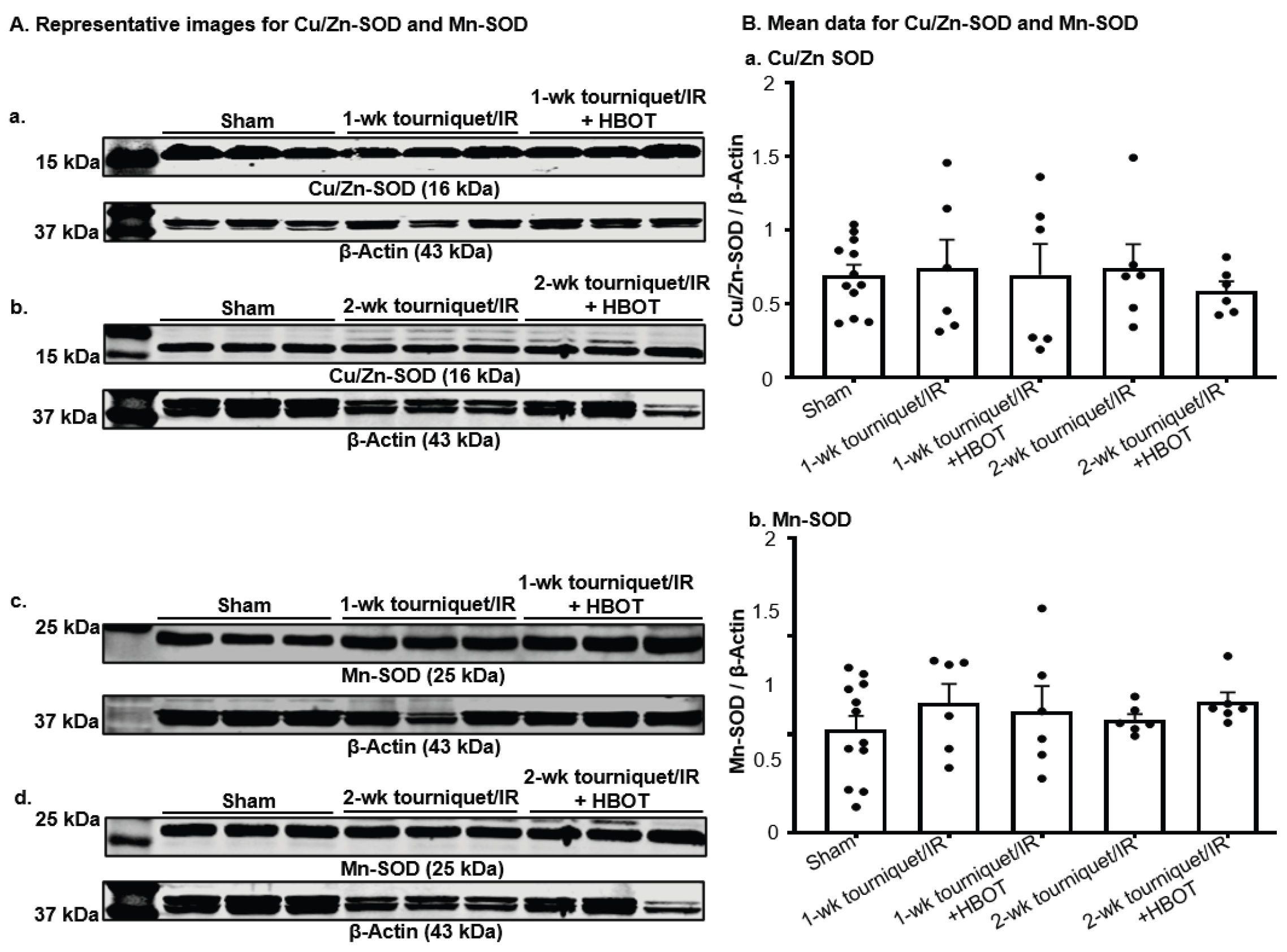
As critical antioxidant enzymes, protein expression of Cu/Zn-SOD and Mn-SOD was assessed (Figure 9). Compared to sham group, Cu/Zn-SOD and Mn-SOD expressed in the gastrocnemius muscle were not significantly changed in 1-wk tourniquet/IR, 1-wk tourniquet/IR+HBOT, 2-wk tourniquet/IR, and 2-wk tourniquet/IR+HBOT groups.
4. Discussion
Due to the increased risk of irreversible ischemic and neurovascular injury associated with prolonged tourniquet use, clinical recommendations emphasize the use of tourniquets for no more than 2 hours [30]. In this present study, three hours of unilateral hindlimb tourniquet/IR resulted in deficits in skeletal muscle structure and function, including NMJs, that persisted up to 2-weeks after injury. It is noteworthy that all mortality observed in the present study occurred within the first 3 days after tourniquet/IR. This finding is consistent with previous studies using a similar murine mouse model of tourniquet/IR, in which mortality primarily occurred within the first 3 days [19]. We previously demonstrated that tourniquet/IR induces significant inflammatory and structural injury to remote organs, especially the lungs, suggesting that the systemic effects of tourniquet/IR, rather than the local hindlimb injury alone, may contribute to early mortality following tourniquet/IR [19].
The NMJ is a specialized synapse that transmits impulses from motor neurons to skeletal muscle fibers to initiate muscle contraction. Similar to findings from our previous studies [29], our present study also found that motor nerve terminal innervation was markedly reduced during 2-wk tourniquet/IR accompanied by diminished nerve terminal occupancy and increased NMJ fragmentation. Although motor nerve innervation and occupancy exhibited moderate restoration at 2-wk tourniquet/IR timepoint, nAChR cluster morphology remained progressively worse. Tu et al. reported complete denervation during the first 3 days after tourniquet/IR, followed by gradual regeneration from 1 to 6 weeks [29]. Postsynaptic nAChR clusters became progressively fragmented during this recovery period [29]. Recent evidence also suggests that postsynaptic nAChR cluster organization can remain disorganized after NMJ injury despite motor nerve innervation [31]. These data suggest that restoration of motor nerve innervation does not explicitly result in concurrent recovery of postsynaptic NMJ architecture. Consistent with this structural dissociation, EPP amplitudes in our previous study increased progressively but remained lower than sham levels even at 6 weeks [29]. Similarly, the NMJ morphological abnormalities in this present study were accompanied by reduced EPP amplitudes in the present study. HBOT improved tourniquet/IR-reduced EPP amplitudes at 1-wk and 2-wk timepoints. These findings indicate that the incomplete recovery of neuromuscular transmission contributes to the persistent motor nerve and nAChR functional deficits observed after tourniquet/IR, and HBOT may accelerate NMJ functional recovery as early as at the 1-wk tourniquet/IR timepoint.
Histological analysis demonstrated that tourniquet/IR induced significant skeletal muscle remodeling, evidenced by reduced muscle fiber cross-sectional area, increased centrally nucleated muscle fibers, and increased muscle fibrosis. Central nucleation is a well-established marker of muscle regeneration which reflects ongoing repair after muscle damage [32]. The increase in muscle fibrosis is consistent with our previous findings of leucocyte infiltration after 3 hours of tourniquet use and 48 hours of reperfusion [22]. Persistent inflammatory cell infiltration has been shown to promote a pro-fibrotic environment through the production of cytokines and sustained deposition of extracellular matrix, ultimately contributing to fibrosis and impaired muscle function [33]. HBOT increased muscle fiber cross sectional area, attenuated muscle fibrosis, and improved skeletal muscle contraction, suggesting that HBOT partially mitigates tourniquet/IR-induced skeletal muscle contractile dysfunction through promoting NMJ functional improvement and muscle structural recovery.
Inflammation contributes significantly to ischemia-reperfusion injury and is also important for skeletal muscle repair. Tu et al. reported that during reperfusion after skeletal muscle injury, inflammatory cells phagocytize damaged tissue and release inflammatory mediators which promote myogenesis and neovascularization [25]. However, excessive inflammation promotes proteolysis, destructive metabolism, and impairs tissue recovery. In the present study, protein expressions of IL-1β and TNF-α were elevated at 1-wk and 2-wk tourniquet/IR, which indicates a persistent inflammatory response during the early phase of tourniquet/IR. Treatment with HBOT did not sufficiently alter IL-1β and TNF-α expression during tourniquet/IR. From these data, we assume that inflammation (IL-1β and TNF-α) is not a mediator to be involved in HBOT-improved NMJ and muscle function. Importantly, Tu et al. emphasized that complete suppression of inflammation during skeletal muscle recovery may not necessarily be beneficial, since a controlled inflammatory response is needed for the transition from tissue injury to tissue repair [25].
Oxidative stress is another important mediator of ischemia-reperfusion injury. Restoration of blood flow to previously ischemic tissues can result in excessive generation of ROS and contribute to cellular damage and impaired tissue regeneration. Nicotinamide Adenine Dinucleotide Phosphate (NADPH) oxidase is a major source of ROS in ischemic tissues, with NOX2 serving as the catalytic subunit, p67-phox as the catalytic activator, and p47-phox serving as the organizer subunit of the NOX-2 containing NADPH oxidase complex [34]. In the present study, we demonstrated an increase in NOX2, p67-phox, and p-47phox during tourniquet/IR. Haddad et al. reported increased NOX2 and p47 expression in ischemic hindlimbs, showing that genetic deficiency of NOX2 reduced oxidative stress while improving blood flow recovery and neovascularization [35]. Together, these findings suggest that activation of NADPH oxidase pathway may contribute to oxidative stress and subsequent tissue injury following tourniquet/IR. HBOT did not have any significant effect on NADPH subunits at either 1-wk or 2-wk tourniquet/IR timepoint, which also rules out the involvement of NADPH oxidase-related oxidative stress in HBOT-improved NMJ and muscle function.
Mitochondrial dysfunction is an important contributor to tourniquet/IR. During ischemia, oxygen deprivation disrupts oxidative phosphorylation and alters mitochondrial bioenergetics, and subsequent reperfusion promotes excessive mitochondrial ROS production [36,37]. Using a similar tourniquet/IR model, one previous study from Tran et al. demonstrated acute reductions in the expression and activities of ETC complexes I, II, and III in tourniquet/IR-injured gastrocnemius muscle lysates [18]. In the present study, ETC complexes I, II, and III were reduced during tourniquet/IR, although the reduction of some ETC complex subunits were not statistically significant. However, protein expression of Mn-SOD and Cu/Zn-SOD (two antioxidant enzymes) was unchanged during tourniquet/IR. Treatment with HBOT did not affect mitochondria-derived oxidative stress and antioxidants in tourniquet/IR-injured muscles at 1-wk and 2-wk timepoints. In addition, HBOT has been shown to exert beneficial or deleterious effects based on the timing of treatments [26]. Daily HBO treatment for ≤1 week was associated with increased oxidative stress and reduced mitochondrial activity, likely because of the transient increase in oxygen availability and thus ROS production [26]. Conversely, daily HBO treatment for ≥4 weeks was associated with the improvement of mitochondrial function, likely from activation of endogenous antioxidant responses such as Nrf2-mediated antioxidant signaling [38]. Future studies will be required to assess whether long-term treatment with HBOT can inhibit mitochondrial oxidative stress.
HBOT involves the delivery of pure (100%) oxygen at two to three times the normal atmospheric pressure to sustain oxygen delivery to tissues and preserve oxidative phosphorylation in ischemic conditions [39]. Plasma hemoglobin is almost completely saturated at sea level. However, under hyperbaric conditions, the partial pressure of oxygen increases and creates a steep gradient that allows more oxygen to dissolve into blood and reach damaged tissues [40]. Dissolved oxygen content increases according to Henry’s Law [40]. The therapeutic efficacy of HBOT seems to depend on the timing and period of treatment. In the present study, HBOT administered after tourniquet/IR partially mitigates tourniquet/IR-induced skeletal muscle contractile dysfunction via promoting NMJ functional improvement and muscle structural recovery. In addition, HBOT did not affect the pro-inflammatory cytokines, NADPH oxidase/mitochondria-derived oxidative stress, and some antioxidants in tourniquet-injured muscles. These results strongly eliminate the involvement of anti-inflammation and anti-oxidative stress in HBOT-improved NMJ and skeletal muscle function during tourniquet/IR, which is similar to the previous report that demonstrates a dissociation between biochemical and morphological effects of HBOT on the skeletal muscle [22]. Therefore, some alternative mechanisms should be considered. First, Oyaizu et al., demonstrated that HBOT increased proliferation and differentiation of satellite cells, inducing regeneration of skeletal muscle fibers [41]. Second, Tu et al. reported that myokines such as IL-6 facilitate skeletal muscle repair by promoting satellite cell expansion and myoblast proliferation through STAT3 signaling, while macrophage-derived growth factors such as insulin-like growth factor 1 (IGF-1) promote muscle regeneration. These findings suggest that HBOT may enhance recovery after tourniquet/IR by promoting regenerative pathways involved in skeletal muscle remodeling and repair. Third, HBOT may also promote functional recovery through macrophage-derived vascular endothelial growth factor (VEGF) signaling, which supports angiogenesis, revascularization and NMJ regeneration after injury [25]. Fourth, the attenuation of tourniquet/IR-induced fibrosis by HBOT suggests that HBOT may suppress pro-fibrotic signaling pathways such as TGF-β mediated extracellular matrix remodeling [42]. These potential mechanisms underlying the protective effects of HBOT against tourniquet/IR-induced NMJ and skeletal muscle dysfunction will be explored in future studies.
Other factors could explain the limited efficacy of HBOT demonstrated in the present study. Optimizing timepoints and periods for tourniquet/IR and HBOT treatment may detect a sustained therapeutic response to HBOT. Our previous studies demonstrated that NMJ regeneration continues from 1 to 6 weeks, with NMJ transmission still impaired at the 6-week time point [29]. Thus, the 2-wk duration selected in the present study likely captured the active phase of degeneration/regeneration and remodeling after tourniquet/IR, rather than the recovery phase. Extending the observation period beyond 2 weeks may determine whether the benefits of HBOT eventually translate into the significantly morphological and functional recovery of the NMJ and skeletal muscle after tourniquet/IR.
5. Conclusions
In conclusion, tourniquet/IR caused persistent skeletal muscle and neuromuscular injury characterized by muscle fiber atrophy, NMJ dysfunction, and impaired motor function. HBOT partially improved NMJ and skeletal muscle function in tourniquet/IR-injured muscles. However, HBOT did not affect tourniquet/IR-induced inflammation and oxidative stress. These findings suggest that, under the treatment conditions and time points evaluated in the present study, anti-inflammation and anti-oxidative stress are not mediators to contribute to the protective effects of HBOT against tourniquet/IR-induced NMJ and skeletal muscle injury. Future studies extending the recovery period and optimizing the timing of HBOT are needed to clarify HBOT benefits in long-term tourniquet/IR-injured NMJ and muscle functional recovery and potential cellular and molecular mechanisms.
Supplementary Materials
The following supporting information can be downloaded at the website of this paper posted on Preprints.org.
Author Contributions
Conceptualization, Y.-L.L.; methodology, E.A.A., H.T., L.W., and Y.-L.L.; validation, E.A.A., H.T., A.B., M.W., and Y.-L.L.; investigation, E.A.A., H.T., and Y.-L.L.; writing—original draft preparation, E.A.A., H.T., and Y.-L.L.; writing—review and editing, E.A.A., A.B., M.W., and Y.-L.L.; supervision, Y.-L.L.; project administration, Y.-L.L.; funding acquisition, Y.-L.L. All authors have read and agreed to the published version of the manuscript.
Funding
This research was funded by the National Institute of Health, Bethesda, MD, USA (R01GM145736 to Y.-L.L.).
Institutional Review Board Statement
This study conforms to the guidelines for the Care and Use of Laboratory Animals and was approved by the Institutional Animal Care and Use Committee (IACUC) at the University of Nebraska Medical Center (IACUC number: 23-011-03-FC).
Informed Consent Statement
Not applicable.
Data Availability Statement
The original contributions presented in this study are included in the article/supplementary materials. Further inquiries can be directed to the corresponding author.
Conflicts of Interest
The authors declare no conflicts of interest.
References
- Tuncali, B.; Karci, A.; Tuncali, B.E.; Mavioglu, O.; Ozkan, M.; Bacakoglu, A.K.; Baydur, H.; Ekin, A.; Elar, Z. A New Method for Estimating Arterial Occlusion Pressure in Optimizing Pneumatic Tourniquet Inflation Pressure. Anesth. Amp Analg. 2006, 102, 1752–1757. [Google Scholar] [CrossRef] [PubMed]
- Kauvar, D.S.; Dubick, M.A.; Walters, T.J.; Kragh, J.F. Systematic review of prehospital tourniquet use in civilian limb trauma. J. Trauma Acute Care Surg. 2018, 84, 819–825. [Google Scholar] [CrossRef] [PubMed]
- Welling, D.R.; McKay, P.L.; Rasmussen, T.E.; Rich, N.M. A brief history of the tourniquet. J. Vasc. Surg. 2012, 55, 286–290. [Google Scholar] [CrossRef] [PubMed]
- Zideman, D.A.; De Buck, E.D.J.; Singletary, E.M.; Cassan, P.; Chalkias, A.F.; Evans, T.R.; Hafner, C.M.; Handley, A.J.; Meyran, D.; Schunder-Tatzber, S.; et al. European Resuscitation Council Guidelines for Resuscitation 2015 Section 9. First aid. Resuscitation 2015, 95, 278–287. [Google Scholar] [CrossRef] [PubMed]
- Radulovic, A.; Cerovac, S. The history of tourniquet use in limb surgery. Int. Orthop. 2024, 48, 603–609. [Google Scholar] [CrossRef] [PubMed]
- Rudolf, R. Avoiding long-term muscle damage upon ischaemia–reperfusion. Acta Physiol. 2017, 219, 343–345. [Google Scholar] [CrossRef] [PubMed]
- Knudson, M.M.; Velmahos, G.; Cooper, Z.R. Response to mass casualty events: from the battlefield to the Stop the Bleed campaign. Trauma Surg. Amp Acute Care Open 2016, 1, e000023. [Google Scholar] [CrossRef] [PubMed]
- Jacobs, L.M.; Wade, D.; McSwain, N.E.; Butler, F.K.; Fabbri, W.; Eastman, A.; Conn, A.; Burns, K.J. Hartford Consensus: A Call to Action for THREAT, a Medical Disaster Preparedness Concept. J. Am. Coll. Surg. 2014, 218, 467–475. [Google Scholar] [CrossRef] [PubMed]
- Kragh, J.F.; Walters, T.J.; Baer, D.G.; Fox, C.J.; Wade, C.E.; Salinas, J.; Holcomb, J.B. Survival With Emergency Tourniquet Use to Stop Bleeding in Major Limb Trauma. Ann. Surg. 2009, 249, 1–7. [Google Scholar] [CrossRef] [PubMed]
- Hamawandi, S.A.; Amin, H.I.; Al-Humairi, A.K. Effects of the Use of Tourniquet in Total Knee Arthroplasty on the Clinical and Functional Outcomes with 5 Years of Follow-up: A Randomized Controlled Trial. J. Knee Surg. 2023, 36, 222–230. [Google Scholar] [CrossRef] [PubMed]
- Shulman, B.S.; Rettig, M.; Yang, S.S.; Sapienza, A.; Bosco, J.; Paksima, N. Tourniquet Use for Short Hand Surgery Procedures Done Under Local Anesthesia Without Epinephrine. J. Hand Surg. 2020, 45, 554.e551–554.e556. [Google Scholar] [CrossRef] [PubMed]
- Sato, J.; Ishii, Y.; Noguchi, H.; Takeda, M. Safety and efficacy of a new tourniquet system. BMC Surg. 2012, 12, 17. [Google Scholar] [CrossRef] [PubMed]
- Doyle, G.S.; Taillac, P.P. Tourniquets: A Review of Current Use with Proposals for Expanded Prehospital Use. Prehospital Emerg. Care 2008, 12, 241–256. [Google Scholar] [CrossRef] [PubMed]
- Corrick, R.M.; Tu, H.; Zhang, D.; Barksdale, A.N.; Muelleman, R.L.; Wadman, M.C.; Li, Y.-L. Dexamethasone Protects Against Tourniquet-Induced Acute Ischemia-Reperfusion Injury in Mouse Hindlimb. Front. Physiol. 2018, 9, 244. [Google Scholar] [CrossRef] [PubMed]
- Xacur-Trabulce, A.; Casas-Fuentes, G.; Ruiz-Vasconcelos, V.; Reitz, M.M.; Henry, S.M.; Scalea, T.M.; Ribeiro, M.A.F., Jr. Tourniquet-related complications in extremity injuries: a scoping review of the literature. World J. Emerg. Surg. 2025, 20, 57. [Google Scholar] [CrossRef] [PubMed]
- Hamad, F.; Rossiter, N. Tourniquet use in trauma and orthopaedics, how and when: current evidence. Surgery 2023, 41, 637–641. [Google Scholar] [CrossRef]
- An, P.; An, Y.; Chen, M.; Wu, L.; Wang, R. Ischemia–Reperfusion Injury: Molecular Mechanisms and Therapeutic Interventions. MedComm 2026, 7, e70822. [Google Scholar] [CrossRef] [PubMed]
- Tran, T.P.; Tu, H.; Liu, J.; Muelleman, R.L.; Li, Y.-L. Mitochondria-Derived Superoxide Links to Tourniquet-Induced Apoptosis in Mouse Skeletal Muscle. PLoS ONE 2012, 7, e43410. [Google Scholar] [CrossRef] [PubMed]
- Xu, L.; Whitney, L.; Tu, H.; Evans, A.J.; Klammer, A.; Barksdale, A.N.; Wadman, M.C.; Li, Y.-L. Different inflammatory responses in the remote organs after tourniquet-induced ischemia-reperfusion in mouse hindlimb. Injury 2026, 57, 113060. [Google Scholar] [CrossRef] [PubMed]
- Lukiianchuk, V.; Linchevskyy, O.; Dorlac, W.C.; Russo, R.M.; Andreatta, P.B.; Aarabi, S.; Patel, S.; Butler, F.K.; Polk, T.M.; Holcomb, J.B. Morbidity and mortality associated with ischemia-reperfusion injury after prolonged tourniquet use: A wartime single-center treatment algorithm. J. Trauma Acute Care Surg. 2025, 99, S79–S85. [Google Scholar] [CrossRef] [PubMed]
- Asim, M.; Amin, F.; El-Menyar, A. Multiple organ dysfunction syndrome: Contemporary insights on the clinicopathological spectrum. Qatar Med. J. 2020, 2020, 22. [Google Scholar] [CrossRef] [PubMed]
- Frisby, D.M.; Tu, H.; Qian, J.; Zhang, D.; Barksdale, A.N.; Wadman, M.C.; Cooper, J.S.; Li, Y.-L. Hyperbaric oxygen therapy does not alleviate tourniquet-induced acute ischemia-reperfusion injury in mouse skeletal muscles. Injury 2022, 53, 368–375. [Google Scholar] [CrossRef] [PubMed]
- Tran, T.P.; Tu, H.; Pipinos, I.I.; Muelleman, R.L.; Albadawi, H.; Li, Y.-L. Tourniquet-induced acute ischemia–reperfusion injury in mouse skeletal muscles: Involvement of superoxide. Eur. J. Pharmacol. 2011, 650, 328–334. [Google Scholar] [CrossRef] [PubMed]
- Tu, H.; Qian, J.; Zhang, D.; Barksdale, A.N.; Wadman, M.C.; Pipinos, I.I.; Li, Y.-L. Different responses of skeletal muscles to femoral artery ligation-induced ischemia identified in BALB/c and C57BL/6 mice. Front. Physiol. 2022, 13, 1014744. [Google Scholar] [CrossRef] [PubMed]
- Tu, H.; Li, Y.-L. Inflammation balance in skeletal muscle damage and repair. Front. Immunol. 2023, 14, 1133355. [Google Scholar] [CrossRef] [PubMed]
- Schottlender, N.; Gottfried, I.; Ashery, U. Hyperbaric Oxygen Treatment: Effects on Mitochondrial Function and Oxidative Stress. Biomolecules 2021, 11, 1827. [Google Scholar] [CrossRef] [PubMed]
- Woo, J.; Min, J.-H.; Lee, Y.-H.; Roh, H.-T. Effects of Hyperbaric Oxygen Therapy on Inflammation, Oxidative/Antioxidant Balance, and Muscle Damage after Acute Exercise in Normobaric, Normoxic and Hypobaric, Hypoxic Environments: A Pilot Study. Int. J. Environ. Res. Public Health 2020, 17, 7377. [Google Scholar] [CrossRef] [PubMed]
- Yamamoto, N.; Oyaizu, T.; Enomoto, M.; Horie, M.; Yuasa, M.; Okawa, A.; Yagishita, K. VEGF and bFGF induction by nitric oxide is associated with hyperbaric oxygen-induced angiogenesis and muscle regeneration. Sci. Rep. 2020, 10, 2744. [Google Scholar] [CrossRef] [PubMed]
- Tu, H.; Zhang, D.; Corrick, R.M.; Muelleman, R.L.; Wadman, M.C.; Li, Y.-L. Morphological Regeneration and Functional Recovery of Neuromuscular Junctions after Tourniquet-Induced Injuries in Mouse Hindlimb. Front. Physiol. 2017, 8, 207. [Google Scholar] [CrossRef] [PubMed]
- Miller, T.; Polk, T.; Henry, S.M. Do tourniquets for extremity injuries save lives? Trauma Surg. Amp Acute Care Open 2026, 11, e002341. [Google Scholar] [CrossRef] [PubMed]
- Zelada, D.; Bermedo-García, F.; Mella, J.; Olguín, H.C.; Henríquez, J.P. Early postsynaptic instability and acetylcholine receptor compartmentalization precede neuromuscular synapse dismantling. Commun. Biol. 2026, 9, 576. [Google Scholar] [CrossRef] [PubMed]
- Roman, W.; Gomes, E.R. Nuclear positioning in skeletal muscle. Semin. Cell Amp Dev. Biol. 2018, 82, 51–56. [Google Scholar] [CrossRef] [PubMed]
- Gong, J.; Xu, J.; Zhang, J.; Shen, Y.; Sun, H.; Chen, B. Molecular mechanisms of skeletal muscle fibrosis and potential targeted therapeutic strategies. Front. Immunol. 2026, 17, 1714238. [Google Scholar] [CrossRef] [PubMed]
- El-Benna, J.; Dang, P.M.-C.; Gougerot-Pocidalo, M.-A.; Marie, J.-C.; Braut-Boucher, F. p47phox, the phagocyte NADPH oxidase/NOX2 organizer: structure, phosphorylation and implication in diseases. Exp. Mol. Med. 2009, 41, 217. [Google Scholar] [CrossRef] [PubMed]
- Haddad, P.; Dussault, S.; Groleau, J.; Turgeon, J.; Michaud, S.-E.; MéNard, C.; Perez, G.; Maingrette, F.; Rivard, A. Nox2-Containing NADPH Oxidase Deficiency Confers Protection From Hindlimb Ischemia in Conditions of Increased Oxidative Stress. Arterioscler. Thromb. Vasc. Biol. 2009, 29, 1522–1528. [Google Scholar] [CrossRef] [PubMed]
- Zhou, H.; Toan, S. Pathological Roles of Mitochondrial Oxidative Stress and Mitochondrial Dynamics in Cardiac Microvascular Ischemia/Reperfusion Injury. Biomolecules 2020, 10, 85. [Google Scholar] [CrossRef] [PubMed]
- Naito, H.; Nojima, T.; Fujisaki, N.; Tsukahara, K.; Yamamoto, H.; Yamada, T.; Aokage, T.; Yumoto, T.; Osako, T.; Nakao, A. Therapeutic strategies for ischemia reperfusion injury in emergency medicine. Acute Med. Surg. 2020, 7, e501. [Google Scholar] [CrossRef] [PubMed]
- Dhamodharan, U.; Karan, A.; Sireesh, D.; Vaishnavi, A.; Somasundar, A.; Rajesh, K.; Ramkumar, K.M. Tissue-specific role of Nrf2 in the treatment of diabetic foot ulcers during hyperbaric oxygen therapy. Free Radic. Biol. Med. 2019, 138, 53–62. [Google Scholar] [CrossRef] [PubMed]
- Hentia, C.; Rizzato, A.; Camporesi, E.; Yang, Z.; Muntean, D.M.; Săndesc, D.; Bosco, G. An overview of protective strategies against ischemia/reperfusion injury: The role of hyperbaric oxygen preconditioning. Brain Behav. 2018, 8, e00959. [Google Scholar] [CrossRef] [PubMed]
- Ortega, M.A.; Fraile-Martinez, O.; García-Montero, C.; Callejón-Peláez, E.; Sáez, M.A.; Álvarez-Mon, M.A.; García-Honduvilla, N.; Monserrat, J.; Álvarez-Mon, M.; Bujan, J.; et al. A General Overview on the Hyperbaric Oxygen Therapy: Applications, Mechanisms and Translational Opportunities. Medicina 2021, 57, 864. [Google Scholar] [CrossRef] [PubMed]
- Oyaizu, T.; Enomoto, M.; Yamamoto, N.; Tsuji, K.; Horie, M.; Muneta, T.; Sekiya, I.; Okawa, A.; Yagishita, K. Hyperbaric oxygen reduces inflammation, oxygenates injured muscle, and regenerates skeletal muscle via macrophage and satellite cell activation. Sci. Rep. 2018, 8, 1288. [Google Scholar] [CrossRef] [PubMed]
- Mayorca-Guiliani, A.E.; Leeming, D.J.; Henriksen, K.; Mortensen, J.H.; Nielsen, S.H.; Anstee, Q.M.; Sanyal, A.J.; Karsdal, M.A.; Schuppan, D. ECM formation and degradation during fibrosis, repair, and regeneration. npj Metab. Health Dis. 2025, 3, 25. [Google Scholar] [CrossRef] [PubMed]
Figure 1.
Experimental design, hindlimb perfusion, and survival following tourniquet-induced ischemia/reperfusion (tourniquet/IR) and hyperbaric oxygen therapy (HBOT). (A) Schematic of study design and treatment protocol, including the use of McGivney Hemorrhoidal Ligator to place orthodontic rubber band at the left hip joint of C57BL/6 mouse (B) Representative Laser Speckle Doppler perfusion images showing hindlimb blood flow. (C) Kaplan-Meier survival curves for sham, tourniquet/IR, and tourniquet/IR+HBOT groups over the 14-day study period. N = 16 mice per group. Survival data was analyzed using the Gehan-Breslow-Wilcoxon test and shown as survival rate (%). *p < 0.05 vs. sham; #p < 0.05 vs tourniquet/IR.
Figure 1.
Experimental design, hindlimb perfusion, and survival following tourniquet-induced ischemia/reperfusion (tourniquet/IR) and hyperbaric oxygen therapy (HBOT). (A) Schematic of study design and treatment protocol, including the use of McGivney Hemorrhoidal Ligator to place orthodontic rubber band at the left hip joint of C57BL/6 mouse (B) Representative Laser Speckle Doppler perfusion images showing hindlimb blood flow. (C) Kaplan-Meier survival curves for sham, tourniquet/IR, and tourniquet/IR+HBOT groups over the 14-day study period. N = 16 mice per group. Survival data was analyzed using the Gehan-Breslow-Wilcoxon test and shown as survival rate (%). *p < 0.05 vs. sham; #p < 0.05 vs tourniquet/IR.
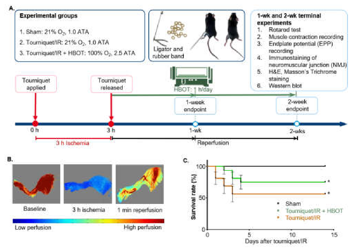
Figure 2.
Morphological changes in neuromuscular junctions (NMJs) of the gastrocnemius muscle following tourniquet/IR. (A) Representative images of NMJs from sham mice and tourniquet/IR mice with/without HBOT at 1 and 2-weeks after tourniquet release. Synaptophysin and neurofilament 200 (red) were used to label presynaptic nerve terminals, while α-bungarotoxin (α-BTX; green) was used to label postsynaptic nicotinic acetylcholine receptor (nAChR) clusters. Mean data for (B) fragments per nAChR cluster, (C) area per fragment, (D) fragmented nAChR clusters/total nAcHR clusters, (E) whole area per nAchR cluster, (F) nerve terminal occupancy, (G) motor nerve innervation to nAChR cluster. Data are mean ± SEM. Sham: 35 NMJs from 3 mice; 1-week tourniquet/IR: 37 NMJs from 3 mice; 1-week tourniquet/IR + HBOT: 39 NMJs from 3 mice; 2-week tourniquet/IR: 58 NMJs from 3 mice; 2-week tourniquet/IR + HBOT: 68 NMJs from 3 mice. Panels B, C, E, F were analyzed using the Kruskal-Wallis test followed by Dunn’s multiple-comparisons test. Panels D and G were analyzed using one-way ANOVA followed by Tukey’s multiple-comparisons test. *p < 0.05 vs sham; #p < 0.05 vs corresponding tourniquet/IR group.
Figure 2.
Morphological changes in neuromuscular junctions (NMJs) of the gastrocnemius muscle following tourniquet/IR. (A) Representative images of NMJs from sham mice and tourniquet/IR mice with/without HBOT at 1 and 2-weeks after tourniquet release. Synaptophysin and neurofilament 200 (red) were used to label presynaptic nerve terminals, while α-bungarotoxin (α-BTX; green) was used to label postsynaptic nicotinic acetylcholine receptor (nAChR) clusters. Mean data for (B) fragments per nAChR cluster, (C) area per fragment, (D) fragmented nAChR clusters/total nAcHR clusters, (E) whole area per nAchR cluster, (F) nerve terminal occupancy, (G) motor nerve innervation to nAChR cluster. Data are mean ± SEM. Sham: 35 NMJs from 3 mice; 1-week tourniquet/IR: 37 NMJs from 3 mice; 1-week tourniquet/IR + HBOT: 39 NMJs from 3 mice; 2-week tourniquet/IR: 58 NMJs from 3 mice; 2-week tourniquet/IR + HBOT: 68 NMJs from 3 mice. Panels B, C, E, F were analyzed using the Kruskal-Wallis test followed by Dunn’s multiple-comparisons test. Panels D and G were analyzed using one-way ANOVA followed by Tukey’s multiple-comparisons test. *p < 0.05 vs sham; #p < 0.05 vs corresponding tourniquet/IR group.
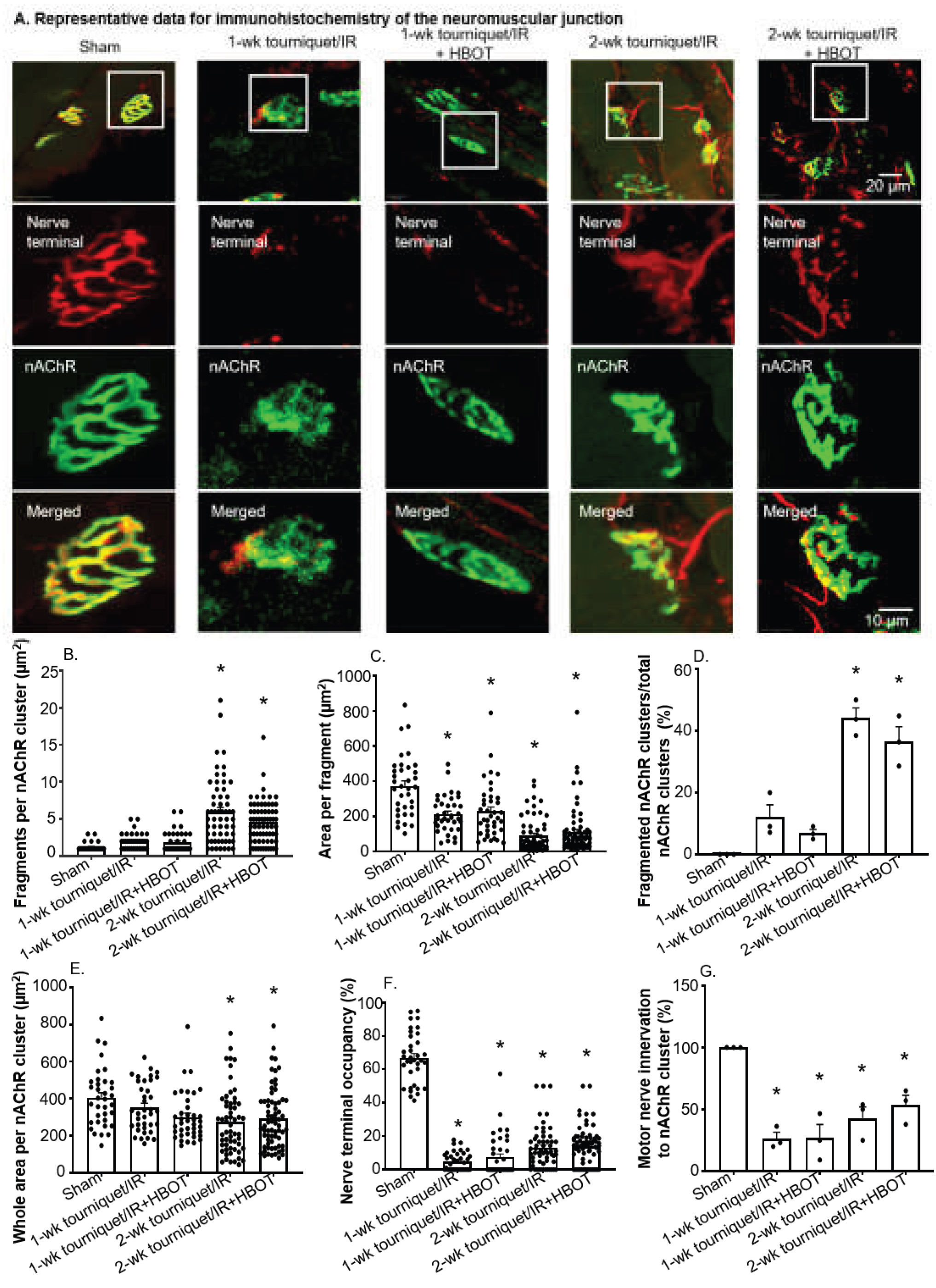
Figure 3.
(A) Representative recording and (B) quantitative data for sciatic nerve-stimulated end plate potentials (EPPs) in the gastrocnemius muscle. Data are mean ± SEM. Sham: 42 endplates from 6 mice; 1-week tourniquet/IR: 25 endplates from 4 mice; 1-week tourniquet/IR + HBOT: 26 endplates from 4 mice; 2-week tourniquet/IR: 30 endplates from 4 mice; 2-week tourniquet/IR + HBOT: 31 endplates from 3 mice. Mean data was analyzed using the Kruskal-Wallis test followed by Dunn’s multiple-comparisons test. *p < 0.05 vs sham; #p < 0.05 vs corresponding tourniquet/IR.
Figure 3.
(A) Representative recording and (B) quantitative data for sciatic nerve-stimulated end plate potentials (EPPs) in the gastrocnemius muscle. Data are mean ± SEM. Sham: 42 endplates from 6 mice; 1-week tourniquet/IR: 25 endplates from 4 mice; 1-week tourniquet/IR + HBOT: 26 endplates from 4 mice; 2-week tourniquet/IR: 30 endplates from 4 mice; 2-week tourniquet/IR + HBOT: 31 endplates from 3 mice. Mean data was analyzed using the Kruskal-Wallis test followed by Dunn’s multiple-comparisons test. *p < 0.05 vs sham; #p < 0.05 vs corresponding tourniquet/IR.
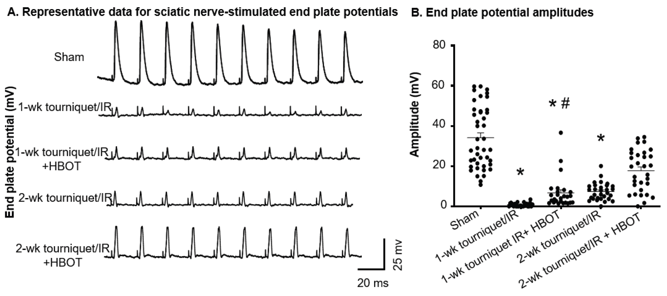
Figure 4.
Morphological changes in the gastrocnemius muscle following 1 and 2-weeks of tourniquet/IR with/without HBOT. Representative histological images of gastrocnemius muscle sections stained with (A) hematoxylin and eosin (H&E) and (B) Masson’s trichrome staining. Quantitative analyses for (C) mean muscle fiber cross-sectional area, (D) distribution of individual myofiber cross sectional area with each point representing an individual muscle fiber, (E) percentage of muscle fibers containing centralized nuclei, and (F) extent of muscle fibrosis. Data was quantified from 9 randomly selected sections per mouse. Data are mean ± SEM, n=3 mice per group. Panel D was analyzed using the Kruskal-Wallis test followed by Dunn’s multiple-comparisons test. Panels C, E, and G were analyzed using one-way ANOVA followed by Tukey’s multiple-comparisons test. *p < 0.05 vs sham; #p < 0.05 vs corresponding tourniquet/IR.
Figure 4.
Morphological changes in the gastrocnemius muscle following 1 and 2-weeks of tourniquet/IR with/without HBOT. Representative histological images of gastrocnemius muscle sections stained with (A) hematoxylin and eosin (H&E) and (B) Masson’s trichrome staining. Quantitative analyses for (C) mean muscle fiber cross-sectional area, (D) distribution of individual myofiber cross sectional area with each point representing an individual muscle fiber, (E) percentage of muscle fibers containing centralized nuclei, and (F) extent of muscle fibrosis. Data was quantified from 9 randomly selected sections per mouse. Data are mean ± SEM, n=3 mice per group. Panel D was analyzed using the Kruskal-Wallis test followed by Dunn’s multiple-comparisons test. Panels C, E, and G were analyzed using one-way ANOVA followed by Tukey’s multiple-comparisons test. *p < 0.05 vs sham; #p < 0.05 vs corresponding tourniquet/IR.
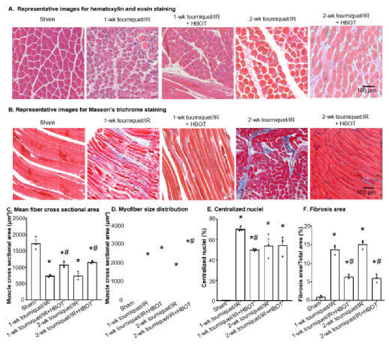
Figure 5.
(A) Representative recordings and (B) mean data for sciatic nerve-stimulated gastrocnemius muscle tetanic contractions in all experimental groups. (C) Average latency to fall off an accelerating rotarod at 1 and 2-wk tourniquet/IR with/without HBOT. Data are mean ± SEM, n = 6 mice per group. Data were analyzed using one-way ANOVA followed by Tukey’s multiple-comparisons test; *p < 0.05 vs sham; #p < 0.05 vs corresponding tourniquet/IR.
Figure 5.
(A) Representative recordings and (B) mean data for sciatic nerve-stimulated gastrocnemius muscle tetanic contractions in all experimental groups. (C) Average latency to fall off an accelerating rotarod at 1 and 2-wk tourniquet/IR with/without HBOT. Data are mean ± SEM, n = 6 mice per group. Data were analyzed using one-way ANOVA followed by Tukey’s multiple-comparisons test; *p < 0.05 vs sham; #p < 0.05 vs corresponding tourniquet/IR.
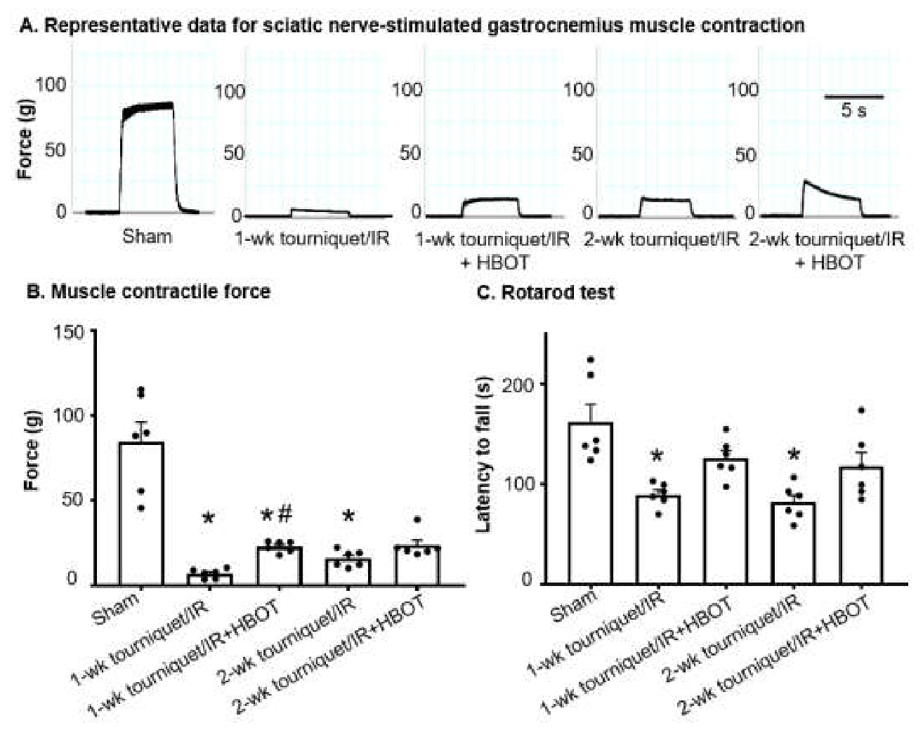
Figure 6.
(A) Representative immunoblots and (B) densitometric quantification of IL-1β and TNF-α in gastrocnemius muscle lysates from all experimental groups. Data are mean ± SEM, n = 6 mice per group. Panel B-a was analyzed using Welch’s one-way ANOVA with Dunnett’s multiple-comparisons test, and Panel B-b was analyzed with Kruskal–Wallis’ test followed by Dunn’s post-hoc test. *p < 0.05 vs sham; #p < 0.05 vs corresponding tourniquet/IR.
Figure 6.
(A) Representative immunoblots and (B) densitometric quantification of IL-1β and TNF-α in gastrocnemius muscle lysates from all experimental groups. Data are mean ± SEM, n = 6 mice per group. Panel B-a was analyzed using Welch’s one-way ANOVA with Dunnett’s multiple-comparisons test, and Panel B-b was analyzed with Kruskal–Wallis’ test followed by Dunn’s post-hoc test. *p < 0.05 vs sham; #p < 0.05 vs corresponding tourniquet/IR.
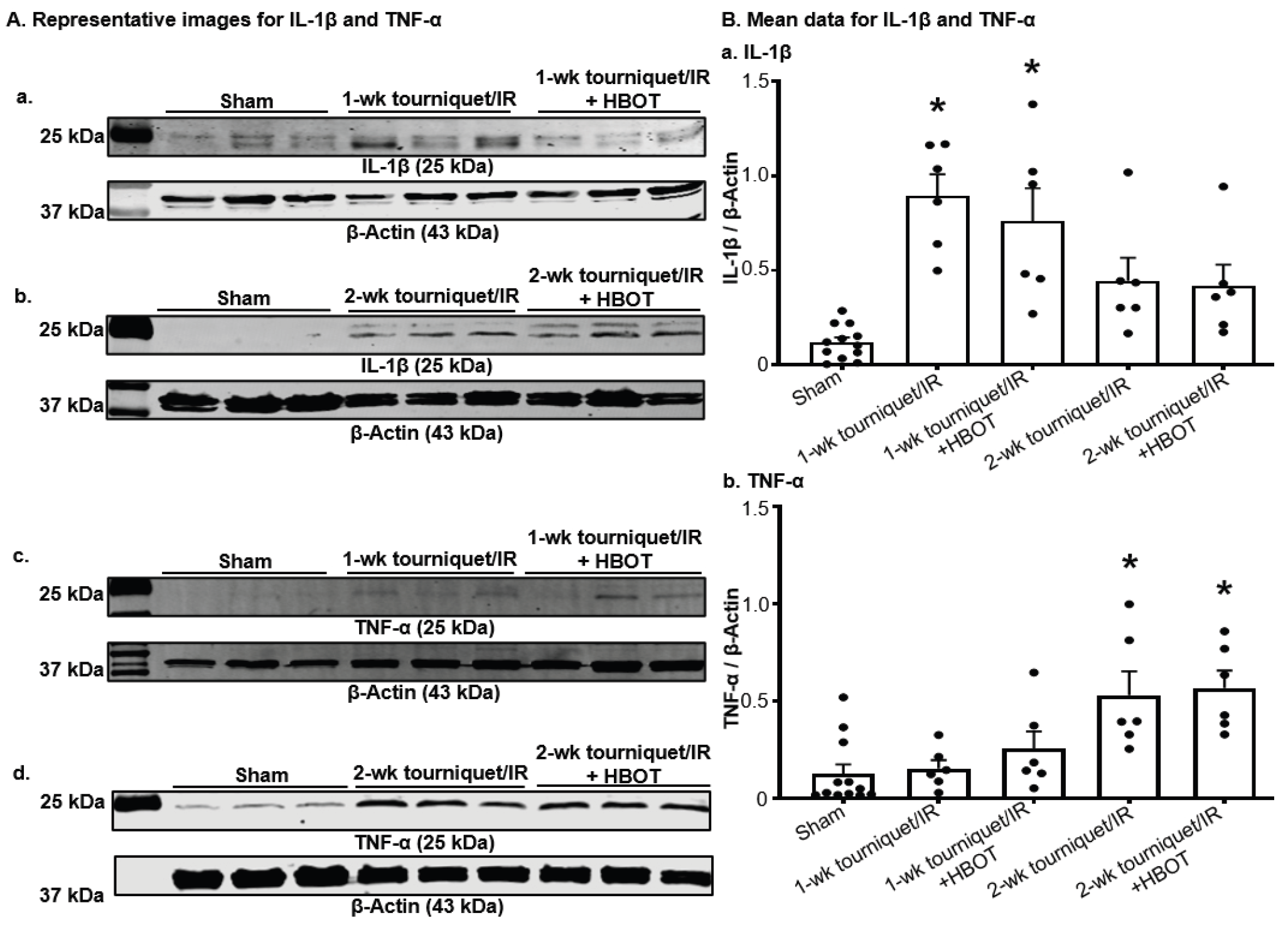
Figure 7.
(A) Representative immunoblots and (B) densitometric quantification of NOX2 (gp91-phox), p67-phox, and P47-phox in gastrocnemius muscle lysates from all experimental groups. Data are mean ± SEM; n=6 mice per group. Panels B-a and B-b were analyzed using Kruskal–Wallis’ test followed by Dunn’s test, and panel B-c was analyzed by one-way ANOVA with Tukey’s post-hoc test. *p < 0.05 vs sham; #p < 0.05 vs corresponding tourniquet/IR.
Figure 7.
(A) Representative immunoblots and (B) densitometric quantification of NOX2 (gp91-phox), p67-phox, and P47-phox in gastrocnemius muscle lysates from all experimental groups. Data are mean ± SEM; n=6 mice per group. Panels B-a and B-b were analyzed using Kruskal–Wallis’ test followed by Dunn’s test, and panel B-c was analyzed by one-way ANOVA with Tukey’s post-hoc test. *p < 0.05 vs sham; #p < 0.05 vs corresponding tourniquet/IR.
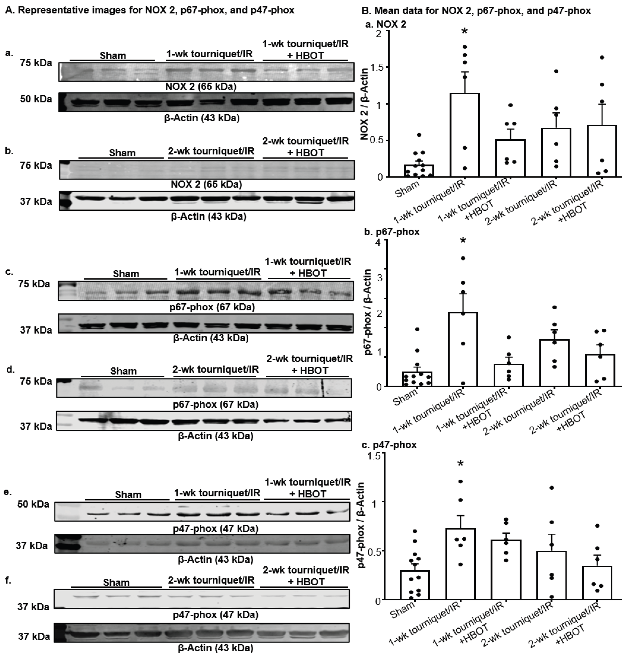
Figure 8.
(A) Representative immunoblots and (B) densitometric quantification of electron transport chain Complex I, Complex II, and Complex III in gastrocnemius muscle lysates from all experimental groups. Data are mean ± SEM; n=6 mice per group. Panel B-a was analyzed using ordinary one-way ANOVA with Tukey’s post-hoc test; panel B-b was analyzed using Welch’s one-way ANOVA with Dunnett’s T3 post-hoc test; and panel B-c was analyzed using the Kruskal–Wallis test followed by Dunn’s post-hoc test. *p < 0.05 vs sham; #p < 0.05 vs corresponding tourniquet/IR.
Figure 8.
(A) Representative immunoblots and (B) densitometric quantification of electron transport chain Complex I, Complex II, and Complex III in gastrocnemius muscle lysates from all experimental groups. Data are mean ± SEM; n=6 mice per group. Panel B-a was analyzed using ordinary one-way ANOVA with Tukey’s post-hoc test; panel B-b was analyzed using Welch’s one-way ANOVA with Dunnett’s T3 post-hoc test; and panel B-c was analyzed using the Kruskal–Wallis test followed by Dunn’s post-hoc test. *p < 0.05 vs sham; #p < 0.05 vs corresponding tourniquet/IR.
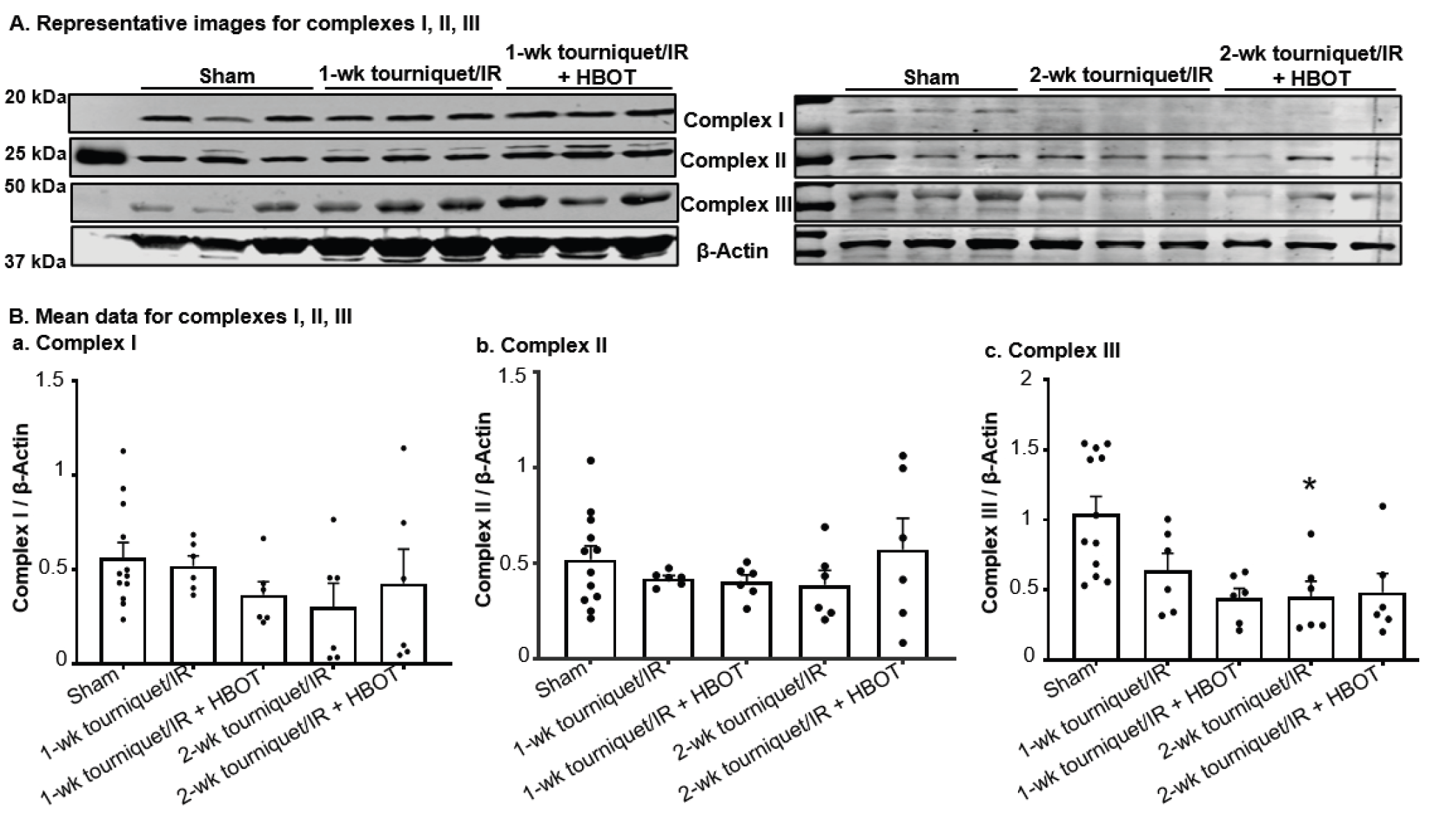
Figure 9.
(A) Representative immunoblots and (B) densitometric quantification of Cu/Zn-SOD and Mn-SOD in gastrocnemius muscle lysates from all experimental groups. Data are mean ± SEM; n=6 mice per group. Data were analyzed using one-way ANOVA followed by Tukey’s multiple-comparisons test. *p < 0.05 vs sham; #p < 0.05 vs corresponding tourniquet/IR.
Figure 9.
(A) Representative immunoblots and (B) densitometric quantification of Cu/Zn-SOD and Mn-SOD in gastrocnemius muscle lysates from all experimental groups. Data are mean ± SEM; n=6 mice per group. Data were analyzed using one-way ANOVA followed by Tukey’s multiple-comparisons test. *p < 0.05 vs sham; #p < 0.05 vs corresponding tourniquet/IR.
Disclaimer/Publisher’s Note: The statements, opinions and data contained in all publications are solely those of the individual author(s) and contributor(s) and not of MDPI and/or the editor(s). MDPI and/or the editor(s) disclaim responsibility for any injury to people or property resulting from any ideas, methods, instructions or products referred to in the content. |
© 2026 by the authors. Licensee MDPI, Basel, Switzerland. This article is an open access article distributed under the terms and conditions of the Creative Commons Attribution (CC BY) license (http://creativecommons.org/licenses/by/4.0/).
Copyright: This open access article is published under a Creative Commons CC BY 4.0 license, which permit the free download, distribution, and reuse, provided that the author and preprint are cited in any reuse.