

Submitted:
23 October 2026
Posted:
27 October 2026
You are already at the latest version
Abstract
Feline leukemia virus (FeLV) and feline immunodeficiency virus (FIV) are important retroviral pathogens affecting domestic and free-roaming cats worldwide. This systematic review and meta-analysis synthesized prevalence, risk factors, diagnostic considerations, and epidemiological trends in North America from 1987 to 2024. Following the PRISMA framework, studies from the United States and Canada were evaluated using data from 285,080 cats for FIV and 183,296 cats for FeLV. Pooled prevalence was 4.02% (95% CI: 3.60–4.49%) for FIV and 3.57% (95% CI: 3.13–4.07%) for FeLV, with substantial heterogeneity among studies. Older age, male sex, intact male status, outdoor access, poor health, and feral or shelter status were associated with increased infection prevalence. Prevalence remained relatively stable overall but varied geographically and across population types. Diagnostic limitations, particularly detection of regressive FeLV infection, may influence prevalence estimates. These findings support integrated surveillance, improved diagnostic strategies, vaccination, and targeted population management.
Keywords:
feline leukemia virus (FeLV)
; feline immunodeficiency virus (FIV)
; retroviral infections
; prevalence
; seroprevalence
; risk factors
; molecular epidemiology
; diagnostic testing
; domestic cats
; free-roaming cats
; North America
; meta-analysis
Introduction
Feline leukemia virus (FeLV) and feline immunodeficiency virus (FIV) are globally distributed retroviruses that establish lifelong infections in domestic and wild felids and remain major causes of morbidity, mortality, and population-level disease burden [1,2]. Both viruses are associated with immunosuppression, hematologic disorders, and neoplasia and remain endemic across North America despite available diagnostics and vaccination. The interface between domestic and wild felids further amplifies their importance, with FeLV spillover into North American pumas causing severe and sometimes fatal outbreaks [2,3].
Like all retroviruses, the structural biology of these viruses is generally conserved, consisting of an outermost lipid envelope with glycoprotein spikes, an icosahedral capsid, and a helical RNA genome contained within a nucleocapsid [2,3] FeLV and FIV share conserved virion architecture and genomic organization, including the canonical gag, pol, and env genes [4,5]. Among these, env is a principal determinant of cell tropism, antigenic diversity, and immune escape. The env gene is the primary determinant of cell tropism and antigenic diversity due to its high variability among subtypes and recombinants [6].
FIV is a lentivirus that primarily targets activated CD4+ T lymphocytes and other immune and nervous system cells, leading to progressive immune dysfunction and increased susceptibility to opportunistic infections and malignancies. Infection typically progresses through acute, asymptomatic, and terminal phases [2,7]. FIV is classified into at least five major subtypes (A–E) based on env sequence diversity, with subtypes A and B predominating in North America. FeLV, a gammaretrovirus, displays a broader spectrum of infection outcomes, including abortive, regressive, and progressive infections [8] FeLV-A is the primary horizontally transmissible form, whereas FeLV-B arises through recombination with endogenous FeLV sequences and is associated with altered tissue tropism and lymphoma. FeLV-C results from env mutations affecting receptor usage and is associated with severe non-regenerative anemia [9]
Transmission dynamics for both viruses are closely linked to their dependence on direct contact. FIV is transmitted predominantly through bite wounds, whereas FeLV spreads mainly through prolonged close contact, including mutual grooming and shared food and water [2,10]. Across North America, adulthood, intact male sex, and outdoor or feral status are important risk factors, shaping heterogeneous transmission patterns across different cat populations.
The molecular epidemiology of some common viral diseases of human, livestock animals, birds and campaign animals provides important information to fine-tune the management and control strategies of these important diseases that affect the health and performance of these animals and birds [11,12,13,14,15,16,17,18,19,20,21,22,23,24,25,26].
Molecular epidemiology has identified env as a major focus of surveillance and evolutionary analysis. FIV diversity is driven primarily by mutation and geographic isolation, whereas FeLV diversity is strongly influenced by recombination with endogenous FeLV sequences [27] These mechanisms complicate efforts to infer transmission patterns, estimate prevalence, and develop broadly protective vaccines.
A critical barrier to accurate molecular surveillance is the absence of a single gold-standard diagnostic that reliably identifies all infection states. FIV point-of-care tests primarily detect antibodies and may produce false-positive or false-negative results under certain conditions [28]. FeLV antigen assays detect p27 and are effective for identifying progressive infection but may miss regressive infections in which proviral DNA persists [29,30] Quantitative PCR assays can detect proviral DNA and, when interpreted alongside antigen results and clinical context, help distinguish progressive from regressive states [31]. This diagnostic gap has important implications for epidemiologic inference, risk modeling, and control strategies. The interface between domestic cats and wild felids represents a critical frontier for conservation medicine, with FeLV spillover into pumas providing a prominent example of cross-species transmission [32]. These findings highlight the need for integrated surveillance frameworks linking domestic cat health, wildlife conservation, and landscape-level management.
Despite decades of prevalence studies, gaps remain in understanding feline retroviral epidemiology. Many surveys rely on antigen or antibody testing alone, potentially underestimating FeLV burden and obscuring subtype distributions. Limited subtype resolution and insufficient integration of spatial, ecological, and social-structure data further hinder efforts to link viral genetics with clinical outcomes and transmission patterns. This systematic review and meta-analysis synthesize approximately four decades of North American evidence with three objectives: (1) to estimate pooled FIV and FeLV prevalence and characterize regional and temporal variation; (2) to summarize host- and population-level risk factors and evaluate how diagnostic methods influence reported prevalence; and (3) to discuss implications for domestic-cat health, free-roaming cat management, and wild-felid conservation.
2. Materials and Methods
2.1. Literature Search and Data Acquisition
A systematic review and meta-analysis was conducted to evaluate the prevalence and geographic distribution of feline immunodeficiency virus (FIV) and feline leukemia virus (FeLV) among domestic and free-roaming cats in North America from 1987 to 2024. The review was conducted following the Preferred Reporting Items for Systematic Reviews and Meta-Analyses (PRISMA) framework. Electronic searches were performed in PubMed/MEDLINE, EMBASE, Scopus, and Web of Science using combinations of terms related to cats, FIV, FeLV, prevalence, seroprevalence, serology, and infection. Reference lists of relevant publications and reviews were also screened to identify additional eligible studies.
The search strategy included combinations of terms such as cat, feline, Felis catus, feline immunodeficiency virus, FIV, feline leukemia virus, FeLV, prevalence, seroprevalence, serology, and infection. Records were imported into a reference-management database and duplicates were removed before screening.
2.2. Study Selection and Eligibility Criteria
Studies were included if they evaluated domestic cats in the United States or Canada and reported FIV and/or FeLV infection or seroprevalence using a recognized diagnostic method. Eligible studies were required to provide the number of cats tested and the number of positive animals, or sufficient information to calculate prevalence.
Studies involving experimentally infected animals, case reports, case series, reviews, duplicate datasets, or populations without sufficient diagnostic or geographic information were excluded. Studies specifically targeting highly selected clinical populations were excluded from the primary regional analysis when their sampling framework was not representative of the general cat population.
Data were extracted on publication and sampling year, geographic location, population type, number of cats tested, number of positive cats, diagnostic method, and available demographic or epidemiological characteristics.
2.3. Study Population and Geographic Classification
Cats were categorized as client-owned, shelter, feral/free-roaming, TNR-associated, or mixed populations according to the information provided in each study. Geographic data were standardized by U.S. state and U.S. Census region and by Canadian province or territory. Pooled estimates were subsequently calculated for North America, Canada, the United States, and individual regions where sufficient data were available.
The final analytic datasets comprised 285,080 cats contributing to the FIV synthesis and 183,296 cats contributing to the FeLV synthesis. Of these, 26,268 FIV observations and 24,967 FeLV observations were from Canada, while 44,810 FIV observations and 47,116 FeLV observations were from the United States. The remaining observations came from studies reported at broader geographic levels or from datasets that could not be assigned to a single country-level subgroup. Pooled prevalence estimates and 95% confidence intervals were calculated for North America and, where the available data permitted, for the United States, Canada, and individual regions.
2.4. Diagnostic Methods
Diagnostic methods were recorded for each study and included enzyme-linked immunosorbent assays, point-of-care or lateral-flow assays, rapid immunomigration, immunofluorescence assays, Western blotting, and PCR or qPCR. Commercial point-of-care platforms, including SNAP® and WITNESS®, were classified according to the analyte detected and their intended diagnostic role. Primary and confirmatory test results were extracted separately when both were reported.
Published evidence regarding diagnostic sensitivity and specificity was reviewed to account for potential differences among testing platforms. Factors that could influence diagnostic interpretation, including early infection, vaccination history, assay characteristics, and confirmatory testing, were also considered. FIV serological testing may be affected by vaccination-induced antibodies, while early infection may result in false-negative serological results.
Pooled prevalence estimates were calculated separately for FIV and FeLV using a random-effects meta-analysis because substantial clinical and methodological heterogeneity was expected across studies. Study-specific proportions were synthesized with an appropriate variance-stabilizing approach, and 95% confidence intervals were reported. Statistical heterogeneity was assessed using the I² statistic. Prespecified subgroup analyses considered geographic region, population type, diagnostic method, and study period when sufficient data were available. Studies contributing multiple non-overlapping populations were treated as separate estimates; overlapping samples were retained only once.
The methodological quality of included prevalence studies was evaluated using a standardized appraisal tool appropriate for cross-sectional and prevalence research. Assessments considered sampling-frame representativeness, recruitment procedures, diagnostic validity, completeness of reporting, and appropriateness of statistical analysis. Quality assessments were used to characterize confidence in the evidence and to inform sensitivity analyses. Potential small-study effects and publication bias were evaluated when the number of available estimates was sufficient for meaningful interpretation.
3. Results
3.1. Risk Factors
Host-associated risk factors- such as age, sex, and immune status- play an important role in determining a cat’s susceptibility to infection. Adulthood, when compared to juveniles, is a significant predictor of both FIV and FeLV status [33,34,35,36] Prevalence increases with age, with 16 year or older cats being the most likely to be infected [33]. Male status is also associated with a higher likelihood of infection with FIV and FeLV, with intact males being the most likely to harbor an infection [33,34,35,36]. Sick cats, compared to healthy cats, are also more likely to be infected with FIV or FeLV, though clinical illness is more predictive of FeLV infection [37,38]. Some signs, such as gastrointestinal, oral, ocular, respiratory disease, and bite wounds, are significantly associated with infection of FIV/FeLV [39,40,41]. The relationship between breed and FIV is unclear, with some findings indicating mixed breeds are more susceptible to infection [42,43,44]. Cats with access outdoors are at a significantly higher risk of infection compared to indoors only cats. Feral cats and shelter cats also have a higher prevalence of FIV and FeLV than owned cats [45,46,47,48] Urban areas tend to have higher populations of feral cats, leading to a higher prevalence of FIV/FeLV infected cats in high population density areas [49].

Table 1.
Summary of reported risk factors associated with FIV and FeLV seropositivity in domestic cats in North America.
Table 1.
Summary of reported risk factors associated with FIV and FeLV seropositivity in domestic cats in North America.
| Risk factor | FIV | FeLV |
|---|---|---|
| Older age | Significant | Significant |
| Male sex | Significant | Significant |
| Intact male | Significant | Significant |
| Outdoor access | Significant | Significant |
| Poor health/clinical disease | Significant | Significant |
| Feral/shelter population | Higher prevalence | Higher prevalence |
| Breed | Inconsistent | Inconsistent |
3.2. Seroprevalence
Estimates of seroprevalence for FIV and FeLV provide critical insights into the geographical distribution of retroviral morbidity across domestic cat populations (Figure 1 and Figure 2). Understanding seroprevalence for these retroviruses is particularly important due to the life-long persistence of the viruses, transmission potential, and ability to cause immunosuppression and chronic illness in affected cats. The apparently evident lack of reduction in seroprevalences across North America may indicate the need for improved vaccination strategies, updated shelter-intake policies, and further control of feral cat populations. Monitoring changes in infection rates over time may improve resource allocation and public health interventions in the future [50].
Across the included studies, reported FIV prevalence ranged from 0% to 23%, and FeLV prevalence ranged from 0% to more than 10%. Descriptive sample-size-weighted averages were 3.6% for FIV and 2.8% for FeLV across North America. These descriptive values differ from the random-effects pooled estimates reported in Section 3.3 because the two approaches weight studies differently. Canada showed higher descriptive averages than the United States for both FIV and FeLV, at 4.7% and 3.4%, respectively.
Reported seroprevalence estimates may be influenced by variability in diagnostic tests utilized across studies. Though this review mainly used ELISA diagnostic results, ELISA has been shown to detect false positives in FIV-vaccinated cats and false negatives in the early stages of FIV infection [28,38]. The FIV vaccine was discontinued in North America in 2017 [40], resulting in a possibly higher prevalence of FIV-positive cats from 1987-2017. ELISA-based detection of FIV relies on the presence of antibodies; recent infections may produce a false-negative due to a lack of seroconversion [39]. However, no such trends in seroprevalence were noted over the course of this review. FeLV detection with ELISA utilizes antigen which may be below the detection threshold in the regressive form of the disease or with bone marrow- localized infection, resulting in false negatives [38]. Many studies did not include a confirmatory test (PCR or Western blot), and there is no “gold standard” for FIV or FeLV diagnosis, which may have contributed to inconsistencies in classification of infection status.
The FIV seroprevalence figure contains data from 285,080 domestic and feral cats in North America, with 26,268 samples from Canada and 44,810 from the United States. Data were primarily collected from veterinary clinics, shelters, and trap-neuter-release programs across North America. Study data were excluded from this figure if pertaining to high- or low-risk group prevalences. Regional data represent pooled values from state and broader regional datasets within defined U.S. Census regions. The regions without shading (grey) indicate a lack of data within this study period. The highest rates of FIV were observed in the western region of the United States (4.2%), with individual states in the south, such as Georgia, reaching as high as 10.3%. Canadian provinces demonstrated variability, with Quebec reporting the highest prevalence at 9.6%. The lowest prevalences across North America were reported in the Northeast U.S. and New Brunswick, at 2.1% and 0.8%, respectively. The average seroprevalence was 3.6% across North America, 3.6% in the United States, and 4.7% in Canada. For detailed prevalence values by location, year, and diagnostic method, see supplementary table S1.
The FeLV seroprevalence figure contains data from 183,296 domestic and feral cats in North America, with 24,967 samples from Canada and 47,116 from the United States. Data were primarily collected from veterinary clinics, shelters, and trap-neuter-release programs across North America. Study data were excluded from this figure if pertaining to high- or low-risk group prevalences. Regional data represent pooled values from state and broader regional datasets within defined U.S. Census regions. The regions without shading (grey) indicate a lack of data within this study period. The highest rates of FeLV were observed in the southern region of the United States (3.1%), with individual states in that region, such as Georgia, reaching as high as 7.4%. The western region of the U.S. provided the lowest prevalence, with a high in Washington at 6.6% and a low in California at 1.3%. Canadian provinces were somewhat stable, with higher prevalences belonging to the eastern provinces: reaching a high in Nova Scotia at 6.6%. The lowest prevalence in Canada also lied in an eastern province with New Brunswick having a prevalence of 0%. The average seroprevalence was 2.8% across North America, 3% in the United States, and 3.4% in Canada. For detailed prevalence values by location, year, and diagnostic method, see supplementary table S1.
3.3. Pooled Prevalence of FIV and FeLV in North American Cats
The meta-analysis estimated pooled prevalence at 4.02% (95% CI: 3.60-4.49%) for FIV and 3.57% (95% CI: 3.13-4.07%) for FeLV among cats in North America (Table 2). Prevalence varied substantially across studies and geographic settings, ranging from less than 1% in some populations to 23% for FIV in a feral-cat population in Quebec and from 0% to more than 10% for FeLV in some U.S. populations. Given this variation and the high heterogeneity reported below, the pooled estimates should be interpreted as broad summaries of diverse study populations rather than as prevalence estimates for an average North American cat.
Substantial heterogeneity was observed for both infections (I² ≈ 95.0% for FIV and 93.8% for FeLV), indicating considerable variation among the included populations. This heterogeneity may reflect differences in geographic region, sampling source, cat population characteristics, diagnostic methods, and study period. The dataset includes feral, client-owned, clinic, and shelter populations, as well as studies conducted over several decades. These findings emphasize that the pooled estimates represent an overall prevalence across diverse North American cat populations rather than a uniform prevalence applicable to all cats.
3.4. Longitudinal Trends in the United States and Canada
In the United States, overall FeLV and FIV seroprevalence remained relatively stable at approximately 3.1% and 3.6%, respectively, although regional variation was observed (Table 3). Higher prevalence was reported in the southern and western regions, whereas estimates were generally lower in the Northeast. In Canada, FeLV prevalence ranged from approximately 2.5-3.4% and FIV from 2.5-4.3%, with modest variation between Quebec and Ontario (Table 3). Overall, these findings indicate relatively stable national prevalence but persistent geographic differences, particularly among free-roaming and feral cat populations that may serve as important viral reservoirs [41,51].
Across the four study periods from 1987 to 2024, sample-size-weighted FIV prevalence was 6.60% in 1987–1999, 3.56% in 2000–2009, 6.13% in 2010–2019, and 10.80% in 2020–2024 (Table 4). Corresponding FeLV estimates were 2.37%, 2.50%, 3.87%, and 10.80%. However, the earliest FIV period and the most recent period for both viruses were each represented by a single record, and the number and composition of contributing populations varied among periods. These values therefore describe the available studies but do not establish a temporal increase or decrease in population prevalence.
3.4. Contextual Evidence from South America
Evidence from regions outside North America provides useful context but was not included in the primary systematic review or meta-analysis. Reports from South America, Europe, and Asia therefore should be interpreted as supplementary comparisons rather than as components of the pooled North American estimates.
3.5. Contextual Evidence from Europe
3.6. Impact of Clinical Status
Clinical status is an important source of variation in apparent FeLV prevalence. Studies based on clinically ill or referral populations may yield substantially higher estimates than studies of healthy shelter or community cats because animals are selected for testing according to illness or perceived risk. Such estimates should not be interpreted as population prevalence and should be analyzed separately from broadly sampled cat populations.
Discussion
Spatiotemporal Stability and Regional "Hotspots"
The available studies suggest that FIV and FeLV prevalence has remained within a similar range in North America since the late 1980s, although direct temporal comparisons are limited by differences in study populations, diagnostic methods, geographic coverage, and sampling intensity. Higher estimates reported in parts of the southern United States may reflect variation in outdoor access, cat-population structure, preventive-care coverage, and the proportion of clinically ill, shelter, or free-roaming cats included. These explanations remain hypotheses because the included prevalence studies were not designed to isolate the effects of climate, socioeconomic conditions, or veterinary access.
Demographic Risk Architecture
The consistent association between intact male status and FIV positivity is biologically plausible because FIV is transmitted predominantly through deep bite wounds. Territorial aggression and fighting therefore provide a mechanism linking sex, reproductive status, outdoor access, and infection risk. However, the strength of these associations may vary by population and study design, and the review should distinguish unadjusted prevalence differences from independently estimated risk factors.
Conversely, the data for FeLV show a broader demographic spread, including younger cats and social groups. Because FeLV is transmitted through "friendly" contact (mutual grooming, shared water bowls), its presence in a colony suggests a breakdown in social isolation or the introduction of a "progressive" shedder. The peak prevalence in cats aged 6-10 years suggests a cumulative exposure risk, where the probability of encountering a positive individual increases linearly with time spent in unmanaged outdoor environments.
The Regressive Diagnostic Gap
A critical point of analysis arising from the recent molecular data is the discrepancy between ELISA (antigen/antibody) and qPCR (proviral) results. Current surveillance suggests that standard point-of-care tests may be underestimating the "true" viral burden by as much as 20–30% due to regressive infections.
Regressive FeLV-infected cats represent a unique challenge for population modeling. While they are often transiently non-viremic and less likely to transmit the virus than progressive carriers, they remain "proviral-positive." The potential for these individuals to reactivate the virus during periods of stress or co-infection (such as with the recently detected Feline Hepadnavirus [46] means they serve as a silent reservoir that can re-ignite outbreaks in supposedly "cleared" colonies.
Molecular Evolution of the Env Gene
Sequence variation in the env gene is relevant to subtype classification, cell tropism, antigenic diversity, and molecular epidemiology. However, the studies synthesized here do not establish that vaccination pressure is driving antigenic change or that observed env variation has reduced the performance of p27 antigen tests or current vaccines. Future surveillance should combine representative sampling, whole-genome or env sequencing, clinical metadata, vaccination history, and standardized diagnostic testing to evaluate these hypotheses.
Interspecies Implications and Conservation
Finally, the data concerning the domestic-wildlife interface (particularly pumas and Florida panthers) underscore the "One Health" importance of feline retrovirology. The high mortality rates in wild felids following spillover events from domestic cats suggest that while domestic populations have reached a level of endemic stability with these viruses, wild populations remain immunologically naive. Mathematical modeling of these spillover events is no longer a theoretical exercise but a conservation necessity to prevent the extinction of fragmented wild felid populations.
Stability of Infection Rates and Environmental Reservoirs
The synthesis of data from 1987 to 2024 suggests a concerning lack of reduction in seroprevalence across North America. Despite the discontinuation of the FIV vaccine in North America in 2017 and advancements in FeLV vaccination, the viruses remain endemic. This stability likely points to the role of unmanaged feral cat populations as persistent reservoirs. The higher prevalence in Canada (FIV 4.7% vs U.S. 3.6%) may reflect differences in population management or environmental factors that influence cat density and interaction.
The Diagnostic Gap: Regressive and Early Infections
Differences between antigen-, antibody-, and proviral-DNA-based testing complicate comparisons across FeLV studies. Antigen assays are useful for identifying many progressive infections but may not detect regressive infections in which proviral DNA persists without sustained antigenemia. PCR-based detection can identify proviral DNA, but a positive result does not by itself establish active viral shedding or equivalent transmission risk. Prevalence estimates should therefore be interpreted according to the analyte measured, the timing of sampling, and the confirmatory algorithm used.
Pathogenic Evolution and Molecular Surveillance
The env gene's high variability makes it a critical focus for molecular surveillance. As the main determinant of antigenic diversity, mutations in env facilitate immune escape and may influence cell tropism. The discovery of domestic cat Hepadnavirus in Texas further complicates the clinical picture, suggesting that co-infections may exacerbate the immunosuppressive effects of retroviruses.
The Socioeconomic Drivers of Prevalence
International differences in reported FeLV prevalence may reflect variation in cat-population structure, vaccination coverage, access to veterinary care, diagnostic methods, study design, and sampling of owned, shelter, stray, or clinically ill cats. Because the present review did not formally model national income or veterinary-service indicators, it cannot determine whether socioeconomic conditions independently explain geographic variation. Cross-country comparisons should therefore be treated as descriptive and interpreted cautiously.
Regressive Infection: The Hidden Reservoir
Regressive FeLV infection is an important diagnostic consideration because proviral DNA may persist in cats that test negative for circulating antigen. These cats generally pose a lower transmission risk than cats with progressive infection, although viral reactivation may occur under some conditions. The prevalence and epidemiologic importance of regressive infection cannot be quantified from the present synthesis unless studies using comparable paired antigen and proviral-PCR testing are analyzed separately.
Evolutionary Pressure and the Env Gene
The mutation rates within the env gene suggest that FeLV is undergoing rapid adaptation. In regions with high vaccination pressure, we observe "mutational drift" in the surface subunit (SU) that may eventually compromise diagnostic sensitivity. Furthermore, the high prevalence of Subtype B in South America suggests that recombination with endogenous sequences is a frequent and clinically significant event, directly increasing the risk of lymphoma in those populations.
Future Directions and Public Health Considerations
Continued surveillance is needed to determine whether FIV and FeLV prevalence changes over time and to identify population groups in which prevention and testing will have the greatest benefit. Priorities include standardized reporting of population source and health status, diagnostic algorithms that distinguish progressive from regressive FeLV infection, confirmatory testing when results are discordant or clinically consequential, and vaccination and risk-reduction strategies tailored to individual cats and local epidemiology. Shelter and free-roaming cat programs should integrate testing decisions with animal-welfare considerations, available resources, and current professional guidance.
Author Contributions
T.M.: Methodology, data analysis, writing first draft, writing final draft; CV.: Methodology, data analysis, writing first draft; M.G.H.: Conceptualization, methodology, data analysis, funding, writing first draft, writing final draft. All authors have read and agreed to the published version of the manuscript.
Funding
This work was supported by a Morris Animal Foundation Veterinary Student Scholar Grant (Grant: 25FEANVS-0005).
Institutional Review Board Statement
Not applicable.
Informed Consent Statement
Not applicable.
Acknowledgments
We thank the Morris Animal Foundation for providing a student scholarship to Tyler Michalka, DVM student at Lewyt College of Veterinary Medicine, Long Island University.
Conflicts of Interest
The authors declare no conflicts of interest.
References
- Hartmann, K. Clinical aspects of feline retroviruses: a review. Viruses 2012, 4(11), 2684–710. [Google Scholar] [CrossRef]
- Siebelink, K.H.; Chu, I.H.; Rimmelzwaan, G.F.; Weijer, K.; van Herwijnen, R.; Knell, P.; et al. Feline immunodeficiency virus (FIV) infection in the cat as a model for HIV infection in man: FIV-induced impairment of immune function. AIDS Res. Hum. Retroviruses 1990, 6(12), 1373–8. [Google Scholar] [CrossRef]
- Neil, J.C.; Onions, D.E. Feline leukaemia viruses: molecular biology and pathogenesis. Anticancer Res. 1985, 5(1), 49–63. [Google Scholar]
- Elder, J.H.; Lerner, D.L.; Hasselkus-Light, C.S.; Fontenot, D.J.; Hunter, E.; Luciw, P.A.; et al. Distinct subsets of retroviruses encode dUTPase. J. Virol. 1992, 66(3), 1791–4. [Google Scholar] [CrossRef]
- Tomasselli, A.G.; Heinrikson, R.L. Specificity of retroviral proteases: an analysis of viral and nonviral protein substrates. Methods Enzymol. 1994, 241, 279–301. [Google Scholar] [CrossRef]
- Samman, A.; McMonagle, E.L.; Logan, N.; Willett, B.J.; Biek, R.; Hosie, M.J. Phylogenetic characterisation of naturally occurring feline immunodeficiency virus in the United Kingdom. Vet. Microbiol. 2011, 150(3-4), 239–47. [Google Scholar] [CrossRef]
- Phillips, T.R.; Prospero-Garcia, O.; Wheeler, D.W.; Wagaman, P.C.; Lerner, D.L.; Fox, H.S.; et al. Neurologic dysfunctions caused by a molecular clone of feline immunodeficiency virus, FIV-PPR. J. Neurovirol 1996, 2(6), 388–96. [Google Scholar] [CrossRef]
- Burkhard, M.J.; Dean, G.A. Transmission and immunopathogenesis of FIV in cats as a model for HIV. Curr. HIV Res. 2003, 1(1), 15–29. [Google Scholar] [CrossRef]
- Pedersen, N.C.; Ho, E.W.; Brown, M.L.; Yamamoto, J.K. Isolation of a T-lymphotropic virus from domestic cats with an immunodeficiency-like syndrome. Science 1987, 235(4790), 790–3. [Google Scholar] [CrossRef]
- Cattori, V.; Weibel, B.; Lutz, H. Inhibition of Feline leukemia virus replication by the integrase inhibitor Raltegravir. Vet. Microbiol. 2011, 152(1-2), 165–8. [Google Scholar] [CrossRef]
- Al-Mubarak, A.I.A.; Hussen, J.; Kandeel, M.; Al-Kubati, A.A.G.; Falemban, B.; Hemida, M.G. Avian encephalomyelitis virus in backyard chickens. Vet. World 2023, 16(9), 1866–70. [Google Scholar] [CrossRef]
- Al-Mubarak, A.I.A.; Hussen, J.; Kandeel, M.; Al-Kubati, A.A.G.; Falemban, B.; Hemida, M.G. Evidence of the circulation of avian metapneumovirus in domestic backyard chickens in Eastern Saudi Arabia in 2019. Vet. World 2023, 16(6), 1246–51. [Google Scholar] [CrossRef]
- Alnaeem, A.A.; Hemida, M.G. Surveillance of the equine infectious anemia virus in Eastern and Central Saudi Arabia during 2014-2016. Vet. World 2019, 12(5), 719–23. [Google Scholar] [CrossRef]
- Ba Abduallah, M.M.; Hemida, M.G. Comparative analysis of the genome structure and organization of the Middle East respiratory syndrome coronavirus (MERS-CoV) 2012 to 2019 revealing evidence for virus strain barcoding, zoonotic transmission, and selection pressure. Rev. Med. Virol. 2021, 31(1), 1–12. [Google Scholar] [CrossRef]
- Hemida, M.G.; Al-Ghadeer, H.M. Evidence of Peste des petits Ruminants' Virus in Dromedary Camels in the Kingdom of Saudi Arabia between 2014 and 2016. Vet. Med. Int. 2019, 2019, 4756404. [Google Scholar] [CrossRef]
- Hemida, M.G.; Al-Hammadi, M.A.; Daleb, A.H.S.; Gonsalves, C.R. Molecular characterization and phylogenetic analyses of virulent infectious bronchitis viruses isolated from chickens in Eastern Saudi Arabia. Virusdisease 2017, 28(2), 189–99. [Google Scholar] [CrossRef]
- Hemida, M.G.; Alhammadi, M.; Daleb, A.; Alnaeem, A. Molecular and serological surveillance of African horse sickness virus in eastern and central Saudi Arabia. Rev. Sci. Tech 2017, 36(3), 889–98. [Google Scholar] [CrossRef]
- Hemida, M.G.; Chu, D.; Abdelaziz, A.; Alnaeem, A.; Chan, S.M.S.; Peiris, M. Molecular characterisation of an avian influenza (H5N8) outbreak in backyard flocks in Al Ahsa, Eastern Saudi Arabia, 2017-2018. Vet. Rec. Open 2019, 6(1), e000362. [Google Scholar] [CrossRef]
- Hemida, M.G.; Chu, D.K.W.; Perera, R.; Ko, R.L.W.; So, R.T.Y.; Ng, B.C.Y.; et al. Coronavirus infections in horses in Saudi Arabia and Oman. Transbound. Emerg. Dis. 2017, 64(6), 2093–103. [Google Scholar] [CrossRef]
- Hemida, M.G.; Perera, R.; Chu, D.K.W.; Alnaeem, A.A.; Peiris, M. Evidence of equine influenza A (H3N8) activity in horses from Eastern and Central Saudi Arabia: 2013-2015. Equine Vet. J. 2019, 51(2), 218–21. [Google Scholar] [CrossRef]
- Hemida, M.G.; Perera, R.; Chu, D.K.W.; Ko, R.L.W.; Alnaeem, A.A.; Peiris, M. West Nile virus infection in horses in Saudi Arabia (in 2013-2015). Zoonoses Public Health 2019, 66(2), 248–53. [Google Scholar] [CrossRef]
- Hemida, M.G.; Rizk El-Ghareeb, W.; Al-Hizab, F.; Ibrahim, A. Foot-and-mouth disease virus O/ME-SA/Ind 2001 lineage outbreak in vaccinated Holstein Friesian cattle in Saudi Arabia in 2016. Vet. Q. 2018, 38(1), 88–98. [Google Scholar] [CrossRef]
- Michalka, T.; Shah, A.U.; Liang, T.; Hemida, M.G. Multi-Epitope DNA-Based Feline Immunodeficiency Virus Vaccine Construct Designed by Immunoinformatic and Machine Learning Tools as a Surrogate Model for HIV Vaccine Development. Pathogens 2026, 15(3). [Google Scholar] [CrossRef]
- Shah, A.U.; Esparza, B.; Illanes, O.; Hemida, M.G. Comparative Genome Sequencing Analysis of Some Novel Feline Infectious Peritonitis Viruses Isolated from Some Feral Cats in Long Island. Viruses 2025, 17(2). [Google Scholar] [CrossRef]
- Shah, A.U.; Gauger, P.; Hemida, M.G. Isolation and molecular characterization of an enteric isolate of the genotype-Ia bovine coronavirus with notable mutations in the receptor binding domain of the spike glycoprotein. Virology 2025, 603, 110313. [Google Scholar] [CrossRef]
- Shah, A.U.; Vagra, C.; Moawad, N.; Rosen, M.; Levitan, D.; Hemida, M.G. Molecular epidemiological surveillance of the feline coronavirus reveals novel recombinant FCoV strains with evidence of ongoing viral evolution. Virus Res. 2026, 373, 199802. [Google Scholar] [CrossRef]
- Ngo, M.H.; AbuEed, L.; Kawasaki, J.; Oishi, N.; Pramono, D.; Kimura, T.; et al. Multiple recombination events between endogenous retroviral elements and feline leukemia virus. J. Virol. 2024, 98(2), e0140023. [Google Scholar] [CrossRef]
- Krick, E.L.; Little, L.; Patel, R.; Shofer, F.S.; Sorenmo, K.; Clifford, C.A.; et al. Description of clinical and pathological findings, treatment and outcome of feline large granular lymphocyte lymphoma (1996-2004). Vet. Comp. Oncol. 2008, 6(2), 102–10. [Google Scholar] [CrossRef]
- Little, S.; Levy, J.; Hartmann, K.; Hofmann-Lehmann, R.; Hosie, M.; Olah, G.; et al. 2020 AAFP Feline Retrovirus Testing and Management Guidelines. J. Feline Med. Surg. 2020, 22(1), 5–30. [Google Scholar] [CrossRef]
- Wardrop, K.J.; Birkenheuer, A.; Blais, M.C.; Callan, M.B.; Kohn, B.; Lappin, M.R.; et al. Update on Canine and Feline Blood Donor Screening for Blood-Borne Pathogens. J. Vet. Intern Med. 2016, 30(1), 15–35. [Google Scholar] [CrossRef]
- Marenzoni, M.L.; Lauzi, S.; Miglio, A.; Coletti, M.; Arbia, A.; Paltrinieri, S.; et al. Comparison of three blood transfusion guidelines applied to 31 feline donors to minimise the risk of transfusion-transmissible infections. J. Feline Med. Surg. 2018, 20(8), 663–73. [Google Scholar] [CrossRef]
- Poulet, H.; Thibault, J.C.; Masias, A. Immunosuppression in a Comparative Study of Feline Leukemia Virus Vaccines. Clin. Vaccine Immunol. 2015, 22(12), 1294–5. [Google Scholar] [CrossRef]
- Ravi, M.; Wobeser, G.A.; Taylor, S.M.; Jackson, M.L. Naturally acquired feline immunodeficiency virus (FIV) infection in cats from western Canada: Prevalence, disease associations, and survival analysis. Can. Vet. J. 2010, 51(3), 271–6. [Google Scholar]
- Levy, J.K.; Scott, H.M.; Lachtara, J.L.; Crawford, P.C. Seroprevalence of feline leukemia virus and feline immunodeficiency virus infection among cats in North America and risk factors for seropositivity. J. Am. Vet. Med. Assoc. 2006, 228(3), 371–6. [Google Scholar] [CrossRef]
- Chhetri, B.K.; Berke, O.; Pearl, D.L.; Bienzle, D. Comparison of risk factors for seropositivity to feline immunodeficiency virus and feline leukemia virus among cats: a case-case study. BMC Vet. Res. 2015, 11, 30. [Google Scholar] [CrossRef]
- Burling, A.N.; Levy, J.K.; Scott, H.M.; Crandall, M.M.; Tucker, S.J.; Wood, E.G.; et al. Seroprevalences of feline leukemia virus and feline immunodeficiency virus infection in cats in the United States and Canada and risk factors for seropositivity. J. Am. Vet. Med. Assoc. 2017, 251(2), 187–94. [Google Scholar] [CrossRef]
- Edinboro, C.H. Feral Cat Populations and Feline Retrovirus Prevalence in San Mateo County, California in Three Time Periods between 2001 and 2016; Animals (Basel), 2022; 24, p. 12. [Google Scholar]
- Little, S. A review of feline leukemia virus and feline immunodeficiency virus seroprevalence in cats in Canada. Vet. Immunol. Immunopathol. 2011, 143(3-4), 243–5. [Google Scholar] [CrossRef]
- Hosie, M.J.; Addie, D.; Belak, S.; Boucraut-Baralon, C.; Egberink, H.; Frymus, T.; et al. Feline immunodeficiency. ABCD guidelines on prevention and management. J. Feline Med. Surg. 2009, 11(7), 575–84. [Google Scholar] [CrossRef]
- Nehring, M.; Dickmann, E.M.; Billington, K.; VandeWoude, S. Study of feline immunodeficiency virus prevalence and expert opinions on standards of care. J. Feline Med. Surg. 2024, 26(7), 1098612X241245046. [Google Scholar] [CrossRef]
- Yamamoto, J.K.; Pu, R.; Sato, E.; Hohdatsu, T. Feline immunodeficiency virus pathogenesis and development of a dual-subtype feline-immunodeficiency-virus vaccine. AIDS 2007, 21(5), 547–63. [Google Scholar] [CrossRef]
- Biezus, G.; Grima de Cristo, T.; da Silva Casa, M.; Lovatel, M.; Vavassori, M.; Bruggemann de Souza Teixeira, M.; et al. Progressive and regressive infection with feline leukemia virus (FeLV) in cats in southern Brazil: Prevalence, risk factors associated, clinical and hematologic alterations. Prev. Vet. Med. 2023, 216, 105945. [Google Scholar] [CrossRef]
- Cohen, N.D.; Carter, C.N.; Thomas, M.A.; Lester, T.L.; Eugster, A.K. Epizootiologic association between feline immunodeficiency virus infection and feline leukemia virus seropositivity. J. Am. Vet. Med. Assoc. 1990, 197(2), 220–5. [Google Scholar] [CrossRef]
- Liem, B.P.; Dhand, N.K.; Pepper, A.E.; Barrs, V.R.; Beatty, J.A. Clinical findings and survival in cats naturally infected with feline immunodeficiency virus. J. Vet. Intern Med. 2013, 27(4), 798–805. [Google Scholar] [CrossRef]
- Gallina, L.; Facile, V.; Roda, N.; Sabetti, M.C.; Terrusi, A.; Urbani, L.; et al. Molecular investigation and genetic characterization of feline leukemia virus (FeLV) in cats referred to a veterinary teaching hospital in Northern Italy. Vet. Res. Commun. 2024, 48(4), 2683–9. [Google Scholar] [CrossRef]
- Martins, N.B.; de Almeida, J.C.N.; Goncalves, M.S.S.; Gila, L.I.; Yogui, D.R.; Alves, M.H.; et al. Occurrence of Typical Domestic Animal Viruses in Wild Carnivorans: An Emerging Threat to the Conservation of Endangered Species. Transbound. Emerg. Dis. 2024, 2024, 3931047. [Google Scholar] [CrossRef]
- Petch, R.J.; Gagne, R.B.; Chiu, E.; Mankowski, C.; Rudd, J.; Roelke-Parker, M.; et al. Feline Leukemia Virus Frequently Spills Over from Domestic Cats to North American Pumas. J. Virol. 2022, 96(23), e0120122. [Google Scholar] [CrossRef]
- Tandon, R.; Cattori, V.; Willi, B.; Lutz, H.; Hofmann-Lehmann, R. Quantification of endogenous and exogenous feline leukemia virus sequences by real-time PCR assays. Vet. Immunol. Immunopathol. 2008, 123(1-2), 129–33. [Google Scholar] [CrossRef]
- Candela, M.G.; Fanelli, A.; Carvalho, J.; Serrano, E.; Domenech, G.; Alonso, F.; et al. Urban landscape and infection risk in free-roaming cats. Zoonoses Public Health 2022, 69(4), 295–311. [Google Scholar] [CrossRef]
- Bendavid, E. Seroprevalence Studies Are Critical Early Pandemic Tools, and They Were Underappreciated During COVID-19. Am. J. Public Health 2023, 113(5), 523–4. [Google Scholar] [CrossRef]
- Chen, M.C.; Cavasin, J.P.; Tu, T.; Van Brussel, K.; Nekouei, O.; Cullen, J.M.; et al. Domestic cat hepadnavirus is detected infrequently in feline blood and liver samples submitted for diagnostic testing in Texas, USA. Virology 2025, 607, 110506. [Google Scholar] [CrossRef]
Figure 1.
Illustration of an average of FIV seroprevalence across North America from 1987-2024, visualized by province, region, and state when applicable. Data were compiled from peer-reviewed sources utilizing ELISA, qPCR, western blot, and IFA.
Figure 1.
Illustration of an average of FIV seroprevalence across North America from 1987-2024, visualized by province, region, and state when applicable. Data were compiled from peer-reviewed sources utilizing ELISA, qPCR, western blot, and IFA.
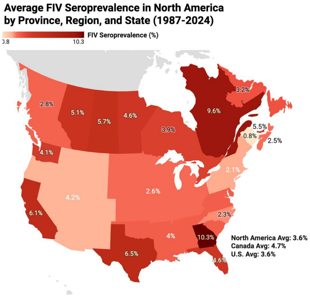
Figure 2.
Illustration of an average of FeLV seroprevalence across North America from 1987-2022, visualized by province, region, and state when applicable. Data were compiled from peer-reviewed sources utilizing ELISA, qPCR, western blot, and IFA.
Figure 2.
Illustration of an average of FeLV seroprevalence across North America from 1987-2022, visualized by province, region, and state when applicable. Data were compiled from peer-reviewed sources utilizing ELISA, qPCR, western blot, and IFA.
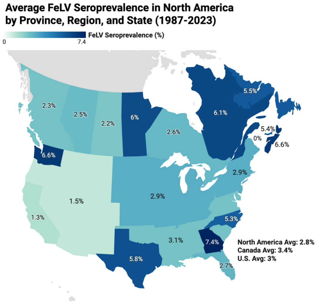
Table 2.
Meta-analysis estimates of pooled feline immunodeficiency virus and feline leukemia virus prevalence in North American cats.
Table 2.
Meta-analysis estimates of pooled feline immunodeficiency virus and feline leukemia virus prevalence in North American cats.
| Infection | Estimates | Pooled prevalence | 95% CI | I² |
|---|---|---|---|---|
| FIV | 40 | 4.02% | 3.60–4.49% | ~95.0% |
| FeLV | 37 | 3.57% | 3.13–4.07% | ~93.8% |
The table presents the number of studies included in the meta-analysis, pooled prevalence estimates with 95% confidence intervals (CIs), and the corresponding heterogeneity statistics (I²).
Table 3.
Detailed FeLV/FIV Prevalence in North America (Regional Breakdown).
| Region | Sample Size (n) | FeLV Prevalence (%) | FIV Prevalence (%) |
|---|---|---|---|
| USA Overall | 47,116 | 3.1 | 3.6 |
| U.S. South (Georgia) | 8,420 | ~3.5–4.5 | ~4.0–5.5 |
| U.S. Northeast | 12,150 | ~2.0–2.8 | ~2.5–3.5 |
| U.S. West (California) | 9,840 | ~3.2–4.2 | ~3.8–5.0 |
| Canada Overall | 24,967 | ~2.5–3.4 | ~2.5–4.3 |
| Quebec | 4,200 | ~2.3–3.0 | ~2.5–3.5 |
| Ontario | 7,150 | ~2.8–3.4 | ~3.0–4.3 |
Table 4.
Temporal trends in FIV and FeLV prevalence in the United States and Canada from 1987 to 2024.
Table 4.
Temporal trends in FIV and FeLV prevalence in the United States and Canada from 1987 to 2024.
| Virus | Period | Records | Total recorded N* | Arithmetic mean | Sample-size weighted mean |
|---|---|---|---|---|---|
| FIV | 1987–1999 | 1 | 587 | 6.60% | 6.60% |
| FIV | 2000–2009 | 21 | 39,093 | 4.98% | 3.56% |
| FIV | 2010–2019 | 6 | 5,168 | 5.70% | 6.13% |
| FIV | 2020–2024 | 1 | 346 | 10.80% | 10.80% |
| FeLV | 1987–1999 | 4 | 4,063 | 4.25% | 2.37% |
| FeLV | 2000–2009 | 25 | 101,222 | 3.18% | 2.50% |
| FeLV | 2010–2019 | 4 | 5,306 | 4.68% | 3.87% |
| FeLV | 2020–2024 | 1 | 346 | 10.80% | 10.80% |
Table 5.
Global prevalence of Feline Leukemia Virus (FeLV) across different regions and population types.
Table 5.
Global prevalence of Feline Leukemia Virus (FeLV) across different regions and population types.
| Country/Region | Population Type | Dominant Variant |
|---|---|---|
| Brazil (Urban) | Domestic/Stray | FeLV-A/B |
| Ecuador (Quito) | Clinical | FeLV-A |
| Portugal | Clinical | FeLV-A |
| Thailand | Mixed | FeLV-A |
| Italy | Stray/Owned | FeLV-A |
| Germany | Clinical | FeLV-A |
Disclaimer/Publisher’s Note: The statements, opinions and data contained in all publications are solely those of the individual author(s) and contributor(s) and not of MDPI and/or the editor(s). MDPI and/or the editor(s) disclaim responsibility for any injury to people or property resulting from any ideas, methods, instructions or products referred to in the content. |
© 2026 by the authors. Licensee MDPI, Basel, Switzerland. This article is an open access article distributed under the terms and conditions of the Creative Commons Attribution (CC BY) license.
Copyright: This open access article is published under a Creative Commons CC BY 4.0 license, which permit the free download, distribution, and reuse, provided that the author and preprint are cited in any reuse.