

Submitted:
25 October 2026
Posted:
26 October 2026
You are already at the latest version
Abstract
Hypothalamic tanycytes have traditionally been viewed as metabolic sensors, structural components of hypothalamic barriers, and transporters of circulating signals. However, accumulating evidence indicates that they also translate metabolic, hormonal, and inflammatory inputs into bioactive signals that coordinate communication with neurons, glia, blood vessels, and cerebrospinal fluid. Here, we introduce the term tanykines to describe bioactive molecules whose extracellular availability depends on their regulated generation, release, processing, or bioactivation by tanycytes and that modify tanycyte function or other target cells. We propose a mechanism-based taxonomy comprising: (i) primary tanykines, directly generated and released by tanycytes, including ATP, lactate, β-hydroxybutyrate, prostaglandin E₂, and VEGF-A; (ii) secondary processed tanykines, generated extracellularly from a primary tanykine, exemplified by ATP-derived adenosine; and (iii) bioactivated tanykines, produced through tanycyte-dependent transformation of a transported precursor, exemplified by T4-to-T3 conversion. We further distinguish established, probable, and candidate or disputed tanykines according to tanycytic origin, regulated extracellular availability, target-cell identity, mechanism of action, and physiological causality. This framework separates tanykines from molecules merely sensed, transported, or expressed by tanycytes and reframes these cells as context-dependent signal generators coordinating metabolic and neuroendocrine homeostasis.
Keywords:
tanycytes
; tanykines
; hypothalamus
; glia–neuron communication
; metabolic sensing
; purinergic signaling
; neuroendocrine homeostasis
1. Introduction
Glial cells actively participate in neural communication and homeostasis. Astrocytes, microglia and oligodendrocytes sense changes in neuronal activity, metabolic state and inflammatory conditions and translate these inputs into signals that influence neuronal function, vascular responses and tissue plasticity [1,2].Within the hypothalamus, tanycytes represent a particularly specialized glial population. Their cell bodies line the walls and floor of the third ventricle, while their processes extend toward hypothalamic nuclei and the fenestrated capillary network of the median eminence [3]. This organization places tanycytes at the interface between cerebrospinal fluid, circulating signals, blood vessels and neuronal circuits controlling energy balance and neuroendocrine function [4].
Tanycytes have traditionally been described as structural components of hypothalamic barriers, transporters of circulating hormones and metabolic sensors. They express transporters, enzymes, receptors and ion channels that enable them to detect glucose, amino acids, fatty acids and hormonal or inflammatory signals [5,6,7,8,9]. Their responses include changes in intracellular Ca²⁺, metabolic flux, gene expression, proliferation and barrier organization [10,11,12,13,14]. However, this sensor-centered interpretation does not fully explain how information detected by tanycytes is transmitted to neighboring neurons, glial cells or vascular elements.
Accumulating evidence indicates that tanycytes actively generate and release a chemically diverse repertoire of bioactive signals. These include metabolites such as lactate [15,16] and β-hydroxybutyrate [17,18], purines such as ATP [13,19,20]; lipid mediators including prostaglandin E₂ and 2-arachidonoylglycerol, and proteins such as VEGF-A [9,10,21,22,23,24,25,26]. Tanycytes can also create new extracellular signals by processing a released precursor, as illustrated by the conversion of ATP into adenosine, or bioactivate an imported precursor through intracellular enzymatic transformation, as in the conversion of thyroxine into triiodothyronine [27,28,29,30]. Nevertheless, these molecules are generally discussed within separate metabolic, neuroendocrine, inflammatory or vascular contexts. A unifying framework for understanding tanycytes as signal-generating cells is currently lacking.
Here, we introduce the term tanykines to describe bioactive molecules whose extracellular availability depends directly on their regulated generation, release, processing or bioactivation by tanycytes and that modify tanycyte function or other target cells. This definition intentionally excludes molecules that tanycytes merely detect, transport without modification or express without demonstrated extracellular availability. It also distinguishes the biological mechanism generating the signal from the strength of the supporting evidence. Accordingly, we classify tanykines as established, probable, candidate or disputed according to evidence of tanycytic origin, regulated extracellular availability, target-cell identity, mechanism of action and physiological causality.
We organize the tanykine repertoire into three mechanistic categories. Primary tanykines are generated within tanycytes and released directly through regulated exocytosis, membrane channels, transporters or other export mechanisms. They include metabolic tanykines, such as lactate and β-hydroxybutyrate; the purinergic tanykine ATP; lipid-derived tanykines, including prostaglandin E₂ and 2-arachidonoylglycerol; and protein tanykines, represented most convincingly by VEGF-A. Secondary processed tanykines are generated extracellularly from a primary tanykine through tanycyte-associated enzymatic machinery, as illustrated by ATP-derived adenosine. Bioactivated tanykines arise when tanycytes import and enzymatically transform a less active precursor, exemplified by T4-to-T3 conversion. Molecules supported primarily by transcriptional expression, pharmacological administration or disputed cell-of-origin evidence are considered separately as candidate or controversial tanykines [12,31,32].
This Review critically evaluates the experimental evidence supporting each category, identifies unresolved mechanisms and examines how nutritional state, inflammation, circadian or seasonal timing, sex and tanycyte subtype may shape the tanykine repertoire. We conclude that tanycytes should be understood not only as metabolic sensors or transport interfaces but also as context-dependent signal-generating cells that coordinate neuronal, glial, vascular and neuroendocrine adaptations. The tanykine framework provides testable criteria for distinguishing genuine tanycyte-derived signals from molecules that are merely sensed or transported by these cells and identifies priorities for future experimental validation.
2. Primary Tanykines
2.1. Metabolic Tanykines
2.1.1. Lactate: From Metabolic Substrate to Multimodal Tanykine
The conceptual origins of lactate-mediated glia-neuron communication precede the recognition of tanycytes as metabolic signaling cells. Pellerin and Magistretti first demonstrated that glutamate uptake stimulates aerobic glycolysis in astrocytes, providing the experimental basis for the astrocyte-neuron lactate shuttle hypothesis. According to this model, neuronal activity promotes astrocytic glucose utilization and lactate production, and the resulting lactate is transferred to neurons as an oxidative energy substrate [33,34]. Although developed in the context of astrocyte-neuron coupling, this model provided an important conceptual framework for considering whether other specialized glial cells could convert extracellular glucose fluctuations into metabolically encoded signals [33].
An anatomical foundation for such a mechanism in the basal hypothalamus emerged with the demonstration that tanycytes express GLUT2, a low-affinity, high-capacity glucose transporter associated with glucosensing, and subsequently glucokinase and other components of the glucose-sensing machinery [5,6]. Their strategic position between the cerebrospinal fluid, fenestrated vessels of the median eminence, and arcuate neurons suggested that tanycytes could detect changes in glucose availability and transmit this information to neuronal circuits controlling energy balance. This possibility became mechanistically plausible when Cortés-Campos et al. demonstrated that tanycytes express functional MCT1 and MCT4 and release lactate in response to physiological glucose concentrations [7]. A complementary study then localized the high-affinity lactate transporter MCT2 predominantly to arcuate neurons, including approximately 60% of POMC neurons and 90% of AgRP neurons, thereby revealing a spatially complementary arrangement consistent with tanycyte-to-neuron lactate transfer [35].
Functional evidence emerged when inhibition of hypothalamic MCT1 disrupted normal glucose-dependent regulation of orexigenic and anorexigenic neuropeptides and increased food intake and body-weight gain [14]. Subsequent inhibition of MCT4, either alone or together with MCT1, produced distinct alterations in meal structure, satiety and neuropeptide expression, suggesting that these transporters do not necessarily have interchangeable functions and may regulate the extracellular availability of different monocarboxylates under different metabolic conditions [11]. These experiments connected tanycytic monocarboxylate transport with feeding behavior, although transporter manipulation alone could not establish that lactate was the only bioactive molecule responsible for the resulting phenotypes.
Building on this evidence, Lhomme et al. showed that tanycyte-derived lactate contributes to the tonic activity of POMC neurons. Their experiments indicated that tanycytes metabolize glucose into lactate and distribute it across extensive Cx43-coupled tanycytic networks. Pharmacological inhibition of lactate production or monocarboxylate transport reduced POMC neuronal activity, whereas disruption of Cx43-mediated tanycytic coupling impaired the glucose-dependent activation of these neurons [15]. Together with the complementary localization of tanycytic MCT1/MCT4 and neuronal MCT2 demonstrated in earlier studies, these findings support a causal route through which tanycytes convert glucose availability into a lactate signal that contributes to POMC neuronal activity.
The physiological relevance of neuronal monocarboxylate uptake was subsequently reinforced by Coca et al. Selective MCT2 knockdown in the arcuate nucleus increased food intake and body-weight gain in female rats [36]. This result is consistent with a contribution of neuronal MCT2 to lactate uptake, although the broader cellular distribution of MCT2 and the accompanying reduction in MCT1 prevent the phenotype from being attributed exclusively to POMC neurons. Taken together, the studies support molecularly distinct tanycytic export and neuronal uptake steps without implying that the same transporter operates on both sides of the pathway.
However, lactate is not decoded exclusively through neuronal uptake and oxidation. Órdenes and collaborators found that lactate depolarized most recorded POMC neurons even when monocarboxylate transport was inhibited, whereas only a smaller neuronal subset exhibited a transporter-dependent response. The HCAR1 agonist 3Cl-HBA reproduced lactate-induced excitation, but HCAR1 was not detected in POMC neurons or tanycytes and was instead localized to neighboring astrocytes. These observations suggested that lactate could activate POMC neurons through two distinct mechanisms: direct MCT-dependent metabolic transfer and an indirect, HCAR1-dependent intercellular pathway involving astrocytes [37]. Importantly, this work identified the cellular paradox, astrocytic localization of HCAR1 despite the neuronal electrophysiological response, that motivated the search for an intervening glial signal.
Central to this state-dependent metabolic choice is glucokinase regulatory protein (GKRP), the only known endogenous regulator of glucokinase (GK) localization and activity. GK commits glucose to intracellular metabolism by phosphorylating it to glucose-6-phosphate. In tanycytes, GK localization depends on the relative abundance of GKRP and differs from the classical hepatic pattern: increasing glucose promotes nuclear GK accumulation [38], whereas GKRP overexpression restores a more liver-like pattern characterized by preferential nuclear sequestration under low-glucose conditions [39]. Functional manipulation subsequently showed that reducing GKRP favors GK-dependent glucose phosphorylation and lactate release, whereas increasing GKRP under low-glucose conditions promotes βHB output [17]. GKRP should therefore be viewed not as a strictly binary switch, but as a regulator that biases tanycytic metabolic flux toward alternative monocarboxylate outputs according to glucose availability.
Manipulation of tanycytic GKRP further linked glucose-dependent lactate generation and MCT-mediated export to POMC activity, satiety and feeding behavior, whereas low glucose favored β-hydroxybutyrate generation. These findings revealed a state-dependent shift between alternative metabolic tanykines [17].
The receptor-mediated pathway was finally resolved by demonstrating a complete tanycyte-astrocyte-neuron signaling relay. Tanycyte-derived lactate activates HCAR1 in arcuate astrocytes, induces intracellular Ca²⁺ signaling and promotes glutamate release. Astrocytic glutamate subsequently generates NMDA receptor-dependent slow inward currents and increases POMC neuronal excitability. Astrocyte-selective HCAR1 silencing and pharmacological interruption of the glutamatergic pathway disrupted successive steps of this response, establishing the causal sequence from tanycytic lactate release to POMC activation [16].
An additional anatomical substrate for extended glial communication is provided by the hypothalamic panglial network. Tracer-coupling experiments demonstrated that tanycytes are strongly interconnected and form Cx43-dependent gap-junctional contacts with parenchymal astrocytes and, less frequently, oligodendrocytes [40]. This organization could distribute metabolites and second messengers beyond individual tanycyte domains and coordinate information arriving from the CSF and circulation. However, panglial coupling has not yet been shown to mediate the extracellular lactate–HCAR1 relay directly. Gap-junctional panglial coupling and receptor-mediated extracellular signaling should therefore be considered complementary routes of tanycyte-glial communication.
Thus, tanycyte-derived lactate is neither merely a glycolytic end product nor exclusively a neuronal fuel. It is an established primary metabolic tanykine whose biological effects are decoded through at least two complementary mechanisms: direct MCT-dependent metabolic transfer to arcuate neurons and receptor-mediated signaling through an astrocytic glutamatergic relay. The relative contribution of these pathways is likely determined by the target-cell population, local lactate concentration, nutritional state and spatial organization of the tanycytes-astrocyte-neuron network.
2.1.2. β-Hydroxybutyrate: A Fasting-Associated Probable Tanykine
Ketone bodies have traditionally been regarded as liver-derived alternative fuels that sustain brain metabolism during glucose scarcity. During fasting, carbohydrate restriction, ketogenic diets and prolonged exercise, circulating β-hydroxybutyrate (βHB) rises and enters the brain through monocarboxylate transporters, providing an efficient oxidative substrate [41,42]. However, βHB is now recognized as a pleiotropic metabolite whose actions extend well beyond ATP production. Depending on its concentration, duration of exposure, and cellular context, βHB can regulate neuronal excitability, gene expression, synaptic plasticity, inflammation, and cellular resistance to metabolic stress[43,44,45,46].
An important example of this signaling capacity was provided by Sleiman et al. (2016), who identified βHB as a molecular link between prolonged exercise and brain-derived neurotrophic factor (BDNF) expression. Exercise-induced βHB inhibited histone deacetylases HDAC2 and HDAC3, activated specific Bdnf promoters and enhanced BDNF/TrkB-dependent neurotransmitter release in the hippocampus. This finding established that βHB can function as an activity-dependent epigenetic signal rather than solely as an energy substrate[47]. More recently, Holstein et al. (2025) showed that βHB enhances cerebral metabolic function under both normoglycemic and hyperglycemic conditions, attenuates the acute metabolic disturbances caused by excessive glucose and reduces infarct size in a mouse model of ischemic stroke [48]. Together, these studies illustrate the capacity of βHB to couple systemic metabolic states, including exercise, dietary carbohydrate restriction and metabolic stress, to neuronal plasticity and cerebroprotection.
The brain may also generate ketone bodies locally. Astrocytes possess ketogenic machinery and can produce ketone bodies from fatty acids, establishing the principle of cell-specific cerebral ketogenesis [49,50]. In the ventromedial hypothalamus, astrocyte-derived ketone bodies have been implicated in the adaptation of food intake to high-fat feeding [51]. Moreover, tanycytes efficiently take up and differentially process saturated and unsaturated fatty acids, providing an anatomical and metabolic basis for local lipid-derived signaling in the mediobasal hypothalamus [8]. Nevertheless, fatty-acid uptake or oxidation alone does not demonstrate βHB generation, and this study should therefore be considered supportive evidence of tanycytic ketogenic competence rather than direct evidence of βHB release.
Hypothalamic studies further indicate that βHB is a nutrient signal adept at modifying neuronal activity, neuropeptide expression, food intake and peripheral glucose homeostasis [51,52]. Acute or prolonged cerebral exposure to βHB increases food intake and stimulates hypothalamic Npy and Agrp expression, although the direction and duration of these effects vary according to the experimental paradigm, concentration, exposure time, diet and presumed cellular source [51,52,53,54]. These apparently divergent responses suggest that circulating, astrocyte-derived and tanycyte-derived βHB may not be functionally equivalent. Rather, the biological outcome may depend on where βHB is generated, which neuronal or glial populations encounter it, and whether it is decoded through transport and oxidation, cell-surface receptors, or intracellular signaling and epigenetic mechanisms.
Direct evidence for tanycyte-derived βHB was provided by Salgado et al. (2022). Primary tanycytes expressed key components of the ketogenic machinery and released βHB preferentially under low-glucose conditions [17]. This response was associated with AMPK activation, modulated by glucokinase regulatory protein (GKRP), and reduced by interference with monocarboxylate transport. The absence of detectable Oxct1 mRNA, which encodes the enzyme required for terminal ketone-body oxidation, also supported a cellular phenotype oriented toward βHB production and export rather than local utilization. Thus, tanycytes appear capable of translating reduced glucose availability into a diffusible monocarboxylate signal.
Nevertheless, βHB does not yet meet all the criteria for an established physiological tanykine. Its production and regulated release by tanycytes have been demonstrated in vitro, while exogenous βHB affects POMC neurons, hypothalamic neuropeptide expression and feeding behavior [17,52]. However, a causal in vivo connection between selective tanycytic ketogenesis, endogenous βHB release, a defined receptor or transport mechanism in target cells, and the resulting behavioral response remains to be established. In particular, it will be necessary to determine whether selective inhibition of tanycytic ketogenesis prevents fasting-associated changes in POMC or AgRP/NPY activity and food intake.
βHB should therefore be classified as a probable primary metabolic tanykine and a priority candidate for experimental validation. Its study also expands the tanykine concept beyond molecules that simply transmit nutrient availability: βHB illustrates how a locally generated metabolite may integrate fasting, dietary composition and exercise with neuronal excitability, epigenetic regulation, plasticity and cellular protection.
Recent single-cell transcriptomic evidence further supports the metabolic plasticity of tanycytes during fasting. Brunner et al. (2024) showed that 12- and 24-hour fasting induce progressive and spatially organized transcriptional changes across tanycyte populations, with α2-tanycytes displaying particularly marked responsiveness [18]. Fasting caused a dorsoventral redistribution of gene-expression programs associated with lipid and cholesterol metabolism toward tanycytes facing the arcuate nucleus, accompanied by changes in FASN distribution, free cholesterol content, and predicted tanycyte-neuron and tanycyte-synapse interactions. Although this study did not directly demonstrate tanycytic ketogenesis or βHB release, it established that fasting promotes coordinated remodeling of lipid processing and intercellular communication in tanycytes in vivo[18]. These adaptations provide an appropriate physiological framework for the generation of lipid-derived tanykines during energy scarcity. Collectively, these findings support a model in which tanycytes shift their primary metabolic tanykine output from lactate during glucose availability to βHB during glucose scarcity (Figure 1).
2.2. Purinergic Tanykines
2.2.1. ATP: A Primary Purinergic Tanykine Linking Nutrient Sensing to Intercellular Signaling
ATP provides a paradigmatic example of how tanycytes convert nutrient detection into extracellular communication. Although ATP is universally required as an intracellular energy currency, its regulated release allows it to act as a primary purinergic tanykine. This distinction is particularly relevant in tanycytes, which are highly glycolytic cells strategically positioned to sample glucose and other nutrients from the cerebrospinal fluid and blood-facing domains of the mediobasal hypothalamus[5,7,55]. Rather than passively reflecting cellular energy metabolism, extracellular ATP is an actively regulated signal that can amplify nutrient detection, propagate Ca²⁺ responses across the tanycytic network, regulate tanycyte plasticity, and generate downstream purinergic signals [13,19,20,27,56,57].
Metabolic glucosensing generates a releasable ATP pool
An initial mechanistic framework emerged from the observation that tanycytes express GLUT2, glucokinase and ATP-sensitive K⁺ channels, components analogous to the canonical glucosensing machinery of pancreatic β-cells [5,6]. Frayling et al. (2011) demonstrated in acute hypothalamic slices that stimulation of the ventricular surface with glucose evokes ATP release and propagating Ca²⁺ waves within the tanycytic layer[13]. These responses were reproduced by non-metabolizable glucose analogues, suggesting that tanycytes possess more than one mechanism for recognizing extracellular sugars and raising the possibility that receptor-mediated detection may coexist with metabolic glucosensing [58].
Orellana et al. (2012) subsequently defined a metabolically dependent pathway linking glucose uptake to ATP release. In cultured tanycytes, increasing glucose from 2 to 10 mM was sufficient to activate Cx43 hemichannels and increase extracellular ATP, with the magnitude of the response increasing as glucose concentrations rose. These experiments establish concentration-dependent glucosensitivity, although the activation threshold operating in vivo may differ between the CSF-facing and blood-facing domains of tanycytes. Glucose entry through facilitative transporters and phosphorylation by glucokinase increased ATP generation predominantly through glycolysis. Inhibition of glycolysis, but not mitochondrial oxidative phosphorylation, prevented the glucose-induced Ca²⁺ response, consistent with the highly glycolytic phenotype of tanycytes. The resulting increase in intracellular ATP contributed to KATP-channel closure and was associated with increased opening of connexin 43 hemichannels already present at the plasma membrane. Glucose increased extracellular ATP from approximately 1 to 45 pmol per 10⁶ cells, and this response was prevented by inhibition of glucose transport, glucokinase, glycolysis, KATP-dependent signaling or Cx43 hemichannels. Once released, ATP activated tanycytic P2Y1 receptors, promoted IP₃-dependent Ca²⁺ release from intracellular stores and amplified the original glucose response through autocrine and paracrine signaling. Thus, the pathway can be represented as[20]:
Glucose → GLUT/GK → glycolytic ATP generation → KATP-dependent signaling → Cx43 hemichannel opening → ATP release → P2Y1/IP₃/Ca²⁺ signaling.
This mechanism reveals a distinctive feature of ATP as a tanykine: the same molecule first reports intracellular glucose metabolism and subsequently becomes an extracellular signal that recruits neighboring tanycytes. ATP release therefore transforms the metabolic response of individual cells into a coordinated multicellular signal.
Sweet taste receptors provide a parallel glucose-sensing pathway
The observation that non-metabolizable glucose analogues can trigger ATP-dependent Ca²⁺ signals in tanycytes prompted investigation of a receptor-mediated glucosensing mechanism [13]. Benford et al. demonstrated that tanycytes respond not only to glucose but also to non-nutritive sweeteners through the sweet taste receptor heterodimer TAS1R2/TAS1R3. Sweetener-induced responses required extracellular Ca²⁺, stimulated ATP release and generated a propagating Ca²⁺ signal mediated by P2Y1 receptors. In Tas1r2-null mice, the proportion of glucose-unresponsive tanycytes was markedly increased, although a subset retained normal glucose sensitivity, indicating the existence of additional glucosensing mechanisms [58]. These findings indicate that tanycytic glucosensing is mechanistically heterogeneous. While one population detects glucose through TAS1R2/TAS1R3-dependent signaling, another remains responsive through metabolic or alternative sensing pathways. Importantly, both mechanisms converge upon ATP release and purinergic amplification [20,58]. ATP is therefore not simply the terminal product of one glucosensing pathway; it is a common extracellular output through which metabolically and receptor-initiated signals can be integrated across the tanycytic network.
The relative contribution of these pathways probably depends on tanycyte subtype, glucose concentration, cellular polarity and experimental preparation. Metabolic glucosensing is supported by the dependence on GLUTs, glucokinase and glycolysis observed in cultured tanycytes[20], whereas receptor-mediated sensing is particularly evident following stimulation of the CSF-facing apical surface in acute slices[13,58]. Rather than being mutually exclusive, these mechanisms may allow tanycytes to detect both the metabolic value and the sensory identity of extracellular carbohydrates.
Tanycytes distinguish glucose from fructose
The relationship between intracellular ATP availability and tanykine release is further illustrated by the differential responses to glucose and fructose. Although tanycytes express transporters capable of handling both hexoses [5], glucose and fructose produce markedly different metabolic and behavioral effects. Central glucose administration generally promotes satiety, whereas central fructose lowers the ATP/AMP ratio, activates AMPK and favors feeding [59].
Recabal et al. (2022) directly compared the effects of these sugars on tanycytic hemichannel activity and extracellular ATP. Glucose increased Cx43 hemichannel activity and ATP release, whereas fructose alone failed to increase hemichannel activity and produced substantially lower extracellular ATP availability. A glucose-fructose mixture retained the capacity to enhance the hemichannel activity, indicating that the glucose component was sufficient to trigger the response. Furthermore, tanycytes exposed to fructose remained viable and responded when glucose was subsequently applied, showing that the lack of a fructose response was not due to irreversible cellular dysfunction [60].
These findings suggest that tanycytes discriminate between closely related hexoses at the level of metabolic coupling to Cx43 hemichannel opening and ATP release. Although intracellular ATP production was not directly quantified in this study, the deficient extracellular ATP response to fructose is consistent with the lower ATP/AMP ratio and reduced energetic signaling previously associated with cerebral fructose metabolism [59]. Therefore, ATP release may encode not merely the presence of sugar, but its metabolic consequences for the tanycyte.
This nutrient selectivity is conceptually important for the tanykine framework. A bioactive molecule becomes informative not simply because it is released, but because the magnitude and mechanism of its release reflect the identity and metabolic value of the initiating nutrient. Glucose efficiently recruits the Cx43-ATP-P2Y1 pathway, whereas fructose fails to engage this purinergic amplification mechanism to the same extent [20,60]. The resulting difference could contribute to the opposing effects of these sugars on hypothalamic signaling and feeding behavior.
Amino-acid sensing converges on ATP through a different release machinery
Beyond carbohydrate sensing, Lazutkaite et al. (2017) demonstrated that tanycytes detect several L-amino acids, including arginine, lysine, alanine and serine, when these nutrients are applied to their CSF-facing apical surface. Amino-acid stimulation evoked Ca²⁺ signals and ATP release through pannexin 1 and CALHM1 channels, followed by P2 receptor-dependent amplification and propagation of the Ca²⁺ response. The involvement of the umami taste receptor TAS1R1/TAS1R3 and mGluR4 further indicated that tanycytes employ receptor-mediated mechanisms to discriminate protein-derived nutrients [19]. Genetic deletion of Tas1r1 reduced tanycyte responses to lysine and a subset of responses to arginine, supporting a role for the TAS1R1/TAS1R3 heterodimer. However, the persistence of several amino-acid responses indicated additional receptor mechanisms. Pharmacological experiments implicated metabotropic glutamate receptor 4, particularly in detecting alanine and lysine [19]. Tanycytes therefore appear to use overlapping combinations of TAS1R1/TAS1R3 and mGluR4 to detect different amino acids.
Crucially, amino-acid-induced ATP release did not depend primarily on Cx43 hemichannels. Arginine responses involved pannexin 1, whereas alanine-induced ATP release was sensitive to inhibition of calcium homeostasis modulator 1 (CALHM1). The extracellular ATP signal was then decoded through combinations of P2X and P2Y receptors, rather than exclusively through P2Y1. ATP could be detected near both tanycyte cell bodies and their processes, with a temporal delay compatible with signal propagation from the ventricular surface into the hypothalamic parenchyma [19].
These observations suggest that tanycytes use stimulus-specific release machinery:
Glucose metabolism → Cx43 hemichannels → ATP → predominantly P2Y1 [20].
Sweet taste receptor activation → ATP release → P2Y1-dependent amplification [58].
Amino-acid/umami receptor activation → Panx1 and CALHM1 → ATP → combined P2X/P2Y signaling [19].
Thus, ATP functions as a common tanykine language, while the nutrient-sensing receptor, release channel and receptor repertoire provide molecular specificity. Different nutrients do not necessarily generate identical purinergic messages; instead, they may produce distinct spatial and temporal patterns of ATP availability.
ATP as an autocrine and mitogenic tanykine
The biological functions of ATP extend beyond the acute propagation of nutrient-induced Ca²⁺ signals [13,19,20]. Recabal et al. (2021) demonstrated that FGF2 stimulates tanycyte proliferation through a Cx43 hemichannel- and purinergic-dependent pathway. FGF2 increased Cx43 hemichannel activity, ATP-dependent Ca²⁺ signaling and ERK1/2 activation, and the proliferative response was reduced by Cx43 hemichannel or P2Y1-receptor inhibition both in cultured tanycytes and in vivo. In this context, FGF2 is not itself established as a tanykine because it acts as an input to the tanycyte [56]. ATP is the tanycyte-derived output that translates mitogenic receptor activation into an autocrine proliferative program:
FGF2 → FGFR → Cx43 hemichannel opening → ATP release → P2Y1/Ca²⁺/ERK → tanycyte proliferation [56].
ATP is therefore a pleiotropic tanykine. During acute nutrient sensing, it amplifies and propagates Ca²⁺ signals [13,19,20]; over longer timescales, it contributes to the structural and cellular plasticity of the tanycytic population [56]. This temporal range links rapid metabolic detection with adaptive remodeling of hypothalamic circuits.
From ATP release to neuronal signaling
The identity of the physiological targets of tanycyte-derived ATP remains more complex. P2X and P2Y receptors are expressed by multiple hypothalamic cell populations, including tanycytes, astrocytes and defined neuronal subsets [56,61,62]. P2X4 reporter studies identified receptor expression in tanycytes and AgRP/NPY-associated circuits, including functional presynaptic P2X4 receptors whose activity is regulated by fasting and leptin [62]. However, these findings do not demonstrate that tanycyte-derived ATP is the endogenous ligand reaching those receptors. Consequently, direct ATP-mediated tanycyte-to-neuron communication remains a plausible mechanism rather than an established causal pathway.
Collectively, these findings establish ATP as a multimodal primary tanykine that integrates metabolic, sensory and trophic inputs. Its actions range from local amplification of Ca²⁺ signaling [20] and tanycyte proliferation [56,60] to the extracellular generation of adenosine [27], a secondary processed tanykine discussed in Section 3.1.
2.3. Lipid-Derived Tanykines
Lipid-derived tanykines are synthesized on demand from membrane-associated precursors, allowing tanycytes to translate metabolic, inflammatory and neuronal inputs into spatially restricted signals. Current evidence identifies PGE₂ and 2-AG as the best-supported members of this category.
2.3.1. PGE₂ and the Downstream TGF-β1 Relay
Prostaglandin E₂ (PGE₂) represents the best-supported lipid tanykine identified to date. Early studies implicated tanycyte-derived PGE₂ in the structural remodeling of the median eminence that facilitates gonadotropin-releasing hormone (GnRH) secretion. Activation of ErbB1 signaling by TGF-α stimulated PGE₂ release from cultured tanycytes and initiated a downstream pathway involving TGF-β1 release and retraction of tanycytic processes [21]. Estradiol further promoted this neurovascular plasticity by increasing COX-1 and COX-2 expression in tanycytes and enhancing their response to endothelial nitric oxide. The resulting retraction of tanycytic processes was COX-dependent and could be mimicked by exogenous PGE₂, supporting the involvement of a prostaglandin-mediated mechanism without directly demonstrating increased PGE₂ release following estradiol exposure [22,63]. More recently, PGE₂ was shown to translate systemic inflammation into a behavioral response: circulating IL-1β activated NF-κB signaling and induced COX-2 expression and PGE₂ release in tanycytes, whereas tanycyte-selective disruption of NF-κB signaling attenuated inflammation-induced anorexia [9]. Together, these findings identify PGE₂ as a primary lipid tanykine whose regulated production enables tanycytes to convert reproductive and inflammatory inputs into local structural remodeling and changes in feeding behavior. However, the receptors and target cells mediating some of these context-dependent effects remain to be fully defined.
In the reproductive context, PGE₂ also initiates a sequential tanykine response. Activation of ErbB1 signaling in cultured tanycytes increased PGE₂ release, which was required for the subsequent extracellular release of TGF-β1 [21]. TGF-β1 then promoted the retraction of tanycytic processes through a metalloproteinase-dependent mechanism, potentially facilitating the access of GnRH neurosecretory terminals to the portal vasculature. Thus, TGF-β1 may represent a protein tanykine operating downstream of PGE₂ in a tanykine relay that couples extracellular stimulation to structural remodeling of the median eminence. However, because its regulated release and functional effects have been demonstrated primarily in cultured tanycytes, whereas tanycyte-specific physiological causality remains unconfirmed in vivo, TGF-β1 should currently be classified as a probable rather than an established tanykine.
2.3.2. 2-AG: An Endocannabinoid Tanykine Linking Metabolic Sensing to Neuroendocrine Output
Endocannabinoids are lipid mediators synthesized on demand from membrane phospholipid precursors and are therefore well suited to translate acute changes in cellular metabolism into local intercellular signals. Anatomical studies initially identified diacylglycerol lipase-α (DAGLα), the principal biosynthetic enzyme for 2-arachidonoylglycerol (2-AG), in hypothalamic tanycytes, indicating that these cells possess key biosynthetic machinery for endocannabinoid production [64]. Palma-Chávez et al. subsequently showed that DAGLα is expressed in both α- and β-tanycytes and that its abundance increases with extracellular glucose availability. Tanycyte-targeted knockdown of Dagla disrupted the normal glucose-induced changes in arcuate Npy and Pomc expression, providing initial functional evidence that tanycytic 2-AG synthesis contributes to the transmission of metabolic information within hypothalamic circuits [23]. Although this study did not directly measure 2-AG release or identify its immediate cellular targets, it established a glucose-sensitive tanycytic biosynthetic pathway upstream of neuronal responses to glucose.
Functional evidence for a neuroendocrine role of this pathway was subsequently obtained in the median eminence. Farkas et al. showed that DAGLα-positive tanycytic processes closely associate with CB1-expressing hypophysiotropic TRH axon terminals and that endocannabinoid signaling tonically inhibits TRH release. Glutamate released from TRH axons activates AMPA/kainate receptors and glutamate transport mechanisms in β-tanycytes, inducing membrane depolarization, intracellular Ca²⁺ signaling and increased 2-AG levels in the median eminence. Tanycyte-derived 2-AG then acts retrogradely on presynaptic CB1 receptors to restrain TRH secretion, establishing a reciprocal glutamate–2-AG microcircuit between tanycytes and neuroendocrine terminals [24]. However, the tissue-level measurement of 2-AG did not directly resolve its release from tanycytes.
The physiological relevance of tanycytic endocannabinoid synthesis was further supported by tanycyte-specific Dagla deletion. Kővári et al. reported an approximately twofold increase in pituitary Tshb expression and increased circulating free T4 without changes in hypothalamic Trh expression, consistent with disinhibition of TRH release rather than increased TRH synthesis. The mutant mice also exhibited an increased fat-mass ratio and reduced blood glucose levels, linking tanycytic DAGLα-dependent signaling to hypothalamic–pituitary–thyroid axis activity and systemic metabolic homeostasis [25].
Within the proposed evidence framework, these convergent findings support the classification of 2-AG as an established primary lipid-derived tanykine, while leaving its cell-specific release dynamics unresolved. This classification rests on the identification of tanycytic biosynthetic machinery, an anatomically and functionally defined CB1-dependent neuroendocrine circuit, and tanycyte-specific loss of biosynthetic function with physiological consequences. Unlike TGF-β1, for which evidence remains primarily culture-based, 2-AG therefore has cell-specific functional support in vivo. Nevertheless, direct measurement of extracellular 2-AG originating from tanycytes remains an important experimental goal.
2.4. Protein Tanykines
Protein tanykines are regulated secretory signals through which tanycytes can influence vascular, neuronal and glial targets. Among the proposed members of this class, VEGF-A currently provides the strongest evidence of tanycytic origin, regulated release, defined target-cell receptors and physiological causality.
2.4.1. VEGF-A: A Polarized Tanykine Coordinating Vascular and Neuronal Adaptations
VEGF-A provides a compelling example of a primary protein tanykine that remodels the hypothalamic neurovascular interface according to nutritional state. Langlet et al. demonstrated that fasting, through the accompanying reduction in circulating glucose, increases VEGF-A expression in tanycytes of the median eminence. Tanycyte-derived VEGF-A acts primarily through endothelial VEGFR2 to increase fenestration in median eminence capillary loops extending toward the arcuate nucleus, while promoting coordinated reorganization of tanycytic tight junctions. This structural remodeling increases blood-hypothalamus barrier permeability and facilitates the access of circulating metabolic signals, including glucose and leptin, to arcuate neuronal circuits. The response was mimicked by glucoprivation and reversed by glucose administration or refeeding. Moreover, pharmacological inhibition of VEGF receptor signaling with axitinib prevented fasting-induced capillary fenestration and tight-junction reorganization, whereas tanycyte-targeted deletion of Vegfa established the cellular origin of the signal. Interference with VEGF signaling also impaired the normal feeding response following fasting, supporting a causal connection between tanycytic VEGF-A, neurovascular plasticity and metabolic adaptation [26,65]. These findings identify VEGF-A as an established vasocrine tanykine through which tanycytes convert glucose scarcity into a transient restructuring of the vascular gateway to the hypothalamus.
Although these findings initially positioned tanycyte-derived VEGF-A as a vascular remodeling signal, subsequent evidence showed that its tanykine actions extend beyond the endothelium. Benevento et al. (2024) demonstrated that, during heat exposure, glutamatergic projections from the parabrachial nucleus activate hypothalamic α-tanycytes, increasing VEGF-A production and its polarized release along basal processes through vesicle-associated membrane protein 2 (VAMP2), a vesicular SNARE required for regulated exocytosis[10].
Rather than acting primarily on the vasculature in this context, VEGF-A engages VEGFR1 (Flt1)-expressing AgRP and tyrosine hydroxylase-positive neurons, increases their action-potential threshold and reduces neuronal excitability. Tanycyte-specific Vegfa knockdown or disruption of VAMP2-dependent tanycytic exocytosis attenuated heat-induced hypophagia, establishing a causal link between regulated tanycytic VEGF-A release and feeding suppression. Notably, POMC neurons did not appear to be the principal targets of this pathway, underscoring the cellular specificity of VEGF-A action [10].
Together, these studies establish VEGF-A as a polarized primary tanykine capable of mediating vascular or neuronal adaptations according to physiological context.
3. Secondary Processed Tanykines
Secondary processed tanykines are bioactive molecules generated extracellularly through the enzymatic processing of a primary tanykine by tanycyte-associated machinery, thereby producing a chemically and functionally distinct signal. Accordingly, these molecules are not released by tanycytes in their final bioactive form.
3.1. Adenosine: A Secondary Processed Tanykine
The extracellular fate of ATP provides the first demonstrated example of secondary tanykine generation. Translocator protein (TSPO) had previously been implicated in tanycytic lipid handling, as its inhibition promoted lipophagy and improved energy balance under conditions of overnutrition [66]. Building on this metabolic model, Kim et al. (2025) showed that tanycyte-specific deletion of Tspo increased intracellular ATP levels, followed by ATP release through Cx43 hemichannels and its sequential extracellular conversion into adenosine by tanycyte-associated ectonucleotidases: ENTPD1 hydrolyzes ATP through ADP to AMP, whereas NT5E converts AMP into adenosine. The resulting adenosine activates adenosine A1 receptors (A1R) expressed by adjacent AgRP/NPY neurons, decreases ERK phosphorylation and suppresses Agrp and Npy expression. Pharmacological inhibition of ATP-to-adenosine conversion or A1R blockade reversed the reductions in food intake and body weight observed in tanycyte-specific Tspo-knockout mice, supporting a causal tanycyte–adenosine–neuron pathway [27]. Importantly, AgRP-neuron-targeted knockdown of Adora1, the gene encoding the adenosine A1 receptor, in the arcuate nucleus abolished the adenosine-induced reductions in food intake and body weight and prevented the associated decrease in ERK phosphorylation. This cell-specific loss-of-function experiment establishes A1R on AgRP neurons as a necessary mediator of the response, providing causal evidence for the receptor and target-cell specificity of adenosine signaling [26].
Adenosine therefore differs mechanistically from primary tanykines: it is not released directly by tanycytes in its final signaling form but is generated extracellularly from tanycyte-derived ATP. This pathway illustrates how extracellular enzymatic processing can transform a primary tanykine into a chemically and functionally distinct signal. However, because the mechanism was identified primarily under tanycytic TSPO deficiency and high-fat feeding, its contribution under unmanipulated physiological conditions and across other nutritional states remains to be established.
The stimulus-dependent mechanisms of ATP release and the subsequent diversification of purinergic tanykine signaling through direct ATP actions and extracellular adenosine generation are summarized in Figure 2.
4. Bioactivated Tanykines
Bioactivated tanykines are generated through tanycyte-dependent enzymatic conversion of an extracellular or transported precursor into a more biologically active signal. Unlike secondary processed tanykines, which are generated extracellularly, bioactivated tanykines are produced within tanycytes before becoming available to neighboring target cells.
4.1. T3: An Established Bioactivated Tanykine
Thyroid-hormone signaling in the brain depends not only on circulating hormone concentrations but also on cell-specific transport and metabolism. Although T4 possesses intrinsic hormonal activity, it functions predominantly as a prohormone whose conversion by type 2 iodothyronine deiodinase (DIO2) generates the more biologically active hormone T3 [67]. Within the mediobasal hypothalamus, DIO2 is highly enriched in tanycytes, which constitute a critical site of T4-to-T3 conversion involved in the negative-feedback regulation of hypophysiotropic TRH neurons and pituitary TSH secretion [28].
This bioactivation capacity is sustained by a tanycyte-specific regulatory mechanism. Whereas T4 exposure promotes DIO2 ubiquitination and loss of enzymatic activity in cortical astrocytes, tanycytes preserve DIO2 activity through USP33-dependent deubiquitination. Consequently, tanycytes maintain T4-to-T3 conversion and progressively amplify local T3 signaling during continued T4 exposure [29]. This mechanism positions tanycytes as specialized amplifiers of thyroid-hormone signaling rather than passive recipients of circulating T4.
For locally generated T3 to function as an intercellular signal, it must exit the tanycyte and become available to neighboring target cells. Monocarboxylate transporter 8 (MCT8), encoded by SLC16A2, is a bidirectional transporter of T4 and T3 expressed in hypothalamic tanycytes. Studies of the human hypothalamus identified MCT8 in both DIO2-expressing glial cells and thyroid-hormone-responsive neurons, supporting a model in which T4 is taken up and converted into T3 by tanycytes before locally generated T3 becomes available to hypophysiotropic TRH neurons [68]. More recent genetic mapping using an inducible Mct8-CreERT2 reporter confirmed prominent Mct8 expression in adult mouse tanycytes, including their cell bodies and radial processes [30]. Thus, the combined expression of MCT8 and DIO2 provides tanycytes with the molecular machinery required to couple thyroid-hormone transport with local bioactivation and intercellular T3 availability.
The tanycytic thyroid-hormone machinery is also dynamically regulated. In seasonal hamsters, photoperiodic and photorefractory activation of the reproductive axis is associated with coordinated changes in tanycytic Dio2, Dio3 and Mct8 expression, indicating that local T3 availability depends on the integrated regulation of precursor transport, activation and inactivation rather than on DIO2 activity alone [69]. T3 therefore represents an established bioactivated tanykine: unlike adenosine, which is generated extracellularly from ATP, T3 is produced intracellularly through tanycyte-dependent enzymatic conversion of a transported precursor. Nevertheless, the precise route, polarity and physiological regulation of T3 export remain to be fully resolved.
5. Candidate and Disputed Tanykines
Candidate and disputed tanykines are molecules for which tanycytic expression, production or responsiveness has been reported, but for which regulated release, cellular origin or physiological causality remains incomplete or contested. This category distinguishes evidence that tanycytes respond to a signal from evidence that they produce and release it.
5.1. FGF21: A Tanykine or a Hepatokine Sensed by Tanycytes?
FGF21 represents a particularly informative boundary case for the tanykine concept. Geller et al. reported that fasting and palmitate induce Fgf21 expression and FGF21 secretion from tanycytes through fatty-acid oxidation and ROS/p38-MAPK signaling. They further showed that tanycyte-targeted deletion of Fgf21 alters peripheral lipid mobilization and energy expenditure [31]. These findings initially supported classifying FGF21 as a protein tanykine linking hypothalamic lipid sensing to systemic metabolic adaptation.
However, an independent study failed to detect Fgf21 expression in hypothalamic tanycytes using an endogenous reporter, RNAscope and single-cell transcriptomic datasets, and questioned the specificity of some of the antibody-based and molecular approaches used in the original study [32]. Although tanycytes possess FGFR1-dependent signaling machinery and loss of FGFR1 affects their morphology and proliferative capacity, these effects cannot be specifically attributed to FGF21. FGFR1 responds to several FGF ligands, and the expression and functional requirement of β-Klotho, the obligate co-receptor that enables canonical FGF21 signaling through FGFR1c, were not established in this context [12].
Moreover, the predominant physiological source of circulating FGF21 is the liver, and liver-derived FGF21 can signal to the central nervous system [70]. Thus, current evidence supports tanycytes as potential targets of FGF signaling but does not conclusively establish them as an endogenous source of FGF21. FGF21 should therefore remain a disputed candidate tanykine until its tanycytic transcription, regulated secretion and cell-specific physiological function are independently demonstrated.
The proposed mechanistic taxonomy and the evidence supporting the classification of established, probable, candidate, and disputed tanykines are summarized in Table 1.
6. Discussion
This review introduces the term tanykines to designate bioactive molecules whose extracellular availability depends directly on their regulated generation, release, processing, or bioactivation by tanycytes and that modify the function of tanycytes themselves or other target cells. This definition deliberately excludes molecules that tanycytes merely sense, transport, or express without evidence that these cells control their extracellular availability. The proposed taxonomy positions tanycytes, given their privileged location at the interfaces among the cerebrospinal fluid, portal circulation, and hypothalamic parenchyma[3,4,65], as active translators of nutritional, hormonal, neuronal, and inflammatory information. From this perspective, tanycytes not only detect changes in the internal environment but also convert them into signals capable of coordinating feeding, systemic metabolism, and neuroendocrine responses.
6.1. The Biological Meaning of a Tanykine Depends on its Cellular Context
Lactate provides the most complete example of the transition from metabolic sensing to intercellular signaling. Tanycyte-derived lactate can be decoded through at least two modalities: direct metabolic transfer mediated by monocarboxylate transporters [15] and receptor-mediated signaling involving an astrocytic glutamatergic relay [7,16,35]. In the first mode, lactate exported from tanycytes through MCT1/MCT4 is taken up by neurons through MCT2 and contributes to the maintenance of POMC neuronal activity[11,14,17,36]. In the second, lactate activates HCAR1 in astrocytes, triggering Ca²⁺ signaling and glutamate release that increases POMC neuronal excitability[16,37]. These mechanisms are not necessarily mutually exclusive. Their relative contributions are likely to depend on the concentration and duration of the lactate signal, nutritional state, target-cell identity, and spatial organization of the tanycyte–astrocyte–neuron network.
Panglial coupling through gap junctions and extracellular receptor-mediated signaling may represent complementary routes through which tanycytes distribute metabolic information within the hypothalamus. Cx43-coupled networks enable metabolites and second messengers to propagate among tanycytes and potentially toward other glial components, whereas extracellular release permits selective communication determined by the distribution of transporters and receptors [15,40]. Lactate therefore demonstrates that the biological meaning of a tanykine cannot be inferred from its chemical identity alone. It also depends on its extracellular concentration, route of release, signaling distance, and the decoding machinery expressed by the receiving cells [14,15,16,17,36].
Lactate and β-hydroxybutyrate belong to the same mechanistic category of metabolic tanykines, but they have not yet reached the same level of physiological validation. For lactate, causal evidence connects tanycytic production, MCT1/MCT4-mediated export, direct or indirect reception by target cells, and effects on neuronal activity, feeding, and energy homeostasis [11,14,16,36]. For βHB, its production and release by tanycytes under low-glucose conditions have been demonstrated [17], as have its inhibitory effects on POMC neurons and its effects on feeding following exogenous administration [17,51,52,53,54]. However, it remains to be shown in vivo that selective manipulation of tanycytic ketogenesis modifies endogenous βHB availability and produces physiological effects through a defined target-cell mechanism [41,43,49,50]. βHB should therefore be considered a probable metabolic tanykine, whereas lactate represents an established tanykine. This comparison also raises the possibility of a nutritional-state-dependent switch in which glucose availability modifies not only the amount of signal released but also the chemical identity of the metabolite made available by tanycytes to hypothalamic circuits.
6.2. A single Molecule can Encode Different Signals
ATP occupies a distinctive position because it initially reflects the intracellular energetic state and, after its release, acquires an extracellular signaling function. Its biological meaning therefore depends on the coupling among cellular metabolism, membrane-channel opening, extracellular persistence, and purinergic receptor distribution. Stimulus identity may also be encoded by the magnitude, timing, polarity, and molecular mechanism of ATP release [57]. Glucose and FGF2 recruit Cx43 hemichannels [20,56], whereas some amino-acid responses involve Panx1 or CALHM1 [19]. ATP thus represents a common purinergic output generated through partially distinct mechanisms, upon which the distribution of P2X and P2Y receptors confers cellular and functional specificity[61,62].
ATP signaling also connects responses operating over different timescales. ATP can propagate Ca²⁺ waves among tanycytes within seconds or minutes [20,40], while also activating ERK-dependent pathways that modify tanycyte proliferation and plasticity[56]. Whether this adaptive response becomes proinflammatory during sustained hyperglycemia remains unresolved. One possibility deserving investigation is that chronic or excessive ATP release reaches concentrations sufficient to activate lower-affinity receptors, particularly P2X7, thereby linking glucose sensing to hypothalamic inflammation. Testing this hypothesis will require direct measurements of extracellular ATP and selective manipulation of purinergic receptors under chronic hyperglycemic conditions.
The ATP–adenosine pathway further demonstrates that the proposed categories are mechanistically connected rather than biologically isolated. ATP functions as a primary tanykine when released directly from tanycytes, but it also serves as the extracellular precursor of adenosine, a secondary processed tanykine. The outcome of tanycytic purinergic signaling consequently depends not only on the amount of ATP released but also on its extracellular lifetime, the local distribution of ENTPD1 and NT5E, and the relative availability of P2 and adenosine receptors [27].
Adenosine currently constitutes the clearest example of a secondary processed tanykine, although the physiological scope of this pathway remains uncertain. The mechanism was identified primarily under conditions of tanycytic TSPO deficiency and high-fat feeding, which increase intracellular ATP and may enhance substrate availability for extracellular adenosine generation [27,66]. It remains to be established whether physiological nutrient sensing produces sufficient ATP release and ectonucleotidase activity to activate the same tanycyte–adenosine–AgRP/NPY pathway in unmanipulated animals [27].
6.3. Molecular Relays, Release Polarity, and Target-Cell Diversity
The PGE₂–TGF-β1 pathway illustrates why tanykines should be classified according to the mechanism by which they become extracellularly available rather than solely according to the biochemical identity of the molecules participating in a signaling sequence [21]. Although TGF-β1 is a protein, discussing it together with PGE₂ is appropriate because the available evidence places its release downstream of a tanycytic relay initiated by this prostaglandin. This does not classify TGF-β1 as a lipid-derived or secondary processed tanykine. Instead, it demonstrates that signals belonging to different molecular classes can operate sequentially within a common functional pathway [22,63].
The mechanism responsible for PGE₂ export from tanycytes remains unresolved [21]. Evidence from other cell types suggests that Cx43 hemichannels may participate in this process: nitric oxide can enhance Cx43 hemichannel activity through S-nitrosylation, and mechanically activated Cx43 hemichannels have been associated with PGE₂ release. Determining whether inflammatory or endothelial nitric oxide similarly regulates Cx43 hemichannels in tanycytes could reveal a point of convergence between purinergic and lipid tanykine signaling. However, this possibility should currently be regarded as a testable hypothesis rather than a mechanism demonstrated in tanycytes.
The evidence surrounding 2-AG illustrates how a molecule can progress from a candidate to an established tanykine [64]. Identification of DAGLα and the glucose responsiveness of this biosynthetic machinery provided the initial evidence but did not by itself demonstrate intercellular signaling [23]. Subsequent identification of the glutamate–2-AG–CB1 circuit, together with the neuroendocrine and metabolic effects of tanycyte-specific Dagla manipulation, provided both an immediate receptor-expressing target and physiological causality[24,25]. This progression illustrates the value of the proposed evidence framework: expression of biosynthetic machinery establishes candidacy, whereas regulated extracellular availability, identification of a receiving cell, and cell-specific manipulation of tanycytic origin support recognition as an established tanykine.
VEGF-A demonstrates that a tanykine cannot necessarily be assigned a single target or physiological function. During fasting, tanycyte-derived VEGF-A acts predominantly through endothelial VEGFR2 to remodel the blood–hypothalamus barrier[26]. During heat exposure, by contrast, its polarized VAMP2-dependent release acts on VEGFR1-expressing neuronal populations and contributes to feeding suppression. The biological meaning of VEGF-A therefore depends on the initiating stimulus, tanycyte subtype, polarity and mechanism of release, and receptor repertoire of the target cells [10]. This spatial flexibility allows the same molecule to coordinate vascular or neuronal adaptations without requiring a single fixed function.
6.4. Mechanistic Classification Should be Separated from the Level of Evidence
A central contribution of this review is the distinction between the biological classification of a tanykine and the strength of evidence supporting its inclusion. The primary, secondary processed, and bioactivated categories describe how a signal is generated, but do not imply that all molecules within these categories have achieved the same degree of experimental validation. Independently, the established, probable, and candidate or disputed categories reflect evidence for tanycytic origin, regulated extracellular availability, target-cell identity, receptor or effector mechanism, and physiological causality.
T3 exemplifies this distinction. Mechanistically, it belongs to the bioactivated tanykines because tanycytes convert the transported precursor T4 into a more biologically active signal through DIO2. However, the route, polarity, and physiological regulation of T3 export remain experimental gaps [68]. Tanycytic bioactivation is strongly supported, but the intercellular transfer of locally generated T3 requires greater spatial and temporal resolution [69].
FGF21 represents the converse problem and demonstrates why the presence of receptors or cellular responsiveness does not prove that tanycytes are the source of a ligand. Although tanycytic FGF21 synthesis and secretion have been proposed, other studies have failed to detect Fgf21 in these cells, and the liver remains its predominant physiological source. The expression of FGFR1 and tanycytic responsiveness to FGF signals do not specifically establish local FGF21 production, particularly in the absence of evidence for the obligate co-receptor β-Klotho [12,31,32,70]. FGF21 should therefore remain a disputed candidate tanykine until its tanycyte-specific transcription, secretion, and physiological function are independently confirmed.
Finally, T4-to-T3 conversion demonstrates that tanykines should not be defined exclusively by de novo synthesis. Tanycytes may also control the identity and local availability of a signal through enzymatic transformation of a precursor. Analogously, extracellular conversion of ATP into adenosine shows that tanycyte-dependent regulation can extend beyond the moment of release and determine the chemical fate of a signal within the extracellular space.
7. Concluding Perspective
Collectively, the tanykine taxonomy reframes tanycytes as signal-generating, signal-transforming, and signal-distributing cells at hypothalamic interfaces. Their signaling repertoire is not static, but is shaped by nutritional, inflammatory, hormonal, and thermal states, as well as by tanycyte subtype, release polarity, and target-cell identity. The value of this taxonomy lies precisely in classifying the mechanism of signal generation separately from the strength of the available evidence. This distinction allows chemically diverse signals to be integrated within a common conceptual framework without equating transcriptomic observations, cellular responses, and causal physiological demonstrations, while providing explicit experimental criteria for identifying and validating future tanykines.
Author Contributions
Conceptualization, M.A.G.-R.; methodology, M.A.G.-R.; investigation, M.A.G.-R., V.S., D.V., S.d.S., S.L., C.V. and J.C.S.; resources, M.A.G.-R., V.S., D.V., S.d.S., S.L., C.V. and J.C.S.; writing—original draft preparation, M.A.G.-R.; writing—review and editing, M.A.G.-R., V.S., D.V., S.d.S., S.L., C.V. and J.C.S.; visualization, M.A.G.-R.; supervision, M.A.G.-R.; project administration, M.A.G.-R.; funding acquisition, M.A.G.-R. All authors have read and agreed to the published version of the manuscript.
Funding
This research was funded by the Agencia Nacional de Investigación y Desarrollo (ANID), FONDECYT Regular grant No. 1261709 to M.A.G.-R., and by the ANID Millennium Science Initiative Program, Millennium Institute Centro Interdisciplinario de Neurociencia de Valparaíso, grant No. ICN2025_026 to J.C.S. and M.A.G.-R.
Institutional Review Board Statement
Not applicable. This review did not involve new studies with human participants or animals.
Informed Consent Statement
Not applicable.
Data Availability Statement
No new data were created or analyzed in this study. Data sharing is not applicable to this article.
Acknowledgments
During the preparation of this manuscript, the authors used ChatGPT (OpenAI, accessed October 2026) for language editing, organizational support, and assistance in preparing schematic figures. The authors reviewed and edited all outputs and take full responsibility for the scientific content of the manuscript.
Conflicts of Interest
The authors declare no conflicts of interest.
References
- Allen, N.J.; Lyons, D.A. Glia As Archit. Cent. Nerv. Syst. Form. Funct. Sci. 2018, 362(6411), 181–185.
- Argente-Arizon, P.; et al. Glial Cells Energy Balance J. Mol. Endocrinol. 2017, 58(1), R59–R71. [PubMed]
- Rodriguez, E.M.; et al. Hypothal. Tanycytes A Key Compon. Brain-Endocr. Interact. Int. Rev. Cytol. 2005, 247, 89–164.
- Langlet, F. Tanycytes A Gatew. To Metab. Hypothal. J. Neuroendocrinol. 2014, 26(11), 753–60.
- García, M.A.; et al. Hypothalamic ependymal-glial cells express the glucose transporter GLUT2, a protein involved in glucose sensing Journal of Neurochemistry 2003, 86(3), 709–724. [PubMed]
- Millán, C.; et al. Glial glucokinase expression in adult and post-natal development of the hypothalamic region ASN Neuro 2010, 2(3), e00035. [PubMed]
- Cortés-Campos, C.; et al. MCT expression and lactate influx/efflux in tanycytes involved in glia-neuron metabolic interaction PLoS One 2011, 6(1), e16411. [PubMed]
- Hofmann, K.; et al. Tanycytes A Differ. Fat. Acid. Metab. Hypothal. Glia 2017, 65(2), 231–249.
- Bottcher, M.; et al. NF-kappaB Signal. Tanycytes Mediat. Inflamm.-Induc. Anorex. Mol. Metab. 2020, 39, 101022.
- Benevento, M.; et al. A brainstem-hypothalamus neuronal circuit reduces feeding upon heat exposure Nature 2024, 628(8009), 826–834. [PubMed]
- Elizondo-Vega, R.; et al. Inhibition of Hypothalamic MCT4 and MCT1-MCT4 Expressions Affects Food Intake and Alters Orexigenic and Anorexigenic Neuropeptide Expressions Mol Neurobiol 2020, 57(2), 896–909. [PubMed]
- Esteve, N.A. Tanycyte radial morphology and proliferation are influenced by fibroblast growth factor receptor 1 and high-fat diet Eur J Neurosci 2024, 60(5), 5000–5018. [PubMed]
- Frayling, C.; Britton, R.; Dale, N. ATP-Mediat. Glucosensing By Hypothal. Tanycytes J. Physiol. 2011, 589 Pt 9, 2275–2286.
- Elizondo-Vega, R.; et al. Inhibition of hypothalamic MCT1 expression increases food intake and alters orexigenic and anorexigenic neuropeptide expression Sci Rep 2016, 6, 33606. [PubMed]
- Lhomme, T.; et al. Tanycytic networks mediate energy balance by feeding lactate to glucose-insensitive POMC neurons Journal of Clinical Investigation 2021, 131(18), e140521. [PubMed]
- López, S. Tanycyte-derived lactate activates astrocytic HCAR1 to modulate glutamatergic signaling and POMC neuron excitability Proceedings of the National Academy of Sciences of the United States of America 2026, 123(15), e2537810123. [PubMed]
- Salgado, M.; et al. GKRP-dependent modulation of feeding behavior by tanycyte-released monocarboxylates Theranostics 2022, 12(4), 1518–1536. [PubMed]
- Brunner, M.; et al. Fasting induces metabolic switches and spatial redistributions of lipid processing and neuronal interactions in tanycytes Nature Communications 2024, 15(1), 6604. [PubMed]
- Lazutkaite, G.; et al. Amino acid sensing in hypothalamic tanycytes via umami taste receptors Molecular Metabolism 2017, 6(11), 1480–1492. [PubMed]
- Orellana, J.A.; et al. Glucose increases intracellular free Ca2+ in tanycytes via ATP released through connexin 43 hemichannels Glia 2012, 60(1), 53–68. [PubMed]
- Prevot, V.; et al. Activation of erbB-1 signaling in tanycytes of the median eminence stimulates transforming growth factor beta1 release via prostaglandin E2 production and induces cell plasticity J Neurosci 2003, 23(33), 10622–32. [PubMed]
- Prevot, V. Gonadotrophin-releasing hormone nerve terminals, tanycytes and neurohaemal junction remodelling in the adult median eminence: functional consequences for reproduction and dynamic role of vascular endothelial cells J Neuroendocrinol 2010, 22(7), 639–49. [PubMed]
- Palma-Chavez, A.; et al. Glucose Increase DAGLalpha Levels in Tanycytes and Its Inhibition Alters Orexigenic and Anorexigenic Neuropeptides Expression in Response to Glucose Front Endocrinol 2019, 10, 647.
- Farkas, E.; et al. A Glial-Neuronal Circuit in the Median Eminence Regulates Thyrotropin-Releasing Hormone-Release via the Endocannabinoid System iScience 2020, 23(3), 100921. [PubMed]
- Kovari, D. Tanycyte specific ablation of diacylglycerol lipase alpha stimulates the hypothalamic-pituitary-thyroid axis by decreasing the endocannabinoid mediated inhibition of TRH release J Neuroendocrinol 2022, 34(1), e13079. [PubMed]
- Langlet, F.; et al. Tanycytic VEGF-A boosts blood-hypothalamus barrier plasticity and access of metabolic signals to the arcuate nucleus in response to fasting Cell Metab 2013, 17(4), 607–17.
- Kim, N. Adenosine transmission from hypothalamic tanycytes to AGRP/NPY neurons regulates energy homeostasis Exp Mol Med 2025, 57(5), 1047–1063. [PubMed]
- Fonseca, T.L.; et al. Coordination of hypothalamic and pituitary T3 production regulates TSH expression J Clin Invest 2013, 123(4), 1492–500. [PubMed]
- Sinko, R. Variable transduction of thyroid hormone signaling in structures of the mouse brain Proc Natl Acad Sci U S A 2025, 122(6), e2415970122. [PubMed]
- Molenaar, A.; et al. A novel tamoxifen-inducible Mct8-CreERT2 mouse model for targeted studies of Mct8-expressing cells and thyroid hormone transport and function Transgenic Res 2025, 34(1), 50. [PubMed]
- Geller, S.; et al. Tanycytes Regulate Lipid Homeostasis by Sensing Free Fatty Acids and Signaling to Key Hypothalamic Neuronal Populations via FGF21 Secretion Cell Metab 2019, 30(4), 833–844 e7. [PubMed]
- Zhou, B.; et al. Cent. FGF21 Prod. Regul. Mem. But Not. Peripher. Metab. Cell Rep. 2022, 40(8), 111239.
- Pellerin, L.; Magistretti, P.J. Glutamate uptake into astrocytes stimulates aerobic glycolysis: a mechanism coupling neuronal activity to glucose utilization Proceedings of the National Academy of Sciences of the United States of America 1994, 91(22), 10625–10629. [PubMed]
- Pellerin, L.; Magistretti, P.J. Excitatory amino acids stimulate aerobic glycolysis in astrocytes via an activation of the Na+/K+ ATPase Developmental Neuroscience 1996, 18(5-6), 336–342.
- Cortes-Campos, C.; et al. MCT2 expression and lactate influx in anorexigenic and orexigenic neurons of the arcuate nucleus PLoS One 2013, 8(4), e62532. [PubMed]
- Coca, A. Knocking down the neuronal lactate transporter MCT2 in the arcuate nucleus of female rats increases food intake and body weight Sci Rep 2025, 15(1), 7497. [CrossRef] [PubMed]
- Órdenes, P.; et al. Lact. Act. Hypothal. POMC Neurons By Intercell. Signal. Sci. Rep. 2021, 11(1), 21644.
- Salgado, M.; et al. Dynamic localization of glucokinase and its regulatory protein in hypothalamic tanycytes PLoS One 2014, 9(4), e94035. [PubMed]
- Salgado, M.; et al. When a Little Bit More Makes the Difference: Expression Levels of GKRP Determines the Subcellular Localization of GK in Tanycytes Front Neurosci 2019, 13, 275.
- Recabal, A.; et al. Connexin-43 gap junctions are responsible for the hypothalamic tanycyte-coupled network Frontiers in Cellular Neuroscience 2018, 12, 406. [PubMed]
- Newman, J.C. Verdin. Β-Hydroxybutyrate A Signal. Metab. Annu. Rev. Nutr. 2017, 37, 51–76. [Google Scholar]
- Pellerin, L.; et al. Cellular and subcellular distribution of monocarboxylate transporters in cultured brain cells and in the adult brain Journal of Neuroscience Research 2005, 79(1-2), 55–64. [PubMed]
- Newman, J.C.; Verdin, E. Ketone Bodies As Signal. Metab. Trends Endocrinol. Metab. 2014, 25(1), 42–52. [PubMed]
- Shimazu, T.; et al. Suppression of oxidative stress by β-hydroxybutyrate, an endogenous histone deacetylase inhibitor Science 2013, 339(6116), 211–214. [PubMed]
- Youm, Y.-H.; et al. Ketone Metab. Β-Hydroxybutyrate Blocks NLRP3 Inflammasome-Mediat. Inflamm. Dis. Nat. Med. 2015, 21(3), 263–269.
- Zampieri, T.T. Exposure to β-hydroxybutyrate reduces the operating set point and increases excitability in hippocampal circuitry of healthy mice Frontiers in Pharmacology 2025, 16, 1557612. [PubMed]
- Sleiman, S.F.; et al. Exerc. Promot. Expr. Brain Deriv. Neurotrophic Factor (BDNF) through the action of the ketone body β-hydroxybutyrate eLife 2016, 5, e15092.
- Holstein, D.M.; et al. β-Hydroxybutyrate enhances brain metabolism in normoglycemia and hyperglycemia, providing cerebroprotection in a mouse stroke model Journal of Cerebral Blood Flow and Metabolism 2025, 45(8), 1493–1506.
- Blázquez, C.; et al. The AMP-activated protein kinase is involved in the regulation of ketone body production by astrocytes Journal of Neurochemistry 1999, 73(4), 1674–1682. [PubMed]
- Blázquez, C.; et al. Role of carnitine palmitoyltransferase I in the control of ketogenesis in primary cultures of rat astrocytes Journal of Neurochemistry 1998, 71(4), 1597–1606. [PubMed]
- Le Foll, C.; et al. Regulation of hypothalamic neuronal sensing and food intake by ketone bodies and fatty acids Diabetes 2014, 63(4), 1259–1269. [PubMed]
- Carneiro, L.; et al. Evidence for hypothalamic ketone body sensing: impact on food intake and peripheral metabolic responses in mice American Journal of Physiology. Endocrinology and Metabolism 2016, 310(2), E103–E115. [PubMed]
- Carneiro, L.; et al. Hypothalamic sensing of ketone bodies after prolonged cerebral exposure leads to metabolic control dysregulation Scientific Reports 2016, 6, 34909. [PubMed]
- Carneiro, L. Elevation of hypothalamic ketone bodies induces a decrease in energy expenditures and an increase risk of metabolic disorder Molecular Metabolism 2024, 83, 101926.
- Garcia, M.A.; et al. Elevated expression of glucose transporter-1 in hypothalamic ependymal cells not involved in the formation of the brain-cerebrospinal fluid barrier J Cell Biochem 2001, 80(4), 491–503.
- Recabal, A.; et al. The FGF2-induced tanycyte proliferation involves a connexin 43 hemichannel/purinergic-dependent pathway Journal of Neurochemistry 2021, 156(2), 182–199. [PubMed]
- Salgado, M.; García-Robles, M.Á.; Sáez, J.C. Purinergic signaling in tanycytes and its contribution to nutritional sensing Purinergic Signalling 2021, 17(4), 607–618. [PubMed]
- Benford, H.; et al. A Sweet Tast. Recept.-Depend. Mech. Glucosensing Hypothal. Tanycytes Glia 2017, 65(5), 773–789.
- Cha, S.H.; et al. Differential effects of central fructose and glucose on hypothalamic malonyl-CoA and food intake Proceedings of the National Academy of Sciences of the United States of America 2008, 105(44), 16871–16875. [PubMed]
- Recabal, A.; et al. A Short-Term Sucrose Diet Impacts Cell Proliferation of Neural Precursors in the Adult Hypothalamus Nutrients 2022, 14(13). [PubMed]
- Pollatzek, E.; et al. Functional expression of P2 purinoceptors in a primary neuroglial cell culture of the rat arcuate nucleus Neuroscience 2016, 327, 95–114.
- Xu, J.; et al. P2X4 receptor reporter mice: sparse brain expression and feeding-related presynaptic facilitation in the arcuate nucleus The Journal of Neuroscience 2016, 36(34), 8902–8920. [PubMed]
- de Seranno, S.; et al. Role of estradiol in the dynamic control of tanycyte plasticity mediated by vascular endothelial cells in the median eminence Endocrinology 2010, 151(4), 1760–72. [PubMed]
- Suarez, J.; et al. Endocannabinoid system in the adult rat circumventricular areas: an immunohistochemical study J Comp Neurol 2010, 518(15), 3065–85. [PubMed]
- Langlet, F.; et al. Tanycyte-like cells form a blood-cerebrospinal fluid barrier in the circumventricular organs of the mouse brain J Comp Neurol 2013, 521(15), 3389–405.
- Kim, S.; et al. Tanycytic TSPO inhibition induces lipophagy to regulate lipid metabolism and improve energy balance Autophagy 2020, 16(7), 1200–1220. [PubMed]
- Bernal, J.; Guadano-Ferraz, A.; Morte, B. Perspectives in the study of thyroid hormone action on brain development and function Thyroid 2003, 13(11), 1005–12.
- Fliers, E.; Unmehopa, U.A.; Alkemade, A. Functional neuroanatomy of thyroid hormone feedback in the human hypothalamus and pituitary gland Mol Cell Endocrinol 2006, 251(1-2), 1–8. [PubMed]
- Milesi, S.; Simonneaux, V.; Klosen, P. Downregulation of Deiodinase 3 is the earliest event in photoperiodic and photorefractory activation of the gonadotropic axis in seasonal hamsters Sci Rep 2017, 7(1), 17739.
- Prida, E.; et al. Liver Brain Interactions: Focus on FGF21 a Systematic Review Int J Mol Sci 2022, 23(21). [PubMed]
Figure 1.
Nutritional-state-dependent switching of primary metabolic tanykines. During glucose availability, tanycytes take up glucose through GLUT2 and metabolize it through a glucokinase/GKRP-regulated glycolytic pathway, generating lactate. Tanycyte-derived lactate activates POMC neurons through complementary mechanisms involving direct MCT-dependent metabolic transfer and an astrocytic HCAR1-dependent glutamatergic relay, thereby promoting satiety. Under low-glucose conditions, AMPK activation and lipid metabolism favor tanycytic β-hydroxybutyrate production and release. βHB acutely inhibits POMC neurons and may contribute to transient orexigenic responses during energy scarcity. Solid arrows represent experimentally supported pathways; dashed arrows indicate proposed mechanisms requiring further validation.
Figure 1.
Nutritional-state-dependent switching of primary metabolic tanykines. During glucose availability, tanycytes take up glucose through GLUT2 and metabolize it through a glucokinase/GKRP-regulated glycolytic pathway, generating lactate. Tanycyte-derived lactate activates POMC neurons through complementary mechanisms involving direct MCT-dependent metabolic transfer and an astrocytic HCAR1-dependent glutamatergic relay, thereby promoting satiety. Under low-glucose conditions, AMPK activation and lipid metabolism favor tanycytic β-hydroxybutyrate production and release. βHB acutely inhibits POMC neurons and may contribute to transient orexigenic responses during energy scarcity. Solid arrows represent experimentally supported pathways; dashed arrows indicate proposed mechanisms requiring further validation.
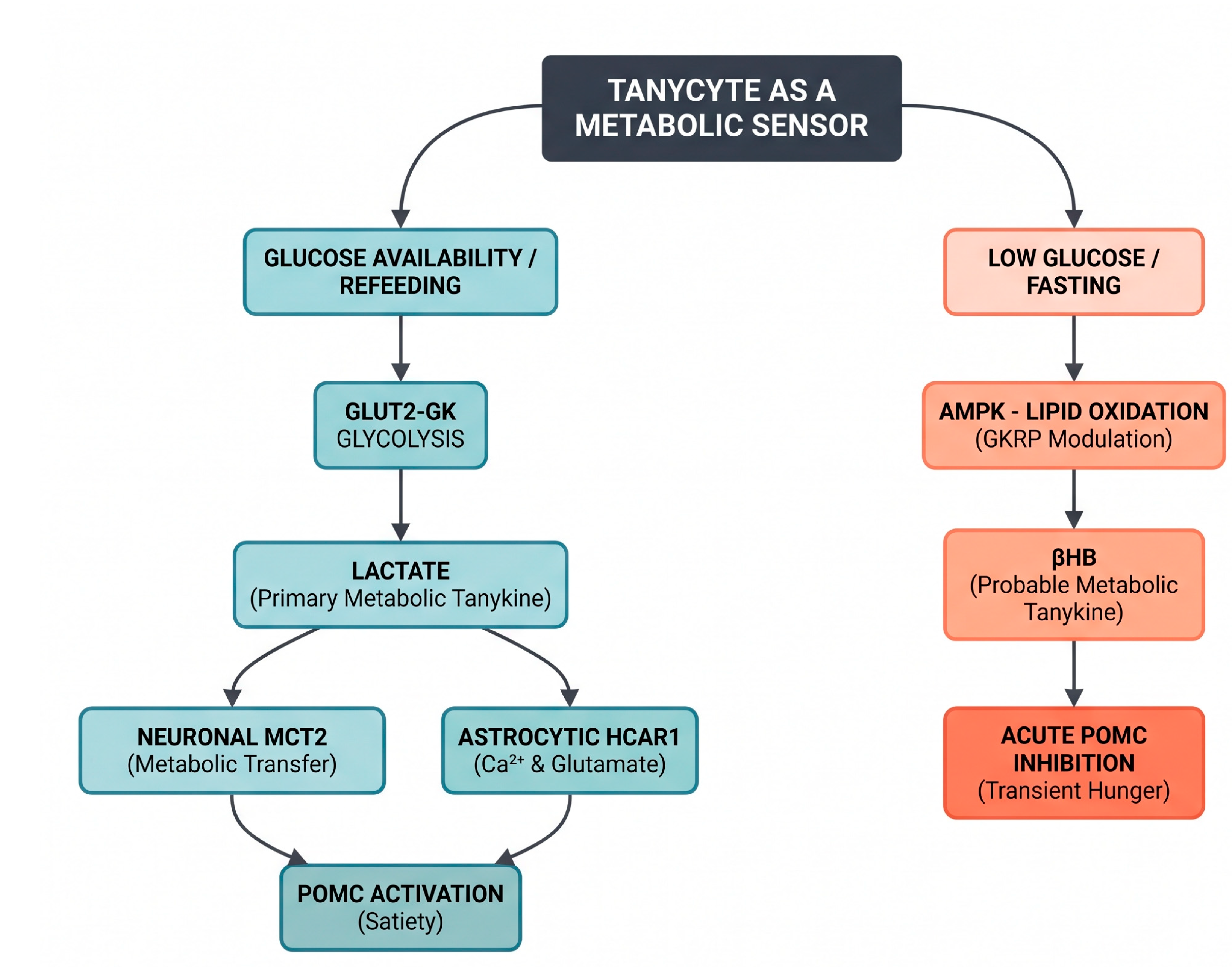
Figure 2.
ATP as a primary tanykine and precursor of secondary adenosine signaling. Left, schematic organization of tanycytes in the mediobasal hypothalamus, illustrating their ventricular cell bodies and processes extending toward hypothalamic neurons and the median eminence vasculature. Right, nutrient- and growth-factor-dependent pathways regulating tanycytic ATP release. Glucose uptake and metabolism increase intracellular ATP availability and promote connexin 43 (Cx43) hemichannel opening. Sweet compounds activate TAS1R2/TAS1R3-dependent signaling, although the ATP release mechanism remains unresolved. Amino acids recruit TAS1R1/TAS1R3 and/or mGluR4, with arginine responses involving pannexin 1 (Panx1) and alanine responses involving calcium homeostasis modulator 1 (CALHM1). FGF2 activates FGF receptor signaling and recruits Cx43-dependent ATP release. Extracellular ATP acts as a primary tanykine, activating tanycytic P2Y1 receptors to amplify intracellular Ca²⁺ signaling; FGF2-evoked purinergic signaling also promotes tanycyte proliferation. ATP additionally serves as a precursor for extracellular adenosine generation: ENTPD1 sequentially hydrolyzes ATP to ADP and AMP, and NT5E/CD73 converts AMP to adenosine. Adenosine acts as a secondary processed tanykine through A1 receptors on AgRP/NPY neurons, reducing ERK1/2 phosphorylation and Agrp/Npy expression. This adenosine pathway was demonstrated under tanycytic TSPO deficiency and high-fat feeding; its contribution under unmanipulated physiological conditions remains unresolved. Channel placement is schematic and does not establish the subcellular localization of each release pathway. Solid arrows indicate experimentally supported relationships, whereas dashed arrows indicate unresolved mechanisms. CSF, cerebrospinal fluid; 3V, third ventricle.
Figure 2.
ATP as a primary tanykine and precursor of secondary adenosine signaling. Left, schematic organization of tanycytes in the mediobasal hypothalamus, illustrating their ventricular cell bodies and processes extending toward hypothalamic neurons and the median eminence vasculature. Right, nutrient- and growth-factor-dependent pathways regulating tanycytic ATP release. Glucose uptake and metabolism increase intracellular ATP availability and promote connexin 43 (Cx43) hemichannel opening. Sweet compounds activate TAS1R2/TAS1R3-dependent signaling, although the ATP release mechanism remains unresolved. Amino acids recruit TAS1R1/TAS1R3 and/or mGluR4, with arginine responses involving pannexin 1 (Panx1) and alanine responses involving calcium homeostasis modulator 1 (CALHM1). FGF2 activates FGF receptor signaling and recruits Cx43-dependent ATP release. Extracellular ATP acts as a primary tanykine, activating tanycytic P2Y1 receptors to amplify intracellular Ca²⁺ signaling; FGF2-evoked purinergic signaling also promotes tanycyte proliferation. ATP additionally serves as a precursor for extracellular adenosine generation: ENTPD1 sequentially hydrolyzes ATP to ADP and AMP, and NT5E/CD73 converts AMP to adenosine. Adenosine acts as a secondary processed tanykine through A1 receptors on AgRP/NPY neurons, reducing ERK1/2 phosphorylation and Agrp/Npy expression. This adenosine pathway was demonstrated under tanycytic TSPO deficiency and high-fat feeding; its contribution under unmanipulated physiological conditions remains unresolved. Channel placement is schematic and does not establish the subcellular localization of each release pathway. Solid arrows indicate experimentally supported relationships, whereas dashed arrows indicate unresolved mechanisms. CSF, cerebrospinal fluid; 3V, third ventricle.
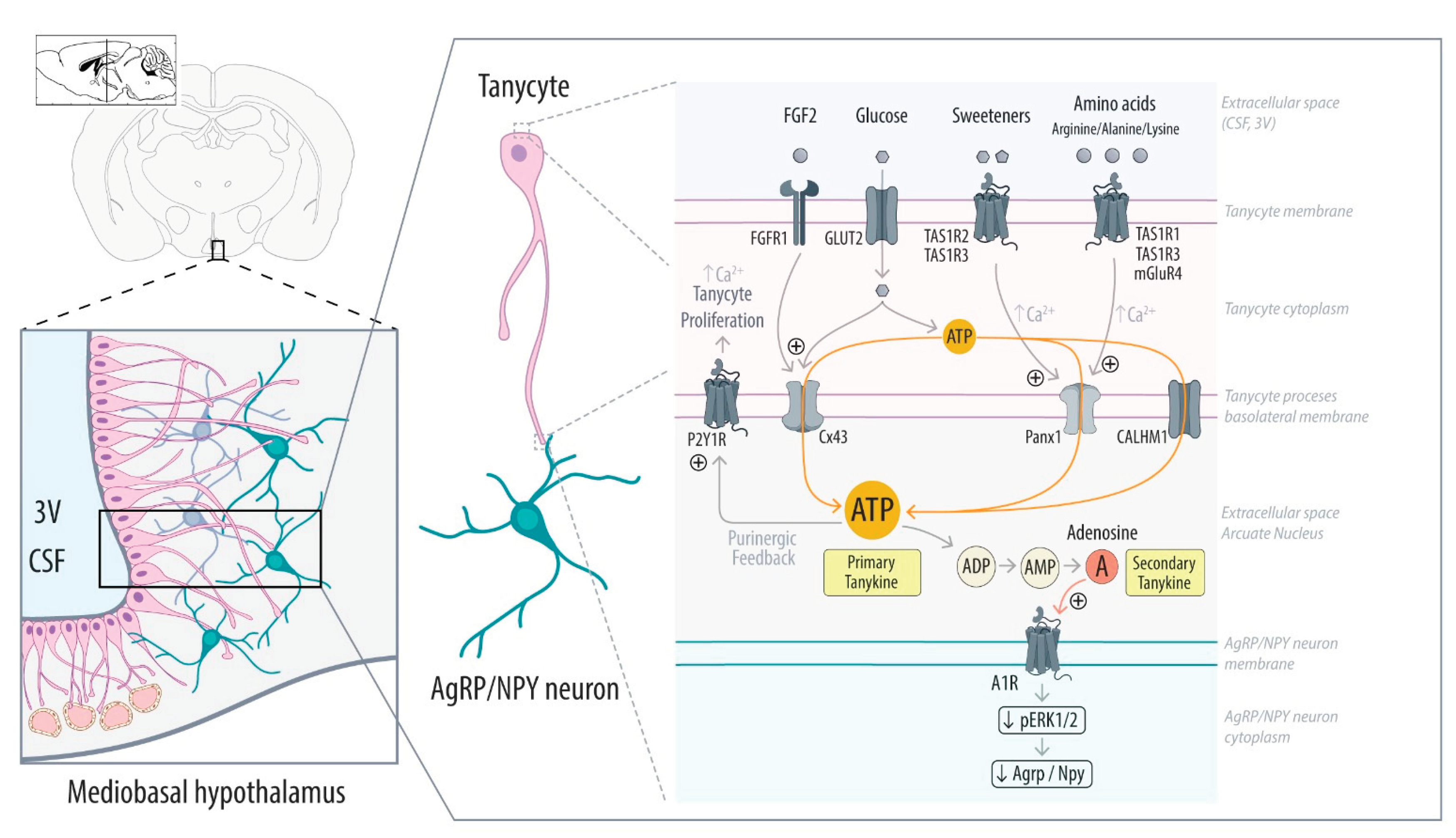

Table 1.
Mechanism-based taxonomy and evidence framework for established, probable, candidate, and disputed tanykines.
Table 1.
Mechanism-based taxonomy and evidence framework for established, probable, candidate, and disputed tanykines.
| Category | Mediator | Biosynthesis / Release / Bioactivation Route | Target Cells & Receptors | Primary Functional Effect | Validation Status | Selected References |
| Primary | Lactate | Glycolytic conversion (GLUT2/GK); export via MCT1/4. | Arcuate POMC neurons (MCT2); Astrocytes (HCAR1). | POMC depolarization; astrocytic glutamate relay; satiety. | Established | [7,15,16] |
| Primary | β-hydroxybutyrate (βHB) | Low-glucose-associated ketogenesis, modulated by AMPK and GKRP; MCT1/4-sensitive export. | POMC neurons; the immediate target-cell receptor remains unresolved. |
POMC hyperpolarization; transient increase in feeding following central administration of exogenous βHB. |
Probable | [17,18] |
| Primary | ATP | Regulated export via Cx43 hemichannels, Panx1, or CALHM1. | Tanycytes (P2Y1); direct neuronal P2X/P2Y targets remain unresolved. | Propagation of intercellular Ca2+ waves; autocrine proliferation. | Established | [13,19,20,56] |
| Primary | Prostaglandin E₂ (PGE2) | COX-1/2-dependent production downstream of ErbB1, estradiol, or IL-1β/NF-κB signaling. | Tanycytes; Median eminence neurovascular unit. | Tanycytic end-feet retraction (GnRH release); inflammatory anorexia. | Established | [9,21,22] |
| Primary | 2-Arachidonoylglycerol (2-AG) | Glucose-dependent on-demand synthesis via DAGLα. | CB1-expressing hypophysiotropic TRH terminals; arcuate neuronal circuits | Retrograde inhibition of TRH release; modulation of thyroid-axis and metabolic function | Probable | [23,24,25] |
| Primary | VEGF-A | Regulated exocytosis (VAMP2-dependent) or fasting induction. | ME Endothelium (VEGFR2); AgRP/TH neurons (VEGFR1) | Capillary fenestration / barrier remodeling; heat-induced hypophagia. | Established | [10,26] |
| Primary | TGF-β1 | Sequential release downstream of erbB1/PGE2 signaling. | Tanycytes (autocrine). | MMP-dependent process retraction in median eminence. | Probable | [21] |
| Secondary | Adenosine | Extracellular cleavage of tanycytic ATP by ENTPD1 and NT5E. | Arcuate AgRP/NPY neurons (A1 receptor). | Inhibition of ERK phosphorylation; Agrp/Npy suppression. | Established, context-dependent | [27] |
| Bioactivated | Triiodothyronine (T3) | MCT8 uptake of T4 /DIO2 conversion (USP33-stabilized). | Hypophysiotropic TRH neurons and other thyroid-hormone-responsive hypothalamic cells. | TRH/TSH negative feedback; photoperiodic adaptations. | Established bioactivation; export route unresolved | [28,29,30] |
| Candidate / Disputed | FGF21 | Fasting / lipid stimulation (disputed local transcription). | Putative CNS targets; canonical FGF21 signaling requires FGFR1c/β-Klotho. | Central regulation of energy expenditure (source controversial). | Disputed | [12,31,32] |
Disclaimer/Publisher’s Note: The statements, opinions and data contained in all publications are solely those of the individual author(s) and contributor(s) and not of MDPI and/or the editor(s). MDPI and/or the editor(s) disclaim responsibility for any injury to people or property resulting from any ideas, methods, instructions or products referred to in the content. |
© 2026 by the authors. Licensee MDPI, Basel, Switzerland. This article is an open access article distributed under the terms and conditions of the Creative Commons Attribution (CC BY) license.
Copyright: This open access article is published under a Creative Commons CC BY 4.0 license, which permit the free download, distribution, and reuse, provided that the author and preprint are cited in any reuse.