

Submitted:
23 October 2026
Posted:
26 October 2026
You are already at the latest version
Abstract
Background and Clinical Significance: Testicular germ cell tumors (GCTs) represent the most common solid malignancy in young adult males, with a rising incidence in industrialized countries. Postpubertal-type mixed GCTs, composed of more than one invasive germ cell neoplasm—seminomatous and/or non-seminomatous—intermingled in a single tumoral nodule, reflect the pluripotential differentiation capacity of their common precursor, the Germ Cell Neoplasia In Situ (GCNIS); Case Presentation: A 29-year-old man presenting with painless right testicular nodule of recent onset was found to have a solid hypoechoic lesion suspicious for malignancy in testicular ultrasonography. Serum tumor markers showed marked elevation of β-human chorionic gonadotropin levels. Radical inguinal orchiectomy demonstrated a postpubertal-type mixed germ cell tumor composed of five distinct histological components: seminoma, embryonal carcinoma, yolk sac tumor, choriocarcinoma, and teratoma, associated with adjacent GCNIS; Conclusions: This exceptional case illustrates the remarkable malignant transformative potential of GCNIS, a clinically silent precursor capable of differentiating into multiple invasive tumor phenotypes. Furthermore, it underscores the importance of meticulous histopathological assessment to identify all components, as accurate quantification directly impacts risk stratification, surveillance strategies, and therapeutic decision-making.
Keywords:
choriocarcinoma
; embryonal carcinoma
; germ cell neoplasia in situ
; mixed testicular germ cell tumor
; seminoma
; teratoma
; uropathology
; yolk sac tumor
1. Introduction and Clinical Significance
Testicular cancer is the most common solid malignancy in adolescent and young adult males, particularly between 15 and 40 years of age. Its incidence has increased over recent decades with more than 72.000 new cases globally each year, especially in industrialized countries, suggesting a relevant role for environmental, endocrine, and genetic factors in its pathogenesis [1,2]. Although it accounts for approximately 1% of all male malignancies, it is responsible for a disproportionate number of life-years lost, as it predominantly affects individuals in their reproductive years [3].
Germ cell tumors (GCTs) account for 90–95% of testicular tumors and are generally divided into seminomatous and non-seminomatous categories, based on their histopathological characteristics and clinical behavior. This heterogeneous group of histological subtypes derived from a common precursor —the germ cell neoplasia in situ (GCNIS)— may coexist within a single lesion, giving rise to the so-called postpubertal-type mixed germ cell tumor (PTMGCT), which represent approximately 30–40% of all GCTs [4,5].
Accurate identification and quantification of all histological components in a PTMGCT is not merely of academic interest but directly influences staging, risk stratification, and therapeutic decision-making. Tumors composed by five distinct histological components are exceptionally rare, accounting for <1% of all testicular GCTs [6]. In this context, we report an unusual case of a PTMGCT composed of seminoma, embryonal carcinoma, teratoma, yolk sac tumor, and choriocarcinoma in a 29-year-old patient.
2. Case Presentation
A 29-year-old man with no relevant medical history other than seasonal asthma and tobacco smoking presented with a painless nodule in the upper pole of the right testis, incidentally detected by the patient. Physical examination revealed increased consistency in the upper region of the right testis, without signs of local inflammation or involvement of the epididymis or spermatic cord. Examination of the contralateral testis was normal.
Scrotal ultrasonography demonstrated a solid, heterogeneous, hypoechoic lesion measuring approximately 2 cm in diameter in the upper pole of the right testis, with internal vascularity on color Doppler imaging (Figure 1). No sonographic evidence of epididymal or spermatic cord involvement was observed. A small adjacent satellite nodule measuring approximately 3 mm was also identified.
Serum tumor markers showed elevated β-human chorionic gonadotropin (β-hCG) levels (20.3 mIU/mL), with alpha-fetoprotein (AFP; 7.5 ng/mL) and lactate dehydrogenase (LDH; 165 U/L) within normal limits. Computed tomography of the chest, abdomen, and pelvis revealed no evidence of distant metastases or retroperitoneal lymphadenopathy, consistent with clinical stage I disease.
The patient underwent right radical inguinal orchiectomy with placement of a testicular prosthesis. Gross examination of the surgical specimen revealed a testis measuring 4.2 × 3 cm attached to an 8-cm long spermatic cord. Sectioning disclosed a firm, whitish nodular mass with focal hemorrhagic areas measuring 2 × 1.8 cm, abutting but not invading the tunica albuginea (Figure 2). Notably, a 3-mm satellite nodule was also observed.
Histopathological analysis of the orchiectomy specimen demonstrated a postpubertal-type mixed GCT composed of five distinct neoplasms within a single nodular lesion (Figure 3). Each component was morphologically and immunophenotypically consistent with its pure counterpart, and their relative proportions were estimated based on the total tumor area as follows:
- Embryonal carcinoma (60%): solid growth pattern with pleomorphic cells, prominent nuclei, and high mitotic activity; positive for CD30 and pancytokeratin (AE1/AE3).
- Postpubertal mature and immature teratoma (15%): tissues derived from all three germ layers, including keratinizing squamous epithelium, immature cartilage, and smooth muscle.
- Postpubertal yolk sac tumor (10%): glomeruloid structures consistent with Schiller–Duval bodies; AFP-positive.
- Classic seminoma (10%): large cells with clear cytoplasm, round nuclei with prominent nucleoli, and lymphocyte-rich stroma; tumor cells expressed CD117 (c-KIT) and OCT3/4.
- Choriocarcinoma (5%): hemorrhagic areas containing syncytiotrophoblastic and cytotrophoblastic cells with β-hCG expression.
Adjacent testicular parenchyma, including the 3-mm satellite nodule, demonstrated germ cell neoplasia in situ (GCNIS). No lymphovascular invasion or involvement of the epididymis or rete testis was identified.
Postoperative follow-up demonstrated progressive normalization of serum tumor markers (β-hCG <2 mIU/mL, AFP 3.4 ng/mL, LDH 145 U/L) and no evidence of local recurrence or distant disease after 18 months of surveillance.
3. Discussion
Most testicular GCT arise from primordial germ cells (gonocytes) that remain developmentally arrested and fail to differentiate into spermatogonia due to a combination of genetic susceptibility and prenatal environmental exposures (e.g., maternal exposure to estrogen-like compounds or organochlorine substances). These factors lead to dysfunction of androgen or anti-Müllerian hormone signaling pathways, resulting in the persistence of a latent totipotent developmental potential in these cells residing within the spermatogonial niche of the seminiferous tubules [7]. Epigenetic dysregulation of these cells results in transformation into germ cell neoplasia in situ (GCNIS), also known as intratubular germ cell neoplasia, which represents the common precursor of both seminomatous and non-seminomatous postpubertal-type GCT [8,9].
GCNIS may remain clinically silent for years until the acquisition of additional genetic abnormalities, most notably gain of chromosome 12p (isochromosome i(12p)), that triggers progression to invasive disease [10]. Initial differentiation typically gives rise to seminoma or embryonal carcinoma, with subsequent potential transformation into choriocarcinoma, postpubertal yolk sac tumor, or postpubertal teratoma [7,8]. Teratomas are composed of at least two embryonic germ layers —ectoderm, mesoderm, and/or endoderm— and may, in rare instances, undergo transformation into a secondary somatic-type malignancy (i.e., the development of a non–germ cell malignant neoplasm such as sarcoma, adenocarcinoma, or embryonic-type neuroectodermal tumor arising within a GCT)[5,10]. Therefore, presence of GCNIS in adjacent parenchyma supports a multifocal progressive transformation process, and justifies close surveillance of the contralateral testis in young patients at increased risk [11].
Mixed GCT may contain several components in varying proportions. However, the individual elements are histologically and immunophenotypically identical to their pure counterparts [4]. The recognition of extreme histological heterogeneity within a single PTMGCT is of paramount importance, as each component carries distinct biological behavior, metastatic potential, and response to therapy. For instance, tumors dominated by aggressive non-seminomatous elements such as embryonal carcinoma or choriocarcinoma are associated with a higher risk of early systemic dissemination, whereas teratomatous components may confer resistance to chemotherapy and require surgical management [7,10]. Nevertheless, the amount of each component also provides critical prognostic information beyond mere histological presence, as some studies have found that a ≤5% choriocarcinoma component may not significantly worsen prognosis [12].
Early diagnosis of testicular GCT relies on clinical vigilance in the evaluation of any testicular abnormality, even in the absence of pain. Diagnostic assessment includes physical examination, scrotal ultrasonography, and measurement of serum tumor markers such as AFP, β-hCG, and LDH, which are particularly valuable for post-treatment monitoring [13]. AFP is elevated in approximately 50-70% of non-seminomatous germ cell tumors (NSGCT), almost exclusively reflecting the presence of yolk sac tumor differentiation, while it is never elevated in pure seminoma [14]. β-hCG is increased in approximately 40–60% of NSGCT and in 10–30% of seminomas, typically due to syncytiotrophoblastic elements [15]. LDH, although associated with tumor burden, shows low specificity and may be elevated in a wide range of non-malignant conditions [15]. At least 60% of patients with testicular cancer present with elevation of at least one serum marker at diagnosis [15]; however, the unaltered values of AFP in our case can be explained by the limited proportion of yolk sac tumor (approximately 10%), as AFP secretion correlates more closely with the volume of AFP-producing cells rather than their mere presence, highlighting that normal markers do not exclude malignancy [16]. Overall, while marker elevation patterns often reflect the dominant tumor component, definitive classification and risk stratification requires a thorough histopathological and immunohistochemical evaluation [5,9] (Table 1).
From a clinical perspective, radical inguinal orchiectomy remains the cornerstone of initial management, allowing definitive diagnosis and pathological staging. Afterwards, follow-up with serial serum tumor marker assessment and imaging studies are essential. In patients with stage I disease, choice between subsequent active surveillance or adjuvant treatment should be based on up-to-date clinical guidelines, but also on multidisciplinary discussion and patient preference [13]. Mixed tumors with a dominant non-seminomatous component may warrant a more aggressive therapeutic approach depending on patient risk factors (e.g., lymphovascular invasion, direct invasion of the spermatic cord or scrotum), as up to 30% of patients with stage I NSGCT may relapse during the first two years (early relapses) [17]. In stage I NSGCT patients without risk factors and postoperative normalization of tumor markers, prognosis is generally favorable and active surveillance is recommended as an appropriate management [13,17].
4. Conclusions
Postpubertal mixed testicular germ cell tumors represent a biologically complex entity demanding a comprehensive clinicopathological approach that combines clinical evaluation, testicular ultrasonography, serum tumor markers, and thorough histopathological examination. This case, featuring the simultaneous presence of seminoma, embryonal carcinoma, choriocarcinoma, yolk sac tumor, and teratoma, illustrates the full malignant differentiation spectrum of GCNIS and reinforces its role as a precursor lesion and marker of bilateral disease risk, supporting careful follow-up of the contralateral testis. Accurate identification and quantification of each tumoral component are essential not only for prognostic assessment but also for guiding surveillance intensity and therapeutic decision-making.
Author Contributions
Conceptualization and methodology: A.A.-S., F.M.-D. and F.G.-M.; formal analysis and investigation: A.A.-S., and F.M.-D.; writing—original draft preparation: A.A.-S.; writing—review and editing: all authors; visualization: A.A.-S. and F.M.-D.; supervision: F.G.-M. All authors have read and agreed to the published version of the manuscript.
Funding
This research received no external funding.
Institutional Review Board Statement
Ethical review and approval were not required for this single-patient case report in accordance with local legislation and institutional requirements for case reports based on fully anonymized clinical information. The study was conducted in accordance with the principles of the Declaration of Helsinki.
Informed Consent Statement
Written informed consent was obtained from the subject involved in the study.
Data Availability Statement
All data involved in this study are included in this published article. Additional anonymized pictures are available from the corresponding author upon reasonable request.
Conflicts of Interest
The authors declare no competing interests.
Abbreviations
The following abbreviations are used in this manuscript:
| AFP | Alpha-fetoprotein |
| β-hCG | Beta–human chorionic gonadotropin |
| CC | Choriocarcinoma |
| CK7 | Cytokeratin 7 |
| D2-40 | Podoplanin (D2-40 antibody) |
| EC | Embryonal carcinoma |
| GCNIS | Germ cell neoplasia in situ |
| GCT | Germ cell tumor |
| LDH | Lactate dehydrogenase |
| NSGCT | Non-seminomatous germ cell tumor |
| OCT3/4 | Octamer-binding transcription factor 3/4 |
| PLAP | Placental alkaline phosphatase |
| PTMGCT | Postpubertal-type mixed germ cell tumor |
| SALL4 | Sal-like protein 4 |
| WHO | World Health Organization |
| YST | Yolk sac tumor |
References
- Bray, F.; Laversanne, M.; Sung, H.; Ferlay, J.; Siegel, R.L.; Soerjomataram, I.; Jemal, A. Global Cancer Statistics 2022: GLOBOCAN Estimates of Incidence and Mortality Worldwide for 36 Cancers in 185 Countries. CA A Cancer J. Clin. 2024, 74, 229–263. [Google Scholar] [CrossRef] [PubMed]
- McHugh, D.J.; Gleeson, J.P.; Feldman, D.R. Testicular Cancer in 2023: Current Status and Recent Progress. CA A Cancer J. Clin. 2024, 74, 167–186. [Google Scholar] [CrossRef] [PubMed]
- Yu, S.; Guo, Z.; Qiu, Z.; Wang, L.; Chen, X.; Xuan, F. Global Burden and Trends of Testicular Cancer in Adolescents and Young Adults from 1990 to 2021, with Predictions to 2035. Sci. Rep. 2024, 14, 31787. [Google Scholar] [CrossRef]
- WHO Classification of Tumors. Urinary and Male Genital Tumors, 5th ed.; International Agency for Research on Cancer (IARC): Lyon (France), 2022; ISBN 978-92-832-4512-4.
- Ulbright, T.M.; Kao, C.-S.; Williamson, S.R.; Idrees, M.T. Tumors and Tumor-Like Lesions of the Testis and Adjacent Tissues; American Registry of Pathology, 2022; ISBN 978-1-933477-22-0. [Google Scholar]
- Stang, A.; Rusner, C.; Trabert, B.; Oosterhuis, J.W.; McGlynn, K.A.; Heidinger, O. Incidence of Testicular Tumor Subtypes According to the Updated WHO Classification, North Rhine-Westphalia, Germany 2008–2013. Andrology 2019, 7, 402–407. [Google Scholar] [CrossRef] [PubMed]
- Singla, N.; Bagrodia, A.; Baraban, E.; Fankhauser, C.D.; Ged, Y.M.A. Testicular Germ Cell Tumors: A Review. JAMA 2025, 333, 793–803. [Google Scholar] [CrossRef] [PubMed]
- Fichtner, A.; Nettersheim, D.; Bremmer, F. Pathogenesis and Pathobiology of Testicular Germ Cell Tumours: A View from a Developmental Biological Perspective with Guidelines for Pathological Diagnostics. Histopathology 2024, 85, 701–715. [Google Scholar] [CrossRef] [PubMed]
- Ortuño Pacheco, G.; Martínez Diaz, F.; Pastor Quirante, F. Lecciones de Anatomía Patológica General Para Estudiantes de Medicina y Odontología; 3a; Diego Marín (DM): Murcia, 2008; ISBN 978-84-8425-678-6. [Google Scholar]
- Katabathina, V.S.; Vargas-Zapata, D.; Monge, R.A.; Nazarullah, A.; Ganeshan, D.; Tammisetti, V.; Prasad, S.R. Testicular Germ Cell Tumors: Classification, Pathologic Features, Imaging Findings, and Management. RadioGraphics 2021, 41, 1698–1716. [Google Scholar] [CrossRef] [PubMed]
- Dieckmann, K.-P.; Kulejewski, M.; Pichlmeier, U.; Loy, V. Diagnosis of Contralateral Testicular Intraepithelial Neoplasia (TIN) in Patients with Testicular Germ Cell Cancer: Systematic Two-Site Biopsies Are More Sensitive Than a Single Random Biopsy. Eur. Urol. 2007, 51, 175–185. [Google Scholar] [CrossRef] [PubMed]
- Hassan, O.; Epstein, J.I. The Clinical Significance of a Small Component of Choriocarcinoma in Testicular Mixed Germ Cell Tumor (MGCT). Am. J. Surg. Pathol. 2018, 42, 1113–1120. [Google Scholar] [CrossRef] [PubMed]
- Testicular Cancer— NCCN 2026 Guidelines. Available online: https://www.nccn.org/guidelines/guidelines-detail?category=1&id=1468 (accessed on 4 January 2026).
- Sykes, J.; Kaldany, A.; Jang, T.L. Current and Evolving Biomarkers in the Diagnosis and Management of Testicular Germ Cell Tumors. J. Clin. Med. 2024, 13, 7448. [Google Scholar] [CrossRef] [PubMed]
- Mir, M.C.; Pavan, N.; Gonzalgo, M.L. Current Clinical Applications of Testicular Cancer Biomarkers. Urol. Clin. N. Am. 2016, 43, 119–125. [Google Scholar] [CrossRef] [PubMed]
- Ma, C.; Jin, Y.; Wang, Y.; Xu, H.; Zhang, J. Beyond Liver Cancer, More Application Scenarios for Alpha-Fetoprotein in Clinical Practice. Front. Oncol. 2023, 13. [Google Scholar] [CrossRef] [PubMed]
- Daugaard, G.; Gundgaard, M.G.; Mortensen, M.S.; Agerbæk, M.; Holm, N.V.; Rørth, M.; von der Maase, H.; Christensen, I.J.; Lauritsen, J. Surveillance for Stage I Nonseminoma Testicular Cancer: Outcomes and Long-Term Follow-Up in a Population-Based Cohort. J. Clin. Oncol. 2014. [Google Scholar] [CrossRef] [PubMed]
Figure 1.
Testicular ultrasonography. (A) B-mode ultrasound showing a heterogeneous hypoechoic mass within the right testis. (B) Color Doppler imaging demonstrating increased internal vascularity, findings suspicious for a testicular germ cell tumor.
Figure 1.
Testicular ultrasonography. (A) B-mode ultrasound showing a heterogeneous hypoechoic mass within the right testis. (B) Color Doppler imaging demonstrating increased internal vascularity, findings suspicious for a testicular germ cell tumor.
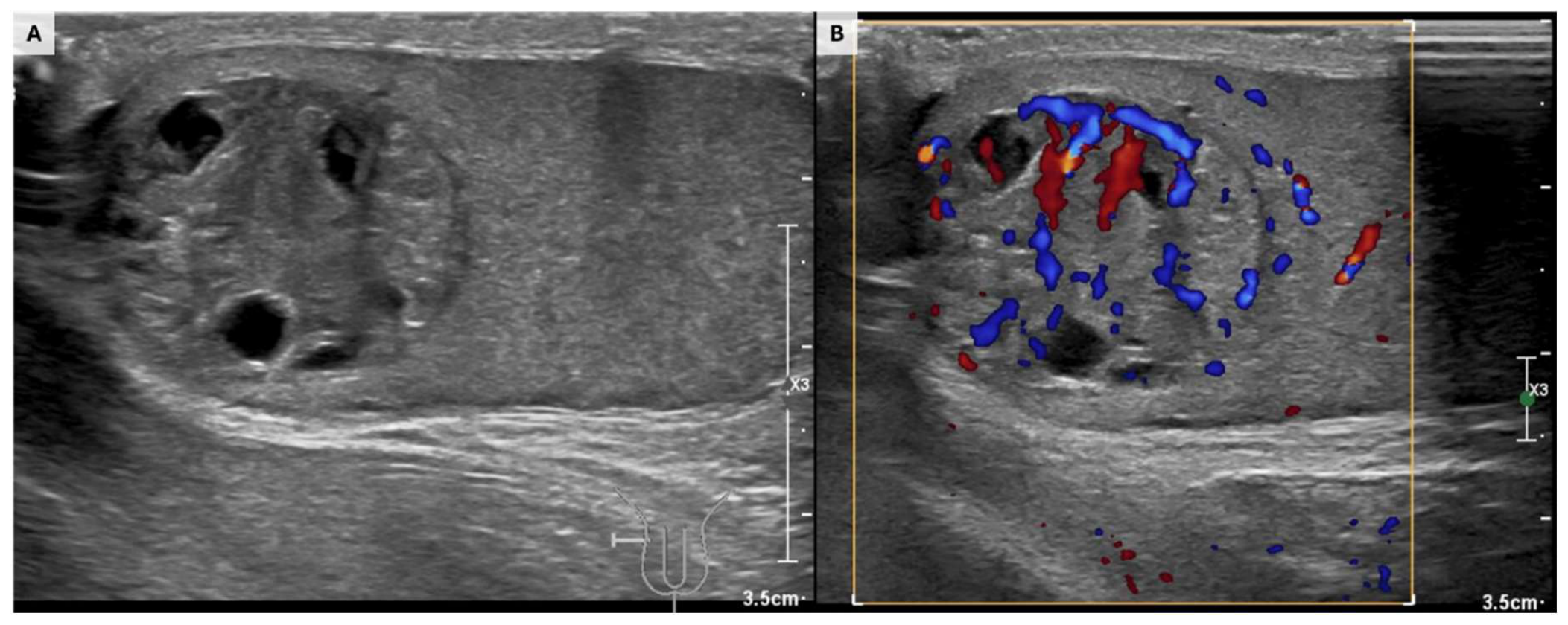
Figure 2.
Gross pathological examination of the right testis after radical orchiectomy. (A) Sagittal section showing a main whitish, heterogeneous nodular mass with areas of hemorrhage and central necrosis located in the upper pole of the testis, with an adjacent smaller whitish satellite nodule (blue arrow). (B, C) Pearly white areas with central necrosis consistent with embryonal carcinoma, cystic areas corresponding to teratoma, hemorrhagic regions suggestive of a choriocarcinoma component, and a gray-yellow area compatible with yolk sac tumor.
Figure 2.
Gross pathological examination of the right testis after radical orchiectomy. (A) Sagittal section showing a main whitish, heterogeneous nodular mass with areas of hemorrhage and central necrosis located in the upper pole of the testis, with an adjacent smaller whitish satellite nodule (blue arrow). (B, C) Pearly white areas with central necrosis consistent with embryonal carcinoma, cystic areas corresponding to teratoma, hemorrhagic regions suggestive of a choriocarcinoma component, and a gray-yellow area compatible with yolk sac tumor.
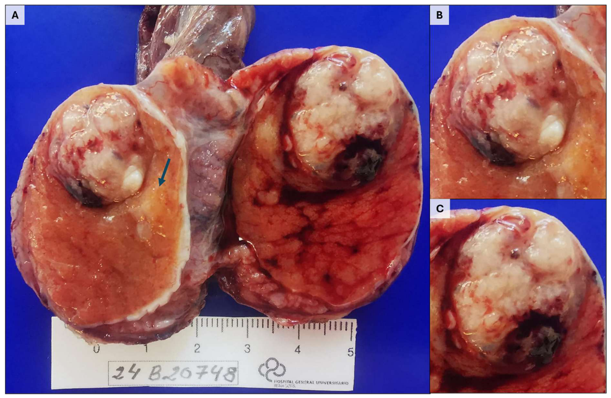
Figure 3.
Morphological heterogeneity of a mixed germ cell tumor within a single lesion. (A) Yolk sac tumor with reticular pattern, palisading tumor cells, and Schiller–Duval bodies, positive for alpha-fetoprotein (AFP). (B) Classic seminoma composed of large cells with clear cytoplasm and prominent nucleoli arranged in nests within a lymphocyte-rich stroma, expressing c-KIT (CD117). (C) Embryonal carcinoma with solid growth pattern, pleomorphic cells, hyperchromatic nuclei, and frequent mitotic figures, showing diffuse membranous and cytoplasmic positivity for CD30. (D) Choriocarcinoma with multinucleated syncytiotrophoblastic cells in a hemorrhagic background, showing strong positivity for β-human chorionic gonadotropin (β-hCG). (E) Teratoma components: (E.1) immature cartilaginous tissue; (E.2) keratinizing squamous epithelium; (E.3, E.4) mesenchymal tissue with smooth muscle differentiation, positive for smooth muscle actin. (F) Adjacent germ cell neoplasia in situ (GCNIS) characterized by enlarged atypical germ cells with clear cytoplasm and centrally placed nuclei occupying the seminiferous tubules. (G) Immunohistochemical staining for c-KIT (CD117) highlighting the cytoplasmic and membranous positivity of GCNIS cells along the basal compartment of seminiferous tubules.
Figure 3.
Morphological heterogeneity of a mixed germ cell tumor within a single lesion. (A) Yolk sac tumor with reticular pattern, palisading tumor cells, and Schiller–Duval bodies, positive for alpha-fetoprotein (AFP). (B) Classic seminoma composed of large cells with clear cytoplasm and prominent nucleoli arranged in nests within a lymphocyte-rich stroma, expressing c-KIT (CD117). (C) Embryonal carcinoma with solid growth pattern, pleomorphic cells, hyperchromatic nuclei, and frequent mitotic figures, showing diffuse membranous and cytoplasmic positivity for CD30. (D) Choriocarcinoma with multinucleated syncytiotrophoblastic cells in a hemorrhagic background, showing strong positivity for β-human chorionic gonadotropin (β-hCG). (E) Teratoma components: (E.1) immature cartilaginous tissue; (E.2) keratinizing squamous epithelium; (E.3, E.4) mesenchymal tissue with smooth muscle differentiation, positive for smooth muscle actin. (F) Adjacent germ cell neoplasia in situ (GCNIS) characterized by enlarged atypical germ cells with clear cytoplasm and centrally placed nuclei occupying the seminiferous tubules. (G) Immunohistochemical staining for c-KIT (CD117) highlighting the cytoplasmic and membranous positivity of GCNIS cells along the basal compartment of seminiferous tubules.
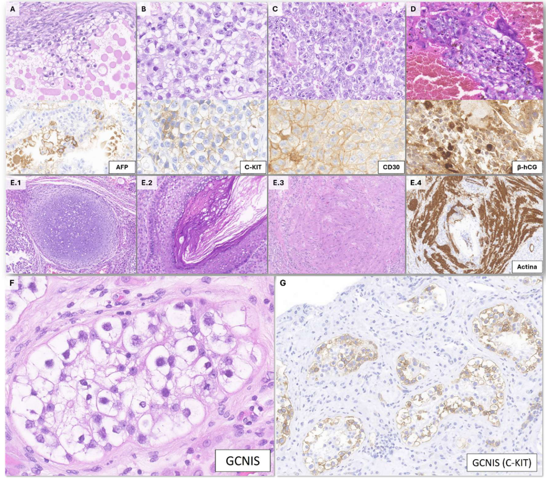

Table 1.
Association of serum tumor markers and immunohistochemical markers with the different subtypes of testicular germ cell tumors and germ cell neoplasia in situ (GCNIS), including their main gross and microscopic features.
Table 1.
Association of serum tumor markers and immunohistochemical markers with the different subtypes of testicular germ cell tumors and germ cell neoplasia in situ (GCNIS), including their main gross and microscopic features.
| GCNIS | Seminoma | Teratoma | EC | YST | CC | |
|---|---|---|---|---|---|---|
| Gross appearance | Not visible | White-gray, glistening, homogeneous | Solid–cystic, ± hair, cartilage, etc. | Grayish, hemorrhagic, areas of necrosis | Gray-yellow, heterogeneous | Markedly hemorrhagic |
| Microscopic features | Atypical germ cells within seminiferous tubules | Large cells with clear cytoplasm and one or more prominent nucleoli; fibrous septa with lymphocytes ± syncytiotrophoblast | Components derived from the three germ layers | Pleomorphic cells, prominent nucleoli, moderate amphophilic cytoplasm, increased mitotic activity | Schiller–Duval bodies; hyaline globules | Syncytiotrophoblastic and cytotrophoblastic cells |
| Marcadores séricos | Not elevated in the initial phase |
↑ LDH, β-hCG (mild) | Normal | ↑ AFP, ↑ β-hCG, ↑ LDH | ↑ AFP | ↑↑ β-hCG |
| CD30 | - | - | - | + | - | - |
|
CD117 (c-Kit) |
+ | + | - | - | - | - |
| OCT3/4 | + | + | - | + | - | - |
| AE1/AE3 | + | - | + (epithelial components) | + | + | - |
| CK7 | ± | - | - | - | - | + (syncytiotrophoblast) |
| P63 | - | - | - | - | - | + (trophoblast) |
| D2-40 | + | + | - | ± | - | - |
| AFP | - | - | - | - | ± | - |
| β-hCG | - | - | - | - | - | + |
| Inhibina | - | - | - | - | - | + |
| Glypican-3 | - | - | ± (immature elements) | - | + | + |
| SALL4 | + | + | + | + | + | |
| GATA3 | - | - | + | - | + | + |
| PLAP | + | + | - | + | ± | ± |
Notes: “+” indicates positivity in ≥80% of cases; “±” positivity in 20–80% of cases; “–” positivity in <20% of cases. Serum tumor markers may be normal despite the presence of the corresponding histological component, particularly when the proportion of marker-producing cells is limited. Abbreviations: EC, embryonal carcinoma; YST, yolk sac tumor; CC, choriocarcinoma. Immunohistochemical markers: SALL4, stem cell marker positive in most germ cell tumors; OCT3/4, transcription factor; CD117 (c-KIT), tyrosine kinase receptor; AE1/AE3, pancytokeratin; CK7 and p63, markers useful for identifying trophoblastic differentiation.
Disclaimer/Publisher’s Note: The statements, opinions and data contained in all publications are solely those of the individual author(s) and contributor(s) and not of MDPI and/or the editor(s). MDPI and/or the editor(s) disclaim responsibility for any injury to people or property resulting from any ideas, methods, instructions or products referred to in the content. |
© 2026 by the authors. Licensee MDPI, Basel, Switzerland. This article is an open access article distributed under the terms and conditions of the Creative Commons Attribution (CC BY) license (http://creativecommons.org/licenses/by/4.0/).
Copyright: This open access article is published under a Creative Commons CC BY 4.0 license, which permit the free download, distribution, and reuse, provided that the author and preprint are cited in any reuse.