

Submitted:
23 October 2026
Posted:
26 October 2026
You are already at the latest version
Abstract
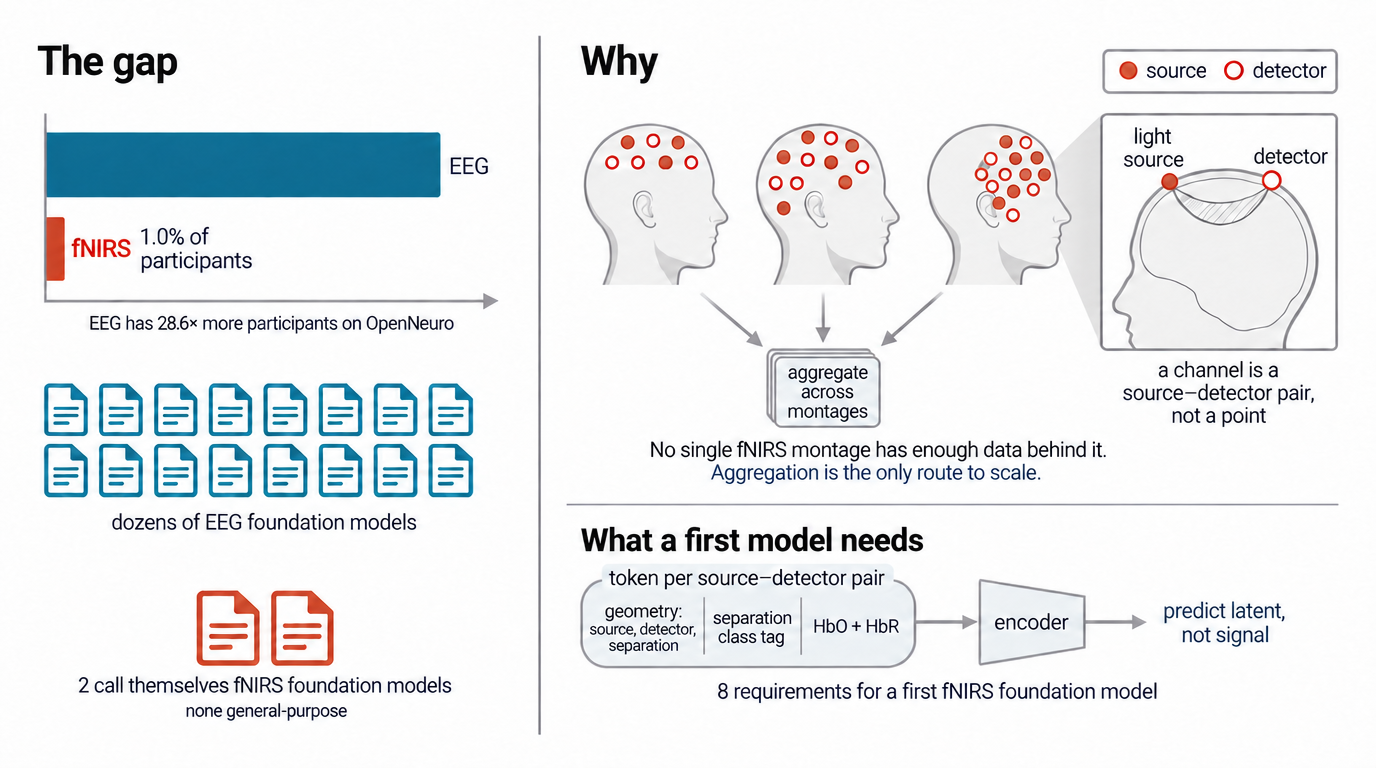
Functional near-infrared spectroscopy (fNIRS) is almost absent from the foundation-model literature. On OpenNeuro, the largest open BIDS-structured neuroimaging repository, fNIRS accounts for 1.0% of participants and 1.6% of datasets, against 28.6 times more participants for EEG. We ask why, and what a first fNIRS foundation model would require. The paper is a review and a roadmap: its contribution is an inventory, a synthesis of evidence from adjacent modalities, and a set of requirements, not new model results.We argue that the barrier is not primarily data structure. The fNIRS data standards already require sensor positions to be recorded, so the laborious step of reconstructing where each sensor sat, which dominates the equivalent EEG models, is largely unnecessary here. The barrier is corpus scale, compounded by the absence of any clinical archive comparable to the Temple University Hospital EEG corpus, and by the recency of NIRS-BIDS, published in 2025 against 2019 for EEG. Because no single fNIRS corpus is large enough, aggregation across heterogeneous montages is the most plausible route to pretraining scale, which makes montage-agnostic architecture a precondition rather than an optimization.Reviewing four EEG foundation models that solve montage heterogeneity, we find that the two available ablations of both components rank spatial aggregation above the choice of positional encoding. Several models without a unifying spatial abstraction fall to near-chance under stringent cross-montage evaluation, mainly when encoders are frozen or tested on layouts they were not trained on. In hemodynamic and multimodal work, the one foundation model borrowed from another domain gave no uplift on fNIRS, while joint pretraining with EEG did. In the one available ablation, though, that benefit tracked the mutual information between modalities and, at the larger shot counts, fell below fNIRS-only pretraining at the lowest level tested. From functional magnetic resonance imaging (fMRI) we take a direction fNIRS has not yet followed: recent work there has moved from signal reconstruction toward latent prediction, because the signal is noisy and off-the-shelf evaluation matters. That rationale may be particularly relevant to fNIRS, where some of the systemic physiological confounds, particularly the Mayer wave, overlap the band in which the hemodynamic signal is analyzed. We chart the nine fNIRS-relevant works we located. We identify three especially consequential properties of fNIRS with no electrophysiological counterpart (source–detector geometry, chromophore choice and hemodynamic timescale) and set out eight requirements for a first fNIRS foundation model, on which work is under way in our laboratory.
Keywords:
functional near-infrared spectroscopy
; foundation models
; self-supervised learning
; representation learning
; optode montage
; transfer learning
1. Why fNIRS Lacks a General-Purpose Foundation Model
1.1. The Claim, and Why It Needs Care
A foundation model is a single large network pretrained once on a broad corpus without task labels, then adapted to many downstream tasks with little labeled data [1]. The approach reorganized natural language processing and computer vision before reaching biomedical signals, and its appeal for neuroimaging is specific: labeled recordings are scarce and expensive, whereas unlabeled ones accumulate, so a representation learned once and reused across tasks is worth more here than in domains where labels are plentiful. Within neuroimaging, however, it has arrived unevenly. Electroencephalography (EEG) has produced dozens [2,3]; functional near-infrared spectroscopy (fNIRS) has produced very few. Two of the works we located describe themselves as fNIRS foundation models, and Section 4 charts these alongside the rest.
fNIRS measures brain activity optically. Near-infrared light is delivered through the scalp, and the amount absorbed along the path to a nearby detector tracks changes in oxygenated and deoxygenated hemoglobin, and so, indirectly, the hemodynamic response to neural activity. It therefore measures the same physiological quantity as functional magnetic resonance imaging (fMRI). The two agree, though not uniformly: in the largest quantitative comparison across cognitive tasks, the mean correlation between the oxygenated-hemoglobin signal and the fMRI BOLD signal was 0.26 across 1,176 channel instances, rising to 0.74 among the most strongly correlated, with oxygenated hemoglobin tracking BOLD more closely than deoxygenated [4]. What fNIRS gives up in depth and spatial resolution it returns in portability. It is safe for repeated and prolonged use, tolerant of movement, and can be battery-powered and worn, which has made it the modality of choice where a scanner cannot go: infant and developmental research, psychiatry, stroke and neurorehabilitation, brain–computer interfaces, hyperscanning of interacting people, and recordings made while participants walk outdoors or drive [5]. That breadth is why its absence from the foundation-model literature is worth explaining.
A claim about absence is indistinguishable from a failure to look. This paper is a critical review and roadmap rather than a meta-analysis, and Section 4.1 states how we searched, what we found and what the search is likely to have missed.
Three independent lines of evidence corroborate the scarcity. Jung, Lee and An [6], introducing their own fNIRS model recently, report that no foundation model designed for fNIRS preceded it and benchmark against EEG and multimodal models instead; a PRISMA-based survey of 64 brain-signal foundation-model studies [3] names fNIRS in its Boolean query yet charts no fNIRS model at all; and a 2026 mini-review of inner-speech decoding across four modalities [7] gives fNIRS a single row of its comparison table.
1.2. The Corpus, Measured
OpenNeuro [8] publishes per-modality participant and dataset counts, giving a single-source measurement made identically across modalities on the same day, 11 August 2026 (Figure 1).
Against a platform total of 86,508 participants across 1,855 datasets, NIRS accounts for 1.0% of participants and 1.6% of datasets. EEG has 28.6 times more participants. The openfnirs community database [9], which indexes datasets on NITRC, OSF, Zenodo and figshare as well as OpenNeuro, adds 16 datasets totaling 255 participants where participant counts are reported. OpenNeuro’s 886 NIRS participants (Figure 1A) and these 255 sum to 1,141 without deduplication, and fewer once the overlap between the two venues is removed.
For comparison, REVE [2], the largest EEG pretraining effort published to date, assembled 61,415 hours from 24,274 subjects across 92 sources. The entire NIRS holdings of the largest open neuroimaging repository amount to under 4% of the subjects behind a single EEG foundation model.
Converting the curated database to channel-hours, the unit the EEG literature reports, sharpens rather than softens this. Seven of the sixteen entries report subjects, duration and channel count together (Table 1):
The comparison that follows is with electrophysiology, where channel-hours originate. BrainBERT [10], an intracranial model, has the smallest pretraining corpus among the ten EEG and intracranial models reviewed by Kuruppu et al. [11], at 4.5k channel-hours; LaBraM [12] used 80k, the Temple University Hospital EEG corpus holds 541k and BrainWave 878k. The apparent proximity of the fNIRS total to BrainBERT is misleading. One high-density diffuse optical tomography (HD-DOT) dataset contributes 57% of it from nine subjects and ninety minutes of recording. HD-DOT places optodes in a dense overlapping grid so that images can be reconstructed rather than individual channels sampled, which yields hundreds or thousands of channels from a handful of participants; removing that one dataset leaves roughly 2,100 channel-hours. Channel-hours flatter fNIRS because channel density inflates the figure without adding subjects or time.
We therefore report subjects, hours and channels separately throughout, and recommend that others do the same. The consequences of not doing so are visible in the literature. EFRM’s pretraining corpus is imbalanced about 7:1 in favor of EEG counted by recordings, the authors’ figure, about 6:1 by participants, but only about 2.4:1 by hours, so which figure is quoted materially changes how severe the imbalance appears. Hours can also overstate diversity: 312 of EFRM’s 364 fNIRS hours come from a single 19-participant neurofeedback dataset.
A caveat in the other direction deserves equal weight. Not every fNIRS dataset is short: a multi-session NIRS brain–computer interface (BCI) stroke dataset [13] comprises 15 participants but 237 sessions, over 50 hours and 5,296 trials, averaging 3.3 hours per participant. Longitudinal and clinical BCI protocols carry far more recording time per subject than the task-block designs that dominate the curated database, so the total that can be assembled may exceed what participant counts alone suggest. Inter-individual diversity remains a separate axis that hours cannot substitute for.
1.3. There Is No Temple University Hospital Corpus for fNIRS
Volume alone understates the problem. The EEG literature does not rest on volume spread evenly across sources but rather, disproportionately, on a single corpus. The Temple University Hospital (TUH) EEG corpus, 541k channel-hours from a clinical archive, is the pretraining substrate [14] for EEG foundation models including Neuro-GPT [15], EEGFormer [16], NeuroLM [17], FoME [18] and BrainWave [19], among others. Two subsets derived from it [14], TUAB (abnormality detection) and TUEV (event classification), have become the field’s de facto benchmarks. Even REVE, the most diverse corpus in this review, draws 62% of its subjects and 44% of its hours from TUH.
Nothing comparable exists for fNIRS, and nothing comparable is in prospect: there is no clinical fNIRS archive because fNIRS has no routine clinical indication generating incidental data at scale. The public landscape is instead many small, well-documented, task-specific datasets; the Tufts fNIRS2MW mental-workload dataset [20] reports 68 participants and describes itself as the largest for that task by a factor of 2.5.
This has a consequence that organizes the rest of this paper:
Because no single fNIRS corpus is large enough to pretrain on, aggregation across many small, heterogeneous datasets is currently the most plausible route to pretraining scale. And aggregation across heterogeneous montages is exactly what the architectural developments of Section 2 make possible.
The corpus problem and the architecture question are the same problem seen from two directions. A fixed-montage fNIRS foundation model cannot be trained at scale, because no single montage has enough data behind it.
1.4. Standardization Arrived Recently
A timing explanation accounts for much of the lag without appeal to community size. The Brain Imaging Data Structure (BIDS) [21] extension for near-infrared spectroscopy [22], which defines how fNIRS datasets should be organized and mandates the optode geometry discussed in Section 1.5, was published in 2025; BIDS for EEG has existed since 2019. The EEG foundation models of 2023 and 2024 were built on data standardized for years, whereas the fNIRS equivalent is under two years old, and community resources are still redirecting contributors from ad hoc hosting toward BIDS-compliant deposit.
This account is checkable, and it makes a prediction: the fNIRS corpus position should improve rapidly. That prediction is the principal argument for treating this as a roadmap worth acting on rather than a diagnosis of permanent deficit.
1.5. What Is Not the Barrier
It would be reasonable to suspect that fNIRS datasets publish montage diagrams rather than usable coordinates, which would block the coordinate-based methods of Section 2 at the data layer. For standards-compliant data this is false, and the situation is in one respect better than EEG’s.
The Shared Near Infrared Spectroscopy Format (SNIRF) [23] requires optode positions: at least one of 2D or 3D source positions must be present, and likewise for detectors. NIRS-BIDS recommends rather than mandates an optodes.tsv, but where one is present it must carry 3D locations, falling back to two dimensions, with the third left unspecified, only where 3D positions are absent, and must be accompanied by a coordsystem.json, in which the coordinate system and units are required and fiducials and anatomical landmarks recommended. Where no cap was used, NIRSPlacementScheme records a standardized placement system such as 10-20, or an array of position names, so positions remain recoverable without digitization; where a cap was used, the cap manufacturer and model are recorded instead. Both arrangements are common in practice. Our own recordings use a custom headband positioned by hand against 10-20 landmarks, with channel locations then modelled on a 3D head template rather than measured per participant [24,25].
By contrast, a leading heterogeneous EEG model, HEAR [26], devotes a substantial component of its method to a global channel dictionary of 1,132 unique electrodes, mapping every naming scheme its authors could find onto canonical 3D coordinates: 10-20, 10-10 and 10-5 extensions, EGI 256, BioSemi 128, MNE numerical and alphabetic indices, region-based labels and standard reference points. Electrodes whose names cannot be resolved are excluded. REVE faced the same problem, inferring positions from standard labels where coordinates were absent and contending with 396 distinct electrode names across 92 sources.
All of that apparatus exists for one reason: EEG datasets ship electrode names, not electrode positions, so the positions must be reconstructed from the names. Standards-compliant fNIRS ships the positions themselves, which makes the most laborious component of such a model unnecessary here. The usual intuition is therefore inverted. fNIRS has no agreed naming convention for its channels, which looks like a handicap, but a naming convention only matters when names are the route to position; where coordinates travel with the data as metadata, the lack of one costs nothing. One qualification, stated here rather than only in Section 7: the optode file is recommended rather than required, so the mandate binds only the data that carry it and only data deposited under the standard, much of the available fNIRS corpus predates NIRS-BIDS or exists in vendor-native formats, and we have not audited geometry availability dataset by dataset. The advantage is real for modern deposits and should grow, but it is not inherited automatically by the historical corpus.
One heterogeneity axis must be handled rather than assumed away: the standards distinguish measured positions, for example from a 3D digitizer, a stylus-based instrument that logs each optode’s location on the participant’s head, from idealized template positions computed on a sphere, and permit 2D-only geometry. A pretraining pipeline will meet all three, and all three already occur in the published record: a recent multi-analyst reproducibility study registered each participant’s optode positions with an electromagnetic digitizer [27], whereas the headband recordings above carry template positions derived from a 10-20 placement. REVE’s Gaussian coordinate-noise augmentation [2], motivated by robustness to head size and placement variability, is the natural response.
1.6. One Convention That Is Genuinely Missing
There is, however, a definitional gap in the standard with direct consequences for pretraining. BIDS states that short-separation source–detector pairs are included to measure systemic rather than neural activity, and then explicitly declines to define them: what constitutes a short channel is placed beyond the specification’s scope, with acknowledged variation across manufacturers. A community glossary drafted by consensus across the field defines the short-separation channel inside its entry for channel, and gives the general rule that a channel’s depth sensitivity is approximately half its source–detector distance [28]. That is a terminological resource rather than part of the data standard, and no metadata field follows from it.
The field’s own data standard therefore records that a category of channel measures something other than brain activity while leaving that category’s boundary undefined. Any aggregated fNIRS pretraining corpus will contain cortical and extracerebral measurements mixed together, with no standard metadata field separating them.
The consequence is observable. Subedi et al.’s montage includes eight short-separation detectors at 8 mm; across the full text that hardware description is the only mention of them, and the paper never states whether short channels entered the model, were excluded, or served as regressors. Since its channel attention is interpreted anatomically, attributing weight to named prefrontal subregions, whether extracerebral channels were among those attended is not a minor omission. MindSpeech, by contrast, states its policy explicitly: short-channel regression in preprocessing, and averaged short-channel sequences as nuisance regressors with only channels beyond 10 mm entering the general linear model.
No standard field will record this decision. It must be stated by the model builder, and Section 6 makes that a requirement.
2. What Electrophysiology Solved, and How
2.1. The Problem EEG Had
EEG foundation models face a version of the fNIRS montage problem, and the field’s response is the most directly transferable body of work available.
The difficulty is stated plainly across this literature: EEG datasets vary in electrode configuration, producing a fragmented ecosystem of datasets and task-specific models. Earlier foundation models, BIOT [29], LaBraM [12], CBraMod [30] and Neuro-GPT [15], relied heavily on the TUH database with its fixed 19- or 21-channel montage and generalized poorly beyond it. Existing responses were to train a model per montage, or to retain only shared electrodes, which, as LUNA notes, can discard up to 80% of available data.
The mechanism of failure is specific. LaBraM encodes spatial position through a learned embedding table indexed by electrode identity, and an embedding table cannot place an electrode it never saw: where the downstream montage differs, the spatial representation has no defined value. CBraMod instead derives positional information from the input by convolution, which lets it accept any channel count (Section 2.6) but makes no reference to where the electrodes are, and its frozen representation still fails under montage change (Section 2.4).
2.2. Four Convergent Solutions
Four independently developed models solve this by the same principle with different mechanisms.
MMM [31] maps any montage onto a unified topology of 17 anatomically defined scalp regions. Region tokens are appended to channel tokens, a fixed attention mask lets each region token interact only with its own region’s channels, and the decoder sees only the region tokens, a bottleneck forcing the unified representation to carry everything. Positional encoding is sinusoidal over 2D scalp coordinates, and MMM operates on differential-entropy features rather than raw signal.
REVE [2] computes a 4D Fourier positional encoding from each electrode’s 3D coordinate plus patch timestep, summed with a learned linear projection of the same coordinates. It is pretrained on 61,415 hours from 24,274 subjects across 92 sources.
LUNA [32] applies NeRF-style sinusoidal encoding to normalized 3D coordinates through a multilayer perceptron (MLP), then compresses a variable channel set into a fixed latent via Q learned queries and cross-attention. Temporal attention cost therefore scales linearly rather than quadratically in channel count, giving a 300-fold reduction in FLOPs and near-constant cost as channels grow.
HEAR [26] projects 3D coordinates through a spatial MLP into the embedding space, applies attention across channels within each temporal slice, and computes pairwise coordinate differences transformed by a shared MLP into a per-head attention bias. Its dataset spans 8,782 hours from 20 public datasets and more than 150 distinct layouts, mapped onto the 1,132-electrode dictionary of Section 1.5; no single dataset in it exceeds 128 channels. Of these, 13 datasets and about 8,590 hours are used for pretraining and the rest for evaluation.
A fifth model, PopT [33], aggregates features from a frozen channel-independent temporal encoder and encodes each electrode’s position as a sinusoidal embedding of its three anatomical coordinates, concatenated with an ensemble-membership term. Removing that encoding degrades downstream decoding in its ablation, and its authors note that the approach presumes meaningful spatial coordinates are available and that extending it to settings without them remains open, the same dependency Section 1.5 identifies for fNIRS.
The shared principle is that spatial representation is derived from sensor position rather than sensor identity, so it is defined for positions never seen in pretraining. That is what makes pooling across heterogeneous datasets possible at all.
2.3. Aggregation Matters More Than Encoding
Three of these papers report spatial ablations (Table 2). Two of them, MMM and HEAR, remove both components and agree on something that should temper any architectural enthusiasm; LUNA did not test removal of its positional encoding, and its comparison of two forms of aggregation found no difference beyond seed noise. Figure 2 summarizes how the principal models in this review represent sensor position.
How sensor geometry is encoded matters much less than whether the model aggregates channels into a shared spatial structure at all: within the available ablations, removing the mechanism that creates that shared representation degrades performance more than removing the positional-encoding scheme does. These are separate studies on different datasets, architectures and metrics rather than a controlled comparison, so the finding should be read as a consistent direction rather than a quantitative result.
MMM’s region ablation refines this. On three-class emotion recognition in the SEED dataset, accuracy is 94.61% with its 17 anatomically defined regions, 93.96% with 17 groups formed without regard to spatial relationships, 93.95% with a single region, and 94.53% with 62 regions, one per channel. Grouping that ignores anatomy performs about as well as no grouping at all, which suggests that the anatomical validity of the division matters more than the number of tokens, although the variants span less than 0.7 percentage points and are reported as single values without variance.
2.4. What Happens Without a Unifying Abstraction
The failures are not marginal. Three independent demonstrations make the point, each from a different model’s own evaluation (Figure 3).
The first is frozen-encoder collapse: In REVE’s evaluation [2], on four-class motor imagery with the encoder frozen, chance 0.25, CBraMod reaches 0.385, BIOT 0.370 and LaBraM 0.372, while REVE-Base reaches 0.537. REVE’s separate linear-probing comparison, under a different protocol, puts CBraMod at 0.531 on the same task, so the size of this gap depends on how the frozen encoder is probed. Under linear probing on binary abnormality detection, CBraMod, as reproduced by REVE’s authors, reaches exactly 0.500, chance, while REVE-Large reaches 0.821.
The second is failure to transfer to an unseen layout: In HEAR’s evaluation [26], fine-tuning on one electrode subset of a three-electrode motor-imagery dataset and testing on the disjoint subset, HEAR-tiny reaches 0.719 and 0.707 in the two directions, EEGPT [34] 0.629, BIOT 0.513 and 0.517, and LaBraM 0.494 and 0.497, chance on a binary task.
The third is cross-montage transfer under a frozen encoder: In MMM’s evaluation [31], pretraining on 32-channel subsets and probing on 62 channels, MMM loses 2.5–3.4 pp against full fine-tuning while a plain masked autoencoder loses 16.1–17.5 pp, falling below its own untrained encoder. A frozen, untrained MMM encoder loses only 2.75 pp, so in this test MMM’s advantage comes mainly from its architecture rather than from pretraining.
These models are not weak. Fully fine-tuned, their published results are competitive. What collapses is the representation when the montage differs from the one it was trained on, most visibly when the encoder is frozen: fine-tuning on the target montage repairs a spatial mismatch the pretrained representation never resolved.
For fNIRS this is close to decisive. Downstream fNIRS datasets are small, which is precisely the regime where frozen encoders, linear probes and few-shot adaptation are the realistic deployment mode. A model whose representations are useful only after full fine-tuning on a large labeled set is not useful in the setting fNIRS occupies.
2.5. Coordinates Alone Do Not Deliver Generalization
One qualification matters for what follows: LUNA, an EEG model, is pretrained on over 21,900 hours spanning only three montages (20, 22 and 29 channels). Evaluated on an unseen 62-channel layout it underperforms CBraMod by 2–3 pp, and the authors attribute this to reliance on positional encodings learned during pretraining, noting that their montage diversity is limited and proposing multi-dataset pretraining and randomized channel dropout as remedies. REVE, pretrained across 92 sources and 396 distinct electrode names, shows the strong frozen transfer reported above.
Coordinate-based encoding is what makes montage heterogeneity representable. Montage diversity in the pretraining corpus is what makes it generalizable. Mechanism without diversity does not transfer.
This matters more for fNIRS than for EEG, and favorably. fNIRS has the opposite profile to LUNA’s pretraining set: high montage diversity, low total volume. Every dataset added brings a new geometry.
2.6. Adaptation, and When It Backfires
Where a pretrained model must reach a montage it was not trained on, a systematic channel-adaptation benchmark [35] (four adaptation methods across five foundation models spanning 5M to 157M parameters, five tasks, two training regimes, 10–15 seeds and over 200 conditions) provides direct guidance.
Its primary finding is a rigid/flexible dichotomy: BENDR [36] and Neuro-GPT expect fixed montages and require external adaptation, BENDR being unable to run at all on mismatched input, while EEGPT, LUNA and CBraMod handle arbitrary channel counts natively. The authors conclude that this dichotomy and the training regime are the primary deployment decisions, not which adaptation method is chosen: in 41% of conditions multiple methods fall within one point of the best.
The second finding is a probe–fine-tuning asymmetry. External adaptation on flexible models causes negative transfer during fine-tuning in 24.8% of paired experiments, and the failures are severe; CBraMod with Riemannian re-centering on TUEV collapses from 39.4% under probing to 16.7% under fine-tuning, exactly chance. The mechanism given is that adapter and encoder co-adapt destructively once both are unfrozen.
Taken with Section 2.4, this inverts the usual assumption for a low-data modality: frozen-encoder deployment is not merely the fallback when labels are scarce; under external adaptation it is sometimes the better regime outright, a conclusion several independent lines of evidence converge on, including frozen-encoder transfer in EEG–fNIRS work (Section 3.2).
A third option deserves attention for fNIRS specifically. Source-space decomposition projects signals onto 25 spherical-harmonic coefficients, giving a representation independent of channel count. The benchmark finds it preserves global patterns (hemispheric asymmetries, anterior–posterior gradients) but destroys focal activity below roughly 4 cm, so it excels on depression screening (+16 pp for LUNA under probing) and fails on motor imagery. fNIRS is spatially coarse by construction, frequently prefrontal-only, and its clinical targets are often diffuse, so for typical montages discarding detail below 4 cm loses less than it would for a higher-resolution modality while removing the montage problem entirely. High-density arrays, which do resolve finer detail, are the exception.
2.7. Scale Is Not the Lever It Appears to Be
Four independent findings temper any expectation that scale alone resolves the fNIRS deficit. Kuruppu et al. find, across ten EEG foundation models spanning 4.5k to 878k channel-hours and 3.3M to 1.7B parameters, that evidence for data or model scaling is weak and inconclusive, with LaBraM’s gains sharpest below roughly 1,000 hours and plateauing thereafter. The FOMO25 brain-MRI challenge [37] reports no meaningful downstream gain from larger models or longer pretraining on 60,529 scans. HEAR-base gains little over HEAR-tiny, which its authors attribute to saturation of available training data, and HEAR-tiny at 3.1M parameters outperforms EEGPT on five benchmarks. The channel-adaptation benchmark finds CBraMod, the smallest model it evaluates, best on four of five datasets, beating models up to 31 times larger, although its authors note that architecture is confounded with pretraining corpus here. REVE is a partial counterexample, its frozen embeddings improving from Base to Large, so the evidence on scale is mixed rather than uniformly negative.
If returns plateau early and compact architectures suffice, the fNIRS corpus deficit of Section 1.2 is considerably less disqualifying than it first appears. This is the strongest available argument that an fNIRS foundation model is worth attempting now rather than after another decade of collection.
3. What Transfers from Other Modalities
3.1. Direct Transfer from Outside fNIRS Has Not Yet Shown Benefit
PainFormer [38] is a vision foundation model with multi-task supervised pretraining on 14 datasets totaling 10.9 million samples, eleven of them facial-image datasets and the remaining three EEG, Silent-EMG and ECG corpora. It satisfies a standard operational definition of a foundation model, but contains no fNIRS in pretraining; fNIRS appears only downstream, rendered as per-channel waveform images.
Its benchmark is the AI4Pain Grand Challenge 2024 dataset, a three-class pain-intensity task (no pain, low pain, high pain) recorded with fNIRS and facial video from 65 volunteers under a fixed train/validation/test split supplied by the organizers. PainFormer reaches 52.60% accuracy using fNIRS alone at test, against 53.67% for RGB video and 55.69% for the two fused. A bespoke fNIRS method, empirically transformed energy patterns [39], reports 53.66% on the same benchmark. A foundation model pretrained on 10.9 million samples is edged out on fNIRS by a hand-crafted modality-specific method.
3.2. Joint Pretraining with a Neighboring Modality Can Help
The two modalities measure different things on different timescales. EEG records electrical potentials at the scalp, generated by synchronized postsynaptic currents, and resolves events in milliseconds. fNIRS records how much near-infrared light is absorbed along a path through the head, which tracks changes in blood oxygenation following neural activity over several seconds. They are coupled through neurovascular physiology rather than measuring the same quantity, which is what makes the amount of information they share a variable rather than a constant. EFRM [40] learns three domains simultaneously: EEG-specific and fNIRS-specific representations via masked autoencoders reconstructing masked patches within each modality, and a shared domain via CLIP-style contrastive learning between paired EEG and fNIRS embeddings. Inputs include EEG-only, fNIRS-only and paired data, so unpaired corpora contribute. Pretraining spans nine public datasets, 918 participants and approximately 1,250 hours across both modalities, of which the fNIRS component is five datasets totaling 364 hours; the shared domain itself is trained on a single paired motor-imagery dataset of 29 participants and 15.5 hours. In transfer, both encoders are frozen, embeddings are read out after global average pooling, and only a newly added linear layer is trained.
On the fNIRS-only downstream task, accuracy improves with the number of labeled examples, or shots, available to the linear readout (Table 3).
The authors note that EFRM outperforms the MAE baseline even though both share the same encoder architecture, significantly at three of the four shot counts (p = 0.096 at six shots). The difference between them is multimodal pretraining with the shared-domain objective, so the comparison isolates that combination, though not the objective from the added data (Section 3.3). Against the specialized supervised model fNIRS-T [42], which reaches 0.88 at 24 shots, EFRM reaches 0.94 at 8, roughly a threefold reduction in labeled data. These are strong numbers, and they raise the obvious question of why a separate fNIRS foundation model is needed at all. Three reasons. EFRM requires paired or at least concurrent EEG to obtain them, which most fNIRS datasets and most fNIRS applications do not have. It standardizes every montage to 64 HbO and 64 HbR slots filled by arbitrarily selected channels, so it carries no spatial identity that survives a change of montage (Section 4.3). And every task it is evaluated on is binary or ternary (Section 4.3), which is the regime in which a representation can look general without being so.
On the evidence available, what transfers into fNIRS is a corpus rather than a pretrained model: adding EEG recordings to fNIRS pretraining helped, whereas importing a model pretrained outside fNIRS altogether did not (Section 3.1).
Independent support comes from CHTLM [43], a cross-modal heterogeneous transfer model that transfers task knowledge from EEG to fNIRS for motor imagery in stroke patients. It reports 0.831 accuracy pre-rehabilitation and 0.913 post, with area under the curve (AUC) of 0.887 and 0.930, improving on five baselines by 8.6–10.5 pp pre-rehabilitation and 11.3–15.7 pp post. The same study reports that replacing its EEG source with ImageNet lowers accuracy by about 9–11 pp, although the cohort is eight patients and no significance test is reported.
3.3. But the Benefit Is Bounded, and Can Reverse
EFRM’s shared-domain analysis is the essential qualification. Varying the EEG bandpass to control how much information the modalities share, mutual information falls from 0.769 (0.5–50 Hz) to 0.625 (0.5–13 Hz) to 0.486 (0.5–4 Hz), and downstream performance falls with it. Critically, the model pretrained with EEG restricted to 0.5–4 Hz performs worse than the fNIRS-only model at six and eight shots, though not at two or four, in a comparison reported as one run per condition without variance [40].
The claim is therefore not that EEG helps fNIRS generally. Cross-modal alignment may cease to help when the shared information is insufficient, though the existence and location of any general threshold remain unknown, and one ablation on one dataset cannot establish one. Mutual information between candidate modalities is a plausible instrument for checking this before committing to a corpus.
A confound remains, stated by EFRM’s authors: their pretraining corpus is imbalanced roughly 7:1 in favor of EEG by recordings, which they plan to improve. Whether the benefit derives from the shared-domain objective transferring structure from a larger corpus, or from EEG contributing something fNIRS lacks irrespective of volume, has not been separated experimentally. The bandwidth result suggests the latter, since narrowing the band rather than the volume destroyed the benefit, but that is inference.
3.4. What fMRI Contributes
EEG supplies the sensor-array problem; fMRI supplies the physics. It measures the same hemodynamic response fNIRS measures, on a similar timescale, and its foundation models have already worked through problems fNIRS is about to meet.
Tokenization of a slow signal. BrainLM [44], pretrained on 6,700 hours across 77,298 recordings from UK Biobank and the Human Connectome Project, compresses each recording to 424 parcels, trims to 200 timepoints, splits each parcel’s timeseries into patches of 20 timepoints, and adds spatial and temporal embeddings to the resulting 4,240 tokens. The paper describes both as learnable; in the released implementation the spatial embedding is a learned linear projection of each parcel’s 3D coordinates and the temporal encoding is sinusoidal. Pretraining is masked prediction, of random tokens or future timepoints. The scheme is spatial aggregate × temporal patch, not raw channel × sample, the same shape as MMM’s regional tokens (Section 2.2), arrived at independently for a hemodynamic signal. Its 20 timepoints are roughly 15 s at UK Biobank’s repetition time, a figure Section 5.3 uses to anchor the patch-duration question.
The move fNIRS has not yet made. Brain-JEPA [45] replaces reconstruction with latent prediction: a Vision Transformer encoder, a joint embedding predictive architecture (JEPA) predictor and an exponential moving average target encoder, trained to predict the latent representations of masked targets rather than the masked signal itself. The stated rationale is that operating in latent space yields higher effective signal-to-noise after compression. It is explicitly a response to BrainLM, which Brain-JEPA’s authors criticize on several counts: that direct reconstruction is unsuited to inherently noisy data of low information density, since it complicates the separation of noise from signal; that generative architectures such as masked autoencoders give suboptimal linear-probing performance, which they present as an established finding in the self-supervised learning literature rather than their own observation; and that BrainLM consequently requires computationally intensive end-to-end fine-tuning, with a three-layer head, to reach its best results. The first two are properties of the objective rather than of fMRI, which is why they may transfer to a modality with similar signal characteristics. Brain-JEPA reports state-of-the-art results under fine-tuning together with strong linear probing and generalizability across ethnic groups.
The mechanism is worth making explicit, because it is sharper in fNIRS than in fMRI. A reconstruction objective is indifferent to the origin of the variance it reproduces: the model is rewarded for predicting whatever dominates the signal, and in fNIRS that is frequently not neural. Cardiac pulsation near 1 Hz, respiration near 0.2–0.3 Hz, and Mayer waves near 0.1 Hz are present in every optical recording. The last overlaps the upper edge of the 0.01–0.1 Hz band in which the hemodynamic response is analyzed, and which EFRM, among the models of Section 4, uses as its filter passband. Band-pass filtering therefore cannot separate the Mayer wave from the response, and short-channel regression addresses only the extracerebral component (Section 1.6). A masked autoencoder trained on such a signal will tend to allocate capacity to systemic oscillations roughly in proportion to their amplitude, which in fNIRS is often comparable to or greater than the evoked response. A latent predictive objective is not obliged to reproduce them: it need only predict the representation of the masked region, and any variance the encoder has learned to discard is variance the predictor is not penalized for missing. The same argument holds for contrastive objectives on the shared domain, which may bear on EFRM’s result (Section 3.2), although that result does not separate the contrastive objective from the added EEG data (Section 3.3).
Recent fMRI foundation-model work has begun moving from direct signal reconstruction toward latent prediction, on the grounds that the signal is noisy and that off-the-shelf evaluation matters. Every self-supervised fNIRS model in our inventory (Jung et al., Subedi et al. and EFRM) reconstructs. fNIRS depends at least as heavily on frozen-encoder deployment (Section 2.4), and per the mechanism above one of its systemic confounds, the Mayer wave, overlaps its analysis band. The rationale that motivated Brain-JEPA may therefore be particularly relevant to fNIRS, and no fNIRS model has yet acted on it.
Two mechanisms worth borrowing. Brain Gradient Positioning constructs a functional coordinate system for positional encoding of parcels, encoding functional relationships among regions rather than anatomical position, a third kind of spatial representation beyond the anatomical coordinates of Section 2 and the learned per-channel tables they replaced. For fNIRS it sidesteps Section 5.1 entirely: position channels by functional gradient rather than optode geometry, at the cost of requiring a functional atlas.
Spatiotemporal Masking divides the input, excluding the observation block, into three non-overlapping target regions (Cross-Time, Cross-ROI and Double-Cross), forcing the model to forecast in time, to generalize across unseen regions, and to predict timeseries for regions it cannot see. The rationale is that random masking lets a model learn shortcuts such as interpolating the timeseries. Cross-region masking is training for montage generalization: predicting signal at a location the model cannot observe is exactly the capability Section 2.5 identifies as necessary for pooling heterogeneous montages, and it manufactures some of that diversity from within a fixed corpus. No EEG or fNIRS model in this review uses it as an explicit pretraining objective.
Both mechanisms are ablated rather than merely proposed: gradient positioning against sinusoidal encoding and anatomical region-of-interest locations, the masking scheme against standard multi-block target sampling, with the proposed versions preferred.
3.5. Where the Two Donors Converge
The EEG–fNIRS work of Section 3.2 recapitulates a division already established in EEG–fMRI. The first kind improves representations through joint pretraining, as in MCSP, a multimodal cross-domain self-supervised model fusing EEG and fMRI across spatial, temporal and frequency domains under contrastive learning [46]; the second performs cross-modal generation, as in EEG-to-fMRI synthesis that fine-tunes a pretrained EEG foundation model, LaBraM, alongside a multi-scale spectral encoder [47]. EFRM is the first kind; the EEG-to-fNIRS generation models of Section 4 are the second. fNIRS is arriving at a pattern the field has seen before, with the same open question about which objective buys representation quality.
Two observations follow. First, the efforts at unified representation across several modalities at once that we examined, aligning EEG, magnetoencephalography and fMRI [48], or spanning EEG, fMRI, spiking data and calcium imaging [49], include no fNIRS: it is largely absent not only from single-modality foundation models but from cross-modality unification. The one exception we located maps EEG, fMRI and fNIRS into a shared representation, but it has pretrained feature extractors only for EEG and fMRI and treats fNIRS as a target generated from EEG rather than as a pretraining modality in its own right [50]. Second, since paired data are scarce and EFRM’s shared domain requires paired recordings, pairing-free alignment is worth considering: NeuroLM [17] aligns EEG tokens with text embeddings by adversarial training without paired data at all, and an intermediate route is MCSP’s own, which cross-matches different fMRI and EEG runs from the same subject rather than requiring a paired recording [46].
4. What Exists in fNIRS
4.1. The Inventory
Nine fNIRS-relevant works were located, plus two reviews. Table 4 has ten rows: the nine located works, and MRLMC, which is task-specific rather than pretrained and is listed only because Section 5.2 draws on its preprocessing chain.
This is a narrative inventory rather than a systematic review, and we do not present it as one: searching was targeted rather than protocol-registered. Candidate works were sought with deliberately varied vocabulary (pretraining, self-supervision, representation learning, transfer) rather than only the phrase “foundation model,” across arXiv and the indexed literature, with citation chasing from every work located. Recall is therefore not established. An AI-mediated literature search over the same field returned two of the nine works, one of which we found only during revision, and the PRISMA-based survey of brain-signal foundation models discussed in Section 1.1 names fNIRS in its Boolean query yet returned no fNIRS model at all. We report this because the same shortfall should be expected of dataset discovery, which Section 7 returns to.
4.2. Two Observations About the Inventory
The best-charted model does not call itself an fNIRS foundation model, and the one that does fails the transfer criterion. Subedi et al. pretrain by masked segment reconstruction on 141 participants spanning three surgical procedures, then adapt to a held-out procedure using a sub-3,000-parameter adapter with as few as 24 labeled samples, reaching AUC above 85%. Critically, the held-out procedure used a different optode montage, making this the only demonstration of cross-montage transfer in fNIRS we located. Jung, Lee and An pretrain across five datasets with channel counts of 14, 44, 44, 20 and 45, clear corpus breadth, but evaluate on a single task in a single 8-participant dataset [6].
One group’s trajectory is worth following, because it shows how the field has moved in a single line of work. Jung, Lee and An first published an fNIRS-only masked autoencoder on 364 hours from 115 participants. Their group later published a multimodal model that adds EEG to the same five fNIRS datasets, for a corpus of 918 participants and approximately 1,250 hours. EFRM frames the addition as exploiting information shared between the modalities; that fNIRS-only pretraining had reached its limit is our reading of this trajectory, not a conclusion the authors state.
4.3. Reporting Practices
Three limitations recur across the works in the inventory, and each bears on the requirements set out in Section 6.
Spatial information is discarded (Figure 2). The mechanisms differ, but the conclusion is uniform. EFRM standardizes to 64 oxygenated hemoglobin (HbO) and 64 deoxygenated hemoglobin (HbR) channels by randomly selecting N consecutive channels or duplicating existing ones, so its per-slot positional embeddings carry no consistent spatial meaning across datasets. TADM reduces each channel to the 2D midpoint between source and detector. Subedi et al. learn attention weights indexed by channel rather than derived from coordinates. Yao et al. treat each fNIRS channel as a sampling point onto which EEG is interpolated by inverse-square-distance weighting. No fNIRS model encodes full source–detector geometry, and those using optode coordinates at all reduce each channel to a single point.
Chromophore handling is inconsistent and untested. At least one work, MRLMC, uses oxygenated hemoglobin only (Section 5.2); EFRM, MindSpeech, CHTLM and Yao et al. carry both, and Subedi et al. work on optical density. No study reports testing whether the deoxygenated channel contributes complementary information.
Downstream classification tasks are binary or ternary. EFRM’s authors state this limitation directly, noting their downstream datasets are largely sleep-related and low-cardinality, and across the pretrained models in the inventory the same holds: none demonstrates transfer to a difficult multi-class problem. The field cannot currently distinguish a genuinely general fNIRS representation from one that separates two well-separated states.
5. What Does Not Transfer
Three properties of fNIRS have no counterpart in the models of Section 2, and each requires a decision that cannot be borrowed. They are not the only sources of domain shift a pretraining corpus will meet: wavelength sets, continuous-wave against frequency- and time-domain instruments, differential pathlength assumptions, units and scaling, motion correction and scalp-coupling practice, and adult against pediatric head anatomy all vary across datasets. Head anatomy interacts with the first property below, since penetration depth for a given separation depends on scalp and skull thickness.
5.1. A Channel Is Not a Point
Every model in Section 2 assumes a sensor is a point with one coordinate. REVE’s positional input is a C×3 matrix, one coordinate per channel; LUNA, HEAR and MMM make the same assumption in three or two dimensions.
An fNIRS channel is not a point. It is a source–detector pair, and its measurement is a path-integrated absorption change over a banana-shaped sensitivity volume spanning both optodes. Two consequences follow. First, the geometry requires more than three numbers (a source position, a detector position and their separation), and collapsing a channel to its midpoint discards the information determining what it measures. Second, separation largely determines depth sensitivity, together with scalp and skull thickness, since short-separation channels sample predominantly extracerebral tissue; a positional encoding that cannot distinguish a short channel from a long one represents scalp hemodynamics and cortical response as the same kind of measurement at the same location. The geometry itself is recoverable: where optode coordinates are present, source–detector separation can simply be computed. What the metadata do not supply is the semantic classification, since no standard defines the separation at which a channel ceases to be treated as cortical (Section 1.6).
We therefore propose extending coordinate-based encoding from a point to a source–detector geometry. Concretely, a channel’s positional input would carry both optode positions and their separation, [xs, ys, zs, xd, yd, zd, dSD], optionally with the midpoint and an orientation vector as derived features. This is distinct from, and composes with, HEAR’s pairwise-difference attention bias: HEAR’s differences express relations between sensors, whereas source–detector separation is a relation within a channel. The two operate at different levels, and a model can use within-channel geometry to describe each channel and between-channel differences to relate channels to one another.
We are deliberate about the strength of this claim. Given Section 2.3, the EEG evidence does not support expecting large accuracy gains from the encoding scheme itself. The argument is correctness, representing what the sensor physically measures, not performance. Where accuracy is concerned, the region-based approach of MMM may serve as well and is cheaper: fNIRS montages are typically designed around a region of interest, so a channel need only be located within a region rather than embedded as a coordinate.
5.2. Wavelength Has No Electrophysiological Analogue
fNIRS records at two or more wavelengths, conventionally converted to oxygenated and deoxygenated hemoglobin concentrations via the modified Beer–Lambert law. BIDS defines an fNIRS channel as a source–detector pair at a single wavelength, so the metadata channel count is a multiple of the spatial channel count.
Whether a model should ingest optical density per wavelength or converted concentrations is open, and existing practice diverges. Subedi et al. work on optical density with deliberately minimal preprocessing. EFRM, MindSpeech and MRLMC, a multimodal representation-learning model using multiscale contrasting [55], convert to hemoglobin concentrations. MRLMC then uses HbO alone, a choice its authors attribute to prevailing fNIRS practice rather than to any test of whether HbR contributes. The choice interacts with tokenization. HbO and HbR are estimated jointly from the same multiwavelength measurements and typically show complementary, often anticorrelated, physiological dynamics, so treating them as unrelated channels discards a known relationship, while collapsing them to one series discards their relative dynamics. A third option is more natural for a foundation model: retain one token per spatial channel and carry the chromophores, or the wavelength-specific optical densities, as a multivariate feature vector within that token. This keeps the spatial and spectral factors separate, which matters because requirement 1 (Section 6) already asks the spatial dimension to carry source–detector geometry. EFRM’s fNIRS encoder already takes this form, entering HbO and HbR as two input planes of its patch embedding, although its text describes them as 64 HbO and 64 HbR channels. No study has compared this arrangement with the alternatives.
5.3. Timescale and Sampling Rate
EEG foundation models operate at 200–256 Hz with patches of 1–6 seconds and context windows up to 90 seconds. These do not transfer numerically, and the fNIRS literature now supplies first empirical estimates.
EFRM’s ablation finds optimal patch sizes of 0.25 s for EEG and 2 s for fNIRS (Figure 4), reasoning that EEG is high-frequency and requires local extraction while fNIRS’s slow hemodynamics rely on global-level information. These are the smallest and largest values tested (0.25–1 s for EEG, 0.5–2 s for fNIRS). Its fNIRS-only runs at 1 s and 2 s differ only at the two-shot condition, so the optimum may lie outside that range. BrainLM’s fMRI patches of roughly 15 s complete a ladder (0.25 s, 2 s, 15 s) consistent with patch duration tracking the timescale of the signal, placing fNIRS between its two donors. The ladder combines one ablation optimum per modality with one adopted value, and the EEG foundation models above use patches of 1–6 s, so it indicates a direction rather than settled values.
MindSpeech determines the hemodynamic delay empirically with a per-participant finite impulse response model, finding peak HbO response at 4–6 s for three participants and 6–8 s for a fourth. It adopts 6 s, noting that this differs from the 8 s conventional in fMRI decoding and performed better. Masking differs too: EFRM finds performance peaking at ratios of 0.3–0.5 and degrading sharply above, while Jung et al. find larger mask blocks (1×32) superior, attributing this to fNIRS being dominated by low-frequency components. Together these results favor coarse blocks at a moderate ratio.
Public fNIRS sampling rates span 5–100 Hz across datasets, and the distribution within that range matters for a pretraining pipeline. Among the datasets used by the works in our inventory, acquisition near 10–16 Hz is the most common case: the five sets behind the fNIRS-only pretraining of Jung, Lee and An were acquired at 16, 20, 10, 13.3 and 25 Hz and resampled to 16 Hz [6], CHTLM records at 11 Hz [43], and SCDM’s fNIRS at 12.5 Hz [53]. The tails are wide but sparsely populated, from 7.8 Hz in one of TADM’s six datasets [52] to 50 Hz in AI4Pain [38] and 100 Hz in MRLMC [55] and another of TADM’s sets. Rates below 10 Hz are therefore the exception rather than the norm, and a resampling target in the low tens of Hz, as every one of these works adopts, discards little. One route around this is MMM’s: a fixed non-parametric spectral feature of the kind described in Section 2.2 has no learnable parameters and so removes sampling-rate variation entirely, letting features transfer across time resolutions. The cost is that it forgoes raw-signal modeling, but for fNIRS’s narrow analysis band (0.01–0.1 Hz in EFRM) it is a legitimate option.
6. Requirements for a First fNIRS Foundation Model
The eight requirements below follow from the evidence assembled above rather than from general good practice, and each names the section it derives from. Requirements 1 to 3 and 6 to 8 are adapted from EEG and fMRI; requirements 4 and 5 have no counterpart in those fields and arise from properties of fNIRS itself.
- Requirement 1. A unifying spatial abstraction, with source–detector geometry represented. Per Section 2.3 and Section 5.1. Learned per-channel embedding tables are ruled out: they cannot generalize to unseen montages, and per Section 1.3 no single montage carries enough data. Whether the abstraction is coordinate-based, region-based or query-based matters less than that it exists, but it should encode source position, detector position and separation, so that depth sensitivity is expressed rather than discarded.
- Requirement 2. Pretraining by aggregation, with deliberate montage diversity. Per Section 1.3 and Section 2.5. The target is the union of the public holdings of Section 1.2 plus institutional data, pooled across montages. Requirement 1 is the precondition for this one. Randomized channel dropout is a cheap augmentation for generalization to unseen layouts. Batching heterogeneous montages efficiently, by grouping samples that share an electrode configuration, is an implementation detail worth planning for.
- Requirement 3. Optimize and report for the frozen-encoder regime. Per Section 2.4 and Section 2.6. Representation quality should be assessed by k-nearest neighbors, linear evaluation and full fine-tuning, the three protocols already standard in brain-signal self-supervised learning, with linear probing and few-shot results as primary outcomes rather than appendix material. Reporting fine-tuning alone is a departure from established practice, not a stylistic choice.
- Requirement 4. An explicit, stated policy on short channels, and a token type to carry it. Per Section 1.6. Since no metadata field records the decision and no standard defines the category, we propose that fNIRS foundation models adopt a two-part convention. First, report the separation threshold used and the number of channels falling below it, as an acquisition parameter alongside sampling rate and wavelength; the threshold should be reported rather than fixed by convention, for two reasons. Manufacturers differ, with values in the models we reviewed spanning 8–10 mm. More fundamentally, the depth a given separation samples is not a property of the separation alone: penetration scales with separation but also with scalp and skull thickness, so a separation that samples only extracerebral tissue in an adult may reach cortex in an infant. A threshold fixed in millimeters would mislabel channels in any corpus spanning age groups, precisely the heterogeneity an aggregated corpus is assembled to exploit. Reporting the threshold, and the population it was chosen for, keeps the decision auditable where fixing it would not. Second, rather than discarding short channels or silently mixing them with long ones, admit them with a distinct token type, tagging each channel token with a learned embedding indicating whether its separation places it above or below the reported threshold. We would avoid labeling the resulting token type as nuisance or artifact. The extracerebral signal is a measurement of a different tissue compartment rather than noise, and it carries the systemic physiology that Section 3.4 identifies as a major confound for reconstruction objectives. Naming it nuisance would encode a decision to discard information the model may be better placed to use. A neutral tag denoting separation class preserves the distinction without prejudging its use. This makes the channel’s status explicit to the attention mechanism, and may allow a single model to learn extracerebral regression across montages whose short-channel geometry differs. It follows from requirement 1: if separation is encoded as part of the channel’s geometry, the distinction is already available and need only be made salient.
- Requirement 5. Report scale in subjects, hours and channels separately. Per Section 1.2. Channel-hours alone materially overstate fNIRS pretraining volume when high-density systems are included, and the choice of unit changes how corpus imbalance appears.
- Requirement 6. A shared benchmark, harder than current tasks. Per Section 4.3. The EEG field converged on TUAB and TUEV largely by default rather than by design, because they derived from the corpus everyone had pretrained on; fNIRS has no such default and will not acquire one the same way. Kuruppu et al. report that only four of ten EEG models could be ranked against each other even on those two tasks, and warn that benchmarks must be hard enough to leave room for improvement. fNIRS starts weaker, since every downstream classification task in the inventory is binary or ternary. A benchmark should span multiple task families, include multi-class problems and regression as well as classification, report frozen-encoder, few-shot and fine-tuned performance, and hold out splits along the axes that matter for this modality: subject, dataset, montage, device and site. Robustness to missing channels is worth measuring directly, since montages in deployment rarely match those in pretraining.
- Requirement 7. Dataset-level provenance and leakage control. Per Section 1.3 and Section 7. A corpus aggregated from many small datasets makes contamination easy and hard to detect: a benchmark set, or individual participants within it, may have entered pretraining unnoticed. Provenance should be tracked at dataset and participant level, and benchmark sets excluded from pretraining explicitly rather than by assumption. This becomes more pressing as the corpus grows, and the EEG literature has not consistently met it: REVE and LUNA exclude their downstream datasets from pretraining, whereas HEAR lists TUAB among its pretraining datasets and also reports downstream results on it.
- Requirement 8. Consider latent prediction rather than signal reconstruction. Per Section 3.4. Every self-supervised fNIRS model in our inventory reconstructs masked signal. Recent fMRI work has moved toward latent-space prediction on the grounds that noisy, sparsely informative signals reconstruct poorly and that the deficit shows up most under linear probing, the regime requirement 3 makes primary. Cross-region masking, which trains prediction of signal at unobserved locations, additionally manufactures montage-generalization pressure from a fixed corpus (Section 2.5).
Table 5 summarizes the eight requirements, the modality each is derived from, and the evidence supporting it.
Parameter choices follow from Section 5 rather than from these requirements. They are: a pretraining patch of roughly 2 s (Figure 4), a starting point rather than a settled value; coarse masking blocks at a ratio near 0.3–0.5; a hemodynamic delay estimated per participant rather than assumed; and an explicit choice between optical density and hemoglobin concentrations. Figure 5 draws these choices together with requirements 1, 4 and 8.
One further point concerns modality pooling. Where EEG data are added (Section 3.2), the mutual information between modalities under the intended preprocessing may provide a useful diagnostic, since in the one ablation available the benefit tracked it and reversed at the lowest level (Section 3.3).
7. Future Directions
The requirements of Section 6 are not offered only as advice to others. Work toward them is under way in our laboratory, and we describe the direction here.
We are assembling a pretraining corpus that combines public holdings with fNIRS recordings held on servers in the Division of Interdisciplinary Brain Sciences at Stanford, using automated search alongside conventional queries to reach datasets that keyword searching misses. The retrieval shortfall described in Section 4.1 is part of the motivation: searching by mechanism rather than by label matters as much for data as for literature. Candidate datasets are verified by a researcher before inclusion, and access follows each dataset’s license and consent terms, so part of an aggregated corpus may be usable for pretraining without being redistributable.
We are curating that corpus into a common structure. Per Section 1.5 this should be more tractable for fNIRS than the equivalent work in EEG, since optode geometry is mandatory in SNIRF and NIRS-BIDS rather than reconstructed from sensor names; whether the advantage holds for data deposited before NIRS-BIDS is a question the curation itself will answer, and we intend to report the proportion of datasets whose optode positions are measured, template-derived or recovered from placement labels, since the standards treat the three differently. Two decisions are recorded by no metadata field: how short-separation channels are treated, and whether the model is trained on optical density or on hemoglobin concentrations. We intend to state both explicitly.
From there the aim is to pretrain from scratch rather than adapt an electrophysiological model, on the evidence of Section 3.1, varying data volume and model size to test whether the scaling plateau of Section 2.7 holds for fNIRS. Alongside retrospective aggregation we also plan to collect recordings prospectively with a wearable system that allows repeated naturalistic sessions, which adds recording hours faster than it adds participants or montages and so changes the corpus along the axes requirement 5 asks to be reported separately.
We set this out because the value of Section 6 depends on whether its requirements survive contact with data. Several are directly testable: whether an aggregated corpus reaches the scale at which the plateau effects of Section 2.7 make a compact model sufficient; whether coordinate-based encoding extended to source–detector geometry behaves as Section 2.3 predicts, which is to say that it matters less than having any unifying spatial abstraction at all; and whether latent prediction outperforms reconstruction under linear probing at fNIRS signal-to-noise ratios. Where the requirements prove wrong, that will be worth reporting too.
8. Conclusion
fNIRS is not behind because its data are badly structured. It is behind because not enough of it has been shared in one place: recordings exist in individual laboratories, but comparatively little is public or aggregated. It is also behind because the two problems that follow, aggregating across montages and learning from a noisy signal without labels, have been addressed elsewhere and not yet applied here. The eight requirements of Section 6 are a single argument seen from different angles. A corpus assembled from many small heterogeneous datasets demands four things: a spatial abstraction that survives montage change, an evaluation protocol that reports what a frozen encoder has learned, an objective that does not spend capacity reconstructing systemic physiology, and a benchmark hard enough to tell whether any of it worked. None requires waiting a decade for further data collection. Each is testable now, and we intend to test them.
9. Limitations
Four limitations qualify the claims above. They concern, in turn, how the inventory was assembled, how the corpus was counted, how thoroughly the cited work was read, and the status of the proposal we make in Section 5.1.
The inventory rests on the targeted search described in Section 4.1 rather than on a registered protocol, so its recall is unestablished rather than demonstrated.
The corpus figures of Section 1.2 are repository tallies and a curated community index, not a deduplicated census; the two venues overlap by an unmeasured amount. Channel-hour conversions were computed from secondary metadata rather than from the data, and nine of sixteen entries in the curated database could not be converted because the index’s own duration or channel fields are blank.
Values for models cited through Kuruppu et al. are as reported there. Several further fMRI models bearing on acquisition-protocol heterogeneity, the fMRI analogue of the montage problem, are not cited. Where a paper’s own authors state a limitation bearing on a claim we make from it, we have reported it alongside the claim rather than in isolation.
The proposal of Section 5.1 has not been implemented or tested. It follows from a physical property of fNIRS channels and from a published mechanism, but Section 2.3 gives reason to expect its effect on accuracy to be modest, and we have framed it accordingly.
Use of generative AI
Generative AI tools were used in the preparation of this manuscript for literature search, for summarizing and cross-checking primary sources, for drafting and language editing, and in preparing the figures. The figures were drawn programmatically with AI assistance from the values reported in the text. The exception is the head illustration in panel A of Figure 5, which was produced with an image-generation tool. Sources were obtained and checked by the authors, to the extent described in Section 9. The authors take full responsibility for the content of the manuscript.
References
- Bommasani, R.; et al. On the opportunities and risks of foundation models. arXiv 2021, arXiv:2108.07258. [Google Scholar]
- El Ouahidi, Y.; Lys, J.; Thölke, P.; Farrugia, N.; Pasdeloup, B.; Gripon, V.; et al. REVE: a foundation model for EEG, adapting to any setup with large-scale pretraining on 25,000 subjects. Adv. Neural Inf. Process. Syst. 2025, arXiv:2510.21585. [Google Scholar] [CrossRef]
- Altaheri, H.; Karray, F.; Islam, M.A.; Raju, S.M.T.U.; Karimi, A.H. Bridging brain with foundation models through self-supervised learning. arXiv 2025, arXiv:2506.16009. [Google Scholar]
- Cui, X.; Bray, S.; Bryant, D.M.; Glover, G.H.; Reiss, A.L. A quantitative comparison of NIRS and fMRI across multiple cognitive tasks. NeuroImage 2011, 54(4), 2808–2821. [Google Scholar] [CrossRef] [PubMed]
- Pinti, P.; Tachtsidis, I.; Hamilton, A.; Hirsch, J.; Aichelburg, C.; Gilbert, S.; et al. The present and future use of functional near-infrared spectroscopy (fNIRS) for cognitive neuroscience. Ann. N. Y. Acad. Sci. 2020, 1464(1), 5–29. [Google Scholar] [CrossRef] [PubMed]
- Jung, E.; Lee, H.; An, J. fNIRS foundation model for few-shot based fNIRS classification. 2025 13th Int. Conf. Brain-Comput. Interface (BCI) 2025, 1–4. [Google Scholar] [CrossRef]
- Sümer-Arpak, E.; Saini, R.; Das Chakladar, D.; Varun, S.K.; Simistira Liwicki, F. The current status of foundation models in decoding inner speech from non-invasive brain signals: a mini review. Front. Hum. Neurosci. 2026, 20, 1838064. [Google Scholar] [CrossRef] [PubMed]
- Markiewicz, C.J.; Gorgolewski, K.J.; Feingold, F.; Blair, R.; Halchenko, Y.O.; Miller, E.; et al. The OpenNeuro resource for sharing of neuroscience data. eLife 2021, 10, e71774. [Google Scholar] [CrossRef] [PubMed]
- openfnirs community database. Society for functional Near Infrared Spectroscopy. Available online: https://openfnirs.org/data/ (accessed on 11 August 2026).
- Wang, C.; et al. BrainBERT: self-supervised representation learning for intracranial recordings. arXiv 2023, arXiv:2302.14367. [Google Scholar]
- Kuruppu, G.; Wagh, N.; Kremen, V.; Varatharajah, Y. EEG foundation models: a critical review of current progress and future directions. J. Neural Eng. 2026, 23(2), 021001. [Google Scholar] [CrossRef] [PubMed]
- Jiang, W.-B.; Zhao, L.-M.; Lu, B.-L. Large brain model for learning generic representations with tremendous EEG data in BCI. arXiv 2024, arXiv:2405.18765. [Google Scholar]
- Isaev, M.R.; Mokienko, O.A.; Lyukmanov, R.K.; Ikonnikova, E.S.; Cherkasova, A.N.; Suponeva, N.A.; et al. A multiple session dataset of NIRS recordings from stroke patients controlling brain–computer interface. Sci. Data 2024, 11(1), 1168. [Google Scholar] [CrossRef] [PubMed]
- Obeid, I.; Picone, J. The Temple University Hospital EEG data corpus. Front. Neurosci. 2016, 10, 196. [Google Scholar] [CrossRef] [PubMed]
- Cui, W.; et al. Neuro-GPT: towards a foundation model for EEG. 2024 IEEE Int. Symp. Biomed. Imaging (ISBI) 2024, 1–5. [Google Scholar] [CrossRef]
- Chen, Y.; et al. EEGFormer: towards transferable and interpretable large-scale EEG foundation model. arXiv 2024, arXiv:2401.10278. [Google Scholar]
- Jiang, W.-B.; Wang, Y.; Lu, B.-L.; Li, D. NeuroLM: a universal multi-task foundation model for bridging the gap between language and EEG signals. arXiv 2024, arXiv:2409.00101. [Google Scholar]
- Shi, E.; et al. FoME: a foundation model for EEG using adaptive temporal-lateral attention scaling. arXiv 2024, arXiv:2409.12454. [Google Scholar]
- Yuan, Z.; et al. BrainWave: a brain signal foundation model for clinical applications. arXiv 2024, arXiv:2402.10251. [Google Scholar]
- Huang, Z.; Wang, L.; Blaney, G.; Slaughter, C.; McKeon, D.; Zhou, Z.; et al. The Tufts fNIRS mental workload dataset and benchmark for brain-computer interfaces that generalize. NeurIPS Datasets Benchmarks Track 2021. [Google Scholar]
- Gorgolewski, K.J.; et al. The brain imaging data structure, a format for organizing and describing outputs of neuroimaging experiments. Sci. Data 2016, 3, 160044. [Google Scholar] [CrossRef] [PubMed]
- Luke, R.; Oostenveld, R.; Cockx, H.; Niso, G.; Shader, M.J.; Orihuela-Espina, F.; et al. NIRS-BIDS: brain imaging data structure extended to near-infrared spectroscopy. Sci. Data 2025, 12(1), 159. [Google Scholar] [CrossRef] [PubMed]
- von Lühmann, A.; Franke, R.; Horschig, J.M.; Powell, S.; Oostenveld, R.; Lührs, M.; et al. Introduction to the shared near infrared spectroscopy format (SNIRF). Neurophotonics 2023, 10(1), 013507. [Google Scholar] [CrossRef] [PubMed]
- Rahimpour Jounghani, A.; Kumar, A.; Moreno Carbonell, L.; Lopez Aguilar, E.P.; Picardi, T.B.; Crawford, S.; et al. Wearable fNIRS platform for dense sampling and precision functional neuroimaging. npj Digit. Med. 2025, 8(1), 271. [Google Scholar] [CrossRef] [PubMed]
- Aitken, A.; Rahimpour Jounghani, A.; Moreno Carbonell, L.; Tadeo, D.; Kumar, A.; Crawford, S.; et al. Naturalistic fNIRS assessment reveals decline in executive function and altered prefrontal activation following social media use in college students. Sci. Rep. 2025, 15(1), 36960. [Google Scholar] [CrossRef] [PubMed]
- Chen, Z.; Qin, C.; You, W.; Liu, R.; Chu, C.; Yang, R.; et al. HEAR: an EEG foundation model with heterogeneous electrode adaptive representation. arXiv 2025, arXiv:2510.12515. [Google Scholar]
- Yücel, M.A.; Luke, R.; Mesquita, R.C.; von Lühmann, A.; Mehler, D.M.A.; Lührs, M.; et al. fNIRS reproducibility varies with data quality, analysis pipelines, and researcher experience. Commun. Biol. 2025, 8(1), 1149. [Google Scholar] [CrossRef] [PubMed]
- Stute, K.; Gossé, L.; Montero-Hernandez, S.; Perkins, G.A.; Yücel, M.A.; Cutini, S.; et al. The fNIRS glossary project: a consensus-based resource for functional near-infrared spectroscopy terminology. Neurophotonics 2025, 12(2), 027801. [Google Scholar] [CrossRef] [PubMed]
- Yang, C.; Westover, M.; Sun, J. BIOT: biosignal transformer for cross-data learning in the wild. Adv. Neural Inf. Process. Syst. 2023, 36. [Google Scholar]
- Wang, J.; Zhao, S.; Luo, Z.; Zhou, Y.; Jiang, H.; Li, S.; et al. CBraMod: a criss-cross brain foundation model for EEG decoding. Int. Conf. Learn. Represent. 2025, 75310–75346. [Google Scholar]
- Yi, K.; Wang, Y.; Ren, K.; Li, D. Learning topology-agnostic EEG representations with geometry-aware modeling. Adv. Neural Inf. Process. Syst. 2023, 36, 53875–53891. [Google Scholar] [CrossRef]
- Döner, B.; Ingolfsson, T.M.; Benini, L.; Li, Y. LUNA: efficient and topology-agnostic foundation model for EEG signal analysis. Adv. Neural Inf. Process. Syst. 2025, arXiv:2510.22257. [Google Scholar] [CrossRef]
- Chau, G.; Wang, C.; Talukder, S.; Subramaniam, V.; Soedarmadji, S.; Yue, Y.; et al. Population transformer: learning population-level representations of neural activity. Int. Conf. Learn. Represent. 2025, 69100–69121. [Google Scholar]
- Wang, G.; Liu, W.; He, Y.; Xu, C.; Ma, L.; Li, H. EEGPT: pretrained transformer for universal and reliable representation of EEG signals. Adv. Neural Inf. Process. Syst. 2024, 37, 39249–39280. [Google Scholar] [CrossRef]
- Kokate, K.; Aristimunha, B.; Truong, D.; Delorme, A. Channel adaptation for EEG foundation models: a systematic benchmark across architectures, tasks, and training regimes. arXiv 2026, arXiv:2604.23091. [Google Scholar]
- Kostas, D.; Aroca-Ouellette, S.; Rudzicz, F. BENDR: using transformers and a contrastive self-supervised learning task to learn from massive amounts of EEG data. Front. Hum. Neurosci. 2021, 15, 653659. [Google Scholar] [CrossRef] [PubMed]
- Munk, A.; Cerri, S.; Nersesjan, V.; Krag, C.H.; Ambsdorf, J.; García, P.R.; et al. Towards brain MRI foundation models for the clinic: findings from the FOMO25 challenge. arXiv 2026, arXiv:2604.11679. [Google Scholar]
- Gkikas, S.; Fernandez Rojas, R.; Tsiknakis, M. PainFormer: a vision foundation model for automatic pain assessment. IEEE Trans. Affect. Comput. 2025, 16(4), 3369–3386. [Google Scholar] [CrossRef]
- Khan, M.U.; Aziz, S.; Murtagh, L.; Chetty, G.; Goecke, R.; Fernandez Rojas, R. Empirically transformed energy patterns: a novel approach for capturing fNIRS signal dynamics in pain assessment. Comput. Biol. Med. 2025, 192, 110300. [Google Scholar] [CrossRef] [PubMed]
- Jung, E.; An, J. EFRM: a multimodal EEG–fNIRS representation-learning model for few-shot brain-signal classification. Comput. Biol. Med. 2025, 199, 111292. [Google Scholar] [CrossRef] [PubMed]
- Thapa, R.; He, B.; Kjaer, M.R.; Moore, H.; Ganjoo, G.; Mignot, E.; Zou, J. SleepFM: multi-modal representation learning for sleep across brain activity, ECG and respiratory signals. Proc. 41st Int. Conf. Mach. Learn.>, PMLR 2024, 235, 48019–48037. [Google Scholar]
- Wang, Z.; Zhang, J.; Zhang, X.; Chen, P.; Wang, B. Transformer model for functional near-infrared spectroscopy classification. IEEE J. Biomed. Health Inform. 2022, 26(6), 2559–2569. [Google Scholar] [CrossRef] [PubMed]
- Feng, J.; Li, Y.; Huang, Z.; Chen, Y.; Lu, S.; Hu, R.; et al. Heterogeneous transfer learning model for improving the classification performance of fNIRS signals in motor imagery among cross-subject stroke patients. Front. Hum. Neurosci. 2025, 19, 1555690. [Google Scholar] [CrossRef] [PubMed]
- Ortega Caro, J.; et al. BrainLM: a foundation model for brain activity recordings. Int. Conf. Learn. Represent. 2024. [Google Scholar] [CrossRef]
- Dong, Z.; et al. Brain-JEPA: brain dynamics foundation model with gradient positioning and spatiotemporal masking. Adv. Neural Inf. Process. Syst. 2024, 37, 86048–86073. [Google Scholar] [CrossRef]
- Wei, X.; Zhao, K.; Jiao, Y.; Carlisle, N.B.; Xie, H.; Fonzo, G.A.; Zhang, Y. Multi-modal cross-domain self-supervised pre-training for fMRI and EEG fusion. Neural Netw. 2025, 184, 107066. [Google Scholar] [CrossRef] [PubMed]
- Li, Y.; Lou, A.; Xu, Z.; Zhang, S.; Wang, S.; Englot, D.J.; et al. NeuroBOLT: resting-state EEG-to-fMRI synthesis with multi-dimensional feature mapping. Adv. Neural Inf. Process. Syst. 2024, 37. [Google Scholar] [PubMed]
- Ferrante, M.; Boccato, T.; Rashkov, G.; Toschi, N. Towards neural foundation models for vision: aligning EEG, MEG, and fMRI representations for decoding, encoding, and modality conversion. Inf. Fusion 2026, 126, 103650. [Google Scholar] [CrossRef]
- Yang, F.; Feng, C.; Wang, D.; Wang, T.; Zeng, Z.; Xu, Z.; et al. NeuroBind: towards unified multimodal representations for neural signals. arXiv 2024, arXiv:2407.14020. [Google Scholar]
- Yao, W.; Chen, X.; Wang, S. Empowering functional neuroimaging: a pre-trained generative framework for unified representation of neural signals. arXiv 2025, arXiv:2506.02433. [Google Scholar]
- Subedi, A.; De, S.; Cavuoto, L.; Schwaitzberg, S.; Hackett, M.; Norfleet, J. An interpretable transformer-based foundation model for cross-procedural skill assessment using raw fNIRS signals. ACM Trans. Comput. Healthc. 2026, 7(3), 45. [Google Scholar] [CrossRef]
- Yuan, B.; et al. TADM: unified pre-trained framework for EEG-to-fNIRS cross-modal generation in BCI decoding. IEEE Trans. Consum. Electron. 2026, 72(2), 3752–3763. [Google Scholar] [CrossRef]
- Li, Y.; Wang, Y.; Lei, B.; Wang, S. SCDM: unified representation learning for EEG-to-fNIRS cross-modal generation in MI-BCIs. IEEE Trans. Med. Imaging 2025, 44(6), 2384–2394. [Google Scholar] [CrossRef] [PubMed]
- Zhang, S.; et al. MindSpeech: continuous imagined speech decoding using high-density fNIRS and prompt tuning for advanced human-AI interaction. arXiv 2024, arXiv:2408.05362. [Google Scholar]
- Shao, K.; Wang, R.; Hao, Y.; Hu, L.; Chen, M.; Jacobsen, H.A. Multimodal physiological signals representation learning via multiscale contrasting for depression recognition. In Proceedings of the 32nd ACM International Conference on Multimedia, 2024; pp. 5692–5701. [Google Scholar] [CrossRef]
Figure 1.
OpenNeuro holdings by modality on 11 August 2026: participants (A) and datasets (B), on logarithmic scales. Counts are as published by the repository, whose label for the modality is NIRS; platform totals are not the sums of the rows shown, because a dataset can contain several modalities.
Figure 1.
OpenNeuro holdings by modality on 11 August 2026: participants (A) and datasets (B), on logarithmic scales. Counts are as published by the repository, whose label for the modality is NIRS; platform totals are not the sums of the rows shown, because a dataset can contain several modalities.
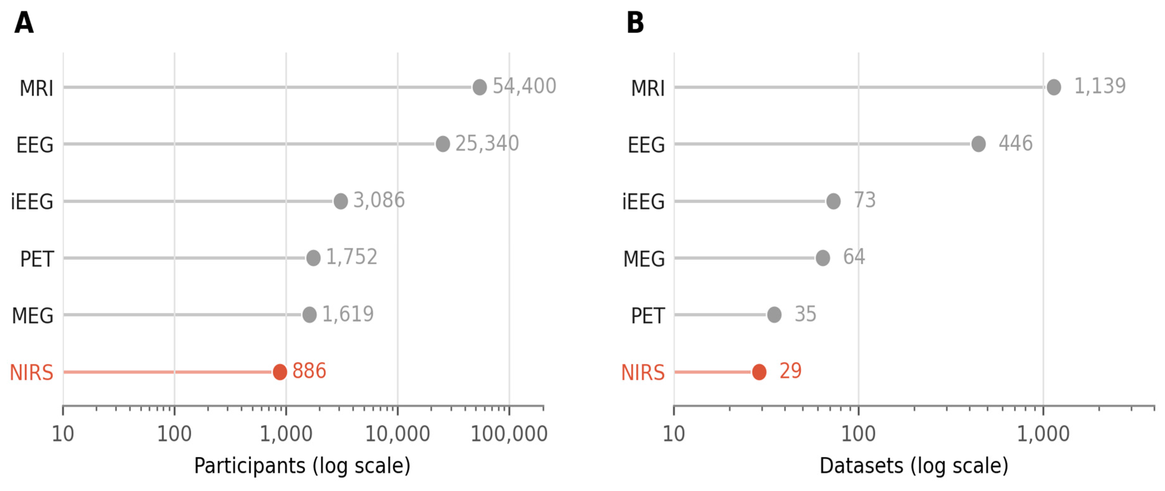
Figure 2.
How the principal models in this review represent where a sensor is. The EEG and fMRI models are described inSection 2 and Section 3.4 and the fNIRS works in Section 4; the figure is placed here because the contrast it draws is the subject of this section. The legend gives the two keys: marker fill for how coordinates are used (full, partial, or used only to place channels and not encoded), and color for modality, which the column grouping also carries. MMM appears twice because it combines scalp regions with a sinusoidal encoding of 2D electrode coordinates. No fNIRS model encodes full source–detector geometry: TADM reduces each channel to a 2D source–detector midpoint, Yao et al. use channel positions only to interpolate EEG onto fNIRS channels, and Subedi et al. learn attention weights indexed by channel. PainFormer, Jung et al. and EFRM carry no consistent spatial identity (EFRM’s fixed per-slot encoding is filled by arbitrarily selected channels). CBraMod derives position from the input by convolution rather than from electrode identity or coordinates, and BrainLM, in its released implementation, projects each parcel’s 3D coordinates.
Figure 2.
How the principal models in this review represent where a sensor is. The EEG and fMRI models are described inSection 2 and Section 3.4 and the fNIRS works in Section 4; the figure is placed here because the contrast it draws is the subject of this section. The legend gives the two keys: marker fill for how coordinates are used (full, partial, or used only to place channels and not encoded), and color for modality, which the column grouping also carries. MMM appears twice because it combines scalp regions with a sinusoidal encoding of 2D electrode coordinates. No fNIRS model encodes full source–detector geometry: TADM reduces each channel to a 2D source–detector midpoint, Yao et al. use channel positions only to interpolate EEG onto fNIRS channels, and Subedi et al. learn attention weights indexed by channel. PainFormer, Jung et al. and EFRM carry no consistent spatial identity (EFRM’s fixed per-slot encoding is filled by arbitrarily selected channels). CBraMod derives position from the input by convolution rather than from electrode identity or coordinates, and BrainLM, in its released implementation, projects each parcel’s 3D coordinates.
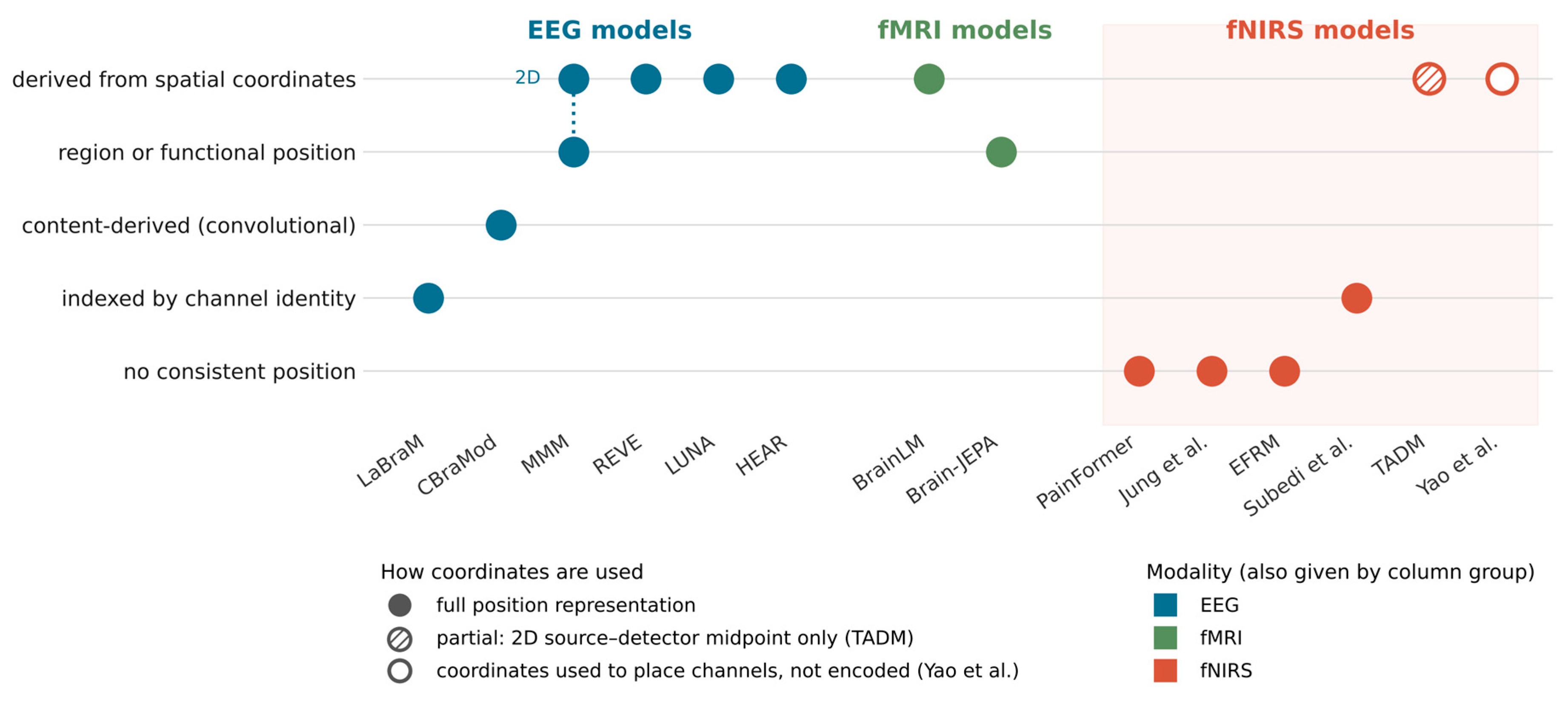
Figure 3.
EEG evidence that pretrained representations can fail when the encoder is frozen or the electrode layout changes, from the evaluations reported by REVE [2] (A, B), HEAR [26] (C) and MMM [31] (D). (A) Four-class motor imagery with the encoder frozen; REVE’s separate linear-probing comparison, under a different protocol, places CBraMod at 0.531 on the same task. (B) Binary abnormality detection under linear probing; the CBraMod value is as reproduced by REVE’s authors and is 0.500 with or without pooling, and the REVE-Large value uses attention pooling. (C) Accuracy on a three-electrode motor-imagery dataset, fine-tuned on one electrode subset and tested on the disjoint subset, in both directions. (D) Accuracy lost against full fine-tuning when a model pretrained on 32-channel subsets is probed on 62 channels; bars span the three pretraining datasets and open circles mark an untrained encoder of the same architecture. Dashed lines mark chance. Models that derive position from sensor coordinates or scalp regions are drawn in blue with their names in bold, so the distinction does not rest on color alone; the legend below the panels gives both keys.
Figure 3.
EEG evidence that pretrained representations can fail when the encoder is frozen or the electrode layout changes, from the evaluations reported by REVE [2] (A, B), HEAR [26] (C) and MMM [31] (D). (A) Four-class motor imagery with the encoder frozen; REVE’s separate linear-probing comparison, under a different protocol, places CBraMod at 0.531 on the same task. (B) Binary abnormality detection under linear probing; the CBraMod value is as reproduced by REVE’s authors and is 0.500 with or without pooling, and the REVE-Large value uses attention pooling. (C) Accuracy on a three-electrode motor-imagery dataset, fine-tuned on one electrode subset and tested on the disjoint subset, in both directions. (D) Accuracy lost against full fine-tuning when a model pretrained on 32-channel subsets is probed on 62 channels; bars span the three pretraining datasets and open circles mark an untrained encoder of the same architecture. Dashed lines mark chance. Models that derive position from sensor coordinates or scalp regions are drawn in blue with their names in bold, so the distinction does not rest on color alone; the legend below the panels gives both keys.
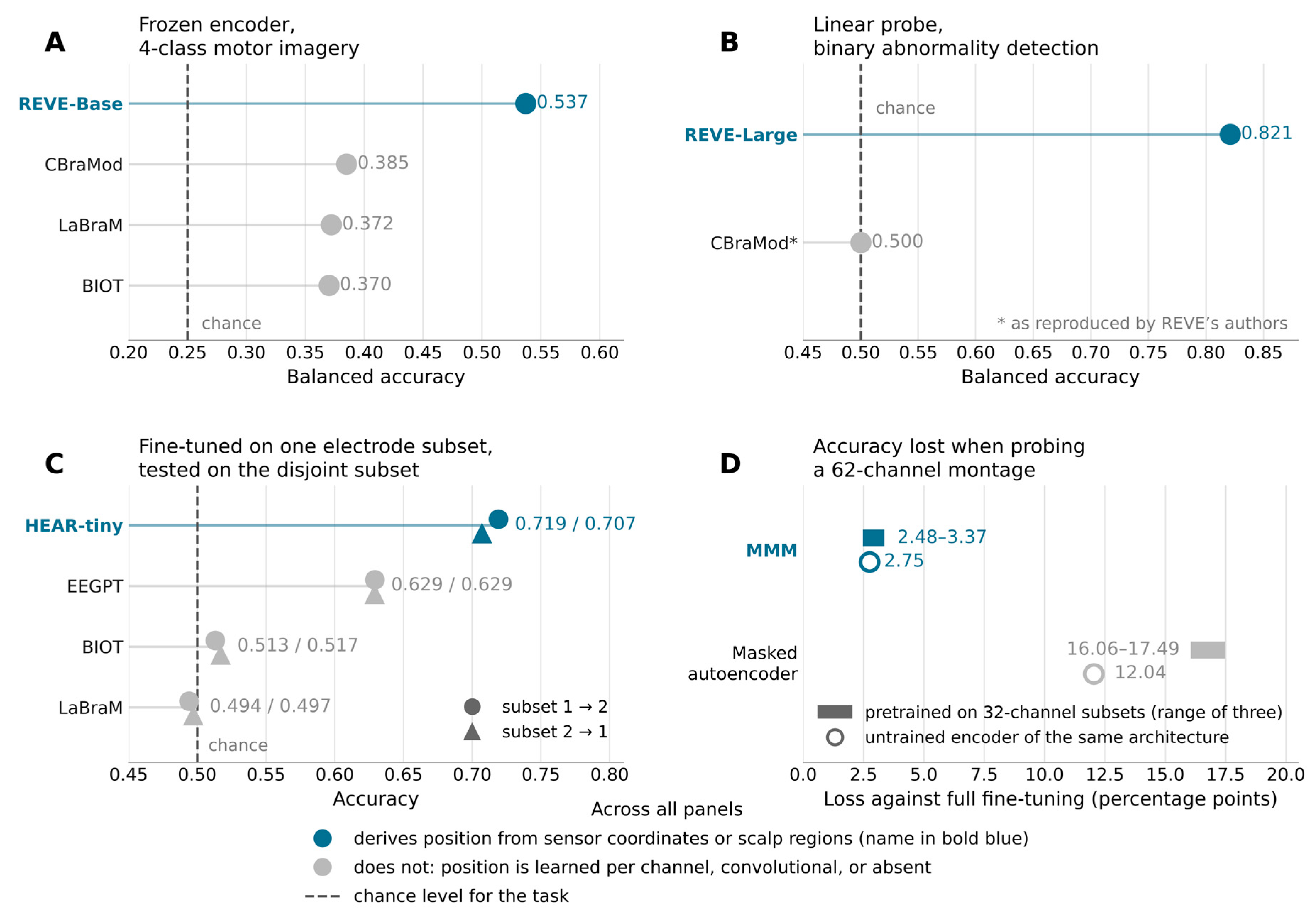
Figure 4.
Pretraining patch durations against the timescales of the hemodynamic signal. Horizontal lines show the durations tested in EFRM’s ablation [40], with the value the authors selected filled and the other tested values open; for both modalities the selected value lies at the edge of the tested range, and dashed arrows mark untested durations. BrainLM’s 20-timepoint patch [44] is adopted rather than ablated. Bands mark the per-participant peak of the hemodynamic response reported by MindSpeech [54] (4–8 s) and the 0.01–0.1 Hz analysis band expressed as periods (10–100 s); the vertical line marks the Mayer wave near 0.1 Hz.
Figure 4.
Pretraining patch durations against the timescales of the hemodynamic signal. Horizontal lines show the durations tested in EFRM’s ablation [40], with the value the authors selected filled and the other tested values open; for both modalities the selected value lies at the edge of the tested range, and dashed arrows mark untested durations. BrainLM’s 20-timepoint patch [44] is adopted rather than ablated. Bands mark the per-participant peak of the hemodynamic response reported by MindSpeech [54] (4–8 s) and the 0.01–0.1 Hz analysis band expressed as periods (10–100 s); the vertical line marks the Mayer wave near 0.1 Hz.
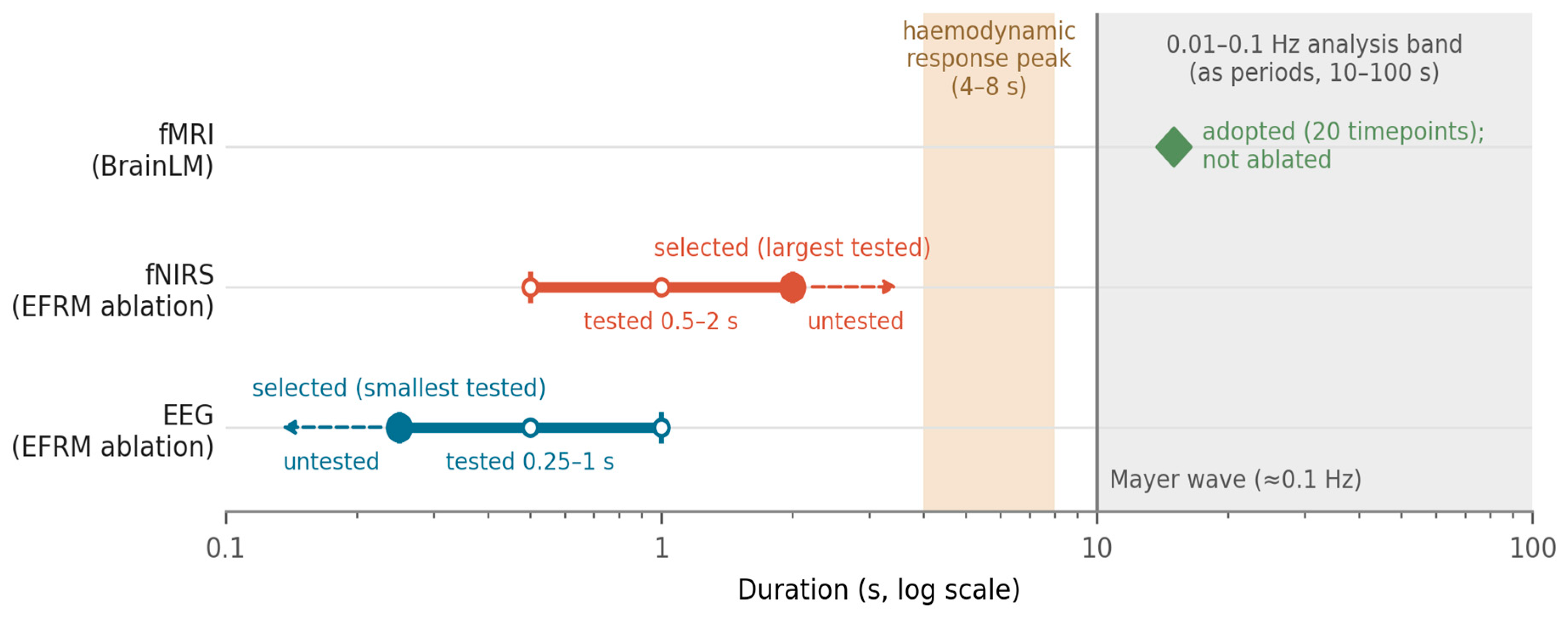
Figure 5.
Proposed input representation and pretraining objective for an fNIRS foundation model, drawing onSection 3.4, Section 5.1 and Section 5.2, and on requirements 1, 4 and 8 of Section 6. (A) Each channel is a source–detector pair whose measurement is integrated over a banana-shaped sensitivity volume between the two optodes; the inset marks the region shown in section. Short-separation channels sample predominantly extracerebral tissue, and the depth reached increases with separation and also varies with scalp and skull thickness. (B) One token per source–detector pair and temporal patch of roughly 2 s. Its positional input, [xs, ys, zs, xd, yd, zd, dSD], describes geometry within the channel; a learned embedding tags its separation class relative to a reported threshold; and the chromophores, or wavelength-specific optical densities, form a feature vector within the token rather than separate channels. (C) Geometry between channels enters the context encoder as a pairwise attention bias of the kind used in HEAR [26]. Following Brain-JEPA [45], the context encoder sees visible tokens and a target encoder updated as its exponential moving average sees all tokens. A predictor is trained to match the target encoder’s latent representation of the masked tokens under an L2 loss, rather than reconstructing the signal as the self-supervised fNIRS models of Section 4 do. A latent objective is not required to reproduce systemic oscillations, of which Mayer waves near 0.1 Hz overlap the 0.01–0.1 Hz analysis band. Colors follow Figure 2. The geometric encoding and separation-class token are untested. The within-token feature vector is used by EFRM [40] but has not been compared with alternatives. No fNIRS model has yet used a latent predictive objective, and the 2 s patch is the largest value tested in a single ablation [40], a starting point rather than a settled value.
Figure 5.
Proposed input representation and pretraining objective for an fNIRS foundation model, drawing onSection 3.4, Section 5.1 and Section 5.2, and on requirements 1, 4 and 8 of Section 6. (A) Each channel is a source–detector pair whose measurement is integrated over a banana-shaped sensitivity volume between the two optodes; the inset marks the region shown in section. Short-separation channels sample predominantly extracerebral tissue, and the depth reached increases with separation and also varies with scalp and skull thickness. (B) One token per source–detector pair and temporal patch of roughly 2 s. Its positional input, [xs, ys, zs, xd, yd, zd, dSD], describes geometry within the channel; a learned embedding tags its separation class relative to a reported threshold; and the chromophores, or wavelength-specific optical densities, form a feature vector within the token rather than separate channels. (C) Geometry between channels enters the context encoder as a pairwise attention bias of the kind used in HEAR [26]. Following Brain-JEPA [45], the context encoder sees visible tokens and a target encoder updated as its exponential moving average sees all tokens. A predictor is trained to match the target encoder’s latent representation of the masked tokens under an L2 loss, rather than reconstructing the signal as the self-supervised fNIRS models of Section 4 do. A latent objective is not required to reproduce systemic oscillations, of which Mayer waves near 0.1 Hz overlap the 0.01–0.1 Hz analysis band. Colors follow Figure 2. The geometric encoding and separation-class token are untested. The within-token feature vector is used by EFRM [40] but has not been compared with alternatives. No fNIRS model has yet used a latent predictive objective, and the 2 s patch is the largest value tested in a single ablation [40], a starting point rather than a settled value.
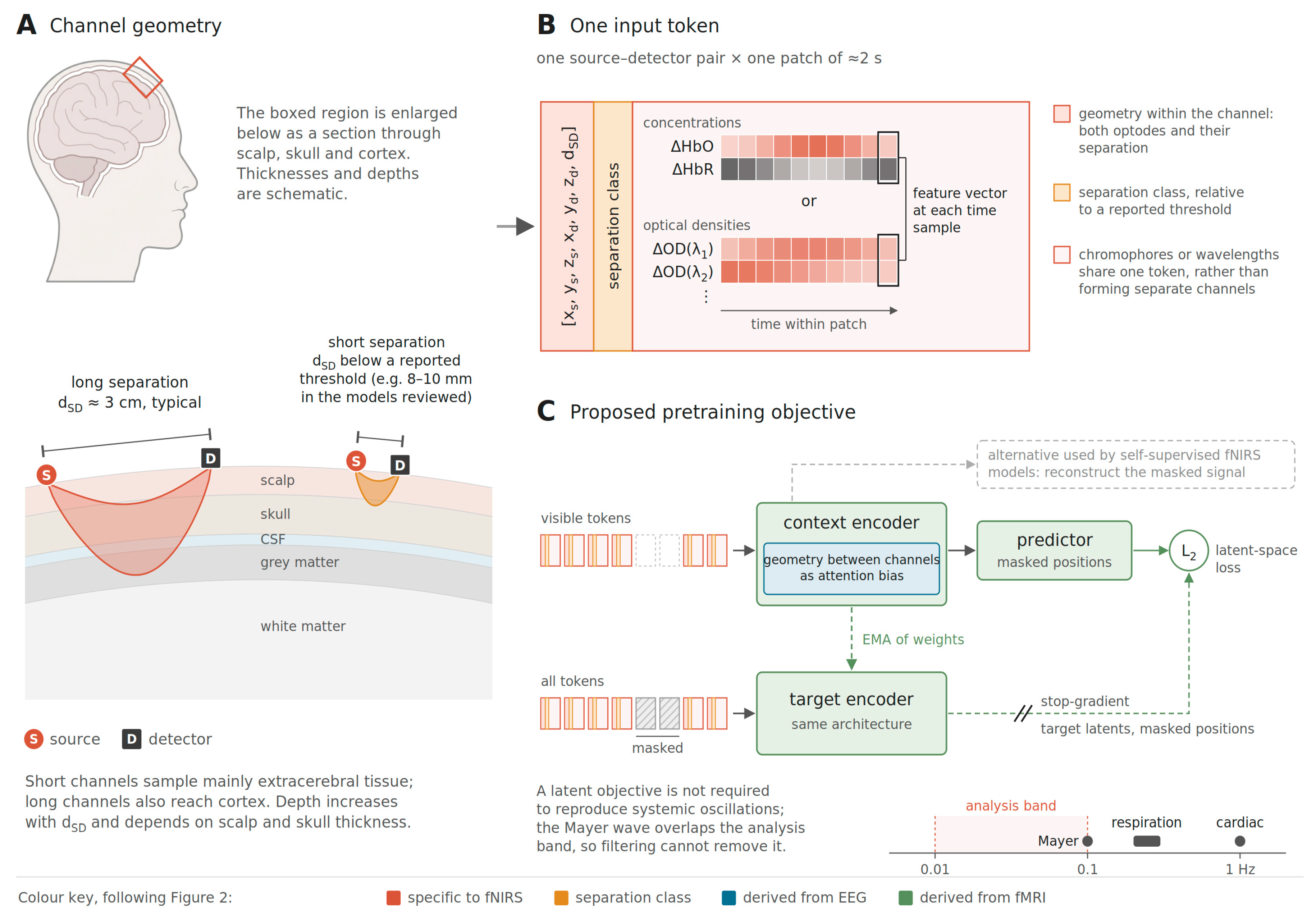

Table 1.
Curated openfnirs datasets that report subjects, duration and channel count together, with the resulting channel-hours. HD-DOT, high-density diffuse optical tomography.
Table 1.
Curated openfnirs datasets that report subjects, duration and channel count together, with the resulting channel-hours. HD-DOT, high-density diffuse optical tomography.
| Dataset | Subjects | Duration | Channels | Channel-hours |
| Luhmann2020synhrf-1 | 14 | 5 min | 28 | 33 |
| Luhmann2020synhrf-2 | 14 | 10 min | 56 | 131 |
| Li2020mirror | 18 | 6 min | 60 | 108 |
| goldbeck19rsfc | 43 | 7 min | 52 | 261 |
| Cockx2023 | 24 | 90 min | 36 | 1,296 |
| Eggebrecht2014 (HD-DOT) | 9 | 10 min | 1,842 | 2,763 |
| Guevara2024 | 40 | 19 min | 22 | 279 |
| Total | ≈ 4,870 |
Table 2.
Spatial ablations reported by MMM, HEAR and LUNA. Values are as stated in each paper; the studies differ in dataset, architecture and metric, so the rows are not directly comparable. pp, percentage points.
Table 2.
Spatial ablations reported by MMM, HEAR and LUNA. Values are as stated in each paper; the studies differ in dataset, architecture and metric, so the rows are not directly comparable. pp, percentage points.
| Paper | Positional encoding removed | Spatial aggregation removed |
| MMM | 0.03–0.4 pp | 3.2 pp (region tokens) |
| HEAR | moderate | catastrophic (0.710 → 0.429 on LargeMI) |
| LUNA | not tested | learned queries vs. fixed regions: 0.004–0.006, within seed noise |
Table 3.
Accuracy on the fNIRS-only downstream task by number of labeled shots, as reported by EFRM [40]. Pretrained encoders are frozen and only a linear layer is trained; SleepFM, SSCL and MAE were pretrained on the fNIRS-only data.
Table 3.
Accuracy on the fNIRS-only downstream task by number of labeled shots, as reported by EFRM [40]. Pretrained encoders are frozen and only a linear layer is trained; SleepFM, SSCL and MAE were pretrained on the fNIRS-only data.
| Method | 2 shots | 4 shots | 6 shots | 8 shots |
| SleepFM [41] | 0.75 | 0.62 | 0.60 | 0.58 |
| SSCL | 0.70 | 0.73 | 0.69 | 0.69 |
| MAE | 0.67 | 0.67 | 0.75 | 0.75 |
| EFRM | 0.90 | 0.81 | 0.85 | 0.94 |
Table 4.
The fNIRS-relevant works located by our search, with the category under which each is charted. MRLMC, cited in Section 5.2, is listed for completeness and is not counted among the nine.
Table 4.
The fNIRS-relevant works located by our search, with the category under which each is charted. MRLMC, cited in Section 5.2, is listed for completeness and is not counted among the nine.
| Work | Type | Charted |
| Subedi et al. [51] | Self-supervised masked segment reconstruction on raw fNIRS, cross-procedural skill assessment | Foundation model |
| EFRM [40] | Multimodal EEG–fNIRS masked autoencoder plus contrastive shared domain | Foundation model |
| Jung, Lee & An [6] | fNIRS-only masked autoencoder | FM-adjacent (single downstream task and dataset) |
| TADM [52] | Pretrained topology-adaptive diffusion, EEG→fNIRS generation | Pretrained, generative objective |
| SCDM [53] | Spatio-temporal controlled diffusion, EEG→fNIRS generation | Generative, non-pretrained baseline |
| Yao, Chen & Wang [50] | Pretrained generative framework unifying EEG, fMRI and fNIRS; EEG→fNIRS and EEG→fMRI generation | Generative; pretrained feature extractors for EEG and fMRI only |
| PainFormer [38] | Vision foundation model; fNIRS downstream only | Foundation model, not fNIRS |
| MindSpeech [54] | Prompt tuning of frozen Llama2 for imagined speech | Not a foundation model (no self-supervised pretraining) |
| CHTLM [43] | Heterogeneous EEG→fNIRS transfer, stroke motor imagery | Transfer learning |
| MRLMC [55] | Multimodal fNIRS–EEG depression recognition | Task-specific; cited in Section 5.2 for its preprocessing chain |
Table 5.
Requirements for a first fNIRS foundation model, with the modality each is derived from and the supporting evidence.
Table 5.
Requirements for a first fNIRS foundation model, with the modality each is derived from and the supporting evidence.
| # | Requirement | Derived from | Evidence |
| 1 | A unifying spatial abstraction, with source–detector geometry represented | EEG, fNIRS | Ablations in MMM and HEAR agree that aggregation matters more than the encoding scheme, and LUNA finds one alternative form of aggregation no better than another (Section 2.3); no fNIRS model encodes full source–detector geometry (Figure 2). The extension from a point to source–detector geometry is proposed rather than tested (Section 5.1) |
| 2 | Pretraining by aggregation, with deliberate montage diversity | EEG | LUNA underperforms on an unseen 62-channel layout despite 21,900 hours spanning only three montages; REVE, at 92 sources, transfers (Section 2.5) |
| 3 | Optimize and report for the frozen-encoder regime | EEG | CBraMod reaches chance (0.500) under linear probing, and LaBraM reaches chance (0.494) under montage change; external adaptation causes negative transfer in 24.8% of paired experiments under fine-tuning (Section 2.4 and Section 2.6) |
| 4 | An explicit short-channel policy, carried by a distinct token type | fNIRS | BIDS declines to define short channels; the practice is unreported in the best-charted fNIRS model (Section 1.6) |
| 5 | Report scale in subjects, hours and channels separately | fNIRS | One HD-DOT dataset contributes 57% of curated channel-hours from nine subjects; EFRM’s corpus imbalance reads as 7:1, 6:1 or 2.4:1 depending on the unit (Section 1.2) |
| 6 | A shared benchmark, harder than current tasks | EEG, fNIRS | Only four of ten EEG models are rankable on the two common tasks; every fNIRS downstream classification task in the inventory is binary or ternary (Section 4.3) |
| 7 | Dataset-level provenance and leakage control | EEG, fNIRS | A corpus aggregated from many small datasets is exactly the regime in which contamination is easy and hard to detect (Section 7) |
| 8 | Consider latent prediction rather than signal reconstruction | fMRI | Brain-JEPA moved from reconstruction to latent prediction because noise and sparse information degrade linear probing; the Mayer wave overlaps the fNIRS analysis band (Section 3.4) |
Disclaimer/Publisher’s Note: The statements, opinions and data contained in all publications are solely those of the individual author(s) and contributor(s) and not of MDPI and/or the editor(s). MDPI and/or the editor(s) disclaim responsibility for any injury to people or property resulting from any ideas, methods, instructions or products referred to in the content. |
© 2026 by the authors. Licensee MDPI, Basel, Switzerland. This article is an open access article distributed under the terms and conditions of the Creative Commons Attribution (CC BY) license (http://creativecommons.org/licenses/by/4.0/).
Copyright: This open access article is published under a Creative Commons CC BY 4.0 license, which permit the free download, distribution, and reuse, provided that the author and preprint are cited in any reuse.