

Submitted:
23 October 2026
Posted:
26 October 2026
You are already at the latest version
Abstract
More than 30 years after the discovery of the genetic cause of spinocerebellar ataxia type 1 (SCA1), selective Purkinje cell vulnerability remains a defining feature of disease pathogenesis. Increasing evidence also implicates glial cells, including oligodendroglia, astroglia, and microglia, as important contributors to neurodegeneration in SCA1 and other ataxias. In this review, we summarize current evidence for the mechanisms through which glia contribute to SCA1 progression and pathology. Early oligodendroglial dys-function leads to cell-autonomous myelination deficits and non-cell-autonomous im-pairments in Purkinje cell axonal health. Astroglial dysfunction, particularly in Berg-mann glia, disrupts glutamate homeostasis and is associated with aberrant activation of NF-κβ, JNK, and Wnt/β-catenin signaling pathways. Although microglial involvement in SCA1 remains less well understood, microglial activation appears to be regionally re-stricted to the cerebellum and inferior olive. Studies of microglial depletion have yielded partial and, in some cases, conflicting functional outcomes. Notably, dysregulated Wnt/β-catenin signaling across all three glial subtypes may represent a unifying mechanism. Together, these findings identify glia as active, region-specific contributors to SCA1 pathogenesis and highlight underexplored opportunities for mechanistic and therapeutic investigation in polyglutamine diseases.
Keywords:
spinocerebellar ataxia type 1
; SCA1
; neurodegeneration
; glia
; astroglia
; oligodendroglia
; microglia
1. Introduction
Spinocerebellar ataxia type 1 (SCA1) is one of nine polyglutamine (polyQ) diseases, a group of inherited neurodegenerative disorders that also includes Huntington’s disease (HD), spinal and bulbar muscular atrophy (SBMA), dentatorubral-pallidoluysian atrophy (DRPLA), and several other spinocerebellar ataxias (SCA2, SCA3, SCA6, SCA7, and SCA17) [1]. Although these disorders share prominent motor deficits, they differ in the neuronal populations and central nervous system (CNS) regions that are most vulnerable to degeneration [1, 2]. SCA1 is an autosomal-dominant neurodegenerative disorder caused by a CAG repeat expansion in the ATXN1 gene, resulting in production and accumulation of polyQ-expanded ataxin-1 protein [1, 3]. Whereas wild-type ATXN1 alleles contain 4-36 CAG repeats, disease-causing alleles typically contain more than 39 uninterrupted repeats. Longer repeat expansions are associated with earlier disease onset [3]. Historically, SCA1 has been considered primarily a cerebellar disorder, characterized by selective Purkinje cell (PC) vulnerability and progressive motor impairment. Accordingly, decades of research have focused on PC-intrinsic pathogenic mechanisms. More recent studies demonstrate that SCA1-associated cellular and molecular abnormalities extend beyond cerebellar PCs, affecting multiple non-neuronal cell types and extracerebellar brain regions [4-6]. Because no disease-modifying treatments are currently available, defining the contribution of glial cells to SCA1 pathogenesis is essential. Oligodendroglia, astroglia, and microglia have emerged as potential therapeutic targets across a range of neurodegenerative and neurological conditions. Although glial contributions to polyQ disease pathophysiology have been most extensively characterized in HD [7], their roles in rarer polyQ ataxias, including SCA1, remain less well understood. In this review, we examine the molecular mechanisms through which glia contribute to SCA1 pathogenesis. We discuss the cell-autonomous and non-cell-autonomous roles of each glial subtype in disease progression and identify potential mechanisms shared across glial populations, therby highlighting new directions for research in polyQ disorders.
2. Oligodendroglia
2.1. White Matter Matters: Tracking Tract Abnormalities in SCA1
White matter (WM) consists primarily of axonal tracts ensheathed by myelin, which is generated and maintained by oligodendrocytes (OLs). WM volume loss was first identified as a pathological feature of polyQ disorders in HD in 1988 [8]. MRI subsequently identified WM abnormalities in SCA1 in 2001 [9]. Later quantitative MRI studies established WM degeneration as a prominent feature of SCA1. For example, diffusion tensor imaging (DTI) revealed significant microstructural abnormalities in the middle cerebellar peduncles (MCPs) and cerebellar hemispheric WM that correlated with clinical disease severity [10]. Voxel-based morphometry (VBM) further demonstrated symmetric macroscopic volume loss in peridentate WM and the MCPs, which correlated with disease duration and International Cooperative Ataxia Rating Scale (ICARS) scores [11].
Whole-brain tract-based analyses have also shown that WM damage in SCA1 extends beyond the cerebellum, affecting the corticospinal tracts and other supratentorial structures [12]. Comparative imaging studies of cerebellar ataxias, including SCA1, SCA2, SCA3, and multiple system atrophy with cerebellar features (MSA-C), have identified WM pathology as a shared feature across several ataxias [13, 14]. Within this shared pattern, patients with SCA1 and SCA2 exhibited more severe global WM atrophy than those with SCA3. In addition, cerebellar peduncle damage was most strongly associated with clinical severity in patients with SCA1 [14].
Longitudinal studies further suggest that WM pathology in SCA1 is spatially and temporally dynamic. Although the cerebellar peduncles show the most pronounced abnormalities, longitudinal diffusion MRI has demonstrated early, stage-dependent deterioration of corticospinal and pontine crossing tracts, followed by cortical microstructural changes [15]. Importantly, WM deficits can precede clinical onset. In preataxic individuals, cerebellar WM atrophy strongly predicted conversion to overt ataxia and outperformed clinical measures, including the Scale for the Assessment and Rating of Ataxia (SARA), in predicting subsequent disease progression [16-20].
Metabolic alterations in WM may also contribute to SCA1 progression. Magnetic resonance spectroscopy (MRS) identified significantly reduced total N-acetylaspartate (tNAA), a marker of axonal integrity, in the parietal WM of patients with SCA1 [21]. Lower baseline tNAA and glutamate levels in cerebellar WM also predicted more rapid worsening of SARA scores [22]. Neuropathological and biochemical analyses of postmortem SCA1 tissue corroborate these imaging findings, demonstrating widespread WM degeneration throughout the cerebellum and brainstem, as well as reduced numbers of mature OLs and myelinated axons in the cerebellum [5, 23]. Single-nucleus RNA sequencing (snRNA-seq) of postmortem SCA1 cerebellum further revealed transcriptomic dysregulation in both oligodendrocyte progenitor cells (OPCs) and OLs, including reduced expression of key myelin-related genes in OLs [5]. Together, these findings establish WM pathology as a core feature of SCA1, alongside PC degeneration.
2.2. Oligodendroglial Lineage Development, Myelination, and Functional Heterogeneity
The oligodendroglial lineage comprises OPCs, immature OLs, and mature myelinating OLs. This lineage arises through a tightly regulated developmental program that begins during embryogenesis and continues throughout postnatal life [24]. Neural progenitor cells (NPCs) generate OPCs following induction of the transcription factors OLIG2 and SOX10, which remain expressed throughout lineage progression. OPCs are further characterized by expression of markers such as PDGFRα and CSPG4 (NG2) [25]. These cells are proliferative and migratory, are distributed throughout the CNS, and serve as the principal source of new OLs for developmental myelination, adult remyelination, and adaptive myelination [26, 27].
As OPCs differentiate, they downregulate progenitor-associated programs and upregulate markers including GalC, O4, TCF7L2, and MYRF as they transition to immature OLs [26]. TCF7L2, a Wnt/β-catenin co-effector, has a particularly important role at this stage. Wnt/β-catenin signaling through TCF/LEF family transcription factors can either delay or promote OL differentiation depending on developmental timing and brain region. Thus, endogenous Wnt signaling is highly context dependent and must be precisely regulated to support normal oligodendroglial lineage progression [28].
Mature OLs generate and maintain the multilamellar myelin membrane that wraps internodal axonal segments [29, 30]. Major myelin proteins, including MBP, PLP1, MAG, MOG, and CNP, turn over slowly; for example, PLP1 has a half-life of approximately six months [31-33]. Myelin provides mechanical and biochemical protection to axons and enables rapid saltatory conduction by restricting ionic currents to the nodes of Ranvier [29, 30]. OLs also regulate conduction velocity by modifying internode length and myelin thickness, commonly measured by the g-ratio; both properties vary across CNS regions and neuronal subtypes [34].
In addition to myelination, OLs provide critical metabolic support to axons. They convert glucose to lactate and export it through monocarboxylate transporters (MCT1/MCT2) to support axonal mitochondrial metabolism in an activity-dependent manner [29, 30, 35]. OLs can also enhance axonal ATP production by delivering SIRT2-containing exosomes to axon [36]. Furthermore, they help maintain the periaxonal environment by regulating ionic flux and providing trophic factors that support axonal caliber and mitochondrial density [29, 30].
Single-cell transcriptomics studies have revealed temporally and spatially distinct oligodendroglial populations [37-40]. OLs acquire region-specific identities that help shape local myelin architecture, and disruption of this spatial organization can produce substantial functional impairment [41]. In disease, OLs undergo marked transcriptomic and compositional changes [42-45] and may adopt disease-associated oligodendrocyte (DAO) states that contribute to tissue injury and repair in Alzheimer’s disease (AD) and multiple sclerosis (MS) [46, 47].
2.3. Oligodendroglia Dysfunction and Myelin Pathology in SCA1 Mouse Models
Consistent with the WM abnormalities observed in patients with SCA1, longitudinal snRNA-seq of the Atxn1154Q/2Q knock-in (SCA1-KI) mouse model revealed that oligodendroglial dysfunction and impaired myelination are early transcriptomic events [5]. In this model, polyQ-expanded ataxin-1 is expressed endogenously in all cerebellar cell types [48]. Transcriptomic abnormalities in OPCs and OLs preceded PC degeneration, and OLs showed robust downregulation of myelin-related genes as early as 12 weeks of age [5]. At the same age, SCA1-KI mice exhibited progressive biochemical and ultrastructural demyelination of PC axons and impaired differentiation of OPC into OLs. These changes coincided with the onset of motor phenotypes but preceded overt PC loss [5].
Studies of primary OPC cultures from SCA1-KI mice, as well as an oligodendroglial lineage-specific conditional knock-in model (OL-SCA1-cKI), indicate that deficits in OL differentiation and myelination can occur through cell-autonomous mechanisms [5, 49]. In contrast, the B05 transgenic model (Pcp2-ATXN182Q), which selectively expresses mutant ataxin-1 in PCs, develops severe PC pathology without early demyelination at 15 weeks of age [5]. Although B05 mice show reduced WM area at later disease stages, this finding likely reflects overall cerebellar atrophy or the loss of myelinated axons rather than cell-autonomous OL dysfunction [50].
Importantly, findings from OL-SCA1-cKI mice suggest that OLs contribute to SCA1 pathogenesis beyond demyelination. Intact OLs and myelin are required for normal PC development and function [51-54], whereas SCA1-associated OLs adversely affect PC health through non-cell-autonomous mechanisms. OL-SCA1-cKI mice developed progressive PC axonal pathology, including reduced axonal caliber reduction before the onset of demyelination and, subsequently, axonal torpedoes in the granule cell layer (GCL), without overt PC somata [49]. PC torpedoes are a characteristic pathological feature of SCA1 in humans [5, 23].
OL dysfunction also induced early non-cell-autonomous transcriptomic changes in PCs. snRNA-seq of OL-SCA1-cKI cerebella identified increased expression of genes involved in postsynaptic organization in PCs [49], a signature also observed in constitutive SCA1-KI mice [5]. In addition, PCs in OL-SCA1-cKI mice exhibited increased dendritic length [5, 49]. This phenotype may represent aberrant or compensatory synaptic remodeling in response to OL dysfunction, consistent with evidence that OLs can influence PC dendritic arborization [51] and spine turnover [55]. Notably, dendritic lengthening was absent in posterior cerebellar lobules [49], regions that are preferentially affected in SCA1 [56, 57]. This findings raise the possibility that insufficient region-specific compensatory remodeling in response to OL dysfunction contributes to the selective vulnerability of posterior cerebellar regions.
OL-SCA1-cKI mice also accumulated myelin debris in the cerebellum and hippocampus. Myelin debris can destabilize nodes of Ranvier and exacerbate axonal injury [49, 58]. Although overt cerebellar astrogliosis and microgliosis were not detected in OL-SCA1-cKI mice, snRNA-seq revealed microglial transcriptomic dysregulation, suggesting a more subtle immune response [49]. The accumulation of myelin debris in particularly notable in light of a reported case of co-occurring SCA1 and myelin oligodendrocyte glycoprotein (MOG) antibody-associated disease (MOGAD) [59]. This observation raises the possibility that persistent myelin debris contributes to secondary immune activation. Moreover, wild-type ataxin-1 regulates B-cell function and protects against demyelination in the experimental autoimmune encephalomyelitis (EAE) model [60-62]. Thus, polyQ-expanded ataxin-1 may affect both OL biology and adaptive immunity, potentially amplifying demyelination through a positive-feedback loop.
2.4. Unraveling the Molecular Mechanisms Driving Oligodendrocyte Dysfunction in SCA1
Transcriptomic analyses of SCA1 cerebellum have identified broad dysregulation of genes involved in myelination and axonal maintenance, as well as altered OL subtype composition. These changes suggest impaired neuron-OL communication and diminished neuroprotective signaling [5, 49]. Upstream-regulator analyses have implicated TCF7L2 and huntingtin (HTT) as candidate drivers of these transcriptional abnormalities, pointing to two potentially convergent mechanisms: (1) ataxin-1-specific dysfunction and (2) polyQ-dependent toxicity shared across diseases.
Evidence supports an intrinsic role for wild-type ataxin-1 in oligodendroglial function and WM integrity. Although long CAG expansions (≥39 repeats) cause fully penetrant SCA1 [63], intermediate expansions (32-39 repeats) are associated with an increased risk of sporadic amyotrophic lateral sclerosis (ALS) [64, 65]. In addition, intronic variants that reduce ATXN1 expression have been associated with increased risk of AD [66, 67] and MS [61, 68]. Because OL dysfunction and myelination deficits are also prominent features of these disorders [42, 69-73], these genetic associations suggest that ataxin-1 may have broader roles in myelin maintenance beyond SCA1.
Within the OL lineage, Wnt/β-catenin signaling may be a key ataxin-1-specific mechanism. Both wild-type and polyQ-expanded ataxin-1 physically interact with TCF7L2 [57] (Figure 1), a central transcriptional effector of Wnt/β-catenin signaling and an important regulator of the transition from OPCs to immature OLs [28]. TCF7L2 activity is predicted to be repressed in SCA1 oligodendroglia [49], implicating disruption of the ATXN1-TCF7L2 axis in impaired OL differentiation.
More broadly, OL dysfunction occurs across several polyQ diseases, including SCA2, SCA3, HD, and SBMA, suggesting that polyQ-dependent mechanisms may act across disease contexts independent of the host protein [8, 74-81]. Consistent with this possibility, TCF7L2 and HTT have also been identified as upstream regulators of OL dysfunction in HD and SCA3 [82, 83]. Together, these findings raise the possibility that expanded polyQ tracts converge on a shared transcriptional pathway. Whether this convergence reflects direct effects within OLs, however, remains an open question.
3. Astroglia
3.1. Astroglia Diversity in the Cerebellum: Bergmann Glia in Focus
Astroglia are, named for their star-like morphology, are commonly classified by their location in CNS WM or gray matter (GM). The cerebellum, however, contains several morphologically distinct astroglial subtypes, including radial Bergmann glia (BG) and Fañanas cells, fibrous astrocytes, velate astrocytes, and deep cerebellar nuclei (DCN) astrocytes [84-86]. Although a detailed discussion of cerebellar astroglial markers is beyond the scope of this review, these evolutionarily conserved astroglial populations can be identified histologically using ALDH1L1 and GFAP, together with markers such as S100β, glutamine synthetase (GS), and SOX9 [87]. These subtypes occupy distinct cerebellar microenvironments. BG and Fañanas cells are located in the Purkinje cell layer (PCL) and molecular layer (ML); velate astrocytes localize to the GCL; fibrous astrocytes reside in the WM; and DCN astrocytes occupy the DCN. Because BG are closely associated with PCs, the neuronal population most affected in SCA1, they are well positioned to influence SCA1 pathogenesis.
3.2. Mechanisms of Astroglial Involvement in SCA1 Pathogenesis
In 2006, Custer et al. showed that selective overexpression of mutant ataxin-7 in astroglia was sufficient to induce SCA7-like motor deficits, including hindlimb clasping and impaired performance on an accelerating rotarod [88]. This work, together with studies in other neurodegenerative disorders, helped shift the view of astroglia from passive support cells to potential drivers of disease pathogenesis [88, 89]. Astroglia have since been studied extensively in SCA1, particularly in the SCA1-KI and B05 mouse models [48, 90]. In both models, reactive astrogliosis, typically assessed by increased GFAP expression, occurs early, at 8 weeks in SCA1-KI mice and at 4 weeks in the PC-specific B05 model [6]. Because astroglial reactivity precedes overt PC neurodegeneration in both models [6, 48, 90, 91], astroglia may actively contribute to degeneration rather than simply respond to neuronal dysfunction. However, this possibility requires direct testing. Newly developed ON/OFF systems that enable cell type-specific manipulation of mutant ataxin-1 [49, 92] may help define the contribution of astroglia to SCA1 progression and pathology.
3.2.1. Bergmann Glia as the Cerebellar Glutamate-Recycling Hub
A core homeostatic function of astroglia is the clearance of synaptic glutamate through the transporters GLAST and GLT-1 [93]. In the glutamate-glutamine cycle, astroglial take up glutamate and convert it to glutamine via glutamine synthetase. Glutamine is then returned to presynaptic neurons to sustain neurotransmission [94]. Disruption of astroglial glutamate transporters or glutamine synthetase can therefore promote glutamate spillover, excitotoxicity, and neurodegeneration [93,95].
Reduced GLAST expression has been observed in both an astroglia-conditional ON SCA7 mouse model and in the SCA1 B05 model, suggesting that impaired glutamate recycling may arise through both cell-autonomous and non-cell-autonomous mechanisms [88, 93, 95, 96]. Because GLAST expression is enriched in the PCL and ML [97], glutamate-handling deficits in SCAs are primarily attributed to BG rather than other cerebellar astroglial populations. In vivo GLAST deletion is sufficient to produce mild ataxia, including rotarod impairment [93], and disrupts parallel fiber and climbing fiber innervation of PC dendrites [98].
Although astroglia mediate most glutamate uptake in the cerebellum, PCs also express the glutamate transporter EAAT4, which may partially compensate for glutamate spillover [99]. In addition, PC-expressed Down syndrome cell adhesion molecule (DSCAM) interacts with GLAST to support proper parallel fiber and climbing fiber synaptic organization [100], highlighting close functional coupling between PCs and BG. Loss of DSCAM and associated deficits in excitatory synaptic innervation can be partially rescued by riluzole, an enhancer of glutamate transporter function [101]. Troriluzole a riluzole prodrug [102], activates GLAST and GLT-1 and has advanced to clinical testing for SCAs. In Study 206-RWE, Troriluzole was reported to slow disease progression by 50-70% relative to untreated controls [103].
3.2.2. Stage-Specific Roles of NF-κβ Signaling in SCA1 Astroglia
Beyond synaptic homeostasis, astroglia respond to their microenvironment through signaling pathways such as nuclear factor-κβ (NF-κβ). NF-κβ is a family of transcription factors expressed broadly in CNS cell types, including astroglia, microglia, and OLs [104, 105]. Its activation is commonly triggered by injury or disease and has been investigated as a regulator and therapeutic target of astrogliosis [106, 107]. In the SCA1 B05 model, astroglia isolated from 8-week-old cerebella showed increased NF-κβ phosphorylation, indicating inflammatory pathway activation in response to altered PC health [108]. However, the effects of astroglial NF-κβ modulation appear to be disease- and stage-dependent. Although NF-κβ suppression can reduce astrogliosis and improve outcomes in models of spinal cord injury, MS, glaucoma, vascular dementia [106, 107, 109, 110], timing is critical in SCA1. Conditional NF-κβ silencing in BG at 8 weeks of age in B05 mice (ATXN1[82Q]; IKKβF/F; Slc1a3-cre/ERT) reduced astrogliosis but accelerated ataxia and worsened PC pathology, ML thinning, and climbing fiber innervation of PC dendrites [108] (Figure 2). In contrast, silencing NF-κβ in BG at 16 weeks partially improved motor phenotypes and PC neurodegeneration [108] (Figure 2). Together, these findings suggest a biphasic role for astroglial NF-κβ signaling: it may be protective early in disease abut detrimental at later stages, consistent with observations in ALS and AD models [111,112].
3.2.3. JNK Activation in SCA1 Astroglia
Reactive astrogliosis is also regulated by c-Jun N-terminal kinase (JNK) signaling. The relationship between JNK activation and GFAP accumulation, a hallmark of reactive astrogliosis, was initially investigated in Alexander disease (AxD), an astroglial protein-aggregation disorder [114]. In COS-7 cells, expression of AxD-associated GFAPR239C [115] resulted in intracellular GFAP inclusions and increased JNK phosphorylation [116]. Similarly, activation of upstream JNK kinases, including MLK2, MLK3, and ASK1, increased GFAP accumulation, supporting a reciprocal relationship between JNK signaling and GFAP dysregulation [116].
In the cerebellum, increased c-Jun, a key JNK target, has been observed in mouse models of PC degeneration (pcd) and granule cell degeneration (weaver) [117-119]. In postmortem cerebellar tissue from individuals with SCA1, SCA2, and SCA7, phosphorylated c-Jun accumulated prominently in the PCL and ML [113]. In SCA1-KI mice, c-JUN phosphorylation at serine 63, a JNK target residue, was particularly enriched in BG, as shown by S100β co-staining [113] (Figure 2). Treatment of SCA1-KI mice with the blood-brain barrier-penetrant JNK inhibitor SP600125 reduced BG phospho-c-Jun staining and GFAP fluorescence intensity, improved accelerating rotarod performance, and preserved PC dendritic arborization [113] (Figure 2). These findings implicate JNK signaling in BG reactivity and suggest that JNK inhibition may mitigate both glial and neuronal phenotypes. However, because SP600125 broadly suppresses JNK signaling, cell type-specific genetic studies are needed to determine whether JNK inhibition in BG alone is sufficient to rescue SCA1-associated PC neurodegeneration.
3.3. Developmental Origins of Astroglial Dysfunction in SCA1
Although astroglial reactivity is a hallmark of SCA1 pathogenesis, cerebellar astroglia are also essential for normal cerebellar development. SCA1 onset and severity are closely associated with polyQ tract length, with longer CAG repeats correlating with earlier onset and more severe disease in both patients and mouse models [3, 120]. In addition to intergenerational repeat expansion, the SCA1 CAG repeat is somatically unstable across brain regions and cell types [121], as observed in other polyQ disorders, including HD [122, 123]. These findings underscore the need to examine how CAG-repeat instability influences disease across the lifespan, rather than focusing exclusively on post-symptomatic pathology. Consistent with this view, transcriptomic changes occur across cerebellar cell types in SCA1 models before overt PC degeneration [5, 124, 125]. At postnatal day 7 (P7), SCA1-KI mice show increased cerebellar neural stem-cell proliferation, including elevated Ki67+/Nestin+ progenitor populations relative to wild-type controls [126]. This phenotype is absent in Atxn1-null mice, indicating that it depends on mutant ataxin-1 expression [126].
Although the contribution of these early progenitor changes to later disease onset remains unclear, normal cerebellar development depends on functional BG, positioning astroglia as potential determinants of early SCA1 vulnerability. Rakic’s landmark work established BG as migratory scaffolds for granule cells. In the weaver mouse model, BG hypertrophy precedes abnormal granule-cell migration [127], as well as later neurodegeneration and ataxia [118, 128].
Wnt-1 (int-1) is also required for cerebellar development [129, 130]. During embryogenesis (E14.5), Wnt-1 is expressed in granule-cell progenitors, whereas by P10 its expression is restricted to BG [130]. Conditional disruption of Wnt/β-catenin signaling in BG through Cre-mediated deletion of floxed Ctnnb1 disrupts foliation and lamination, causes astroglial mislocalization, alters granule-cell and PC positioning, and produces ataxia [131, 132]. These findings identify BG-mediated Wnt signaling as a key regulator of cerebellar morphogenesis and a plausible contributor to the vulnerability of SCA1-relevant neuronal populations.
Aberrant Wnt/β-Catenin Activation in SCA1 Astroglia
Wnt/β-catenin signaling is abnormally increased in the SCA1 cerebellum [57]. Expression of the canonical Wnt target genes Ccnd1 and c-Myc increases with age in SCA1-KI cerebella [57] (Figure 1). In HeLa cells, co-expression of ataxin-1 and β-catenin increased Wnt/β-catenin reporter activity in a polyQ length-dependent manner: ataxin-1 [30Q] and ataxin-1 [82Q], but not ataxin-1 [2Q], significantly increased signaling [57]. Similarly, PC-specific SCA1 transgenic model (Pcp2-ATXN163Q) exhibit increased Ccnd1, c-Myc, and active β-catenin expression [57]. In Pcp2-ATXN163Q; TCF/Lef:H2B/GFP reporter mice, GFP signal was increased in PCs and neighboring PCL cells, implicating additional cell types in Wnt-pathway activation [57]. Importantly, PC-specific suppression of Wnt/β-catenin signaling through Ctnnb1 deletion failed to attenuate SCA1 phenotypes, whereas PC-specific pathway hyperactivation through Apc suppression was insufficient to induce disease-associated changes [57]. These findings point to a non-PC contributor to Wnt dysregulation. In contrast, conditional Wnt/β-catenin activation in astroglia under a Gfap promoter induced astroglial reactivity in the PCL and ML and reduced expression of BG-enriched GLAST [57] (Figure 1). Both are established astroglial features of SCA1 [6, 96]. Although it remains unclear whether BG-specific Wnt/β-catenin inactivation can rescue SCA1 neurodegeneration, Ctnnb1 siRNA knockdown in C8-S astrocytes expressing Flag-hATXN1 [82Q] reduced cleaved caspase-3 (CC3) levels in co-cultured N2A cells [57]. Together, these findings support further investigation of astroglial Wnt/β-catenin inhibition as a strategy to slow SCA1 progression and pathology.
4. Microglia
4.1. Microglia: Hero, Villain, or Bystander?
Microglia (MG) are highly heterogeneous, mesoderm-derived glial cells that serve as the resident macrophages of the CNS. Their functions differ from those of circulating monocytes and other tissue-resident macrophages [133]. MG originate from yolk sac progenitors, enter the CNS during early embryonic development, and self-renew locally throughout adulthood [133]. Under homeostatic conditions, they support CNS function through synaptic pruning [134, 135], phagocytosis [136], debris clearance [137], myelin maintainance [138], and regulation of neural circuitry [139]. However, MG also contribute to neuroinflammation following injury and in neurodegenerative disease [138, 140, 141]. Whether a microglial response is beneficial or harmful depends on the local microenvironment, disease stage, and brain region.
Microglial States Beyond the M1/M2 Binary
Historically, activated MG were classified using the M1/M2 polarization framework. M1 MG were associated with pro-inflammatory functions, including reactive oxygen species production [140], whereas M2 MG were associated with phagocytosis [141] and release of anti-inflammatory cytokines, such as IL-4, IL-10, IL-13, TGF-β [142-145]. However, snRNA-seq and single-cell RNA sequencing (scRNA-seq) have shown that this binary model does not adequately capture microglial diversity. MG adopt distinct transcriptional states across development, aging, injury, and disease [146-150] (Table 1).
For example, scRNA-seq of the healthy murine brain identified nine microglial subclusters present from E14.5 through adulthood (P540) [146]. With aging, the proportion of MG clusters expressing inflammatory genes, including Ccl3, Ccl4, and Il1b, increased [146]. Disease can further induce microglial states that differ from both homeostatic and age-associated populations.
Disease-associated microglia (DAM) were first described in the 5xFAD model of AD, where they were proposed to limit plaque accumulation and protect against neurodegeneration [150]. Subsequent studies showed that DAM functions are context-dependent. In early AD, both DAM and homeostatic MG may contribute to synaptic loss and plaque deposition, whereas later-stage DAM may exert protective effects [159-164]. Moreover, pro- and anti-inflammatory DAM-like states can coexist within the same disease stage [164]. Although DAM are often discussed in the context of AD, DAM-like transcriptional programs have also been reported in MS, PD, TBI, and HD. Together, these findings demonstrate that MG adopt disease-, region-, and stage-specific stages that are not captured by the traditional M1/M2 framework.
4.2. Molecular Mechanisms of Microglial Activation and Dysfunction
Although transcriptomic studies have greatly expanded our understanding of MG diversity, the functional consequences of specific migroglial states remain incompletely defined. MG may be protective in one disease context but harmful in another, and their effects can vary by brain region and disease stage [165]. Three pathways recurrently implicated in neurodegeneration are: (1) major histocompatibility complex class II (MHC-II)-mediated antigen presentation [166], (2) complement-dependent synaptic pruning [134], and (3) triggering receptor expressed on myeloid cells 2 (TREM2) signaling [163, 167, 168]. Each pathway can support homeostasis or promote pathology depending on context.
4.2.1. MHC-II-Mediated Antigen Presentation
MHC-II presents peptides derived from extracellular or phagocytosed material to CD4+ T cells [169]. Under homeostatic conditions, MG express low levels of MHC-II and are not considered major antigen-presenting cells (APCs), while border-associated macrophages serve this role more prominently [151]. However, inflammatory cytokines, tissue damage, or infiltrating immune cells can induce MHC-II expression in MG, enabling them to sample and present local antigens, including self-antigens. MHC-II-mediated antigen presentation contributes to normal CNS immune surveillance [170]. In autoimmune diseases such as MS, however, it may become pathogenic when MG present myelin-derived peptides to autoreactive T cells and locally amplify theimmune response [155, 171].
4.2.2. Complement-Dependent Synaptic Pruning
MG also participate in the classical complement cascade, which labels synapses for elimination during development and normal circuit refinement [134]. In neurodegenerative disease, this pathway can be aberrantly reactivated, resulting in excessive or inappropriate synaptic elimination in the adult CNS. In AD models, complement-mediated synapse loss has been proposed as an early contributor to cognitive decline that precedes neuronal death [172]. Thus, a physiological pruning mechanism may become pathogenic when inappropriately activated.
4.2.3. TREM2-Dependent and TREM2-Independent Signaling
TREM2 regulates MG survival, proliferation, phagocytosis, and metabolism and is required for the adoption of disease-responsive states, including DAM-like programs in AD models [151, 167]. In APP/PS1 mice, Trem2 deficiency reduces microglial clustering around plaques and impairs phagocytic clearance, worsening pathology [163]. These findings supports a protective role for TREM2 in plaque containment and clearance. TREM2 may also promote microglial survival through Wnt/β-catenin signaling [173]. TREM2 signals through DAP12 to activate PI3K/Akt, which phosphorylates and inactivates GSK3β. This stabilizes β-catenin and promotes expression of Wnt/β-catenin target genes, including Ccnd1, c-Myc, Bcl-2, that support proliferation and survival (Figure 3). In cultured Trem2-null MG, loss of TREM2 accelerated β-catenin degradation. Pharmacological activation of Wnt signaling with Wnt3a or inhibition of GSK3β with LiCl or TDZD-8 partially restored β-catenin levels, proliferation, and viability [173, 174].
Wnt signaling may also promote MG inflammation through non-canonical pathways that are independent of TREM2 and canonical β-catenin signaling. An early study proposed that Wnt3a binding to Frizzled receptors (FZD4/5/7/8) and LRP5/6 activated canonical Wnt/β-catenin signaling and induced expression of IL-6, IL-12, and TNFα [174]. More recently, Wnt3a was shown to enhance TNFα release in lipopolysaccharide (LPS)-primed MG through non-canonical JNK/ planar cell polarity (PCP) and PLC/Ca²⁺ pathways [175]. Inhibition of canonical Wnt signaling did not block this effect, whereas inhibition of either non-canonical pathway did [175]. Wnt3a alone had no effect in unprimed MG, suggesting that inflammatory priming is required [175].
Together, these findings suggest at least two Wnt-linked programs in MG: a TREM2-dependent pathway that promotes β-catenin-mediated survival and proliferation, and a potentially TREM2-independent pathway in which Wnt ligands amplify inflammatory cytokine release through non-canonical signaling. How these pathways interact in vivo, and whether TREM2 loss shifts MG toward inflammatory signaling, remain unresolved.
4.3. Microglial Contributions to PolyQ Diseases
MG involvement in polyQ diseases, including HD and SCAs, remains understudied. However, emerging evidence supports both cell-autonomous and non-cell-autonomous roles. In HD, mutant HTT is expressed in neuronal and non-neuronal cells. Nuclear mutant HTT inclusions have been observed in MG from the frontal cortex of adult-onset HD and from the frontal cortex and striatum of juvenile-onset HD [176]. Primary MG isolated from mutant HTT (Hdh175Q/175Q) knock-in mice induced apoptosis in embryonic stem cell-derived neurons cultured on wild-type astrocytes, whereas wild-type MG did not [176]. Similarly, mice expressing mutant HTT selectively in MG exhibited greater neuronal death under sterile inflammatory conditions than control mice [176].
Microglial toxicity may also be induced by expanded polyQ tracts independently of the host protein. Conditioned medium from BV2 MG-like cells treated with a 69Q peptide, but not a 15Q peptide, induced neurite retraction, reduced branch points, and impaired neurite outgrowth in PC12 cells [156]. These effects were MG-specific, as conditioned medium from polyQ-treated SH-SY5Y neuroblastoma cells did not reproduce the phenotype [156]. Thus, expanded polyQ sequences may be sufficient to induce MG-derived factors that compromise neuronal process integrity in vitro.
Microglial depletion studies in SCAs have yielded mixed findings. In a SCA3 mouse model, MG displayed activated morphology at a mid-disease stage [157]. Treatment with the CSF1R inhibitor PLX3397 reduced MG numbers by 42–59% in the cerebellum and brainstem but did not improve balance, coordination, gait, strength, or reflexes [157]. Importantly, PLX3397 did not normalize MG morphology in SCA3 mice and induced an activated-like phenotype in MG from wild-type mice. Clustering analyses grouped PLX3397-treated wild-type MG with SCA3 MG [157]. These findings complicate interpretation because CSF1R inhibition may alter the phenotype of residual MG rather than simply restore a homeostatic state.
In SCA1, MG function remains poorly characterized. Early postmortem studies reported gliosis in cerebellar regions but did not identify the predominant glial subtype [177, 178]. The first studies to examine MG in SCA1 assessed activation in SCA1-KI and B05 mice [6, 90]. At 8 and 12 weeks, SCA1-KI mice showed early microgliosis in the cerebellar cortex, including a 43.5% increase in Iba1+ cells relative to wild-type mice. In the PC-specific B05 model, robust MG activation was evident as early as 4 weeks [6]. Because mutant ataxin-1 expression in B05 mice is restricted to PCs, these studies suggest that PC dysfunction may be sufficient to trigger MG activation before overt neuronal death.
The functional impact of MG was later assessed by depleting 70-80% of MG in B05 mice with PLX3397 [158]. Sustained depletion partially improved motor performance at 3 months, as measured by rotarod and balance-beam tests, and reduced TNFα and monocyte chemoattractant protein-1 (MCP-1) levels. However, MG depletion did not reduce astrogliosis or prevent PC loss [158]. PLX3397-treated B05 mice also showed increased levels of the postsynaptic density protein PSD95 without a corresponding increase in the presynaptic protein synapsin [158]. This pattern may reflect selective preservation of postsynaptic structures and could contribute to modest behavioral improvement without restoring synaptic integrity or neuronal survival. These results should be interpreted cautiously. CSF1R inhibitors deplete MG regardless subtype, potentially eliminating both harmful and protective populations. In addition, their effects extend across the brain, which can obscure region-specific MG functions. Consistent with this concern, microglial activation in SCA1 appears region-specific. In a 2022 study of SCA1-KI motor cortex, Luttik et al. found no overall increase in microgliosis at 12 or 30 weeks, despite a trend toward increased MG numbers at 12 weeks [4]. This contrasts with significant microgliosis in the cerebellum and inferior olive at comparable time points [179]. These findings suggest that glial activation may parallel region-specific neuronal vulnerability in SCA1. They also highlight a limitation of relying on pan-microglial markers such as Iba1, which may fail to detect changes in discrete MG subpopulations.
Overall, studies in HD, SCA3, and SCA1 indicate that MG respond to polyQ disease and can influence functional outcomes, but t the molecular programs underlying distinct MG states remain unclear. In SCA1, reduced inflammatory cytokines and increased PSD95 after MG depletion may reflect changes in inflammatory, clearance-related, or synapse-associated MG functions [6, 158]. Whether these effects involve TREM2-dependent pathways, as reported in some AD models [159, 163, 164], remains unknown and should not be assumed given the strong disease-context dependence of MG signaling. A consistent conclusion is that MG activation in SCA1 is not uniform: robust microgliosis in the cerebellum and inferior olive contrasts with minimal microgliosis in the motor cortex at matched time points [4, 179]. Determining how protective and pathogenic MG programs vary across brain regions and disease stages will be essential for defining their role in SCA1.
Author Contributions
Conceptualization, E.D., A.C., C.L., and J.L.; original draft preparation, E.D., A.C., and C.L.; review and editing, E.D., A.C., C.L., and J.L.; figure preparation, E.D., and A.C. All authors have read and approved the published version of the manuscript.
Acknowledgments
The authors used BioRender to generate figures for this manuscript. All figure content was reviewed and edited by the authors, who assume full responsibility for the content of this publication. This work was supported by National Institute of Health grants R01 AG076154 (J.L.), T32 NS007224 (E.D. and A.C.), and F31 NS147424 (A.C.), as well as the Waite-Griffin SBMA Fellowship from the Kennedy’s Disease Association (C.L.).
Conflicts of Interest
The authors declare no conflicts of interest.
References
- Orr, H.T.; Zoghbi, H.Y. Trinucleotide repeat disorders. Annu Rev. Neurosci. 2007, 30, 575–621. [Google Scholar] [CrossRef] [PubMed]
- Shao, J.; Diamond, M.I. Polyglutamine diseases: emerging concepts in pathogenesis and therapy. Hum. Mol. Genet 2007, 16 Spec(No. 2), R115–23. [Google Scholar] [CrossRef] [PubMed]
- Orr, H.T.; et al. Expansion of an unstable trinucleotide CAG repeat in spinocerebellar ataxia type 1. Nat. Genet 1993, 4(3), 221–6. [Google Scholar] [CrossRef]
- Luttik, K.; et al. Identifying Disease Signatures in the Spinocerebellar Ataxia Type 1 Mouse Cortex. Cells 2022, 11(17). [Google Scholar] [CrossRef] [PubMed]
- Tejwani, L.; et al. Longitudinal single-cell transcriptional dynamics throughout neurodegeneration in SCA1. Neuron 2024, 112(3), 362–383.e15. [Google Scholar] [CrossRef]
- Cvetanovic, M.; et al. Early activation of microglia and astrocytes in mouse models of spinocerebellar ataxia type 1. Neuroscience 2015, 289, 289–99. [Google Scholar] [CrossRef] [PubMed]
- Wilton, D.K.; Stevens, B. The contribution of glial cells to Huntington's disease pathogenesis. Neurobiol. Dis. 2020, 143, 104963. [Google Scholar] [CrossRef] [PubMed]
- de la Monte, S.M.; Vonsattel, J.P.; Richardson, E.P., Jr. Morphometric demonstration of atrophic changes in the cerebral cortex, white matter, and neostriatum in Huntington's disease. J. Neuropathol. Exp. Neurol. 1988, 47(5), 516–25. [Google Scholar] [CrossRef] [PubMed]
- Nakayama, T.; et al. Case of spinocerebellar ataxia type 1 showing high intensity lesions in the frontal white matter on T2-weighted magnetic resonance images. Med. Sci. Monit. 2001, 7(2), 299–303. [Google Scholar]
- Mandelli, M.L.; et al. Diffusion tensor imaging of spinocerebellar ataxias types 1 and 2. AJNR Am. J. Neuroradiol. 2007, 28(10), 1996–2000. [Google Scholar] [CrossRef]
- Ginestroni, A.; et al. Brain structural damage in spinocerebellar ataxia type 1: a VBM study. J. Neurol. 2008, 255(8), 1153–8. [Google Scholar] [CrossRef] [PubMed]
- Della Nave, R.; et al. Brain white matter damage in SCA1 and SCA2. An in vivo study using voxel-based morphometry, histogram analysis of mean diffusivity and tract-based spatial statistics. Neuroimage 2008, 43(1), 10–9. [Google Scholar] [CrossRef] [PubMed]
- Ferreira, M.; et al. Cerebellar Volumetry in Ataxias: Relation to Ataxia Severity and Duration. Cerebellum 2024, 23(4), 1521–1529. [Google Scholar] [CrossRef]
- Sha, R.; et al. Global and Regional Brain Grey and White Matter Morphometry Alterations in Type 1, 2, and 3 Spinocerebellar Ataxias (SCAs) Patients. Cerebellum 2024, 24(1), 4. [Google Scholar] [CrossRef]
- Kapteijns, K.C.J.; et al. The pattern and dynamics of white matter alterations in Spinocerebellar ataxia type 1: A diffusion-weighted magnetic resonance imaging study. Neuroimage Clin. 2025, 103783. [Google Scholar] [CrossRef]
- Rezende, T.J.R.; et al. Sensitivity of Advanced Magnetic Resonance Imaging to Progression over Six Months in Early Spinocerebellar Ataxia. Mov. Disord. 2024, 39(10), 1856–1867. [Google Scholar] [CrossRef] [PubMed]
- Rezende, T.J.R.; et al. Value of MRI Outcomes for Preventive and Early-Stage Trials in Spinocerebellar Ataxias 1 and 3. Ann. Clin. Transl. Neurol. 2026, 13(7), 1452–1466. [Google Scholar] [CrossRef]
- Park, Y.W.; et al. Assessment of Cerebral and Cerebellar White Matter Microstructure in Spinocerebellar Ataxias 1, 2, 3, and 6 Using Diffusion MRI. Front Neurol. 2020, 11, 411. [Google Scholar] [CrossRef] [PubMed]
- Chandrasekaran, J.; et al. Clinically Meaningful Magnetic Resonance Endpoints Sensitive to Preataxic Spinocerebellar Ataxia Types 1 and 3. Ann. Neurol. 2023, 93(4), 686–701. [Google Scholar] [CrossRef]
- Arpin, D.J.; et al. Fixel-Based Analysis of Diffusion Imaging as a Quantitative Marker of Disease State in Spinocerebellar Ataxia. Ann. Clin. Transl. Neurol. 2025, 12(9), 1846–1857. [Google Scholar] [CrossRef] [PubMed]
- Doss, S.; et al. Metabolic evidence for cerebral neurodegeneration in spinocerebellar ataxia type 1. Cerebellum 2014, 13(2), 199–206. [Google Scholar] [CrossRef]
- van Prooije, T.H.; et al. Multimodal, Longitudinal Profiling of SCA1 Identifies Predictors of Disease Severity and Progression. Ann. Neurol. 2024, 96(4), 774–787. [Google Scholar] [CrossRef] [PubMed]
- Rüb, U.; et al. Spinocerebellar ataxia type 1 (SCA1): new pathoanatomical and clinico-pathological insights. Neuropathol. Appl. Neurobiol. 2012, 38(7), 665–80. [Google Scholar] [CrossRef] [PubMed]
- Bradl, M.; Lassmann, H. Oligodendrocytes: biology and pathology. Acta Neuropathol. 2010, 119(1), 37–53. [Google Scholar] [CrossRef] [PubMed]
- Elbaz, B.; Popko, B. Molecular Control of Oligodendrocyte Development. Trends Neurosci. 2019, 42(4), 263–277. [Google Scholar] [CrossRef]
- Kuhn, S.; et al. Oligodendrocytes in Development, Myelin Generation and Beyond. Cells 2019, 8(11). [Google Scholar] [CrossRef] [PubMed]
- Bergles, D.E.; Richardson, W.D. Oligodendrocyte Development and Plasticity. Cold Spring Harb. Perspect. Biol. 2015, 8(2), a020453. [Google Scholar] [CrossRef] [PubMed]
- Guo, F.; et al. Canonical Wnt signaling in the oligodendroglial lineage--puzzles remain. Glia 2015, 63(10), 1671–93. [Google Scholar] [CrossRef]
- Nave, K.A.; Werner, H.B. Myelination of the nervous system: mechanisms and functions. Annu Rev. Cell Dev. Biol. 2014, 30, 503–33. [Google Scholar] [CrossRef] [PubMed]
- Simons, M.; Gibson, E.M.; Nave, K.A. Oligodendrocytes: Myelination, Plasticity, and Axonal Support. Cold Spring Harb. Perspect. Biol. 2024, 16(10). [Google Scholar] [CrossRef]
- Lüders, K.A.; et al. Maintenance of high proteolipid protein level in adult central nervous system myelin is required to preserve the integrity of myelin and axons. Glia 2019, 67(4), 634–649. [Google Scholar] [CrossRef]
- Toyama, B.H.; et al. Identification of long-lived proteins reveals exceptional stability of essential cellular structures. Cell 2013, 154(5), 971–982. [Google Scholar] [CrossRef] [PubMed]
- Buscham, T.J.; et al. Turning to myelin turnover. Neural Regen. Res. 2019, 14(12), 2063–2066. [Google Scholar] [CrossRef] [PubMed]
- Osanai, Y.; et al. Heterogeneity and regulation of oligodendrocyte morphology. Front Cell Dev. Biol. 2022, 10, 1030486. [Google Scholar] [CrossRef]
- Lee, Y.; et al. Oligodendroglia metabolically support axons and contribute to neurodegeneration. Nature 2012, 487(7408), 443–8. [Google Scholar] [CrossRef]
- Chamberlain, K.A.; et al. Oligodendrocytes enhance axonal energy metabolism by deacetylation of mitochondrial proteins through transcellular delivery of SIRT2. Neuron 2021, 109(21), 3456–3472.e8. [Google Scholar] [CrossRef]
- Beiter, R.M.; et al. Evidence for oligodendrocyte progenitor cell heterogeneity in the adult mouse brain. Sci. Rep. 2022, 12(1), 12921. [Google Scholar] [CrossRef] [PubMed]
- Marques, S.; et al. Oligodendrocyte heterogeneity in the mouse juvenile and adult central nervous system. Science 2016, 352(6291), 1326–1329. [Google Scholar] [CrossRef] [PubMed]
- Siletti, K.; et al. Transcriptomic diversity of cell types across the adult human brain. Science 2023, 382(6667), eadd7046. [Google Scholar] [CrossRef]
- Su, Y.; et al. A single-cell transcriptome atlas of glial diversity in the human hippocampus across the postnatal lifespan. Cell Stem Cell 2022, 29(11), 1594–1610.e8. [Google Scholar] [CrossRef]
- Foerster, S.; et al. Developmental origin of oligodendrocytes determines their function in the adult brain. Nat. Neurosci. 2024, 27(8), 1545–1554. [Google Scholar] [CrossRef]
- Jäkel, S.; et al. Altered human oligodendrocyte heterogeneity in multiple sclerosis. Nature 2019, 566(7745), 543–547. [Google Scholar] [CrossRef] [PubMed]
- Aboelnour, E.L.; et al. A comparative transcriptomic analysis of mouse demyelination models and multiple sclerosis lesions. Nat. Commun. 2026, 17(1). [Google Scholar] [CrossRef] [PubMed]
- Grubman, A.; et al. A single-cell atlas of entorhinal cortex from individuals with Alzheimer's disease reveals cell-type-specific gene expression regulation. Nat. Neurosci. 2019, 22(12), 2087–2097. [Google Scholar] [CrossRef] [PubMed]
- Martirosyan, A.; et al. Unravelling cell type-specific responses to Parkinson's Disease at single cell resolution. Mol. Neurodegener. 2024, 19(1), 7. [Google Scholar] [CrossRef]
- Pandey, S.; et al. Disease-associated oligodendrocyte responses across neurodegenerative diseases. Cell Rep. 2022, 40(8), 111189. [Google Scholar] [CrossRef] [PubMed]
- Park, H.; et al. Single-cell RNA-sequencing identifies disease-associated oligodendrocytes in male APP NL-G-F and 5XFAD mice. Nat. Commun. 2023, 14(1), 802. [Google Scholar] [CrossRef] [PubMed]
- Watase, K.; et al. A long CAG repeat in the mouse Sca1 locus replicates SCA1 features and reveals the impact of protein solubility on selective neurodegeneration. Neuron 2002, 34(6), 905–19. [Google Scholar] [CrossRef] [PubMed]
- Lee, C.; et al. Oligodendrocyte dysfunction contributes to motor deficits and Purkinje cell axonopathy in spinocerebellar ataxia type 1. J. Clin. Invest 2026, 136(12). [Google Scholar] [CrossRef]
- Liu, C.J.; et al. Polarization-sensitive optical coherence tomography reveals gray matter and white matter atrophy in SCA1 mouse models. Neurobiol. Dis. 2018, 116, 69–77. [Google Scholar] [CrossRef]
- Mathis, C.; Collin, L.; Borrelli, E. Oligodendrocyte ablation impairs cerebellum development. Development 2003, 130(19), 4709–18. [Google Scholar] [CrossRef] [PubMed]
- Collin, L.; et al. Motor training compensates for cerebellar dysfunctions caused by oligodendrocyte ablation. Proc. Natl. Acad. Sci. U S A 2004, 101(1), 325–30. [Google Scholar] [CrossRef] [PubMed]
- Doretto, S.; et al. Oligodendrocytes as regulators of neuronal networks during early postnatal development. PLoS ONE 2011, 6(5), e19849. [Google Scholar] [CrossRef] [PubMed]
- Barron, T.; et al. Myelination of Purkinje axons is critical for resilient synaptic transmission in the deep cerebellar nucleus. Sci. Rep. 2018, 8(1), 1022. [Google Scholar] [CrossRef]
- Xin, W.; et al. Oligodendrocytes and myelin limit neuronal plasticity in visual cortex. Nature 2024, 633(8031), 856–863. [Google Scholar] [CrossRef]
- Hamel, K.; et al. Cerebellar Heterogeneity and Selective vulnerability in Spinocerebellar Ataxia Type 1 (SCA1). Neurobiol. Dis. 2024, 197, 106530. [Google Scholar] [CrossRef] [PubMed]
- Luttik, K.; et al. Differential effects of Wnt-β-catenin signaling in Purkinje cells and Bergmann glia in spinocerebellar ataxia type 1. Proc. Natl. Acad. Sci. U S A 2022, 119(34), e2208513119. [Google Scholar] [CrossRef] [PubMed]
- Chapman, T.W.; Hill, R.A. Myelin plasticity in adulthood and aging. Neurosci. Lett. 2020, 715, 134645. [Google Scholar] [CrossRef]
- Nasello, M.; et al. Co-existence of Myelin Oligodendrocyte Glycoprotien Antibody-associated Disease (MOGAD) and Spinocerebellar Ataxia type 1 (SCA1): A case report. Neurol. Sci. 2024, 45(8), 4067–4070. [Google Scholar] [CrossRef]
- Didonna, A.; et al. Ataxin-1 regulates B cell function and the severity of autoimmune experimental encephalomyelitis. Proc. Natl. Acad. Sci. U S A 2020, 117(38), 23742–23750. [Google Scholar] [CrossRef] [PubMed]
- Ma, Q.; Didonna, A. The novel multiple sclerosis susceptibility gene ATXN1 regulates B cell receptor signaling in B-1a cells. Mol. Brain 2021, 14(1), 19. [Google Scholar] [CrossRef] [PubMed]
- Ma, Q.; Oksenberg, J.R.; Didonna, A. Epigenetic control of ataxin-1 in multiple sclerosis. Ann. Clin. Transl. Neurol. 2022, 9(8), 1186–1194. [Google Scholar] [CrossRef] [PubMed]
- Zoghbi, H.Y.; Orr, H.T. Pathogenic mechanisms of a polyglutamine-mediated neurodegenerative disease, spinocerebellar ataxia type 1. J. Biol. Chem. 2009, 284(12), 7425–9. [Google Scholar] [CrossRef] [PubMed]
- Conforti, F.L.; et al. Ataxin-1 and ataxin-2 intermediate-length PolyQ expansions in amyotrophic lateral sclerosis. Neurology 2012, 79(24), 2315–20. [Google Scholar] [CrossRef] [PubMed]
- Lattante, S.; et al. ATXN1 intermediate-length polyglutamine expansions are associated with amyotrophic lateral sclerosis. Neurobiol. Aging 2018, 64, 157.e1–157.e5. [Google Scholar] [CrossRef] [PubMed]
- Bertram, L.; et al. Genome-wide association analysis reveals putative Alzheimer's disease susceptibility loci in addition to APOE. Am. J. Hum. Genet 2008, 83(5), 623–32. [Google Scholar] [CrossRef] [PubMed]
- Suh, J.; et al. Loss of Ataxin-1 Potentiates Alzheimer's Pathogenesis by Elevating Cerebral BACE1 Transcription. Cell 2019, 178(5), 1159–1175.e17. [Google Scholar] [CrossRef] [PubMed]
- Multiple sclerosis genomic map implicates peripheral immune cells and microglia in susceptibility. Science 2019, 365(6460). [CrossRef] [PubMed]
- Kang, S.H.; et al. Degeneration and impaired regeneration of gray matter oligodendrocytes in amyotrophic lateral sclerosis. Nat. Neurosci. 2013, 16(5), 571–9. [Google Scholar] [CrossRef] [PubMed]
- Mathys, H.; et al. Single-cell transcriptomic analysis of Alzheimer's disease. Nature 2019, 570(7761), 332–337. [Google Scholar] [CrossRef] [PubMed]
- Mot, A.I.; Depp, C.; Nave, K.A. An emerging role of dysfunctional axon-oligodendrocyte coupling in neurodegenerative diseases. Dialogues Clin. Neurosci. 2018, 20(4), 283–292. [Google Scholar] [CrossRef] [PubMed]
- Schirmer, L.; et al. Neuronal vulnerability and multilineage diversity in multiple sclerosis. Nature 2019, 573(7772), 75–82. [Google Scholar] [CrossRef] [PubMed]
- Wang, J.; et al. Cell-autonomous requirement of TDP-43, an ALS/FTD signature protein, for oligodendrocyte survival and myelination. Proc. Natl. Acad. Sci. U S A 2018, 115(46), E10941–e10950. [Google Scholar] [CrossRef] [PubMed]
- Costa, M.D.C.; et al. In Vivo Molecular Signatures of Cerebellar Pathology in Spinocerebellar Ataxia Type 3. Mov. Disord. 2020, 35(10), 1774–1786. [Google Scholar] [CrossRef] [PubMed]
- Finsterer, J.; Scorza, F.A. Central nervous system abnormalities in spinal and bulbar muscular atrophy (Kennedy's disease). Clin. Neurol. Neurosurg. 2019, 184, 105426. [Google Scholar] [CrossRef] [PubMed]
- Huang, B.; et al. Mutant huntingtin downregulates myelin regulatory factor-mediated myelin gene expression and affects mature oligodendrocytes. Neuron 2015, 85(6), 1212–26. [Google Scholar] [CrossRef] [PubMed]
- Iida, M.; et al. Dysregulated synaptic gene expression in oligodendrocytes of spinal and bulbar muscular atrophy. JCI Insight 2025, 10(12). [Google Scholar] [CrossRef] [PubMed]
- McLoughlin, H.S.; Moore, L.R.; Paulson, H.L. Pathogenesis of SCA3 and implications for other polyglutamine diseases. Neurobiol. Dis. 2020, 134, 104635. [Google Scholar] [CrossRef] [PubMed]
- Myers, R.H.; et al. Decreased neuronal and increased oligodendroglial densities in Huntington's disease caudate nucleus. J. Neuropathol. Exp. Neurol. 1991, 50(6), 729–42. [Google Scholar] [CrossRef] [PubMed]
- Probst-Cousin, S.; et al. Spinocerebellar ataxia type 2 with glial cell cytoplasmic inclusions. J. Neurol. Neurosurg. Psychiatry 2004, 75(3), 503–5. [Google Scholar] [CrossRef] [PubMed]
- Ramani, B.; et al. Comparison of spinocerebellar ataxia type 3 mouse models identifies early gain-of-function, cell-autonomous transcriptional changes in oligodendrocytes. Hum. Mol. Genet 2017, 26(17), 3362–3374. [Google Scholar] [CrossRef] [PubMed]
- Schuster, K.H.; et al. Disease-associated oligodendrocyte signatures are spatiotemporally dysregulated in spinocerebellar ataxia type 3. Front Neurosci. 2023, 17, 1118429. [Google Scholar] [CrossRef] [PubMed]
- Benraiss, A.; et al. A TCF7L2-responsive suppression of both homeostatic and compensatory remyelination in Huntington disease mice. Cell Rep. 2022, 40(9), 111291. [Google Scholar] [CrossRef] [PubMed]
- Cerrato, V.; et al. Multiple origins and modularity in the spatiotemporal emergence of cerebellar astrocyte heterogeneity. PLoS Biol. 2018, 16(9), e2005513. [Google Scholar] [CrossRef] [PubMed]
- Singer, A.; et al. A first morphological and electrophysiological characterization of Fañanas cells of the mouse cerebellum. J. Physiol. 2025, 603(4), 855–871. [Google Scholar] [CrossRef] [PubMed]
- Goertzen, A.; Veh, R.W. Fañanas cells-the forgotten cerebellar glia cell type: Immunocytochemistry reveals two potassium channel-related polypeptides, Kv2.2 and Calsenilin (KChIP3) as potential marker proteins. Glia 2018, 66(10), 2200–2208. [Google Scholar] [CrossRef] [PubMed]
- Hercher, C.; et al. Distribution and morphological features of astrocytes and Purkinje cells in the human cerebellum. Front Neuroanat. 2025, 19, 1592671. [Google Scholar] [CrossRef] [PubMed]
- Custer, S.K.; et al. Bergmann glia expression of polyglutamine-expanded ataxin-7 produces neurodegeneration by impairing glutamate transport. Nat. Neurosci. 2006, 9(10), 1302–11. [Google Scholar] [CrossRef] [PubMed]
- Liddelow, S.A.; Sofroniew, M.V. Astrocytes usurp neurons as a disease focus. Nat. Neurosci. 2019, 22(4), 512–513. [Google Scholar] [CrossRef] [PubMed]
- Burright, E.N.; et al. SCA1 transgenic mice: a model for neurodegeneration caused by an expanded CAG trinucleotide repeat. Cell 1995, 82(6), 937–48. [Google Scholar] [CrossRef] [PubMed]
- Clark, H.B.; et al. Purkinje cell expression of a mutant allele of SCA1 in transgenic mice leads to disparate effects on motor behaviors, followed by a progressive cerebellar dysfunction and histological alterations. J. Neurosci. 1997, 17(19), 7385–95. [Google Scholar] [CrossRef] [PubMed]
- Orengo, J.P.; et al. Reduction of mutant ATXN1 rescues premature death in a conditional SCA1 mouse model. JCI Insight 2022, 7(8). [Google Scholar] [CrossRef] [PubMed]
- Rothstein, J.D.; et al. Knockout of glutamate transporters reveals a major role for astroglial transport in excitotoxicity and clearance of glutamate. Neuron 1996, 16(3), 675–86. [Google Scholar] [CrossRef] [PubMed]
- Martinez-Hernandez, A.; Bell, K.P.; Norenberg, M.D. Glutamine synthetase: glial localization in brain. Science 1977, 195(4284), 1356–8. [Google Scholar] [CrossRef] [PubMed]
- Zou, J.; et al. Glutamine synthetase down-regulation reduces astrocyte protection against glutamate excitotoxicity to neurons. Neurochem Int. 2010, 56(4), 577–84. [Google Scholar] [CrossRef] [PubMed]
- Cvetanovic, M. Decreased expression of glutamate transporter GLAST in Bergmann glia is associated with the loss of Purkinje neurons in the spinocerebellar ataxia type 1. Cerebellum 2015, 14(1), 8–11. [Google Scholar] [CrossRef] [PubMed]
- Schmitt, A.; et al. Cellular and regional distribution of the glutamate transporter GLAST in the CNS of rats: nonradioactive in situ hybridization and comparative immunocytochemistry. J. Neurosci. 1997, 17(1), 1–10. [Google Scholar] [CrossRef] [PubMed]
- Miyazaki, T.; et al. Glutamate transporter GLAST controls synaptic wrapping by Bergmann glia and ensures proper wiring of Purkinje cells. Proc. Natl. Acad. Sci. U S A 2017, 114(28), 7438–7443. [Google Scholar] [CrossRef] [PubMed]
- Takayasu, Y.; et al. Differential roles of glial and neuronal glutamate transporters in Purkinje cell synapses. J. Neurosci. 2005, 25(38), 8788–93. [Google Scholar] [CrossRef] [PubMed]
- Dewa, K.I.; et al. Neuronal DSCAM regulates the peri-synaptic localization of GLAST in Bergmann glia for functional synapse formation. Nat. Commun. 2024, 15(1), 458. [Google Scholar] [CrossRef] [PubMed]
- Fumagalli, E.; et al. Riluzole enhances the activity of glutamate transporters GLAST, GLT1 and EAAC1. Eur. J. Pharmacol. 2008, 578(2-3), 171–6. [Google Scholar] [CrossRef] [PubMed]
- Sevinsky, H.; et al. Next Generation Prodrug Troriluzole: Increased Bioavailability of Riluzole with No Food Effect in Healthy Subjects (S5.006). Neurology 2024, 102((7_) supplement_1, 3259. [Google Scholar] [CrossRef]
- L’Italien, G.; et al. Comparative Effectiveness of Troriluzole Versus Untreated Natural History Cohorts in Spinocerebellar Ataxia Leveraging Propensity Score Matching Methods (P6-5.012). Neurology 2025, 104((7_) Supplement_1, 2252. [Google Scholar] [CrossRef]
- Bethea, J.R.; et al. Traumatic spinal cord injury induces nuclear factor-kappaB activation. J. Neurosci. 1998, 18(9), 3251–60. [Google Scholar] [CrossRef] [PubMed]
- Mattson, M.P.; Camandola, S. NF-kappaB in neuronal plasticity and neurodegenerative disorders. J. Clin. Invest 2001, 107(3), 247–54. [Google Scholar] [CrossRef] [PubMed]
- Brambilla, R.; et al. Transgenic inhibition of astroglial NF-kappa B improves functional outcome in experimental autoimmune encephalomyelitis by suppressing chronic central nervous system inflammation. J. Immunol. 2009, 182(5), 2628–40. [Google Scholar] [CrossRef] [PubMed]
- Brambilla, R.; et al. Inhibition of astroglial nuclear factor kappaB reduces inflammation and improves functional recovery after spinal cord injury. J. Exp. Med. 2005, 202(1), 145–56. [Google Scholar] [CrossRef] [PubMed]
- Kim, J.H.; et al. Astroglia contribute to the pathogenesis of spinocerebellar ataxia Type 1 (SCA1) in a biphasic, stage-of-disease specific manner. Glia 2018, 66(9), 1972–1987. [Google Scholar] [CrossRef] [PubMed]
- Yang, X.; et al. Transgenic inhibition of astroglial NF-κB restrains the neuroinflammatory and neurodegenerative outcomes of experimental mouse glaucoma. J. Neuroinflammation 2020, 17(1), 252. [Google Scholar] [CrossRef] [PubMed]
- Saggu, R.; et al. Astroglial NF-kB contributes to white matter damage and cognitive impairment in a mouse model of vascular dementia. Acta Neuropathol. Commun. 2016, 4(1), 76. [Google Scholar] [CrossRef] [PubMed]
- Ouali Alami, N.; et al. NF-κB activation in astrocytes drives a stage-specific beneficial neuroimmunological response in ALS. Embo J. 2018, 37(16). [Google Scholar] [CrossRef] [PubMed]
- Jong Huat, T.; et al. The impact of astrocytic NF-κB on healthy and Alzheimer's disease brains. Sci. Rep. 2024, 14(1), 14305. [Google Scholar] [CrossRef] [PubMed]
- Edamakanti, C.R.; Mohan, V.; Opal, P. Reactive Bergmann glia play a central role in spinocerebellar ataxia inflammation via the JNK pathway. J. Neuroinflammation 2023, 20(1), 126. [Google Scholar] [CrossRef] [PubMed]
- Alexander, W.S. Progressive fibrinoid degeneration of fibrillary astrocytes associated with mental retardation in a hydrocephalic infant. Brain 3 pl. 1949, 72(3), 373–81. [Google Scholar] [CrossRef] [PubMed]
- Brenner, M.; et al. Mutations in GFAP, encoding glial fibrillary acidic protein, are associated with Alexander disease. Nat. Genet 2001, 27(1), 117–20. [Google Scholar] [CrossRef] [PubMed]
- Tang, G.; Xu, Z.; Goldman, J.E. Synergistic effects of the SAPK/JNK and the proteasome pathway on glial fibrillary acidic protein (GFAP) accumulation in Alexander disease. J. Biol. Chem. 2006, 281(50), 38634–43. [Google Scholar] [CrossRef] [PubMed]
- Gillardon, F.; et al. DNA fragmentation and activation of c-Jun in the cerebellum of mutant mice (weaver, Purkinje cell degeneration). Neuroreport 1995, 6(13), 1766–8. [Google Scholar] [CrossRef] [PubMed]
- Rakic, P.; Sidman, R.L. Weaver mutant mouse cerebellum: defective neuronal migration secondary to abnormality of Bergmann glia. Proc. Natl. Acad. Sci. U S A 1973, 70(1), 240–4. [Google Scholar] [CrossRef] [PubMed]
- Mullen, R.J.; Eicher, E.M.; Sidman, R.L. Purkinje cell degeneration, a new neurological mutation in the mouse. Proc. Natl. Acad. Sci. U S A 1976, 73(1), 208–12. [Google Scholar] [CrossRef] [PubMed]
- Gilliat, S.; et al. Modest Reduction in CAG Repeat Length Rescues Motor Deficits but Not Purkinje Cell Pathology and Gliosis in Spinocerebellar Ataxia Type 1 Mice. Neuroglia 2023, 4(1), 52–68. [Google Scholar] [CrossRef] [PubMed]
- Chong, S.S.; et al. Gametic and somatic tissue-specific heterogeneity of the expanded SCA1 CAG repeat in spinocerebellar ataxia type 1. Nat. Genet 1995, 10(3), 344–50. [Google Scholar] [CrossRef] [PubMed]
- Handsaker, R.E.; et al. Long somatic DNA-repeat expansion drives neurodegeneration in Huntington's disease. Cell 2025, 188(3), 623–639.e19. [Google Scholar] [CrossRef] [PubMed]
- Telenius, H.; et al. Somatic and gonadal mosaicism of the Huntington disease gene CAG repeat in brain and sperm. Nat. Genet 1994, 6(4), 409–14. [Google Scholar] [CrossRef] [PubMed]
- Ingram, M.; et al. Cerebellar Transcriptome Profiles of ATXN1 Transgenic Mice Reveal SCA1 Disease Progression and Protection Pathways. Neuron 2016, 89(6), 1194–1207. [Google Scholar] [CrossRef] [PubMed]
- Lin, X.; et al. Polyglutamine expansion down-regulates specific neuronal genes before pathologic changes in SCA1. Nat. Neurosci. 2000, 3(2), 157–63. [Google Scholar] [CrossRef] [PubMed]
- Edamakanti, C.R.; et al. Mutant ataxin1 disrupts cerebellar development in spinocerebellar ataxia type 1. J. Clin. Invest 2018, 128(6), 2252–2265. [Google Scholar] [CrossRef] [PubMed]
- Rakic, P. Neuron-glia relationship during granule cell migration in developing cerebellar cortex. A Golgi and electronmicroscopic study in Macacus Rhesus. J. Comp. Neurol. 1971, 141(3), 283–312. [Google Scholar] [CrossRef] [PubMed]
- Rakic, P.; Sidman, R.L. Sequence of developmental abnormalities leading to granule cell deficit in cerebellar cortex of weaver mutant mice. J. Comp. Neurol. 1973, 152(2), 103–32. [Google Scholar] [CrossRef] [PubMed]
- McMahon, A.P.; Bradley, A. The Wnt-1 (int-1) proto-oncogene is required for development of a large region of the mouse brain. Cell 1990, 62(6), 1073–85. [Google Scholar] [CrossRef] [PubMed]
- Thomas, K.R.; Capecchi, M.R. Targeted disruption of the murine int-1 proto-oncogene resulting in severe abnormalities in midbrain and cerebellar development. Nature 1990, 346(6287), 847–50. [Google Scholar] [CrossRef] [PubMed]
- Liu, J.; et al. Wnt/β-catenin signalling: function, biological mechanisms, and therapeutic opportunities. Signal Transduct. Target Ther. 2022, 7(1), 3. [Google Scholar] [CrossRef] [PubMed]
- Wen, J.; et al. β-Catenin is critical for cerebellar foliation and lamination. PLoS ONE 2013, 8(5), e64451. [Google Scholar] [CrossRef] [PubMed]
- Hashimoto, D.; et al. Tissue-resident macrophages self-maintain locally throughout adult life with minimal contribution from circulating monocytes. Immunity 2013, 38(4), 792–804. [Google Scholar] [CrossRef] [PubMed]
- Stevens, B.; et al. The classical complement cascade mediates CNS synapse elimination. Cell 2007, 131(6), 1164–78. [Google Scholar] [CrossRef] [PubMed]
- Wake, H.; et al. Resting microglia directly monitor the functional state of synapses in vivo and determine the fate of ischemic terminals. J. Neurosci. 2009, 29(13), 3974–80. [Google Scholar] [CrossRef] [PubMed]
- Brockhaus, J.; Möller, T.; Kettenmann, H. Phagocytozing ameboid microglial cells studied in a mouse corpus callosum slice preparation. Glia 1996, 16(1), 81–90. [Google Scholar] [CrossRef]
- Davalos, D.; et al. ATP mediates rapid microglial response to local brain injury in vivo. Nat. Neurosci. 2005, 8(6), 752–8. [Google Scholar] [CrossRef] [PubMed]
- McNamara, N.B.; et al. Microglia regulate central nervous system myelin growth and integrity. Nature 2023, 613(7942), 120–129. [Google Scholar] [CrossRef] [PubMed]
- Ueno, M.; et al. Layer V cortical neurons require microglial support for survival during postnatal development. Nat. Neurosci. 2013, 16(5), 543–51. [Google Scholar] [CrossRef] [PubMed]
- Ding, X.; et al. Activated microglia induce the production of reactive oxygen species and promote apoptosis of co-cultured retinal microvascular pericytes. Graefes Arch. Clin. Exp. Ophthalmol. 2017, 255(4), 777–788. [Google Scholar] [CrossRef] [PubMed]
- De Simone, R.; Ajmone-Cat, M.A.; Minghetti, L. Atypical antiinflammatory activation of microglia induced by apoptotic neurons: possible role of phosphatidylserine-phosphatidylserine receptor interaction. Mol. Neurobiol. 2004, 29(2), 197–212. [Google Scholar] [CrossRef] [PubMed]
- He, Y.; et al. IL-4 Switches Microglia/macrophage M1/M2 Polarization and Alleviates Neurological Damage by Modulating the JAK1/STAT6 Pathway Following ICH. Neuroscience 2020, 437, 161–171. [Google Scholar] [CrossRef] [PubMed]
- Bogdan, C.; Vodovotz, Y.; Nathan, C. Macrophage deactivation by interleukin 10. J. Exp. Med. 1991, 174(6), 1549–55. [Google Scholar] [CrossRef] [PubMed]
- Choi, J.Y.; et al. M2 Phenotype Microglia-derived Cytokine Stimulates Proliferation and Neuronal Differentiation of Endogenous Stem Cells in Ischemic Brain. Exp. Neurobiol. 2017, 26(1), 33–41. [Google Scholar] [CrossRef] [PubMed]
- Szczepanik, A.M.; et al. IL-4, IL-10 and IL-13 modulate A beta(1--42)-induced cytokine and chemokine production in primary murine microglia and a human monocyte cell line. J. Neuroimmunol. 2001, 113(1), 49–62. [Google Scholar] [CrossRef] [PubMed]
- Hammond, T.R.; et al. Single-Cell RNA Sequencing of Microglia throughout the Mouse Lifespan and in the Injured Brain Reveals Complex Cell-State Changes. Immunity 2019, 50(1), 253–271.e6. [Google Scholar] [CrossRef] [PubMed]
- Pettas, S.; et al. Profiling Microglia through Single-Cell RNA Sequencing over the Course of Development, Aging, and Disease. Cells 2022, 11(15). [Google Scholar] [CrossRef] [PubMed]
- Tay, T.L.; et al. Unique microglia recovery population revealed by single-cell RNAseq following neurodegeneration. Acta Neuropathol. Commun. 2018, 6(1), 87. [Google Scholar] [CrossRef] [PubMed]
- Guo, Y.; et al. Single-Cell RNA Sequencing Characterizes Microglial Phenotypic Heterogeneity Across Different Stroke Subtypes. Aging Dis. 2026. [Google Scholar] [CrossRef] [PubMed]
- Keren-Shaul, H.; et al. A Unique Microglia Type Associated with Restricting Development of Alzheimer's Disease. Cell 2017, 169(7), 1276–1290.e17. [Google Scholar] [CrossRef] [PubMed]
- Silvin, A.; et al. Dual ontogeny of disease-associated microglia and disease inflammatory macrophages in aging and neurodegeneration. Immunity 2022, 55(8), 1448–1465.e6. [Google Scholar] [CrossRef] [PubMed]
- Martins-Ferreira, R.; et al. The Human Microglia Atlas (HuMicA) unravels changes in disease-associated microglia subsets across neurodegenerative conditions. Nat. Commun. 2025, 16(1), 739. [Google Scholar] [CrossRef] [PubMed]
- van der Vliet, D.; et al. Foamy microglia link oxylipins to disease progression in multiple sclerosis. Nat. Neurosci. 2026, 29(7), 1585–1598. [Google Scholar] [CrossRef] [PubMed]
- Absinta, M.; et al. A lymphocyte-microglia-astrocyte axis in chronic active multiple sclerosis. Nature 2021, 597(7878), 709–714. [Google Scholar] [CrossRef] [PubMed]
- Benito-Cuesta, I.; et al. Allogeneic MHC-mismatched microglia-like cell replacement as a therapeutic approach for multiple sclerosis. J. Neuroinflammation 2026, 23(1), 36. [Google Scholar] [CrossRef] [PubMed]
- Owada, R.; et al. Polyglutamine-containing microglia leads to disturbed differentiation and neurite retraction of neuron-like cells. Heliyon 2020, 6(9), e04851. [Google Scholar] [CrossRef] [PubMed]
- Campos, A.B.; et al. Microglial Depletion Has No Impact on Disease Progression in a Mouse Model of Machado-Joseph Disease. Cells 2022, 11(13). [Google Scholar] [CrossRef] [PubMed]
- Qu, W.; et al. Inhibition of colony-stimulating factor 1 receptor early in disease ameliorates motor deficits in SCA1 mice. J. Neuroinflammation 2017, 14(1), 107. [Google Scholar] [CrossRef] [PubMed]
- Baligács, N.; et al. Homeostatic microglia initially seed and activated microglia later reshape amyloid plaques in Alzheimer's Disease. Nat. Commun. 2024, 15(1), 10634. [Google Scholar] [CrossRef] [PubMed]
- Taipa, R.; et al. Inflammatory pathology markers (activated microglia and reactive astrocytes) in early and late onset Alzheimer disease: a post mortem study. Neuropathol. Appl. Neurobiol. 2018, 44(3), 298–313. [Google Scholar] [CrossRef] [PubMed]
- Streit, W.J.; et al. Microglial activation occurs late during preclinical Alzheimer's disease. Glia 2018, 66(12), 2550–2562. [Google Scholar] [CrossRef] [PubMed]
- Sun, N.; et al. Human microglial state dynamics in Alzheimer's disease progression. Cell 2023, 186(20), 4386–4403.e29. [Google Scholar] [CrossRef] [PubMed]
- Sheng, L.; et al. Microglial Trem2 induces synaptic impairment at early stage and prevents amyloidosis at late stage in APP/PS1 mice. Faseb J. 2019, 33(9), 10425–10442. [Google Scholar] [CrossRef] [PubMed]
- Rangaraju, S.; et al. Identification and therapeutic modulation of a pro-inflammatory subset of disease-associated-microglia in Alzheimer's disease. Mol. Neurodegener. 2018, 13(1), 24. [Google Scholar] [CrossRef] [PubMed]
- Grabert, K.; et al. Microglial brain region-dependent diversity and selective regional sensitivities to aging. Nat. Neurosci. 2016, 19(3), 504–16. [Google Scholar] [CrossRef] [PubMed]
- Hayes, G.M.; Woodroofe, M.N.; Cuzner, M.L. Microglia are the major cell type expressing MHC class II in human white matter. J. Neurol. Sci. 1987, 80(1), 25–37. [Google Scholar] [CrossRef] [PubMed]
- Wang, Y.; et al. TREM2 lipid sensing sustains the microglial response in an Alzheimer's disease model. Cell 2015, 160(6), 1061–71. [Google Scholar] [CrossRef] [PubMed]
- Feiten, A.F.; et al. TREM2 expression level is critical for microglial state, metabolic capacity and efficacy of TREM2 agonism. Nat. Commun. 2026, 17(1). [Google Scholar] [CrossRef] [PubMed]
- Cresswell, P. Invariant chain structure and MHC class II function. Cell 1996, 84(4), 505–7. [Google Scholar] [CrossRef] [PubMed]
- Roche, P.A.; Furuta, K. The ins and outs of MHC class II-mediated antigen processing and presentation. Nat. Rev. Immunol. 2015, 15(4), 203–16. [Google Scholar] [CrossRef] [PubMed]
- Schetters, S.T.T.; et al. Neuroinflammation: Microglia and T Cells Get Ready to Tango. Front Immunol. 2017, 8, 1905. [Google Scholar] [CrossRef] [PubMed]
- Hong, S.; et al. Complement and microglia mediate early synapse loss in Alzheimer mouse models. Science 2016, 352(6286), 712–716. [Google Scholar] [CrossRef] [PubMed]
- Zheng, H.; et al. TREM2 Promotes Microglial Survival by Activating Wnt/β-Catenin Pathway. J. Neurosci. 2017, 37(7), 1772–1784. [Google Scholar] [CrossRef] [PubMed]
- Halleskog, C.; et al. WNT signaling in activated microglia is pro-inflammatory: WNT/β-catenin signaling in microglia. Glia 2026, 59(1), 119. [Google Scholar]
- Federici, G.; et al. Wnt-3a exacerbates production of TNF-α in LPS stimulated microglia independent of the β-catenin canonical pathway. Sci. Rep. 2026, 16(1). [Google Scholar] [CrossRef] [PubMed]
- Crotti, A.; et al. Mutant Huntingtin promotes autonomous microglia activation via myeloid lineage-determining factors. Nat. Neurosci. 2014, 17(4), 513–21. [Google Scholar] [CrossRef] [PubMed]
- Genis, D.; et al. Clinical, neuropathologic, and genetic studies of a large spinocerebellar ataxia type 1 (SCA1) kindred: (CAG)n expansion and early premonitory signs and symptoms. Neurology 1995, 45(1), 24–30. [Google Scholar] [PubMed]
- Gilman, S.; et al. Spinocerebellar ataxia type 1 with multiple system degeneration and glial cytoplasmic inclusions. Ann. Neurol. 1996, 39(2), 241–55. [Google Scholar] [CrossRef] [PubMed]
- Rosa, J.G.; et al. Spatial and Temporal Diversity of Astrocyte Phenotypes in Spinocerebellar Ataxia Type 1 Mice. Cells 2022, 11(20). [Google Scholar] [CrossRef] [PubMed]
Figure 1.
Dysregulated Wnt/β-catenin signaling in SCA1 and its consequences for PC health. Top Left: Under homeostatic conditions, Bergmann glia (BG) maintain low Wnt/β-catenin activity through the β-catenin destruction complex, composed of GSK-3β, APC, Axin, and CKIα. This complex phosphorylates β-catenin and targets it for ubiquitin-dependent degradation. In SCA1, aberrant Wnt/β-catenin activation is proposed to promote BG reactivity and heterotopic localization within the molecular layer. Stabilized β-catenin translocates to the nucleus and, where it interacts with TCF/LEF transcription factors to induce target genes, including Myc and Ccnd1. Bottom Left: During normal oligodendroglial development, TCF7L2 expression increases during the transition from OPCs to pre-OLs. PolyQ-expanded ataxin-1 physically interacts with TCF7L2, suggesting a potential mechanism for impaired OL differentiation in SCA1. Right: Aberrant Wnt signaling in BG and OLs is proposed to contribute to PC pathology, including dendritic atrophy, glutamate excitotoxicity, axonal shrinkage and torpedo formation, demyelination, and accumulation of myelin debris [49, 57]. Created in https://BioRender.com.
Figure 1.
Dysregulated Wnt/β-catenin signaling in SCA1 and its consequences for PC health. Top Left: Under homeostatic conditions, Bergmann glia (BG) maintain low Wnt/β-catenin activity through the β-catenin destruction complex, composed of GSK-3β, APC, Axin, and CKIα. This complex phosphorylates β-catenin and targets it for ubiquitin-dependent degradation. In SCA1, aberrant Wnt/β-catenin activation is proposed to promote BG reactivity and heterotopic localization within the molecular layer. Stabilized β-catenin translocates to the nucleus and, where it interacts with TCF/LEF transcription factors to induce target genes, including Myc and Ccnd1. Bottom Left: During normal oligodendroglial development, TCF7L2 expression increases during the transition from OPCs to pre-OLs. PolyQ-expanded ataxin-1 physically interacts with TCF7L2, suggesting a potential mechanism for impaired OL differentiation in SCA1. Right: Aberrant Wnt signaling in BG and OLs is proposed to contribute to PC pathology, including dendritic atrophy, glutamate excitotoxicity, axonal shrinkage and torpedo formation, demyelination, and accumulation of myelin debris [49, 57]. Created in https://BioRender.com.
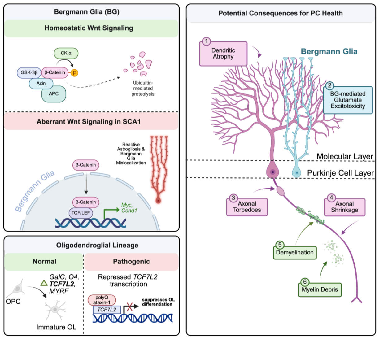
Figure 2.
NF-κβ signaling and JNK signaling in SCA1 pathogenesis. Left Panel: NF-κβ signaling inhibition at 8 weeks reduces astrogliosis but worsens motor coordination, accelarates ML thinning, and reduces climbing fiber innervation. In constrast, NF-κβ inhibition at 16 weeks improves motor coordination and PC health, suggesting that NF-κβ signaling is protective early but detrimental later in disease.Right Panel: Treatment of SCA1-KI mice with the JNK inhibitor SP600125 reduces phospho-c-Jun accumulation and astrogliosis and improves motor coordination and PC health [108, 113]. Created in https://BioRender.com.
Figure 2.
NF-κβ signaling and JNK signaling in SCA1 pathogenesis. Left Panel: NF-κβ signaling inhibition at 8 weeks reduces astrogliosis but worsens motor coordination, accelarates ML thinning, and reduces climbing fiber innervation. In constrast, NF-κβ inhibition at 16 weeks improves motor coordination and PC health, suggesting that NF-κβ signaling is protective early but detrimental later in disease.Right Panel: Treatment of SCA1-KI mice with the JNK inhibitor SP600125 reduces phospho-c-Jun accumulation and astrogliosis and improves motor coordination and PC health [108, 113]. Created in https://BioRender.com.
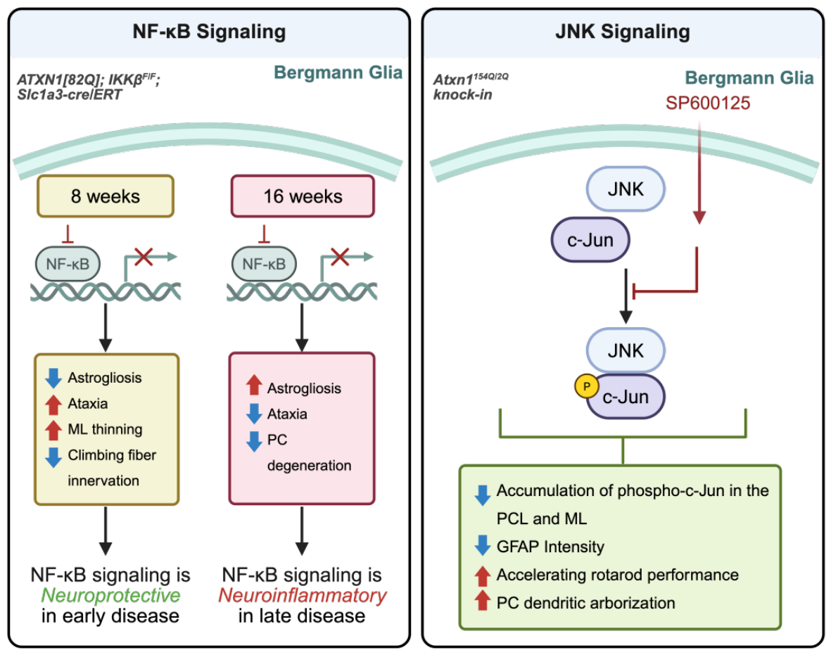
Figure 3.
Proposed TREM2-dependent and TREM2-independent Wnt/β-catenin signaling pathway in microglia. Top Left: In the canonical TREM2-dependent pathway, GSK3β phosphorylation at Ser9 inactivates GSK3β, stabilizes β-catenin, and promotes expression of survival-associated genes, including c-Myc, Ccnd1, Bcl-2. Top Right: A proposed TREM2-independent pathway in which Wnt3a binding to the LRP/Frizzled complex promotes pro-inflammatory cytokine release, including TNFα, through non-canonical JNK/planar cell polarity (PCP) and PLC/Ca²⁺ signaling. Bottom: A hypothetical model in which these pathways compete or act in parallel to influence MG behavior in SCA1 [173-175].Created in https://BioRender.com.
Figure 3.
Proposed TREM2-dependent and TREM2-independent Wnt/β-catenin signaling pathway in microglia. Top Left: In the canonical TREM2-dependent pathway, GSK3β phosphorylation at Ser9 inactivates GSK3β, stabilizes β-catenin, and promotes expression of survival-associated genes, including c-Myc, Ccnd1, Bcl-2. Top Right: A proposed TREM2-independent pathway in which Wnt3a binding to the LRP/Frizzled complex promotes pro-inflammatory cytokine release, including TNFα, through non-canonical JNK/planar cell polarity (PCP) and PLC/Ca²⁺ signaling. Bottom: A hypothetical model in which these pathways compete or act in parallel to influence MG behavior in SCA1 [173-175].Created in https://BioRender.com.
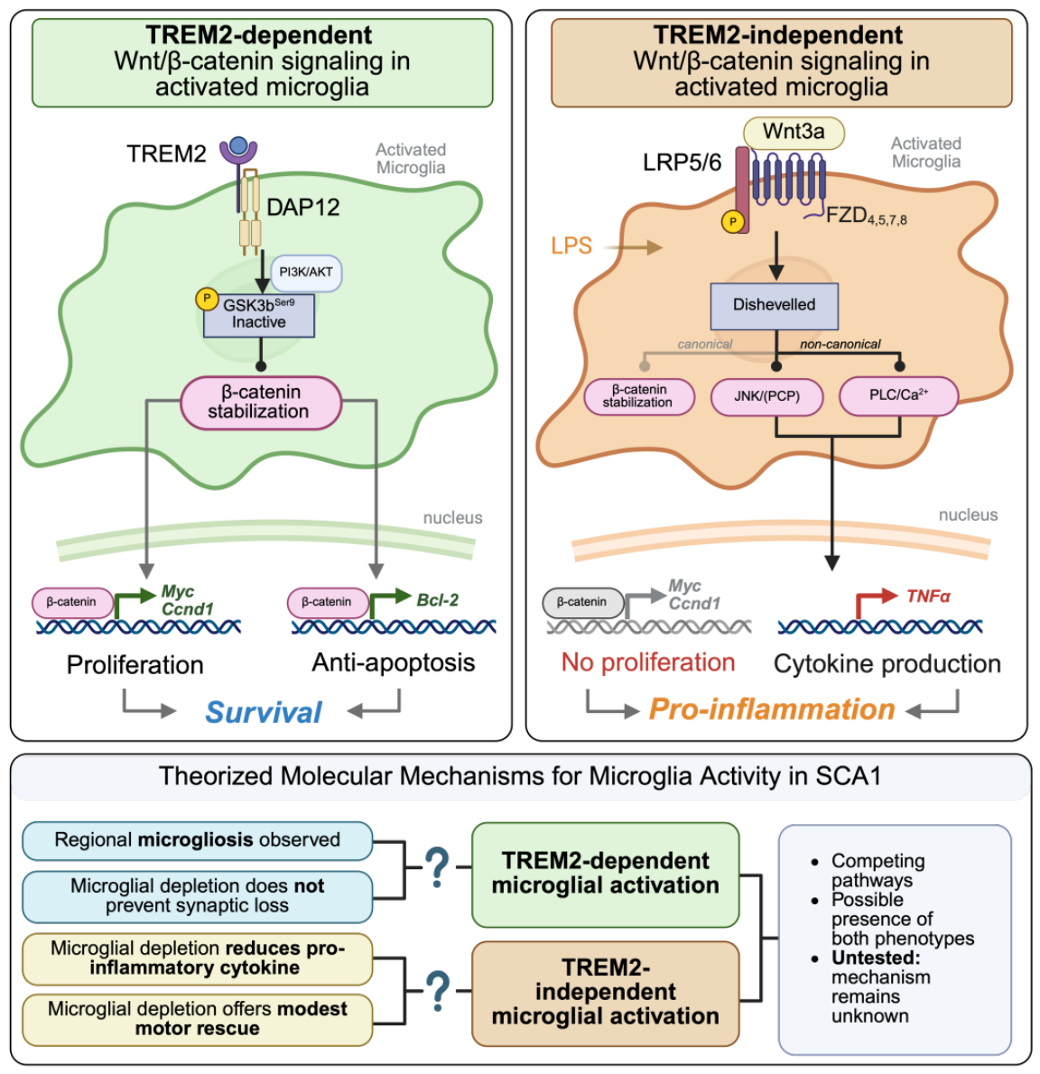

Table 1.
Summary of microglial activation states and their reported roles in homeostasis and disease.
Table 1.
Summary of microglial activation states and their reported roles in homeostasis and disease.
| Disease/models | Microglial subtype | Defining markers/features | Reported functional outcome | Ref. |
|---|---|---|---|---|
| Healthy aged mice | Activated/pro-inflammatory | Ccl4, Ccl3, Cst7, Il1b, Lgals3 | Pro-inflammatory MG are present throughout life and increases with age. | [146] |
| Healthy aged mice | Interferon-responsive | Ifitm3, Rtp4, Oasl2 | This population increases with age and exhibits an inflammatory transcriptional profile. | [146] |
| Healthy early postnatal (P7) mice | Youth-associated microglia (YAM) | Itgax (CD11c) | Transcriptionally similar to DAM; supports developmental programs related to proliferation and metabolism. | [151] |
| AD (human and 5xFAD) | Disease-associated microglia (DAM) | Dkk2, Fabp5, Gpnmb, Igf1, Itgax, Mamdc2, Spp1, and Gm1673 | Often associated with plaque containment, reduced Aβ burden, and fetal-like or anti-inflammatory programs. | [150-152] |
| AD (5xFAD),Trem2-deficient | Disease-inflammatory macrophages (DIM) | Il1a, Il1b, Tnf, Nfkbia, Cd49f, Cd54, and Cd83 | Pro-inflammatory microglia-like cells that promote cytokine release. | [151, 152] |
| MS/EAE | Microglia inflamed in MS – Foamy (MIMS) | Antigen-presentation and lysosomal-dysfunction genes; HLA-A, HLA-B, HLA-DQ, GPNMB, GRN, and CHIT1 | Characterized by altered lipid metabolism; reported as both pro- and anti-inflammatory. | [153] |
| MS/EAE | Microglia inflamed in MS – Iron (MIMS) | Antigen-presentation, ferritin, and complement genes; HLA-DRA, HLA-DPA1, CD74, FTL, FTH1, cC1QA, and C1QB | Associated with heavy-metal sequestration, inflammation, and lesion formation. | [154, 155] |
| HD (Q175/Q175 knock-in; BV2+69Q in vitro) | Mutant HTT-expressing/polyQ-activated MG | Nuclear mutant HTT inclusions in vivo; enlarged soma and shortened, thickened processes in vitro | MG-mediated neuronal toxicity, apoptosis, neurite retraction, and impaired neurite outgrowth. | [156] |
| SCA3/MJD (CMVMJD135) | Activated MG | Reduced ramification, smaller size, and increased soma thickness | Activated at baseline; partial depletion with PLX3397 does not improve motor phenotypes. | [157] |
| SCA1 | Undefined; region-specific microgliosis | Increased Iba1 staining/intensity | MG depletion modestly improves motor behavior but does not prevent PC loss. | [5, 6, 158] |
Disclaimer/Publisher’s Note: The statements, opinions and data contained in all publications are solely those of the individual author(s) and contributor(s) and not of MDPI and/or the editor(s). MDPI and/or the editor(s) disclaim responsibility for any injury to people or property resulting from any ideas, methods, instructions or products referred to in the content. |
© 2026 by the authors. Licensee MDPI, Basel, Switzerland. This article is an open access article distributed under the terms and conditions of the Creative Commons Attribution (CC BY) license (http://creativecommons.org/licenses/by/4.0/).
Copyright: This open access article is published under a Creative Commons CC BY 4.0 license, which permit the free download, distribution, and reuse, provided that the author and preprint are cited in any reuse.