

Submitted:
19 October 2026
Posted:
20 October 2026
You are already at the latest version
Abstract
Human and plants microbiota are different but they also show common specificities, such as regional distribution and a huge diversity of bacteria, fungi, yeasts, viruses, archaea, and even viroids. The plants surface, or phyllosphere commensal microbial population contains a significant number of microbial species with are also present in the human cutaneous microbiota and several of those bacteria have activities in plants that are also of interest for the human skin. Plants also harbor an abundant intra-tissular or endophyte microbiota, suggesting that some vegetal active compounds should be in fact of microbial origin suggesting potential new strategies for the production of active cosmetic and pharmaceutical ingredients. The plant microbiota is at the origin of soluble, but also volatile molecules and exosomes that confer protection against environmental stresses and their activities in now considered for transposition to cosmetic products. Despite the plant phyllosphere is essentially composed of Gram-negative bacterial species while the majority of skin associated bacteria are Gram-positive, they interact following same strategies and, in both cases, communicate with their eucaryotic host through hormones, mimetics compounds and cytokines. Then technics used in phytoremediation could be also applied to preserve skin microbiota homeostasis and skin integrity. In the present review, we investigate the complexity of the plant microbiota in regard of the human counterpart , targeting to establish parallels between microorganisms, molecules and strategies that can be applied in cosmetology and pharmacology.
Keywords:
microbiology
; skin barrier
; claim substantiation
; biomimetic
; plants resources
; innovation
1. Introduction
The skin microbiota is nowadays present in the claims for many cosmetic products and its key role in skin integrity but also pathologies is now well recognized. However, this microbiota is only one of those existing in the human body. Besides the gut and skin microbiota, which are the principal ones, different specific microbiota are also present in the mouth, eye, vagina and in areas generally considered sterile such as the lung or urinary bladder. Increasing evidences on a blood microbiota are even challenging the old concept of sterility of our inner medium [1]. In addition, many species of the human microbiota are also present in the environment including in plants and, as conceptualized under the term of holobiont, constant bidirectional exchanges are occurring between our body and the ecosystem [2]. The human and plants microbiota are showing major differences but also common species, metabolites and mechanisms of action that remain poorly investigated although plants represent a major resource in cosmetic and pharmaceutic industries.
In the present review, we aimed at investigating the specificities and common features of the plant with the human skin microbiota that could inspire new strategies for innovative bio-inspired active compounds.
2. Plant Versus Human Microbiotas
As in humans, the plant microbiome comprises beneficial, neutral, and pathogenic microorganisms. This microbiome is involved in growth, nutrient uptake, and protection against pathogens. Some of these microorganisms, such as Pseudomonas, may individually have beneficial activities, but the role of microbial communities, particularly those present in the soil in plant health is also increasingly recognized. A new paradigm is emerging today following the observation that plants adaptation to their environment should result as much from the plant’s physiology itself and from its microbiota. This level of symbiosis and commensalism between plants and their microbiota likely stems from their static nature, and these relationships seem even more pronounced than in humans.
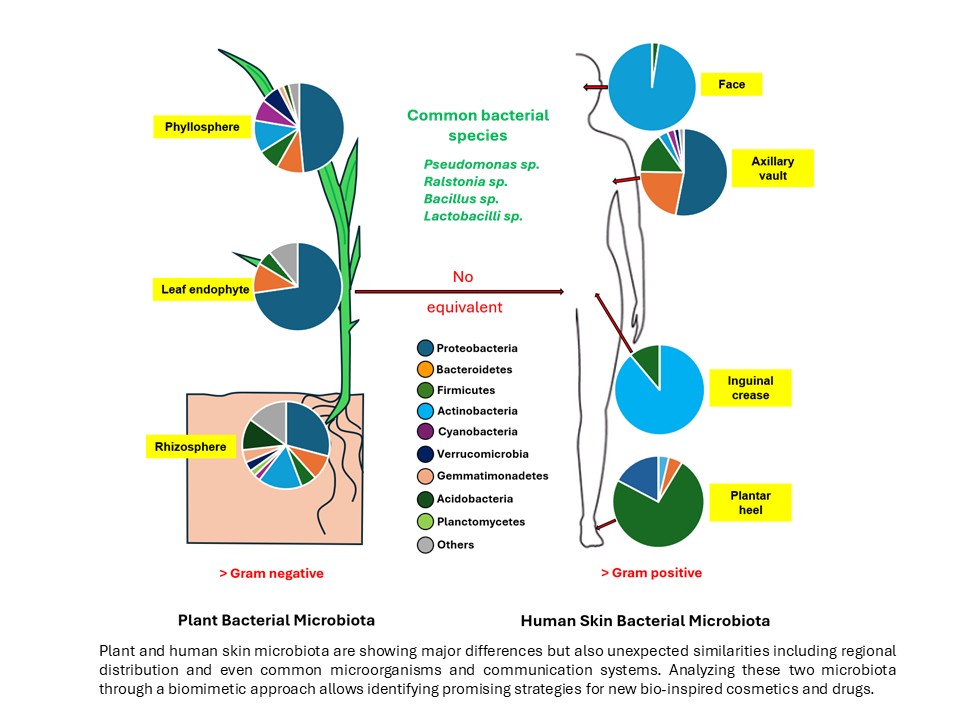
Just like in the human body, plants host different microbiomes following compartments. In surface, we find the phyllosphere microbiota which includes microorganisms associated with surface organs, including leaves, flowers, and fruits. Despite their great diversity, a “core” of bacterial species can be identified in the phyllosphere that appears to be present in most plants: cereals, sugarcane, grapevines, soybeans, or Arabidopsis thaliana. Among these bacteria are Pseudomonas, Agrobacterium, Methylobacterium, Sphingomonas, Erwinia, Cladosporium, Coniothyrium, Resinicium, and Fusarium [3]. Proteobacteria account for 50% of the phyllosphere microbiota. Among the other phyla of the phyllosphere are, in particular, Bacteroidetes, Firmicutes, and Actinomycetes. The phylum Proteobacteria was recently renamed Pseudomonadota and, as the name suggests, this phylum includes Gram-negative bacteria such as Pseudomonas, Vibrio, Rhizobium (involved in nitrogen fixation), Acetobacter, Burkholderia, and Acinetobacter. This represents a significant difference from the human skin microbiome, which, outside of moist areas, is largely dominated by Gram-positive bacteria. However, as on human skin, bacteria of the genera Streptomyces, Bacillus, and even Enterobacteriaceae are also found [3,4].
The rhizosphere microbiota is the second specific plant microbiota. It is corresponding to the microbiota present in surface and at the vicinity of the roots. For that reason, it is influenced by the composition of the soil microbiota but differs from the bulk soil microbiota with an enrichment of fast-growing Gram-negative bacterial species of the Proteobacteria and Bacteroidetes phyla [5]. These bacteria are attracted by plant exudates. In contrast other typical soil bacterial species such as Gemmatimonadota or Acidobacteria are depleted in in this environment. A parallel should be established between the skin and plantar microbiota in human, which also remarkably differ in diversity.
A great difference between plants and human microbiotas is linked to their internal organization and to the absence of inner organs in plants. In addition, whereas in human the inner medium is considered sterile, plants host an abundant population of microorganisms in their tissues. This endophyte microbiota resides in all parts of the plant, with also significant differences between leaf and roots [3]. The bacterial communities of the endophyte are dominated by Proteobacteria and Firmicutes (renamed Bacillota) and, to a lesser extent, by Bacteroidetes, regardless of the composition of the soil microbiota. There is no equivalent in human although living bacteria can be encountered in the skin just to the stratum basalis [6] and increasing evidences (DNA signatures, bacterial like particles) suggest that blood should harbor its own microbiota [1].
Except in cases of infection, the plant fungal community (fungal microbiota) is less diverse than the bacterial community and is dominated by Ascomycetes (higher fungi) and Basidiomycetes (cap mushrooms). The ratio of these two phyla varies within limited proportions between the rhizosphere, the endophyte, and the phyllosphere. Among the representatives of microscopic fungi and yeasts (102 to 108 CFU/g of leaf) are spores of the genera Acremonium, Alternaria, Aspergillus, Cladosporium, Mucor, and Penicillium [4,7]. Yeasts are the primary members of the phyllosphere. Among the main genera are Sporobolomyces, Cryptococcus, and Rhodotorula. The latter two genera are also present in the human skin fungeome, but this one is dominated by the genus Malassezia, with the exception of the foot plantar region [8] while in plants, the composition of the fungeome appears to be primarily linked to that of the surrounding soil [4]. The extent of the interactions between the plant fungal community and plant physiology appears less extensive than with bacteria, even though several genera have been associated with beneficial effects on plant growth and stress resistance [9,10]. In a recent study, inoculation of Asteraceae species with different arbuscular mycorrhizal fungi revealed that plant phenotypic variations, including biomass, vegetative or reproductive traits, can be correlated to the fungal taxon and to their relative phylogenetic distance [11] but the portion of the fungeome that has been studied to date remains limited.
Regarding the plant virome, as in the case of the human skin microbiome, both eukaryotic viruses and bacteriophages are present. There is relatively little data on the eukaryotic virome of plants, although, as in human skin, these viruses do not appear to be limited to pathogenic action but may also maintain commensal or even mutualistic interactions and contribute to the ecological adaptation of plants [12]. DNA viruses are found in a circulating or lysogenic form integrated into the plant genome, as well as RNA viruses [13]. Given the very high specificity of phages, one finds in particular phages targeting rhizosphere bacteria, including Rhizobium, as well as numerous phages of phytopathogenic Gram-negative bacteria such as Erwinia or Xanthomonas, or conversely plant commensals such as Pseudomonas putida, Pseudomonas syringae, or Pseudomonas fluorescens (Phage Φ2, which has been extensively characterized). These phages are typically lysogenic (integrated into the bacterial DNA) or lytic (particulate) [14].
The plant microbiome naturally includes archaea belonging to the phyla Thaumarchaeota, Crenarchaeota, and Euryarchaeota. This community appears to vary depending on the plant region. These archaeal communities possess the ability to degrade glycogen, which is generally released in fungal exudates, and appear to be involved in stress tolerance and nutrient uptake [3]. It is interesting to note that some of these genera of soil archaea (Thaumarchaeota and Euryarchaeota) are also found in the human skin archaeome [15].
Viroids are a class of particles that have long been considered unique to plants. They consist of a circular, linear, or folded RNA strand without any associated capsid or protein. These RNA strands are generally non-coding but exhibit enzymatic activity (ribozymes). Viroids are associated with several plant diseases [16]. However, it has recently been demonstrated that viroid-like particles, known as obelisks, are also present in the human gut and oral microbiota [17]. Their presence in the human skin microbiota was not investigated until now but this hypothesis is highly probable. Unlike plant viroids, obelisks encode proteins in addition to their ribozyme activity and are thought to specifically infect bacteria, modulating their populations in a manner similar to that of phages. As phages, whichare among the most promising tools for human pathogen resistant bacteria elimination, including Staphylococcus aureus and acneic Cutibacterium acnes strains, viroids and obelisks should represent a new generation of biological tools.
Taken together these observation suggest that, given the abundance of bacterial endophytes, i.e.10⁴ to 10⁸ bacteria per gram of root tissue and 106 to 108 bacteria per gram of leaf in the phyllosphere and even more with the fungal endophyte [18], it is highly likely that many of the activities attributed to plants are in fact activities linked to metabolites derived from the endophyte, suggesting that molecules of interest initially identified in plants could subsequently be produced by bio-fermentation using the identified microorganisms or microbial communities.
3. Plant Microorganisms in the Human Skin Microbiome
The existence of a soil-plant-human gut axis is a concept consistent with the notion of holobiont connecting the entire ecosphere [19] and explains the existence of common microorganisms between plants and humans [20]. In the case of bacteria, although a same species should be represented by different strains in plants and humans, that is also indicating that they share a minimum of 96.8% average nucleotide identity in their core genome and therefore essential common physiological properties [21],
Regarding the plant and skin microbiome, several species are shared. Bacillus subtilis plays a key role in plant stress resistance by promoting the growth of Rhizobia, but this bacterium is also capable of producing vitamins and antioxidant metabolites. On the skin, B. subtilis appears capable of antagonizing S. aureus and reducing acne [22]. Similar results, with correction of dysbiotic skin microbiota, have been observed in animals [23,24].
Pseudomonas species, such as Pseudomonas chlororaphis, are capable of inhibiting the development of plant pathogens both through competition and by producing molecules with antimicrobial activity [25]. Certain species, such as P. chlororaphis or P. pergaminensis, exhibit biostimulant and probiotic activities for plants and even in aquaculture [25,26]. At the same time, Pseudomonas species such as P. syringae or P. fluorescens produce biosurfactants that preserve the permeability and hydration of the plant cuticle [27]. A bacterium such as P. syringae also possesses the ability to nucleate water, allowing it to form ice at temperatures above 0 °C, which protects the surface of leaves [28]. Finally, depending on their growth conditions, certain species such as P. fluorescens have the ability to produce pigments providing protective effects against UV radiations [29]. On the skin, Pseudomonas (P. fluorescens in healthy skin) are a minority species except in moist areas where they may constitute the majority of the population [30]. Cutaneous strains of P. fluorescens have very low virulence and exhibit commensal behavior [31]. A study has shown that the Pseudomonas population on the skin is significantly reduced in patients with acne, particularly on the face [32]. This phenomenon is thought to result from competition with C. acnes suggesting that in skin this species should have protective effects since in addition it can produce biosurfactant [33] and could contribute to the regulation of skin hydration.
The genus Ralstonia includes soil-dwelling bacteria known for their phytopathogenic activity, such as Ralstonia solanacearum, but there are also non-pathogenic species such as Ralstonia mannitolilytica, which is capable of producing bacteriocins. The bacteriocins have demonstrated antibiofilm activity effective against catheter-related infections in humans [34]. On the skin of the forehead, the genus Ralstonia may account for up to 8.7% of the skin microbiome [35]. This population appears highly sensitive to the use of cosmetics containing complex polysaccharides [35], and as with Pseudomonas, a decline in the Ralstonia population is observed in acne-prone skin [32].
Lactobacilli, including Lactobacillus plantarum, Lactobacillus paracasei, and Lactobacillus pentosus (now Lactiplantibacillus), are present in the environment and on plants (leaves, fruits), but they are also found in the human digestive tract and, to a lesser extent, on the skin. These microorganisms are known as probiotics or postbiotics, and their beneficial effects are recognized both in the gut [36] and on the skin [37].
Arthrobacter agilis is a rhizosphere bacterium that promotes plant growth by improving iron availability [38]. It is also known as the “snow bacterium” due to its ability to withstand extreme cold, UV radiation, and oxidative stress. These properties are linked to its ability to synthesize red pigments, bacteriorubins, which possess chaperone-like (protein protection) and antioxidant activities [39]. These molecules have been formulated into cosmetic products. It should be noted that the resistance of extremophile bacteria such as Arthrobacter agilis or Deinococcus radiodurans (environmental bacteria found in extreme environments) is the basis of the “metatheory of aging,” which suggests that protecting the proteome alongside combating inflammation is one of the key elements in protecting against the effects of aging [37]. D. radiodurans also expresses exopolysaccharides that inhibit biofilm formation by S. aureus [40].
Other bacteria in the plant microbiota, with the exception of Acinetobacter, which is primarily known as pathogen, are rarely found in the skin microbiota. However, it can be observed here that certain plant-derived species exhibit beneficial effects on the skin microbiome and the skin itself and may lend themselves to the formulation of pro- or postbiotics derived from the plant microbiome, with potential interest in the treatment of acne or even atopic dermatitis.
Few applications of the plant fungeomes are currently known. However, we can cite yeasts of the genus Pichia, particularly Pichia naganishii, isolated from the exudate of Camellia japonica [41], which was patented for its effectiveness in strengthening the skin barrier. This patent is based on a biomimetic approach modeled after the protective action of yeast on plant tissues [37].
For archaea, their increase appears to be correlated with a decrease in skin hydration [15]. Certain halophilic species found on the skin (Halobacteriota) are adapted to the high salt concentrations present in sweat. Thaumarchaeota have the ability to oxidize ammonia under aerobic conditions and produce nitrites. Nitrification activity, and the resulting decrease in pH, could have a positive effect on the skin. Sweat contains urea, which could serve as a metabolite in these reactions, and it has been proposed to apply Thaumarchaeota to the skin (archaeobiotics) to reduce odors and lower skin pH [42]. However, culturing archaea is extremely difficult, and although these species are considered cultivable, to our knowledge, to date no one has claimed to have cultured human skin archaea.
Eukaryotic viruses, due to their very high specificity, are poorly suited for application in other contexts; however, the plant virome contains a wide variety of phages that warrant consideration [43]. Acting primarily on proteobacteria, and thus on species that are essentially commensal and beneficial to the skin (P. fluorescens, Ralstonia sp.), they can serve as models for defining the conditions that reduce their activity in the skin microbiome and protect these beneficial bacteria. Given that phage recognition of the bacterium is stereospecific and highly charge-dependent, the polarity of cosmetic formulations could influence this process. The use of phages themselves remains prohibited in the EU to date [44], which has led to efforts to utilize the endolysin expressed by these phages. However, to date, this strategy employed against S. aureus, including its multidrug-resistant forms, remains under regulatory review in the cosmetics sector.
Viroids are well known as plant pathogens, and the discovery of the human viroidome and obelisks [17] has opened up a new field of research. Their presence in the skin has yet to be demonstrated, but the identified mechanism of action of obelisks is of great interest because it resembles that of phages. Indeed, these ribozymes appear to target bacteria rather than eukaryotic cells. They could therefore play a role identical to that of phages in regulating the microbiome, and potentially the skin microbiome. Furthermore, their simple structure (an unencapsulated RNA loop) could simplify their handling. The question of their regulatory acceptability in cosmetics remains open, especially since, as a recent discovery, we have no historical perspective on their actual potential.
4. Plant Microbiome Metabolism and Its Transposition to the Skin
Bioremediation by soil microorganisms is widely used in environmental pollutants removal. The rhizosphere of buckwheat possesses a specific microbiota that is essential for the plant adaptation to its environment and its resistance to numerous environmental toxins, including heavy metals (copper, zinc, aluminum, or lead) [45]. The soil bacterium Paracoccus aminovorans, which is also found in association with plants, facilitates the degradation of Polycyclic Aromatic Hydrocarbons (PAHs). Applying buckwheat extract to the skin increases the presence of P. aminovorans without inducing skin dysbiosis, suggesting that this bacterium could reduce the skin exposure to PAHs and inflammation associated with this pollutant [37,46].
Among the microorganisms found in or on plants as well as on human skin and capable of bioremediation, we can also mention bacteria of the genus Acinetobacter, which degrade phenol and methanol, Bacillus cereus, which is capable of degrading ethylbenzene; Corynebacteria, which metabolize n-alkanes; and Pseudomonas, which, depending on their culture conditions, metabolize oils, hexadecane, phenanthrene, xylene, or salicylates [4]. The effect of environmental pollutants (NO2 in particular) was also studied on skin strains of Corynebacterium tuberculostearicum, C. acnes, S. aureus, S. epidermidis, S. capitis, and P. fluorescens. Each strain is affected differently in terms of growth and biofilm formation, with P. fluorescens appearing to be the most resistant [47]. The skin microbiota thus appears to serve as the skin first line of defense against environmental pollutants.
The plant microbiome has numerous beneficial effects on plants beyond the direct effects related to nitrogen fixation, the transport of ammonium (NH4+) and nitrate (NO3-) ions, and the well-known symbiotic mechanisms in leguminous plants involving bacteria of the genus Rhizobium. In fact, bacteria in the plant microbiome oxidize and chelate elements such as phosphate, potassium, or iron, making them more accessible to plant cells in the form of organic acids or bound to siderophores [3]. Inspiring from the symbiosis between microorganisms and plants is a potential source of cosmetic active ingredients. For example, the fungus Pseudozyma flocculosa, which has a symbiotic relationship with clover, produces a glycolipid called flocculosin among its secondary metabolites, which has anti-inflammatory and antimicrobial effects of potential interest in cosmetics [37,48].
Exosome-like nanoparticles derived from ginger and containing microRNAs have been shown to be absorbed by Lactobacilli, including L. rhamnosus. One of these microRNAs targets the ycnE monooxygenase and induces the production of indole-3-carboxaldehyde (I3A) and interleukin 22, which is known to positively affect intestinal barrier function [49]. Regarding the skin, the effects on or via the skin microbiota remain to be studied, but these plant-derived exosomes have been shown to have direct protective effects against UV radiation and oxidative stress [50]. They could also serve as carriers for the delivery of peptides in cosmetic products [51].
Plants are also a source of molecules with antimicrobial activity, such as sesquiterpene lactones, which are active against S. aureus [52], flavonoids [53], or polyphenols [54]. Some of these molecules, such as α-mangostin (xanthone), are active against Gram-positive bacteria and are being studied for their applications in cosmetics [37].
Volatile metabolites constitute what is now referred to as the “volatilome,” and every organism and all environments generate volatile molecules that are more or less unique to them. Due to their volatile nature, these molecules remain difficult to detect and characterize, as they require techniques such as headspace analysis coupled with mass spectrometry [55], but both the plant volatilome [56] and the human eukaryotic [57] or microbial microbiota [58] are beginning to be characterized. It often remains difficult to distinguish within a volatilome between molecules produced by the organism (plant or human) and those produced by the associated microbiota, as some molecules are of mixed origin [59]. Some of these molecules may play a central role, such as 1-undecene, which is involved in the communication of P. fluorescens [60] or in the regulation of hair growth [61]. Some of these molecules may possess a fragrance and properties of interest in the cosmetic field.
A recent development related to microbiota metabolism, though not limited to plant or skin microbiota, concerns the concept of cosmetomicrobiomics. This concept derives from that of pharmacomicrobiomics, which studies the effects of gut microbiota metabolism on drugs [62]. Indeed, the enzymatic activity of the gut microbiota can lead to major changes in the structure of orally administered pharmaceutical active molecules, potentially resulting in their cleavage and inactivation even before absorption by the intestine. However, when understood, this metabolism can also be harnessed to generate active ingredients in situ from inactive precursors (prodrugs), thereby protecting fragile molecules until they reach their site of absorption in the intestine. At the skin level, this principle has already been applied in the case of vitamin C, which can be used in cosmetic products in the form of AA2G (ascorbic acid 2-glucoside) or stabilized vitamin C [63]. The same strategy could be applied to other unstable cosmetic active ingredients such as peptides, phenolic compounds, and even pigments.
5. Microbial Communication: From Plants to Human Skin
Intra- and inter-kingdom communication plays a crucial role in plant health. Applying this concept to the human skin must take into account species differences and variations in metabolism across different microbiomes [3,64].
Within plant microbiomes, quorum sensing plays a central role in controlling the growth and expression of different bacterial species, but it should be noted that, since these are primarily Gram-negative bacteria, the intra- and even inter-species communication molecules are mainly N-acyl homoserine lactones (H-AHLs) composed of a lactone ring and a saturated or unsaturated aliphatic chain. Conversely, since the human skin microbiota consists primarily of Gram-positive bacteria, the signaling molecules in these microorganisms are mainly linear or cyclic peptides. Certain molecules, such as autoinducer 2 (AI2), can be expressed in both Gram-positive and Gram-negative bacteria, but their role in population dynamics appears to be less significant [65]. Attempting to stimulate the growth of skin bacteria such as S. epidermidis would therefore require the production of complex polymers, often containing rare amino acids, a typical feature of Gram-positive quorum sensing peptides [66]. On the other hand, to date, the communication mechanisms in some major bacteria of the skin microbiota, such as C. acnes, are unknown. For other microorganisms like P. fluorescens, although it is a Gram-negative bacterium, quorum sensing does not appear to involve N-HSL, but rather a volatile molecule such as 1-undecene [60], or even type VI secretion systems [67]. Interbacterial competition also appears to depend on other types of secretion systems, such as the type VII secretion system, which can be expressed in both Gram-negative [68] and Gram-positive [69] bacteria and membrane extracellular vesicles, which have multiple roles and have promising biotechnological applications [70].
Plant pathogens, such as bacteria of the genera Pectobacterium, Erwinia, or Dickeya also communicate through the production of N-HSL, but it has been observed that these molecules can be hydrolyzed by commensal bacteria, which can inhibit their growth, biofilm formation, and virulence expression. These observations have led to the development of biocontrol strategies to protect plants from diseases such as blackleg in Solanaceae through the selection of natural strains of Rhodococcus erythropolis that secrete enzymes capable of degrading the N-HSLs produced by Pectobacterium [71] or Rhizobium [72]. This mechanism, known as quorum quenching, could be applied to the skin microbiota, subject to structural differences in the molecules involved. Apart from the inactivation of quorum sensing genes, as observed in S. aureus, another strategy for blocking this process is the overexpression of efflux pumps, which would limit the activation of the system by autoinducers [73]. Plant metabolites such as baicalein derived from Scutellaria, a flowering plant species in the Lamiaceae family, as well as from pink stonecrop and Ceylon currant, inhibit the comX and luxS genes involved in the expression of tryglycine B, a quorum sensing peptide in the Gram-positive bacterium Streptococcus mutans [74]. The combination of molecules such as trans-cinnamaldehyde derived from cinnamon with antimicrobials such as chlorhexidine or fluoride enhances the inhibitory effect on the comX and luxS genes involved in quorum sensing in this species [75]. Similarly, walkmycin C, an antibiotic produced by the Streptomyces strain MK632-100F11, as well as probiotics expressed by Lactobacilli, interfere with the quorum sensing signaling pathway in S. mutans [74]. Coumarin and green tea extracts have been tested against the Gram-negative bacterium Porphyromonas gingivalis, demonstrating inhibitory effects on the quorum-sensing. One of the main components of green tea, epigallocatechin-3-gallate, inhibits adhesion and biofilm formation by acting on the AI2 bacterial communication pathway [76]. D-galactose also appears to possess quorum quenching activity on the AI2 pathway, with inhibitory effects on biofilm formation by several microbial species [77]. In the case of the yeast Candida albicans, it has been observed that farnesol, a sesquiterpene with a structure similar to that of dodecanal, and tyrosol, a derivative of tyrosine, inhibit biofilm formation [78]. The issue with most, if not all, of these molecules is that they target the Lux pathway, and the LuxR/S system is involved in quorum sensing in both Gram-positive and Gram-negative bacteria, resulting in limited specificity of action. Furanones, produced by red alga but also higher plant species, can also be cited for their quorum sensing-inhibiting and antibiofilm activity in many microbial species, but their use is limited by their toxicity and carcinogenicity [79].
Inter-kingdom communication enables the development of other strategies for regulating the skin microbiota, and this communication occurs in both directions. The plant microbiota can stimulate plant growth by metabolizing tryptophan and producing molecules that are identical to or related to (molecular mimetics) phytohormones such as auxins, gibberellins, and cytokinins [3,80,81]. Conversely, plants produce signaling molecules capable of influencing microbial physiology. This is particularly true of γ-aminobutyric acid (GABA). GABA is the primary inhibitory neurotransmitter in the human central nervous system, but it also serves as a signaling and alarm molecule in plants. GABA transporters and sensors have been identified in numerous microorganisms such as Escherichia coli, Bacillus subtilis, and Agrobacterium tumefaciens [82]. It has been observed that GABA stimulates biofilm production as well as virulence in P. fluorescens [83] and P. aeruginosa [84], in which GABA also modulates chemotaxis [85]. Similarly, it has been demonstrated that a bacterium used in biocontrol, Serratia plymuthica A153, is capable of detecting and responding to auxin produced by plants [86]. Microbial endocrinology, which studies the effect of hormones and neurohormones on bacteria, is therefore not limited to mammals and humans but also concerns the interaction of plants with their microbiota. It should, of course, be noted that at the skin level, numerous signaling factors such as catecholamines, Substance P, Calcitonin Gene-Related Peptide (CGRP), or natriuretic peptides are capable of modulating the virulence and biofilm production of both Gram-positive and Gram-negative bacteria [87]. Even steroid hormones such as estradiol modulate adhesion and biosurfactant production in Lactobacillus crispatus [88,89], and recent studies suggest that androgens may regulate the virulence of S. aureus at the skin level [90]. The effects of host hormones on microorganisms are not limited to bacteria, as it has been shown that the galanin message-associated peptide, a molecule abundant in the skin, can regulate the growth of C. albicans [91]. Knowledge of these mechanisms was the basis of patents for cosmetics targeting acneic and sensitive skins; however, most of the regulatory pathways involved is skin-microbiota communication have not yet been explored for cosmetic applications.
6. Conclusion
The biosphere, and in particular the plant world, is an infinite source of inspiration for the development of new cosmetic active ingredients and new strategies, and as we can see, we have studied only a tiny fraction of it. Living organisms, beyond the exchange of matter, work, and heat taught by thermodynamics, are characterized first and foremost by the exchange of information and communication. Beyond the sole objective of bringing new cosmetic products to market, the accompanying message will inevitably lead one day to the recognition of this bond that unites all living structures, and there is still time to embrace the concept of the holobiont. As a 2010 article in New Scientist entitled “the real Avatar” already asserted [92], and long before James Lovelock and the Gaia theory [93], reality is very likely far greater than fiction.
Author Contributions
MF: Conceptualization; Writing – review & editing. SD: Writing – review. CL: Writing – review. VS-B: Project administration; Supervision; Validation. GR Sci Adv Borad: Supervision; Validation.
Funding
University of Rouen Normandy, Evreux Agglomeration, Région Normandy, French National Research Agency (ANR), European Union (FEDER) and Groupe Rocher.
Institutional Review Board Statement
Not applicable.
Informed Consent Statement
Not applicable.
Data Availability Statement
Review based on published data analysis.
Conflicts of Interest
The authors declare no conflict of interest.
Acknowledgments
Not applicable.
References
- Cheng, H.S.; Tan, S.P.; Wong, D.M.K.; Koo, W.L.Y.; Wong, S.H.; Tan, N.S. The blood microbiome and health: Current evidence, controversies, and challenges. Int. J. Mol. Sci. 2023, 24, 5633. [Google Scholar] [CrossRef] [PubMed]
- Garg, N. Metabolomics in functional interrogation of individual holobiont members. mSystems 2021, 6, e0084121. [Google Scholar] [CrossRef] [PubMed]
- Trivedi, P.; Leach, J.E.; Tringe, S.G.; Sa, T.; Singh, B.K. Plant-microbiome interactions: From community assembly to plant health. Nat. Rev. Microbiol. 2020, 18, 607–621. [Google Scholar] [CrossRef] [PubMed]
- Bashir, I.; War, A.F.; Rafiq, I.; Reshi, Z.A.; Rashid, I.; Shouche, Y.S. Phyllosphere microbiome: Diversity and functions. Microbiol. Res. 2022, 254, 126888. [Google Scholar] [CrossRef] [PubMed]
- Ling, N.; Wang, T.; Kuzyakov, Y. Rhizosphere bacteriome structure and functions. Nat. Commun. 2022, 13, 836. [Google Scholar] [CrossRef] [PubMed]
- Nakatsuji, T.; Chiang, H.-I.; Jiang, S.B.; Nagarajan, H.; Zengler, K.; Gallo, R.L. The microbiome extends to subepidermal compartments of normal skin. Nat. Commun. 2013, 4, 1431. [Google Scholar] [CrossRef] [PubMed]
- Rodriguez, R.J.; White, J.F.; Arnold, A.E.; Redman, R.S. Fungal endophytes: Diversity and functional roles. New Phytol. 2009, 182, 314–330. [Google Scholar] [CrossRef] [PubMed]
- Underhill, D.M.; Iliev, I.D. The mycobiota: interactions between commensal fungi and the host immune system. Nat. Rev. Immunol. 2014, 14, 405–416. [Google Scholar] [CrossRef] [PubMed]
- Santos, A.; Domingo, M. Killer toxin of Pichia membranifaciens and its possible use as a biocontrol agent against grey mould disease of grapevine. Microbiology 2004, 150, 2527–2534. [Google Scholar] [CrossRef] [PubMed]
- Xia, Y.; Sahib, M.R.; Amna, A.; Opiyo, S.O.; Zhao, A.; Gao, Y.G. Culturable endophytic fungal communities associated with plants in organic and conventional farming systems and their effects on plant growth. Sci. Rep. 2019, 9, 1669. [Google Scholar] [CrossRef] [PubMed]
- Turanoglu, C.; Ancel, H.; Le Lann, C.; Van Baaren, J.; Bazin, O.; Declerck, S.; Fort, F.; Fontaine Breton, T.; Vandenkoornhuyse, P.; Mony, C. Functional traits of Asteraceae species vary with arbuscular mycorrhizal fungal identity and phylogeny. Mycorrhiza 2025, 35, 40. [Google Scholar] [CrossRef] [PubMed]
- Poupin, M.J.; González, B. Embracing complexity in plant-microbiome systems. Environ. Microbiol. Rep. 2024, 16, e70000. [Google Scholar] [CrossRef] [PubMed]
- Aghdam, S.A.; Lahowetz, R.M.; Brown, A.M.V. Divergent endophytic viromes and phage genome repertoires among banana (Musa) species. Front. Microbiol. 2023, 14, 1127606. [Google Scholar] [CrossRef] [PubMed]
- Koskella, B.; Taylor, T.B. Multifaceted impacts of bacteriophages in the plant microbiome. Ann. Rev. Phytopathol. 2022, 56, 17.1–17.20. [Google Scholar]
- Moissl-Eichinger, C.; Probst, A.J.; Birarda, G.; Auerbach, A.; Koskinen, K.; Wolf, P.; Holman, H.-Y.N. Human age and skin physiology shape diversity and abundance of archaea on skin. Sci. Rep. 2017, 7, 4039. [Google Scholar] [CrossRef] [PubMed]
- Tsagris, E.M.; Martínez de Alba, A.E.; Gozmanova, M.; Kalantidis, K. Viroids. Cell. Microbiol. 2008, 10, 2168–2179. [Google Scholar] [CrossRef] [PubMed]
- Zheludev, I.N.; Edgar, R.C.; Lopez-Galiano, M.J.; de la Peña, M.; Babaian, A.; Bhatt, A.S.; Fire, A.Z. Viroid-like colonists of human microbiomes. Cell 2024, 187, 6521–6536.e18. [Google Scholar] [CrossRef] [PubMed]
- Oukala, N.; Aissat, K.; Pastor, V. Bacterial endophytes: The hidden actor in plant immune responses against biotic stress. Plants 2021, 10, 1012. [Google Scholar] [CrossRef] [PubMed]
- Weiland-Bräuer, N. Friends or foes-Microbial interactions in nature. Biology 2021, 10, 496. [Google Scholar] [CrossRef] [PubMed]
- Ma, H.; Cornadó, D.; Raaijmakers, J.M. The soil-plant-human gut microbiome axis into perspective. Nat. Commun. 2025, 16, 7748. [Google Scholar] [CrossRef] [PubMed]
- Chung, M.; Munro, J.B.; Tettelin, H.; Dunning Hotopp, J.-C. Using core genome alignments to assign bacterial species. mSystems 2018, 3, e00236-18. [Google Scholar] [CrossRef] [PubMed]
- Alessandrini, G.; Mercuri, S.R.; Martella, A.; Ferrara, F.; Simonetti, V.; Trifirò, C.; Emanuele, E. Topical application of bacteriocins from Bacillus subtilis promotes Staphylococcus aureus decolonization in acneic skin and improves the clinical appearance of mild-to-moderate acne. Postep. Dermatol. Alergol. 2023, 40, 115–118. [Google Scholar] [CrossRef] [PubMed]
- Moskovicz, V.; Ben-El, R.; Horev, G.; Mizrahi, B. Skin microbiota dynamics following B. subtilis formulation challenge: an in vivo study in mice. BMC Microbiol. 2021, 21, 231. [Google Scholar] [CrossRef] [PubMed]
- Vicente-Gil, S.; Nogales-Mérida, S.; Gonçalves, G.; Serra, C.R.; Camino Ordás, M.; Tafalla, C.; Díaz-Rosales, P. The oral delivery of Bacillus subtilis spores modulates skin and intestinal mucus responses in rainbow trout (Oncorhynchus mykiss). Aquac. Rep. 2024, 37, 102253. [Google Scholar] [CrossRef]
- Anderson, A.J.; Kim, Y.C. Insights into plant-beneficial traits of probiotic Pseudomonas chlororaphis isolates. J. Med. Microbiol. 2020, 69, 361–371. [Google Scholar] [CrossRef] [PubMed]
- Díaz, M.; Bach, T.; González Anta, G.; Agaras, B.; Wibberg, D.; Noguera, F.; Canciani, W.; Valverde, C. Agronomic efficiency and genome mining analysis of the wheat-biostimulant rhizospheric bacterium Pseudomonas pergaminensis sp. nov. strain 1008T. Front. Plant. Sci. 2022, 13, 894985. [Google Scholar] [CrossRef] [PubMed]
- Van Der Wal, A.; Leveau, J.H.J. Modelling sugar diffusion across plant leaf cuticles, the effect of free water on substrate availability to phyllosphere bacteria. Environ. Microbiol. 2011, 13, 792–797. [Google Scholar] [CrossRef] [PubMed]
- Attard, E.; Yang, H.; Delort, A.M.; Amato, P.; Poschl, U.; Glaux, C.; Koop, T.; Morris, C.E. Effects of atmospheric conditions on ice nucleation activity of Pseudomonas. Atmos. Chem. Phys. 2012, 12, 10667–10677. [Google Scholar] [CrossRef]
- Diaz Appella, M.N.; Kolender, A.; Oppezzo, O.J.; López, N.I.; Tribelli, P.M. The structural complexity of pyomelanin impacts UV shielding in Pseudomonas species with different lifestyles. FEBS Lett. 2024, 598, 2702–2716. [Google Scholar] [CrossRef] [PubMed]
- Grice, E.A.; Kong, H.K.; Renaud, G.; Young, A.V.; NISC Comparative Sequencing Program; Bouffard, G.G.; Blakesley, R.W.; Wolfsberg, T.G.; Turner, M.L.; Segre, J.A. A diversity profile of the human skin microbiota. Genome Res. 2008, 18, 1043–1050. [Google Scholar] [CrossRef]
- Hillion, M.; Mijouin, L.; N’Diaye, A.; Percoco, G.; Enault, J.; Follet-Gueye, M.-L.; Yvergnaux, F.; Lefeuvre, L.; Feuilloley, M.G.J. Pseudomonas fluorescens, a forgotten member of the human cutaneous microflora sensible to skin communication and defense peptides. Int. J. Curr. Microbiol. Appl. Sci. 2014, 3, 910–925. [Google Scholar]
- Guo, Z.; Yang, Y.; Wu, Q.; Liu, M.; Zhou, L.; Zhang, L.; Dong, D. New insights into the characteristic skin microorganisms in different grades of acne and different acne sites. Front. Microbiol. 2023, 14, 1167923. [Google Scholar] [CrossRef] [PubMed]
- Duclairoir Poc, C.; Meylheuc, T.; Ngoya, S.; Bodilis, J.; Taupin, L.; Mérieau, A.; Feuilloley, M.; Orange, N.; Groboillot, A. Influence of growth temperature on cyclopeptides production and on adhesion behaviour in environmental strains of Pseudomonas fluorescens. J. Bacteriol. Parasitol. 2011, S1, 002. [Google Scholar]
- Ahmed, M.E.; Aljarbou, A.; Mohammed, H.A.; Khan, R.A. Bacteriocin isolated from Ralstonia mannitolilytica and bacteriocin-capped silver nanoparticles: Comparative effects on biofilm formation and LuxS Gene’s expressions by Proteus mirabilis as an approach to counter MDR catheter infections. Microbial. Pathog. 2025, 204, 107558. [Google Scholar] [CrossRef] [PubMed]
- Cui, S.; Pan, M.; Tang, X.; Liu, G.; Mao, B.; Zhao, J.; Yang, K. Metagenomic insights into the effects of cosmetics containing complex polysaccharides on the composition of skin microbiota in females. Front. Cell. Infect. Microbiol. 2023, 13, 1210724. [Google Scholar] [CrossRef] [PubMed]
- Barache, N.; Belguesmia, Y.; Ladjouzi, R.; Bendali, F.; Drider, D. Clusters of Lactobacillus strains from vegetal origins are associated with beneficial functions: Experimental data and statistical interpretations. Foods 2020, 9, 985. [Google Scholar] [CrossRef] [PubMed]
- Queiroz, A. Biomimétisme et bio-inspiration en cosmétique. Vers une révolution durable. Cosmetic Valley Ed., France. 2025; 289 pages, ISBN 978-2-490639-66-3. [Google Scholar]
- Aviles-Garcia, M.E.; Flores-Cortez, I.; Hernández-Soberano, C.; Santoyo, G.; Valencia-Cantero, E. The plant growth-promoting rhizobacterium Arthrobacter agilis UMCV2 endophytically colonizes Medicago truncatula. Rev. Argent. Microbiol. 2016, 48, 342–346. [Google Scholar] [PubMed]
- Benoit, I.; Burty-Valin, E.; Radman, M.A. Proteome-centric view of ageing, including that of the skin and age-related diseases: Considerations of a common cause and common preventative and curative interventions. Clin. Cosmet. Investig. Dermatol. 2023, 16, 79–85. [Google Scholar] [CrossRef] [PubMed]
- Chen, F.; Zhang, J.; Ji, H.J.; Kim, M.K.; Kim, K.W.; Choi, J.I.; Han, S.H.; Lim, S.; Seo, H.S.; Ahn, K.B. Deinococcus radiodurans exopolysaccharide inhibits Staphylococcus aureus biofilm formation. Front. Microbiol. 2021, 12, 712086. [Google Scholar] [CrossRef] [PubMed]
- Kodama, K.; Kyono, T. Ascosporogenous yeasts isolated from tree exudates in Japan. J. Ferment. Technol. Osaka. 1974, 52, 1–9. [Google Scholar]
- Moissl-Eichinger, C. Humane archaeen: salutogen statt pathogen? BIOspektrum 2015, 21, 709–711. [Google Scholar] [CrossRef]
- Choudhary, M.; Bankole, I.A.; McDuffee, S.T.; Parajuli, A.; Poudel, M.; Balogh, B.; Paret, M.L.; Jones, J.B. Bacteriophages as agents for plant disease control: Where are we after a century? Viruses 2025, 17, 1033. [Google Scholar] [CrossRef] [PubMed]
- Verbeken, G.; Pirnay, J.-P. European regulatory aspects of phage therapy: magistral phage preparations. Curr. Opin. Virol. 2022, 52, 24–29. [Google Scholar] [CrossRef] [PubMed]
- Jacoby, R.; Peukert, M.; Succurro, A.; Koprivova, A.; Kopriva, S. The role of soil microorganisms in plant mineral nutrition. Current knowledge and future directions. Front. Plant. Sci. 2017, 8, 1617. [Google Scholar] [CrossRef] [PubMed]
- Sousa, G.; Teixeira, J.; Delerue-Matos, C.; Sarmento, B.; Morais, S.; Wang, X.; Rodrigues, F.; Oliveira, M. Exposure to PAHs during firefighting activities: A review on skin levels, in vitro/in vivo bioavailability, and health risks. Int. J. Environ. Res. Public. Health 2022, 19, 12677. [Google Scholar] [CrossRef] [PubMed]
- Janvier, X.; Alexandre, S.; Boukerb, A.M.; Souak, D.; Maillot, O.; Barreau, M.; Gouriou, F.; Grillon, C.; Feuilloley, M.G.J.; Groboillot, A. Deleterious effects of an air pollutant (NO2) on a selection of commensal skin bacterial strains, potential contributor to dysbiosis? Front. Microbiol. 2020, 11, 591839. [Google Scholar] [CrossRef] [PubMed]
- Mimee, B.; Pelletier, R.; Bélanger, R.R. In vitro antibacterial activity and antifungal mode of action of floculosin, a membrane-active cellobiose lipid. J. Appl. Microbiol. 2009, 107, 989–996. [Google Scholar] [PubMed]
- Teng, Y.; Ren, Y.; Sayed, M.; Hu, X.; Lei, C.; Kumar, A.; Hutchins, E.; Mu, J.; Deng, Z.; Luo, C.; Sundaram, K.; Sriwastva, M.K.; Zhang, L.; Hsieh, M.; Reiman, R.; Haribabu, B.; Yan, J.; Jala, V.R.; Miller, D.M.; Van Keuren-Jensen, K.; Merchant, M.L.; McClain, C.J.; Park, J.W.; Egilmez, N.K.; Zhang, H.G. Plant-derived exosomal microRNAs shape the gut microbiota. Cell. Host. Microbe. 2018, 24, 637–652.e8. [Google Scholar] [CrossRef] [PubMed]
- Choi, W.; Cho, J.H.; Park, S.H.; Kim, D.S.; Lee, H.P.; Kim, D.; Kim, H.S.; Kim, J.H.; Cho, J.Y. Ginseng root-derived exosome-like nanoparticles protect skin from UV irradiation and oxidative stress by suppressing activator protein-1 signaling and limiting the generation of reactive oxygen species. J. Ginseng. Res. 2024, 48, 211–219. [Google Scholar] [CrossRef] [PubMed]
- Hou, J.; Wei, W.; Geng, Z.; Zhang, Z.; Yang, H.; Zhang, X.; Li, L.; Gao, Q. Developing plant exosomes as an advanced delivery system for cosmetic peptide. ACS Appl. Bio. Mater. 2024, 7, 3050–3060. [Google Scholar] [CrossRef] [PubMed]
- Marín, E.M.; Reyes, M.G.; Audisio, M.C.; Nicotra, V.E.; Uriburu, M.L. Antibacterial sesquiterpene lactones from Kaunia lasiophthalma. Chem. Biodivers. 2024, 21, e202301379. [Google Scholar] [CrossRef] [PubMed]
- Periferakis, A.; Periferakis, K.; Badarau, I.A.; Petran, E.M.; Popa, D.C.; Caruntu, A.; Costache, R.S.; Scheau, C.; Caruntu, C.; Costache, D.O. Kaempferol: Antimicrobial properties, sources, clinical, and traditional applications. Int. J. Mol. Sci. 2022, 23, 15054. [Google Scholar] [CrossRef] [PubMed]
- Sehaki, C.; Jullian, N.; Ayati, F.; Fernane, F.; Gontier, E. A review of Pistacia lentiscus polyphenols: Chemical diversity and pharmacological activities. Plants 2023, 12, 279. [Google Scholar] [CrossRef] [PubMed]
- Beccaria, M.; Franchina, F.A.; Nasir, M.; Mellors, T.; Hill, J.E.; Purcaro, G. Investigating bacterial volatilome for the classification and identification of mycobacterial species by HS-SPME-GC-MS and machine learning. Molecules 2021, 26, 4600. [Google Scholar] [CrossRef] [PubMed]
- Tholl, D.; Hossain, O.; Weinhold, A.; Röse, U.S.R.; Wei, Q. Trends and applications in plant volatile sampling and analysis. Plant. J. 2021, 106, 314–325. [Google Scholar] [CrossRef] [PubMed]
- Drabińska, N.; Flynn, C.; Ratcliffe, N.; Belluomo, I.; Myridakis, A.; Gould, O.; Fois, M.; Smart, A.; Devine, T.; Costello, B.L. A literature survey of all volatiles from healthy human breath and bodily fluids: the human volatilome. J. Breath. Res. 2021, 15, 3. [Google Scholar] [CrossRef] [PubMed]
- Cuervo, L.; Méndez, C.; Olano, C.; Malmierca, M.G. Volatilome: Smells like microbial spirit. Adv. Appl. Microbiol. 2024, 127, 1–43. [Google Scholar] [CrossRef] [PubMed]
- Mitra, A.; Choi, S.; Boshier, P.R.; Razumovskaya-Hough, A.; Belluomo, I.; Spanel, P.; Hanna, G.B. The human skin volatolome: A systematic review of untargeted mass spectrometry analysis. Metabolites 2022, 12, 824. [Google Scholar] [CrossRef] [PubMed]
- Dupont, C.A.; Bourigault, Y.; Osmond, T.; Nier, M.; Barbey, C.; Latour, X.; Konto-Ghiorghi, Y.; Verdon, J.; Merieau, A. Pseudomonas fluorescens MFE01 uses 1-undecene as aerial communication molecule. Front. Microbiol. 2023, 14, 1264801. [Google Scholar] [CrossRef] [PubMed]
- Chéret, J.; Bertolini, M.; Ponce, L.; Lehmann, J.; Tsai, T.; Alam, M.; Hatt, H.; Paus, R. Olfactory receptor OR2AT4 regulates human hair growth. Nat. Commun. 2018, 9, 3624. [Google Scholar] [CrossRef] [PubMed]
- Gronich, N.; Geva-Zatorsky, N.; Herren, R.; Kelly, L.; Cohen, Z.; Zhou, H.; Chen, Y.; Shah, K.; Robinson-Catala, T.A.; Frisby, G.; Karnes, J.H.; Shoda, L. Pharmacomicrobiomics. Clin. Pharmacol. Ther. 2025, 118, 1366–1377. [Google Scholar] [CrossRef] [PubMed]
- Jacques, C.; Genies, C.; Bacqueville, D.; Tourette, A.; Borotra, N.; Chaves, F.; Sanches, F.; Gaudry, A.L.; Bessou-Touya, S.; Duplan, H. Ascorbic acid 2-glucoside: An ascorbic acid pro-drug with longer-term antioxidant efficacy in skin. Int. J. Cosmet. Sci. 2021, 43, 691–702. [Google Scholar] [CrossRef] [PubMed]
- Tiwari, A.; Sharma, A.K.; Pinnak, A.K.; Datta, S. The plant microbiome: A key driver of plant health and productivity. Physiol. Mol. Plant. Pathol. 2025, 139, 102818. [Google Scholar] [CrossRef]
- Reading, N.C.; Sperandio, V. Quorum sensing: the many languages of bacteria. FEMS Microbiol. Lett. 2006, 254, 1–11. [Google Scholar] [CrossRef] [PubMed]
- McBrayer, D.N.; Cameron, C.D.; Tal-Gan, Y. Development and utilization of peptide-based quorum sensing modulators in Gram-positive bacteria. Org. Biomol. Chem. 2020, 18, 7273–7290. [Google Scholar] [CrossRef] [PubMed]
- Gallique, M.; Bouteiller, M.; Merieau, A. The type VI secretion system: A dynamic system for bacterial communication? Front. Microbiol. 2017, 8, 1454. [Google Scholar] [CrossRef] [PubMed]
- Costa, T.R.; Felisberto-Rodrigues, C.; Meir, A.; Prevost, M.S.; Redzej, A.; Trokter, M.; Waksman, G. Secretion systems in Gram-negative bacteria: structural and mechanistic insights. Nat. Rev. Microbiol. 2015, 13, 343–359. [Google Scholar] [CrossRef] [PubMed]
- Bowman, L.; Palmer, T. The type VII secretion system of Staphylococcus. Annu. Rev. Microbiol. 2021, 75, 471–494. [Google Scholar] [CrossRef] [PubMed]
- Sartorio, M.G.; Pardue, E.J.; Feldman, M.F.; Haurat, M.F. Bacterial outer membrane vesicles: From discovery to applications. Annu. Rev. Microbiol. 2021, 75, 609–630. [Google Scholar] [CrossRef] [PubMed]
- Barbey, C.; Crépin, A.; Bergeau, D.; Ouchiha, A.; Mijouin, L.; Taupin, L.; Orange, N.; Feuilloley, M.; Dufour, A.; Burini, J.-F.; Latour, X. In planta biocontrol of Pectobacterium atrosepticum by Rhodococcus erythropolis involves silencing of pathogen communication by the rhodococcal gamma-lactone catabolic pathway. PLoS ONE 2013, 8, e66642. [Google Scholar] [CrossRef] [PubMed]
- Bourigault, Y.; Rodrigues, S.; Crépin, A.; Chane, A.; Taupin, L.; Bouteiller, M.; Dupont, C.; Merieau, A.; Konto-Ghiorghi, Y.; Boukerb, A.M.; Turner, M.; Hamon, C.; Dufour, A.; Barbey, C.; Latour, X. Biocontrol of biofilm formation: Jamming of sessile-associated rhizobial communication by rhodococcal Quorum-Quenching. Int. J. Mol. Sci. 2021, 22, 8241. [Google Scholar] [CrossRef] [PubMed]
- Prazdnova, E.V.; Gorovtsov, A.V.; Vasilchenko, N.G.; Kulikov, M.P.; Statsenko, V.N.; Bogdanova, A.A.; Refeld, A.G.; Brislavskiy, Y.A.; Chistyakov, V.A.; Chikindas, M.L. Quorum-sensing inhibition by Gram-positive bacteria. Microorganisms 2022, 10, 350. [Google Scholar] [CrossRef] [PubMed]
- Wright, P.P.; Ramachandra, S.S. Quorum sensing and quorum quenching with a focus on cariogenic and periodontopathic oral biofilms. Microorganisms 2022, 10, 1783. [Google Scholar] [CrossRef] [PubMed]
- Balasubramanian, A.; Vasudevan, S.; Shanmugam, K.; Lévesque, C.; Solomon, A.; Neelakantan, P. Combinatorial effects of trans-cinnamaldehyde with fluoride and chlorhexidine on Streptococcus mutans. J. Appl. Microbiol. 2021, 130, 382–393. [Google Scholar] [CrossRef] [PubMed]
- Fournier-Larente, J.; Morin, M.P.; Grenier, D. Green tea catechins potentiate the effect of antibiotics and modulate adherence and gene expression in Porphyromonas gingivalis. Arch. Oral. Biol. 2016, 65, 35–43. [Google Scholar] [CrossRef] [PubMed]
- Ryu, E.J.; Sim, J.; Sim, J.; Lee, J.; Choi, B.K. D-Galactose as an autoinducer 2 inhibitor to control the biofilm formation of periodontopathogens. J. Microbiol. 2016, 54, 632–637. [Google Scholar] [CrossRef] [PubMed]
- Sebaa, S.; Boucherit-Otmani, Z.; Courtois, P. Effects of tyrosol and farnesol on Candida albicans biofilm. Mol. Med. Rep. 2019, 19, 3201–3209. [Google Scholar] [CrossRef] [PubMed]
- de Nys, N.; Givskov, M.; Kumar, N.; Kjelleberg, S.; Steinberg, P.D. Furanones. Prog. Mol. Subcell. Biol. 2006, 42, 55–86. [Google Scholar] [CrossRef] [PubMed]
- Costacurta, A.; Vanderleyden, J. Synthesis of phytohormones by plant-associated bacteria. Crit. Rev. Microbiol. 1995, 21, 1–18. [Google Scholar] [CrossRef] [PubMed]
- Kabbara, S.; Bidon, B.; Kilani, J.; Osman, M.; Hamze, M.; Stock, A.M.; Papon, N. Cytokinin sensing in bacteria. Biomolecules 2020, 10, 186. [Google Scholar] [CrossRef] [PubMed]
- Chevrot, R.; Rosen, R.; Haudecoeur, E.; Cirou, A.; Shelp, B.J.; Ron, E.; Faure, D. GABA controls the level of quorum-sensing signal in Agrobacterium tumefaciens. Proc. Natl. Acad. Sci. USA. 2006, 103, 7460–7464. [Google Scholar] [CrossRef] [PubMed]
- Dagorn, A.; Chapalain, A.; Mijouin, L.; Hillion, M.; Duclairoir-Poc, C.; Chevalier, S.; Taupin, L.; Orange, N.; Feuilloley, M.G.J. Effect of GABA, a bacterial metabolite, on Pseudomonas fluorescens surface properties and cytotoxicity. Int. J. Mol. Sci. 2013, 14, 12186–12204. [Google Scholar] [CrossRef] [PubMed]
- Dagorn, A.; Hillion, M.; Chapalain, A.; Lesouhaitier, O.; Duclairoir-Poc, C.; Vieillard, J.; Chevalier, S.; Taupin, L.; Le Derf, F.; Feuilloley, M.G.J. Gamma-aminobutyric acid acts as a specific virulence regulator in Pseudomonas aeruginosa. Microbiology 2013, 159, 339–351. [Google Scholar] [CrossRef] [PubMed]
- Reyes-Darias, J.-A.; Garcia, V.; Rico-Jimenez, M.; Corral Lungo, A.; Lesouhaitier, O.; Juarez Hernandez, D.; Yang, Y.; Feuilloley, M.; Munoz Rojas, J.; Sourjik, V.; Krell, T. Specific gamma-aminobutyrate chemotaxis in Pseudomonads with different lifestyle. Mol. Microbiol. 2015, 97, 488–501. [Google Scholar] [CrossRef] [PubMed]
- Gavira, J.A.; Rico-Jiménez, M.; Ortega, Á.; Petukhova, N.V.; Bug, D.S.; Castellví, A.; Porozov, Y.B.; Zhulin, I.B.; Krell, T.; Matilla, M.A. Emergence of an auxin sensing domain in plant-associated bacteria. mBio 2023, 14, e0336322. [Google Scholar] [CrossRef] [PubMed]
- Racine, P.-J.; Janvier, X.; Clabaut, M.; Catovic, C.; Souak, D.; Boukerb, A.M.; Groboillot, A.; Konto-Ghiorghi, Y.; Duclairoir-Poc, C.; Lesouhaitier, O.; Orange, N.; Chevalier, S.; Feuilloley, M.G.J. Dialog between skin and its microbiota: Emergence of “Cutaneous Bacterial Endocrinology”. Exp. Dermatol. 2020, 29, 790–800. [Google Scholar] [CrossRef] [PubMed]
- Clabaut, M.; Suet, A.; Racine, P.-J.; Tahrioui, A.; Verdon, J.; Barreau, M.; Maillot, O.; Le Tirant, A.; Karsybayeva, M.; Kremser, C.; Redziniak, G.; Duclairoir-Poc, C.; Pichon, C.; Chevalier, S.; Feuilloley, M.G.J. Effect of 17β-estradiol on a human vaginal Lactobacillus crispatus strain. Sci. Rep. 2021, 11, 7133. [Google Scholar] [CrossRef] [PubMed]
- Clabaut, M.; Boukerb, A.M.; Mlouka, A.B.; Suet, A.; Tahrioui, A.; Verdon, J.; Barreau, M.; Maillot, O.; Le Tirant, A.; Karsybayeva, M.; Kremser, C.; Redziniak, G.; Duclairoir-Poc, C.; Pichon, C.; Hardouin, J.; Cosette, P.; Chevalier, S.; Feuilloley, M.G.J. Variability of the response of human vaginal Lactobacillus crispatus to 17β-estradiol. Sci. Rep. 2021, 11, 11533. [Google Scholar] [CrossRef] [PubMed]
- John, M.S.; Chinnappan, M.; Artami, M.; Bhattacharya, M.; Keogh, R.A.; Kavanaugh, J.; Sharma, T.; Horswill, A.R.; Harris-Tryon, T.A. Androgens at the skin surface regulate S. aureus pathogenesis through the activation of agr quorum sensing. bioRxiv 2024, 2024.02, 10.579753. [Google Scholar] [CrossRef] [PubMed]
- Rauch, I.; Lundström, L.; Hell, M.; Sperl, W.; Kofler, B. Galanin message-associated peptide suppresses growth and the budded-to-hyphal-form transition of Candida albicans. Antimicrob. Agents. Chemother. 2007, 51, 4167–4170. [Google Scholar] [CrossRef] [PubMed]
- Brahic, C. The real Avatar: ocean bacteria act as “superorganism”. New. Sci. 2010, 205, 11. [Google Scholar] [CrossRef]
- Lovelock, J. Gaia, a new look at life on earth; Oxford University Press: United Kingdom, 2000; p. 182. [Google Scholar]
Disclaimer/Publisher’s Note: The statements, opinions and data contained in all publications are solely those of the individual author(s) and contributor(s) and not of MDPI and/or the editor(s). MDPI and/or the editor(s) disclaim responsibility for any injury to people or property resulting from any ideas, methods, instructions or products referred to in the content. |
© 2026 by the authors. Licensee MDPI, Basel, Switzerland. This article is an open access article distributed under the terms and conditions of the Creative Commons Attribution (CC BY) license (http://creativecommons.org/licenses/by/4.0/).
Copyright: This open access article is published under a Creative Commons CC BY 4.0 license, which permit the free download, distribution, and reuse, provided that the author and preprint are cited in any reuse.