

Submitted:
17 October 2026
Posted:
20 October 2026
You are already at the latest version
Abstract
Biological systems achieve homeostasis through networked interactions among regulatory, transport, executive, and tuning components. However, existing frameworks typically capture only pairwise or axis-specific relationships, lacking a unified geometric language to characterize how these four functional roles assemble into robust alliances or expose fragile dependencies. Here we introduce the 'Virtual-Real Interface' (VRI)—a qualitative geometric-logical framework derived from the physical operation of folding a triangular sheet into a pseudo-tetrahedron. In this framework, the nervous (A), circulatory (B), target (C), and endocrine (D) systems are mapped onto the four vertices. The two geometrically closed faces (A–B–D and B–C–D) represent functionally self-sufficient subsystem alliances ('actual faces'), while the two open faces (A–C–D and A–B–C) expose systemic vulnerabilities ('virtual faces')—geometric nonexistence mapping onto functional non-autonomy. Using the skeletal system as a demonstrative case, we show that the framework (i) enumerates three etiological subtypes of osteoporosis based on the initial defect locus, (ii) predicts prognostic divergence by distinguishing whether the basal face is actual or virtual, and (iii) proposes multi-target repair strategies for postmenopausal osteoporosis. The framework extends to multi-organ interaction networks via co-point, co-edge, and co-face construction rules. Importantly, VRI is not a biological discovery per se, but a structurally derived and empirically testable methodological tool for network-based disease subtyping and therapeutic prioritization.
Keywords:
systems biology
; methodological framework
; geometric modeling
; multi-system interactions
; osteoporosis
; theoretical biology
1. Introduction
Homeostasis in living organisms relies on networked, synergistic interactions among multiple systems—not linear causality—constituting the core paradigm of systems biology and network medicine [1,2]. In bone biology, concepts such as the “neuro-osseous axis” and the “bone–vasculature axis” have successfully uncovered critical inter-system interactions at the local level [3]. Related studies frequently employ refined mathematical frameworks to characterize dynamic relationships between specific systems [4], thereby giving rise to conceptual frameworks that integrate complex clinical physiology through an interface-based perspective [5]. However, existing frameworks face two structural limitations. First, they predominantly focus on a small number of anatomically adjacent or functionally directly coupled systems, failing to incorporate the endocrine system—as a central, systemic, and temporally precise tuning dimension—as an independent node on equal footing with the nervous, circulatory, and target systems. Second, they are largely ad hoc frameworks tailored to specific contexts, lacking a higher-level meta-framework capable of structurally characterizing how four fundamental functional roles—regulation, transport, execution, and tuning—combine across systems, interdependently form functional alliances, and generate vulnerability points. With advances in complex network science, structurally abstracting multi-scale physiological interactions via geometric topology and network theory has emerged as a promising direction [1,6,7]. The framework proposed herein—the “Virtual-Real Interface”(VRI)—is precisely such a framework. This paper pursues three methodological objectives. First, to provide a topological structure grounded in physical geometric operations.Decompose the support network of any target system into four non-degenerate functional roles, and reveal their virtual-real dependencies through a folding operation. Second, by partitioning the framework into the “actual face” and the “virtual face,” encode both functional homeostasis and pathological vulnerability simultaneously within a single homeostatic tetrahedron—thereby enabling logical deduction of system collapse pathways from topological state transitions. Third, by applying constructive rules such as concurrency of points, edges, and planes, endow the framework with generative extensibility, allowing systematic construction of complex network architectures describing cross-organ interactions. Notably, the ultimate aim of these three objectives extends beyond mere reorganization of existing knowledge: it endows the framework with deductive capacity—namely, once the location of an initial defective node is specified, the topological structure of the framework logically determines repair priorities and identifies candidate pathways not yet reported in the literature. This paper selects the skeletal system as the target and comprehensively demonstrates the framework’s construction logic, static validation, and dynamic deductive capability.Importantly, this framework is not intended as a biological discovery per se, but as a structurally derived and empirically testable methodological tool for network-based disease subtyping and therapeutic prioritization.
2. Framework Construction
2.1. Framework Discovery: From Physical Folding to Medical Abstraction
The construction of this framework originates from an interdisciplinary transfer of a physical geometric operation. When a triangular sheet of paper is folded along its median line, it naturally forms a pseudo-triangular pyramid in three-dimensional space. This simple physical operation yields an inescapable geometric consequence: within the resulting homeostatic tetrahedron, only two faces—the two triangular faces sharing the fold line as their common base edge—are geometrically closed and exist as complete triangles; the other two faces remain open due to vertex separation induced by folding (i.e., when the dihedral angle is not 180°—that is, when the paper is not fully flattened).Vertices A and C are necessarily separated—thus, a closed triangular face cannot be formed geometrically. Although such “faces” do not physically exist, the vertex-combination relationships corresponding to their outlines remain intact: the interrelation among the three vertices can still be named and discussed, albeit without spanning a complete planar surface in space.
We extend this elementary physical phenomenon to the medical system. Four core functional nodes—regulation (A), transport (B), target system (C), and tuning (D)—are embedded within a planar network and subjected to stereoscopic folding along the B–D axis. After folding, A and C become spatially separated in three dimensions, yielding a pseudo-triangular pyramidal structure. Within this structure, the A–B–D face and the B–C–D face—each bounded by three actual edges—are geometrically closed and thus defined as “actual faces.” In contrast, the A–C–D face and the A–B–C face cannot close due to the separation of A and C; the virtual edge A–C—representing the loss of direct physical connectivity induced by folding—defines these as “virtual faces.” Figure 1 illustrates the complete construction process from the planar network to the pseudo-triangular pyramid.
The profundity of this transformation lies in the emergence of an isomorphic relationship between physical space and functional space: an “unclosable surface” in physical space precisely maps onto a “non-autonomous node cluster” in functional space. An actual face corresponds to a functionally robust alliance that remains structurally stable even after folding; a virtual face corresponds to fragile dependencies exposed upon folding. Hereinafter, this geometric theoretical framework—centered on the Virtual-Real Interface—is abbreviated as the VRI framework (Virtual-Real Interface framework).
2.2. Four Functional Nodes
The precise definitions of the four functional nodes—and their instantiation within skeletal biology—are presented in Table 1.
2.3. Biological Plausibility of Folding
Transferring the physical folding operation to medical systems is not an arbitrary analogy but rests on well-defined biological correspondences. Most effective interactions between A and C require mediation through the joint pathway formed by B and D.In planar networks, the A–C edge is not a functionally independent communication pathway but rather reflects a composite dependency on paths B and D. Therefore, folding the planar network along the B–D axis—thereby separating A and C—is precisely the geometric encoding of this asymmetric physical dependency. After folding, the line segment connecting A and C—no longer corresponding to any continuous physical pathway due to the folding—transforms into a dashed line, signifying a virtual connection that must be mediated via the B–D bus. The remaining five edges (A–B, A–D, B–C, B–D, C–D) remain solid lines, representing valid functional pathways. It is precisely this folding operation that endows the “Virtual-Real Interface” with both intuitive clarity and profound semantic depth: the actual face comprises functionally intact subsystems that persist after folding; the virtual face denotes potential vulnerability points exposed upon folding—points that occupy no area in physical space and constitute no independently effective operation in functional space.A clarification is needed regarding the status of direct A–C interactions. At the microscopic level, local contacts between nerve terminals and target tissues do exist. The folding operation does not deny their existence. Rather, it encodes a system-level fact: local direct signaling alone cannot maintain global homeostatic control when transport (B) or tuning (D) is impaired. The virtual edge A–C therefore represents functional insufficiency at the whole-system level, not anatomical absence.
2.4. Derived Analytical Dimensions: Line-Level and Volume-Level
Building upon the static topology of points and faces described above, this framework naturally gives rise to two higher-order analytical dimensions. At the line-level, directed connections among nodes constitute functional pathways. By systematically combining the four nodes, multiple fundamental signal transmission paradigms can be derived, providing clear, visually interpretable trajectory maps for analyzing physiological and pathological mechanisms (specific signal transmission paradigms and biological exemplars are provided in the Supplementary Materials). At the volume-level, the complete tetrahedron formed by the four vertices is regarded as a “homeostatic tetrahedron,” representing the system’s holistic functional integrity. Its geometric variables carry explicit dynamic semantics: vertex height represents the functional strength of the corresponding node.Functional integrity leads to a towering apex, whereas functional failure results in apex collapse. The area of a face reflects either the robustness of a functional alliance (actual face) or the fragility of a dependency relationship (virtual face); the overall volume represents the total output capacity achieved through system-level functional integration and can be directly analogized to the patient’s global physiological functional reserve.
It is worth noting that the aforementioned geometric variables—including apex height, face area, and volume—are employed qualitatively in this paper: their functional semantics derive from topological position rather than numerical definition. Apex collapse signifies functional failure; volume reduction indicates diminished system output. The meaning of these geometric transformations arises entirely from the structure itself and requires no mathematical formula for interpretation. This feature enables the framework to support effective logical reasoning even during theoretical development stages where precise clinical data are unavailable.
3. Framework Compatibility Examination
By concretizing C as bone (C₀), we construct a “neuro-circulatory-bone-endocrine” coupled network and perform stepwise logical deduction and validation on each of the framework’s four faces.
3.1. Actual Face A–B–D (Neuro–Circulatory–Endocrine)
Face A–B–D is bounded by three actual edges—A–B, A–D, and B–D—and all three nodes achieve effective interconnection via the B–D bus. It constitutes one of the two physically realized faces after folding and forms a functionally complete subsystem. Logically, this face fully encompasses three distinct functional roles: command generation (A), long-range targeted delivery (B), and global dynamic tuning (D), thereby independently supporting the high-level regulatory function of “command generation → long-range transmission → global tuning.” This deduction aligns closely with established physiological facts.The hypothalamic-mediated sympathetic nervous system–bone regulatory axis serves as the physiological paradigm of this actual face: specific hypothalamic nuclei (A) integrate metabolic signals and, in turn, enhance sympathetic tone to release neurotransmitters such as norepinephrine; these neurotransmitters are delivered via the circulatory system (B) and act on adrenergic receptors within bone tissue, thereby enabling systemic regulation of bone metabolism [8,9]. Activation and precise modulation of this pathway depend on feedback signals and set-point establishment provided by endocrine hormones such as leptin (D) [3,8,9]. Thus, the A–B–D face constitutes the top-level architectural framework and command chain through which the organism achieves high-order physiological regulation—its physical “existence” in space corresponds precisely to its functional “completeness” in the operational domain.
3.2. Actual Face B–C–D (Circulation–Bone–Endocrine)
The B–C–D face is bounded by three actual edges—B–C, B–D, and C–D—and all three nodes achieve effective interconnection via the B–D bus. It represents another physically instantiated face following structural folding and likewise forms a functionally complete subsystem. This face fully encompasses three functional roles: material transport (B), execution terminal (C), and chemical sensing and regulation (D), thereby independently supporting the homeostatic function of “material transport–storage/release–precise tuning.” The parathyroid hormone (PTH)-mediated calcium homeostasis regulatory axis exemplifies this face: upon decline in serum calcium levels, chief cells of the parathyroid gland (D) secrete PTH, which is transported via the circulatory system (B) to target sites—including osteoblasts and osteocytes—in bone tissue (C), promoting bone resorption and rapidly releasing stored calcium and phosphate from the bone matrix into circulation to correct hypocalcemia [10].This pathway represents the core mechanism by which the body maintains transient mineral homeostasis, exemplifying the classic endocrine regulatory function of bone as a terminal effector [11]. Moreover, osteoblast-derived factors—such as osteocalcin—participate in feedback regulation via this circuit [12]. The B–C–D face constitutes the hardware foundation and execution pathway underpinning structural integrity and internal environmental homeostasis.
3.3. Virtual Face A–C–D (Neuro–Bone–Endocrine)
The A–C–D face includes the virtual edge A–C and lacks node B. In physical geometry, this face cannot close due to the folding-induced separation between A and C; in functional space, this “non-closure” acquires precise biological meaning: although it integrates regulatory sources (A and D) and targets (C), it completely lacks a material transport conduit (B). According to the dependency relationships encoded by folding, all effective interactions between A and C must proceed exclusively via the B–D bus. Thus, the inherent logical vulnerability embedded in this virtual face is as follows: when regulatory sources cannot physically reach the effector terminals through a physical conduit, transmission of any regulatory signal becomes blocked, rendering substantive metabolic regulation of the target system impossible. This inference gains strong empirical support from pathological conditions. Bone tissue metabolism is highly dependent on its blood supply for delivery of oxygen, nutrients, and hormones, as well as removal of metabolic waste products [13]. In chronic kidney disease, skeletal pathology and vascular abnormalities are tightly coupled and mutually exacerbating, forming a pathologic “bone–vasculature axis” vicious cycle [14]. This clinical phenomenon provides compelling counter-evidence to the logical output of the present framework: when the transport function of node B is impaired, the latent vulnerability implicit in the A–C–D face is activated into an overt pathological pathway.This virtual face, acting as a vulnerability probe, precisely highlights the indispensable role of the circulatory system (B) in connecting regulatory sources with executive endpoints.
3.4. Virtual Face A-B-C (Neural-Circulatory-Skeletal)
The A-B-C virtual face likewise incorporates the virtual edge A-C and lacks node D, rendering it another non-closable face following folding. Although this face possesses the physical basis for executing localized, immediate responses—namely, vascular regulation mediated by neural component A and material delivery by circulatory component B, both acting upon skeletal component C—it entirely lacks the global chemical tuning dimension. Its inherent vulnerability lies in the absence of temporally precise calibration provided by the endocrine system: without such calibration, repair responses become disordered and fail to re-establish long-term homeostasis. Bone repair and remodeling are not merely cumulative local reactions but rather a systemic process governed by temporally orchestrated regulation from a complex endocrine network, involving multiple precisely coordinated phases—including inflammation, osteogenesis, mineralization, and remodeling [15]. Numerous hormones (e.g., growth factors, sex hormones, vitamin D metabolites) precisely regulate cellular behavior, timing of inflammation resolution, and the balance between bone formation and resorption [16]. In the absence of chemical signaling guidance from node D, repair responses within the A-B-C virtual face readily become disordered, potentially leading to poor callus formation, heterotopic ossification, or delayed repair. As a vulnerability probe, this virtual face underscores the absolute necessity of the endocrine system (D) as the global temporal calibrator.
4. Clinical Extensions of the Framework: Disease Subtyping, Prognostic Assessment, and Dynamic Simulation
4.1. Etiological Subtyping Based on Defective Nodes
The geometric properties of the Virtual-Real Interface framework naturally yield an etiology-based subtyping system for osteoporosis (OP). The subtyping logic hinges on identifying the node at which functional collapse first occurs; the subtype assignment is then determined by whether the basal face—formed by the remaining three nodes—is an actual face or a virtual face. Because C represents the target organ under study, primary defects are considered only in the three upstream nodes—A, B, and D. Type A Deficiency (OP) arises from spinal cord injury, neuropathy, or central regulatory dysfunction [17]. Within the framework, this type is characterized by an actual face B–C–D as its base, with the deficient node A forming the apex. The more severe the neurologic impairment, the lower the apex height and the more pronounced the volumetric collapse of the homeostatic tetrahedron—reflecting loss of high-level regulatory function.Type B Deficiency (Circulatory-Origin OP) results from local bone perfusion deficits caused by arteriosclerosis, diabetic microangiopathy, etc., with its core pathology being disruption of the tightly coupled “osteogenesis–angiogenesis” relationship between bone and the vascular system [18]. This type adopts a virtual face A–C–D as its base; the greater the circulatory impairment, the lower the apex height and the smaller the framework volume—indicating obstruction of fundamental channels for material and signal transmission.Type D Deficiency (Endocrine-Origin OP) stems from classic endocrine dysregulation, such as postmenopausal estrogen deficiency [19]. This type takes a virtual face A–B–C as its base; the more severe the endocrine disturbance, the lower the apex height—signifying failure of the systemic, fine-tuned regulatory system.
Figure 2 illustrates the geometric collapse patterns corresponding to the healthy state and the three deficiency types.
4.2. Prognostic and Therapeutic Strategy Inference Based on Virtual-Real Interface Substrates
The framework further uncovers critical differences among subtypes in terms of therapeutic difficulty and prognosis. The fundamental criterion is whether the basal face—the face formed by the three nodes remaining after removal of the defective node—is an actual face or a virtual face.
Defects with an actual basal face (Type A) reflect collapse of the node that previously participated in the A–B–D actual face, thereby dismantling that high-level functional alliance. Such defects undermine the top-down design and integrative capacity of physiological function, rendering repair extremely challenging. Neurogenic osteoporosis (OP), for instance, stems pathologically from failure of central nervous system–mediated fine regulation of bone metabolism [20]. Exogenous neurotrophic factor administration represents a potential neuroprotective strategy; however, its clinical efficacy is frequently constrained by delivery efficiency, complexity of downstream signaling networks, and narrow therapeutic time windows—making full restoration of the original regulatory architecture difficult [21]. In the field of bone regeneration, “neuro-osseous tissue engineering” strategies—designed to concurrently promote neural and skeletal regeneration—face equally formidable challenges in spatiotemporal coordination and functional integration [22]. Consequently, prognosis for such defects is often poor; therapeutic emphasis therefore shifts away from complete structural restoration of the network toward functional compensation and systemic support, aiming to sustain essential system-level output.
Defects with a virtual basal face (Types B and D) indicate rupture of a key dependency pathway. Although the impact is substantial, such defects target relatively modularized supporting components; thus, intervention targets are well-defined, and significant therapeutic efficacy is often achievable through pathway substitution, supplementation, or reconstruction.Thus, the prognosis is theoretically superior. In type B deficiency (corresponding to the virtual face A–C–D), the core pathology is disruption of the “osteogenesis–angiogenesis” coupling. Vascular-targeted therapies—such as those enhancing endothelial PDGFR-β signaling to promote H-type angiogenesis—act directly on node B, thereby restoring skeletal blood supply and improving bone formation [23]. This channel-specific repair strategy has a clearly defined target. In type D deficiency (corresponding to the virtual face A–B–C), representative etiologies include sex hormone deficiency or hypothyroidism. Hormone replacement therapy—the standard treatment—directly and effectively restores function at node D, correcting systemic tuning dysregulation and significantly improving bone metabolism [24]. Such node-replacement strategies exemplify precise intervention into the modular tuning system. In summary, compared with interventions targeting actual-face deficiencies—which destabilize functional alliances—therapeutic strategies directed at virtual-face deficiencies are more direct and effective, yielding superior prognoses. This is because the repair target is a relatively independent, functionally well-defined support unit, rather than a highly integrated, difficult-to-reconstruct functional network.
Based on the above distinction between virtual and actual faces, the framework further implies a topological priority for systemic repair: since actual faces constitute the sole geometrically closed structural units after folding, they form the minimal rigid skeleton required for the system to maintain its fundamental output. Therefore, when the basal face is an actual face, the primary therapeutic objective should not be direct repair of the defective node itself—given that its dysfunction fundamentally reflects the collapse of a higher-order alliance—but rather reinforcement of the remaining nodes within the same actual face, thereby preserving the basic integrity of this rigid skeleton. Conversely, when the basal face is a virtual face, this virtual face lacks the node required for closure and therefore constitutes the primary structural deficiency. Therefore, the initial intervention point for repair must focus on completing the missing nodes of that virtual face, thereby converting the virtual face into an actual face and reconstructing the system’s complete topological architecture. This inference possesses clear clinical operability: in Type A deficiency, targeted interventions at nodes D and B are prioritized to compensate for the loss of neural regulation; in Type D deficiency, node D is supplemented first, followed sequentially by restoration of nodes B and A. The geometric blueprint of the repair strategy is entirely determined by the nature of the basal face (i.e., virtual or actual)—not derived empirically through trial-and-error.
4.3. Dynamic Deduction at the Homeostatic Tetrahedron Level: A Case Study of Postmenopausal Osteoporosis
The dynamic evolution of the homeostatic tetrahedron represents the pivotal transition of the Virtual-Real Interface framework from static analysis to dynamic deduction. Below, using a pathophysiological progression model of postmenopausal osteoporosis (PMO), we comprehensively demonstrate the geometric navigation process under this framework. Figure 3 visually illustrates this entire networked pathological progression through geometric structural transformations.
This simulation reveals a critical clinical insight: the disease course of PMO does not terminate with isolated skeletal degeneration. The initial defect at node D propagates through network dependencies, eventually dragging nodes A and B into dysregulated states. This implies that—even if estrogen levels are successfully restored (i.e., elevating vertex D) in late-stage disease—the already impaired neural regulation (node A) and vascular transport (node B) functions will not spontaneously recover. This constitutes the fundamental network-level reason underlying the limited efficacy of single-target therapies in advanced disease stages.
Based on this evolutionary trajectory, the therapeutic objective is not merely to elevate vertex D in isolation, but rather to reconstruct a stable, fully volumetric tetrahedral structure. To achieve this, a synchronized, multi-target, network-based repair strategy is required: Core elevation of node D via hormone replacement therapy to restore the defective basal foundation [25,26]; Functional exercise to unblock and sustain node B [27], combined with blood-activating and stasis-resolving therapies to consolidate critical material transport pathways [28];Neuromodulation techniques to regulate and maintain node A, thereby preserving homeostasis of higher-order neural control [29,30]; Nutritional supplementation to reinforce node C, providing the material basis for target-organ repair. Figure 4 illustrates the complete logical framework—from network-based diagnosis to multi-target repair.
Therapy is a dynamic optimization process grounded in the framework. After initiating networked intervention (e.g., within 3–6 months), multidimensional data—including hormone levels, bone mineral density, perfusion imaging, and neurofunctional rating scales—are collected and used to redraw the tetrahedron within the framework, enabling re-evaluation of height trends at each vertex. If assessment reveals an increase in the volume of the homeostatic tetrahedron but persistent geometric instability (e.g., insufficient improvement at Vertex B, leaving the virtual face A–C–D still fragile), this indicates that therapeutic strategy weights must be dynamically adjusted to strengthen intervention at that vulnerable node. This iterative feedback loop—“assessment (redrawing) → decision-making (adjustment) → re-intervention”—can be repeated as needed. The ultimate therapeutic goal is not infinite optimization of any single metric, but rather guiding the patient’s physiological network toward a relatively stable and plump geometric configuration—that is, a clinically meaningful and durable remission state.
5. Discussion
5.1. Methodological Significance as a Meta-Framework
The core contribution of the Virtual-Real Interface framework lies not in deriving any specific medical conclusion, but in providing a structured thinking tool transposed from physical geometric operations—a pseudo-triangular-pyramidal topology endowed with inferential capacity.It does not represent a novel discovery of any specific pathway, but rather provides a framework enabling researchers to systematically decompose the functional roles of complex systems—identifying critical functional alliances through the actual face and pinpointing lethal dependency bottlenecks through the virtual face.
Compared with localized frameworks focusing on specific pairwise system-axis interactions [3], this framework delivers a macroscopic, structurally organized relational map. This qualitative, structural orientation resonates with the current paradigm shift in complex network science—from describing pairwise relationships to characterizing higher-order network interactions [31]. More critically, unlike flat-topology networks commonly employed in systems biology to identify associative modules yet incapable of expressing directional dependencies, this framework introduces semantics of directionality, hierarchy, and dependency strength into higher-order interactions via physical folding and the natural distinction between the actual face and the virtual face. As a result, the framework transcends mere connection description to reveal *why* certain connections are robust while others are fragile. This is precisely the core value of the Virtual-Real Interface: elevating functional characterization from “association identification” to “vulnerability analysis,” directly illuminating the system’s collapse mechanism.
At the methodological level, the construction logic of this framework deeply aligns with the Artificial systems, Computational experiments, Parallel execution (ACP) framework within the parallel intelligence paradigm [32]. This paradigm advocates managing and optimizing complex systems through three interlinked components: Artificial systems (A), Computational experiments (C), and Parallel execution (P). The geometric theoretical framework of the Virtual-Real Interface serves as the structured artificial system built for the skeletal system; disease subtyping and therapeutic reasoning based thereon constitute computational experiments conducted within this artificial system; and the envisioned dynamic assessment and iterative treatment embody the closed-loop optimization principle of parallel execution.This work can be regarded as a concrete application of the ACP methodology to biomedical complex systems.
It is worth noting that the construction pathway of this framework itself possesses unique methodological significance. It originates from a discovery process spanning physics, geometry, medicine, and systems science—namely, the transfer of the pseudo-triangular-pyramid structure formed by folding paper into an abstract framework for describing multi-system functional interactions. In this transfer, the “nonexistent faces” in physical space become precisely isomorphic to the “relationships incapable of independent operation” in functional space, endowing the geometric variables of the framework with intrinsic functional semantics. It is precisely this directness—from physical manipulation to functional mapping—that obviates the need for elaborate mathematical definitions: the structure itself already articulates its intended meaning. This further validates the self-referential nature of the framework as a meta-framework; specifically, applying it reflexively to the nervous system yields a microscopic tetrahedron comprising the “neural–glymphatic–synaptic–neurochemical microenvironment.” This derivation likewise identifies two actual faces and two virtual faces, thereby leading to the enumeration of three theoretical subtypes of Alzheimer’s disease—each rooted in distinct network-defect-initiating nodes—and their corresponding prognostic differences (see Supplementary Material S1). This demonstrates that the analytical capacity of the Virtual-Real Interface framework is not confined to any specific target system but rather functions as a unified language of functional roles, enabling translation across different hierarchical levels and distinct therapeutic targets.
5.2. Extension Path from Qualitative Framework to Intelligent-Aided Derivation
The VRI framework is currently presented in a qualitative form; however, its topological structure—comprising four nodes, two actual faces, two virtual faces, and directed-path composition rules—naturally constitutes a finite yet enumerable logical state space, well-suited for artificial intelligence systems to assist researchers in conducting high-efficiency logical derivation within this space. Specifically, AI can perform two types of tasks in this structured space. First, systematic enumeration of path combinations: based on the framework’s connection syntax, AI can exhaustively enumerate all directed-path combinations satisfying topological constraints and classify and annotate them as “known,” “candidate,” or “unknown,” thereby relieving researchers from laborious manual path enumeration and enabling them to concentrate on high-value candidate pathways. Second, structured cross-literature knowledge mapping: the four functional roles defined by the framework serve as unified labels, allowing fragmented findings from diverse domains to be mapped onto corresponding nodes and faces of the same geometric structure. Building upon this structured representation, AI can automatically detect gaps and discontinuities in the knowledge graph and generate a candidate list of pathway combinations not yet investigated. This approach aligns closely with the concept of “mechanism-aware AI,” which integrates biological knowledge into computational frameworks [33], and resonates with the “exploration-driven scientific discovery” paradigm—i.e., accelerating scientific discovery through systematic exploration of the hypothesis space [34]. The VRI framework precisely provides a structured search-space skeleton for this paradigm within the biomedical domain.
5.3. Limitations
The current framework is predominantly qualitative and remains at the heuristic and discovery stage. Definitions of nodes and delineations of faces may be refined as understanding deepens.Several predictions derived from the framework’s logical deduction—such as superior prognostic performance of defects anchored on the virtual face, and enhanced therapeutic efficacy of multi-target synergy over single-target intervention—require validation through prospective clinical studies. Furthermore, the discussion of the framework’s “self-referentiality” (i.e., reflexively applying the framework to the nervous system itself) is not elaborated in the main text but presented in the Supplementary Materials. Likewise, advanced syntactic extensions of the framework—including the four-tiered framework system derived from construction rules such as co-point, co-edge, and co-face—are systematically documented in Supplementary Materials S1, S2, and S3. It must be acknowledged that, as a theoretical framework grounded in physical geometric phenomena, its biological validation currently relies primarily on logical alignment with existing literature and conceptual derivation rather than empirical quantification. The core contribution of this paper lies in providing a logically deductive skeleton—not a calibrated quantitative toolkit. Should the four functional nodes eventually be mapped onto clinical biomarkers, and should large-sample data enable determination of weighting schemes and threshold systems, this framework may offer precise quantitative support for cognitive reasoning and diagnostic inference across multiple diseases—though such implementation falls beyond the methodological scope of this paper. Vertex heights, face areas, and volume are used as conceptual placeholders for functional states; no numerical values are assigned. This reflects the current stage of development, where priority is establishing a logically consistent topology. Future work may explore mapping measurable biomarkers to these geometric variables.
6. Conclusion
This paper proposes the Virtual-Real Interface Geometric Theory Framework—a structured analytical framework originating from physical folding operations, designed to characterize the multisystem interaction network of the skeletal system. Within this framework, the nervous, circulatory, skeletal, and endocrine systems are positioned as four core functional roles within a pseudo-triangular pyramid structure generated by folding. The two faces that remain physically realized after folding—the actual faces—represent functionally integrated subsystem alliances; conversely, the two faces that fail to close upon folding—the virtual faces—expose systemic vulnerabilities. Validation centered on osteoporosis demonstrates that…This framework not only enables structural deconstruction of known physiological regulatory pathways and pathological collapse mechanisms but also logically deduces disease subtyping, prognostic heterogeneity, and networked therapeutic strategies starting from initial conditions. As a meta-framework, it possesses the capacity to extend toward multi-organ interaction networks through a generative grammar grounded in co-point, co-edge, and co-face principles; its topological structure provides a structured foundation for subsequent theoretical expansion and furnishes a mechanism-based geometric scaffold for building interpretable AI clinical decision-support systems. We believe this interdisciplinary tool—bridging physics and medicine—will help advance bone biology research from traditional uniaxial thinking toward a more systemic, networked paradigm.
Supplementary Materials
The following supporting information can be downloaded at the website of this paper posted on Preprints.org.
Author Contributions
ZhiXiang Gao: Conceptualization, Writing – original draft, Writing – review & editing.
Data Availability Statement
No data were generated or analyzed in this article. Therefore, data sharing is not applicable.
Conflicts of Interest
The author declares that there are no known competing financial interests or personal relationships that could have appeared to influence the work reported in this paper.
Declaration of Generative AI and AI-Assisted Technologies in the Manuscript Preparation Process: During the preparation of this work, the author used DeepSeek in order to assist with language refinement and format editing. After using this tool/service, the author reviewed and edited the content as needed and takes full responsibility for the content of the published article.
Ethics Statement: This manuscript is a hypothesis-based work and does not include any experiments involving human participants, animals, or identifiable human data. Hence, ethical approval and informed consent were not applicable.
Funding
This research did not receive any specific grant from funding agencies in the public, commercial, or not-for-profit sectors.
References
- Gross, T.; Blasius, B. Adaptive coevolutionary networks: a review. J. R Soc. Interface [Internet] Available from. 2007, 5(20), 259–271. [Google Scholar] [CrossRef]
- Ching, L.M.; Benjamin, B.A.; Stiles, E.G.; Shaw, H.H. Enabling health potential: exploring nonlinear and complex results of osteopathic manual medicine through complex systems theory. J. Osteopath. Med. [Internet] Available from. 2023, 123(4), 207–213. [Google Scholar] [CrossRef]
- Li, J.; Zhang, Z.; Tang, J.; Hou, Z.; Li, L.; Li, B. Emerging roles of nerve-bone axis in modulating skeletal system. Med. Res. Rev. [Internet] Available from. 2024, 44(4), 1867–1903. [Google Scholar] [CrossRef]
- Cao, J.; Grover, P.; Kainerstorfer, J.M. A model of neurovascular coupling and its application to cortical spreading depolarization. J. Theor. Biol. [Internet] Available from. 2023, 572, 111580. [Google Scholar] [CrossRef]
- Rola, P.; Kattan, E.; Siuba, M.T.; Haycock, K.; Crager, S.; Spiegel, R.; et al. Point of View: A Holistic Four-Interface Conceptual Model for Personalizing Shock Resuscitation. JPM [Internet] Available from. 2025, 15(5), 207. [Google Scholar] [CrossRef]
- Deepa Maheshvare, M.; Raha, S.; Pal, D. A Graph-Based Framework for Multiscale Modeling of Physiological Transport. Front Netw. Physiol. [Internet] Available from. 2022, 1. [Google Scholar] [CrossRef]
- Suchacki, K.J.; Alcaide-Corral, C.J.; Nimale, S.; Macaskill, M.G.; Stimson, R.H.; Farquharson, C.; et al. A systems-level analysis of dynamic total-body PET data reveals complex skeletal energy metabolism networks in vivo [Internet]. bioRxiv. openRxiv. Available from. 2021. [CrossRef]
- Dimitri, P.; Rosen, C. The Central Nervous System and Bone Metabolism: An Evolving Story. Calcif. Tissue Int. [Internet] Available from. 2016, 100(5), 476–485. [Google Scholar] [CrossRef]
- Shi, H.; Chen, M. The brain–bone axis: unraveling the complex interplay between the central nervous system and skeletal metabolism. Eur. J. Med. Res. [Internet] Available from. 2024, 29(1). [Google Scholar] [CrossRef]
- Liu, H.; Liu, L.; Rosen, C.J. PTH and the Regulation of Mesenchymal Cells within the Bone Marrow Niche. Cells [Internet] Available from. 2024, 13(5), 406. [Google Scholar] [CrossRef]
- Xiao, Y.; Han, C.; Wang, Y.; Zhang, X.; Bao, R.; Li, Y.; et al. Interoceptive regulation of skeletal tissue homeostasis and repair. Bone Res. [Internet] Available from. 2023, 11(1). [Google Scholar] [CrossRef]
- Verdelli, C.; Tavanti, G.S.; Forno, I.; Vaira, V.; Maggiore, R.; Vicentini, L.; et al. Osteocalcin modulates parathyroid cell function in human parathyroid tumors. Front Endocrinol. [Internet] Available from. 2023, 14. [Google Scholar] [CrossRef]
- Agafonova, A.A.; Krupatkin, A.I.; Dorokhin, A.I. Clinical and pathogenetic significance of the microvascular component of bone tissue. NN Priorov J. Traumatol. Orthop. [Internet] Available from. 2023, 30(3), 357–366. [Google Scholar] [CrossRef]
- Vachey, C.; Candellier, A.; Toutain, S.; Mac-Way, F. The Bone-Vascular Axis in Chronic Kidney Disease: From Pathophysiology to Treatment. Curr. Osteoporos. Rep. [Internet] Available from. 2024, 22(1), 69–79. [Google Scholar] [CrossRef]
- Suchacki, K.J.; Roberts, F.; Lovdel, A.; Farquharson, C.; Morton, N.M.; MacRae, V.E.; et al. Skeletal energy homeostasis: a paradigm of endocrine discovery. J. Endocrinol. [Internet] Available from. 2017, 234(1), R67–R79. [Google Scholar] [CrossRef]
- Miromanov, A.M.; Gusev, K.A. Osteogenesis Hormonal Regulation: Review. Traumatol. Orthop. Russ. [Internet] Available from. 2021, 27(4), 120–130. [Google Scholar] [CrossRef]
- Shams, R.; Drasites, K.P.; Zaman, V.; Matzelle, D.; Shields, D.C.; Garner, D.P.; et al. The Pathophysiology of Osteoporosis after Spinal Cord Injury. IJMS [Internet] Available from. 2021, 22(6), 3057. [Google Scholar] [CrossRef]
- Chen, M.; Li, Y.; Huang, X.; Gu, Y.; Li, S.; Yin, P.; et al. Skeleton-vasculature chain reaction: a novel insight into the mystery of homeostasis. Bone Res. [Internet] Available from. 2021, 9(1). [Google Scholar] [CrossRef]
- Aibar-Almazán, A.; Voltes-Martínez, A.; Castellote-Caballero, Y.; Afanador-Restrepo, D.F.; Carcelén-Fraile, M.; del, C.; López-Ruiz, E. Current Status of the Diagnosis and Management of Osteoporosis. IJMS [Internet] Available from. 2022, 23(16), 9465. [Google Scholar] [CrossRef]
- Liang, T.Z.; Jin, Z.Y.; Lin, Y.J.; Chen, Z.Y.; Li, Y.; Xu, J.K.; et al. Targeting the central and peripheral nervous system to regulate bone homeostasis: mechanisms and potential therapies. Mil. Med. Res. [Internet] Available from. 2025, 12(1). [Google Scholar] [CrossRef]
- Zhao, H.; Alam, A.; San, C.Y.; Eguchi, S.; Chen, Q.; Lian, Q.; et al. Molecular mechanisms of brain-derived neurotrophic factor in neuro-protection: Recent developments. Brain Res. [Internet] Available from. 2017, 1665, 1–21. [Google Scholar] [CrossRef]
- Sun, W.; Ye, B.; Chen, S.; Zeng, L.; Lu, H.; Wan, Y.; et al. Neuro–bone tissue engineering: emerging mechanisms, potential strategies, and current challenges. Bone Res. [Internet] Available from. 2023, 11(1). [Google Scholar] [CrossRef]
- Lin, H.; Lin, R.; Hou, J.; Zhu, C.; Liu, G.; Lin, Y.; et al. Targeting endothelial PDGFR-β facilitates angiogenesis-associated bone formation through the PAK1/NICD axis. J. Cell. Physiol. [Internet] Available from. 2024, 239(8). [Google Scholar] [CrossRef]
- YRKM, S. Exploring the potential of thyroid hormone therapy: Recent advancements and controversies. Int. J. Clin. Endocrinol. Metab. [Internet] Available from. 2024, 10(1), 001–005. [Google Scholar] [CrossRef]
- Chen, X.; Zhu, X.; Hu, Y.; Yuan, W.; Qiu, X.; Jiang, T.; et al. EDTA-Modified 17β-Estradiol-Laden Upconversion Nanocomposite for Bone-Targeted Hormone Replacement Therapy for Osteoporosis. Theranostics [Internet] Available from. 2020, 10(7), 3281–3292. [Google Scholar] [CrossRef]
- Ukon, Y.; Makino, T.; Kodama, J.; Tsukazaki, H.; Tateiwa, D.; Yoshikawa, H.; et al. Molecular-Based Treatment Strategies for Osteoporosis: A Literature Review. IJMS [Internet] Available from. 2019, 20(10), 2557. [Google Scholar] [CrossRef]
- Hu, T.; Xiang, Z.; Zhang, X. The effectiveness of estrogen therapy and aerobic exercise combined with resistance training therapy in postmenopausal women’s osteoporosis. TNS [Internet] Available from. 2023, 17(1), 215–221. [Google Scholar] [CrossRef]
- Munir, M.M.; Zhou, X.; Chang, D. Exploring the pro-angiogenic potential of Chinese herbal medicines: a comprehensive insight into mechanisms. J. Ethnopharmacol. [Internet] Available from. 2025, 351, 120132. [Google Scholar] [CrossRef]
- Davidson, B.; Bhattacharya, A.; Sarica, C.; Darmani, G.; Raies, N.; Chen, R.; et al. Neuromodulation techniques – From non-invasive brain stimulation to deep brain stimulation. Neurotherapeutics [Internet] Available from. 2024, 21(3), e00330. [Google Scholar] [CrossRef]
- Qiu, J.; Xu, J.; Cai, Y.; Li, M.; Peng, Y.; Xu, Y.; et al. Catgut embedding in acupoints combined with repetitive transcranial magnetic stimulation for the treatment of postmenopausal osteoporosis: study protocol for a randomized clinical trial. Front Neurol. [Internet] Available from. 2024, 15. [Google Scholar] [CrossRef]
- Tian, H.; Zafarani, R. Higher-Order Networks Representation and Learning: A Survey. SIGKDD Explor Newsl. [Internet] Available from. 2024, 26(1), 1–18. [Google Scholar] [CrossRef]
- Wang, X.; Yang, J.; Liu, Y.; Wang, Y.; Wang, F.Y.; Kang, M.; et al. Parallel intelligence in three decades: a historical review and future perspective on ACP and cyber-physical-social systems. Artif. Intell. Rev. [Internet] Available from. 2024, 57(9). [Google Scholar] [CrossRef]
- Occhipinti, A.; Verma, S.; Doan, L.M.T.; Angione, C. Mechanism-aware and multimodal AI: beyond model-agnostic interpretation. Trends Cell Biol. [Internet] Available from. 2024, 34(2), 85–89. [Google Scholar] [CrossRef]
- Kitano, H. Nobel Turing Challenge: creating the engine for scientific discovery. npj Syst. Biol. Appl. [Internet] Available from. 2021, 7(1). [Google Scholar] [CrossRef]
Figure 1.
Schematic illustration of the Virtual-Real Interface core framework construction. (Left) Two-dimensional planar functional network: four core functional nodes are interconnected via solid lines, representing their foundational associations. (Right) Three-dimensional homeostatic tetrahedron: the planar network folds stereoscopically along the B–D axis to form a tetrahedral structure. The connection between A and C transforms into a dashed line, signifying its inclusion in the functionally incomplete virtual faces (A–C–D and A–B–C); all other connections remain solid lines, collectively constituting two independently operable actual faces (A–B–D and B–C–D).This geometric construction transforms flat associations into a network structure endowed with hierarchy, directionality, and dependency relationships.
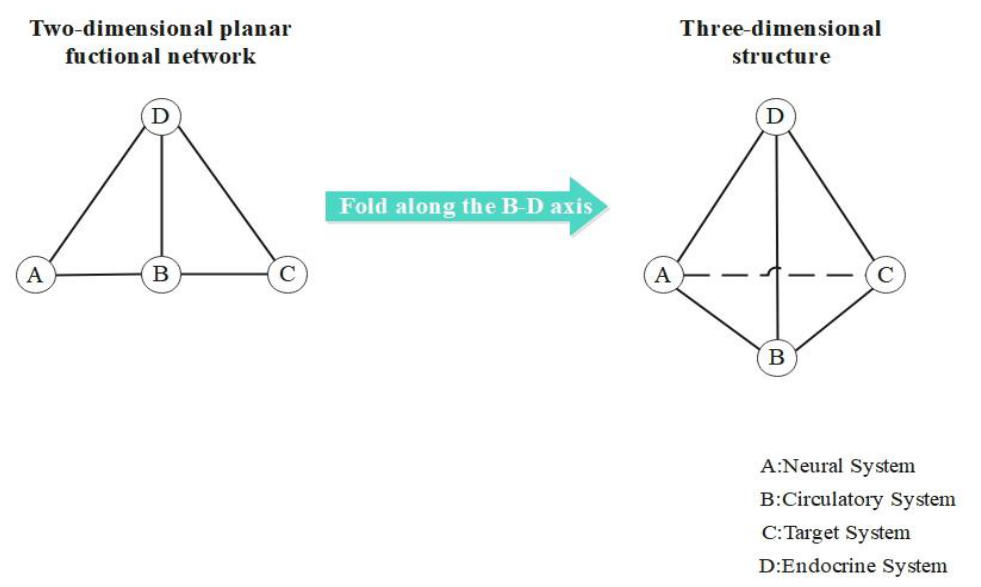
Figure 2.
Disease subtyping and associated geometric collapse patterns based on the Virtual-Real Interface framework. (a) Healthy framework: a symmetrical homeostatic tetrahedron, with edge A–C rendered as a dashed line. (b) Type A deficiency framework: primary collapse of node A (blue arrow), causing overall leftward tilt of the structure and traction of node C (red arrow). (c) Type B deficiency framework: primary collapse of node B, inducing traction of node C. (d) Type D deficiency framework: primary collapse of node D, inducing traction of node C. Node height represents functional strength; distortion of connecting edges reflects imbalance in inter-nodal relationships.Regardless of the subtype, all ultimately manifest as degeneration of the target organ—the skeleton (C)—yet the geometric changes in the framework intuitively reveal fundamentally distinct patterns of network collapse.
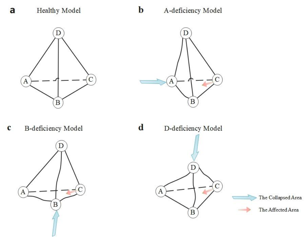
Figure 3.
Networked progression deduction of PMO based on the Virtual-Real Interface framework.(a) Healthy state: A symmetrical and intact tetrahedron, with all vertices functionally intact; edge A–C is rendered as a dashed line.(b) Disease onset (D-node collapse): Primary collapse of node D induces traction on node C, resulting in functional atrophy of the actual face B–C–D and abnormal stretching of the virtual face A–C–D.(c) Stage I of network progression: Deficiency at node D propagates via network dependencies, secondarily disrupting nodes A and B, thereby reducing overall structural stability.(d) Stage II of network progression: Dysregulation of nodes A and B ultimately leads to marked decline in node C.All functional face areas contract, and the volume of the homeostatic tetrahedron undergoes significant collapse. Diagnostic insight summary (right side of figure): Postmenopausal osteoporosis (PMO) is a prototypical network disease originating at node D, triggering sequential dysregulation across multiple nodes via network dependencies, ultimately culminating in global structural collapse.
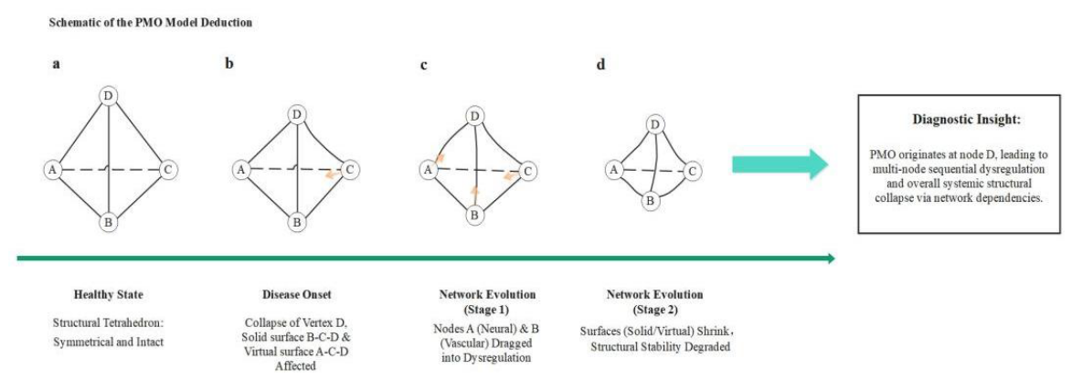
Figure 4.
Three-dimensional therapeutic design for PMO grounded in the Virtual-Real Interface framework. (Left) Pre-treatment network state: a tetrahedron with geometric distortion and instability due to collapse of node D.Four colored arrows point to their respective target sites, corresponding to precise intervention strategies.(Central arrow) Therapeutic intervention: symbolizes the initiation of multi-target synergistic intervention.(Right-side arrow) Post-treatment network target state: following simulated combined intervention, the homeostatic tetrahedron is remodeled into a symmetrical, plump, and structurally stable geometric configuration. This schematic illustrates that precise intervention targeting defective nodes can ultimately enhance the systemic stability of the entire homeostatic tetrahedron by repairing the associated edges and faces—thereby embodying a paradigm shift from single-modality therapy toward systematic, network-based restoration.
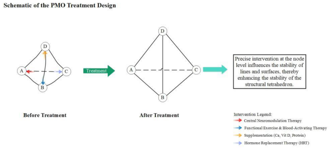

Table 1.
Definition and instantiation of the four core functional nodes within the Virtual-Real Interface framework.
Table 1.
Definition and instantiation of the four core functional nodes within the Virtual-Real Interface framework.
| Node Symbol | Abstract Functional Role | Definition | Instantiation in Bone Metabolism Paradigm |
| A | Regulatory System | Rapid sensing, integration, and command issuance | Nervous system |
| B | Transport System | Delivery and distribution of matter, energy, and signaling molecules | Circulatory system |
| C | Target System | Functional execution endpoint or structural platform | Skeletal tissue |
| D | Tuning System | | Systemic, temporally coordinated, and fine-grained regulation via chemical signals | Endocrine system |
Disclaimer/Publisher’s Note: The statements, opinions and data contained in all publications are solely those of the individual author(s) and contributor(s) and not of MDPI and/or the editor(s). MDPI and/or the editor(s) disclaim responsibility for any injury to people or property resulting from any ideas, methods, instructions or products referred to in the content. |
© 2026 by the author. Licensee MDPI, Basel, Switzerland. This article is an open access article distributed under the terms and conditions of the Creative Commons Attribution (CC BY) license.
Copyright: This open access article is published under a Creative Commons CC BY 4.0 license, which permit the free download, distribution, and reuse, provided that the author and preprint are cited in any reuse.