

Submitted:
08 October 2026
Posted:
19 October 2026
You are already at the latest version
Abstract
Trichomoniasis is a venereally transmitted protozoan disease of bovines that causes vaginitis, early embryonic death and second-trimester abortion, increasing the risk of infertility. Despite its economic and health significance, it has been misdiagnosed or under-reported in Pakistan. This study aimed to describe the molecular epidemiology of Trichomonas in cattle and buffaloes on selected farms in Punjab, Pakistan. Preputial and vaginal secretions (600 samples) were collected from suspected animals across eight districts by purposive sampling, together with placentomes from aborted fetuses. Samples were cultured in InPouch TF kits, and culture-positive samples were subjected to DNA extraction and PCR. Blood samples were tested by latex agglutination. The associations of natural mating, breeding practices, infrastructure and veterinary services with infection were analysed statistically. Overall prevalence was 16.67% (100/600; 95% CI 13.9-19.9%), and differed significantly by district (chi-square = 27.60, df = 7, p = 0.0003), ranging from 0% in Burewala to 30.0% in Bahawalnagar. Natural mating was strongly associated with infection (odds ratio 12.43, 95% CI 6.32-24.46, p < 0.0001) compared with artificial insemination. These results link prevalence to local risk factors and can support control strategies; artificial insemination should be preferred over natural mating.
Keywords:
trichomoniasis
; venereal disease
; Trichomonas fetus
; Trichomonas vaginalis
; natural mating
; InPouch TF
; latex agglutination test
; prevalence
1. Introduction
Protozoa inhabit the digestive tract of animals as a commensal but in clinical cases, certain protozoa cause economic losses. Infected animal expresses dullness, lethargy, pyrexia, and anorexia. Morbidity is comparatively high, but mortality in naturally infected cases is not significant (Rae at al., 2013). Trichomoniasis is a sexually transferred protozoan disease produced by T. fetus and T. vaginalis [1]. In 1836, Donne used prepared vaginal discharge that had been combined with saline to analyze this common parasite in an unstained wet mount. In 1883, Kunkle examined the function of female urinary tract parasites. The name T. fetus was given by Redmullier to this parasite causing infection in cattle. Later on, based on morphology, Wendish and Emmerson gave the name T. fetus to this parasite after 28 years. T. fetus is a major cause of diarrhea in cats. Trichomonas tenax was first the Trichomonas identified within the dental calculus by Muller in 1773 [2].
Animal health problems result in significant financial losses, both directly and indirectly, including the action parameters and restrict international movement, illness treatment, and management tactics. Infectious diseases pose a severe threat to animal health and output in poor countries (Malik et al., 2019). Parasitic infections are also a major concern when it comes to infectious diseases. [3].
There are three main serotypes of T. fetus that has spread globally and all have been reported in Australia such as var. Brisbane, var. Belfast, and var. Manley [4]. There are other types of Trichomonas present in the bovine genital tract, including Pentatrichomonas hominis, Tetratrichomonas (Tt) buttery, Tetratrichomonas (Tt) pavlova, Tritrichomonas enteric and Pseudotrichomonas species (Villarroel et al., 2004).
Prevalence stays high in natural breeding settings, infected bulls were recorded in 15.8% of California cattle herds and 26.74% of Nevada ranches (Kvasnicka et al., 1989). But where natural mating is being practiced, prevalence is high. Trichomoniasis has been found endemic. There are pieces of evidence of cases in many states such as Alabama, California, Idaho, Colorado, Missouri, New Mexico, Nebraska, Nevada, and Oklahoma [5].
The developing countries such as Pakistan and South African countries have high prevalence of Trichomonas because of natural mating. Abortion is causing a huge economical loss to farmers. If animals are not treated, the infection persists and leads to chronic condition. Ultimately, there are chances of coinfection in infected animals. There are different approaches to identify the disease, such as culturing, staining and latex agglutination test. But the culturing through InPouch® kit is the best way to identify the Trichomonas positive animals (Masha et al., 2019).
There has been limited research in Pakistan on the identification of infectious pathogens that cause abortion in cattle and buffaloes. Because most of these diseases are zoonotic, to know completely about the zoonotic and infectious diseases is inevitable for the public health significance and control strategies [6].
2. Materials and Methods
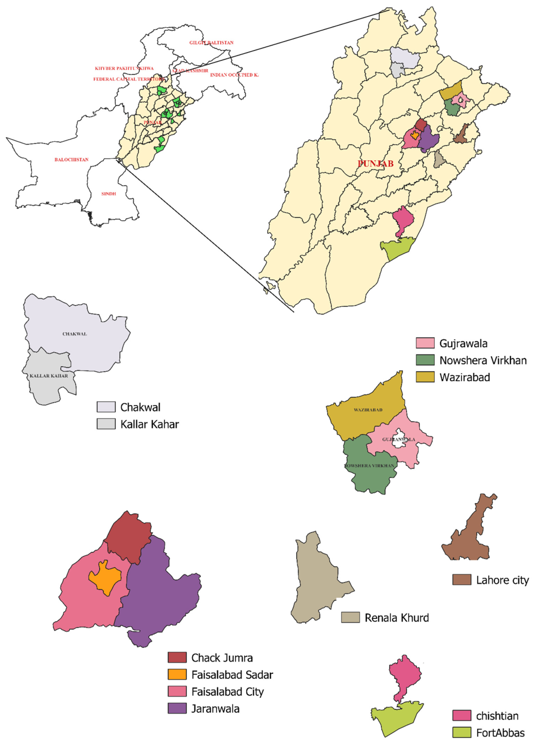
2.1. Study Design and Sampling
Total 280 samples were collected from animals and transported to the Lab of Molecular Parasitology and One Health at the Department of Parasitology, University of Agriculture, Faisalabad. The preputial secretions were collected from bulls and transported into InPouch® kits.
The proposed type of sampling was simple purposive sampling or convenient sampling, measuring the prevalence across the study areas from. The method used for sampling was convenient sampling with purposive selection. The sampling unit was selected by seeing clinical parameters based on purposive sampling. There were three different ways to get samples from bull. Scraping, preputial Lavage, and sponge sampling (Agnelli et al., 2013). Scraping is an antique method for the collection of smegma (Kamath et al., 2016). For, the collection of smegma, penile and preputial mucosa will be vigorously rubbed with the help of an insemination pipette connected with a bulb via a silicon rubber tube to create negative pressure. The collected material was shifted to PBS (Phosphate buffer saline. Preputial flushing is another kind of method. For this, a plastic tube having 50ml PBS was instilled into the prepuce, massaged, and suctioned back.
The Trichomonas positive samples were collected based on clinical signs and history taken from farmers. Then these bulls and affected cows and buffaloes were selected. The study animals were cattle and buffaloes such as bulls mainly and pregnant cows. The bulls and cows were screened for trichomoniasis at least two times during the period of study. One of the criteria of the study, was natural mating in herd and use of cows with the history of abortion and positive samples were processed further for PCR and DNA isolation (Richard et al., 1998; BonDurrant et al., 2005; Yao et al., 2015).
The protocol for the sampling was described by Richard et al. [7].
2.2. Sample Collection
For the collection of samples, the pipette had been inserted into the preputial orifice of bulls and pipette was attached with disposable syringe which was having 50 ml normal saline to flush the tract. After again and again flushing of preputial orifice, smegma was collected. Which was then transported into the InPouch® kit [7].
The modified way of collection was used of metal brushes, 8mm diameter and having a parallel groove which is perpendicular to long axis. The brush was inserted into prepuce and back and forth moved around 20 -25 times. The accumulated material was collected after washing with normal saline (Parker et al., 1999; Tedesco et al., 1999).
The sponge method, the bull will be restrained, and the penis will be extended by the massage on the preputial cavity. Swabbing with a sponge on the glans penis and swabbing down the penile shaft and prepuce will be done. vaginal secretion after washing with PBS will be collected.
In case of abortion, placenta and fetal fluid will be collected. The 5 ml blood sample will be collected from the jugular vein and coccygeal vein and transferred to an anticoagulant containing vacutainers. These vacutainers will be labeled properly with details of the farm and stored at 4°C for transportation (Zajac et al., 2021). The collecting bottles were correctly labeled. The collecting bottles were correctly labeled. (Ernst and Benz, 1986).
The samples were transported at temperature than 20 °C to the Lab (Ali et al., 2014). InPouch® kits were imported from United states of America and incubated into the incubator at 37°C for 72 hours. These were kept over for three days. There was a turbid growth that could be seen in the kits (Nicoletti, 2010).
Figure 2.
Methodology followed during sample collection. (A) Insertion of catheter into the preputial orifice of cow/bull; (B) insertion of catheter into the preputial orifice of a buffalo bull; (C) inoculation of smegma into the InPouch kit; (D) incubation of samples at 37 °C for 48–72 h; (E) positive culture medium; (F) turbid growth.
Figure 2.
Methodology followed during sample collection. (A) Insertion of catheter into the preputial orifice of cow/bull; (B) insertion of catheter into the preputial orifice of a buffalo bull; (C) inoculation of smegma into the InPouch kit; (D) incubation of samples at 37 °C for 48–72 h; (E) positive culture medium; (F) turbid growth.
2.3. InPouch Culture
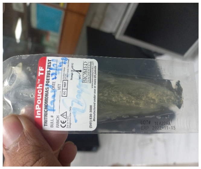
InPouch® kit called a Field culture kit was used for identification of Trichomonas. This had two compartments either for transportation or for culturing of a parasite. The purpose of the InPouch® kit was to screen for T. fetus and T. vaginalis. This was made up of a double chambered plastic pouch. Both chambers were connected by a narrow channel. The functions of these, viewing of samples and culturing (Hanson et al., 2002).
Confirmation was required after isolation of T. fetus and T. vaginalis to discriminate from other Trichomonads in the digestive tract. The diagnostic efforts should be concentrated on bulls, because they were carriers (kyle et al., 2002). A pipette was suctioned while being used to scrape the epithelium of the preputial fornix forcefully. Douching with saline and lactated Ringer's solution (no preservatives) was another option. To study the aspirate, Dark field contrast microscopy was used. So, douches/aspirates that had been concentrated by centrifugation. This was transported to culture medium like a Diamond medium very instantly (Silva et al., 2011).
This was the gold standard for T. vaginalis and used in confirmatory test. The media was known as Diamond media. It was reported, this is selective in nature. Commercial kits are available for Trichomonas vaginalis as well as T. fetus. These kits had advantages over constituted media in laboratory such long term storage. This was highly sensitive for the detection of T. vaginalis. The swab and preputial secretions from vagina and bulls were inserted into InPouch® kits by opening the seal of plastic bag.
With a dry pipette, preputial smegma was collected from the 280 bulls and cultured in an InPouch® Kits TF, T. fetus cultured pouch (BioMed Diagnostics; White City, OR USA).
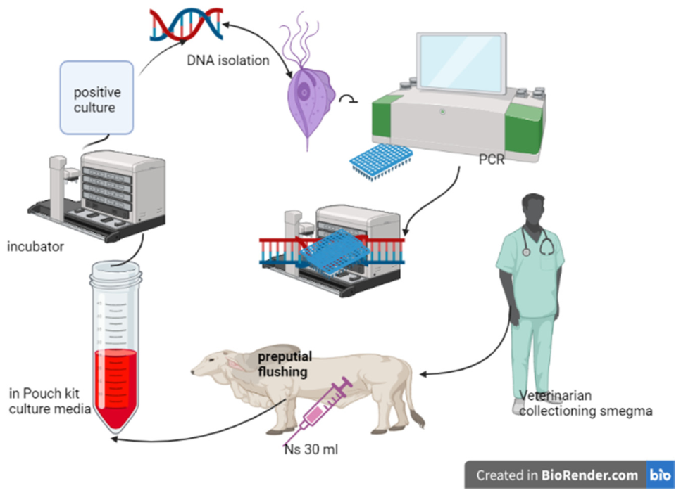
Figure 3.
Experimental protocol for field culture (adapted from Rhyan et al., 2013). (A) Veterinarian; (B) preputial flushing; (C) InPouch kit for culturing; (D) incubation; (E) Thermo Scientific kit for DNA isolation; (F) PCR machine; (G) phylogenetic analysis.
Figure 3.
Experimental protocol for field culture (adapted from Rhyan et al., 2013). (A) Veterinarian; (B) preputial flushing; (C) InPouch kit for culturing; (D) incubation; (E) Thermo Scientific kit for DNA isolation; (F) PCR machine; (G) phylogenetic analysis.
2.4. DNA Extraction
After the culturing, the samples were processed for DNA isolation through the use of Qiagen and Thermo-scientific kits. according to the manufacturer’s instructions.
The growth positive culture media were centrifuged and processed for DNA using thermos-scientific kit. The following protocol was used as 25µl of sample was taken which is intended to DNA isolation and180 µl of digestible solution is added. After this, 20 µl of Proteinase-k was added into the solution and vortex it for 30 seconds. After this, Incubation of this solution was done at 56°C ’for 3hours and 20µl of RNAase was added and incubated at for 10 minutes at 37°C. Then 200µl of lysis solution was added and vortex for 15 minutes and 400 µl of ethanol 50% was added. Then it was transferred to the Column vials and Centrifugate it at 6000g for 1 minute. After this, 500µl of wash buffer-1 was added and vertex 8000g for 1 minute. Then, 500 µl of wash buffer -2 was added and vertex 8000g for 3 minutes. Finally, Elution buffer was added of 200µl and vertex 8000g for 1 minute (Camperio et al., 2011).
2.5. Primer Design and PCR
[ACTION NEEDED: the source text describes PCR three different, mutually exclusive ways (cycle number, extension time, agarose %, and primer targets all differ between the three passages below). Confirm which one matches your lab notebook, delete the other two, and merge into a single protocol paragraph before submission.]
[Version A] After the isolation of DNA from the Trichomonas samples, the DNA was amplified for the conduction of PCR (Polymerase chain reaction). For this purpose, primers were designed using the ITS gene through using the primer blast at NCBI.
PCR was carried out at Molecular Parasitology and One Health Lab at Department of Parasitology, Faculty of Veterinary sciences at University of Agriculture Faisalabad. The primer used in sequence were 5’GATTATTCTTGAAGCCCACTCC-3’in forward direction, TFR 3, and 5’-CTTGCCGGTAACAGTTCTAGC-3’in reverse direction used in gel -PCR. The primers paired with 347 bps DNA fragment of ITS-1 5.8 rRNA ITS2 from trichomonas species, like T. fetus, T. suis, T. mobilensis. These primers did not amplify the genome of others trichomonas species such as T. fetus and T. suis, Tetratrichomonas galinarum, T. gallinae and Pentatrichomonas hominis, T. tenax. The point must say, DNA was required for PCR gel electrophoresis, qPCR and for phylogenetic (Felleisen et al., 1998; Lun et al., 2005; McMillen et al., 2006).
The first samples were processed through three freeze thaw cycles and proteinase-k digestion. DNA concentration was measured by using the spectrophotometric device at 260 nm or 280nm. For molecular diagnostic purposes, a pair of oligonucleotide PCR primers was chosen from the conserved ITS- 1/5.8/ITS-2 region of T. vaginalis and TFR-1 for T. fetus. The reaction of amplification was carried out by using a 44 µl reaction containing100 p mole of each primer as diluted form used 2µl of forward Primer and 2µl of reverse Primer, 20 µl of master mix (MM), and 15µl of water: initial denaturation at 95°C for 3 min., followed by 37 cycles in series of denaturation at 95°C for 15sec., annealing at 60°C for 30 sec, extension at 72°C for 30 sec, and final extension step is at 72°C for 5 minutes. By adding 5 µl of PCR products, electrophoresis will be performed. The addition at 1% (w/w) agarose gel and safe stained for 45 minutes at 120V. Bands visualized by ultraviolet trans illumination (biosystematics Devon UK)[3].
(Nanodrops Thermo-Scientifics, USA) after extraction. COI and ITS2 regions were amplified using PCR. All reactions were completed in a 44µl of volume as 20µl of Vasyme master mix (Themo Fisher Scientific, USA), 2-microliters of each primer, 15µl of PCR-grade water (Themo Fisher Scientific), and 5µl template DNA (DNA-concentration was 100 to 150 ng/L) were used in the PCR process. The study's primers and PCR reaction parameters. However, adapters for COI were generated using NCBI primer blast utilizing the sequence information available. After safe gel staining, the PCR-products were run on an electrophoresis gel and inspected under UV light to check that a single-band without any nonspecific product was evident.
[Version B] Purified DNA from cultivated organisms has been used as a template in diagnostic fragment amplification processes. More recently, a PCR assay using anti hormonal primers and T. fetus and, T. vaginalis specific primers was established. Positive isolates of T. fetus produce an extra amplification produce of 347 bps to the segregation of all other Trichomonads, whereas samples negative for T. fetus create no amplicons. Agarose gels were used to visualize the products. T. fetus and T. vaginalis control efforts were centered on diagnostic assays with improved sensitivity.
If Trichomonad organisms were visible under a microscope, the samples were termed suspicion positive. To validate Trichomonad morphology, smears were made from a drop (25: l) of each sample and left to air dry before inactivation with iodine and staining with Diff-Quick (Lun and Gajadhar, 1999).
The all the processed experimental samples 1-4, PCR was carried out in thermal cycle by using the oligonucleotide primers made for ITS 5.8 rRNA region of T. fetus genome, 5 ml gene amplification buffer, 100 mM of deoxyribonucleotide and .03 µm of each primer, and 0.03µm of DNA sample were taken. The PCR was taken 48 cycles of denaturation at 94°C, annealing for 30 sec at 60 °C, extension at 72 °C for 15 minutes, the bands of deoxyribonucleotide were seen through electrophoresis on 2% agarose gel by using the ethidium bromide stain. The nucleic acid from T. fetus was seen in Figure 4.4, lane 1, T. fetus, lane 2: T. fetus Faisalabad, lane 3: T. vaginalis Lahore, lane 4: control blank, lane 5: markers. the preputial samples were 1, 2, 3, 4, processed and results of these, and negative control of T. fetus was used. Control consists of all reagents without DNA templates. The product size after amplification was 347 bps. The first reports of author knowledge of used this method in Pakistan to detect parasite DNA (Felleisean, 1989; Campero et al., 2003).
[Version C (electrophoresis)] The DNA extracts were run on 1% agarose gel at 120V for 45 min, stained with safe gel stain and visualized under ultraviolet light. In lane A is the ladder (100 bps) and B and C is the product bulls’ isolated DNA of T. fetus with 347 bps.
(McMillian et al., 2006).
Primers were ordered from macrogen® is a South Korean biotechnology company. The condition of PCR had been same mentioned above.
2.6. Gel Extraction and Sequencing
After the running of PCR, the bands were obtained, and size was measured through running ladder. This was having 347 base pairs. The gel was removed to purify the DNA. The gel was cut into small pieces having minimum gel around DNA. Gel slices were weighted into pre-weight 1.5ml tube and weight again. If these were used for cloning reactions, avoids the DNA damaged through UV light exposure. Add 1:1 volume of binding buffer to gel size (volume: weight). Addition of 100 µl of binding buffer for every 100 mg of agarose gel. If the gel concentration was greater than 2%, add 2:1 volume of binding buffer to the gel slices (Ohlemeyer et al., 1988).
Incubation of gel mixture at 50-60°C for 10 minutes or until the gel slices were removed. Spinning tubes for every few minutes to facilitate the melting process. It was ensured that gel was completely dissolved. Then vortex of gel mixture at 12000 rpm for 1 minute. The color was monitored. A yellow color was obtained which indicates optimal pH for DNA bindings. It was recommended, if the product size < 500 bps or > 10kb. Then, use 1 gel volume of 100% isopropanol to the solubilized gel solution e.g., 100µl of isopropanol was added to 100 mg gel slice solubilized in 100 µl of binding buffer. Transfer to 800 µl of solubilized gel solution to the gene jet purification column. Centrifuge for 1 mint. Discard the flow-through and place the column back into the same collection tube.
For the sequencing purposes, add 100µl of binding buffer was added into the Gene jet purification column. Centrifugation was done for 1 minute. Discard the flow through and place the column back to the same collection tube. Added the 700 µl of wash buffer to the Gene Jet purification column. Centrifuge for 1 minute. The additional centrifugation of the empty gene jet was done to completely remove the residual with wash buffer. Then the Gene Jet purification column was transferred to microcentrifuge tube. 50 µl of elution buffer was added to the center of purification column membrane.
Finally discard the gene Jet purification column and stored the DNA at -20°C.
2.7. Statistical Analysis
District-level prevalence was compared using a chi-square test of independence (district x infection status). The association between breeding method (natural mating vs. artificial insemination) and infection status was assessed using a 2x2 contingency table, with the odds ratio and its 95% confidence interval calculated by the Woolf logit method and confirmed by Fisher’s exact test. Wilson score 95% confidence intervals were calculated for all prevalence estimates. A p-value < 0.05 was considered statistically significant. [State the statistical software used (e.g., R, SPSS, GraphPad Prism) and its version.]
3. Results
The present study of frequency, distribution, molecular characterization, and control of Trichomonas fetus in cattle and buffaloes of the farms of Punjab, Pakistan. For this purpose, a total of twenty (20) farms were selected where natural mating was being practiced instead of Artificial insemination (AI). They comprised some local farms and units of intensive natural breeding. It is evident from the presented data that of the sampled 280, considered as dominant and co-dominant from the diversified microflora.
Trichomonas vaginalis was detected in five animals, and positive bands were seen after conduction of PCR, nearly having 347 bps. Furthermore, the clinical status showed the relation between variables [8].
Cattle were found to be positive with Trichomonas, and prevalence is 10% in Faisalabad, 25% Bahawalanagar,11.43% in Gujranwala, 17.78% in Lahore,5.26% Burewala,10% Chakwal, 10% Okara,0% Sahiwal, and chi-square 6.1, odd ratio 0.21 and p- value 0.013.
3.1. Association with Breeding Method
There are certain independent variables which may affect the prevalence of trichomoniasis in livestock farming like veterinary service, infrastructure, awareness of biosecurity, natural mating, methods of cleanings, purchasing of animals, isolation, and quarantine of newly purchased animals [9].
The findings of this investigation indicate that prevalence of T. vaginalis is higher in Bahawalnagar, Faisalabad, Lahore, and Gujranwala. This finding according to BonDurrant, who found majority of cows and young bulls could possibility achieve the spontaneous clearance of infection and highest prevalence in breeder bulls.
[Original Figures 4.1 and 4.2 (risk-factor and prevalence charts) contained no images in the source file. Table 1 and Table 2 above now carry this information instead - add bar charts here only if you want a visual in addition to the tables.]
The sample targeted Faisalabad, Bahawalnagar, Lahore, and Gujranwala etc. The natural breeding pockets, where extensive natural mating was used to breed the cows. Because natural mating is the main source of transmission of infection in naïve animals.
This is thought to be, typically cows cleared their infection within 3 months, or less than that, because immunity developed against T. fetus in less than 6 months. But this statement is inconsistent with the findings of Yao, who states that higher prevalence is in heifers than cows, because cows retain immunity against infection as far as three years. Simply, we can say that immunity prevails up to three years after infection in cows. But it depends upon the immune status of the animals and varies from animal to animal. So, it is recommended, to control the infection of Trichomonas vaginalis in herd, to use of 2-3 years bulls rather than older bulls. In this, study Trichomonas vaginalis was detected to PCR, because of the high sensitivity and specificity of this technique for the detection of the parasite (Yao et al., 2009).
3.2. Molecular and Phylogenetic Analysis
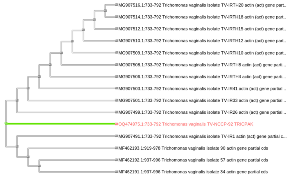
Accession no. OQ474975.
Figure 4.
Agarose gel electrophoresis of PCR products showing the 347 bp amplicon.
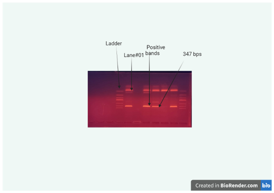
Figure 5.
Phylogenetic tree including isolate OQ474975.
4. Discussion
The idea of employing PCR tests to detect T. fetus and T. vaginalis directly from preputial materials without an intermediary culture has been investigated in studies. Some laboratories utilize these tests to shorten the time it takes to make a conclusive diagnosis of trichomoniasis (Stockdale et al., 2008). The suspected positive bulls will easily identify by using InPouch® kits, it is recommended to use three tests of the suspected positive bulls. Then, if becomes positive on culture media, should be isolated, and culled. For diagnostic purposes vaginal mucus or discharge is helpful. The number of times the bull battery should be tested and cultured to verify the bulls are negative is determined by the cow herd's fetal wastage rate. With more open or late cows, the frequency of tests should rise, increasing the likelihood that bulls should be protozoan free (Stockdale et al., 2007).There is reported about the serology of Trichomonas, but this method has least reliability because immune response is not evident in trichomoniasis. Mucus from vagina is used to identify the presence of Trichomonas. There is specific agglutination upon the conduction of test if sample is taken from positive animal. The dependent variables such as early embryonic mortality, vaginitis, pyometra, congenital abnormalities in calves, and healthy animals. The clinical status and correlation between pathogenic states, statically demonstrate that early embryonic mortality was 17% as compared to congenital anomaly, and vaginitis is 1.7% as compared to pyometra. It is evident that from (Joanna et al., 2006). The monitored clinical associated with T. fetus were range from moderate vaginitis, abortion, pyometra pelvic inflammation. Early embryonic death, abortion had seen at the time of pregnancy. There was seen infertility, and abortion because mucosal membrane is damaged by the parasite of the genital tract which released Cysteine protease, which causes cytopathogenic effect in epithelial cells, oviduct, uterus, vagina, and apoptosis of cells. This is also causing the cleavage of IgG2 and evade the immune responses of the host [1].
The simplest and easiest way to identify is the microscopic examination. The freshly collected positive samples are directly seen under microscope on 100X or 400X. If sample is positive, mortality can be seen having flagellates. For the transportation of the samples, certain media are used such phosphate buffer saline, egg yolk, and egg white. Another method is use of Diamond media which is present in InPouch kit®. The positive samples are inoculated with these kits, and then incubated for 72 hours. There is recommended that inoculated samples directly can been on wet mount slide to confirm the presence of trichomonas. PCR can be used for confirmation with 99% specificity and sensitivity. ITS-1, 5.8S gene, which RNA ribosomal gene is being targeted. The primers are TFITS-F/TFITS-R, TFR-1/TFR2 and NCS5 and NC-2 [10]. With the advancement in biotechnology, studies are conducted utilizing multiplex PCR, nested PCR, PCR-RFL (Restriction length polymorphism), commercial field culture kit, serological testing, and culturing of the organism. The latest method of identification of T. fetus is loop isothermal medium amplification which is based on DNA. This targets the 5.8S ribosomal DNA subunit and B-tublulin gene by usuing TFR3 and TFR4 primers. This method is highly specific, sensitive, and cost effective [11].
5. Conclusions
Overall prevalence of Trichomonas infection was 16.67% across the sampled districts of Punjab, Pakistan, with significant variation by district and a strong, significant association between natural mating and infection (OR = 12.43). These findings support artificial insemination as a practical control measure to reduce the transmission of bovine trichomoniasis in the region.
Author Contributions
Conceptualization, M.T. and M.S.S.; methodology, M.T.; investigation, M.T.; formal analysis, M.T.; writing—original draft preparation, M.T.; writing—review and editing, M.S.S., H.A. and H.D.K.; supervision, M.S.S., H.A. and H.D.K. All authors have read and agreed to the published version of the manuscript. Confirm roles with co-authors.
Data Availability Statement
The sequence generated in this study is available in GenBank under accession number OQ474975.
Conflicts of Interest
The authors declare no conflicts of interest. Confirm with co-authors.
References
- BonDurant, R. H. Venereal diseases of cattle: Natural history, diagnosis, and the role of vaccines in their control. Vet. Clin. North Am. Food Anim. Pract. 2005, 21, 383–408. [Google Scholar] [CrossRef] [PubMed]
- McMillan, L.H.; Brady, E.C.; O’Driscoll, M.P.; Marsh, N.V. A multifaceted validation study of spence and robbins'(2006) workaholism battery. J. Occup. Organ Psychol. 2002, 75, 357–368. [Google Scholar] [CrossRef]
- Jin, Y.; Schumaker, B.; Logan, J.; Yao, C. Risk factors associated with bovine trichomoniasis in beef cattle identified by a questionnaire. J. Med. Microbiol. 2014, 63. [Google Scholar] [CrossRef] [PubMed]
- Yao, C.; Köster, L.S. Tritrichomonas foetus infection, a cause of chronic diarrhea in the domestic cat. Vet. Res. 2015, 46, 35. [Google Scholar] [CrossRef] [PubMed]
- BonDurant, H.; Greenfield, B.; Tse, S.M. Construct validity of the adolescent borderline personality disorder: a review. Child Adolesc. Psychiatr. Rev. 2004, 13, 53. [Google Scholar]
- Yoader, I.; Robertson, L. A comparison of various selective media, including a new selective medium for the isolation of brucellae from milk. J. Appl. Bacteriol. 1972, 35, 625–630. [Google Scholar] [CrossRef] [PubMed]
- Richard, S.J.; Felleisen, N. L.; Bruno, G. Detection of Tritrichomonas foetus by PCR and DNA enzyme immunoassay based on rRNA gene unit sequences. Clin. Microbiol. Rev. 1998, 36, 513–519. [Google Scholar] [CrossRef] [PubMed]
- Stamou, S.C.; Williams, M.L.; Gunn, T.M.; Hagberg, R.C.; Lobdell, K.W.; Kouchoukos, N.T. Aortic root surgery in the United States: A report from the society of thoracic surgeon’s database. J. Thorac. Cardiovasc. Surg. 2015, 149, 116–122. [Google Scholar] [CrossRef] [PubMed]
- Rodning, S.P.; Wolfe, D.F.; Carson, R.L.; Wright, J.C.; Stockdale, H.D.; Pacoli, M.E.; Busby, H.C.; Rowe, S.E. Prevalence of tritrichomonas foetus in several subpopulations of alabama beef bulls. Theriogenology 2008, 69, 212–217. [Google Scholar] [CrossRef] [PubMed]
- Felleisen, R. Comparative genetic analysis of Tritrichomonadid protozoa by the random amplified polymorphic DNA technique. Parasitol. Res. 1998, 84, 153–156. [Google Scholar] [CrossRef] [PubMed]
- Rhyan, J.C.; Wilson, K. L.; Burgess, D.E.; Stackhouse, L. L.; Quinn, W.J. Immunohistochemical detection of Tritrichomonas foetus in formalin-fixed, paraffin-embedded sections of bovine placenta and fetal lung. J. Vet. Diagn. Invest. 1995, 7, 98–101. [Google Scholar] [CrossRef] [PubMed]
- Rae, D.O.; Crews, J. E. Tritrichomonas foetus. Vet. Clin. North Am. Food Anim. Pract. 2006, 22, 595–611. [Google Scholar] [CrossRef] [PubMed]
- Rae, D.O. Impact of trichomoniasis on the cow-calf producer's profitability. J. Am. Vet. Med. Assoc. 1989, 194, 771–775. [Google Scholar] [CrossRef]
- Campero, C.M.; Dubra, C.R.; Bolondi, A.; Cacciato, C.; Cobo, E.; Perez, S.; Odeon, A.; Cipolla, A.; BonDurant, R.H. Two-step (culture and pcr) diagnostic approach for differentiation of non-t. Foetus trichomonads from genitalia of virgin beef bulls in argentina. Vet. Parasitol. 2003, 112, 167–175. [Google Scholar] [CrossRef] [PubMed]
- Corbeil, L.B.; Campero, C.M.; Van Hoosear, K.; Bondurant, R.H. Detection of trichomonad species in the reproductive tracts of breeding and virgin bulls. Vet. Parasitol. 2008, 154, 226–232. [Google Scholar] [CrossRef] [PubMed]
- Grahn, R.A.; BonDurant, R.; Van Hoosear, K.; Walker, R.; Lyons, L.A. An improved molecular assay for Tritrichomonas fetus. Vet. Parasitol. 2005, 127, 33–41. [Google Scholar] [CrossRef] [PubMed]
- BonDurant, R.; Gajadhar, A.; Campero, C.; Johnson, E.; Lun, Z.; Nordhausen, R.; Van Hoosear, K.; Villanueva, M.; Walker, R. Preliminary characterization of a tritrichomonas foetus-like protozoan isolated from preputial smegma of virgin bulls. Bov. Pract. 1999, 10, 124–127. [Google Scholar] [CrossRef]
- Bon Durant, R.H.; Anderson, M. L.; Blanchard, P.; Hird, D.; Danaye-Elmi, C.; Palmer, C.; Sischo, W. M.; Suther, D.; Utterback, W.; Weigler, B. J. Prevalence of trichomoniasis among California beef herds. J. Am. Vet. Med. Assoc. 2005, 196, 1590–1593. [Google Scholar]
- Tedesco, L.; Errico, F.; Baglivi, L.D. Diagnosis of tritrichomonas fetus infection in bulls using two sampling methods and a transport medium. Aust. Vet. J. 1979, 55, 322–324. [Google Scholar] [CrossRef] [PubMed]
- Parker, S.; Lun, Z.R.; Gajadhar, A. Application of a PCR assay to enhance the detection and identification of tritrichomonas foetus in cultured preputial samples. J. Vet. Diagn. Invest. 2001, 13, 508–513. [Google Scholar] [CrossRef] [PubMed]
- Yao, C. Diagnosis of tritrichomonas foetus infected bulls, an ultimate approach to eradicate bovine trichomoniasis in US cattle. J. Med. Microbiol. 2013, 62, 1–9. [Google Scholar] [CrossRef] [PubMed]
- Yao, C.; Bardsley, K.D.; Litzman, E.A.; Hall, M.L.M.; Davidson, M. Tritrichomonas foetus infection in beef bull populations in Wyoming. J. Bacteriol. 2011, 2, 1–6. [Google Scholar]
- Stroud, L.; Šlapeta, J.; Padula, J.; Druery, M. P.; Tsiotsioras, D.; Coorssen, G.; R, J.; Stack, C.M. Comparative proteomic analysis of two pathogenic Tritrichomonas foetus genotypes: there is more to the proteome than meets the eye. Int. J. Parasitol. 2017, 47, 203–213. [Google Scholar] [CrossRef] [PubMed]
- Rhyan, J.; Wilson, K.; Wagner, B.; Anderson, M.L.; BonDurant, R.; Burgess, D.; Mutwiri, G.; Corbeil, L. Demonstration of tritrichomonas foetus in the external genitalia and of specific antibodies in preputial secretions of naturally infected bulls. Vet. Pathol. 1999, 36, 406–411. [Google Scholar] [CrossRef] [PubMed]
- Rhyan, J.C.; Nol, P.; Quance, C.; Gertonson, A.; Belfrage, J.; Harris, L.; Straka, K.; Robbe-Austerman, S. Transmission of brucellosis from elk to cattle and bison, greater yellowstone area, USA. 2002–2012. Emerg. Infect. dis. 2013, 19, 1992. [Google Scholar] [CrossRef] [PubMed]
- Felleisen, R.; Lambelet, N.; Bachmann, P. Detection of Tritrichomonas foetus by PCR and DNA enzyme immunoassay based on rRNA gene unit sequences. J. Clin. Microbiol. 1998, 36, 513–519. [Google Scholar] [CrossRef] [PubMed]
- Cepicka, I.; Hampl, V.; Kulda, J. Critical taxonomic revision of parabasalids with description of one new genus and three new species. Protist 2010, 161, 400–433. [Google Scholar] [CrossRef] [PubMed]
- Cepicka, I.; Hampl, V.; Kulda, J.; Flegr, J. New evolutionary lineages, unexpected diversity, and host specificity in the parabasalid genus Tetratrichomonas. Mol. Phylogent. Evol. 2006, 39, 542–551. [Google Scholar] [CrossRef] [PubMed]

Table 1.
Prevalence of Trichomonas by district (n = 600).
| District | Animals tested (n) | Positive (n) | Prevalence (%) | 95% CI |
| Faisalabad | 100 | 15 | 15.00 | 9.3-23.3% |
| Bahawalnagar | 100 | 30 | 30.00 | 21.9-39.6% |
| Gujranwala | 50 | 10 | 20.00 | 11.2-33.0% |
| Lahore | 50 | 5 | 10.00 | 4.4-21.4% |
| Burewala | 50 | 0 | 0.00 | 0-7.1% |
| Chakwal | 100 | 15 | 15.00 | 9.3-23.3% |
| Okara | 50 | 5 | 10.00 | 4.4-21.4% |
| Sahiwal | 100 | 20 | 20.00 | 13.3-28.9% |
| Total | 600 | 100 | 16.67 | 13.9-19.9% |
Of the 600 animals sampled across eight districts, 100 (16.67%; 95% CI 13.9-19.9%) tested positive for Trichomonas by culture and PCR. Prevalence differed significantly among districts (chi-square = 27.60, df = 7, p = 0.0003), ranging from 0% in Burewala to 30.0% in Bahawalnagar.
Table 2.
Association between breeding method and infection status.
| Breeding method | Positive (n) | Negative (n) | Total | Prevalence (%) |
| Natural mating | 90 | 210 | 300 | 30.00 |
| Artificial insemination | 10 | 290 | 300 | 3.33 |
Animals bred by natural mating had significantly greater odds of infection than those bred by artificial insemination (odds ratio = 12.43, 95% CI 6.32-24.46; chi-square = 74.89, p < 0.0001; Fisher’s exact p < 0.0001).
Disclaimer/Publisher’s Note: The statements, opinions and data contained in all publications are solely those of the individual author(s) and contributor(s) and not of MDPI and/or the editor(s). MDPI and/or the editor(s) disclaim responsibility for any injury to people or property resulting from any ideas, methods, instructions or products referred to in the content. |
© 2026 by the authors. Licensee MDPI, Basel, Switzerland. This article is an open access article distributed under the terms and conditions of the Creative Commons Attribution (CC BY) license.
Copyright: This open access article is published under a Creative Commons CC BY 4.0 license, which permit the free download, distribution, and reuse, provided that the author and preprint are cited in any reuse.