
Submitted:
16 October 2026
Posted:
16 October 2026
You are already at the latest version
Abstract
Malassezia is a common fungus and one of the zoonotic pathogens. It is a common disease among pets including cats, which usually parasitizes the skin surfaces under suitable environmental conditions. Management of Malassezia dermatitis relies mainly on antifungal drugs. Repeated or prolonged usage may be accompanied by reduced susceptibility and concerns about recurrence. Natural compounds may offer advantages in the management of superficial fungal skin disease. In this study, thirty-eight clinical samples were collected from pet cats with Malassezia-associated skin lesions and a KM mouse dermatitis model was established for in vivo evaluation. Three M. pachydermatis isolates were screened against 11 antibiotics and 14 natural compounds. The most resistant Malassezia pachydermis strain SZ06 was selected for minimum inhibitory concentration determination using checkerboard assays, growth-curve analysis, scanning electron microscopy after treatment, and genome annotation. Plant-derived compound allicin, cinnamaldehyde, eugenol, and anisaldehyde showed significant antifungal activity. Checkerboard assays showed the strongest synergistic effect for allicin plus cinnamaldehyde, reducing the minimum inhibitory concentrations of allicin and cinnamaldehyde to one-eighth and one-quarter of their single-agent values, respectively. For the regression mice infected with SZ06, topical treatment with 0.5% allicin plus 0.5% cinnamaldehyde produced the best therapeutic response, improving lesions, fungal clearance, histopathology, and inflammatory cytokine levels. Genome sequencing of SZ06 suggested that this isolate carried multiple candidate genes involved in efflux, membrane transport, and drug-response pathways. The natural compound allicin-cinnamaldehyde combination demonstrated promising antifungal activity, representing a potential topical therapy for Malassezia dermatitis in pet cats.
Keywords:
cat
; Malassezia dermatitis
; allicin
; cinnamaldehyde
; combination therapy
; mouse model
1. Introduction
Malassezia-associated dermatitis is one of the most common recurrent superficial fungal skin diseases in companion animals including cats[1]. It is commonly associated with erythema, scaling, pruritus, malodor, and abnormal sebaceous secretion. Repeated episodes often result in prolonged treatment courses and inconsistent responses to empirical therapy[2].
In pet skin microbiota, Malassezia may exist as an opportunistic commensal organism. It can become pathogenic when the skip barrier is compromised, when local humidity and lipid-rich conditions favor overgrowth, or when host immune status changes[2,3]. Species-level isolation and identification are therefore important for clarifying the predominant pathogen and for guiding susceptibility-based treatment decisions.
Conventional management of Malassezia dermatitis still relies mainly on antifungal drugs[4−6]. However, repeated or prolonged usage may be accompanied by reduced susceptibility, variable treatment response, and concerns about recurrence[7,8]. Plant-derived molecules with antifungal or membrane-disruptive properties have consequently become of interest as adjunctive or alternative topical therapies. Natural compounds may offer advantages in the management of superficial fungal skin disease, including broad antimicrobial activity, multiple cellular targets, and anti-inflammatory effects, which may reduce the selective pressure associated with prolonged exposure to conventional antifungal drugs. Previous studies have reported that certain plant-derived compounds exert antifungal activity through disruption of membrane integrity, alteration of oxidative homeostasis, and interference with fungal metabolism[9,10]. In addition, combination strategies involving natural compounds have attracted attention because they may enhance antifungal efficacy while lowering the concentration required for individual agents. Nevertheless, most available evidence has been generated in non-Malassezia systems, and data regarding antifungal susceptibility and natural compound-based intervention in cat-derived M. pachydermatis remain limited. Plant-derived compound allicin[11], cinnamaldehyde[12], anisaldehyde[13], and eugenol[14] have shown antimicrobial activity in other systems, yet systematic evaluation against cat-derived M. pachydermatis remains limited. Accordingly, this study isolated and identified Malassezia spp. from cat clinical cases in Guangdong Province, China, and screened natural compounds and conventional antifungal agents for activity against M. pachydermatis. This study also focused on the in vitro and in vivo evaluation of plant-derived compound allicin-cinnamaldehyde combination therapy. In addition, a representative isolate was subjected to whole-genome sequencing to provide insights into resistance-related determinants. Host inflammatory responses contribute substantially to the development of Malassezia-associated dermatitis. TNF-α, IL-6, and IL-1β are major pro-inflammatory cytokines involved in cutaneous inflammation and are widely used as indicators of inflammatory activity. Therefore, these cytokines were measured to evaluate the anti-inflammatory effects of different treatments.
2. Materials and Methods
2.1. Materials
Berberine hydrochloride (≥98%), dictamnine (≥98%), paeonol (≥99%), allicin (≥99%), baicalin (≥95%), matrine (≥98%), cantharidin (≥98%), osthole (≥98%), chrysophanol (≥98%), oxymatrine (≥98%), thymol (≥98%), cinnamic acid (98%), eugenol (≥98%), honokiol (≥98%), anisaldehyde (≥98%), curcumin (≥98%), ketoconazole (≥98%), itraconazole (≥98%), fluconazole (≥98%) and terbinafinehydrochloride (≥98%) were all purchased from Macklin Biochemical Co., Ltd. Commercial fungal susceptibility testing plates containing amphotericin B, 5-fluorocytosine, anidulafungin, caspofungin, micafungin, fluconazole, itraconazole, posaconazole, and voriconazole were obtained from Thermo Fisher Scientific.
2.2. Sample Collection
Thirty-eight clinical samples were collected from cats with Malassezia-associated skin lesions in the city of Foshan, Guangzhou, and Shenzhen, Guangdong Province, China. Sampling sites included ear canal, perianal region, lip region, and skin folds. All cats had visible skin lesions and were preliminarily diagnosed as having Malassezia-associated dermatitis by Parker ink staining.
2.3. Ethics Statement
All animal procedures were reviewed and approved by the Institutional Animal Care and Use Committee of Guangdong Academy of Agricultural Sciences (approval no. YC-SPF2023021). All animal handling complied with relevant institutional requirements for laboratory animal care and welfare, and appropriate anesthesia or humane euthanasia was used when procedures were likely to cause pain or distress.
2.4. Isolation and Identification of Malassezia spp.
Clinical samples were inoculated onto modified Dixon (mDixon) medium for fungal isolation. Suspected colonies were purified and characterized by colony morphology, light microscopy, and scanning electron microscopy. Molecular identification was performed by ITS amplification using ITS1/ITS4 primers, followed by sequence comparison and phylogenetic analysis. Species assignment was based on the combined phenotypic and molecular evidence.
2.5. In Vitro Antifungal Susceptibility Screening
Three M. pachydermatis isolates were screened by inhibition-zone assays against 11 conventional antifungal agents (Ciclopirox, Itraconazole, Fluconazole, Terbinafine Hydrochloride, Amphotericin B,5-Fluorocytosine, Anidulafungin, Caspofungin, Micafungin, Posaconazole, Voriconazole)and 16 natural compounds (Berberine hydrochloride, diphtherine, Paeonol, allicin, Gulin, matrine, cantharidin, osthole, chrysophanol, oxymatrine, thymol, cinnamic acid, eugenol, magnolol, anisaldehyde, Curcumin). Agents showing stronger inhibitory activity were selected for further study. Isolate SZ06, which showed comparatively weaker overall drug responsiveness at the phenotypic level, was chosen as the representative strain for subsequent single-agent and combination experiments.
2.6. Combination Assay and Growth-Curve Analysis
Checkerboard microdilution assays[8] were used to assess pairwise interactions among itraconazole, allicin, cinnamaldehyde, eugenol, and anisaldehyde against isolate SZ06. Minimum Inhibitory Concentration (MIC) and Minimum Fungicidal Concentration (MFC) values of selected antifungal agents were determined using a broth microdilution assay in sterile 96-well plates. Two-fold serial dilutions of each compound were prepared in American Type Culture Collection (ATCC) liquid medium. Fungal suspensions were added to each well and plates were incubated under appropriate conditions. MIC values were defined as the lowest concentrations showing complete visible inhibition of fungal growth[15]. MFC values were determined by subculturing aliquots from growth-inhibited wells onto Sabouraud dextrose agar plates, with the MFC defined as the lowest concentration yielding no visible colony growth[15].
The Fractional Inhibitory Concentration Index (FICI) was calculated based on the following formula[16].
where FICA and FICB represent the MICs of each drug in combination divided by the MIC of each drug alone. FICI values were interpreted as follows: synergistic interaction (FICI ≤ 0.5), no interaction (FICI > 0.5–4.0) and antagonistic interaction (FICI > 4.0) [17]. Combinations showing synergistic activity were further evaluated by growth-curve analysis to verify inhibition of fungal proliferation.
2.7. Ultrastructural Observation After Drug Exposure
Scanning electron microscopy was used to observe ultrastructural changes in M. pachydermatis cells after exposure to selected compounds. This analysis was performed to determine whether the tested agents caused visible damage to fungal cell morphology and surface integrity.
2.8. Whole-Genome Sequencing and Resistance-Related Annotation
The representative isolate SZ06 was subjected to combined Illumina and PacBio sequencing, followed by de novo genome assembly. Predicted genes were annotated using functional databases, and resistance-related candidates were explored using a resistance annotation framework including ARO-based classification. This analysis was intended to provide molecular context for the observed susceptibility patterns.
2.9. Mouse Infection Model and In Vivo Therapeutic Evaluation
To minimize pain, suffering, and distress, KM mice were anesthetized with ether before the experimental procedures. Following cardiac blood collection, the mice were immediately euthanized by cervical dislocation while under anesthesia. The mice were monitored daily for general health status, body weight, activity, food and water intake, and the severity of skin lesions throughout the experimental period. Humane endpoints were established for animals showing severe clinical signs, marked weight loss, impaired mobility, or other signs of severe suffering, and such animals would have been euthanized immediately.
A total of 115 healthy male SPF Kunming (KM) mice weighing 25–35 g were purchased from Guangdong Medical Laboratory Animal Center, China. The mice were housed under specific pathogen-free (SPF) conditions in the animal facility of the Guangdong Academy of Agricultural Sciences throughout the experimental period. The housing conditions were maintained at a temperature of 20–25°C and a relative humidity of 40–60%, with a 12h light/12h dark cycle. Standard laboratory chow and drinking water were provided ad libitum. The mice were acclimatized for 3 days before experiments beginning. Following inoculation and model establishment, mice that developed visible skin lesions and met the predefined infection criteria were randomly assigned to 17 groups, while animals that failed to establish infection were excluded. A cutaneous infection model was established by topical inoculation with Malassezia pachydermatis once daily for 7 consecutive days. Successful infection was confirmed by the development of visible skin lesions, including erythema and scaling, together with positive fungal culture[18]. Infected mice were randomly assigned to 17 treatment groups (n = 6 per group, Table 1). The combinations with the best in vitro results were selected for in vivo experiments. These groups received topical treatment using: blank control, 1% DMSO vehicle control, allicin, cinnamaldehyde, or anisaldehyde monotherapy, and allicin–cinnamaldehyde or allicin–anisaldehyde combination. Treatments were administered topically at a dose of 100 μL once daily for 15 consecutive days. Therapeutic efficacy was evaluated based on clinical lesion improvement, fungal culture results, histopathological examination, and serum concentrations of inflammatory markers TNF-α, IL-6, and IL-1β.
2.10. Statistical Analysis
Data were expressed as mean ± standard deviation (SD) and analyzed using GraphPad Prism 8.0 (GraphPad Software, San Diego, CA, USA). Group comparisons were performed using one-way analysis of variance (ANOVA) followed by an appropriate post hoc test. A value of P < 0.05 was considered statistically significant.
3. Results
3.1. Isolation and Species Identification
Six Malassezia isolates were successfully recovered and identified, comprising three M. pachydermatis isolates and three M. nana isolates (Table 2). Colonies of M. pachydermatis measured approximately 1–2 mm in diameter and were creamy white, opaque, smooth, circular, slightly raised centrally, and greasy in texture. Microscopically, the cells were oval, round, or short cylindrical and exhibited the characteristic peanut-shaped budding morphology. Colonies of M. nana were smaller (approximately 0.3–1 mm), pale yellow and translucent, with predominantly spherical spores observed microscopically[19]. Internal Transcribed Spacer (ITS) amplification and phylogenetic analysis further confirmed the species identity of all isolates. Based on the successfully identified isolates, M. pachydermatis was selected for subsequent antifungal evaluation.
3.2. In Vitro Susceptibility Profile of *M. pachydermatis*
Inhibition-zone screening showed that natural compound allicin, cinnamaldehyde, eugenol, and anisaldehyde had obvious activity against the three M. pachydermatis isolates. Among the conventional antifungal agents screened, itraconazole showed the most evident inhibitory activity. Other 30 natural compounds and antifungal agents tested did not show significant effect. For the most resistant Malassezia pachydermis strain SZ06, the Minimum Inhibitory Concentration (MIC) values for itraconazole, allicin, cinnamaldehyde, eugenol, and anisaldehyde were 0.25, 15.6, 31.25,125, and 250 mg/L, respectively. Scanning electron microscopy showed varying degrees of shrinkage, deformation, and surface disruption after exposure 72h to the selected agents, indicating direct structural damage to fungal cells (Figure 1).
3.3. Synergistic Activity and Growth-Curve Analysis
Checkerboard analysis demonstrated different interaction patterns among the tested combinations against M. pachydermatis SZ06. Among all combinations, natural compound allicin plus cinnamaldehyde showed the strongest synergistic activity, with the MIC of allicin reduced to one-eighth and the MIC of cinnamaldehyde reduced to one-quarter of their respective single-agent values (FICI = 0.375). Allicin plus anisaldehyde also exhibited synergy (FICI = 0.5), whereas allicin plus eugenol and anisaldehyde plus cinnamaldehyde showed partial synergistic effects (FICI = 0.75). Allicin combined with itraconazole produced an additive effect (FICI = 1), while the remaining combinations showed indifferent interactions (FICI > 1) (Figure 2a). Growth-curve analysis showed concentration-dependent inhibition of fungal growth by individual compounds and further supported the checkerboard results (Figure 2b). Allicin and cinnamaldehyde exhibited the strongest inhibitory activity, producing sustained suppression at MIC concentrations and delayed entry into the logarithmic growth phase at subinhibitory concentrations. Combination treatment enhanced growth inhibition compared with single-agent exposure, particularly for allicin–cinnamaldehyde and allicin–anisaldehyde combinations, which maintained antifungal activity even at reduced concentrations (Figure 2c). Overall, growth-curve findings were consistent with FICI results and identified allicin–cinnamaldehyde as the most effective combination against M. pachydermatis SZ06.
3.4. Genomic Findings and Resistance-Related Annotation
Whole-genome sequencing of SZ06 provided an overview of its genomic organization and annotated genomic features (Figure 3). Resistance-associated annotation identified 99 putative resistance-related genes distributed across 11 categories, including 29 ATP-binding cassette (ABC) transporter genes and three multidrug-resistance (MDR)-associated genes. Further classification of antibiotic resistance-associated genes identified genes related to multiple antibiotic classes, including peptide antibiotics (10 genes), tetracyclines (7 genes), fluoroquinolones (7 genes), pleuromutilins (6 genes), macrolides (6 genes), nitroimidazoles (6 genes), lincosamides (5 genes), penams (5 genes), glycopeptides (4 genes), and streptogramins (4 genes), together with 39 genes classified as other resistance-associated genes (Figure 4). Compared with homologous strains, SZ06 showed increased annotation of several azole-related candidate genes, suggesting a possible molecular mechanism basis for reduced susceptibility to some azole agents[21]. These genomic annotations indicate that SZ06 possesses a diverse repertoire of genes potentially associated with antimicrobial resistance and drug-response processes. The identified resistance-associated genes therefore provide supportive genomic information that may help contextualize the variable susceptibility of SZ06 to antifungal agents, including azoles.
3.5. Establishment of a Mouse Model and In Vivo Therapeutic Efficacy
After 7 consecutive days of topical inoculation, mice developed typical erythema, scaling, and erosive lesions consistent with M. pachydermatis dermatitis. Large numbers of proliferating M. pachydermatis could still be recovered from the lesions 10 days after inoculation had stopped, indicating that the isolate was pathogenic and that the model was suitable for therapeutic evaluation.
Topical treatment with natural compound allicin, cinnamaldehyde, and anisaldehyde, either alone or in selected combinations, improved lesions to varying degrees. Clinical improvement began by day 4 after treatments, with drying of erosive lesions; by day 10, lesions were largely healed with visible granulation tissue; and by day 15, most treatment groups showed near-complete recovery, whereas the solvent control group showed only limited self-improvement. Overall, higher- and intermediate-concentration regimens performed better than lower-concentration regimens. Mycological examination showed complete culture negativity in all high-concentration groups. In the intermediate-concentration combination groups, the regimen containing 0.5% allicin plus 0.5% cinnamaldehyde, it also achieved zero colony recovery after 15 days, indicating effective eradication of the colonizing fungus.
After 15 days of treatment, the 0.5% allicin plus 0.5% cinnamaldehyde regimen showed the most consistent reduction in serum TNF-α, IL-6, and IL-1β levels compared with the model control group (P < 0.05). Overall, the combined treatment produced a more pronounced reduction in these inflammatory markers than the individual treatments, with the 0.5% allicin plus 0.5% cinnamaldehyde regimen showing the most favorable overall in vivo therapeutic effect. (Figure 5).
Histopathological examination showed that, after 15 days of treatment, the high- and medium-dose groups exhibited substantial restoration of epidermal integrity, with regularly arranged dermal collagen fibers and relatively preserved hair follicles and sebaceous glands. Mild epidermal thickening and occasional thickening of the granular layer were observed in these groups. In contrast, the low-dose group still showed focal epidermal loss, extensive collagen fiber necrosis with nuclear pyknosis, and necrotic and structurally damaged hair follicles. The model control group showed focal epidermal loss, occasional necrotic debris, epidermal thickening, and reduced numbers of hair follicles and sebaceous glands, accompanied by mild lymphocytic infiltration. The blank control group showed intact epidermal architecture, regularly arranged collagen fibers, and normal skin appendages. Overall, the histopathological findings were consistent with the therapeutic outcomes, with the 0.5% allicin plus 0.5% cinnamaldehyde regimen showing the most favorable restoration of skin structure. (Figure 6).
4. Discussion
Malassezia-associated dermatitis is a common and often recurrent superficial skin disorder in companion animals, and its treatment can be complicated by chronicity, repeated drug exposure, and variable treatment response [22−24]. In this study, clinical isolates obtained from cats in three regions of Guangdong Province, China were identified as Malassezia pachydermatis and M. nana. Domestic and foreign strains were selected for homology comparison, and the isolated Malassezia pachydermis and Nana Malassezia showed no obvious species or regional characteristics. Based on the successfully isolates recovered and identified, M. pachydermatis was selected for subsequent antifungal evaluation. Species-level identification remains clinically relevant because different Malassezia species may vary in host adaptation, pathogenicity, and antifungal susceptibility[25−27]. The present findings therefore provide additional evidence supporting the investigation of M. pachydermatis as a relevant experimental isolate for evaluating antifungal strategies against feline Malassezia-associated dermatitis.
The in vitro susceptibility data showed that both conventional antifungal agents and selected natural compounds merit consideration in the management of cat Malassezia infections. Among the tested agents, itraconazole retained measurable activity, whereas several natural compounds, particularly allicin, cinnamaldehyde, anisaldehyde, and eugenol, also demonstrated significant inhibitory effects against the representative M. pachydermatis isolate. These observations are important because recurrent fungal dermatitis is often treated empirically, yet susceptibility patterns may vary among isolates and may influence therapeutic outcome[28,29]. In this context, screening natural compounds is useful not only for identifying alternative antifungal candidates but also for selecting drugs that may interact synergistically and improve efficacy while potentially reducing the concentration required for each component[30].
One of the most important findings of the present study was the synergistic interaction observed in the combination assays. Among the tested pairings, allicin combined with cinnamaldehyde showed the strongest inhibitory interaction, whereas allicin combined with anisaldehyde and anisaldehyde combined with cinnamaldehyde also demonstrated synergistic or partially synergistic effects. These findings are of practical application significance because combination therapy may be especially valuable in chronic or relapsing fungal skin disease, in which monotherapy can be limited by incomplete response or the need for prolonged administration [31,32]. Synergy between natural compounds may result from complementary effects on fungal membranes, oxidative balance, cell wall integrity, or intracellular metabolic pathways [33,34]. The checkerboard and growth-curve results consistently suggested that allicin-based combinations, particularly with cinnamaldehyde, were more effective in suppressing fungal proliferation than allicin alone.
The ultrastructural observations provided additional support for the antifungal effects detected in the susceptibility assays. Drug-treated fungal cells showed visible structural alterations compared with untreated controls, indicating that the active compounds were associated with direct injury to fungal cell. Electron microscopy provided biologically meaningful evidence that complemented the MIC- and FICI-based findings and supported the proposed antifungal effects of the allicin–cinnamaldehyde combination, although further studies are warranted to elucidate the underlying biochemical mechanisms [35]. In the present study, the morphological damage observed after exposure to active agents supports the interpretation that the inhibitory effects of allicin- and cinnamaldehyde-containing regimens reflected genuine fungal injury rather than only numerical differences in growth measurements.
A major strength of this work is that the in vitro findings were further examined in a mouse model of Malassezia dermatitis. After 7 days of inoculation, mice developed erythema, scaling, and skin lesions consistent with fungal skin infection. Viable fungi remained detectable after cessation of inoculation, indicating that the cat-derived isolate had sustained pathogenic potential in vivo. This is important because compounds that appear promising under in vitro conditions do not always perform equally well in living tissue, where host immunity, barrier disruption, and local inflammatory responses may substantially influence treatment outcome [3,18]. In the present study, however, the in vivo data generally paralleled the in vitro screening results. Natural compound allicin, cinnamaldehyde, and anisaldehyde each showed therapeutic activity to varying degrees, and the allicin–cinnamaldehyde combination emerged as the most favorable treatment overall when lesion recovery, fungal clearance, histopathological improvement, and inflammatory markers were considered together.
The genomic analysis of SZ06 identified multiple candidate genes related to efflux and transport, including ABC transporter genes and MRD genes. The observed variability in susceptibility, particularly to azole agents, suggests potential differences in antifungal resistance among the isolates, although further studies are required to elucidate the underlying molecular mechanisms.[21,36,37]. Functional annotation provided supportive evidence that the isolate may possess a molecular background associated with reduced azole susceptibility. Resistance-related mechanisms deserve further investigation.
5. Conclusions
Clinical isolates obtained from cats in Guangdong Province; China were identified as Malassezia pachydermatis and M. nana. Based on the successfully identified isolates, M. pachydermatis was selected for subsequent antifungal evaluation. In vitro screening demonstrated significant activity for natural compound allicin, cinnamaldehyde, anisaldehyde, and eugenol against M. pachydermatis, and the combination of allicin and cinnamaldehyde showed the strongest synergistic effect. The mouse experiment further showed that 0.5% allicin plus 0.5% cinnamaldehyde produced the best overall response in terms of lesion recovery, fungal clearance, histopathological improvement, and reduction in inflammatory cytokines. Accordingly, the allicin-cinnamaldehyde combination may represent a promising candidate topical strategy for cat Malassezia dermatitis, especially in recurrent or difficult-to-manage cases. The genomic analysis of SZ06 identified multiple candidate genes related to efflux and transport, including ABC transporter genes and MRD genes.
Acknowledgments
The authors are grateful to Guangzhou Chongyuan Animal Hospital, Shenzhen Luno Pet Hospital, and Foshan Guchong Pet Hospital for providing clinical Malassezia isolates and samples used in this study. This study was supported by the Guangdong Provincial Scientific Observation and Research Station for Animal Disease Control (Grant No. 2021B1212050021) (Z.X.) and the Disciplinary Team Project of the Guangdong Academy of Agricultural Sciences (Grant No. 202122TD) (Z.X.).
Ethics Statement
All animal experiments were reviewed and approved by the Institutional Animal Care and Use Committee of Guangdong Academy of Agricultural Sciences (approval no. YC-SPF2023021). All procedures complied with institutional requirements for laboratory animal care and welfare.
Data Availability Statement
The data supporting the findings of this study are available from the corresponding author upon reasonable request.
Conflicts of Interest
The authors declare no conflict of interest related to this study.
References
- Hobi, S.; et al. Malassezia dermatitis in dogs and cats. Vet. J. (Lond. Engl. 1997), 2024, 304, 106084. [Google Scholar] [CrossRef] [PubMed]
- Ordeix, L.; et al. Malassezia spp. overgrowth in allergic cats. Vet. Dermatol. 2007, 18(5), 316–323. [Google Scholar] [CrossRef] [PubMed]
- Sachivkina, N.; et al. A rabbit model of ear otitis established using the Malassezia pachydermatis strain C23 from dogs. Vet. World 2023, 16(11), 2192–2199. [Google Scholar] [CrossRef] [PubMed]
- Hald, M.; et al. Evidence-based Danish guidelines for the treatment of Malassezia-related skin diseases. Acta Derm.-Venereol. 2015, 95(1), 12–19. [Google Scholar] [CrossRef] [PubMed]
- Saunte, D.M.L.; Gaitanis, G.; Hay, R.J. Malassezia-Associated Skin Diseases, the Use of Diagnostics and Treatment. Front. Cell. Infect. Microbiol. 2020, 10, 112. [Google Scholar] [CrossRef] [PubMed]
- Nenoff, P.; Reinl, P.; Haustein, U.F. The yeast fungus Malassezia: pathogen, pathogenesis and therapy. Der Hautarzt Z. Fur Dermatol. Venerol. Und Verwandte Geb. 2001, 52(1), 73–86. [Google Scholar]
- Nenoff, P.; Krüger, C.; Mayser, P. Cutaneous Malassezia infections and Malassezia associated dermatoses: An update. Der Hautarzt Z. Fur Dermatol. Venerol. Und Verwandte Geb. 2015, 66(6), 465–486. [Google Scholar]
- Kano, R.; et al. Multi-azole-resistant strain of Malassezia pachydermatis isolated from a canine Malassezia dermatitis. Med. Mycol. 2019, 57(3), 346–350. [Google Scholar] [CrossRef] [PubMed]
- Ankri, S.; Mirelman, D. Antimicrobial properties of allicin from garlic. Microbes Infect. 1999, 1(2), 125–129. [Google Scholar] [CrossRef] [PubMed]
- Schier, C.; et al. Allicin as a Volatile or Nebulisable Antimycotic for the Treatment of Pulmonary Mycoses: In Vitro Studies Using a Lung Flow Test Rig. Int. J. Mol. Sci. 2022, 23(12), 6607. [Google Scholar] [CrossRef] [PubMed]
- Sharifi-Rad, J.; et al. Antimicrobial Synergic Effect of Allicin and Silver Nanoparticles on Skin Infection Caused by Methicillin-Resistant Staphylococcus aureus spp. Ann. Med. Health Sci. Res. 2014, 4(6), 863–868. [Google Scholar] [CrossRef] [PubMed]
- Ooi, L.S.M.; et al. Antimicrobial activities of cinnamon oil and cinnamaldehyde from the Chinese medicinal herb Cinnamomum cassia Blume. Am. J. Chin. Med. 2006, 34(3), 511–522. [Google Scholar] [CrossRef] [PubMed]
- Nagy-Radványi, L.; et al. Biofilm Inhibition Activity of Fennel Honey, Fennel Essential Oil and Their Combination. Microorganisms 2024, 12(11), 2309. [Google Scholar] [CrossRef] [PubMed]
- Pires Costa, E.; et al. Antioxidant and Anti-inflammatory Activity of Eugenol, Bis-eugenol, and Clove Essential Oil: An In Vitro Study. ACS Omega 2025, 10(28), 31033–31045. [Google Scholar] [CrossRef] [PubMed]
- Standards, N.C.F.C. CLSI M27A3 reference method for broth dilution antifungal susceptibility testing of yeasts. Approved Standard-Third edition. 2008; pp. M27–A3 [S].
- Gómara, M.; Ramón-García, S. The FICI paradigm: Correcting flaws in antimicrobial in vitro synergy screens at their inception. Biochem. Pharmacol. 2019, 163, 299–307. [Google Scholar] [CrossRef] [PubMed]
- Odds, F.C. Synergy, antagonism, and what the chequerboard puts between them. J. Antimicrob. Chemother. 2003, 52(1), 1. [Google Scholar] [CrossRef] [PubMed]
- Torres, M.; Ramírez, A.M.C. Standardization of Galleria mellonella as an Infection Model for Malassezia furfur and Malassezia pachydermatis. In Methods in molecular biology; Clifton, N.J., 2023; Volume 2667, pp. 15–29. [Google Scholar]
- Castellá, G.; et al. Molecular characterization of Malassezia nana isolates from cats. Vet. Microbiol. 2011, 148(2-4), 363–367. [Google Scholar] [CrossRef] [PubMed]
- Maynard, L.; Rème, C.A.; Viaud, S. Comparison of two shampoos for the treatment of canine Malassezia dermatitis: a randomised controlled trial. J. Small Anim. Pract. 2011, 52(11), 566–572. [Google Scholar] [CrossRef] [PubMed]
- Kano, R.; et al. High multi-azole-resistant Malassezia pachydermatis clinical isolates from canine Malassezia dermatitis. Med. Mycol. 2020, 58(2), 197–200. [Google Scholar] [CrossRef] [PubMed]
- Wikramanayake, T.C.; et al. Seborrheic dermatitis-Looking beyond Malassezia. Exp. Dermatol. 2019, 28(9), 991–1001. [Google Scholar] [CrossRef] [PubMed]
- Velegraki, A.; et al. Malassezia infections in humans and animals: pathophysiology, detection, and treatment. PLoS Pathog. 2015, 11(1), e1004523. [Google Scholar] [CrossRef] [PubMed]
- Hamdino, M.; et al. Identification of Malassezia species isolated from some Malassezia associated skin diseases. J. De Mycol. Medicale 2022, 32(4), 101301. [Google Scholar] [CrossRef] [PubMed]
- Saad, M.; et al. Molecular epidemiology of Malassezia globosa and Malassezia restricta in Sudanese patients with pityriasis versicolor. Mycopathologia 2013, 175(1-2), 69–74. [Google Scholar] [CrossRef] [PubMed]
- Cafarchia, C.; et al. Azole susceptibility of Malassezia pachydermatis and Malassezia furfur and tentative epidemiological cut-off values. Med. Mycol. 2015, 53(7), 743–748. [Google Scholar] [CrossRef] [PubMed]
- Li, W.; et al. Molecular epidemiology, in vitro susceptibility and exoenzyme screening of Malassezia clinical isolates. J. Med. Microbiol. 2020, 69(3), 436–442. [Google Scholar] [CrossRef] [PubMed]
- Rathie, B.; et al. Antimicrobial Susceptibility Testing for Three Malassezia Species. Microbiol. Spectr. 2023, 11(4), e0507622. [Google Scholar] [CrossRef] [PubMed]
- Rojas, F.D.; et al. Antifungal susceptibility testing of Malassezia yeast: comparison of two different methodologies. Mycoses 2017, 60(2), 104–111. [Google Scholar] [CrossRef] [PubMed]
- Di Santo, R. Natural products as antifungal agents against clinically relevant pathogens. Nat. Product. Rep. 2010, 27(7), 1084–1098. [Google Scholar] [CrossRef] [PubMed]
- Sun, Y.; et al. Antifungal activity of antifungal drugs, as well as drug combinations against Exophiala dermatitidis. Mycopathologia 2011, 171(2), 111–117. [Google Scholar] [CrossRef] [PubMed]
- Stefanovi, O.D.; Stanojevi, D.D.; Comi, L.R. Synergistic antibacterial activity of Salvia officinalis and Cichorium intybus extracts and antibiotics. Acta Pol. Pharm. 2012, 69(3), 457–463. [Google Scholar]
- Yang, X.; et al. Antifungal Activity and Potential Action Mechanism of Allicin against Trichosporon asahii. Microbiol. Spectr. 2023, 11(3), e0090723. [Google Scholar] [CrossRef] [PubMed]
- Zhang, L.; et al. Research progress of trans-cinnamaldehyde pharmacological effects. Zhongguo Zhong yao za zhi, Zhongguo zhongyao zazhi, China journal of Chinese materia medica 2015, 40(23), 4568–4572. [Google Scholar] [CrossRef]
- Goldberg, M.W. Immunolabeling for scanning electron microscopy (SEM) and field emission SEM. Methods Cell Biol. 2008, 88, 109–130. [Google Scholar] [CrossRef] [PubMed]
- Kano, R. N. Murayama, Rapid Molecular Detection of Antifungal-Resistant Strains of Malassezia pachydermatis. Med. Mycol. J. 2022, 63(2), 53–56. [Google Scholar] [CrossRef] [PubMed]
- Díaz, L.; et al. ERG11 Gene Variability and Azole Susceptibility in Malassezia pachydermatis. Mycopathologia 2023, 188(1-2), 21–34. [Google Scholar] [CrossRef] [PubMed]
Figure 1.
Representative scanning electron microscopy images of Malassezia pachydermatis following treatment with different antifungal agents or natural compounds. After 72h, treated cells showed different degrees of surface damage compared with the untreated control, including shrinkage, wrinkling, indentation, and deformation. Red arrows indicate representative ultrastructural alterations induced by each treatment. (a) Control; (b) cinnamaldehyde;(c) anisaldehyde; (d) itraconazole; (e) eugenol; and (f) allicin.
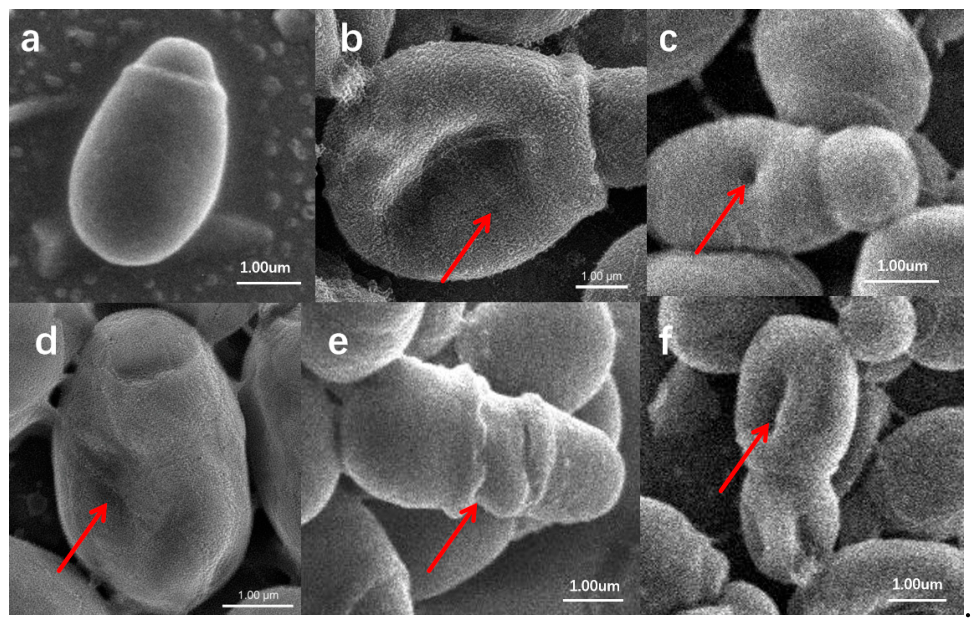
Figure 2.
Checkerboard interaction analysis and growth-curve validation of selected combinations. ALI: allicin; ANI: anisaldehyde; CIN: cinnamaldehyde; EUG: eugenol; ITZ: itraconazole. (a) Checkerboard broth microdilution heat maps showing the interactions between pairwise combinations of natural compounds and antifungal agents against Malassezia pachydermatis SZ06. The color scale represents relative fungal growth (%), where 0% indicates complete growth inhibition, 50% indicates partial growth and 100% indicates growth equivalent to the untreated growth control. (b) Growth curves of Malassezia pachydermatis SZ06 following treatment with 4 natural compounds. M. pachydermatis SZ06 was exposed to 4 natural compounds at MIC, 1/2 MIC, and 1/4 MIC. Fungal growth was monitored by OD600. Untreated cultures served as the growth control. Values represent the mean ± SD (n = 3). (c) Growth curves of Malassezia pachydermatis SZ06 following combination treatment with allicin and selected natural compounds, at MIC, 1/2 MIC, and 1/4 MIC. Fungal growth was assessed by OD600. Data are presented as mean ± SD (n = 3).
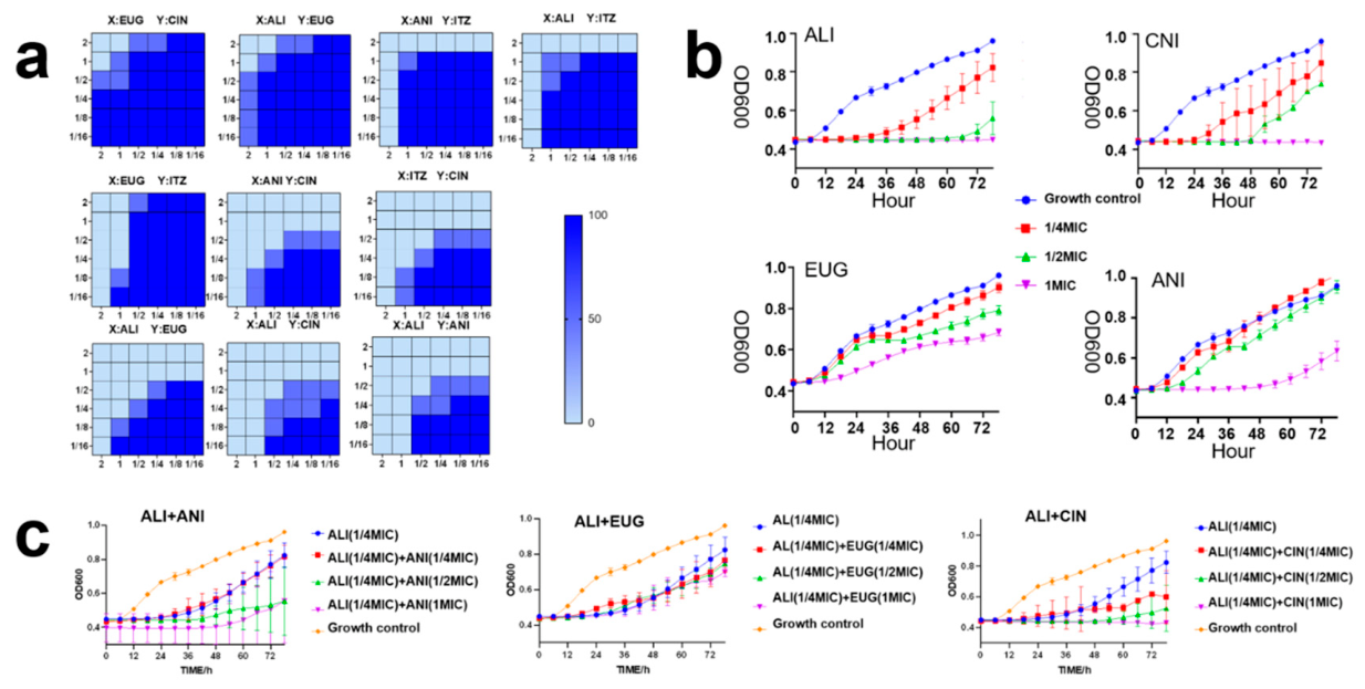
Figure 3.
Whole genome circle of Malassezia pachydermatis SZ06. Circular genome map of Malassezia pachydermatis SZ06. From the innermost to the outermost rings, the tracks indicate GC content, GC skew, the distribution of glycosidase (gray), pectinase (blue), cellulase (green), and xylanase (red) genes across scaffolds, and scaffold organization.
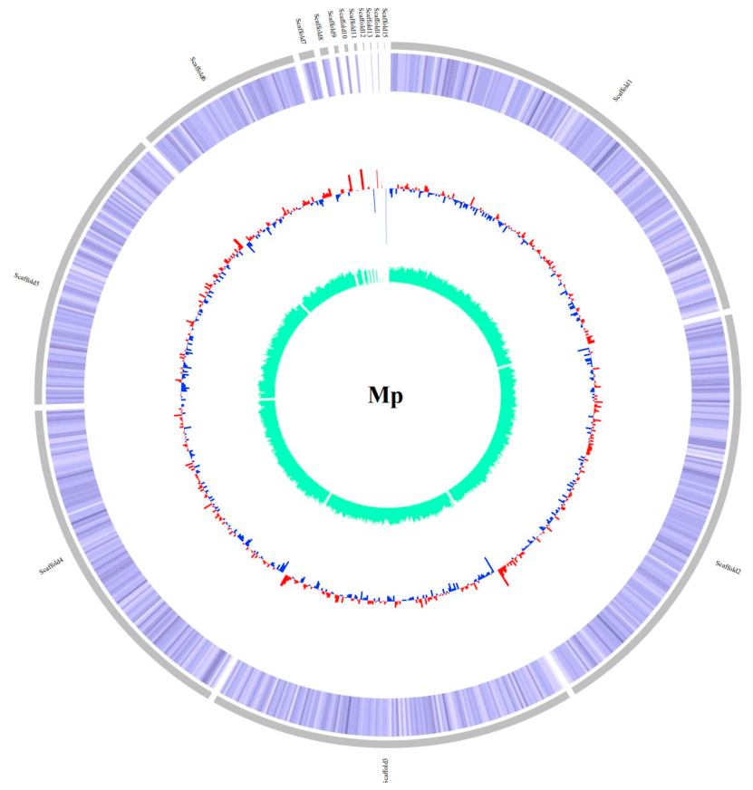
Figure 4.
Classification of antibiotic resistance-associated genes identified in Malassezia pachydermatis SZ06.
Figure 4.
Classification of antibiotic resistance-associated genes identified in Malassezia pachydermatis SZ06.
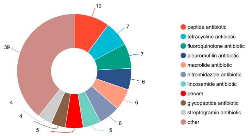
Figure 5.
Serum levels of inflammatory markers IL-1β, IL-6, and TNF-α in different treatment groups after 15 days treatment. ALI: allicin; ANI: anisaldehyde; CIN: cinnamaldehyde; EUG: eugenol; ITZ: itraconazole. Data are presented as mean ± SD.
Figure 5.
Serum levels of inflammatory markers IL-1β, IL-6, and TNF-α in different treatment groups after 15 days treatment. ALI: allicin; ANI: anisaldehyde; CIN: cinnamaldehyde; EUG: eugenol; ITZ: itraconazole. Data are presented as mean ± SD.
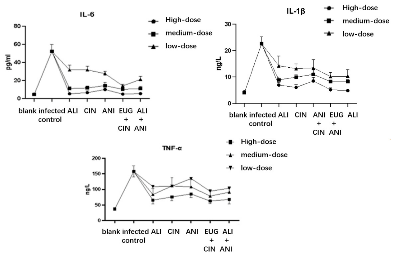
Figure 6.
Representative H&E-stained skin sections after 15 days treatment. (a) blank group, (b) high-dose treatment group, (c) medium-dose treatment group, (d) low-dose treatment group, and (e) infected control group. Black arrows indicate focal epidermal loss; brown arrows indicate focal epidermal thickening; orange arrows indicate thickening of the stratum granulosum; red arrows indicate dermal collagen degeneration/necrosis with nuclear pyknosis; green arrows indicate hair follicle (adnexal) damage; blue arrows indicate necrotic debris; purple arrows indicate elongation of rete ridges; and yellow arrows indicate lymphocytic infiltration. Left panels, ×20 (scale bar = 1000 μm); right panels, ×200 (scale bar = 100 μm).
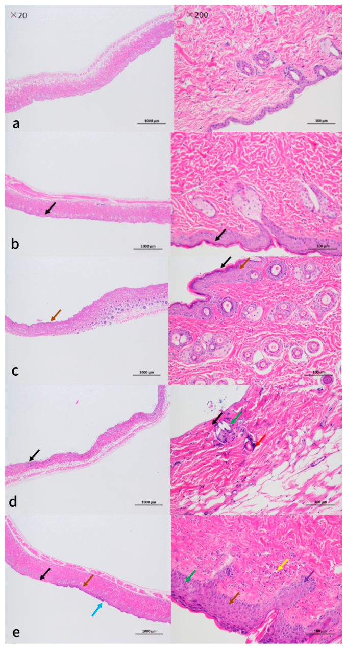

Table 1.
Group allocation and topical treatment used in mouse experiments.
| Classification | Group | Medication |
|---|---|---|
| High-dose group | Group 1 | 2% allicin |
| Group 2 | 2% eugenol | |
| Group 3 | 2% anisaldehyde | |
| Group 4 | 1% allicin+1% eugenol | |
| Group 5 | 1% allicin+1% anisaldehyde | |
| Medium-dose group | Group 6 | 1% allicin |
| Group 7 | 1% eugenol | |
| Group 8 | 1% anisaldehyde | |
| Group 9 | 0.5% allicin+0.5% eugenol | |
| Group 10 | 0.5% allicin+0.5% anisaldehyde | |
| Low-dose group | Group 11 | 0.5% allicin |
| Group 12 | 0.5% eugenol | |
| Group 13 | 0.5% anisaldehyde | |
| Group 14 | 0.25% allicin+0.25% eugenol | |
| Group 15 | 0.25% allicin+0.25% anisaldehyde | |
| Control group | Group 16 | 1% DMSO |
| Blank group | Group 17 | No treatment |
No obvious adverse effects, body weight loss, or mortality were observed during model establishment. Successful infection was established in 102 of 115 male KM mice (88.7%). The successfully infected mice were randomly allocated to 17 experimental groups (n = 6 per group) before treatment initiation.
Table 2.
Information table of isolates.
| Strain Number | Host | Isolation Site | Severity of the onset | Location | Isolation Strain Type |
|---|---|---|---|---|---|
| FS01 | Chinese Li Hua | internal acoustic meatus | Mild | FoShan | M. nana |
| FS02 | British shorthair | perianal region | Mild | FoShan | |
| GZ03 | Blue cat | Lip area | Moderate | GuangZhou | |
| FS05 | Hairless cat | skin folds | Moderate | FoShan | M. pachydermatis |
| SZ06 | Chinese Li Hua | skin folds | Moderate | ShenZhen | |
| GZ07 | Ragdoll | internal acoustic meatus | Moderate | GuangZhou |
Skin lesions were evaluated according to erythema, scaling, crust formation, and skin thickening. Each parameter was graded on a 0–3 severity scale (0 = absent, 1 = mild, 2 = moderate, and 3 = severe), and cumulative scores were used to assess overall lesion severity [20].
Publisher’s Note: MDPI stays neutral with regard to jurisdictional claims in published maps and institutional affiliations. |
© 2026 by the authors. Licensee MDPI, Basel, Switzerland. This article is an open access article distributed under the terms and conditions of the Creative Commons Attribution (CC BY) license (http://creativecommons.org/licenses/by/4.0/).
Copyright: This open access article is published under a Creative Commons CC BY 4.0 license, which permit the free download, distribution, and reuse, provided that the author and preprint are cited in any reuse.