

Submitted:
14 October 2026
Posted:
15 October 2026
You are already at the latest version
Abstract
Ten commercial mushroom products, sourced from resellers and labelled for veterinary use, were subjected to simple quantitative and qualitative polysaccharide analysis methods to differentiate products containing only mushrooms (the fruiting bodies of the fungal life-cycle) from those that include the whole solid fermentation resulting from the fungal mycelium of cereal grain. The therapeutic benefits of fungi are often attributed to their β-glucan content, which, unlike cereal grain β glucans, is uniquely characterized by 1,3-1,6 β glycosidic linkages. Fungal polysaccharide markers were used to distinguish mushroom-containing products from grain-based mycelial products in this study, which highlighted the compositional variability in marketplace products. Ten products labelled for pet use, and one for human use, were subjected to a multifaceted analytical approach by enzymatic quantification of α- and β-glucans and starch, and Lugol’s iodine staining for starch detection. The resulting analyses identified two distinct product profiles: 1) polysaccharide profiles consistent with mushrooms, exhibiting high β-glucan and low α-glucan content and 2) products consistent with fermentation of cereal grains by fungal mycelium that have lower levels of β-glucans, higher levels of α-glucans, and a strong positive iodine-starch test. This study provides objective, evidence-based criteria for differentiation of these product profiles; some products labelled as mushroom-derived contained predominantly grain. This method can be adapted to assess other fungal-derived products and verify the authenticity of mushroom-based veterinary products.
Keywords:
turkey tail
; Trametes versicolor
; β-glucan. α-glucan
; mycelium-fermented grain
; Lugol’s iodine
; veterinary supplements
; companion animals
; label compliance
; polysaccharide analysis
1. Introduction
There has been a significant increase in the application of functional mushrooms as nutritional supplements over the past few years due to the rising trend in the utilization of natural solutions for enhancing the health of both humans and animals [1]. The coronavirus pandemic has seen this development escalate even further as public consciousness regarding immune health increased and as people kept more pets as companions when locked down at home [2]. Dogs have become a fundamental part of the family unit, as they assume the role of family members. This is a trend that has been noted in the past ten years as the “humanization of pets.” This trend is due in part to the fact that many families choose to delay childbearing and instead choose to adopt a pet. Additionally, many families have become “empty nesters,” and choose to add pets to their household as their four-legged children [32]. For these reasons, many pet “parents” desire to provide their dogs and cats with the numerous health benefits from mushrooms, determined by their own successful use and from others’ experiences with using mushrooms.
Despite this growing interest, the landscape of functional mushroom supplementation for companion animals remains relatively underexplored in scientific literature. While numerous studies have documented the health benefits of mushrooms in laboratory animals and humans, peer-reviewed research on their effects in dogs, cats, and horses is limited, but growing. The current understanding of mushroom use in pets is largely extrapolated from human and laboratory animal studies, as well as anecdotal clinical reports from veterinarians and pet owners.
Functional mushrooms contain a variety of secondary metabolites and active ingredients that define their biological activity. Of these active constituents, β-glucans are considered to be the most studied among the many active elements in these mushrooms. Β-glucans serve in the fungal cell as structural elements, strengthened by their combination in the fungal cell wall with chitin and glycoproteins. Β-glucans are polymers of monosaccharides that are linked by glycosidic bonds, between the 1,3 and 1,6 linkages in fungi, and between the 1,3 and 1,4 linkages in cereal grains. Their strong immunomodulating properties in animals result from immunocyte detection of the pathogen-associated molecular pattern (PAMP) of the β glucan molecule physical structure. This immune response is derived from early, archaic animal immune system encounters with fungal pathogen PAMPs from the β glucans in the fungal cell wall. Mushrooms are not fungal pathogens, but their β glucan PAMPs stimulate a robust immune response.
Β-glucans are the most thoroughly studied and therapeutically significant constituents of fungi, primarily known for their potent immune-modulating effects [3,4]. Other secondary metabolites found in fungi, similar to the β-glucans, also have strong biological activities. These metabolites include, but are not limited to, the terpenoids, nucleosides, cordycepin, ergothioneine, ergosterol, phenolic compounds, and peptides [5].
Mushrooms, known in the three-part fungal life cycle as “fruiting bodies,” are one of the three life stages for the fungus. The biological function of mushrooms is to grow the haploid spores, protect the spores while they are developing, and then disseminate the spores widely through their release into the air. The haploid spores germinate into thin hyphal tubes and then “mate” with another compatible hyphal tube to form the mycelium, which is diploid in its chromosome complement.
Mycelium is the vegetative part of the fungal lifecycle. The mycelium elaborates powerful enzymes that digest the substrate it grows upon, which then nourishes further mycelial growth. The mycelium portion of the fungal lifecycle is considered the primary stage that persists in the environment over time. Typically, in the commercial cultivation of mushrooms, the mycelium is first grown on sterile grain. This growth on a solid medium is termed “solid-state fermentation”. The mycelium grows until it visually appears to have completely effaced the cereal grain. Mycelial fermented grain (MFG), which is also called “grain spawn” in the context of cultivation, is then used to inoculate the substrate.
The inoculated substrate becomes myceliated, much like the grain spawn had been, and, when environmental conditions are suitable, the mycelial growth in the substrate will organize itself into a mushroom. This cycle repeats itself as the mushroom (fruiting body) produces the spores, again, and the cycle continues. Traditional mushroom cultivation results in the production of mushrooms from grain spawn-inoculated substrate, using the substrate most appropriate for that mushroom species. Mushroom substrates typically are wood-based for most mushroom species but can be compost/peat-based for a few mushroom species, such as the common button mushrooms (Agaricus bisporus) [6].
In the past 40 years, interest in discovering novel protein sources led researchers to explore the protein-generating properties of mycelium grown on cereal grain or legumes [7]. This novel approach to creating a new category of proteinaceous food has been the source of several commercial food products derived from mycelium fermented substrates that have entered the marketplace. Two examples of these novel foods are Quorn™, which is mycoprotein grown from mycelium, and tempeh, which is myceliated soybeans. These are foods that are consumed in servings of several ounces at a time. These are foods that are consumed in servings of several ounces at a time. This process of creating a novel, protein-rich food from myceliated substrates also introduced this technology to the functional mushroom industry [7].
This technology, originally intended as a protein source for food, has also been used as an ingredient in health supplements. MFG has been found to be a good, novel source of protein that is high quality and vegan. This technology is a new, more rapid, and cost-effective form of cultivation for fungal food products, by skipping that last step in the traditional mushroom cultivation process.
Generally, in cultivation, the grain spawn inoculates an appropriate substrate, such as wood, and continues to grow on this natural substrate until it produces mature mushrooms. It can take as long as 6–9 months for mushrooms cultivated this way to grow to maturity for harvest [8]. By skipping this important step in mushroom cultivation, this new method can allow the myceliated grain spawn to produce mushrooms from the myceliated cereal grain substrate. The MFG is then dried, powdered, and packaged with a label that sometimes describes this novel food as a mushroom product. This MFG product may contain just a few mushrooms, but it is not only mushrooms, as produced from traditional mushroom cultivation practices. Additionally, those mushrooms from MFG are not grown on their native wood substrate, but only on cereal grain, so they do not have the same potency as they would on their natural substrate, wood [8].
One concern, when comparing products that are derived from MFG versus authentic mushrooms, is the amount of grain remaining in the mycelial biomass. For dogs who have allergic sensitivities to cereal grain, the presence of cereal grain in a product can be a serious concern. The actual mycelium-to-grain ratio in the MFG biomass is estimated at no more than 30% [9]. This estimate, published in a patent filing, may be higher than the actual amount of cereal grain converted to fungal biomass.
A recent study quantified the amount of cereal grain that is converted to mycelium. The study estimated the amount of cereal grain remaining after total solid-state colonization of grain flour by fungal mycelium [10]. It determined that the resulting fermented flour remained carbohydrate-dominant, on a dry matter basis, despite the visual appearance of complete colonization. Ergosterol measurements were used as a proxy for the fungal biomass, since ergosterol is present in all fungi and not present at all in cereal grains. Using ergosterol measurements, this study determined that the mycelial biomass in this study was approximately 18%. Other independent studies found comparable results when measuring the amount of mycelium in a solid-state fermentation biomass [11,12]. Typical values range from 15–35% myceliation of the cereal grain [13,14].
Mycelium has very strong biological activities when properly prepared. Reishi, shiitake, lion’s mane and turkey tail fungal species all have mycelium-derived, patented extracts produced from the submerged liquid culture of their mycelium [1,9,15]. Turkey tail PSP and PSK polysaccharide fractions are examples of potent mycelial-derived constituents [16,17]. However, it is important to note that these patented mycelial extracts were derived from mycelium grown in a liquid medium, not on grain [9]. Mycelium can be very easily extracted from a liquid medium and then can be further concentrated into a concentrated fraction [15]. However, when mycelium ferments a solid medium, like cereal grain, it is impossible to separate the mycelium from the grain, and the potency of the mycelium is diluted by the grain. The presence of the grain also makes it difficult to extract and concentrate the mycelium to take advantage of its potent biological activity [10,15,18].
A critical distinction exists between authentic mushroom products and those derived from MFG, with the latter lacking the full complement of bioactive compounds found in mature fruiting bodies. Analytical studies consistently reveal that functional mushrooms are characterized by high levels of β-glucans, which are considered the primary therapeutic compound in fungi. Secondary metabolites are also critical elements of the biological activity of fungi. On the other hand, MFG products are dominated by α-glucans, which are predominantly digestible starch molecules and frequently lack therapeutic levels of key bioactive compounds. This compositional disparity has important implications for both efficacy and labelling accuracy [6,15,19,20,21,22,24,25].
FDA guidelines for mycelium-containing products differ significantly from FDA guidelines for mushroom-containing products. FDA guidelines state clearly that mycelium-based products should be clearly labelled, and the company should not mislabel the products as mushroom-containing [19]. One example of this can be stated as follows: if the container of the MFG product bears a picture of a mushroom, then this is an implied misrepresentation, as it suggests that the product is composed exclusively of mushrooms and not the MFG that it is.
Regarding the fundamental compositional differences that influence biological activity, products with polysaccharide profiles consistent with functional mushrooms are characterized by low average α-glucan content (0.41% ± 0.23% [8]) and high average β-glucan content (60.71% ± 11.80% [22]). In stark contrast, cereal grains typically exhibit high average α-glucan levels (53.6–69% [23]) and significantly lower average β-glucan content (1–7% [24]). These distinct polysaccharide profiles serve as critical markers for product differentiation.
The rapid expansion of the commercial functional mushroom market, now valued in billions of dollars, has prompted concerns regarding product authenticity, mislabeling, and consumer trust [2,20,23,25]. Recent investigations of products labeled for human use have demonstrated that a significant proportion of products marketed as mushroom supplements are, in fact, composed primarily of MFG, with only a minority containing mushroom biochemical profiles [20,27]. This makes it clear that there is a need for robust analytical techniques and proper labelling practices so that both consumers and veterinarians are able to make well-informed choices regarding their use of mushroom products for their pets’ welfare.
In this study, we attempt to overcome these hurdles associated with the proper authentication of a product as mushroom or MFG by using standardized techniques for the measurement of α- and β-glucan concentrations along with a qualitative test for starch. These procedures will be used to differentiate between polysaccharide profiles that are characteristic of mushrooms and those that have mycelium-fermented grain profiles.
These findings will provide a foundation for evidence-based recommendations and quality assurance in the use of functional mushrooms for companion animal health and further equip practitioners with accessible bench-top tools, such as the Lugol’s iodine starch test, to independently verify product composition and avoid mislabeled MFG alternatives.
2. Materials and Methods
2.1. Product Selection and Procurement
Turkey tail (Trametes versicolor) mushroom products labelled for pet use were selected for analytical assessment based on their widespread commercial availability and popular interest as a result of two published trials in canine hemangiosarcoma splenic cancer [16,17]. Of all the mushroom species, turkey tail has emerged as the mushroom species most familiar to the pet consumer, due in part to these cancer studies in the dog.
Ten products, specifically labelled for pet use, and one product labelled for human consumption, were procured via online purchase from Amazon.com™ in quadruplicate to ensure adequate sample material for multi-laboratory analyses. Products were selected based on label claims of containing turkey tail mushrooms or mushroom blends that incorporated turkey tail. Lot numbers were recorded at receipt for traceability. Due to complete blinding during analysis, products were not stratified or analyzed by lot, and all results reflect blinded sample codes rather than lot-specific comparisons.
2.2. Sample Preparation and Blinding Protocol
All product labels were completely obscured and replaced with numerical codes to ensure double-blind analysis conditions for both analytical laboratories and research investigators; blinding and code assignment were performed by an individual not involved in the study. (Figure 1) The code assignment was kept hidden until the analysis and data gathering were completed. This blinding technique ensured that there were no sources of bias involved during the sample analysis that was conducted at multiple analytical laboratories.
2.3. Analytical Methodologies
Three complementary methodologies were employed for the quantification and qualitative assessment of α-glucans, β-glucans, and starch. Starch presence was evaluated qualitatively using Lugol’s iodine staining, while α-glucan and β-glucan contents were quantified using the Megazyme Yeast and Mushroom β-Glucan Assay Kit according to the manufacturer’s protocol. The third test employed a digestible starch assay, namely Total Starch Quantification (AOAC 996.11) (K-TSTA-100A; AOAC 996.11), to ensure accurate measurement of digestible starch. Appropriate reagent blanks, positive controls, and kit standards were included [15,28].
2.3.1. Β-Glucan Quantification (K-YBGL)
Β-glucan content was quantified using the Megazyme Mushroom and Yeast β-Glucan Assay kit (K-YBGL, 08-23) according to the manufacturer’s protocol. In this method, total glucan (the sum of α + β) is measured after acid hydrolysis and enzymatic conversion to glucose using exo-1,3-β-glucanase and β-glucosidase, whereas α-glucans (derived from starch, sucrose, maltodextrin, and trehalose) are quantified by enzymatic hydrolysis with amyloglucosidase, invertase, and trehalase. β-Glucan content is then calculated indirectly as the difference between total glucan and α-glucan. Controls followed the kit specifications and included blank reagent controls and supplied reference standards. Performance was verified with a yeast control with standardized β-glucan percentages. Results are reported as percent β-glucan (w/w) on a dry-weight basis. This test was developed by McCleary when it became clear that the existing analysis for cereal grain 1,3-1,4 β-glucans was not able to measure the 1,3-1,6 β-glucans in mushrooms, yeast, and mycelial products [15].
2.3.2. Total Starch Quantification (Aoac 996.11)
Starch was quantified using the Total Starch (α-amylase/amyloglucosidase) assay kit (K-TSTA-100A; AOAC 996.11) per the manufacturer’s protocol. Assay controls included reagent blanks and kit standards. Method suitability was checked with mushroom-derived cereal grain material as a matrix-appropriate positive control. Results are reported as percent starch (w/w) on a dry-weight basis.
2.3.3. Qualitative Starch Assessment (Lugol’s Iodine)
Qualitative screening employed Lugol’s iodine staining with visual classification of color response and α-amylase digestion to confirm enzyme-susceptible α-glucans. The staining of the positive and negative controls was conducted simultaneously with each set of sample analyses.
2.4. Multi-Laboratory Corroboration and Harmonization
Experiments were conducted in our laboratory (Nammex) with verification of results affirmed by using other accredited commercial laboratories using the same analytical tests. Multiple independent laboratories were used to analyze each product so the results could be compared across laboratories, rather than to assess analytical precision through replicate testing within a single laboratory.
Participating facilities and their specific roles included:
- SGS Burnaby, BC: Performed K-YBGL β-glucan.
- SGS Guelph, ON: Conducted Total Starch (K-TSTA-100A; AOAC 996.11).
- Eurofins Madison, WI: Provided K-YBGL β-glucan, Total Starch (K-TSTA-100A; AOAC 996.11).
- Nammex, Gibsons, BC: Performed K-YBGL β-glucan and Lugol’s staining.
These laboratories were selected to provide cross-platform corroboration of the analytical findings using established methods appropriate to each analyte. Each sample is automatically analyzed in duplicate as part of the test manufacturer’s validated kit protocol, and the reported value is the average of those technical replicates. Where multiple results are reported for the same product from a given laboratory, those were repeat analyses rather than planned study replicates. In a few cases results differed between laboratories more than expected, so the respective laboratory repeated the analysis to confirm the findings.
Concordance among results from the participating laboratories strengthened confidence in the reproducibility of the polysaccharide profiles observed across products and supported the distinction between samples consistent with mushroom fruiting body material and those more consistent with MFG compositions. A summary of participating laboratory accreditation status, method scope, and assay assignments is provided in Supplementary Table S1.
2.5. Statistical Analysis
All quantitative data, including β-glucan, α-glucan, and starch concentrations, were compiled and analyzed using Microsoft Excel. For each product, mean values and standard deviations (SD) were calculated from the analytical measurements reported by the participating laboratories. Where multiple laboratory measurements were available, the variability among laboratory results was used to describe analytical dispersion. Descriptive comparisons of polysaccharide content were performed across product categories (mushroom versus MFG).
3. Results
Orthogonal measurements of starch, α-glucans, and β-(1,3)(1,6)-glucans from multiple independent laboratories of eleven products (PET-011 to PET-021) revealed two predominant compositional clusters: high β-glucan/low α-glucan/low starch consistent with mushroom-derived polysaccharide profiles and low β-glucan/high α-glucan/high starch consistent with MFG polysaccharide profiles. A minority of products showed intermediate values. These patterns form the basis for composition-based classification of mushroom vs MFG derived products. The tabulated raw results and averaged results with label analysis are provided in Supplementary Tables S2 and S3.
3.1. Lugol’s Iodine Testing of Controls and Product Samples
To detect the presence of starch in both control and commercial samples, Lugol’s iodine staining was performed on a panel of reference materials and the eleven commercial products labeled as mushroom products. Control samples yielded expected results: water and mushroom-derived controls (chaga, cordyceps, turkey tail, lion’s mane, reishi) showed no color change (Figure 2 and Figure 3), indicating the absence of starches, while cereal grains (potato starch, brown rice, oats, sorghum) stained blue, consistent due to the presence of starch. Maltodextrin stained deep red, as anticipated (Figure 2).
Among the product samples, PET-012, PET-014, PET-021, and PET-020 exhibited no significant color change upon iodine addition, before or after α-amylase treatment, indicating the absence of starch or maltodextrin. In contrast, PET-013, PET-015, PET-016, PET-017, PET-018, and PET-019 produced a blue color with Lugol’s iodine, which disappeared after α-amylase digestion, confirming the presence of starch (Figure 4). Notably, PET-016 and PET-017 showed the most intense blue coloration, suggesting the highest starch content.
3.2. Quantification of - and -Glucans in Commercial Pet Products
As shown in Figure 5, PET-012, PET-020, PET-014, and PET-021 display high β-glucan (>19.5%), low α-glucan (<8.3%), and low digestible starch (<2.73%), consistent with mushroom-derived polysaccharide profiles. PET-011, PET-013, PET-015, PET-016, PET-017, PET-018, and PET-019 display low β-glucan (<8.5%), high α-glucan (>11.2%), and high digestible starch (>9.85%), characteristic of MFG product profiles.
These percentage thresholds were derived from the observed distribution of values across all analyzed samples and are consistent with previously reported compositional ranges for mushroom-derived products and cereal grain-based substrates, where mushrooms are characterized by high β-glucan and low α-glucan content, while cereal-fermented materials exhibit the inverse profile.
To show another perspective of the data, a three dimensional scatter plot (see Figure 6) including digestible starch was created. The same groupings seen in the two dimensional scatter plots were retained. High levels of β-glucan and low levels of α-glucan in a given product were associated with lower amounts of digestible starch. Products that have the opposite of this profile would be grouped to include products that had higher levels of α-glucan and total starch.
3.3. Starch and Glucan Composition Across Pet Products
Comparative analysis of starch and glucan content in commercial PET mushroom products is shown in Figure 7.
3.4. Label Analysis and Product Classification
A comparison of product label descriptions with analytical results revealed discrepancies in several cases (Table 1). Two products that were marked with terms such as “mushroom” or “mushroom blend” without any reference to mycelium or cereal grains turned out to contain predominantly cereal grain starch. On the contrary, products that mentioned β-glucan content without claiming either mycelium or fillers had mushroom origins based on their chemical analysis.
3.5. Interpretation
The combined results of Lugol’s iodine staining, Megazyme β-glucan assays, and starch assays provide a means of identifying grain adulteration in commercial mushroom products, labeled as mushrooms, based on α- and β-glucan and starch content. Authentic mushroom products are characterized by high β-glucan and low α-glucan and starch content, whereas MFG products show the opposite trend. Two of the eleven products were found to be mislabeled, with label claims inconsistent with their actual compositions. These findings underscore the value of accessible, orthogonal assays for detecting grain-based adulteration in mushroom products and for aligning label claims with composition in veterinary practice. While most quantitative analyses require specialized laboratory setups, the qualitative Lugol’s iodine starch test stands out as a practical preliminary screening tool that can be implemented in-house in virtually any setting, including a standard domestic kitchen, or veterinary clinic, without the need for specialized laboratory infrastructure.
4. Discussion
4.1. Overview of Analytical Approach and Methodology
We developed an objective, comprehensive, analytical framework to differentiate mushroom-derived products from mycelium-fermented grain formulations for veterinary applications by employing multiple complementary methodologies. This approach addresses labelling complexities through validated quantification of α- and β-glucans and total starch. Sample handling followed assay-specific, standardized protocols to minimize matrix-driven variance; β-glucan was calculated using the Megazyme Mushroom and Yeast β-Glucan kit (K-YBGL) with manufacturer-specified hydrolysis and enzymatic digestion, starch (amylose/amylopectin) was quantified using the Total Starch assay (AOAC 996.11), and qualitative Lugol’s iodine staining with optional α-amylase enzymatic digestion provided an orthogonal screen for α-glucans.
The assessment step of this work evaluated 11 internet identified products based on their β-glucan to α-glucan ratios. These ratios were compared with the established compositional profiles of mushrooms and MFG substrates. Finally, we established objective criteria to classify products as either mushroom-derived or MFG-based, using quantitative thresholds derived from analytical measurements.
Our analytical platform was comprised of five main components: (1) validated enzymatic assays for direct quantification of α-glucans and starch; (2) indirect (by-difference) quantification of β-glucans using the K-YBGL method; (3) qualitative identification of starch using the visual data derived from Lugol’s iodine testing, combined with α-glucan enzymatic digestion to verify the presence of α glucans; (4) standardized sample preparation protocols to ensure reproducibility across diverse product matrices; and (5) comparative analysis frameworks used to differentiate product categories based on compositional profiles.
Enzyme-based methods accurately identify the polysaccharide fractions being measured. The enzyme-based platform is modular and can be extended to quantify additional carbohydrate fractions, provided each added analyte is separately validated within the same analytical framework. Moreover, the method can analyze samples from mushroom fruiting bodies, fermented grains containing mycelium, or other mixtures of ingredients [15].
4.2. Differentiation of Authentic Mushrooms from Mycelium-Fermented Grains
A quantitative profile was evident in the analytical assessment carried out by Windsor et al. when comparing the content of α-glucan and β-glucan found in genuine chaga canker, chaga mycelium, chaga myceliated grain, and cereal grains. this showed a distinct separation among products containing authentic chaga, mycelium, and cereals [20].
Through the analysis, we observed significant differences in the products’ composition, which is in alignment with products made from actual mushrooms. MFG products could be separated from those made from mushrooms due to compositional differences in their polysaccharide components. These findings are in line with published analyses of products derived mainly from cereal grain [28]. Our analyses demonstrated that products sourced from mushrooms had β-glucan levels above those of digestible starch and α-glucans. The converse is true with products sourced from MFG, were found to have a digestible starch and α-glucan content greater than their β-glucan content, due to the contribution of cereal grain.
The disparities in structural organization of branched fungal (1,3)(1,6)-β-glucan and cereal grain (1,3)(1,4)-β-D-glucan were used in the design of our method of analysis. These structural differences result in bioactivity differences with fungal β-glucans having greater neutrophil phagocytosis and cytokine production capacity than the β-glucans found in cereal grains [30].
4.3. Clinical Implications and Risk Assessment for Veterinary Applications
This study demonstrated that MFG products may be mislabeled by virtue of being identified as mushroom-derived. MFG products consist mainly of fermented cereal grain without substantial amounts of the β-glucan polysaccharides that are present in actual mushroom products. Veterinarians and animal owners administer mushroom-based products due to the therapeutic potential of mushroom constituents such as fungal β-glucans. The unintended administration of mislabeled mushroom products might lead to a delay in therapy or have no therapeutic effect at all when the desired activity is derived from actual mushroom constituents.
In addition to the potential lack of therapeutic effects, the use of mislabeled products could lead to potential adverse effects among pets with certain health problems. This includes the development of grain allergy or grain-induced hypersensitivity among patients on special dietary restrictions to manage dermatological or gastrointestinal problems. This suggests that mislabeled MFG products not only do not contain mushrooms, but could potentially produce harm among specific patients
Products falsely marketed as containing mushrooms, yet containing primarily grain carbohydrates, do not possess sufficient levels of β-glucans and secondary metabolites required for eliciting intended biological effects associated with mushroom use. Although cereals such as oats and barley typically contain β-glucans at concentrations ranging between 2% and 7%, these differ structurally from the immunomodulatory fungal β-glucans in that the former comprises β-(1→3)(1→4) linkages, while the latter is mainly comprised of β-(1→3)(1→6) bonds. Mushrooms typically contain 5–10 times as much β-glucan as cereal grain [15].
The analysis strategy adopted in this study takes into account these structural differences by examining the glucan profile based on the anticipated signature of fungi containing the (1,3)(1,6) glycosidic linkages. Although grain products may contain β-glucans, these lack the immunogenic attributes of the fungal β-glucans. [21,29,30].
4.4. Inter-Laboratory Validation and Analytical Consistency
For assessing the reproducibility and accuracy of our analysis procedures in different testing environments, we used identical, validated techniques in several certified laboratories using the same set of samples. The testing performed at SGS Guelph and Eurofins Madison under standard protocols produced numerically similar results for most of the samples, with an acceptable level of variability for the remaining samples (less than the usual analytical tolerance of 10%). However, large differences were noted compared to the usual range of variability for some specific samples (PET-011 and PET-012), especially regarding the digestible starch content, necessitating further evaluation [21,29,30].
In general, inter-laboratory differences may be caused by heterogeneous samples resulting from different batches of the products or interfering substances that may have affected the Megazyme assay, such as cellulose from the herbs used as additional ingredients. Where multiple results are reported for the same product from a given laboratory, those were repeat analyses rather than planned study replicates. In a few cases results differed between laboratories more than expected, so the respective laboratory repeated the analysis to confirm the findings We also sought to understand the possibility of matrix interference with the measurements of the digestible starch levels in the given products, as well as the effect of possible variability of their composition.
Accreditation details of the participating laboratories, together with the analytical methods used in this study, are provided in Supplementary Table S1.
4.5. Limitations and Methodological Considerations
Our current analytical approach presents several limitations including that while our analytical framework effectively differentiates between polysaccharide profiles consistent with mushroom materials and those characteristic of MFGs, it does not confirm the presence of any specific fungal species in a given product. Our classifications have been based on the overall polysaccharide profile (high beta-glucan/low alpha-glucan for mushrooms, and the inverse for grain-based products), rather than species-specific markers.
A second limitation was our inability to use ergosterol testing to differentiate fungal and cereal grain products. This stemmed from the variable composition of the products recruited and analysed. Ergosterol testing, which is a routine method of detection of fungal contaminants in stored grain, would have added an extra level of differentiation between cereal grain versus fungal containing products.
The extensively validated Megazyme Mushroom and Yeast β-glucan assay (K-YBGL), indirectly measures β-glucan content through calculations rather than direct quantification. This may introduce systematic errors with complex matrices containing non-hydrolysable α-glucans or interfering compounds, such as cellulose from herbal components. Additionally, the assay cannot differentiate between various types of α-glucans. We implemented complementary enzymatic methodologies (K-YBGL for mushroom and yeast β-glucans and the Total Starch assay for alpha-glucans/starch), and qualitative Lugol’s iodine staining. This approach was designed to enable the differentiation of mushroom-derived from grain-derived products based on their distinct polysaccharide profiles, rather than providing a comprehensive analysis of all possible polysaccharide types.
The existence of other ingredients, such as herbs, palatants, and excipients, in commercial veterinary formulations could affect the outcome of our analysis. In addition, the present study examined randomly purchased products from the internet and not the whole range of commercially available veterinary products labeled as containing mushrooms. The possibility of having variations between batches for single products is another problem that must be considered when attempting to set universal standards for quality control in commercial fungal preparations.
4.6. Implications for Veterinary Practice and Consumer Education
Due to the substantial compositional differences we identified between products with polysaccharide profiles consistent with mushrooms and those consistent with MFG alternatives, our research provides critical guidance for evidence-based product selection in veterinary practice. The objective analytical criteria we established enables veterinarians and pet owners to make informed decisions when selecting fungal products based on intended therapeutic applications rather than marketing claims or product labelling alone. Our findings translate into a practical selection framework that identifies unreliable labels by prioritizing the objective composition markers identified in this study. Veterinarians and pet parents seeking authentic mushroom products should favor products measured to have high β-glucan and low α-glucan and total starch profiles consistent with mushrooms as verified by method-specified assays. In the clinic, veterinarians can employ the use of a simple Lugol’s iodine benchtop check of putative mushroom products to detect starch in a product.
Products reporting on their labels only “total polysaccharides,” with no β-glucan analysis reported, and showing starch-positive reactions, or labelled as “mycelium-fermented grain”, “myceliated grain”, or “mycelial biomass”, will often list the species of cereal grain used which will identify these products as containing diluted fungal bioactives. To better identify these cereal-grain-based products, veterinarians should request batch-specific β-glucan data, screen product labels for clear mushroom designation, and maintain a clinic formulary of vetted mushroom products that have used starch analysis methodologies and β-glucan to α-glucan profiles to ensure consistent, evidence-based product selection when sourcing mushroom products. Our framework demonstrates that the β-glucan to α-glucan ratios found in products with polysaccharide profiles consistent with containing mushrooms correspond to the compositional features associated with the known immunomodulatory and therapeutic benefits of fungal bioactives derived from mushrooms.
Manufacturers bear responsibility to align labels with composition. The path to achieve this is by implementing validated multi-method polysaccharide QC (β-glucan quantification with method disclosure, α-glucan/starch profiling, confirmatory iodine testing, measuring ergosterol levels when possible, publishing batch CoAs, and clearly identifying fungal part(s) used) so that “mushroom” claims are reserved for polysaccharide profiles consistent with fruiting-body material containing high β-glucans and low starch signatures rather than polysaccharide profiles consistent with MFG matrices. Regulatory bodies should codify definitions distinguishing mushroom from mycelium for pet supplements, require method-referenced β-glucan quantitation for immunity-related claims, and enforce corrective actions for misbranding, including rapid relabeling or recall when products fail compositional thresholds consistent with mushroom benchmarks.
5. Conclusions
We have provided objective, evidence-based criteria to differentiate polysaccharide profiles consistent with mushroom products from those consistent with MFG products for veterinary applications, emphasizing the critical role β-glucan content plays for therapeutic efficacy as well as for its value in differentiation between authentic mushrooms and MFG products.
This study has the following four advantages: (i) it provides detailed, objective evidence through polysaccharide analysis, defining the significant compositional differences between these two categories of fungal products; (ii) it reveals that two products in this 11-product analysis were erroneously labelled, consisting mostly of carbohydrates from cereal grains rather than ß-glucans found in mushrooms; (iii) it highlights the potential clinical risk of using mislabeled products, which may interfere with the intended clinical benefits they were initially acquired to address; and finally, and probably most important, (iv) these data empower veterinarians and pet parents to make informed, evidence-based decisions when selecting a fungal product.
Clinicians and consumers should look for products that provide information about the products’ content of β-glucan, what assay methods were employed in the analysis, and which fungal parts were used in the supplement in order to avoid acquiring products with a high proportion of grain when the bioactivity of mushrooms is counted upon. Lugol’s iodine test may become a good starting point, with products producing a blue, brown, or purple color as an indicator of cereal grain content.
The current study considered product composition in terms of polysaccharides. A comparative study of secondary metabolites would also show differences in their content when derived from mushrooms versus myceliated grain. Additional future research should include clinical trials to investigate the connection between product composition and therapeutic effects.
Lastly, the enforcement of existing regulations regarding the appropriate labelling of mushroom supplements to distinguish them from myceliated-grain supplements would go a long way toward re-establishing consumer confidence in commercial fungal products. Extending the application of these regulations to include veterinary mushroom supplements would be a good first step to take in the direction of product transparency and truth in labelling for this category of mushroom supplements.
Supplementary Materials
The following supporting information can be downloaded at the website of this paper posted on Preprints.org. Table S1: Participating laboratories, AOAC certification status, and analytical methods used in this study. A table of the raw data can be found in Table S2, and a table of the averaged data in Table S3.
Author Contributions
Conceptualization, R.J.S; methodology, R.J.S; software, R.J.S and D.A.; formal analysis, D.A, and R.J.S; investigation, R.J.S; resources, R.J.S and data curation, D.A.; writing original draft preparation, D.A. and R.J.S; writing, review and editing, R.J.S, and D.A; visualization, D.A and R.J.S; supervision, R.J.S; project administration, R.J.S, and D.A. All authors have read and agreed to the published version of the manuscript.
Funding
The study was supported by Nammex™ and Real Mushrooms™.
Institutional Review Board Statement
Not applicable. This study did not involve humans or animals; only commercially available pet supplement products were analysed.
Informed Consent Statement
Not applicable.
Data Availability Statement
The data presented in this study are available within the article and its Supplementary Materials. Additional raw data are available from the corresponding author upon request.
Acknowledgments
The authors thank M. Ischii, A.E Kreynes, and N. Boulton for their invaluable assistance to this study, and to the companies, Nammex and Real Mushrooms for their financial and moral support in this work.
Conflicts of Interest
R.J.S is employed by the companies Real Mushrooms™ and Nammex™. D.A. declares no competing interests.
References
- Niego, A.G.; Rapior, S.; Thongklang, N.; Raspé, O.; Jaidee, W.; Lumyong, S.; Hyde, K.D. Macrofungi as a Nutraceutical Source: Promising Bioactive Compounds and Market Value. J. Fungi 2021, 7, 397. [Google Scholar] [CrossRef] [PubMed]
- Kamel, M.S.; El-Sayed, A.A.; Munds, R.A.; Verma, M.S. Interactions between Humans and Dogs during the COVID-19 Pandemic: Recent Updates and Future Perspectives. Animals 2023, 13, 524. [Google Scholar] [CrossRef] [PubMed]
- Blumfield, M.; Abbott, K.; Duve, E.; Cassettari, T.; Marshall, S.; Fayet-Moore, F. Examining the Health Effects and Bioactive Components in Agaricus bisporus Mushrooms: A Scoping Review. J. Nutr. Biochem. 2020, 84, 108453. [Google Scholar] [CrossRef] [PubMed]
- Boulaka, A.; Christodoulou, P.; Vlassopoulou, M.; Koutrotsios, G.; Bekiaris, G.; Zervakis, G.I.; Mitsou, E.K.; Saxami, G.; Kyriacou, A.; Zervou, M.; et al. Genoprotective Properties and Metabolites of β-Glucan-Rich Edible Mushrooms Following Their In Vitro Fermentation by Human Faecal Microbiota. Molecules 2020, 25, 3554. [Google Scholar] [CrossRef] [PubMed]
- Soares, A.A.; de Sá-Nakanishi, A.B.; Bracht, A.; da Costa, S.M.G.; Koehnlein, E.A.; de Souza, C.G.M.; Peralta, R.M. Hepatoprotective Effects of Mushrooms. Molecules 2013, 18, 7609–7630. [Google Scholar] [CrossRef] [PubMed]
- Stamets, P.; Chilton, J.S. The Mushroom Cultivator: A Practical Guide to Growing Mushrooms at Home; Agarikon Press: Olympia, WA, USA, 1983; 415 p. [Google Scholar]
- Quintieri, L.; Nitride, C.; De Angelis, E.; Lamonaca, A.; Pilolli, R.; Russo, F.; Monaci, L. Alternative Protein Sources and Novel Foods: Benefits, Food Applications and Safety Issues. Nutrients 2023, 15, 1509. [Google Scholar] [CrossRef] [PubMed]
- Clark, A.J.; Soni, B.K.; Sharkey, B.; Acree, T.; Lavin, E.; Bailey, H.M.; et al. Shiitake Mycelium Fermentation Improves Digestibility, Nutritional Value, Flavor and Functionality of Plant Proteins. LWT 2022, 156, 113065. [Google Scholar] [CrossRef]
- Stamets, P.E. WO 2016/161138 A1; Antiviral Activity from Medicinal Mushrooms and Their Active Constituents. World Intellectual Property Organization (WIPO): Geneva, Switzerland, 2016.
- Stoffel, F.; et al. Chemical Features and Bioactivity of Grain Flours Colonized by Macrofungi as a Strategy for Nutritional Enrichment. Food Chem. 2019, 297, 124988. [Google Scholar] [CrossRef] [PubMed]
- Agosin, E.; Odier, E. Structural Changes in Wheat Straw Components during Decay by Lignin-Degrading White-Rot Fungi in Relation to Improvement of Digestibility for Ruminants. J. Sci. Food Agric. 1985, 36, 397–403. [Google Scholar] [CrossRef]
- Locci, E.; et al. Wheat Bran Biodegradation by Pleurotus ostreatus: A Solid-State Carbon-13 NMR Study. Bioresour. Technol. 2008, 99, 4279–4284. [Google Scholar] [CrossRef] [PubMed]
- Steudler, S.; Bley, T.; Lindner, P. Better One-Eyed Than Blind—Challenges and Opportunities of Biomass Measurement during Solid-State Fermentation of Basidiomycetes. 2015. [Google Scholar] [CrossRef] [PubMed]
- Agosin, E.; Odier, E. Structural Changes in Wheat Straw Components during Decay by Lignin-Degrading White-Rot Fungi. J. Sci. Food Agric. 1985, 36, 397–403. [Google Scholar] [CrossRef]
- McCleary, B.V.; Draga, A. Measurement of β-Glucan in Mushrooms and Mycelial Products. J. AOAC Int. 2016, 99, 364–373. [Google Scholar] [CrossRef] [PubMed]
- Brown, D.C.; Reetz, J. Single Agent Polysaccharopeptide Delays Metastases and Improves Survival in Naturally Occurring Hemangiosarcoma. Evid.-Based Complement. Altern. Med. 2012, 2012, 1–8. [Google Scholar] [CrossRef] [PubMed]
- Gedney, A.; Salah, P.; Mahoney, J.A.; Krick, E.; Martins, R.; Scavello, H.; Lenz, J.A.; Atherton, M.J. Evaluation of the Anti-Tumour Activity of Coriolus versicolor Polysaccharopeptide (I’m-Yunity) Alone or in Combination with Doxorubicin for Canine Splenic Hemangiosarcoma. Vet. Comp. Oncol. 2022, 20, 688–696. [Google Scholar] [CrossRef] [PubMed]
- Postemsky, P.D.; Curvetto, N.R. Solid-State Fermentation of Cereal Grains and Sunflower Seed Hulls by Grifola gargal and Grifola sordulenta. Int. Biodeterior. Biodegrad. 2015, 100, 52–61. [Google Scholar] [CrossRef]
- U.S. Food and Drug Administration. CPG Sec. 585.525: Mushroom Mycelium—Fitness for Food; Labelling. Available online: https://www.fda.gov/regulatory-information/search-fda-guidance-documents/cpg-sec-585525-mushroom-mycelium-fitness-food-labeling (accessed on 15 August 2025).
- Windsor, C.; Kreynes, A.E.; Chilton, J.S.; Chioffi, W.A.; Krishnamurthy, A.; Ishii, M. Comparative Study of Chaga (Inonotus obliquus) Dietary Supplements Using Complementary Analytical Techniques. Int. J. Mol. Sci. 2025, 26, 2970. [Google Scholar] [CrossRef] [PubMed]
- Sari, M.; Prange, A.; Lelley, J.I.; Hambitzer, R. Screening of Β-Glucan Contents in Commercially Cultivated and Wild Growing Mushrooms. Food Chem. 2017, 216, 45–51. [Google Scholar] [CrossRef] [PubMed]
- Aaman, P.; Hesselman, K. Analysis of Starch and Other Main Constituents of Cereal Grains. Swed. J. Agric. Res. 1984. [Google Scholar]
- Lante, A.; Canazza, E.; Tessari, P. Β-Glucans of Cereals: Functional and Technological Properties. Nutrients 2023, 15, 2124. [Google Scholar] [CrossRef] [PubMed]
- You, H.; Abraham, E.J.; Mulligan, J.; Zhou, Y.; Montoya, M.; Willig, J.; Chen, B.-K.; Wang, C.-K.; Wang, L.-S.; Dong, A.; et al. Label Compliance for Ingredient Verification: Regulations, Approaches, and Trends for Testing Botanical Products Marketed for “Immune Health” in the United States. Crit. Rev. Food Sci. Nutr. 2024, 64, 2441–2460. [Google Scholar] [CrossRef] [PubMed]
- Nussbaumer, M.; Karl, T.; Benz, J.P. Quantification of Fungal Biomass in Mycelium Composites Made from Diverse Biogenic Side Streams. Fungal Biol. Biotechnol. 2024, 11, 20. [Google Scholar] [CrossRef] [PubMed]
- Liu, J.; Sun, J.; He, R.; Xia, J.; He, P. The Situation of Counterfeited and Mislabeled Commercialized Edible Mushrooms in China and the Development of Possible Controls. Foods 2024, 13, 3097. [Google Scholar] [CrossRef] [PubMed]
- Verduzco-Oliva, R.; Gutierrez-Uribe, J.A. Beyond Enzyme Production: Solid State Fermentation (SSF) as an Alternative Approach to Produce Antioxidant Polysaccharides. Sustainability 2020, 12, 495. [Google Scholar] [CrossRef]
- Zielke, C.; Kosik, O.; Ainalem, M.L.; Lovegrove, A.; Stradner, A.; Nilsson, L. Characterization of Cereal β-Glucan Extracts from Oat and Barley and Quantification of Proteinaceous Matter. PLoS ONE 2017, 12, e0172034. [Google Scholar] [CrossRef] [PubMed]
- Wu, D.-T.; Deng, Y.; Chen, L.-X.; Zhao, J.; Bzhelyansky, A.; Li, S.-P. Evaluation on Quality Consistency of Ganoderma lucidum Dietary Supplements Collected in the United States. Sci. Rep. 2017, 7, 7792. [Google Scholar] [CrossRef] [PubMed]
- Caseiro, C.; Dias, J.N.R.; de Andrade Fontes, C.M.G.; Bule, P. From Cancer Therapy to Winemaking: The Molecular Structure and Applications of β-Glucans and β-1,3-Glucanases. Int. J. Mol. Sci. 2022, 23, 3156. [Google Scholar] [CrossRef] [PubMed]
- Flores, G.A.; Cusumano, G.; Venanzoni, R.; Angelini, P. The Glucans Mushrooms: Molecules of Significant Biological and Medicinal Value. Polysaccharides 2024, 5, 212–224. [Google Scholar] [CrossRef]
- Martos Martinez-Caja, A.; De Herdt, V.; Enders-Slegers, M.-J.; Moons, C.P.H. Pets and Their Owners during the First COVID-19 Lockdown Period: Perceived Changes in Routines and Emotions—An Exploratory Study. J. Vet. Behav. 2022, 48, 86–91. [Google Scholar] [CrossRef] [PubMed]
- Dietary Supplement Health and Education Act of 1994, Pub. L. No. 103-417, 108 Stat. 4325 (1994). Available online: https://www.congress.gov/bill/103rd-congress/senate-bill/784 (accessed on 20 May 2026).
- U.S. Food and Drug Administration. Dietary Supplements. Available online: https://www.fda.gov/food/dietary-supplements (accessed on 20 May 2026).
Figure 1.
Photograph of sample product blinding method.
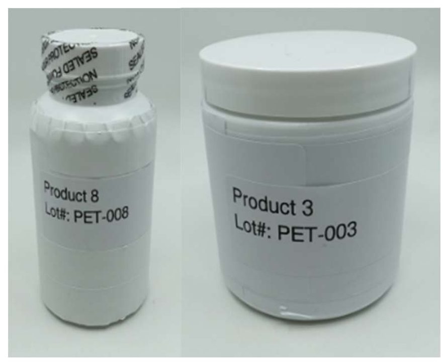
Figure 2.
Lugol’s iodine testing of starch and non-starch control samples. Water showed no color change, indicating the absence of starch. Known starch-containing controls (potato starch, brown rice, oats, sorghum) exhibited a blue color, consistent with the presence of starch. Maltodextrin stained deep red, as anticipated. The introduction of α-amylase, which digests any starch present (third row), did not cause any color changes due to the absence of starch in these samples.
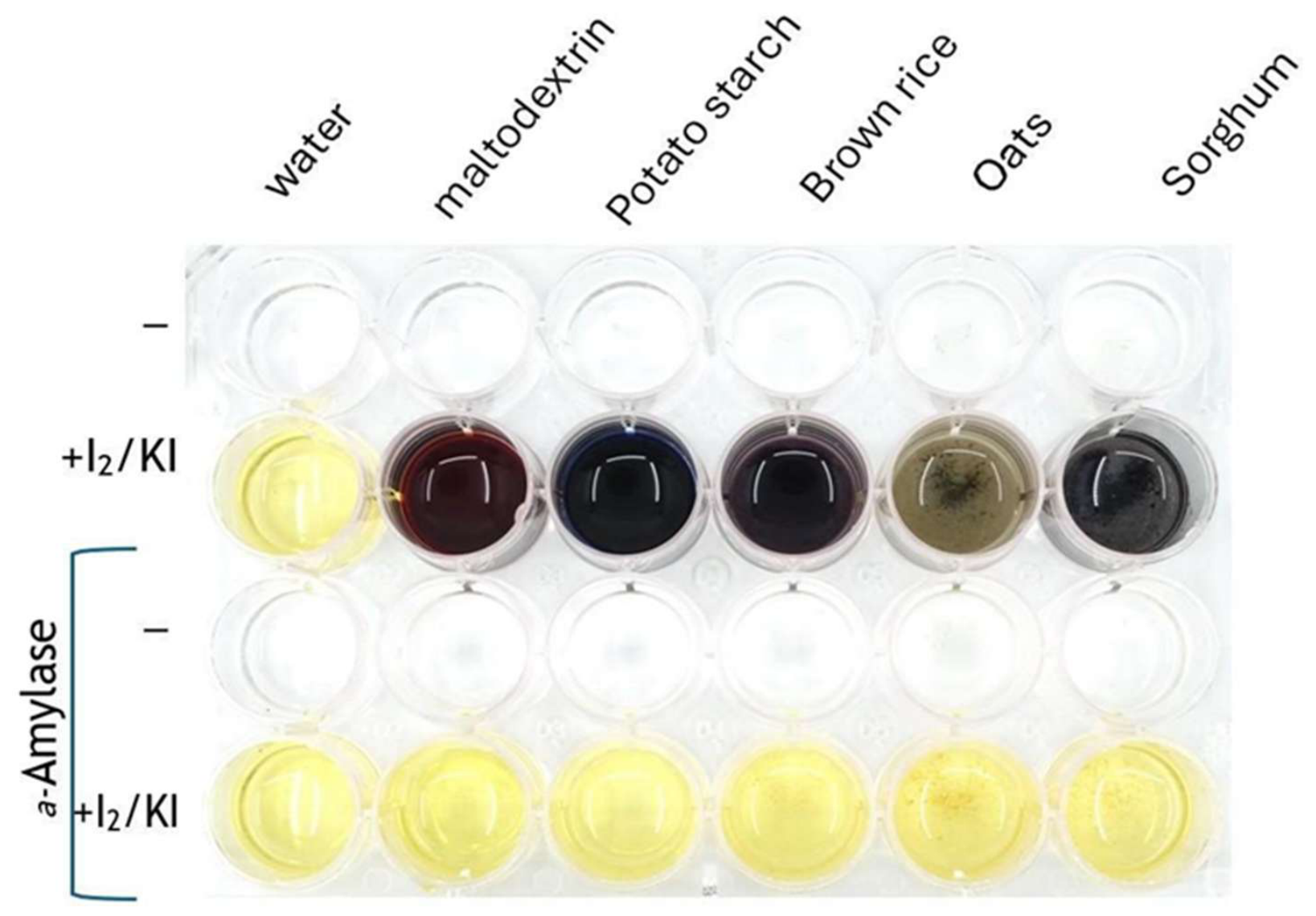
Figure 3.
Lugol’s iodine testing of mushroom control samples. Mushroom-derived controls (chaga, cordyceps, turkey tail, lion’s mane, reishi) showed no color change, indicating the absence of starch. The introduction of the enzyme α-amylase, which digests any starch present (third row), did not cause any color changes due to the absence of starch in these samples.
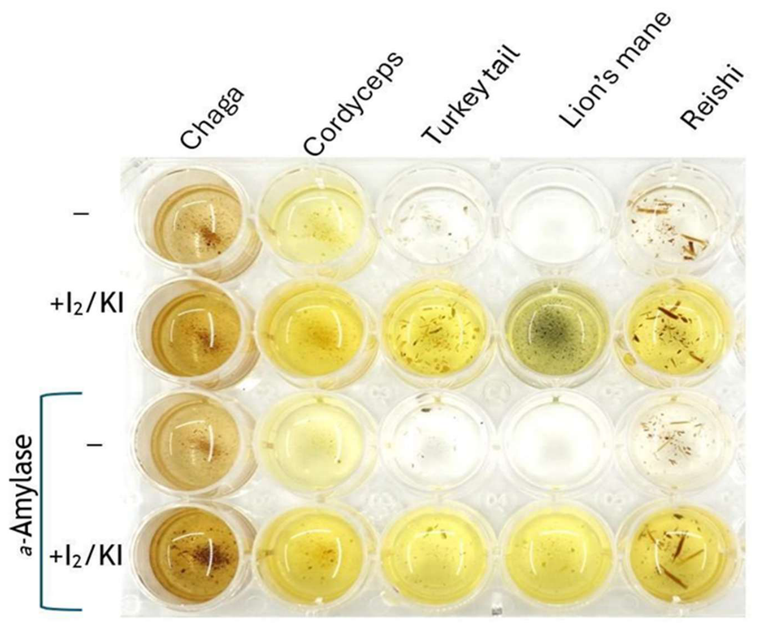
Figure 4.
Lugol’s iodine testing of pet product samples. PET-011: addition of iodine (second row) resulted in the formation of brown color, which is not observed after treatment with α-amylase. This suggests either the presence of glycogen (mushroom α-glucan) or a small amount of maltodextrin (a degradation product of starch). PET-012, PET-014, PET-020, PET-021: no significant color change on addition of iodine, before or after treatment with α-amylase, indicates that there is no starch, maltodextrin, or glycogen present in the sample. PET-013, PET-015, PET-016, PET-017, PET-018, PET-019: addition of iodine (second row) resulted in the formation of blue color, which is not observed after treatment with α-amylase. This indicates the presence of starch in the sample. Samples PET-016 and PET-017 appear significantly darker than other samples on the addition of iodine, these samples likely have the highest levels of starch.
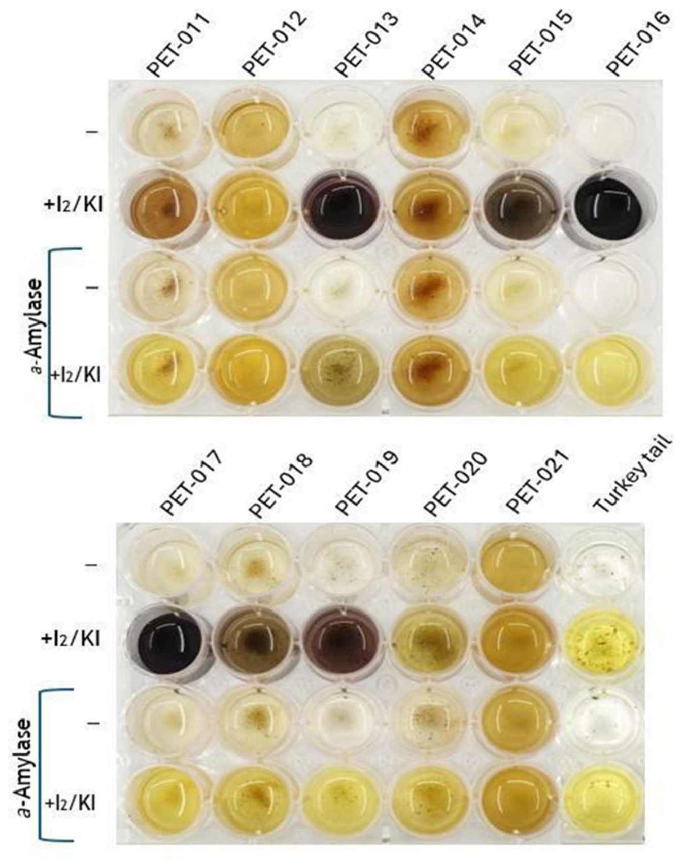
Figure 5.
Scatter quadrant plot of PET-labeled products showing compositional patterns. Numeric thresholds used for classification were defined based on observed compositional ranges in this study and literature reports. The vertical and horizontal lines represent the mean values of α-glucan and β-glucan, respectively. Products located in the upper-left quadrant exhibit relatively higher β-glucan and lower α-glucan values, which may indicate higher fungal polysaccharide content and lower starch contamination. These thresholds are intended solely for visualization and composition-based classification purposes and do not imply species-level authentication or biological activity.
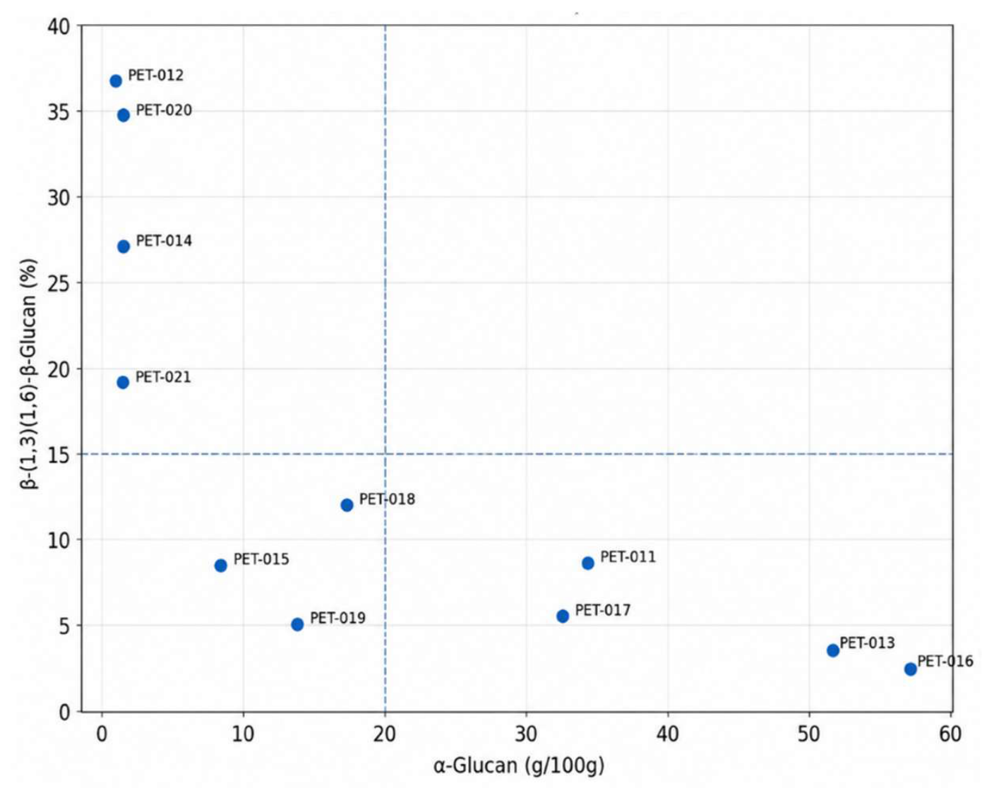
Figure 6.
Three-dimensional scatter plot of α-glucan, β-(1,3)(1,6)-glucan, and digestible starch profiles for PET-labelled products. The 3D view is consistent with the 2D α-/β-glucan separation in Figure 4 and further shows that products with higher α-glucan generally also exhibit higher digestible starch, whereas products with higher β-glucan remain low in digestible starch. In the 3D projection, PET-012, PET-014, PET-020, and PET-021 group on the high-β/low-α/low-starch side, whereas PET-013, PET-016, PET-017, and most of PET-011, PET-015, PET-018, and PET-019 fall toward the higher-α/higher-starch/lower-β side.
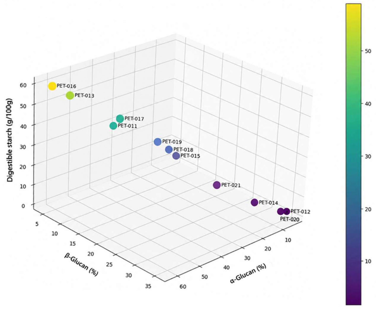
Figure 7.
Comparative starch and glucan composition across commercial PET mushroom supplement product samples. (A) Starch content (g/100 g) measured by the Total Starch assay (K-TSTA-100A; AOAC 996.11); (B) α-glucan percentage (%) measured by the Megazyme Mushroom and Yeast β-Glucan Assay (K-YBGL); and (C) β-(1,3)(1,6)-glucan percentage (%) measured by the Megazyme Mushroom and Yeast β-Glucan Assay (K-YBGL), were measured in eleven PET product samples (PET-011 to PET-021). Data were obtained from multiple laboratories (SGS Guelph, SGS Burnaby, Eurofins, and Nammex), and average values are also indicated. Each symbol represents the result from a specific laboratory as explained in the legend. This analysis reveals substantial variation in starch and glucan profiles across different commercial products and measurement sources.
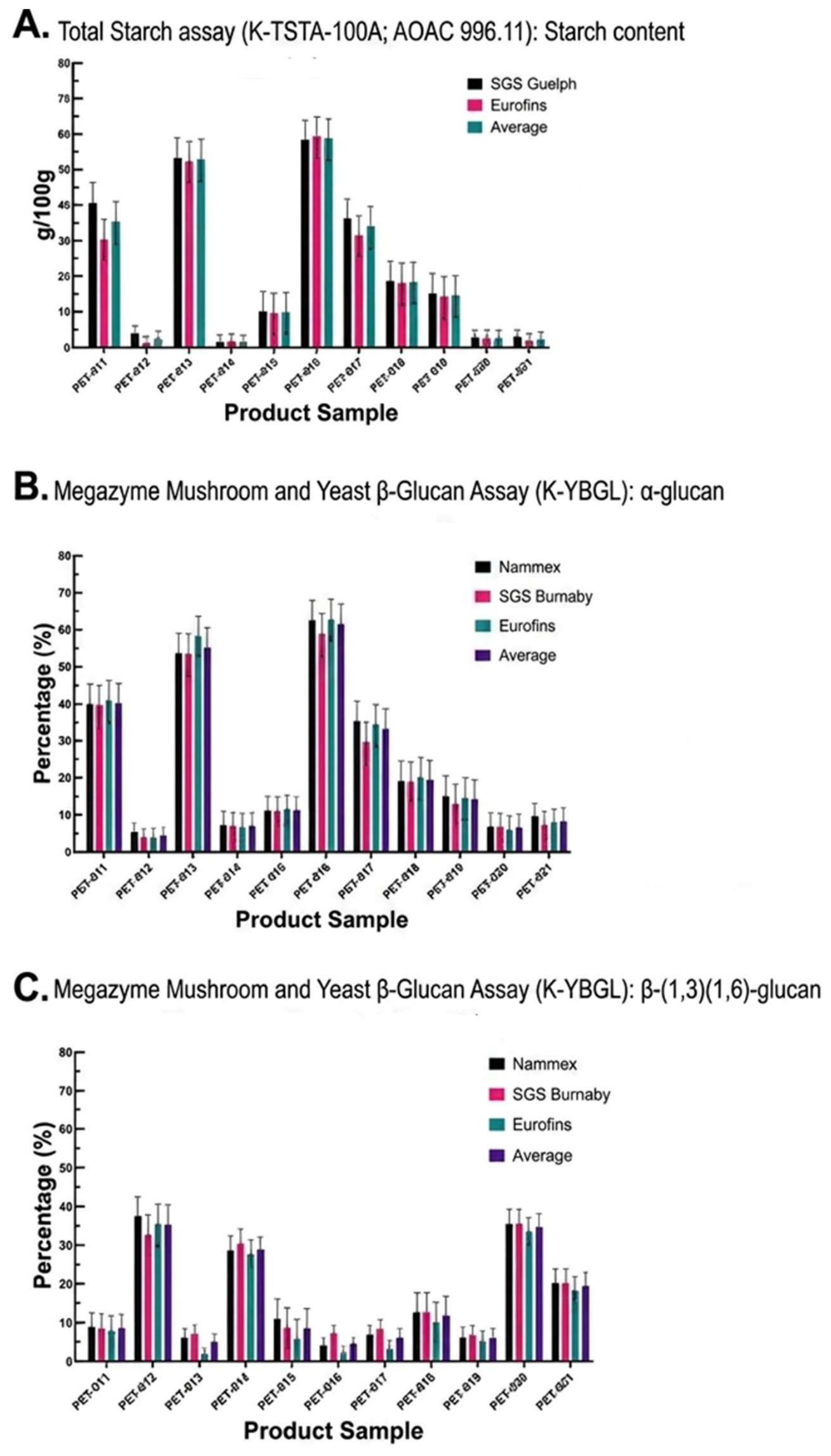

Table 1.
Comparison of product labels with polysaccharide profile classifications.
| Product | Label Description | Comments on Label Description of Ingredients |
Polysaccharide Profile Classification * | Accurate Label Description? |
|---|---|---|---|---|
| PET-011 | “Turkey tail mushroom” | Powder; no mycelium mentioned |
Profile consistent with MFG | NO |
| PET-012 | “Turkey tail pet mushroom” | Capsules; β-glucan statement | Profile consistent with mushroom fruiting bodies | YES |
| PET-013 | “Turkey tail” capsules | Mycelium; biomass statement |
Profile consistent with MFG | YES |
| PET-014 | “Multiple mushrooms | Turkey tail mushroom extract |
Profile consistent with mushroom fruiting bodies | YES |
| PET-015 | “Turkey tail mushroom” | Mycelial biomass and primordia |
Profile consistent with MFG | YES |
| PET-016 | “Mushrooms” | Cereal grain, full-spectrum mushrooms; mycelium, primordia, fruitbodies, extracellular compounds |
Profile consistent with MFG | YES |
| PET-017 | “Mushroom blend” | Mycelial biomass | Profile consistent with MFG | YES |
| PET-018 | “Turkey tail mushrooms” | Fermented grain |
Profile consistent with MFG | YES |
| PET-019 | “Mushroom blend” | Mushroom pictures, no mycelium statement | Profile consistent with MFG | NO |
| PET-020 | “Mushroom blend” | Β-glucan, no mycelium or fillers | Profile consistent with mushroom fruiting bodies | YES |
| PET-021 | “Mushroom pet capsules” | Β-glucan statement | Profile consistent with mushroom fruiting bodies | YES |
* Products classified as “Profile consistent with mushroom fruiting bodies” exhibited high β-glucan and low α-glucan content, while “Profile consistent with MFG” products showed the inverse, as determined by analytical testing.
Disclaimer/Publisher’s Note: The statements, opinions and data contained in all publications are solely those of the individual author(s) and contributor(s) and not of MDPI and/or the editor(s). MDPI and/or the editor(s) disclaim responsibility for any injury to people or property resulting from any ideas, methods, instructions or products referred to in the content. |
© 2026 by the authors. Licensee MDPI, Basel, Switzerland. This article is an open access article distributed under the terms and conditions of the Creative Commons Attribution (CC BY) license (http://creativecommons.org/licenses/by/4.0/).
Copyright: This open access article is published under a Creative Commons CC BY 4.0 license, which permit the free download, distribution, and reuse, provided that the author and preprint are cited in any reuse.