

Submitted:
12 October 2026
Posted:
14 October 2026
You are already at the latest version
Abstract
The advent of the genetic code and translation would have been the most important event in the early evolutionary stage of primordial cells. Although many theories on the origin of the genetic code have been proposed, no definitive answer has yet been reached. In 2001, I have proposed the RNA/protein symmetry theory for the origin of life, in which both RNAs and proteins coevolved forming a stable metabolic complex from the very beginning of the origin of life. In this theory, the genetic information can be generated from existing cellular proteins through reverse translation, which proceeds in the 3′ to 5′ direction using a set of reverse transfer RNAs (rtRNAs). Here, the original model for reverse translation was revised by modifying rtRNAs to naturally include forward translation. Inspired by the structures of the modified rtRNAs, I analyzed the relation between the sizes of the 20 proteinogenic amino acids and the stability of each codon/anticodon pair for each amino acid. I found the strong correlations between the molecular weights of the 18 amino acids excluding tryptophan and arginine and the ΔG values for the formation of each codon/anticodon duplex. This correlation may partly explain the origin of the genetic code.
Keywords:
RNA/protein symmetry theory
; RNA world theory
; reverse translation
; the origin of the genetic code
; reverse transfer RNA
1. Introduction
How life on Earth began would be one of the most curious mysteries for mankind. Solving the mystery would be almost impossible in principle because the history of the Earth cannot be repeated, but, in the future, we may be able to create the same type of life by reproducing the environmental conditions of the ancient Earth and to solve the mystery. Although the path forward may be challenging, scientists from a large range
of fields have been theoretically and experimentally investigating various aspects of the origin of life such as prebiotic synthesis of mono-nucleotides and amino acids and their polymerization, production of chemical energy, integrated metabolic networks, formation of lipid membranes, and replication of genetic materials (1,2). The discovery that RNA can function as a catalyst has led researchers to think that primordial cells might have been made primarily of RNA molecules, which can work both as information carriers and as biocatalysts (3,4). Extensive and intensive investigations have been conducted based on this RNA world theory.
The advent of the genetic code and translation would have been the most important event in the early evolutionary stage of primordial cells. Many theories on the origin of the genetic code, which are not necessarily mutually exclusive, have been proposed: the frozen accident theory, the stereochemical theory, the error minimization theory, and the coevolution theory (5–8). However, no definitive answer has yet been reached.
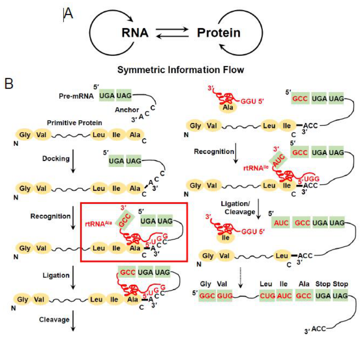
In 2001, I have proposed the RNA/protein symmetry theory for the origin of life, in which both RNAs and proteins coevolved forming a stable metabolic complex from the very beginning of the origin of life and the genetic information flowed symmetrically in primordial cells (Figure 1A) (9). In this theory, the genetic information can be generated from existing cellular proteins through reverse translation. The reverse translation proceeds in the 3′ to 5′ direction using a set of reverse transfer RNAs (rtRNAs) that bind to their specific amino acid residue and carry their corresponding codon (Figure 1B). In addition, I have experimentally demonstrated some basic steps of reverse translation using rtRNAArg. Twenty-five years later, Shi et al. have experimentally advanced the reverse translation process a step further (10).
I recently realized that the existence of reverse translation may be intrinsically linked to the origin of the genetic code. In this paper, I propose modified rtRNAs for reverse translation and show that proto-tRNAs that can be derived from their corresponding modified rtRNAs might have been used for forward translation. Furthermore, inspired by the structures of the modified rtRNAs, I analyzed the relation between the sizes of proteinogenic amino acids and the stability of each codon/anticodon pair for each amino acid and found a curious correlation. Importantly, this correlation may partly explain the origin of the genetic code.
2. Materials and Methods
Correlation Plots
The Gibbs free energy change (ΔG) for the formation of each codon/anticodon duplex was calculated using the Turner 2004 parameters (https://rna.urmc.rochester.edu/NNDB/rna_2004/rna_2004_watson_crick_helices.html and https://rna.urmc.rochester.edu/NNDB/rna_2004/rna_2004_gu_pairs.html). The duplex includes a codon/anticodon pair possessing the G-U or U-G base pair between the third letter of the codon and the first letter of the anticodon. The values of the hydropathy index for the 20 proteinogenic amino acids were obtained from Kyte and Doolittle (11). Mean ΔG values (kcal/mol) for each amino acid were plotted against the molecular weights (Da) of the amino acids and against the hydropathy indexes. All the ΔG values for each codon were also plotted against the molecular weights of the amino acids. In addition, the total numbers of hydrogen bonds to form each codon/anticodon duplex were plotted against the molecular weights of the amino acids. Pearson correlation coefficients and their corresponding P values were calculated using Microsoft Excel.
3. Results and Discussion
3.1. Revised Model
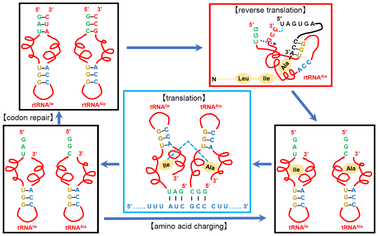
The original model for reverse translation was revised to naturally include forward translation as described below. I came up with the idea that, by modifying the 5′-terminal sequence of the previously proposed rtRNAs, forward translation may be able to be accomplished with proto-tRNAs that are derived from their corresponding modified rtRNAs (Figure 2). Specifically, the 5′-terminal sequence 5′-UGG-3′, which was complementary to the 3′-terminal anchor sequence 5′-CCA-3′ of pre-mRNA, is replaced with the anticodon sequence of each rtRNA containing its codon sequence at the 3′ terminus (Figure 2, the upper-left panel). The 5′-terminal sequence 5′-UGG-3′ is moved to the middle of each rtRNA and placed to form Watson-Crick-Franklin base-pairings with the endogenous sequence 5′-CCA-3′, although the latter is not necessarily needed. rtRNAAla and rtRNAIle are exemplified in the upper-left panel of Figure 2.
In the revised model, reverse translation proceeds as in the original model, but with the modified rtRNAs (Figure 2, the upper-right panel). rtRNAAla binds to the C-terminal alanine side chain and the 3′-terminal anchor sequence 5′-CCA-3′ of the protein/pre-mRNA conjugate through its alanine recognition domain and the 5′-UGG-3′ sequence, respectively. The 3′-terminal C of rtRNAAla and the 5′-terminal U of the pre-mRNA are joined by a phosphodiester bond and, subsequently, the 3′-terminal codon sequence 5′-GCC-3′ is transferred to the 5′-terminus of the pre-mRNA by cleaving the RNA at the 5′ side of the first codon nucleotide G. The rtRNAAla that lost its 3′-terminal codon sequence dissociates together with the C-terminal alanine from the protein/pre-mRNA conjugate and a new conjugate of the protein shortened by one amino acid and the pre-mRNA extended by one codon sequence is formed. These steps are repeated until all the amino acids are transformed into their codon sequences. The reverse translation occurs on a scaffold made of RNAs and/or proteins, which also possesses catalytic activities.
The reverse translation process generates rtRNAs that lack their 3′-terminal codon sequence and carry their corresponding amino acid as byproducts, which can be regarded as prototypes of aminoacyl-tRNAs. That is, rtRNAs that lack their 3′-terminal codon sequence are regarded as proto-tRNAs. proto-tRNAAla carrying alanine and proto-tRNAIle carrying isoleucine are exemplified in the lower-right panel of Figure 2.
Forward translation proceeds by decoding mRNA using the amino-acid-carrying proto-tRNAs possessing their corresponding anticodon at the 5′-terminus (Figure 2, the middle panel). Peptide bond formation occurs on proto-ribosome, which is different from the scaffold for reverse translation.
Proto-tRNAs that supplied their corresponding amino acid for peptide elongation are recharged with the amino acid. Or they undergo codon repair by using the 5′-terminal anticodon sequence to regenerate rtRNAs (Figure 2, the left panels).
The structures of the modified rtRNAs not only naturally incorporate forward translation into the revised model, but also provide clues to understanding the origin of the genetic code as described below.
3.2. A Codon for a Larger Amino Acid Tends to Form a Less Stable Duplex with Its Anticodon
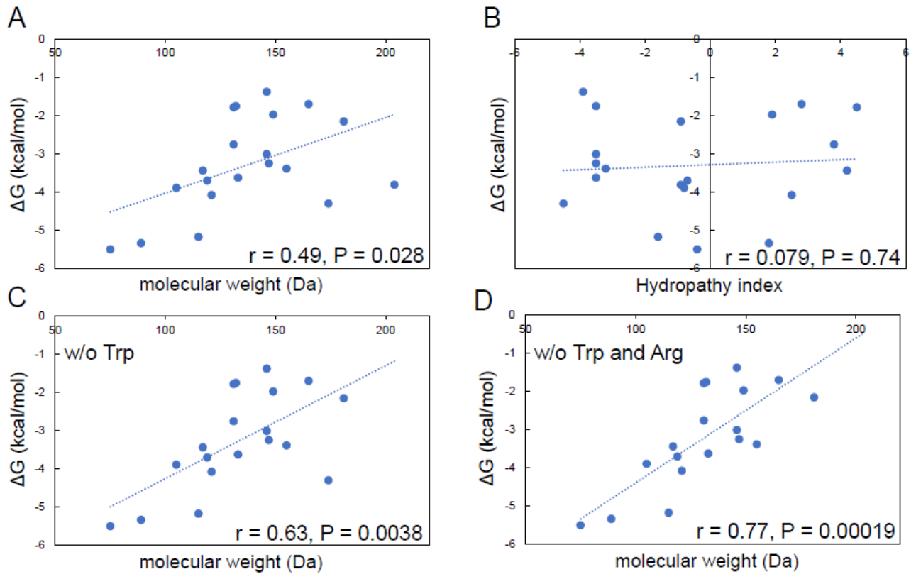
Inspired by the structures of the modified rtRNAs, I analyzed the relation between the sizes of the 20 proteinogenic amino acids and the stability of each codon/anticodon pair for each amino acid. The ΔG value for the formation of each codon/anticodon duplex was calculated using the Turner 2004 parameters. I plotted mean ΔG values for each amino acid against the molecular weights of the amino acids, and found that there is a moderate positive Pearson linear correlation between them (r = 0.49, P = 0.028) (Figure 3A). Mean ΔG values were also plotted against the hydropathy indexes of the amino acids, but there was no significant correlation (r = 0.079, P = 0.74) (Figure 3B).
Since the data point for tryptophan was an outlier, mean ΔG values were re-plotted against the molecular weights of the 19 amino acids without tryptophan, and the plot showed a strong positive linear correlation (r = 0.63, P = 0.0038) (Figure 3C). Furthermore, since the data point for arginine was another outlier, mean ΔG values were re-plotted against the molecular weights of the 18 amino acids without tryptophan and arginine (Figure 3D). Again, a strong positive linear correlation (r = 0.77, P = 0.00019) was observed.
I also plotted all the ΔG values for each codon/anticodon pair against the molecular weights of the amino acids and observed a moderate linear correlation (r = 0.42, P = 3.1 × 10⁻⁵) (Figure S1). A moderate linear correlation (r = 0.46, P = 5.1 × 10⁻⁶) was also observed by excluding the data points for tryptophan, while a strong linear correlation (r = 0.69, P = 1.0 × 10⁻¹²) was observed by excluding the data points for tryptophan and arginine (Figure S1).
The same analyses as described above were carried out with the same data sets of ΔG values but excluding those for the duplexes with the G-U or U-G base pair, and very similar positive linear correlations were observed (Figure S2).
Furthermore, to analyze the relation between the sizes of the 20 amino acids and the total numbers of hydrogen bonds to form each codon/anticodon duplex, the data were plotted in the same fashion as above (Figures S3 and S4). Linear correlations similar to those shown above (Figure 3, Figures S1 and S2) were observed, although, on the whole, the correlations were weaker.
3.3. Correlations for the 10 Prebiotically Synthesized Amino Acids
I performed the same analyses as described above for 10 amino acids, Gly, Ala, Asp, Glu, Val, Ser, Ile, Leu, Pro, and Thr, which can be prebiotically synthesized relatively easily (12). The strengths of the correlations between the ΔG values and the molecular weights of the amino acids under different conditions were almost the same as those for the 18 amino acids other than tryptophan and arginine (Figure 3, Figure S1, Figures S2 and S5). And the strengths of the correlations between the total numbers of hydrogen bonds and the molecular weights of the amino acids were slightly higher than those for the 18 amino acids other than tryptophan and arginine (Figure S3, Figures S4 and S6).
3.4. Insights Into the Origin of the Genetic Code
The strong correlations between the molecular weights of the 18 amino acids excluding tryptophan and arginine and the ΔG values (Figure 3, Figures S1 and S2) or the total numbers of hydrogen bonds (Figures S3 and S4) may provide new insights into the origin of the genetic code. Assuming the existence of the proposed reverse translation, the observed strong correlations could be explained as follows: rtRNAs for large amino acids need to transiently unwind their codon/anticodon duplex to recognize and bind to the C-terminal amino acid of a pre-mRNA/protein conjugate, while rtRNAs for small amino acids do not because the amino-acid recognition pockets of the rtRNAs can recognize and bind to the small amino acids relatively easily without perturbation of their codon/anticodon duplex. In addition, the strong codon/anticodon duplexes of rtRNAs for small amino acids help to form a stable complex of the rtRNA with a pre-mRNA/protein conjugate given that the interaction points between the amino acid and the recognition pocket of the rtRNA are limited, while the strong codon/anticodon duplexes are not needed for rtRNAs for large amino acids because they have a relatively large number of the interaction points between the amino acid and the recognition pocket. The genetic code might have been assigned during the early evolution according to the rule that rtRNAs for larger amino acids need weaker codon/anticodon pairs and that rtRNAs for smaller amino acids need stronger codon/anticodon pairs.
The observation that the strengths of the correlations between the ΔG values and the molecular weights of the 10 prebiotically synthesized amino acids were almost the same as those for the 18 amino acids excluding tryptophan and arginine (Figure 3, Figure S1, Figures S2 and S5) suggests that the rule to assign the genetic code holds all the 20 amino acids other than tryptophan and arginine. The codons for these two amino acids might have been assigned after the other 18 amino acids obtained their codons.
3.5. Creation of mRNAs with a Large Variety of Sequences by Reverse Translation
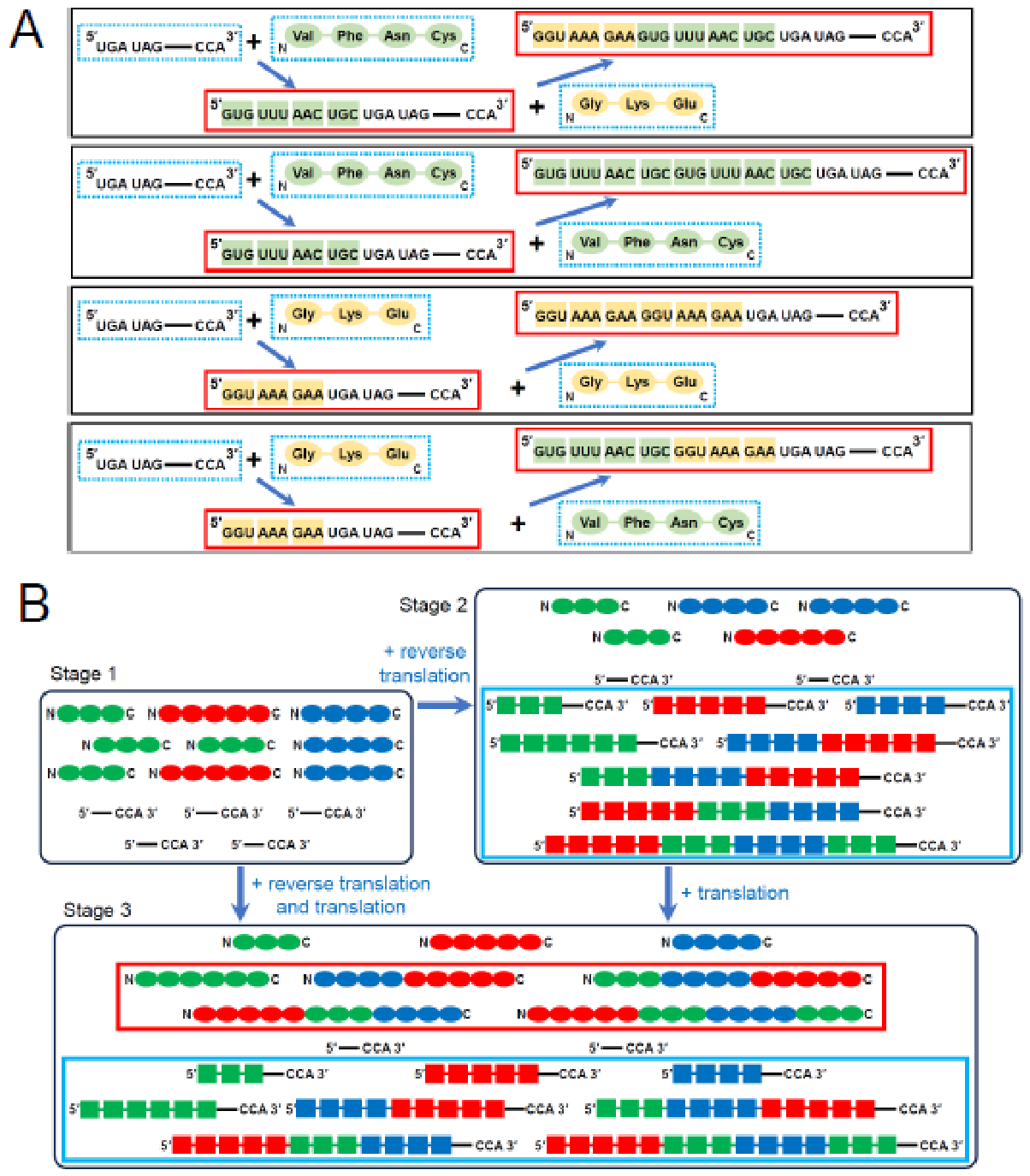
The reverse translation of a primitive protein can create a fused mRNA by using an mRNA generated by the reverse translation of another primitive protein as a pre-mRNA (Figure 4A). These two primitive proteins can be the same or different.
The RNA/protein symmetry theory suggests the following early evolutionary stages (Figure 4B). Primordial cells at the very early stage might have been composed of relatively small RNAs and small proteins (Stage 1). The advent of reverse translation might have created mRNAs with a large variety of sequences (Stage 2). After some period of time, forward translation might have been established and generated a wide variety of newly created fusion proteins (Stage 3). Alternatively, reverse translation and forward translation might have emerged simultaneously without going through Stage 2.
3.6. Superiority of the RNA/Protein Symmetry Theory
There are two important questions about the origin of the genetic code: How advantageous was the emergence of the genetic code and the translation machinery? How did primordial cells generate translatable mRNAs whose translation products were advantageous?
According to the RNA world theory, the answers would be that it is hardly advantageous without the presence of translatable mRNAs and that primordial cells needed to search an enormous RNA sequence space from scratch.
Based on the RNA/protein symmetry theory, the answer to the first question would be that it is highly advantageous because primordial cells can produce many copies of primitive functional proteins and newly created fusion proteins from mRNAs with a large variety of sequences in concert with reverse translation (Figure 4B). The answer to the second one would be that primordial cells generate translatable mRNAs by the reverse translation of primitive functional proteins.
3.7. Perspectives
The revised model for reverse/forward translation with modified rtRNAs based on the RNA/protein symmetry theory appears a bit far-fetched but may provide clues to understanding the origin of translation. And the observation that there is the strong correlation between the stability of each codon/anticodon pair for each amino acid and the molecular weights of the 18 amino acids excluding tryptophan and arginine is very curious and may provide new insights into the origin of the genetic code.
These hypotheses could be tested, albeit not rigorously, as follows: First of all, the reverse translation process could be shown by using rtRNAs, enzyme(s) that can remove the C-terminal amino acid and rejoin the protein to the pre-mRNA, and a scaffold where reverse translation occurs, although artificially synthesizing these molecules would be very difficult. The forward translation process could be shown by using proto-tRNAs that can be derived from the corresponding rtRNAs by removing their 3′-terminal codon and another scaffold as a proto-ribosome.
Finally, if the reverse translation process is established, we could examine if a significant difference in the reverse translation efficiency can be observed between with the normal rtRNAs and with altered rtRNAs, in which more stable codon/anticodon pairs of rtRNAs for smaller amino acids are swapped for less stable codon/anticodon pairs of rtRNAs for larger amino acids. If this is the case, the relationship between the stability of each codon/anticodon pair for each amino acid and the molecular weights of the 18 amino acids excluding tryptophan and arginine may be not merely correlational but causal. And this would shed light on the origin of the genetic code.
Data Availability Statement
All relevant data are within the manuscript and its Supporting Information file.
Conflicts of Interest
The author is an advisor of Veritas In Silico Inc., and owns stock of the company.
References
- Trevors, J.T. Early assembly of cellular life. Prog. Biophys. Mol. Biol. 2003, 81(3), 201–17. [Google Scholar] [CrossRef] [PubMed]
- Xavier, J.C.; Hordijk, W.; Kauffman, S.; Steel, M.; Martin, W.F. Autocatalytic chemical networks at the origin of metabolism. Proc. Biol. Sci. 2020, 287(1922), 20192377. [Google Scholar] [CrossRef] [PubMed] [PubMed Central]
- Robertson, M.P.; Joyce, G.F. The origins of the RNA world. Cold Spring Harb. Perspect. Biol. 2012, 4(5), a003608. [Google Scholar] [CrossRef] [PubMed] [PubMed Central]
- Higgs, P.G.; Lehman, N. The RNA World: molecular cooperation at the origins of life. Nat. Rev. Genet. 2015, 16(1), 7–17. [Google Scholar] [CrossRef] [PubMed]
- Crick, F.H. The origin of the genetic code. J. Mol. Biol. 1968, 38(3), 367–79. [Google Scholar] [CrossRef] [PubMed]
- Yarus, M.; Widmann, J.J.; Knight, R. RNA-amino acid binding: a stereochemical era for the genetic code. J. Mol. Evol. 2009, 69(5), 406–29. [Google Scholar] [CrossRef] [PubMed]
- Koonin, E.V.; Novozhilov, A.S. Origin and Evolution of the Universal Genetic Code. Annu Rev. Genet. 2017, 51, 45–62. [Google Scholar] [CrossRef] [PubMed]
- Di Giulio, M. Theories of the origin of the genetic code: Strong corroboration for the coevolution theory. Biosystems Epub. 2024, 239, 105217. [Google Scholar] [CrossRef] [PubMed]
- Nashimoto, M. The RNA/protein symmetry hypothesis: experimental support for reverse translation of primitive proteins. J. Theor. Biol. 2001, 209(2), 181–7. [Google Scholar] [CrossRef] [PubMed]
- Shi, L.; Senissar, M.; Leistner, K.; Jers, C.; Mijakovic, I. From peptides to DNA: All required steps can be catalyzed. Proc. Natl. Acad. Sci. U S A 2026, 123(9), e2534387123. [Google Scholar] [CrossRef] [PubMed] [PubMed Central]
- Kyte, J.; Doolittle, R.F. A simple method for displaying the hydropathic character of a protein. J. Mol. Biol. 1982, 157(1), 105–32. [Google Scholar] [CrossRef] [PubMed]
- Ring, D.; Wolman, Y.; Friedmann, N.; Miller, S.L. Prebiotic synthesis of hydrophobic and protein amino acids. Proc. Natl. Acad. Sci. U S A 1972, 69(3), 765–8. [Google Scholar] [CrossRef] [PubMed] [PubMed Central]
Disclaimer/Publisher’s Note: The statements, opinions and data contained in all publications are solely those of the individual author(s) and contributor(s) and not of MDPI and/or the editor(s). MDPI and/or the editor(s) disclaim responsibility for any injury to people or property resulting from any ideas, methods, instructions or products referred to in the content. |
© 2026 by the author. Licensee MDPI, Basel, Switzerland. This article is an open access article distributed under the terms and conditions of the Creative Commons Attribution (CC BY) license.
Copyright: This open access article is published under a Creative Commons CC BY 4.0 license, which permit the free download, distribution, and reuse, provided that the author and preprint are cited in any reuse.