

Submitted:
11 October 2026
Posted:
12 October 2026
You are already at the latest version
Abstract
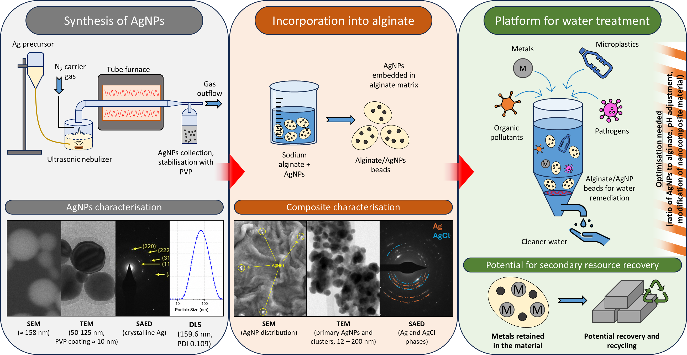
This study presents the development and characterization of a polymer-immobilized silver nanoparticle (AgNP) composite for potential application in water remediation and metal recycling. AgNPs were synthesized by ultrasonic spray pyrolysis (USP) using silver nitrate as a precursor and polyvinylpyrrolidone (PVP) as a stabilizing agent, followed by incorporation into a sodium alginate matrix to form separable alginate/AgNP beads. The nanoparticles were characterized by scanning and transmission electron microscopy (SEM, TEM), energy-dispersive X-ray spectroscopy (EDS), selected-area electron diffraction (SAED), dynamic light scattering (DLS), and zeta-potential measurements. The AgNPs were predominantly spherical and crystalline, with a TEM observed average diameter of approximately 90 nm and most particles between 50 and 125 nm. A PVP surface layer of up to approximately 10 nm was observed. DLS measurements showed a hydrodynamic diameter of 159.6 nm. AgNP incorporation into the alginate matrix was confirmed by microscopy and elemental analysis. AgCl formation inside parts of the composite was observed, indicating chemical transformation of silver and the possibility of affected stability and performance due to interactions with the surrounding environment. The dried composite exhibited a low specific surface area of 0.05 m²/g and a pore size of approximately 2 nm, indicating limited accessible porosity. Results indicate a required further optimization of AgNP loading and alginate structure, with preliminary tests of microplastic (MP) interactions with alginate/AgNP beads showing a limited interaction under the tested conditions. The developed composite provides a basis for multifunctional water treatment materials targeting microbial, metallic, organic, and MP contaminants and subsequent metal recycling.
Keywords:
silver nanoparticles
; ultrasonic spray pyrolysis
; alginate
; microplastics
1. Introduction
Safe and clean drinking water is a basic essential need for humans and other life forms, as well as an essential prerequisite for a healthy environment. The lack of safe drinking water remains a global concern in low-income and developing countries, in regions of Southeast Asia, the Middle East, and North Africa [1,2,3,4]. Increasing industrialization, urbanization, agriculture, and other human activities have further contributed to the global contamination of water resources. Recycling of materials, industrial waste, and improper waste disposal may also contribute to contaminants that are difficult to degrade in the environment. Various techniques are being researched in water treatment to adsorb contaminants and recycling residues with varying efficiency, environmental impact, and cost-effectiveness [5,6,7].
Water remediation and the development of affordable, efficient, and environmentally sustainable water-treatment technologies are thus important for protecting public health and supporting sustainable development. Water contaminants include a broad range of pollutants, including inorganic contaminants such as toxic metals, recycling remains, organic contaminants including pesticides and pharmaceuticals, microbial pathogens such as bacteria, viruses, and fungi, and emerging pollutants such as micro- and nanoplastics [1,8,9]. Numerous technologies have been developed for the removal or degradation of these contaminants, with nanomaterials increasingly attracting attention because of their high surface area, tunable physicochemical properties, and multifunctional activity [8,10,11].
Silver nanoparticles (AgNPs) have emerged as versatile nanomaterials for water remediation owing to their physicochemical, optical, catalytic, and antimicrobial properties. Their high surface-to-volume ratio, chemical stability, and ease of synthesis make them attractive candidates for the detection, removal, and degradation of several types of water contaminants. AgNPs exhibit strong oligodynamic activity against Gram-positive and Gram-negative bacteria, including multidrug-resistant strains, enabling efficient water disinfection at relatively low concentrations [12,13]. AgNPs have also demonstrated remarkable catalytic and photocatalytic performance in the degradation of organic pollutants, including dyes such as methylene blue and rhodamine B, as well as pharmaceutical contaminants [10]. These processes are further enhanced when AgNPs are incorporated into composite materials with graphene, metal oxides, hydrogels, or other functional supports, resulting in improved adsorption capacity, charge transfer, and catalytic efficiency [10]. In addition, AgNPs have been employed as surface modifiers to enhance the adsorption of heavy metals, such as Pb, Cd, Hg or Cu, and other pollutants, while their localized surface plasmon resonance has enabled their application as colorimetric sensors and as signal amplifiers in surface-enhanced spectroscopic techniques for environmental monitoring [12]. Owing to this multifunctionality, AgNP-based materials represent promising platforms for integrated water treatment systems capable of simultaneously disinfecting water, degrading organic contaminants, enhancing pollutant adsorption, and facilitating contaminant detection [8,10,12,13].
Water purification from microplastics (MP) is also a major challenge. MPs are emerging contaminants that pose significant environmental and ecological risks due to their persistence, widespread occurrence, and bioaccumulation in aquatic environments. Most of the research published on MPs focus on polyethylene and polystyrene, while it must be emphasized that one of the major MPs plastic constituents relevant from the environmental aspect is polyamide [14]. Various removal methods, including coagulation, adsorption, membrane filtration, photocatalytic degradation, and biodegradation, have been investigated [15,16]. Although AgNPs are not direct sorbents for MPs, their integration into multifunctional water-treatment systems offers potential for the simultaneous removal of MPs and other contaminants. The performance of AgNPs strongly depends on physicochemical characteristics such as particle size, morphology, surface chemistry, and colloidal stability, dependent on the precise control over nanoparticle synthesis and functionalization, to maximize remediation efficiency. Among the several available methods for their production, ultrasonic spray pyrolysis (USP) has the potential to produce AgNPs suitable for water remediation.
USP is a relatively inexpensive, continuous aerosol-based synthesis technique used for the preparation of thin films, coatings, and functional powders. In USP, high-frequency ultrasonic vibrations generate fine droplets, typically smaller than 20 μm, from low-viscosity precursor solutions [17,18]. The fine droplets are transported by a carrier gas into a heating zone for particle and film formation from the precursor. Compared with other spray pyrolysis methods, USP offers reproducibility, operation under atmospheric conditions, compatibility with a wide range of precursor solutions, and control over material composition, morphology, and stoichiometry. The process parameters, including precursor composition and concentration, solvent type, carrier gas flow, solution feed rate, reaction or deposition temperature, and retention time, can be adjusted to produce dense or porous films, multilayer coatings, and multicomponent particles with tailored properties [19,20]. Due to its versatility, scalability, and ability to synthesize materials with narrow size distributions and controlled morphology, USP is an attractive technique for the preparation of advanced functional materials, such as metal nanoparticles for environmental and biomedical applications [21,22].
The stability of AgNPs in dispersions can be further increased with various stabilizers (e.g., citrates and different polymers (polyvinylpyrrolidone (PVP), or polyethylene glycol (PEG)), which adsorb onto the surface of the nanoparticles, preventing or reducing their tendency to assemble [23]. The stabilized AgNPs may then act as a platform for additional surface modification. Attaching Na-alginate to the stabilized AgNPs can provide functionality useful for water remediation. Na-alginate is a biopolymer usually obtained from seaweed (a polysaccharide of brown algal extract [24]), which can act as a toxic metal’s sorbent as well as a flocculant/sorbent of MP particles. Although AgNPs have been individually explored for antimicrobial applications, their incorporation into alginate–matrices using green synthesis processes remains rarely investigated [25].
Adsorption of metals on the Na-alginate with incorporated AgNPs provides an additional possibility for secondary material sourcing and recovery of valuable metals from water remediation. The adsorption of metal ions, such as iron, cobalt, zinc, copper, nickel, arsenic, mercury, lead, chromium, and cadmium, occurs via ion exchange, as the sodium ions (Na+) within the hydrogel matrix are replaced by heavy metal ions [5,6,26]. Additionally, the adsorption is aided by the porous structure of the alginate, providing a large surface area for electrostatic interactions and physical adsorption [6]. Following adsorption, the retained metals can be recovered from the alginate-based material through appropriate further treatment, allowing their subsequent reuse or processing. Such a use of the proposed alginate composite can supplement and contribute to metal recycling efforts, where water is used as a treatment medium.
The aim of this study is the development of environmentally friendly, polymer functionalized AgNPs, combining antimicrobial properties, adsorption of heavy metals and MPs for the integrated removal of up to four pollutant categories (microbial pathogens, organic pollutants, toxic metals, and MPs) in wastewater treatment. The AgNPs were synthesized using USP as a method employable for flexible, scalable, reproducible, and controllable synthesis of nanoparticles with a narrow size distribution and controllable shape. AgNPs were then incorporated into alginate beads due to easier separation of beads from the mentioned pollutant categories and their reuse.
2. Materials and Methods
2.1. AgNP Synthesis
AgNPs were synthesized using the USP method. The precursor solution was converted into an aerosol using an ultrasonic generator, which was then transported through the reaction zones of the USP system (Figure 1 a). The resulting AgNPs were collected in collection bottles with a PVP solution (Figure 1 b). The synthesis parameters are shown in Table 1.
The precursor was prepared from silver nitrate (AgNO₃) and deionized water at a concentration of 1 g/L. A total of 1 L of precursor solution was prepared, with approximately 600 mL being used during the synthesis.
A solution of deionized water and polyvinylpyrrolidone (PVP) at a concentration of 2.5 g/L was used in the collection bottles. Two collection bottles were prepared with PVP solution to capture and stabilize the synthesized AgNPs. The third bottle contained 0.05 M sodium hydroxide (NaOH) solution, which served to neutralize acid gases and reaction products generated during the USP process.
The silver concentration in the AgNPs dispersion after USP synthesis and rotary evaporation was determined using an Agilent 5800 VDV ICP-OES spectrometer. The measured silver concentration in the synthesized dispersion was 1.4 µg/mL. The obtained concentration was relatively low for further experimental work and preparation of composite materials for water purification, so it was concentrated using a rotary evaporator. The purpose of the procedure was to remove part of the solvent and increase the concentration of nanoparticles without changing their chemical composition. The parameters used for the rotary evaporation were pressure – 25 mPa, evaporation flask rotation speed – 200 rpm, heating bath temperature – 40 °C.
ICP–OES analysis showed that the silver concentration after concentration increased to 169.5 µg/mL. Compared to the initial concentration (1.4 µg/mL), this represents an approximately 121-fold increase in concentration. The concentration using a rotary evaporator was very effective and a dispersion with a significantly higher AgNPs content was prepared, suitable for further research and the preparation of polymer-covered AgNP for water purification.
2.2. Scanning Electron Microscopy
The samples were examined using a scanning electron microscope JSM-IT800SHL (JEOL, Tokyo, Japan), equipped with an energy-dispersive X-ray spectrometer (EDS) Ultim Max 100 (Oxford Instruments, UK). The AgNP dispersion and alginate/AgNP beads were prepared separately, each being mounted on an individual SEM stub covered with carbon tape and dried in a desiccator at room temperature. The SEM-EDS analysis was performed at an accelerating voltage of 10 kV and a working distance of 10 mm.
2.3. Transmission Electron Microscopy
Both AgNP/PVP and alginate/AgNP dispersions were homogenized using an ultrasonic bath for 10 min before sample preparation. The homogeneous dispersion samples were then drop-cast onto copper TEM grids coated with a thin amorphous carbon film and dried at room temperature.
The characterization of the samples was performed using a Thermo Fisher Talos F200X (Thermo Fisher Scientific Inc., USA) transmission electron microscope at an accelerating voltage of 200 kV. Images were acquired in transmission electron microscopy (TEM) mode and scanning transmission electron microscopy (STEM) mode using a bright field (BF) and a high-angle annular dark field (HAADF) detector. Selected-area electron diffraction (SAED) was performed in TEM mode using apertures with diameters of 10, 40, and 200 µm. Elemental mapping was carried out using a segmented Super-X EDS, with an acquisition time of 300 s at currents between 30 and 70 pA. The TEM analysis and data processing were done in Velox Software ver. 3.22 (Thermo Fisher Scientific Inc., USA). Additional data analysis of SAED diffraction pattern was done in CrysTBox Software [27].
Based on the acquired images, the morphological and structural properties of the nanoparticles were determined, including their shape, size and crystal structure. The size distribution was obtained by measuring the particle size in TEM images. A total of 300 particles were measured across multiple areas at various magnifications on individual samples.
2.4. DLS and Zeta Potential Measurements
DLS and zeta potential measurements were performed using a Malvern Zetasizer Nano ZS instrument (Malvern Panalytical, Worcestershire, UK) equipped with Zetasizer Software version 7.13. An Omega cuvette (Anton Paar GmbH, Graz, Austria; Material No. 225288) was used for the measurements. The measurements were performed at a temperature of 25 °C.
2.5. Preparation of Alginate/AgNP Beads and Specific Surface Analysis
Alginate/AgNP beads were prepared with synthesized AgNPs (Figure 2). Firstly, 2% solution of sodium alginate (100 mL) in distilled water using a magnetic stirrer was prepared. Then 2 g of the concentrated AgNP dispersion was added to the solution used for the synthesis of alginate beads. The sodium alginate solution was then added dropwise using a plastic dropper into a 2 % CaCl2 solution, while stirring gently with a magnetic stirrer. Alginate beads formed in the beaker. They were stored in CaCl2 for characterization. Specific surface area was determined by N₂ adsorption–desorption at 77 K using a Micromeritics TriStar II 3020 porosimeter. Samples (≈0.5 g) were oven-dried and degassed on a VacPrep unit at 40 °C under vacuum (<10 Pa) for 24 h to remove physisorbed species. Specific surface area was calculated by selecting the linear region according to Rouquerol’s consistency criteria. Reported values of surface area are averages of duplicate runs with instrument repeatability checks.
2.6. Preliminary Tests of MP Interactions with Alginate/AgNP Beads
MP in form of polyamide (PA) Microfibers, with standard length of 50 µm, and average diameter of 10 µm from Microplastic Solution (Toulouse, France) was used. First, 30 mg of PA MPs were weighed, followed by addition of 100 mL of tap water to a conical flask. After 30 min of stirring 10.5 mg of alginate/AgNPs beads were added into dispersion. Three replicates were made. The dispersions were mixed using a magnetic stirrer for 2 h, 4 h, and 24 h. Immediately after the stirring was completed, each dispersion was drained through a 2 mm sieve to separate the beads from PA. The remaining dispersion was filtered using a pre-dried (105 °C) and pre-weighed polyester sulfonyl filter. After filtration, the beads were stored in a refrigerator and overlaid with stock solution for storage (for possible reuse). The filters were dried again with a microfiber filter cake at 105 °C for 4 h, cooled in a desiccator, and re-weighed. The mass difference would show if there were any interactions between fibres and alginate/AgNPs under natural conditions in tap water.
3. Results
3.1. AgNPs Electron Microscopy Results
SEM analysis of the dried AgNPs showed a predominantly spherical shape of the particles with sizes between 16 and 265 nm (Figure 3). The average size of the analyzed particles was approximately 158 nm. A more detailed size measurement was performed in the TEM analysis. SEM-EDS analysis confirmed the presence of silver as the dominant element in the analyzed particles, confirming the successful synthesis of AgNPs. The presence of carbon (C) mainly results from the conductive carbon adhesive tape used to prepare the sample on the SEM holder. The PVP medium used for stabilization of the nanoparticles also contributed to the carbon and oxygen content in the EDS analysis (Figure 3).
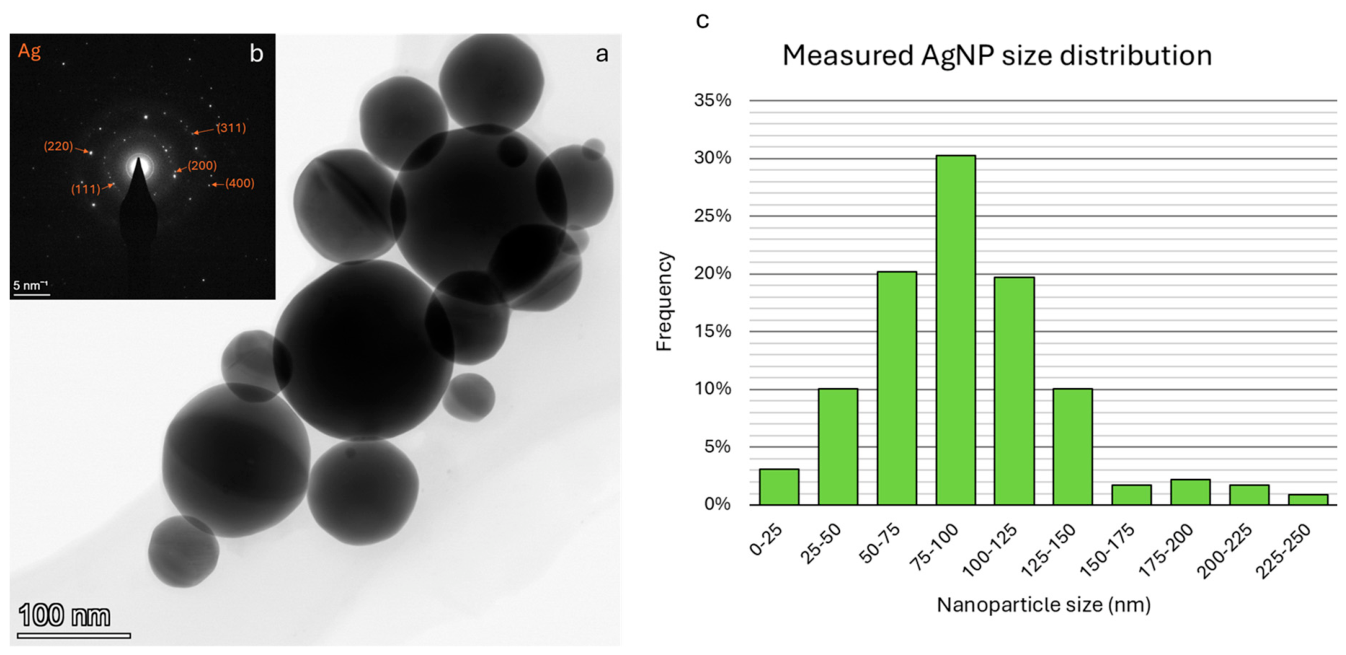
TEM analysis revealed that the synthesized AgNPs were predominantly spherical in shape. The histogram of the size distribution of individual AgNPs (Figure 5) shows a distribution with the highest frequency in the size class of 75–100 nm. Most particles have a diameter between 50 and 125 nm, while particles with a diameter above 150 nm are represented only in a small number. In TEM images, a surface layer with a thickness of up to approximately 10 nm is visible around the AgNPs (Figure 4), attributed to the PVP coating used to stabilize the nanoparticles. The particles are well dispersed in the solution, as reflected in the arrangement of individual particles on the TEM grid. However, aggregates and agglomerates of nanoparticles also occur, as can be seen from Figure 4a-b. AgNPs are connected via an amorphous surface layer, while aggregates show connections via “necks” due to coalescence (Figure 4d).
SAED analysis confirmed the crystallinity of the synthesized AgNPs (Figure 5b), with the SAED pattern exhibiting distinct diffraction spots and rings corresponding to the crystallographic planes for face-centered cubic (FCC) silver (111), (200), (220), (311), and (400).
Figure 4.
TEM images of the, as-synthesized AgNPs stored in PVP solution. Images (a) and (b) show agglomerates of particles of various sizes; (c) a detail of particles coated with a thin coating of PVP; and (d) a detail of particles showing neck formation due to coalescence.
Figure 4.
TEM images of the, as-synthesized AgNPs stored in PVP solution. Images (a) and (b) show agglomerates of particles of various sizes; (c) a detail of particles coated with a thin coating of PVP; and (d) a detail of particles showing neck formation due to coalescence.
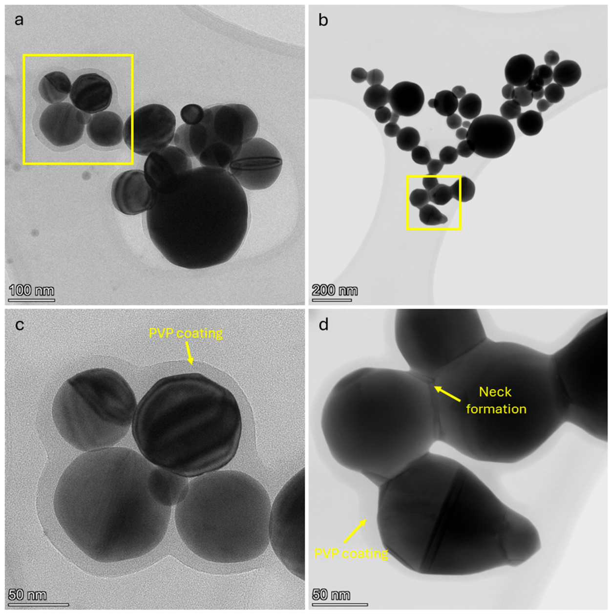
Figure 5.
TEM image of AgNPs (a) with electron diffraction pattern (b) indicating good crystallinity of synthesized particles. The histogram (c) shows the particle size distribution from the performed size measurements.
Figure 5.
TEM image of AgNPs (a) with electron diffraction pattern (b) indicating good crystallinity of synthesized particles. The histogram (c) shows the particle size distribution from the performed size measurements.
3.2. Alginate/AgNP Beads Electron Microscopy Results
SEM analysis of the dried alginate/AgNP beads showed visible embedded AgNPs on the surface of the sodium alginate matrix (Figure 6). The AgNP single particles and clusters were confirmed by EDS analysis, with a high Ag content in the point analysis at the visible AgNP sites. The shapes and sizes of the AgNPs were highly obscured during imaging by the organic alginate matrix. TEM analysis shows a more suitable determination of the AgNP features.
The distribution of AgNPs across the alginate matrix surface was random, with areas of higher or lower concentration of particles. A similar distribution may be estimated across the sodium alginate bead volume.
The analyzed sample contained alginate microspheres (or microstructures) up to a few micrometers in size (Figure 7). EDS analysis of these structures confirms a high mass fraction of carbon and oxygen, reflecting the organic nature of the matrix. Additionally, the elemental composition includes N, Na, P, S, Cl, and Ca. Nanoscale regions within the microspheres have a distinct co-localization of Ca and P.
The size of the AgNPs suspended in alginate solution ranges from 12 to 200 nm, with larger structures often composed of coalesced primary nanoparticles (Figure 8). Particle shape varies from spherical to irregular, a characteristic strongly dependent on size; as diameter increases, particle morphology becomes more complex, and crystal twins become more pronounced. A substantial proportion of the AgNPs examined in the sample showed secondary alteration, consistent with the high affinity of Ag for reaction with Cl. SAED analysis of AgNPs from alginate dispersion confirms a polycrystalline structure with clearly defined diffraction rings characteristic of FCC silver (111), (200), (220), and (311) planes (Figure 8c). In specific regions of particle agglomerates (Figure 8d), the diffraction patterns show reflections characteristic of AgCl (111), (200), and (311) planes (Figure 8f).
Due to the presence of silver chloride phases, the characterization of these agglomerates was challenging. The silver chloride phase was highly sensitive to electron-beam irradiation and degraded rapidly despite the use of a reduced beam current during analysis. In some cases, decomposition of the secondary phase preserved only individual AgNPs, whereas in others, beam-induced secondary Ag crystallites formed during analysis. An example of the beam-induced degradation of the silver chloride phase during EDS analysis is presented in Figure 9.
3.3. DLS and Zeta Potential Measurement Results
The DLS and zeta potential results are presented in Figure 10, where Figure 10a shows the particle size distribution determined by DLS and Figure 10b the corresponding zeta potential distribution.
DLS measurements revealed a mean hydrodynamic diameter of 159.6 nm and a polydispersity index (PDI) of 0.109 (Figure 10a). The particle size distribution exhibited a single dominant peak, indicating a relatively homogeneous AgNP population with no evidence of significant aggregation. The low PDI value further confirmed the narrow particle size distribution and good colloidal homogeneity of the dispersion.
The zeta potential measurements demonstrated good colloidal stability of the synthesized AgNP dispersion. The AgNPs exhibited a zeta potential of −16.1 mV, with a single dominant peak centered at approximately −16 mV (Figure 10b). Although this value corresponds to only moderate electrostatic stabilization, PVP is known to stabilize nanoparticles predominantly through steric rather than electrostatic repulsion. Consequently, the relatively low absolute zeta potential does not necessarily imply poor colloidal stability. This interpretation is supported by the DLS results, which showed a narrow particle size distribution, a low PDI, and no evidence of significant aggregation.
3.4. Specific Surface Analysis
Specific surface analysis revealed that the alginate/AgNPs beads possess a specific surface area of only 0.05 m²/g and a pore size of 2 nm, which is significantly lower than anticipated. Consequently, the adsorption and intraparticle diffusion of contaminants would be highly restricted [28]. Drying-induced shrinkage and pore collapse likely reduced the number of gas-accessible pores. Strong crosslinking of the alginate network may have additionally limited diffusion of nitrogen molecules into the bead interior. Similar behavior has been reported for hydrogel-based beads, where specific surface area reflects only the externally accessible surface rather than the total internal reactive surface available under aqueous conditions [29]. SEM analysis of the dried alginate/AgNPs beads (Figure 7) did not show a spherical shape as in case of AgNPs (Figure 3 and Figure 4). The average size of the individual alginate/AgNPs microbeads was approximately 2 µm. As shown in Figure 7, microbead aggregates were formed. Adding a relatively high concentration of nanoparticles increases the viscosity of the alginate solution before it gels. This can alter the gelation mechanism, often leading to a much denser, more compact structure with minimal porosity in accordance with the literature [30]. It could be suggested that the binding interactions between AgNPs and sodium alginate were stronger, increasing the structural stability of beads. This also affected the sphericity of the beads, while the share of solid matter is higher compared to bound water share [31].
Due to low specific surface area values future development on alginate/AgNPs beads should pivot toward improvement of material properties in a controlled atmosphere, by reducing the ratio of added AgNPs particles to alginate, investigating the effect of pH and modification of nanocomposite material.
3.5. Preliminary Tests of MP Interactions with Alginate/AgNP Beads
Figure 11 shows the filtrate of MP after different contact times of MP with alginate/AgNPs. It was hard to distinguish some differences in residual MP forms. Also, the measurement results were similar. The mass of filter residue after 2 h and 4 h was 6.6% lower compared to the initial MP mass in both cases. After 24h, the percentage did not increase. The interactions of MP and alginate/AgNPs was lower than expected.
4. Discussion
The present study demonstrates that ultrasonic spray pyrolysis is a suitable method for synthesizing spherical silver nanoparticles with a relatively narrow particle size distribution and good colloidal stability. The combination of ICP–OES, TEM, SEM, DLS, and zeta potential measurements provided complementary information regarding the morphology, composition, crystallinity, and dispersion stability of the synthesized AgNPs.
TEM analysis revealed that the average particle diameter of the as-synthesized AgNPs was approximately 90 nm (92±40 nm), whereas SEM and DLS measurements yielded larger average particle sizes of 157.6 nm and 159.6 nm, respectively. These differences are expected because each characterization technique measures different particle characteristics. TEM provides direct visualization of the metallic nanoparticle core and therefore reflects the actual physical particle diameter. In contrast, DLS measures the hydrodynamic diameter of particles dispersed in a liquid medium, including the PVP stabilizing layer together with the surrounding solvation shell. Consequently, DLS generally reports larger particle sizes than TEM. Similarly, SEM measurements were performed on dried nanoparticles, where partial aggregation, particle overlap, and neck formation during solvent evaporation may contribute to an apparent increase in particle size.
TEM observations also showed an amorphous layer of up to 10 nm thickness around AgNPs, which is attributed to adsorbed PVP molecules acting as a steric stabilizer during synthesis. DLS measurements support the effectiveness of PVP stabilization., showing a single dominant particle population without pronounced secondary peaks, indicating that significant aggregation was not present. Although some AgNPs aggregates and agglomerates were observed by electron microscopy, these structures were relatively limited and may have formed during sample preparation (e.g., dispersion drying on TEM grids) for microscopic analysis.
SAED analysis confirmed that the synthesized nanoparticles possess the face-centered cubic (fcc) crystal structure characteristic of metallic silver. The presence of well-defined diffraction spots and rings corresponding to the (111), (200), (220), (311), and (400) crystallographic planes demonstrates that the USP process produced highly crystalline AgNPs without detectable crystalline impurity phases. High crystallinity is particularly desirable for applications involving antimicrobial activity and catalytic processes, where the surface structure of silver nanoparticles significantly influences their performance.
A part of the AgNPs underwent secondary chemical alteration in alginate/CaCl2 solution, due to the high affinity of dissolved silver for chlorine. Silver readily formed insoluble silver chloride, which was confirmed by EDS and SAED analysis. This observation should be considered when evaluating the composite’s properties, as the presence of AgCl may influence surface chemistry, silver availability, antimicrobial activity, and long-term stability of the material.
The measured zeta potential of −16.1 mV indicates moderate electrostatic stabilization of the AgNPs dispersion. However, this value should be interpreted together with the presence of PVP, which stabilizes nanoparticles predominantly through steric rather than electrostatic repulsion. Consequently, the relatively low absolute zeta potential does not necessarily imply poor colloidal stability. This interpretation is supported by the DLS results, which revealed a narrow particle size distribution, a low polydispersity index (PDI = 0.109), and no evidence of significant aggregation. These findings indicate that steric stabilization provided by PVP effectively maintained a stable nanoparticle dispersion despite the moderate zeta potential.
ICP–OES analysis showed that the as-synthesized nanoparticle dispersion contained only a small amount of silver (1.4 µg mL⁻¹), which is typical for laboratory-scale USP synthesis performed under dilute precursor conditions. However, rotary evaporation proved to be a highly efficient concentration method, increasing the silver concentration by approximately 121-fold without altering the chemical composition of the nanoparticles. This concentration step is particularly important for the subsequent preparation of AgNP-based composite materials, where higher nanoparticle loadings are required to achieve sufficient antimicrobial activity and functional performance.
Overall, the obtained results confirm that the synthesized AgNPs possess the morphological, structural, and colloidal characteristics required for incorporation into polymer-based composite materials intended for water treatment applications. The combination of relatively uniform particle morphology, high crystallinity, effective steric stabilization, and successful post-synthesis concentration provides a solid foundation for the preparation of sodium alginate-based AgNP composites, which will be investigated in future studies.
The main reason for low interactions between MP and alginate/AgNPs could be attributed to electrostatic repulsion [28]. The zeta potential of the particles was measured at -15.1 mV as shown in Figure 6, and for alginate/AgNPs at -22 mV, which is in accordance with the literature [32]. MP in the form of PA also develops a negative zeta potential in water, depending on pH [14]. In our experiment, tap water with pH 7.4 was used, which is above the point of zero charge for PA, determined at pH 6.5; thus, it is negatively charged. Consequently, electrostatic repulsion reduces the frequent contact between MP and AgNPs, strongly influencing adsorption compared to the surface area of nanoparticles alone. Another reason for insufficient interactions could be pore size of alginate/AgNPs, which plays an important role in gaining strong interactions [33]. Alginate/AgNP pore size was determined at 2 nm, while the MP size was measured at 50 µm in the present study. Insufficient contact area influences the attachment between the particles, especially if the MP surface is smooth. Some studies emphasized that contact area controls interaction efficiency [28]. In the process of MP adhesion to NPs, pH was the most significant influencing factor and had the most crucial effect on MP and NPs interactions [34]. Recent developments favor hybrid approaches that synergize the strengths of multiple techniques [35]. A novel pre-treatment step should be carefully considered to optimize process efficiency of alginate/AgNPs material.
5. Conclusions
In this study, PVP-stabilized AgNPs were successfully synthesized by USP and incorporated into a sodium alginate matrix to develop a multifunctional material with potential application in water remediation and metal recycling.
- The synthesized AgNPs were predominantly spherical and crystalline, with a TEM observed average diameter of approximately 90 nm and most particles between 50 and 125 nm. USP is demonstrated as suitable for the preparation of AgNPs for incorporation into an alginate-based composite as a promising basis for water remediation, combining the functional properties of AgNPs with the practical advantage of incorporating them into a larger, more easily handled and potentially separable material.
- The formation of AgCl in parts of the composite was observed, indicating that the chemical state of silver can be affected by interactions with the surrounding environment. Changes of the chemical state of silver within the alginate material should be considered when evaluating its stability and performance in aqueous environments.
- The dried alginate/AgNP beads exhibited a very low specific surface area (0.05 m²/g) and a pore size of approximately 2 nm, indicating restricted accessible porosity, with a need for further optimization of the composite structure by changing of the AgNP-to-alginate ratio, investigating the effect of pH and modification of nanocomposite material.
- The developed alginate/AgNP material represents a promising platform for multifunctional water-treatment systems of microbial pathogens, organic pollutants, metals, and MPs, with a potential for secondary resource recovery, as metals retained during water treatment could potentially be concentrated in the material and subsequently recovered. Metal recovery and recycling were not demonstrated in the present study and require dedicated investigation.
- Based on the preliminary results, future studies will be focused on pH value adjustment during experiments as well as the material and size of MP would be carefully chosen.
Author Contributions
Conceptualization, R.R. and P.M.; methodology, R.R.; software, P.M.; validation, P.M, R.R. and M.S.; formal analysis, P.M, L.K., S.K and M.S.; investigation, P.M, L.K., S.K and M.S.; resources, R.R. and Š.I.; data curation, P.M, L.K., S.K and M.S.; writing—original draft preparation, P.M. and L.K; writing—review and editing, R.R.; visualization, P.M, L.K., S.K and M.S; supervision, R.R.; project administration, R.R. and Š.I.; funding acquisition, R.R. and Š.I. All authors have read and agreed to the published version of the manuscript.
Funding
This research was funded by the Slovenian Research Agency ARIS: P2-0120 and P4-0446 Research Programs and Infrastructure program I0-0005. This research was also funded by the EUREKA: REC-MET project 2024-20740/NP/Western Balkan Call1, funded by Ministry of Science of the Republic of Montenegro and the Bilateral cooperation project “Maritime Industry in Circular Economy” BI-ME/25-27-014 within the Bilateral Scientific and Technological Cooperation Programme with the Republic of Slovenia and Montenegro.
Institutional Review Board Statement
Not applicable.
Informed Consent Statement
Not applicable.
Data Availability Statement
The original contributions presented in this study are included in the article. Further inquiries can be directed to the corresponding author.
Conflicts of Interest
The authors, Peter Majerič and Rebeka Rudolf, were employed by the company Zlatarna Celje d.o.o. The remaining authors declare that this research was conducted in the absence of any commercial or financial relationships that could be construed as a potential conflict of interest.
Abbreviations
The following abbreviations are used in this manuscript:
| AgNP | Silver nanoparticle |
| USP | Ultrasonic spray pyrolysis |
| PVP | Polyvinylpyrrolidone |
| MP | Microplastic |
| PA | Polyamide |
| SEM | Scanning electron microscopy |
| TEM | Transmission electron microscopy |
| STEM | Scanning transmission electron microscopy |
| EDS | Energy-dispersive X-ray spectroscopy |
| SAED | Selected-area electron diffraction |
| BF | Bright field |
| HAADF | High-angle annular dark field |
| DLS | Dynamic light scattering |
References
- Shayo, G.M.; Elimbinzi, E.; Fabian, C.; Kibona, T.E.; Shao, G.N. Enhancing Water Quality at the Point of Use through Fabrication of Silver Nanoparticles Impregnated Ceramic Water Filters. J. Indian Chem. Soc. 2025, 102, 101882. [Google Scholar] [CrossRef]
- Margareth, M.; Fortunatus, J.; Ally, M. Development of a Filter System Using Silver Nanoparticles Modified Silica Sand for Drinking Water Disinfection. Afr. J. Environ. Sci. Tech. 2023, 17, 146–156. [Google Scholar] [CrossRef]
- Wafy, K.R.; El-Aswar, E.I.; Mohamed, W.S.E.; El-Sabbagh, S.M. Water Disinfection Using Durable Ceramic Filter Coated with Silver Nanoparticles Synthesized Using Actinomycetes. Appl. Water Sci. 2023, 13, 140. [Google Scholar] [CrossRef]
- Mecha, A.C.; Chollom, M.N.; Babatunde, B.F.; Tetteh, E.K.; Rathilal, S. Versatile Silver-Nanoparticle-Impregnated Membranes for Water Treatment: A Review. Membranes 2023, 13, 432. [Google Scholar] [CrossRef] [PubMed]
- Wang, B.; Wan, Y.; Zheng, Y.; Lee, X.; Liu, T.; Yu, Z.; Huang, J.; Ok, Y.S.; Chen, J.; Gao, B. Alginate-Based Composites for Environmental Applications: A Critical Review. Crit. Rev. Environ. Sci. Technol. 2019, 49, 318–356. [Google Scholar] [CrossRef] [PubMed]
- Ling Felicia, W.X.; Rovina, K.; Supri, S.; Matanjun, P.; Mohd Amin, S.F.; Abdul Rahman, M.N. Next-Generation Sodium Alginate Hydrogels for Heavy Metal Ion Removal: Properties, Dynamic Adsorption–Desorption Mechanisms, and Sustainable Application Potential. Polym. Bull. 2025, 82, 10587–10637. [Google Scholar] [CrossRef]
- Jimoh, A.A.; Azeez, S.O.; Jamiu, W. Recent Developments in Heavy Metal Remediation from Polluted Water: Sources, Environmental Impacts, and Sustainable Hybrid Treatment Strategies. Discov. Chem. 2026, 3, 279. [Google Scholar] [CrossRef]
- Krishna, S.B.N.; Sheik, A.G.; Pillay, K.; Ahmed Hamza, M.; Mohammed Elamir, M.Y.; Selim, S. Nanotechnology in Action: Silver Nanoparticles for Improved Eco-Friendly Remediation. PeerJ 2024, 12, e18191. [Google Scholar] [CrossRef] [PubMed]
- Syafiuddin, A.; Salmiati, S.; Hadibarata, T.; Kueh, A.B.H.; Salim, M.R.; Zaini, M.A.A. Silver Nanoparticles in the Water Environment in Malaysia: Inspection, Characterization, Removal, Modeling, and Future Perspective. Sci. Rep. 2018, 8, 986. [Google Scholar] [CrossRef] [PubMed]
- Palani, G.; Trilaksana, H.; Sujatha, R.M.; Kannan, K.; Rajendran, S.; Korniejenko, K.; Nykiel, M.; Uthayakumar, M. Silver Nanoparticles for Waste Water Management. Molecules 2023, 28, 3520. [Google Scholar] [CrossRef] [PubMed]
- Kumar, A.; Kumari, S.; Ghosh, A. Application of Silver Nanoparticles in Drinking Water Purification. In Proceedings of the Proceedings of MOL2NET’22, Conference on Molecular, Biomedical & Computational Sciences and Engineering, 8th ed.; MOL2NET: FROM MOLECULES TO NETWORKS: Basel, Switzerland; MDPI, 28 May 2022; p. 12641. [Google Scholar]
- Silva-Holguín, P.N.; Garibay-Alvarado, J.A.; Reyes-López, S.Y. Silver Nanoparticles: Multifunctional Tool in Environmental Water Remediation. Materials 2024, 17, 1939. [Google Scholar] [CrossRef] [PubMed]
- Junejo, Y.; Safdar, M.; Ozaslan, M. Synthesis and Characterization of Silver Nanoparticles for Water Purification. Eurasia Proc. Health Environ. Life Sci. 2025, 17, 22–26. [Google Scholar] [CrossRef]
- Pizzichetti, A.R.P.; Pablos, C.; Álvarez-Fernández, C.; Reynolds, K.; Stanley, S.; Marugán, J. Evaluation of Membranes Performance for Microplastic Removal in a Simple and Low-Cost Filtration System. Case Stud. Chem. Environ. Eng. 2021, 3. [Google Scholar] [CrossRef]
- Gao, W.; Zhang, Y.; Mo, A.; Jiang, J.; Liang, Y.; Cao, X.; He, D. Removal of Microplastics in Water: Technology Progress and Green Strategies. Green Anal. Chem. 2022, 3, 100042. [Google Scholar] [CrossRef]
- Cai, T.; Tang, Z.; Gu, T.; Tong, K.; Wang, X.; Chen, H.; Zhou, X.; Long, Z.; Hao, C.; Chen, C.; et al. Microplastics in Drinking Water: A Review of Sources, Removal, Detection, Occurrence, and Potential Risks. Toxics 2025, 13, 782. [Google Scholar] [CrossRef] [PubMed]
- Rahemi Ardekani, S.; Sabour Rouh Aghdam, A.; Nazari, M.; Bayat, A.; Yazdani, E.; Saievar-Iranizad, E. A Comprehensive Review on Ultrasonic Spray Pyrolysis Technique: {Mechanism}, Main Parameters and Applications in Condensed Matter. J. Anal. Appl. Pyrolysis 2019, 141, 104631. [Google Scholar] [CrossRef]
- Tsai, S.C.; Song, Y.L.; Tsai, C.S.; Yang, C.C.; Chiu, W.Y.; Lin, H.M. Ultrasonic Spray Pyrolysis for Nanoparticles Synthesis. J. Mater. Sci. 2004, 39, 3647–3657. [Google Scholar] [CrossRef]
- Nguyen, T.H.A.; Quang, D.T.; Van Tan, L.; Vo, T.K. Ultrasonic Spray Pyrolysis Synthesis of TiO 2 /Al 2 O 3 Microspheres with Enhanced Removal Efficiency towards Toxic Industrial Dyes. RSC Adv. 2023, 13, 5859–5868. [Google Scholar] [CrossRef] [PubMed]
- Wang, T.; Xue, B.; Cui, H.; Zhang, Y.; Liu, M.; Chen, J.; Wen, M.; Wang, W.; Sun, X.; Liu, S. Ultrasonic Spray Pyrolysis Synthesis and Photoluminescence of LuAG:Ce Thin Films. Materials 2022, 15, 2577. [Google Scholar] [CrossRef] [PubMed]
- Palacio-Sifuentes, D.I.; Escobedo-Márquez, D.L.; Castillo-Campos, N.A.; Pérez-García, L.A.; Martínez-López, R.; Álvarez-Macías, C. Performance and Validation of a Sustainable Ultrasonic Spray Pyrolysis Reactor Made from Recycled Materials, through the Production of SnO Thin Films. In Proceedings of the 2025 IEEE 53rd Photovoltaic Specialists Conference (PVSC); IEEE, 8 June 2025; pp. 1301–1305. [Google Scholar]
- Majerič, P.; Rudolf, R. Advances in Ultrasonic Spray Pyrolysis Processing of Noble Metal Nanoparticles—Review. Materials 2020, 13, 3485. [Google Scholar] [CrossRef] [PubMed]
- Tejamaya, M.; Römer, I.; Merrifield, R.C.; Lead, J.R. Stability of Citrate, PVP, and PEG Coated Silver Nanoparticles in Ecotoxicology Media. Environ. Sci. Technol. 2012, 46, 7011–7017. [Google Scholar] [CrossRef] [PubMed]
- Shao, Y.; Wu, C.; Wu, T.; Yuan, C.; Chen, S.; Ding, T.; Ye, X.; Hu, Y. Green Synthesis of Sodium Alginate-Silver Nanoparticles and Their Antibacterial Activity. Int. J. Biol. Macromol. 2018, 111, 1281–1292. [Google Scholar] [CrossRef] [PubMed]
- Rakshith, N.; Deeksha, K.J.; Anush, S.M.; Suvetha, K.; Vishalakshi, B. Development of Core-Shell Silver Nanocomposite Beads of Alginate-Chitosan for Prolonged Release of Biopesticide. J. Environ. Chem. Eng. 2026, 14. [Google Scholar] [CrossRef]
- Elwakeel, K.Z.; Ahmed, M.M.; Akhdhar, A.; Sulaiman, M.G.M.; Khan, Z.A. Recent Advances in Alginate-Based Adsorbents for Heavy Metal Retention from Water: A Review. Desalin. Water Treat. 2022, 272, 50–74. [Google Scholar] [CrossRef]
- Klinger, M.; Jäger, A. Crystallographic Tool Box (CrysTBox): Automated Tools for Transmission Electron Microscopists and Crystallographers. J. Appl. Crystallogr. 2015, 48, 2012–2018. [Google Scholar] [CrossRef] [PubMed]
- Hu, L.; Zhou, J.; Kitazawa, D. Electrostatic Interactions Override Surface Area Effects in Size-Dependent Adsorptive Removal of Microplastics by Fe3O4 Nanoparticles. Sustainability 2025, 17. [Google Scholar] [CrossRef]
- Boontheekul, T.; Kong, H.J.; Mooney, D.J. Controlling Alginate Gel Degradation Utilizing Partial Oxidation and Bimodal Molecular Weight Distribution. Biomaterials 2005, 26, 2455–2465. [Google Scholar] [CrossRef] [PubMed]
- Kalogeropoulou, F.; Papailiou, D.; Protopapa, C.; Siamidi, A.; Tziveleka, L.A.; Pippa, N.; Vlachou, M. Design and Development of Low- and Medium-Viscosity Alginate Beads Loaded with Pluronic® F-127 Nanomicelles. Materials 2023, 16. [Google Scholar] [CrossRef] [PubMed]
- Sosa Lucio, M.D.; Oh, E.J.; Ha, J.H.; Lee, J.; Lee, H.J.; Song, I.H. Effects of Processing Conditions on the Properties of Porous Diatomite Granules Prepared by Sodium Alginate Gelation. Appl. Sci. 2023, 13. [Google Scholar] [CrossRef]
- Turasan, H.; Bonilla, J.; Bozkurt, F.; Maldonado, L.; Li, X.; Yilmaz, T.; Sadeghi, R.; Kokini, J. Comparison of the Fabrication Methods, Formation Dynamics, Structure, and Delivery Performance of Solid Nanoparticles and Hollow Layer-by-Layer Edible/Biodegradable Nanodelivery Systems. J. Food Process Eng. 2020, 43. [Google Scholar] [CrossRef]
- Joshua, F.N.U.; Li Sip, Y.Y.; Biswas, A.; Gray, V.; Chanda, D.; Zhai, L. Contact-Accessible Silver Nanoparticle-Decorated Electrospun Carbon Fibers for Microplastics Detection by SERS. Materials 2026, 19. [Google Scholar] [CrossRef] [PubMed]
- Zhao, M.; Huang, L.; Babu Arulmani, S.R.; Yan, J.; Wu, L.; Wu, T.; Zhang, H.; Xiao, T. Adsorption of Different Pollutants by Using Microplastic with Different Influencing Factors and Mechanisms in Wastewater: A Review. Nanomaterials 2022, 12. [Google Scholar] [CrossRef] [PubMed]
- Li, L.; Luo, Y.; Li, R.; Zhou, Q.; Peijnenburg, W.J.G.M.; Yin, N.; Yang, J.; Tu, C.; Zhang, Y. Effective Uptake of Submicrometre Plastics by Crop Plants via a Crack-Entry Mode. Nat. Sustain. 2020, 3, 929–937. [Google Scholar] [CrossRef]
Figure 1.
Setup of the USP device for AgNP synthesis, with the AgNP dispersion in PVP solution seen in the collection bottles.
Figure 1.
Setup of the USP device for AgNP synthesis, with the AgNP dispersion in PVP solution seen in the collection bottles.
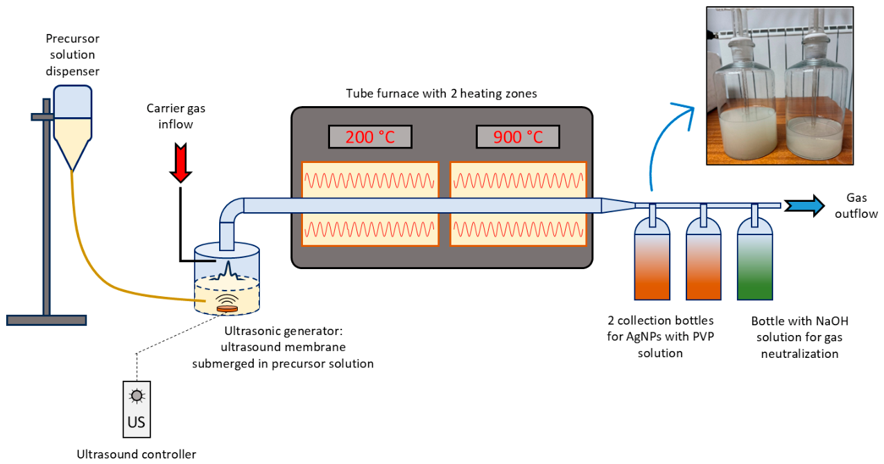
Figure 2.
Alginate/AgNP beads.
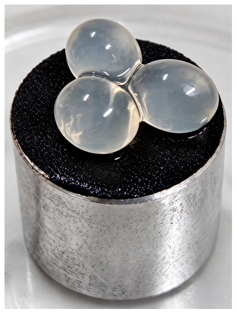
Figure 3.
SEM images of the dried AgNPs dispersion with the performed EDS analysis.
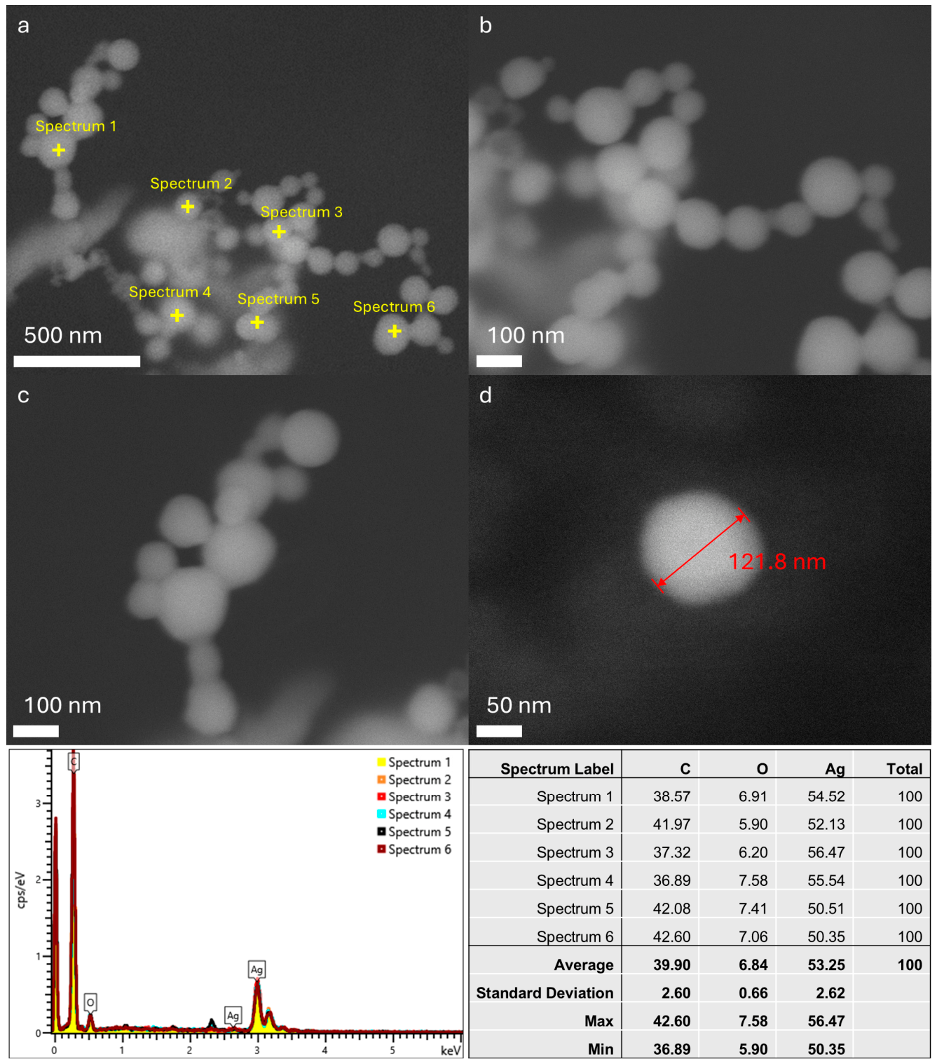
Figure 6.
SEM images of the dried alginate/AgNPs beads with the performed EDS analysis.
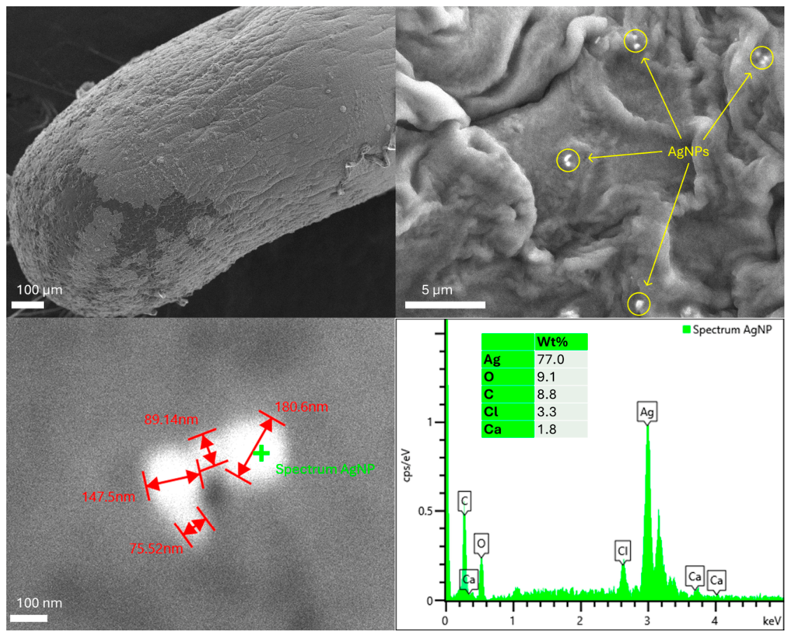
Figure 7.
STEM HAADF images of alginate microspheres.
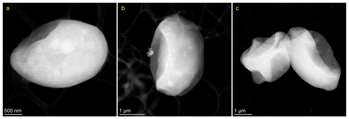
Figure 8.
(a-c) TEM images of a large agglomerate of AgNPs from an alginate dispersion, with diffraction pattern indicating the polycrystalline Ag FCC phase. (d-g) TEM image of silver particles showing localized recrystallization of Ag into AgCl (area II). The corresponding SAED patterns reveal diffraction rings characteristic of metallic Ag and AgCl.
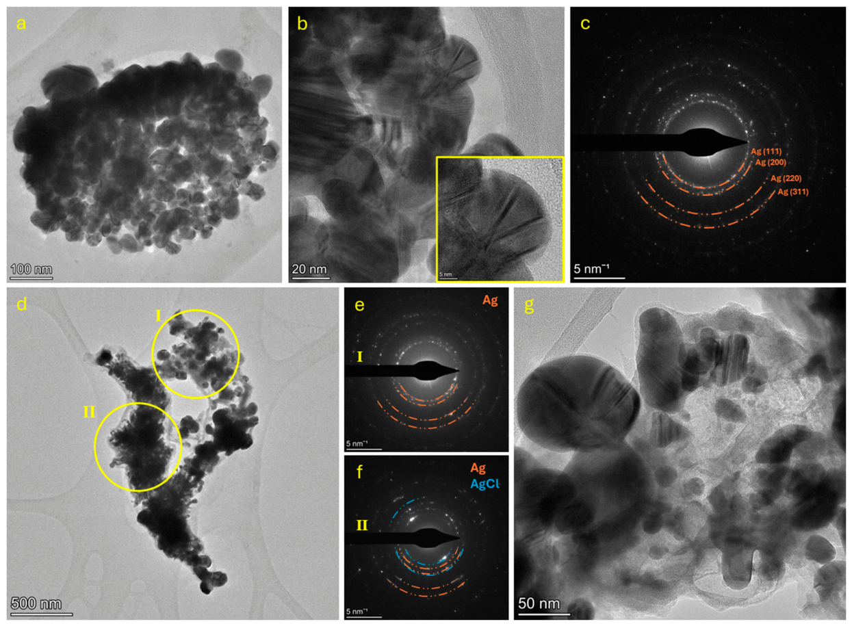
Figure 9.
STEM HAADF images of AgNPs with secondary AgCl (a-b), and the same aggregate after EDS mapping (c-d). The EDS elemental map (e-f) reveals a Cl-rich matrix and Ag enrichment at the nanoparticle sites. Following electron-beam-induced modification, Cl is removed, and fine Ag particles are formed.
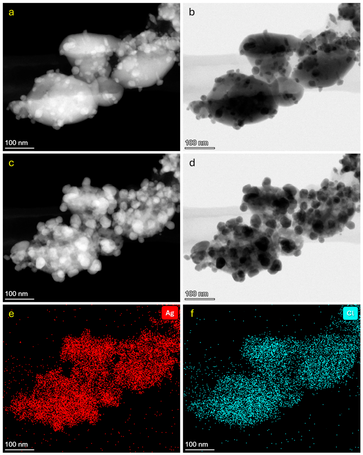
Figure 10.
(a) Dynamic light scattering (DLS) particle size distribution and (b) zeta potential distribution.
Figure 10.
(a) Dynamic light scattering (DLS) particle size distribution and (b) zeta potential distribution.
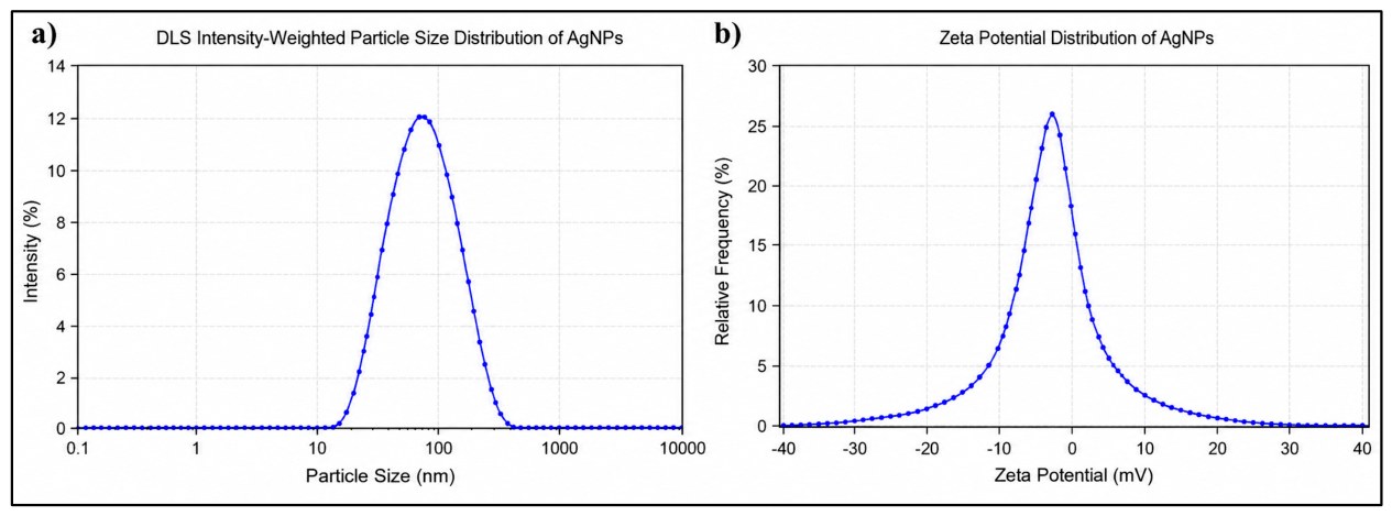
Figure 11.
Filtrate of MP after filtration of remaining PA fibres after a) 2h, b) 4 h and c) 24 h.
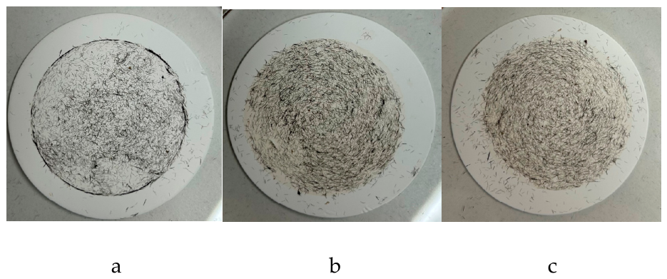

Table 1.
USP technological parameters for AgNP synthesis.
| USP parameter | Value |
|---|---|
| Precursor | 150 mL, 1 g/L AgNO3 |
| Temperature in the evaporation zone | 200 °C |
| Temperature in the reaction zone | 900 °C |
| Carrier gas flow (N2) | 5 L/min |
| Frequency of the ultrasonic generator | 1,65 MHz |
| Collection bottle 1 | 300 mL, 2,5 g/L PVP |
| Collection bottle 2 | 300 mL, 2,5 g/L PVP |
| Collection bottle 3 | 0,05 M NaOH |
| Total synthesis time | 5 h |
Disclaimer/Publisher’s Note: The statements, opinions and data contained in all publications are solely those of the individual author(s) and contributor(s) and not of MDPI and/or the editor(s). MDPI and/or the editor(s) disclaim responsibility for any injury to people or property resulting from any ideas, methods, instructions or products referred to in the content. |
© 2026 by the authors. Licensee MDPI, Basel, Switzerland. This article is an open access article distributed under the terms and conditions of the Creative Commons Attribution (CC BY) license (http://creativecommons.org/licenses/by/4.0/).
Copyright: This open access article is published under a Creative Commons CC BY 4.0 license, which permit the free download, distribution, and reuse, provided that the author and preprint are cited in any reuse.