

Submitted:
10 October 2026
Posted:
12 October 2026
You are already at the latest version
Abstract
Inflammatory bowel disease (IBD) is a chronic intestinal inflammatory disorder characterized by epithelial barrier disruption, excessive immune activation and gut microbial dysbiosis. Dietary amino acids have emerged as potential regulators of intestinal homeostasis; however, whether L-proline (Pro), a functional amino acid involved in cellular metabolism and tissue repair, modulates intestinal inflammation through microbiota–host interactions remains unclear. In this study, we investigated the protective effects and underlying mechanisms of Pro supplementation in dextran sulfate sodium (DSS)-induced colitis. Pro administration significantly ameliorated colitis-associated phenotypes, including body weight loss, disease activity index elevation, colon shortening and histological damage. Furthermore, Pro improved intestinal barrier integrity by enhancing mucus secretion and restoring tight junction proteins, including Zonula Occludens-1 (ZO-1), Claudin-11 and Occludin. Transcriptomic analysis revealed that Pro supplementation induced extensive transcriptional remodeling and was associated with the regulation of inflammation-related pathways. Consistently, Pro modulated the expression of pro-inflammatory cytokines and attenuated activation of NF-κB/STAT3 signaling in colonic tissues. Gut microbiota profiling demonstrated that Pro reshaped DSS-induced microbial dysbiosis by enriching beneficial bacterial taxa, including Lactobacillus, Lachnospiraceae_NK4A136_group, Ruminococcus, Christensenellaceae_R-7_group and Odoribacter, while reducing inflammation-associated taxa such as Escherichia-Shigella. Importantly, fecal microbiota transplantation (FMT) experiments showed that microbiota from Pro-treated donors conferred protective effects against colitis in recipient mice, supporting the functional contribution of Pro-associated microbial remodeling to intestinal protection. Collectively, our findings reveal that Pro alleviates intestinal inflammation through coordinated regulation of gut microbiota composition, epithelial barrier integrity and inflammatory responses. This study provides new insights into the role of Pro in modulating microbiota–host interactions and highlights Pro as a potential nutritional strategy for maintaining intestinal health.
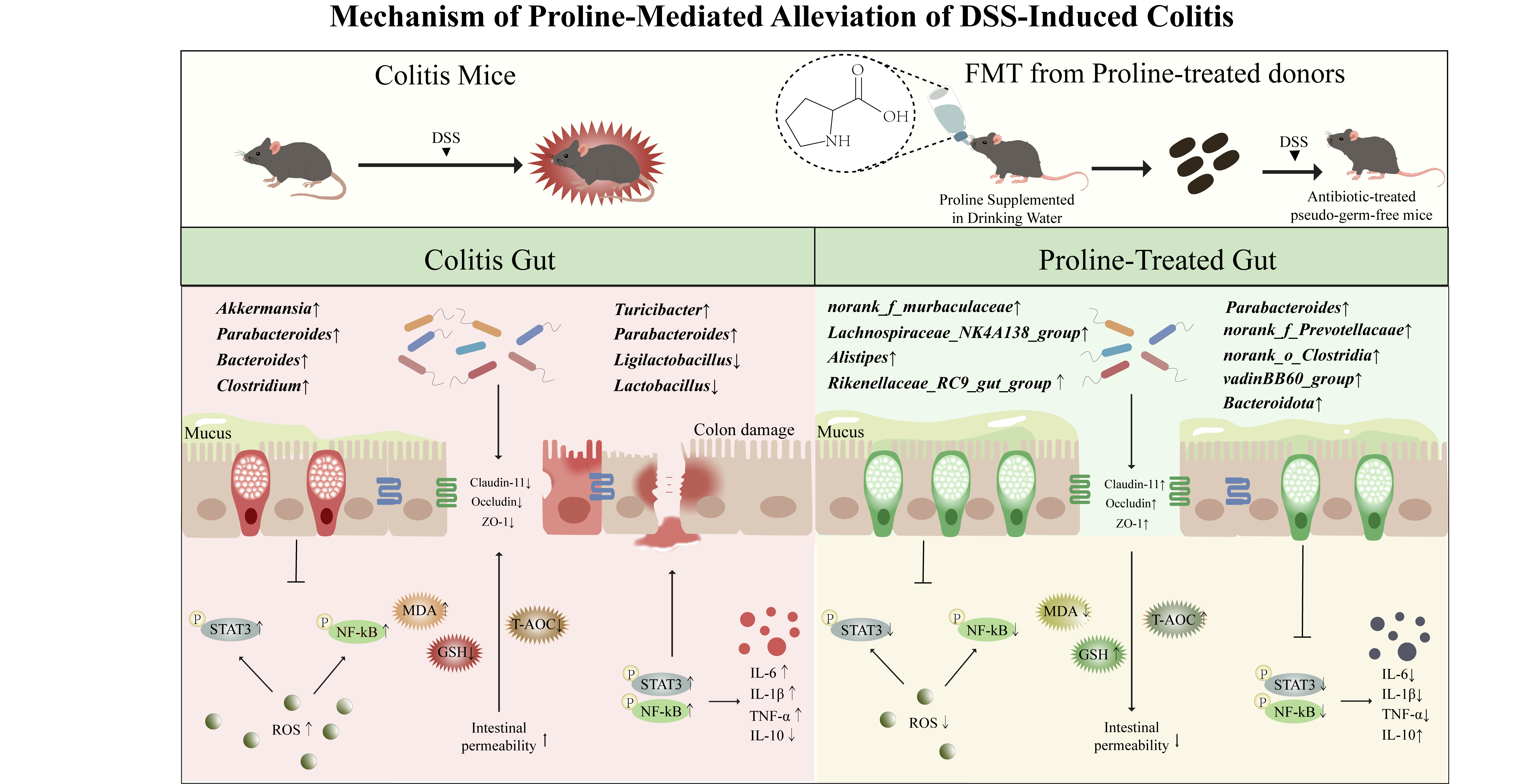
Keywords:
L-proline
; gut microbiota
; fecal microbiota transplantation
; NF-κB/STAT3 signaling pathway
; colitis
1. Introduction
Inflammatory bowel disease (IBD), including ulcerative colitis (UC) and Crohn’s disease (CD), is a chronic relapsing inflammatory disorder of the gastrointestinal tract that arises from complex interactions among host genetics, environmental factors, immune dysregulation and gut microbial imbalance [1]. Despite substantial advances in biological therapies, long-term disease remission remains challenging because current treatments primarily suppress inflammation without fully restoring intestinal homeostasis [2]. Increasing evidence indicates that disruption of the intestinal epithelial barrier and gut microbial ecology is a pivotal event in IBD pathogenesis, allowing luminal microorganisms and their products to penetrate the mucosa, thereby perpetuating chronic immune activation [3]. Consequently, nutritional strategies capable of simultaneously restoring epithelial barrier integrity and microbial homeostasis have emerged as promising complementary approaches for the prevention and management of IBD [4]. The intestinal epithelium functions as the first line of defense against luminal antigens through coordinated interactions between epithelial cells, mucus layers and tight junction proteins [5]. During intestinal inflammation, excessive production of reactive oxygen species (ROS) overwhelms endogenous antioxidant defenses, leading to oxidative damage of cellular macromolecules and disruption of epithelial integrity [6]. Beyond their direct cytotoxic effects, ROS act as important intracellular signaling molecules that activate the nuclear factor kappa-B (NF-κB) pathway, a central regulator of inflammatory responses [7]. Activated NF-κB promotes the transcription of multiple pro-inflammatory cytokines, including tumor necrosis factor-α (TNF-α) and interleukin-6 (IL-6), which subsequently trigger signal transducer and activator of transcription 3 (STAT3) signaling [8,9]. Persistent activation of the NF-κB/STAT3 axis amplifies inflammatory responses, impairs mucosal repair and further compromises the expression of tight junction proteins such as Zonula Occludens-1 (ZO-1), Claudin-11 and Occludin, thereby establishing a self-perpetuating inflammatory cycle that accelerates disease progression [10,11]. Accumulating evidence further suggests that these pathological processes are closely intertwined with alterations in the gut microbiota. Rather than serving as passive inhabitants of the intestine, gut microorganisms actively regulate epithelial barrier function, host immunity and inflammatory signaling through the production of diverse bioactive metabolites. In healthy individuals, a balanced microbial ecosystem dominated by members of the Bacillota and Bacteroidetes phyla contributes to intestinal homeostasis through the generation of short-chain fatty acids (SCFAs), polyamines and other metabolites that reinforce epithelial integrity and suppress excessive immune activation [12,13,14]. In contrast, intestinal inflammation is frequently accompanied by profound microbial dysbiosis characterized by depletion of beneficial commensals and expansion of opportunistic microorganisms. Such ecological disruption not only weakens barrier function but also potentiates ROS accumulation and aberrant activation of the NF-κB/STAT3 signaling pathway, thereby creating a reciprocal interaction between microbial dysbiosis and host inflammatory responses [15,16]. These observations have shifted current therapeutic concepts from simply suppressing inflammation toward restoring host–microbiota homeostasis through targeted nutritional interventions.
Among various nutritional interventions, functional amino acids have attracted increasing attention because they not only serve as substrates for protein synthesis but also participate in the regulation of immunometabolism, redox homeostasis and intestinal barrier function [17]. Several amino acids, including tryptophan and arginine, have been shown to alleviate intestinal inflammation through modulation of host immune responses and microbial metabolism [18]. As a member of the arginine family [19], L-proline (Pro) is traditionally recognized for its indispensable role in collagen synthesis and tissue repair [20]. More recently, accumulating evidence has suggested that Pro also functions as an important regulator of cellular redox balance, mitochondrial metabolism and epithelial integrity, suggesting that its biological activities extend far beyond protein biosynthesis [20,21]. Emerging studies further indicate that Pro may participate in the bidirectional communication between the host and the gut microbiota. Unlike many other amino acids, Pro can be utilized by a broad spectrum of intestinal microorganisms and is closely associated with microbial energy metabolism and ecological fitness [22]. Consequently, alterations in Pro availability may reshape microbial community composition and metabolic output, thereby indirectly influencing host immune responses and epithelial barrier function. This concept is supported by metagenomic studies demonstrating that microbial pathways involved in Pro metabolism are widely distributed within the intestinal microbiome and closely associated with microbial diversity and metabolic activity [23]. Because microbial metabolites, particularly short-chain fatty acids and other bioactive compounds, are important regulators of epithelial integrity and inflammatory signaling, modulation of microbial Pro metabolism may represent a previously underappreciated mechanism linking nutritional intervention with intestinal immune homeostasis [24]. Nevertheless, several critical questions remain unresolved. Although previous studies have demonstrated the antioxidant and anti-inflammatory properties of Pro, most have focused primarily on its direct effects on host cells. Whether the protective effects of Pro are mediated, at least in part, through remodeling of the gut microbiota remains largely unknown. More importantly, the causal contribution of microbiota remodeling to Pro-mediated protection has not been rigorously evaluated. Fecal microbiota transplantation (FMT), which enables the transfer of donor microbial communities to recipient animals, provides a powerful experimental strategy for distinguishing microbiota-dependent effects from direct host responses [25]. Integrating FMT with transcriptomic and microbial profiling therefore offers an opportunity to elucidate the mechanistic relationship between nutritional intervention, gut microbial ecology and host inflammatory signaling [26].
Based on these considerations, we hypothesized that Pro supplementation alleviates DSS-induced colitis through coordinated regulation of gut microbial ecology and host inflammatory responses. Specifically, we proposed that Pro-driven remodeling of the gut microbiota contributes to restoration of intestinal barrier integrity and attenuation of aberrant NF-κB/STAT3 signaling, thereby interrupting the self-amplifying inflammatory cascade characteristic of colitis. To test this hypothesis, we established a dextran sulfate sodium (DSS)-induced murine colitis model and comprehensively evaluated the effects of Pro using physiological assessment, histopathological analysis, transcriptome sequencing, gut microbiota profiling and fecal microbiota transplantation. Our findings provide mechanistic evidence supporting the microbiota-mediated protective effects of Pro and highlight its potential as a precision nutritional strategy for the prevention and management of IBD.
2. Results
2.1. Pro Ameliorates DSS-Induced Colitis Symptoms and Intestinal Barrier Injury
To assess the therapeutic potential of Pro, a DSS-induced colitis mouse model was employed (Figure 1A). A preliminary dose-optimization study identified 1% as the effective dose. Mice co-treated with DSS and Pro exhibited significantly reduced body weight loss compared to the vehicle-treated DSS group (Figure 1B). Consistent with these findings, clinical severity, as measured by the DAI, was significantly alleviated (Figure 1C), and colonic shortening, an indicator of inflammation severity, was markedly prevented (Figure 1D). Histopathological analysis supported these macroscopic observations: while the DSS group exhibited severe epithelial damage, extensive inflammatory infiltration, and goblet cell depletion, Pro treatment effectively preserved colonic mucosal architecture and reduced histological damage (Figure 1E-G). These results suggest that Pro confers protection against DSS-induced pathological changes. Considering that intestinal barrier dysfunction is a hallmark of IBD pathogenesis, it was hypothesized that Pro alleviates colitis by reinforcing epithelial integrity. To test this, the expression of key tight junction proteins was analyzed by Western blotting. DSS treatment caused significant depletion of ZO-1 and Claudin-11 (Figure 1H, I). In contrast, Pro supplementation substantially restored the expression of these proteins, indicating a potent protective effect against DSS-induced barrier disruption.
2.2. Pro Attenuates DSS-Induced Colitis by Reprogramming Inflammatory and Metabolic Associated Transcriptomes
To elucidate the molecular mechanisms underlying the therapeutic effects of Pro, RNA sequencing (RNA-seq) was performed on colonic tissues. Pro supplementation induced a substantial shift in the colonic transcriptome, identifying 54 significantly upregulated genes and 75 downregulated genes compared to the DSS group (Figure 2A). Principal Component Analysis (PCA) revealed distinct clustering of biological replicates, highlighting the reproducibility and the divergent transcriptomic profile induced by Pro (Figure 2B). To identify key effectors, a screening of differentially expressed genes (DEGs) was conducted (Figure 2C). RT-qPCR validation confirmed the regulation of several critical candidates involved in barrier restoration and immune modulation, including S100a8, Wfdc21, Hbb-bs, and Fabp1 (Figure 2E–H). KEGG pathway enrichment analysis further revealed that Pro predominantly modulated pathways linked to the inflammatory “vicious cycle”, such as Cytokine-cytokine receptor interaction, IL-17 signaling, TNF signaling, and Chemokine signaling (Figure 2D and Table. S4). Notably, these enriched pathways showed significant overlap with the STAT3 signaling axis (Table. S5). Furthermore, the enrichment of NF-κB signaling and Arginine and Pro metabolism suggests that Pro may simultaneously disrupt the ROS/NF-κB/STAT3 feed-forward loop while optimizing amino acid metabolism to promote mucosal healing. Collectively, these transcriptomic findings indicate that Pro exerts protective effects by systemically suppressing pro-inflammatory cascades and restoring metabolic homeostasis. Oxidative stress is considered an important contributor to inflammatory injury[27]. To confirm that Pro quenches oxidative stress and disrupts the NF-κB/IL-6/STAT3 axis, redox markers were quantified. Pro supplementation significantly reduced DSS-induced MDA accumulation (Figure 3A), a marker of lipid peroxidation, while restoring redox balance, as evidenced by a marked increase in the GSH/GSSG ratio (Figure 3B, C), and significantly enhanced the overall antioxidant capacity (Figure 3D). Consistent with these biochemical findings, RT-qPCR analysis demonstrated that Pro significantly downregulated the mRNA levels of IL-1β and IL-6 and upregulated mRNA levels of IL-10 and IL-4 (Figure 3E). Finally, to mechanistically validate the inhibition of the core inflammatory axis, the activation of NF-κB and STAT3 was assessed via Western blot. In line with the transcriptomic data, Pro treatment significantly reduced the phosphorylation of the NF-κB subunit and STAT3 (Figure 3F-H). These results demonstrate that Pro mitigates colitis by effectively repressing the post-translational activation of these master transcription factors, thereby interrupting the feed-forward inflammatory signaling cascade.
2.3. Pro Reverses Dysbiosis and Restores Gut Microbiota Homeostasis
To determine whether the therapeutic effects of Pro are mediated through gut microbial remodeling, 16S rRNA gene sequencing was performed on fecal samples. Analysis of α-diversity metrics revealed that DSS treatment led to a significant reduction in microbial richness and evenness, as indicated by a reduced Chao1 index and an increased Shannon index (Figure 4A). Importantly, Pro supplementation effectively counteracted this loss of diversity, restoring these indices to levels comparable to the Control group. In terms of microbial community structure, Principal Coordinate Analysis (PCoA) combined with Analysis of Similarity (ANOSIM; R = 0.6992, P = 0.001) demonstrated clear separation between the Control and DSS groups, confirming the establishment of a dysbiotic state (Figure 4B). However, the microbiota of Pro-treated mice showed a significant shift in ordination space, clustering away from the DSS group and aligning more closely with the healthy phenotype. These results suggest that Pro exerts a strong regulatory effect, promoting a structural reconfiguration of the gut microbiome from dysbiosis to a more balanced state. At the phylum level, taxonomic profiling identified Bacillota (Firmicutes), Bacteroidetes, Verrucomicrobia and Pseudomonadota (Proteobacteria) as the dominant taxa (Figure 4C). At the genus level, DSS treatment induced a distinct dysbiotic signature, marked by a reduction in Lactobacillus and an expansion of Akkermansia compared to the Control group (Figure 4D). Notably, Pro supplementation reversed these compositional changes, restoring the abundance of these key genera to baseline levels (Figure 4E). To identify the specific phylotypes driving these community shifts, Linear Discriminant Analysis Effect Size (LEfSe) was applied. Pro intervention orchestrated a targeted microecological restoration, significantly enriching a consortium of functional commensals (Figure 4F). Specifically, members of the phylum Bacteroidota, including Bacteroides, Parabacteroides, and Alistipes, emerged as dominant biomarkers in the treatment group. Concurrently, core butyrate-producing bacteria within the phylum Bacillota, such as the [Eubacterium] siraeum group, Ruminococcus, and Odoribacter, exhibited robust recovery and significant enrichment following Pro administration. Furthermore, taxa closely associated with the maintenance of metabolic homeostasis, notably the Rikenellaceae_RC9_gut_group, were also prominently expanded, indicating a comprehensive structural reversal of the DSS-disrupted dysbiosis.
2.4. Fecal Microbiota Transplantation Recapitulates the Therapeutic Effects of Pro
Given the pivotal role of dysbiosis in colitis pathogenesis, FMT was conducted to determine whether the gut microbiota mediates the therapeutic effects of Pro. Fecal microbiota from Pro-supplemented donors were transplanted into antibiotic-treated pseudo-germ-free mice to rigorously examine the causal contribution of the reshaped microbiome to colitis alleviation (Figure 5A), the effect of intestinal microbiota depletion is shown in Figure S1A. Phenotypically, mice receiving microbiota from Pro-treated donors (FMT-Pro group) showed substantial mitigation of DSS-induced injury. This was evidenced by the significant attenuation of body weight loss (Figure 5B) and the prevention of colon shortening (Figure 5C). Furthermore, histological analysis revealed that the FMT-Pro group exhibited preserved mucosal architecture, characterized by reduced crypt distortion, diminished epithelial erosion, decreased inflammatory infiltration, and maintained mucin abundance (Figure 5D-F).
To characterize the microbial community responsible for these effects, 16S rRNA gene sequencing was performed on the recipients. Although alpha-diversity indices (Chao1 and Simpson) showed no significant differences between the FMT and DSS groups (Figure 6B), β-diversity analysis revealed a striking structural shift. PCoA and ANOSIM (R = 0.9451, P = 0.001) showed that the microbiome of FMT recipients clustered distinctly away from the DSS group and closely resembled the healthy Control group (Figure 6A), suggesting that the beneficial features of the donor microbiota successfully colonized the recipients.
Taxonomic profiling (Figure 6C) identified Bacillota (Firmicutes), Bacteroidetes, Actinobacteria, Thermodesulfobacteria, and Verrucomicrobia as the dominant phyla. Notably, at the genus level, the depletion of Lactobacillus observed in DSS-treated mice was effectively reversed in the FMT-Pro group, restoring its abundance to baseline control levels (Figure 6D, E). LEfSe analysis further confirmed that the alleviation of colitis was associated with the specific enrichment of beneficial taxa (Figure 6F). Specifically, the FMT-Pro group exhibited enrichment of taxa linked to complex carbohydrate degradation and metabolic homeostasis, notably Rikenellaceae_RC9_gut_group and Prevotellaceae (norank_f_Prevotellaceae). In summary, these results indicate that Pro intervention establishes a beneficial, transmissible microbial signature instrumental in conferring protection against colitis.
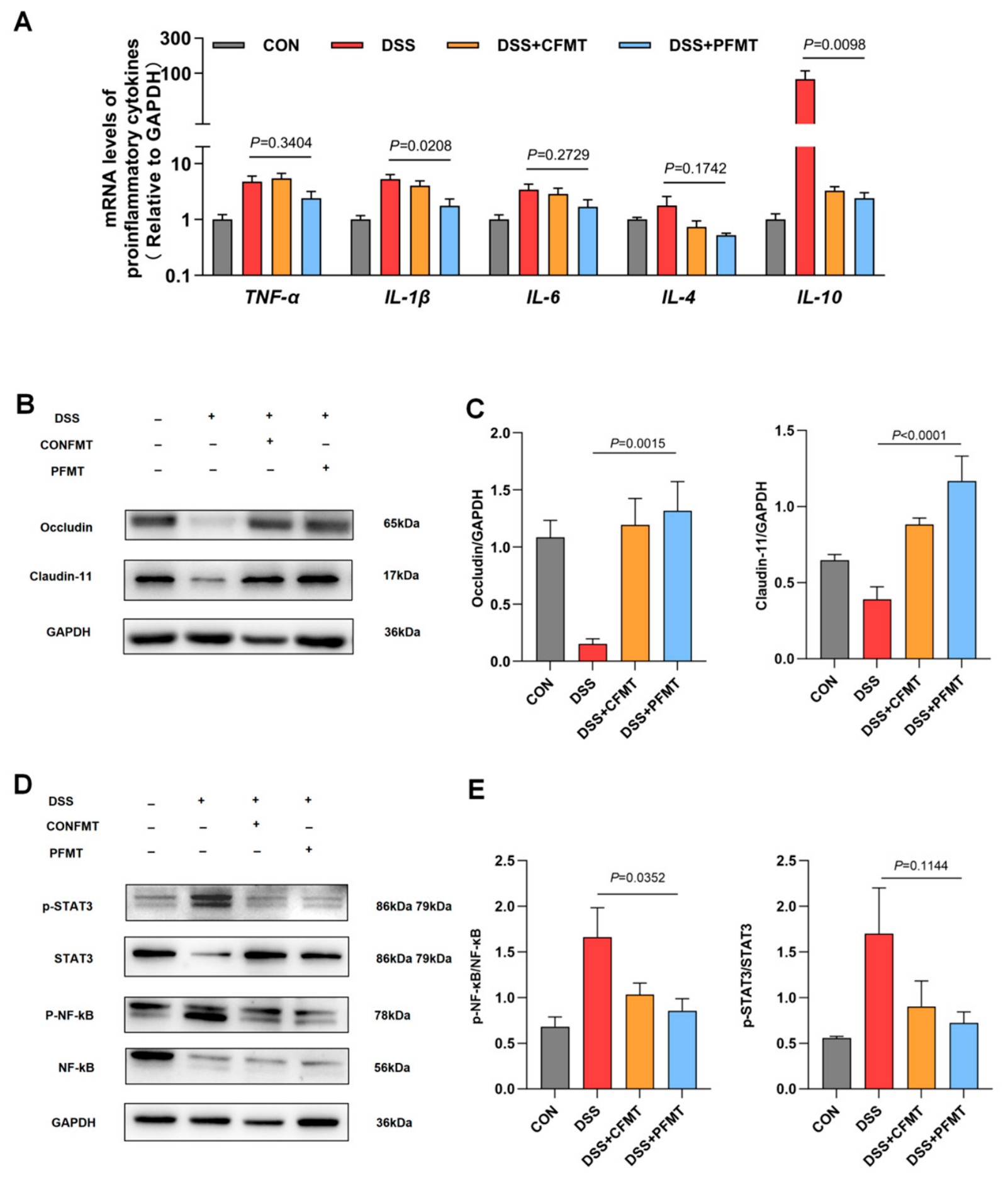
At the molecular level, the protective phenotype was accompanied by a marked reduction in the colonic inflammatory response (Figure 7A). Western blot analysis demonstrated that DSS treatment downregulated the tight junction protein Occludin and Claudin-11 (Figure 7B, C), but transplantation of the Pro-modulated microbiota significantly restored its expression, reinforcing the epithelial barrier. Moreover, the transplanted microbiota effectively suppressed host inflammatory signaling, as evidenced by the significant reduction in NF-κB and STAT3 phosphorylation (Figure 7D, E). These results indicate that the altered microbiota alone can reverse barrier defects and disrupt the pro-inflammatory signaling cascade (P < 0.05). In conclusion, these findings provide direct evidence that Pro-induced gut microbiota remodeling, particularly the restoration of beneficial genera such as Lactobacillus and butyrate-producing bacteria (Odoribacter, [Eubacterium] brachy group), serves as a causal mechanism driving the recovery of intestinal homeostasis and the suppression of host inflammation.
3. Discussion
IBD remains a major global health challenge because current therapeutic strategies primarily focus on controlling inflammation rather than restoring intestinal homeostasis[28,29]. Increasing evidence indicates that effective intervention should simultaneously target epithelial barrier integrity, immune dysregulation, and gut microbial imbalance, which together constitute the pathological basis of chronic intestinal inflammation[30,31]. In the present study, we demonstrated that Pro markedly alleviated DSS-induced colitis by improving clinical symptoms, preserving intestinal barrier integrity, attenuating inflammatory responses, and remodeling the gut microbial community. Importantly, fecal microbiota transplantation further provided functional evidence supporting the contribution of the Pro-associated microbiota to intestinal protection. Collectively, these findings expand current understanding of the biological functions of Pro and suggest that microbiota remodeling represents an important component of its protective effects against experimental colitis. Disruption of the intestinal epithelial barrier is considered one of the earliest pathological events during colitis[32,33], allowing luminal microorganisms and microbial products to penetrate the mucosa and perpetuate chronic immune activation. Consistent with previous studies demonstrating the cytoprotective properties of Pro, our results showed that Pro effectively preserved colonic architecture, reduced epithelial damage, and restored crypt organization following DSS administration. More importantly, Pro markedly increased goblet cell abundance and enhanced the expression of the tight junction proteins, indicating coordinated restoration of both the mucus barrier and the epithelial mechanical barrier. Goblet cells continuously secrete mucins that physically separate intestinal microorganisms from epithelial cells, whereas tight junction proteins maintain epithelial permeability and prevent excessive antigen translocation[34]. Therefore, simultaneous recovery of these complementary barrier systems is likely to reduce exposure of the intestinal immune system to luminal inflammatory stimuli, thereby contributing to attenuation of intestinal inflammation[35,36]. These findings further support the concept that maintenance of barrier integrity represents a critical therapeutic target in IBD.
Beyond restoration of epithelial barrier integrity, excessive activation of inflammatory signaling pathways represents a major driver of intestinal injury during colitis. Transcriptomic analysis performed in the present study revealed extensive transcriptional remodeling following Pro supplementation and identified inflammation-related pathways, particularly NF-κB and JAK/STAT signaling pathways, as major biological processes associated with the protective effects of Pro[37,38]. These transcriptomic observations were further supported by molecular validation, showing that Pro reduced the expression of pro-inflammatory cytokines, including TNF-α, IL-1β and IL-6, while increasing anti-inflammatory cytokine expression. Moreover, Pro supplementation was associated with reduced phosphorylation of NF-κB and STAT3, indicating attenuation of inflammatory pathway activation[39]. The NF-κB/STAT3 signaling network represents a central regulatory axis involved in the initiation and persistence of intestinal inflammation. Under physiological conditions, NF-κB activation is tightly controlled to maintain immune surveillance and epithelial homeostasis. However, during DSS-induced colitis, excessive oxidative stress and microbial-derived inflammatory stimuli promote persistent NF-κB activation, leading to increased production of inflammatory cytokines. Among these cytokines, IL-6 serves as an important mediator linking innate immune activation with STAT3 signaling, and sustained STAT3 activation contributes to inflammatory amplification and impaired mucosal repair[40]. Therefore, the reduced activation of NF-κB and STAT3 observed after Pro supplementation provides a potential molecular explanation for the improved inflammatory status and barrier function. Nevertheless, the beneficial effects of Pro rather than representing a definitive causal mechanism. Future studies using selective inhibitors, conditional knockout models or targeted intervention approaches will be necessary to further clarify the precise contribution of this signaling pathway[41].
Increasing evidence indicates that gut microbiota plays an essential role in intestinal homeostasis. Recent studies have further demonstrated that microbiota remodeling serves as an important mediator linking dietary or environmental factors to host physiological responses. For example, Elemicin exposure altered gut microbial composition, which subsequently contributed to changes in host lipid metabolism, highlighting the critical role of gut microbiota in mediating host responses to exogenous compounds[42]. In addition to directly affecting epithelial barrier function, intestinal microorganisms produce a wide range of bioactive metabolites, including short-chain fatty acids (SCFAs), indole derivatives and polyamines, which participate in immune regulation and epithelial repair[43]. In the present study, Pro supplementation markedly reshaped the dysbiotic microbial community induced by DSS treatment, suggesting that the protective effects of Pro may involve regulation of the intestinal microbial ecosystem. Notably, Pro enriched several bacterial taxa previously associated with intestinal homeostasis, including Lactobacillus, Ruminococcus, Christensenellaceae_R-7_group, Allistipes, Odoribacter and Lachnospiraceae_NK4A136_group. These microbial members have been implicated in maintaining intestinal health through diverse mechanisms. Lactobacillus species can enhance epithelial barrier function by promoting mucus secretion, regulating tight junction expression and producing antimicrobial metabolites. Members of Lachnospiraceae and Ruminococcaceae families are recognized as important SCFA producers, particularly butyrate-producing bacteria[44], which provide energy substrates for colonocytes and suppress excessive inflammatory responses through epigenetic and receptor-mediated mechanisms. Odoribacter has also been reported to produce beneficial metabolites associated with immune regulation and intestinal metabolic homeostasis. Therefore, enrichment of these bacteria following Pro supplementation may contribute to the restoration of a more favorable intestinal microenvironment. Conversely, Pro treatment reduced the abundance of inflammation-associated microbial taxa, particularly Escherichia-Shigella, which has frequently been linked to intestinal barrier disruption and inflammatory activation. Expansion of opportunistic Proteobacteria is commonly observed during intestinal inflammation and is often considered a hallmark of microbial imbalance. The suppression of these potentially harmful bacteria suggests that Pro may help restore microbial equilibrium by simultaneously promoting beneficial microorganisms and limiting the expansion of inflammation-associated taxa. In addition, Pro significantly rescued the DSS-induced depletion of Lactobacillus, a genus vital for reinforcing mucosal defenses. We have discovered a paradoxical phenomenon, DSS also triggered an overrepresentation of Akkermansia. While Akkermansia is generally recognized as beneficial under physiological conditions, its aberrant expansion during acute colitis often reflects a compensatory response to severe mucin depletion and compromised barrier integrity[45]. Pro intervention effectively normalized these compositional shifts. This restoration suggests that Pro not only replenishes essential probiotic populations but also helps preserve the structural integrity of the mucus layer, thereby eliminating the pathological microenvironment that drives the abnormal proliferation of Akkermansia. This shift from a pro-inflammatory to a homeostatic microbiome suggests that Pro acts as a prebiotic-like modulator, fostering a metabolic environment that is hostile to colitis progression.
Although microbiota sequencing provides important information regarding compositional changes, alterations in microbial abundance alone cannot determine whether these changes are biologically meaningful. FMT experiments addressed this limitation by demonstrating that transplantation of microbiota from Pro-treated donors significantly alleviated colitis severity, improved intestinal barrier integrity and reduced inflammatory responses in recipient mice. These findings provide functional evidence supporting an important role of the Pro-associated microbiota in intestinal protection[46]. Importantly, the protective phenotype observed following FMT does not exclude the possibility that Pro directly regulates host physiology[47]. As an amino acid involved in collagen synthesis, redox regulation and cellular metabolism, Pro may exert microbiota-independent effects on intestinal epithelial cells and immune cells. In addition, microbial metabolites generated after Pro supplementation may represent another layer of communication between dietary intervention and host responses. Therefore, the biological effects of Pro are likely mediated through coordinated interactions between host metabolic regulation and microbiota remodeling rather than through a single pathway.
From a nutritional perspective, the present findings highlight the potential of functional amino acids as dietary regulators of intestinal health[48]. Traditionally, amino acids have been primarily considered as substrates for protein synthesis and energy metabolism; however, accumulating evidence indicates that specific amino acids also function as signaling molecules that regulate immunity, oxidative balance and host–microbiota interactions. Compared with conventional anti-inflammatory therapies that mainly target downstream inflammatory mediators, nutritional interventions based on functional amino acids may provide a more physiological approach by simultaneously regulating epithelial integrity, immune homeostasis and microbial ecology. Pro represents a particularly interesting candidate because of its unique biological properties. As a major component of collagen, Pro contributes to extracellular matrix formation and tissue repair, which are essential processes during intestinal mucosal regeneration. In addition, Pro participates in cellular redox regulation and metabolic adaptation, processes that are frequently disrupted during inflammatory conditions. The present study extends the beneficial effects of Pro are not limited to direct host responses but also involve remodeling of the intestinal microbial ecosystem. This interaction between dietary amino acids and gut microbiota provides a potential framework for developing precision nutritional strategies for intestinal inflammatory disorders.
4. Materials and Methods
4.1. Drugs and Chemicals
DSS was procured from MP Biomedicals (Santa Ana, CA, USA). Pro was obtained from Sigma (St. Louis, MO, USA). Primary antibodies targeting NF-κB, p-NF-κB (Ser536), STAT3, and p-STAT3 (Tyr705), as well as the loading control GAPDH, were supplied by Cell Signaling Technology (Beverly, MA, USA). Horseradish peroxidase (HRP)-labeled secondary antibodies and Hematoxylin and eosin (H&E) were purchased from Huaxingbio Biotechnology Co., Ltd (Beijing, China). TRIzol Reagent was obtained from Thermo Fisher Scientific (Beijing, China). The FastQuant RT Kit (with gDNase) and SuperReal PreMix Plus (SYBR Green) were purchased from TIANGEN Biotech (Beijing, China). Alcian blue was procured from Solarbio (Beijing, China). Urine and Stool Occult Blood Test Kits were obtained from Nanjing Jiancheng Bioengineering Institute (Nanjing, China). Assay kits for malondialdehyde (MDA), glutathione (GSH), oxidized glutathione disulfide (GSSG) and total antioxidant capacity (T-AOC) were purchased from Beyotime Biotechnology (Haimen, China).
4.2. Animals Treatments and Sample Collection
Male C57BL/6J mice (6 – 8 weeks old, 20 – 22 g) were purchased from Sibeifu (Beijing, China) and housed under specific pathogen-free (SPF) conditions (22 ± 2 °C, 55 ± 10% relative humidity, 12 h light/dark cycle) with free access to standard laboratory chow and drinking water. All experimental procedures were approved by the Institutional Animal Care and Use Committee of China Agricultural University (Approval No. AW20206202-1-15).
Following one week of acclimatization, mice were randomly assigned into the experimental groups. To determine the optimal supplementation level, a preliminary dose-screening experiment was conducted in DSS-induced colitis mice receiving Pro in the drinking water at concentrations of 0.5%, 1%, 2%, or 4% (w/v). Disease severity was evaluated according to changes in body weight, disease activity index (DAI), and colon length. Based on the overall protective efficacy, 1% (w/v) Pro was selected for all subsequent mechanistic experiments (Supplementary Figure 1A-C).
Experimental colitis was induced using DSS administration. Following the acclimatization period, mice in the DSS and DSS + Pro groups received 2% (w/v) DSS in drinking water for 7 consecutive days, whereas mice in the Control group received normal drinking water throughout the experiment. To evaluate the preventive effect of Pro, mice in the DSS + Pro group were supplemented with 1% (w/v) Pro in drinking water for 7 days before DSS administration and continuously throughout DSS administration and the subsequent recovery period. The detailed experimental schedule is shown in Figure 1A. Body weight, stool consistency and fecal bleeding were recorded daily throughout the experimental period, and the DAI was calculated according to Table.S1. At the experimental endpoint, mice were euthanized after blood collection. The entire colon was carefully excised from the ileocecal junction to the anus, and colon length was immediately measured as a macroscopic indicator of colitis severity. Colon tissues were fixed in 4% paraformaldehyde for histopathological examination, whereas the remaining colon tissues were rapidly frozen in liquid nitrogen and stored at −80 °C for RNA extraction, western blot analysis and transcriptomic sequencing.
4.3. Fecal Microbiota Transplantation
To determine whether the protective effects of Pro were mediated by alterations in the gut microbiota, FMT was performed using fecal samples collected from donor mice in the Control and 1%Pro groups. Prior to FMT, to establish a microbiota-depleted model, mice received 200 μL per mouse of daily oral gavage of an antibiotic cocktail comprising 10 mg each of ampicillin, neomycin, metronidazole, and vancomycin dissolved in 2 mL phosphate-buffered saline (PBS) for five consecutive days. To verify successful microbial clearance, fecal pellets were gathered on the fifth day. These samples were homogenized and filtered, spread onto LB agar plates, and incubated under both aerobic and anaerobic conditions at 37 °C for 48 h. Following antibiotic treatment, recipient mice were randomly assigned into the experimental groups. Fresh fecal pellets were aseptically collected from donor mice immediately before each transplantation. To minimize inter-individual variation in microbial composition, fecal samples obtained from all donor mice within the same treatment group were pooled and thoroughly homogenized under sterile conditions. The pooled feces were suspended in sterile PBS at a concentration of 100 mg/mL, vortexed thoroughly and allowed to settle briefly. The resulting supernatant containing the bacterial community was collected and immediately used for transplantation. Fresh fecal suspensions were prepared daily throughout the transplantation period to preserve bacterial viability. Recipient mice received 200 μL of freshly prepared fecal suspension by oral gavage once daily for 21 consecutive days. Mice from different treatment groups were housed separately throughout the experiment to minimize microbial cross-contamination. The overall FMT procedure is illustrated in Figure 5A.
At the end of the transplantation period, recipient mice were euthanized, and serum, colonic tissues and colonic contents were collected as described above. Histopathological evaluation was performed by investigators blinded to the treatment groups according to predefined scoring criteria. Colon tissues were subsequently subjected to histological, biochemical and molecular analyses.
4.4. Histological Evaluation
Colonic segments were fixed in 4% paraformaldehyde for 24 h, followed by sequential dehydration and standard paraffin embedding. To evaluate the structural integrity and mucosal damage of the colon, 5-μm-thickness slices were sectioned using a Thermo Fisher Scientific microtome (San Jose, CA, USA) and subsequently stained with H&E and AB-PAS. The microscopic lesions were then assessed by an independent pathologist blinded to the treatment groups, utilizing established scoring guidelines[49]. The colitis score (maximum = 12) was the sum of three features: (1) Loss of surface epithelial cells; (2) Destruction of crypts and; (3) infiltration.
4.5. RNA Extraction and RT-qPCR
Total RNA was extracted from frozen colon tissues using TRIzol reagent (Invitrogen, Carlsbad, CA, USA) according to the manufacturer’s instructions. RNA concentration and purity were determined using a NanoDrop 2000 spectrophotometer (Thermo Fisher Scientific, Wilmington, DE, USA), and RNA integrity was verified by agarose gel electrophoresis.
Complementary DNA (cDNA) was synthesized from 1 μg of total RNA using a reverse transcription kit according to the manufacturer’s protocol. Quantitative real-time PCR (RT-qPCR) was performed using SYBR Green Master Mix on a QuantStudio Real-Time PCR System (Applied Biosystems, Foster City, CA, USA). Relative gene expression levels were calculated using the 2^−ΔΔCt method with GAPDH as the internal reference gene[50]. Primer sequences are listed in Supplementary Table S2.
4.6. Western Blot Analysis
Western blotting was performed to evaluate changes in intestinal barriers and inflammation-related signaling proteins, following previously established protocols for detecting pathway-associated proteins in mouse tissues[51]. Total protein was extracted from frozen colon tissues using RIPA lysis buffer supplemented with protease and phosphatase inhibitors. Protein concentrations were determined using a bicinchoninic acid (BCA) protein assay kit (Beyotime Biotechnology, Shanghai, China). Equal amounts of protein (20–30 μg) were separated by 10% sodium dodecyl sulfate-polyacrylamide gel electrophoresis (SDS-PAGE) and subsequently transferred onto polyvinylidene fluoride (PVDF) membranes (Millipore, Billerica, MA, USA). After blocking with 5% non-fat milk for 1 h at room temperature, membranes were incubated overnight at 4 °C with primary antibodies against ZO-1, Claudin-11, Occludin, NF-κB p65, p-NF-κB, STAT3, p-STAT3 and GAPDH. Following incubation with horseradish peroxidase-conjugated secondary antibodies for 1 h at room temperature, protein bands were visualized using an enhanced chemiluminescence (ECL) detection system. Band intensities were quantified using ImageJ software (National Institutes of Health, Bethesda, MD, USA). The expression levels of target proteins were normalized to GAPDH, whereas phosphorylated proteins were normalized to their corresponding total proteins.
4.7. RNA Sequencing
Total RNA was isolated from colon tissues of mice using TRIzol reagent (Invitrogen, Carlsbad, CA, USA). RNA purity and concentration were assessed using a NanoDrop 2000 spectrophotometer (Thermo Fisher Scientific, Wilmington, DE, USA), and RNA integrity was evaluated using an Agilent 2100 Bioanalyzer (Agilent Technologies, Santa Clara, CA, USA). Only RNA samples with an RNA integrity number (RIN) greater than 7.0 were used for library construction. RNA sequencing libraries were prepared using a standard mRNA library preparation kit according to the manufacturer’s instructions and sequenced on the Illumina NovaSeq 6000 platform (Illumina, San Diego, CA, USA) by Majorbio Bio-Pharm Technology Co., Ltd. (Shanghai, China). Raw sequencing reads were subjected to quality control by removing adaptor sequences, low-quality reads and reads containing ambiguous bases to obtain clean reads. Clean reads were aligned to the mouse reference genome GRCm39/mm39 using HISAT2. Gene expression levels were quantified as fragments per kilobase of transcript per million mapped reads (FPKM). Differentially expressed genes (DEGs) between groups were identified using DESeq2, with an adjusted P value < 0.05 and |log2 fold change| ≥ 1 considered statistically significant. Functional enrichment analyses of Gene Ontology (GO) terms and Kyoto Encyclopedia of Genes and Genomes (KEGG) pathways were subsequently performed to investigate the biological functions and signaling pathways associated with the identified DEGs.
4.8. Gut Microbiota Analysis
Total microbial genomic DNA was extracted using the FastDNA® SPIN Kit for Soil (MP Biomedicals, Solon, OH, USA) according to the manufacturer’s instructions. DNA quality and concentration were assessed by agarose gel electrophoresis and NanoDrop 2000 spectrophotometry (Thermo Fisher Scientific, Wilmington, DE, USA). The V3–V4 hypervariable regions of the bacterial 16S rRNA gene were amplified using the universal primer pair 338F (5′-ACTCCTACGGGAGGCAGCAG-3′) and 806R (5′-GGACTACHVGGGTWTCTAAT-3′). Amplicons were purified, quantified, pooled in equimolar concentrations and sequenced on the Illumina MiSeq PE300 platform (Illumina, San Diego, CA, USA) by Majorbio Bio-Pharm Technology Co., Ltd. (Shanghai, China).
Raw sequencing reads were quality-filtered and denoised using the DADA2 pipeline implemented in QIIME2 to generate amplicon sequence variants (ASVs). Chimeric sequences were removed during the denoising process. Taxonomic assignment was performed against the SILVA 138 reference database. The resulting ASV table was used for all subsequent microbiome analyses, including taxonomic composition, diversity analysis and functional prediction.
4.9. Bioinformatics and Statistical Analysis
Bioinformatic analyses of the gut microbiota were performed using the Majorbio Cloud Platform (https://cloud.majorbio.com). Alpha diversity indices, including Chao1 richness and Shannon diversity, were calculated to evaluate microbial richness and diversity. Beta diversity was assessed by principal coordinate analysis (PCoA) based on Bray–Curtis distances, and differences in microbial community structure among groups were evaluated using permutational multivariate analysis of variance (PERMANOVA). Differentially abundant bacterial taxa were identified using linear discriminant analysis effect size (LEfSe), with an LDA score >2.0 considered statistically significant. Functional profiles of the gut microbiota were predicted using PICRUSt2 based on ASV representative sequences.
Transcriptomic data were analyzed using the DESeq2 package in R. Differentially expressed genes were identified using an adjusted P value <0.05 and |log2 fold change| ≥1 as the significance thresholds. Functional enrichment analyses of GO terms and KEGG pathways were performed to investigate the biological significance of differentially expressed genes.
Data are presented as the means ±SEMs. Statistical analyses were performed using GraphPad Prism 9.0 (GraphPad Software, San Diego, CA, USA) and SPSS 26.0 (IBM Corp., Armonk, NY, USA). Statistical differences among groups at each time point were evaluated using one-way ANOVA followed by Tukey’s multiple comparison test. Differences were considered statistically significant at P <0.05.
5. Conclusions
In conclusion, this study demonstrates that Pro supplementation protects against DSS-induced colitis through coordinated regulation of intestinal barrier integrity, inflammatory responses and gut microbial homeostasis. Transcriptomic and molecular analyses suggest that attenuation of NF-κB/STAT3 signaling is associated with the beneficial effects of Pro, while microbiota profiling and FMT experiments provide functional evidence supporting an important contribution of microbial remodeling. These findings provide new insights into the role of dietary amino acids in regulating host–microbiota interactions and support the potential application of Pro as a nutritional strategy for maintaining intestinal health and alleviating inflammation-associated intestinal disorders.
Supplementary Materials
The following supporting information can be downloaded at the website of this paper posted on Preprints.org, Figure S1; Table S1:Assessment of Disease Activity Index1,2 ; Table S2:Quantitative PCR primer sequences; Table S3:Antibody information; Table S4:KEGG analysis at level 3 was conducted to compare the effects of proline treatment in mice with those of DSS-infected mice; Table S5:Stat3-related signaling pathways that overlap with KEGG analysis in proline treatment mice compared to DSS-infected mice.
Author Contributions
Conceptualization, S.T. and N.L.; methodology, S.T., Y.Y. and N.L.; validation, Y.Y., S.W. and C.W.; formal analysis, S.T., Y.Z. and C.Z.; investigation, S.W., C.W., B.H., C.Z., J.L. and X.M.; resources, Z.W., Y.W., Y.Q. ; data curation, S.T., Y.Y. and S.W.; writing—original draft preparation, S.T.; writing—review and editing, S.T., Y.W., Y.Q. and N.L.; visualization, S.T., Y.Y. and S.W.; supervision, Y.Q. and N.L.; project administration, N.L.; funding acquisition, Y.W., Y.Q. and N.L. All authors have read and agreed to the published version of the manuscript.
Funding
This work was supported by the National Key Research and Development Program of China (2023YFD1302005), National Natural Science Foundation of China (No.32000082), Earmarked Fund for Modern Agro-Industry Technology Research System (CARS-43), the 2115 Talent Program of China Agricultural University and Chinese Universities Scientific Fund (2025TC011, 2026TC056), the Open Project Program of the State Key Laboratory of Animal Nutrition and Feeding, China Agricultural University (Grant No. 2004DA125184H2510).
Institutional Review Board Statement
All animal experimental procedures were conducted in strict accordance with the principles of the ARRIVE guidelines and approved by the Animal Ethics Committee of China Agricultural University (protocol code: AW20206202-1-15). All animal care and experimental protocols complied with the guidelines for laboratory animal welfare and ethics.
Informed Consent Statement
Not applicable.
Data Availability Statement
The RNA-seq and 16S rRNA sequencing data presented in this study will be deposited in the NCBI Sequence Read Archive (SRA) upon acceptance of the manuscript, and the accession numbers will be provided in the final version.
Acknowledgments
During the preparation of this manuscript, the authors used ChatGPT (OpenAI, GPT-5.6) for language editing and drafting assistance. The authors reviewed and edited the output and take full responsibility for the content of this publication.
Conflicts of Interest
The authors declare no conflicts of interest.
References
- Le Berre, C.; Honap, S.; Peyrin-Biroulet, L. Ulcerative colitis. The Lancet 2023, 402, 571–584. [Google Scholar] [CrossRef] [PubMed]
- Gisbert, J.P.; Chaparro, M. Combination therapy with biologics and/or small molecules in inflammatory bowel disease: a comprehensive review. J. Crohn’s Colitis 2025, 19, jjaf161. [Google Scholar] [CrossRef] [PubMed]
- Neurath, M.F. Targeting immune cell circuits and trafficking in inflammatory bowel disease. Nat. Immunol. 2019, 20, 970–979. [Google Scholar] [CrossRef] [PubMed]
- Turner, J.R. Intestinal mucosal barrier function in health and disease. Nat. Rev. Immunol. 2009, 9, 799–809. [Google Scholar] [CrossRef] [PubMed]
- Chelakkot, C.; Ghim, J.; Ryu, S.H. Mechanisms regulating intestinal barrier integrity and its pathological implications. Exp. Mol. Med. 2018, 50, 1–9. [Google Scholar] [CrossRef] [PubMed]
- Vona, R.; Pallotta, L.; Cappelletti, M.; Severi, C.; Matarrese, P. The Impact of Oxidative Stress in Human Pathology: Focus on Gastrointestinal Disorders. Antioxidants 2021, 10, 201. [Google Scholar] [CrossRef] [PubMed]
- Liu, T.; Zhang, L.; Joo, D.; Sun, S.-C. NF-κB signaling in inflammation. Signal Transduct. Target. Ther. 2017, 2, 17023. [Google Scholar] [CrossRef] [PubMed]
- Taniguchi, K.; Karin, M. NF-κB, inflammation, immunity and cancer: coming of age. Nat. Rev. Immunol. 2018, 18, 309–324. [Google Scholar] [CrossRef] [PubMed]
- Xu, M.; Wei, D.; Zhong, S.; Liu, W.; Ye, M.; Luo, J.; Zhu, S.; Lai, Z.; Zheng, Z.; Hu, R.; et al. A hyperactive splice variant of STAT3 promotes colonic inflammation-associated tumorigenesis in mice. Sci. Transl. Med. 2025, 17, eadu8484. [Google Scholar] [CrossRef] [PubMed]
- Cao, S.; Tai, Y.; Li, S.; Liu, X.; Zheng, Y.; Han, J.; Chen, W.; Han, J.; Zuo, H.X.; Li, M.Y.; et al. Network pharmacology and metabolomics reveal Jingfang Granules alleviates DSS-induced colitis via the NF-κB/NLRP3/IL-6/STAT3 pathway. Phytomedicine 2026, 150, 157745. [Google Scholar] [CrossRef] [PubMed]
- Parikh, K.; Antanaviciute, A.; Fawkner-Corbett, D.; Jagielowicz, M.; Aulicino, A.; Lagerholm, C.; Davis, S.; Kinchen, J.; Chen, H.H.; Alham, N.K.; et al. Colonic epithelial cell diversity in health and inflammatory bowel disease. Nature 2019, 567, 49–55. [Google Scholar] [CrossRef] [PubMed]
- Ma, J.; Piao, X.; Mahfuz, S.; Long, S.; Wang, J. The interaction among gut microbes, the intestinal barrier and short chain fatty acids. Anim. Nutr. 2022, 9, 159–174. [Google Scholar] [CrossRef] [PubMed]
- Zeng, L.; Qian, Y.; Cui, X.; Zhao, J.; Ning, Z.; Cha, J.; Wang, K.; Ge, C.; Jia, J.; Dou, T.; et al. Immunomodulatory role of gut microbial metabolites: mechanistic insights and therapeutic frontiers. Front. Microbiol. 2025, 16–2025. [Google Scholar] [CrossRef] [PubMed]
- Zhang, C.; Zhen, Y.; Weng, Y.; Lin, J.; Xu, X.; Ma, J.; Zhong, Y.; Wang, M. Research progress on the microbial metabolism and transport of polyamines and their roles in animal gut homeostasis. J. Anim. Sci. Biotechnol. 2025, 16, 57. [Google Scholar] [CrossRef] [PubMed]
- Rice, T.A.; Bielecka, A.A.; Nguyen, M.T.; Rosen, C.E.; Song, D.; Sonnert, N.D.; Yang, Y.; Cao, Y.; Khetrapal, V.; Catanzaro, J.R.; et al. Interspecies commensal interactions have nonlinear impacts on host immunity. Cell Host Microbe 2022, 30, 988–1002.e1006. [Google Scholar] [CrossRef] [PubMed]
- Zhong, W.; Wu, K.; Long, Z.; Zhou, X.; Zhong, C.; Wang, S.; Lai, H.; Guo, Y.; Lv, D.; Lu, J.; et al. Gut dysbiosis promotes prostate cancer progression and docetaxel resistance via activating NF-κB-IL6-STAT3 axis. Microbiome 2022, 10, 94. [Google Scholar] [CrossRef] [PubMed]
- Starke, S.; Harris, D.M.M.; Zimmermann, J.; Schuchardt, S.; Oumari, M.; Frank, D.; Bang, C.; Rosenstiel, P.; Schreiber, S.; Frey, N.; et al. Amino acid auxotrophies in human gut bacteria are linked to higher microbiome diversity and long-term stability. ISME J. 2023, 17, 2370–2380. [Google Scholar] [CrossRef] [PubMed]
- Canè, S.; Geiger, R.; Bronte, V. The roles of arginases and arginine in immunity. Nat. Rev. Immunol. 2025, 25, 266–284. [Google Scholar] [CrossRef] [PubMed]
- Gao, J.; Xu, K.; Liu, H.; Liu, G.; Bai, M.; Peng, C.; Li, T.; Yin, Y. Impact of the Gut Microbiota on Intestinal Immunity Mediated by Tryptophan Metabolism. Front. Cell. Infect. Microbiol. 2018, Volume 8 - 2018. [Google Scholar] [CrossRef] [PubMed]
- Shen, L.; Yu, Y.; Zhou, Y.; Pruett-Miller, S.M.; Zhang, G.-F.; Karner, C.M. SLC38A2 provides proline to fulfill unique synthetic demands arising during osteoblast differentiation and bone formation. eLife 2022, 11, e76963. [Google Scholar] [CrossRef] [PubMed]
- Phang, -J.M. Proline Metabolism in Cell Regulation and Cancer Biology: Recent Advances and Hypotheses. Antioxid. Redox Signal. 2019, 30, 635–649. [Google Scholar] [CrossRef] [PubMed]
- Pedersen, H.K.; Gudmundsdottir, V.; Nielsen, H.B.; Hyotylainen, T.; Nielsen, T.; Jensen, B.A.H.; Forslund, K.; Hildebrand, F.; Prifti, E.; Falony, G.; et al. Human gut microbes impact host serum metabolome and insulin sensitivity. Nature 2016, 535, 376–381. [Google Scholar] [CrossRef] [PubMed]
- Lloyd-Price, J.; Arze, C.; Ananthakrishnan, A.N.; Schirmer, M.; Avila-Pacheco, J.; Poon, T.W.; Andrews, E.; Ajami, N.J.; Bonham, K.S.; Brislawn, C.J.; et al. Multi-omics of the gut microbial ecosystem in inflammatory bowel diseases. Nature 2019, 569, 655–662. [Google Scholar] [CrossRef]
- Ni, J.; Wu, G.D.; Albenberg, L.; Tomov, V.T. Gut microbiota and IBD: causation or correlation? Nat. Rev. Gastroenterol. Hepatol. 2017, 14, 573–584. [Google Scholar] [CrossRef] [PubMed]
- Paramsothy, S.; Kamm, M.A.; Kaakoush, N.O.; Walsh, A.J.; van den Bogaerde, J.; Samuel, D.; Leong, R.W.L.; Connor, S.; Ng, W.; Paramsothy, R.; et al. Multidonor intensive faecal microbiota transplantation for active ulcerative colitis: a randomised placebo-controlled trial. The Lancet 2017, 389, 1218–1228. [Google Scholar] [CrossRef] [PubMed]
- Kellermayer, R. Fecal microbiota transplantation: great potential with many challenges. Transl. Gastroenterol. Hepatol. 2019, 4. [Google Scholar] [CrossRef] [PubMed]
- Cong, Y.; Hong, Y.; Wang, D.; Cheng, P.; Wang, Z.; Xing, C.; Sun, W.; Xu, G. 2,3,7,8-Tetrachlorodibenzo-p-dioxin induces liver lipid metabolism disorder via the ROS/AMPK/CD36 signaling pathway. Toxicol. Sci. 2023, 191, 276–284. [Google Scholar] [CrossRef] [PubMed]
- Villablanca, E.J.; Selin, K.; Hedin, C.R.H. Mechanisms of mucosal healing: treating inflammatory bowel disease without immunosuppression? Nat. Rev. Gastroenterol. Hepatol. 2022, 19, 493–507. [Google Scholar] [CrossRef] [PubMed]
- Zhang, Y.; Thomas, J.P.; Korcsmaros, T.; Gul, L. Integrating multi-omics to unravel host-microbiome interactions in inflammatory bowel disease. Cell Rep. Med. 2024, 5, 101738. [Google Scholar] [CrossRef] [PubMed]
- Ha, C.; Kornbluth, A. Mucosal Healing in Inflammatory Bowel Disease: Where Do We Stand? Curr. Gastroenterol. Rep. 2010, 12, 471–478. [Google Scholar] [CrossRef] [PubMed]
- Kotla, N.G.; Rochev, Y. IBD disease-modifying therapies: insights from emerging therapeutics. Trends Mol. Med. 2023, 29, 241–253. [Google Scholar] [CrossRef] [PubMed]
- Dunleavy, K.A.; Raffals, L.E.; Camilleri, M. Intestinal Barrier Dysfunction in Inflammatory Bowel Disease: Underpinning Pathogenesis and Therapeutics. Dig. Dis. Sci. 2023, 68, 4306–4320. [Google Scholar] [CrossRef] [PubMed]
- Guan, Q. A Comprehensive Review and Update on the Pathogenesis of Inflammatory Bowel Disease. J. Immunol. Res. 2019, 2019, 7247238. [Google Scholar] [CrossRef] [PubMed]
- Birchenough, G.M.H.; Johansson, M.E.; Gustafsson, J.K.; Bergström, J.H.; Hansson, G.C. New developments in goblet cell mucus secretion and function. Mucosal Immunol. 2015, 8, 712–719. [Google Scholar] [CrossRef] [PubMed]
- Al-Sadi, R.; Dharmaprakash, V.; Nighot, P.; Guo, S.; Nighot, M.; Do, T.; Ma, T.Y. Bifidobacterium bifidum Enhances the Intestinal Epithelial Tight Junction Barrier and Protects against Intestinal Inflammation by Targeting the Toll-like Receptor-2 Pathway in an NF-κB-Independent Manner. Int. J. Mol. Sci. 2021, 22, 8070. [Google Scholar] [CrossRef] [PubMed]
- Ma, T.; Nighot, P.; Al-Sadi, R. Tight Junctions and the Intestinal Barrier; 2018; pp. 587–639. [Google Scholar]
- Guo, Q.; Jin, Y.; Chen, X.; Ye, X.; Shen, X.; Lin, M.; Zeng, C.; Zhou, T.; Zhang, J. NF-κB in biology and targeted therapy: new insights and translational implications. Signal Transduct. Target. Ther. 2024, 9, 53. [Google Scholar] [CrossRef] [PubMed]
- Meng, X.; Zhou, J.; Zhao, C.-N.; Gan, R.-Y.; Li, H.-B. Health Benefits and Molecular Mechanisms of Resveratrol: A Narrative Review. Foods 2020, 9, 340. [Google Scholar] [CrossRef] [PubMed]
- Schett, G.; McInnes, I.B.; Neurath, M.F. Reframing Immune-Mediated Inflammatory Diseases through Signature Cytokine Hubs. N. Engl. J. Med. 2021, 385, 628–639. [Google Scholar] [CrossRef] [PubMed]
- Ruder, B.; Atreya, R.; Becker, C. Tumour Necrosis Factor Alpha in Intestinal Homeostasis and Gut Related Diseases. Int. J. Mol. Sci. 2019, 20, 1887. [Google Scholar] [CrossRef] [PubMed]
- Cui, J.; Qiu, Y.; Kang, N.; Lu, J.; Zheng, L. Correlations of PCSK9 and LDLR Gene Polymorphisms and Serum PCSK9 Levels With Atherosclerosis and Lipid Metabolism in Patients on Maintenance Hemodialysis. J. Clin. Pharmacol. 2023, 63, 1430–1437. [Google Scholar] [CrossRef] [PubMed]
- Zhang, Y.; Ji, M.; Gu, Z.; Pei, W.; Zhu, J.; Wu, Q.; Li, L.; Zhang, Z. Elemicin exposure induced aberrant lipid metabolism via modulation of gut microbiota in mice. Toxicology 2022, 467, 153088. [Google Scholar] [CrossRef] [PubMed]
- Li, M.; Ding, Y.; Wei, J.; Dong, Y.; Wang, J.; Dai, X.; Yan, J.; Chu, F.; Zhang, K.; Meng, F.; et al. Gut microbiota metabolite indole-3-acetic acid maintains intestinal epithelial homeostasis through mucin sulfation. Gut Microbes 2024, 16, 2377576. [Google Scholar] [CrossRef] [PubMed]
- Fitzgerald, B.G.; Govind, M.; Firth, I.J.; Herold, L.; Froment, J.; Vancuren, S.J.; Allen-Vercoe, E.; Kimber, M.S.; Sorbara, M.T. Members of Lachnospiraceae produce valerate and caproate in response to short-chain fatty acids. Microbiome 2025, 13, 201. [Google Scholar] [CrossRef] [PubMed]
- Mo, C.; Lou, X.; Xue, J.; Shi, Z.; Zhao, Y.; Wang, F.; Chen, G. The influence of Akkermansia muciniphila on intestinal barrier function. Gut Pathog. 2024, 16, 41. [Google Scholar] [CrossRef] [PubMed]
- Peery, A.F.; Kelly, C.R.; Kao, D.; Vaughn, B.P.; Lebwohl, B.; Singh, S.; Imdad, A.; Altayar, O. AGA Clinical Practice Guideline on Fecal Microbiota–Based Therapies for Select Gastrointestinal Diseases. Gastroenterology 2024, 166, 409–434. [Google Scholar] [CrossRef] [PubMed]
- Feng, J.; Chen, Y.; Liu, Y.; Lin, L.; Lin, X.; Gong, W.; Xia, R.; He, J.; Sheng, J.; Cai, H.; et al. Efficacy and safety of fecal microbiota transplantation in the treatment of ulcerative colitis: a systematic review and meta-analysis. Sci. Rep. 2023, 13, 14494. [Google Scholar] [CrossRef] [PubMed]
- Wu, G. Functional amino acids in nutrition and health. Amino Acids 2013, 45, 407–411. [Google Scholar] [CrossRef] [PubMed]
- Erben, U.; Loddenkemper, C.; Doerfel, K.; Spieckermann, S.; Haller, D.; Heimesaat, M.M.; Zeitz, M.; Siegmund, B.; Kühl, A.A. A guide to histomorphological evaluation of intestinal inflammation in mouse models. Int. J. Clin. Exp. Pathol. 2014, 7, 4557–4576. [Google Scholar] [PubMed]
- Chen, B.; Cheng, C.; Wu, Y.; Li, S.; Han, M.; Zhen, L.; Peng, Y.; Guo, S.; Shen, K.; Gao, X.; et al. PGC-1α-mediated imbalance of mitochondria-lipid droplet homeostasis in neomycin-induced ototoxicity and nephrotoxicity. Acta Pharm. Sin. B 2024, 14, 4413–4430. [Google Scholar] [CrossRef] [PubMed]
- Ling, J.; Hua, L.; Qin, Y.; Gu, T.; Jiang, S.; Zhao, J. Perfluorooctane sulfonate promotes hepatic lipid accumulation and steatosis in high-fat diet mice through AMP-activated protein kinase/acetyl-CoA carboxylase (AMPK/ACC) pathway. J. Appl. Toxicol. 2023, 43, 312–322. [Google Scholar] [CrossRef] [PubMed]
Figure 1.
Pro supplementation ameliorates clinical symptoms and pathological damage in DSS-induced colitis. (A)Schematic diagram of the experimental design. Mice were administered 2% DSS in drinking water for 7 days, with or without concomitant 1% Pro supplementation. (B) Changes in body weight changes presented as a percentage of the initial weight (n = 9). (C) Daily monitoring of Disease Activity Index (DAI) scores and representative macroscopic images of fecal occult blood status. (D) Representative macroscopic images of colons and quantification of colon length. (E) Representative H&E-stained and AB PAS-stained colonic sections showing mucosal architecture and inflammatory infiltration (Scale bar: 20 μm). Data are presented as mean ± SEM. (F) The tissue damage score, with evaluation parameters corresponding to Supplementary Table. S1. (G) Goblet cell count detection: Calculate the absolute number of goblet cells per unit area. (H) Representative Western blot images showing tight junction proteins Zonula occludens-1 (ZO-1) and Claudin-11. (I) Densitometric quantification of ZO-1 and Claudin-11 normalized to GAPDH.
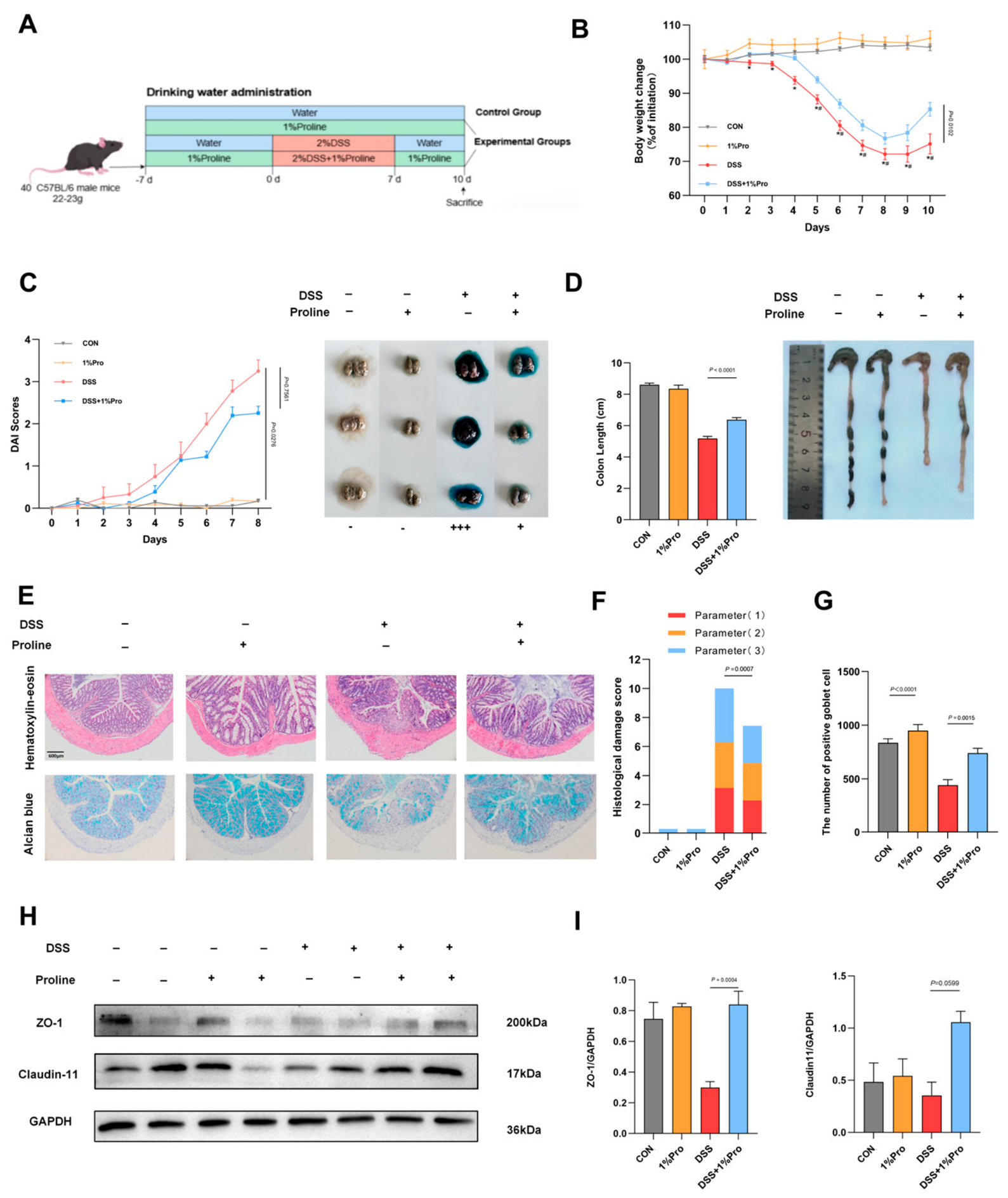
Figure 2.
Pro reprograms the colonic transcriptome and suppresses inflammatory signaling pathways. (A)Volcano plot showing differentially expressed genes (DEGs) between DSS and Pro groups (54 up-regulated, 75 down-regulated). (B) Principal Component Analysis (PCA) demonstrating distinct transcriptomic profiles. (C) Heatmap illustrating core candidate genes with significant differences among groups (P < 0.05). (D) KEGG pathway enrichment analysis of DEGs. (E–H) RT-qPCR validation of key DEGs involved in barrier function and immune modulation. Data are presented as mean ± SEM (n = 6).
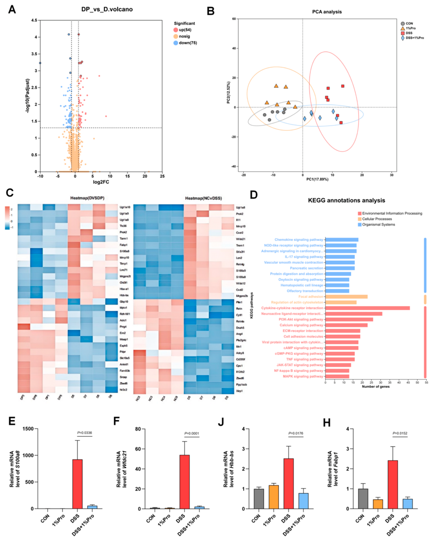
Figure 3.
Pro restores redox homeostasis, reinforces the intestinal barrier, and inhibits the NF-κB/STAT3 axis. (A-D) Assessment of oxidative stress markers: Malondialdehyde (MDA) levels, the GSH/GSSG ratio and Trolox-Equivalent Antioxidant Capacity(T-AOC) in colonic tissues. (E) Relative mRNA expression of pro-inflammatory cytokines and the anti-inflammatory cytokine. (F) Representative Western blots displaying phosphorylation levels of NF-κB and STAT3. (G, H) Quantitative analysis of p-NF-κB/NF-κB and p-STAT3/STAT3 ratios. Data are presented as mean ± SEM.
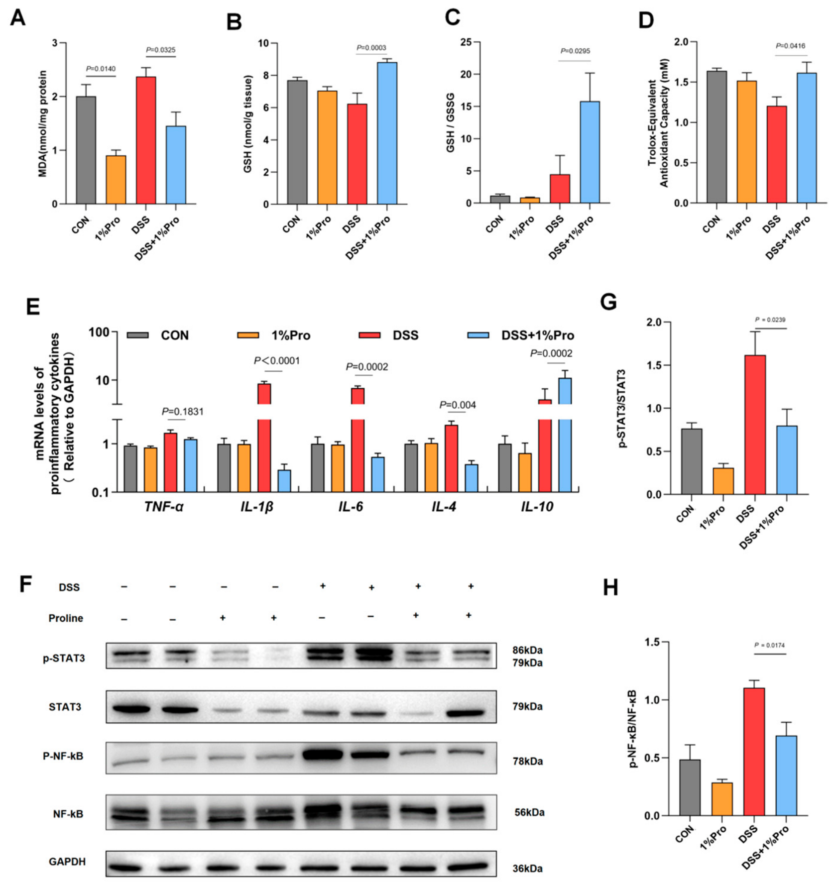
Figure 4.
Pro treatment reverses gut dysbiosis and enriches beneficial commensals. (A) Alpha-diversity analysis using Chao1 (richness) and Simpson (evenness) indices. (B) Principal Coordinate Analysis (PCoA) based on Bray-Curtis distances, showing structural separation between groups (ANOSIM R = 0.6992, P = 0.001). (C) Relative abundance of bacterial taxa at the phylum level. (D, E) Relative abundance of key genera altered by DSS and restored by Pro. (F) LEfSe analysis was performed to identify discriminative taxa among groups. Taxa with an LDA score >4.0 and P <0.05 were considered potential biomarkers.
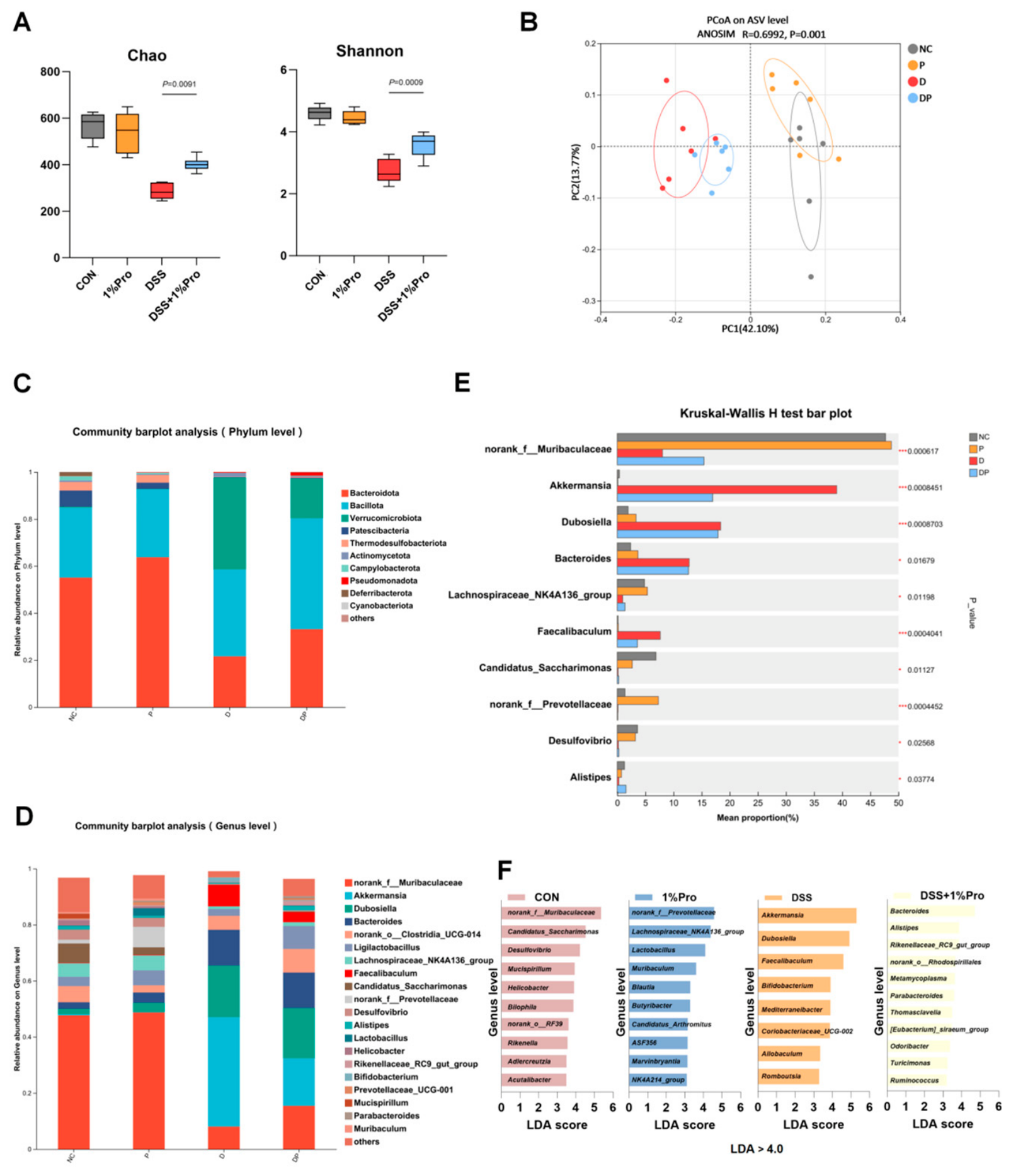
Figure 5.
Fecal Microbiota Transplantation (FMT) from Pro-treated donors recapitulates the protective phenotype. (A) Schematic of the FMT experimental workflow involving antibiotic-treated pseudo-germ-free mice. (B) Changes in body weight changes presented as a percentage of the initial weight (n = 9). (C) Colon length quantification and representative macroscopic images. (D) Histological evaluation of colonic tissues. Representative images of H&E staining and Alcian Blue (AB) staining for mucin assessment (Scale bar: 20 μm). (E) Histological scores quantifying epithelial damage and inflammation. (F) Goblet cell count detection: Calculate the absolute number of goblet cells per unit area. Data are presented as mean ± SEM.
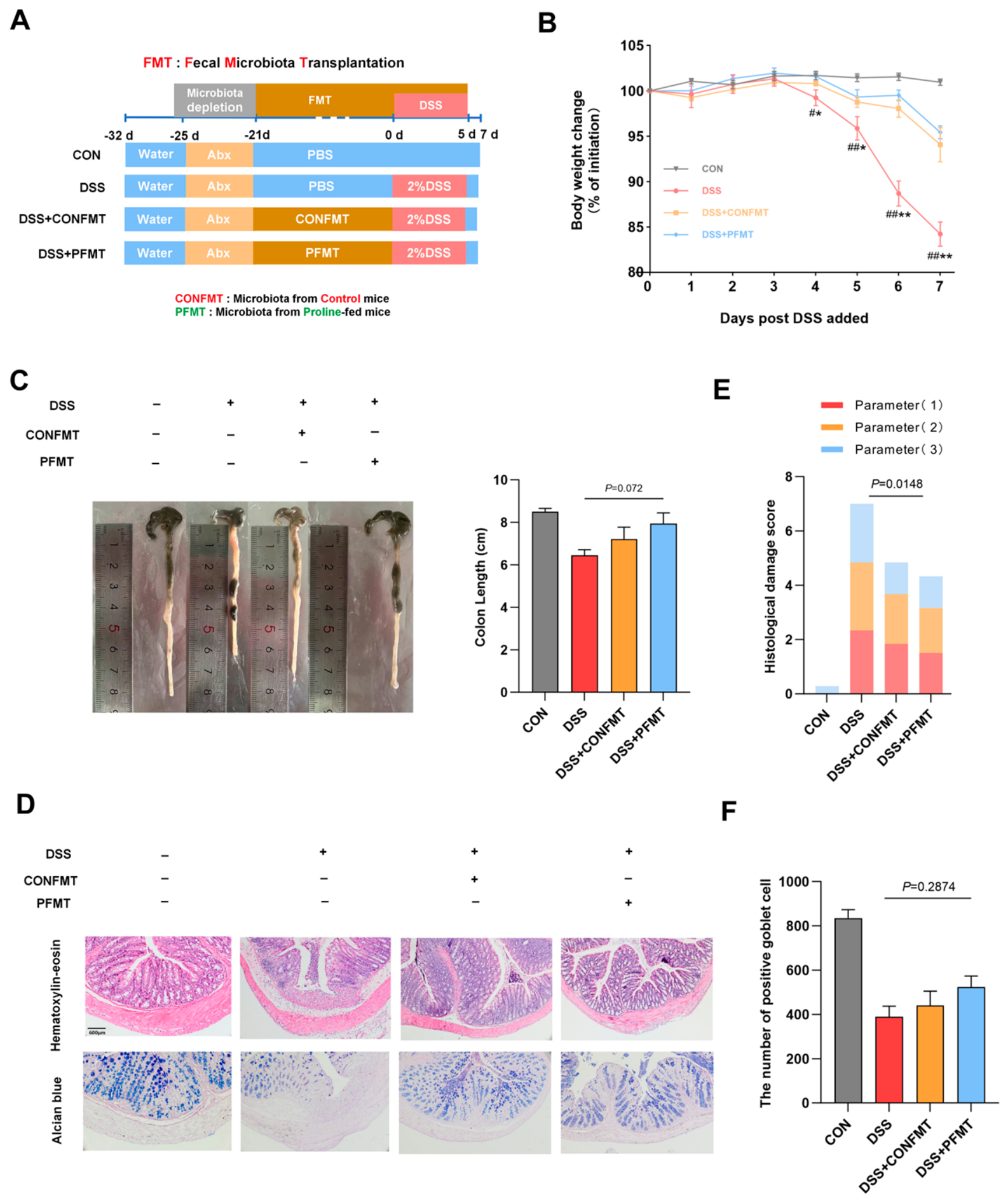
Figure 6.
FMT recipients acquire a restored gut microbial community structure resembling healthy controls. (A) PCoA plot of fecal microbiota from FMT recipients, showing clustering of the Pro-FMT group with the Control group (ANOSIM R = 0.9451, P = 0.001). (B) Alpha-diversity indices (Chao1 and Simpson) showing no significant differences in richness. (C) Taxonomic composition at the phylum level in recipient mice. (D, E) Relative abundance of specific genera. (F) LEfSe analysis identifying differentially abundant taxa in FMT recipients.
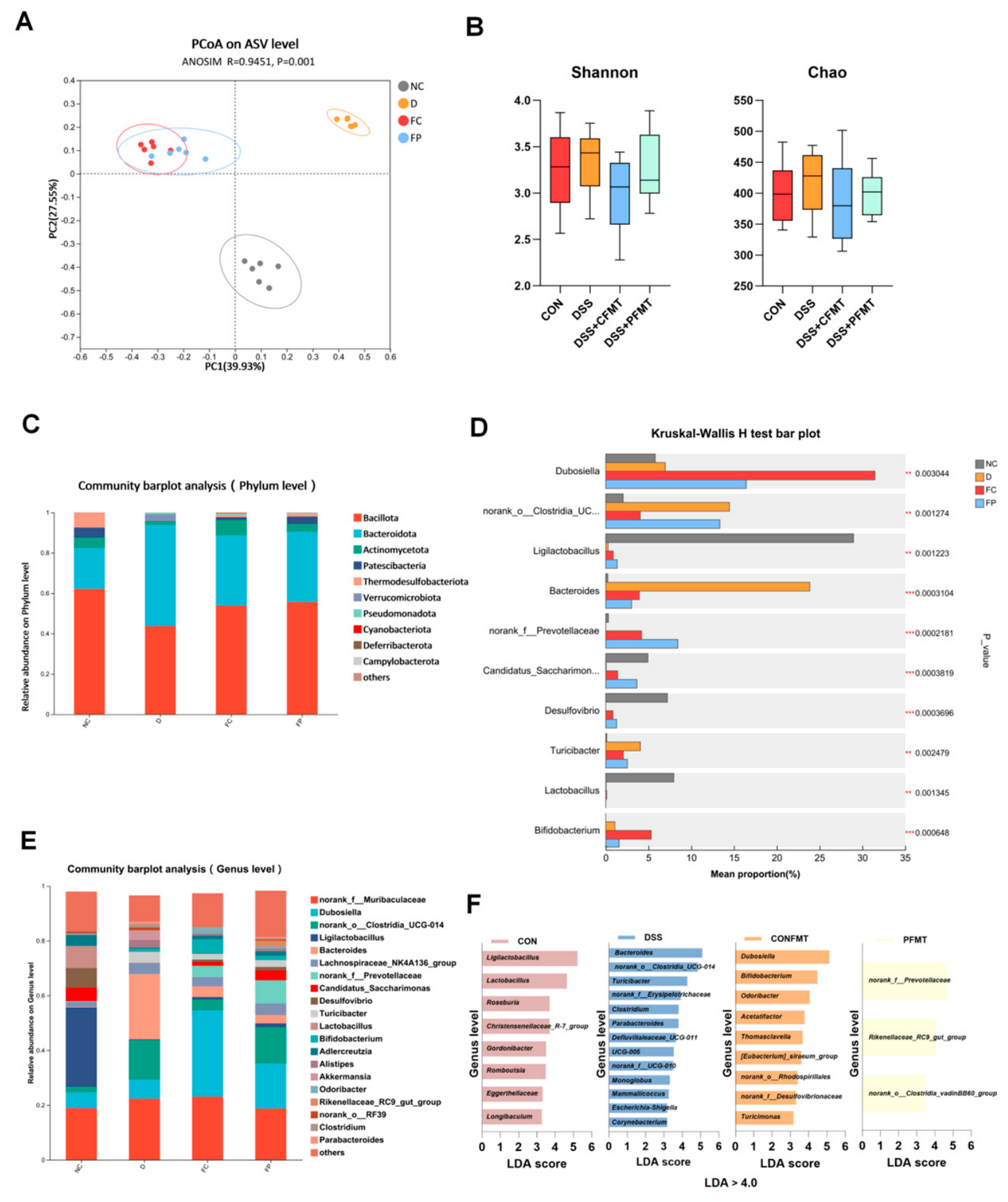
Figure 7.
Transferred microbiota is sufficient to restore barrier integrity and dampen inflammatory signaling. (A) Evaluation of general colonic inflammatory status. (B) Representative Western blot bands for the tight junction protein Occludin and Claudin-11 in FMT recipients. (C) Densitometric quantification of Occludin and Claudin-11 expression. (D) Western blot analysis of p-NF-κB and p-STAT3 in FMT recipients. (E) Quantification of phosphorylation ratios. Transplanting Pro-modulated microbiota significantly suppresses the activation of the NF-κB/STAT3 axis. Data are presented as mean ± SEM (n = 6).
Disclaimer/Publisher’s Note: The statements, opinions and data contained in all publications are solely those of the individual author(s) and contributor(s) and not of MDPI and/or the editor(s). MDPI and/or the editor(s) disclaim responsibility for any injury to people or property resulting from any ideas, methods, instructions or products referred to in the content. |
© 2026 by the authors. Licensee MDPI, Basel, Switzerland. This article is an open access article distributed under the terms and conditions of the Creative Commons Attribution (CC BY) license (http://creativecommons.org/licenses/by/4.0/).
Copyright: This open access article is published under a Creative Commons CC BY 4.0 license, which permit the free download, distribution, and reuse, provided that the author and preprint are cited in any reuse.