

Submitted:
09 October 2026
Posted:
10 October 2026
You are already at the latest version
Abstract
Fish scales are a calcium-rich biogenic waste source for hydroxyapatite (HAp), but the influence of processing route on material characteristics and calcium-release behaviour remains insufficiently understood. This study compared fish-scale-derived HAp produced by calcination and wet-chemical processing. Pretreated fish scales were processed by calcination at 1200°C (cnHAp) or acid-assisted ultrasonic extraction followed by alkaline precipitation (wnHAp). The materials were characterised using Fourier-transform infrared spectroscopy, X-ray diffraction, scanning electron microscopy–energy-dispersive X-ray spectroscopy and high-resolution transmission electron microscopy. Calcium concentrations were measured by inductively coupled plasma optical emission spectrometry at 0, 24 and 72 h and analysed using two-way mixed-design repeated-measures analysis of variance. Both materials exhibited characteristic HAp functional groups and diffraction features but differed in crystallographic ordering and morphology. cnHAp showed greater crystallographic ordering and a smaller mean particle size (42.41 ± 2.00 nm) than wnHAp (181.09 ± 3.64 nm), with Ca/P atomic ratios of 1.87 and 1.57, respectively. Calcium concentration differed significantly by processing group, time and their interaction (all p < .001), with cnHAp consistently showing higher concentrations. Processing route substantially influenced the physicochemical characteristics and temporal calcium-release behaviour of fish-scale-derived HAp.
Keywords:
fish scales
; hydroxyapatite
; nanohydroxyapatite
; calcium release
; calcium phosphate
; biomaterials
; ICP-OES
; calcination
; wet-chemical synthesis
; waste valorisation
1. Introduction
Hydroxyapatite (HAp; Ca₁₀(PO₄)₆(OH)₂) is a calcium phosphate ceramic widely investigated for biomedical applications because of its chemical and structural similarity to the mineral phase of human bone and teeth [1]. This biomimetic character contributes to its biocompatibility, bioactivity and osteoconductivity, supporting applications in bone grafts, implant coatings, tissue engineering, dental materials, drug delivery and regenerative medicine [2,3,4,5]. However, the intrinsic brittleness and limited mechanical properties of HAp restrict its use in applications involving substantial mechanical loading [6]. At the nanoscale, hydroxyapatite exhibits a high specific surface area and enhanced surface activity, which have contributed to the development of nanohydroxyapatite (nHAp) for applications including drug delivery, dental remineralisation, tissue engineering and bone regeneration [1,7,8,9].
Despite its functional potential, conventional nHAp production commonly relies on chemically derived precursors and synthesis routes that may involve costly reagents, substantial energy inputs and potentially hazardous chemicals [10]. These limitations have encouraged the development of more sustainable production strategies consistent with green chemistry and circular-economy principles [11]. In particular, the conversion of biogenic waste into calcium phosphate biomaterials offers a dual benefit by reducing dependence on conventional raw materials while valorising otherwise underutilised waste streams. Marine and agricultural wastes, including eggshells, fish scales, fish bones and oyster shells, contain calcium-rich mineral fractions that can be converted into HAp through thermal, chemical and combined processing routes [12,13].
Among these resources, fish scales represent a promising precursor because of their mineral content and widespread availability. Studies have demonstrated the feasibility of producing nanoscale HAp from fish scales using both thermal and wet-chemical approaches [14,15,16]. The resulting materials may retain trace elements such as magnesium, sodium and strontium derived from the biological precursor, potentially contributing to compositional differences from chemically synthesised HAp [15,17]. More importantly, the processing route can substantially modify the structural characteristics of the recovered material. Thermal treatment promotes transformation of the mineral fraction towards crystalline apatite, may also form [15,17,18]. Conversely, wet-chemical precipitation can produce poorly crystalline or amorphous calcium phosphate under comparatively mild conditions, with ultrasonication capable of influencing nucleation, particle growth and agglomeration [16]. Thus, processing history is an important determinant of the final physicochemical state of fish-scale-derived nHAp.
The structural characteristics established during processing are directly relevant to the aqueous behaviour of HAp. Crystallinity is a major determinant of dissolution, with highly crystalline HAp generally exhibiting greater chemical stability and lower dissolution than poorly crystalline or amorphous calcium phosphate phases [19,20,21]. Particle dimensions and surface characteristics may further influence dissolution through their effects on surface area, surface energy and accessibility of reactive sites, although nanoscale size effects are not governed by surface area alone [22]. These factors determine the extent to which calcium and phosphate ions become available in the surrounding medium. Consequently, processing-induced differences in structure may translate into distinct calcium-release profiles. This is particularly relevant for biomedical applications because ion release contributes to mineralisation, remineralisation and bioresorption, whereas excessive dissolution may reduce material persistence. Conversely, excessive stability may restrict ionic availability. The functional significance of this balance is supported by evidence that differently processed HAp materials can exhibit distinct dissolution, calcium and phosphorus release, bioresorption and biomineralisation behaviour [23].
Although the physicochemical characteristics of waste-derived nHAp have received increasing attention, the relationship between processing route and the time-dependent calcium release of fish-scale-derived nHAp remains insufficiently established. Previous work has shown that precursor origin can influence the crystallinity, particle size, morphology, Ca/P ratio and remineralisation performance of nHAp [14], while investigations of fish-scale-derived HAp have established the effects of calcination conditions on crystallinity, crystallite size and phase composition [15,17,18]. However, these studies provide limited insight into how contrasting processing routes influence the temporal release of calcium when the resulting materials are exposed to an aqueous environment. Resolving this relationship is important because calcium release provides a measurable indication of the dissolution behaviour arising from differences in material structure and processing history.
The present study therefore investigated the time-dependent calcium-release behaviour of fish-scale-derived nHAp over 72 h. A thermally processed crystalline cnHAp preparation was compared with a wet-chemically precipitated amorphous wnHAp preparation, alongside a calcium phosphate reference material. Calcium released into the aqueous medium was quantified at predetermined intervals using inductively coupled plasma optical emission spectrometry (ICP-OES). Calcium concentration was used as the experimental measure of calcium release and as an indicator of dissolution behaviour. The study specifically examined whether the contrasting processing routes produced different temporal calcium-release profiles in fish-scale-derived nHAp.
2. Results
2.1. Fourier-Transform Infrared (FTIR) Spectroscopy Analysis
Fourier-transform infrared (FTIR) spectroscopy was used to identify the functional groups and vibrational modes present in samples (a), (b), and (c) over the investigated wavenumber range (Figure 1). Spectra (a) and (b) exhibited the characteristic vibrational features of hydroxyapatite (HAp). The prominent absorption bands in the approximately 1,030–1,100 cm⁻¹ region were assigned to the ν₃ asymmetric stretching vibrations of PO₄³⁻, while the band near 960 cm⁻¹ corresponded to the ν₁ symmetric stretching mode. The bands in the 560–610 cm⁻¹ region were attributed to the ν₄ bending vibrations of PO₄³⁻, confirming the characteristic phosphate groups of the apatite structure [24].
Additional bands in the approximately 1,410–1,450 cm⁻¹ region and near 870–880 cm⁻¹ were assigned to carbonate (CO₃²⁻) vibrations. These features are characteristic of carbonate-containing apatite and have been reported specifically in fish-scale-derived HAp [25,26]. The presence of carbonate is consistent with the naturally occurring carbonate substitution commonly associated with biological apatite. Where the characteristic carbonate bands are present without additional structural evidence, it is preferable to describe the material as carbonate-containing HAp rather than definitively assigning B-type substitution solely from FTIR.
A band near 3,570 cm⁻¹ can be assigned to the stretching vibration of structural OH⁻, whereas the broad band around 3,200–3,600 cm⁻¹ and the absorption near 1,630–1,650 cm⁻¹ are associated primarily with adsorbed water [27]. The more clearly resolved phosphate bands in spectrum (b) indicate greater definition of the apatite vibrational features; however, differences in crystallinity should be assessed primarily using XRD, as FTIR band resolution provides qualitative rather than definitive quantitative evidence of crystallinity.
In contrast, spectrum (c), representing the commercially purchased calcium dihydrogen phosphate (CAH), Ca(H₂PO₄)₂, exhibited vibrational features characteristic of the H₂PO₄⁻ phosphate precursor rather than organic components. The bands in the approximately 950–1100 cm⁻¹ region were associated with P–O stretching vibrations, while additional bands around 1200 cm⁻¹ can be attributed to P–O–H in-plane bending. Lower-wavenumber bands are associated with phosphate bending vibrations.
2.2. X-ray Diffraction (XRD) Analysis
X-ray diffraction (XRD) patterns were acquired over a 2θ range of 20–90° to investigate the phase composition and crystallographic structure of samples (a), (b), and (c) (Figure 2). Pattern (a) exhibited sharp and highly intense diffraction maxima, indicating a well-crystallised structure with prominent reflections corresponding to the identified crystallographic planes. Pattern (b) displayed the characteristic diffraction reflections of hexagonal hydroxyapatite (HAp; JCPDS No. 09-0432), including the reflections associated with the (002), (211), (112), and (300) planes at approximately 25.9°, 31.7–31.8°, 32.1–32.2°, and 32.8–32.9°, respectively. These reflections are consistent with the reference diffraction pattern of crystalline HAp.
The comparatively broad diffraction peaks and lower maximum intensity observed in pattern (b) are consistent with reduced crystallite dimensions and/or increased structural disorder, characteristic of nanocrystalline HAp. The characteristic HAp reflections are consistent with those previously reported for biologically derived HAp. In a previous study of eggshell-derived nanohydroxyapatite, Onwubu, et al. [28] similarly used XRD to confirm the formation of nanocrystalline HAp and identified the characteristic reflections associated with the apatite phase.
Pattern (c) also exhibited the characteristic reflections of hexagonal HAp, including the (002), (211), (112), and (300) planes. Compared with pattern (b), pattern (c) showed sharper and better-resolved diffraction maxima with higher intensities, indicating greater structural ordering and larger coherent crystallite dimensions following calcination. The absence of additional prominent reflections attributable to detectable secondary crystalline phases further supports the formation of predominantly HAp.
The degree of crystallinity (Xc) was calculated using the empirical relationship based on the intensity of the (300) reflection (I₃₀₀) and the intensity of the valley between the (112) and (300) reflections (V₁₁₂/₃₀₀) (Equation 1). This approach is widely used for assessing the relative crystallinity of HAp from XRD patterns. Sample (a) demonstrated the highest degree of crystalline ordering (Xc≈95%), as reflected by its sharp and intense diffraction maxima. Sample (c) exhibited a well-crystallised HAp structure with a crystallinity of (Xc≈72%), whereas sample (b) showed a comparatively lower crystallinity of (Xc≈38%), consistent with its broader diffraction peaks and smaller coherent crystallite dimensions.
2.3. Morphological Characterization
The surface morphology, microstructure, and elemental composition of the synthesised hydroxyapatite (HAp) samples were evaluated using scanning electron microscopy (SEM) coupled with energy-dispersive X-ray spectroscopy (EDX) and elemental mapping (Figure 3). The high-magnification SEM micrographs (10.00 kX, scale bar (1 μm) revealed distinct morphological differences between samples (a) and (b). Micrograph (a) exhibited an irregular and highly agglomerated surface morphology consisting of clustered, non-uniform particles separated by porous interstitial regions. In contrast, micrograph (b) displayed a comparatively denser and more defined granular network composed of interconnected quasi-spherical to submicron grains, with discernible grain boundaries and localised micro-flake structures. Such morphological variability is consistent with previous reports of fish-scale-derived HAp, which have described distinct morphologies, including flat-plate nanocrystals, depending on the processing approach used for HAp extraction and preparation [29].
2.4. Elemental Composition and Mapping
The corresponding EDX spectra confirmed that calcium (Ca), phosphorus (P), and oxygen (O) were the principal elements detected in both samples, supporting the formation of a calcium-phosphate-based HAp phase. Carbon (C) was also detected, together with minor signals corresponding to sodium (Na) and magnesium (Mg). This elemental profile is consistent with recent characterisation of fish-scale-derived HAp, in which Ca, P, and O were identified as the principal elements, while Na, Mg, and C were detected as additional components [30]. The detection of Na and Mg is particularly relevant to biologically derived HAp because these elements may originate from the natural mineral composition of fish scales and may substitute for Ca²⁺ within the apatite structure. However, their detection by EDX alone does not establish their specific crystallographic location or confirm lattice substitution. Evidence from complementary techniques such as XPS has supported the incorporation of Na⁺ and Mg²⁺ into Ca sites in fish-scale-derived HAp, although Na may also be introduced during alkaline processing through the use of NaOH [30].
The EDX elemental mapping panels for wnHAp and cnHAp demonstrated a broadly continuous spatial distribution of Ca, P, and O throughout the analysed regions. The strong spatial association between Ca and P supports the presence of a calcium-phosphate phase, while the relatively continuous distribution of O is consistent with its contribution to the phosphate-containing apatite structure. Comparable elemental mapping of fish-scale-derived HAp has demonstrated strong spatial correlation between Ca and P, with O distributed relatively uniformly across the analysed regions, providing spatial evidence for the homogeneous distribution of the principal constituent elements [30]. The identification of HAp as the predominant crystalline phase, however, is supported by the corresponding XRD analysis rather than by EDX mapping alone.
The presence of carbon requires cautious interpretation. Although carbon may occur in biologically derived HAp through carbonate incorporation, EDX alone cannot distinguish carbonate-associated carbon from adventitious carbon or carbon originating from the carbon-based substrate used during SEM analysis. In a recent comparative study of fish-scale-derived HAp, Bin Mobarak, Chowdhury and Ahmed [30] similarly reported that the carbon detected by EDX could originate from atmospheric CO₂ or the carbon tape used as the sample substrate. Therefore, the EDX carbon signal should not, by itself, be interpreted as definitive evidence of carbonate substitution within the HAp lattice. Rather, the presence of carbonate-containing HAp is more appropriately supported by the corresponding FTIR bands, particularly the carbonate vibrations in the approximately 1410–1450 and 870–880 cm⁻¹ regions. Fish-scale-derived HAp has previously been shown to exhibit these characteristic carbonate bands, with FTIR providing evidence for carbonate substitution in the apatite structure [29].
The quantitative EDX results (Table 2) further demonstrated that Ca and P were the predominant elements in both wnHAp and cnHAp, confirming their calcium-phosphate-rich composition. Minor Mg was detected at 0.40 wt% in wnHAp and 0.36 wt% in cnHAp, indicating that a small proportion of magnesium was retained following processing. The relatively similar Mg contents between the two samples suggest that calcination produced only a limited change in the detectable Mg concentration. The presence of minor Na and Mg is consistent with the naturally derived origin of the material, as biologically sourced apatites commonly contain trace ionic constituents that may substitute for Ca²⁺ within the apatite structure [30].
The calculated Ca/P atomic ratios were 1.57 for wnHAp and 1.87 for cnHAp. The wnHAp value was below, whereas the cnHAp value was slightly above, the theoretical Ca/P ratio of 1.67 for stoichiometric HAp. These deviations indicate compositional non-stoichiometry and may reflect ionic substitutions, including carbonate, Mg²⁺ and Na⁺, as well as differences in calcium–phosphate composition associated with the biological precursor and processing conditions. Such compositional variability is characteristic of biogenic HAp, in which ionic substitutions and processing conditions can produce Ca/P ratios that differ from the theoretical stoichiometric value [30,31]. Thus, the EDX findings demonstrate deviations from ideal stoichiometric HAp composition; however, EDX alone cannot establish phase purity or identify secondary crystalline phases. The corresponding XRD patterns therefore provide the appropriate evidence for determining whether the samples comprise predominantly HAp or contain additional crystalline phases.
2.5. HRTEM Morphology and Particle-Size Distribution
HRTEM revealed distinct morphological differences between the water-bath/ultrasonication-extracted nanohydroxyapatite (wnHAp) and calcined nanohydroxyapatite (cnHAp) (Figure 4). The wnHAp exhibited relatively broad, irregular and overlapping structures with heterogeneous contrast, indicative of an aggregated and less-defined morphology (Figure 4a). In contrast, cnHAp predominantly exhibited elongated rod- to needle-like nanoparticles with more clearly defined particle boundaries (Figure 4b). Similar elongated morphologies have been reported for fish-scale-derived HAp, with HRTEM demonstrating rod-shaped nanoparticles approximately 50–100 nm in length and 8–12 nm in diameter [32].
The observed morphological differences are consistent with the different processing conditions. The water-bath/ultrasonication treatment promotes extraction and dispersion of the mineral phase, whereas calcination facilitates thermal decomposition of organic constituents and reorganisation of the calcium-phosphate phase. The observed morphological differences between wnHAp and cnHAp are consistent with the findings of Eknapakul, et al. [33], who demonstrated that pretreatment and calcination conditions can markedly alter the morphology and particle size of fish-scale-derived HAp. Their study showed that different pretreatments produced distinct particle morphologies, while increasing calcination temperature promoted particle growth. Their calcined samples exhibited either rod-like or agglomerated morphologies depending on the pretreatment route, confirming the strong influence of processing conditions on HAp structure.
Particle-size distributions were evaluated by Gaussian fitting of the size-frequency histograms (Figure 5). The cnHAp exhibited a relatively narrow distribution, predominantly between 20 and 70 nm, with a mean particle size of 42.41 ± 2.00 nm (Figure 5b). In contrast, wnHAp displayed a broader distribution extending from approximately 70 to 300 nm, with a mean particle size of 181.09 ± 3.64 nm (Figure 5a). Thus, wnHAp had a mean particle size approximately 4.27 times that of cnHAp, indicating substantially finer particles after calcination.
The particle size obtained for cnHAp is consistent with the nanoscale dimensions reported for fish-scale-derived HAp. Eknapakul, Kuimalee, Sailuam, Daengsakul, Tanapongpisit, Laohana, Saenrang, Bootchanont, Khamkongkaeo and Yimnirun [33] reported particle sizes ranging from approximately 30 to 112 nm depending on the pretreatment and calcination conditions, while Athinarayanan, Periasamy and Alshatwi [32] reported rod-shaped HAp nanoparticles approximately 50–100 nm in length and 8–12 nm in diameter. The present cnHAp therefore falls within the nanoscale range reported for fish-scale-derived HAp. The broader particle-size distribution of wnHAp may be associated with greater particle aggregation and the retention of matrix-associated components following the lower-temperature extraction process. In contrast, the narrower distribution observed for cnHAp suggests a more uniform population of nanoscale particles following thermal treatment. Similar processing-dependent changes in particle size and morphology have been reported for fish-scale-derived HAp [33].
2.6. In Vitro Calcium-Release Behaviour
The calcium release behaviour is given in Table 4.3. The two-way mixed-design repeated-measures ANOVA demonstrated significant effects of Time, F(2, 12) = 1083.09, p < .001, partial η² = .995, and Group, F(2, 6) = 11,182.85, p < .001, partial η² = 1.000. Importantly, the Time × Group interaction was also significant, F(4, 12) = 196.85, p < .001, partial η² = .985, indicating that changes in calcium concentration over time differed significantly among the three groups. Calcium concentration decreased across time in the calcium phosphate group and cnHAp group, whereas the wnHAp group decreased from 20.97 at 0 h to 5.69 at 24 h before increasing to 11.57 at 72 h. Bonferroni-adjusted comparisons showed significant differences between time points for all groups except the 0–24 h comparison for calcium phosphate (p = .282).
Figure 6 provides a graphical representation of the repeated mixed-model findings, showing clear material- and time-dependent differences in dissolution. cnHAp consistently exhibited the highest dissolution, with its 95% confidence intervals remaining distinct from those of CAH and wnHAp across the monitoring period. The figure also demonstrates the different temporal trajectories: cnHAp declined progressively but stabilised after 24 h, CAH showed a delayed decline, while wnHAp decreased markedly at 24 h before a modest increase at 72 h.
3. Discussion
The present study demonstrates that processing route is a major determinant of the physicochemical characteristics and time-dependent calcium-release behaviour of fish-scale-derived hydroxyapatite (HAp). The central finding was the significant interaction between material group and incubation time, indicating that the three calcium-phosphate materials did not follow a common release trajectory. This distinction is important because it shows that conversion of fish-scale waste into HAp is not simply a matter of recovering calcium phosphate; the processing history determines structural and morphological attributes that subsequently influence its behaviour in an aqueous environment. Previous studies of fish-scale-derived HAp similarly demonstrate that pretreatment and thermal-processing conditions can substantially influence crystal structure, morphology and particle dimensions [32,34], with Eknapakul, Kuimalee, Sailuam, Daengsakul, Tanapongpisit, Laohana, Saenrang, Bootchanont, Khamkongkaeo and Yimnirun [33] additionally demonstrating effects on structural defects.
The spectroscopic and diffraction findings collectively indicate that both extraction routes produced calcium-phosphate materials with HAp characteristics, but with different degrees of structural organisation. The sharper XRD reflections observed for cnHAp indicate greater crystallographic ordering than the broader reflections of wnHAp. This distinction is consistent with the established influence of thermal treatment on apatite crystallinity. In fish-scale-derived HAp, processing conditions can modify not only crystallinity but also defects, phase composition and morphology; Eknapakul, Kuimalee, Sailuam, Daengsakul, Tanapongpisit, Laohana, Saenrang, Bootchanont, Khamkongkaeo and Yimnirun [33], for example, demonstrated that different pretreatment procedures produced markedly different HAp structures and morphologies, including differences in phase composition after calcination. Similar processing-dependent differences have been reported for HAp obtained from different fish species. For example, acid–alkali treatment followed by high-temperature calcination produced highly crystalline HAp from fish scales, but β-tricalcium phosphate (β-TCP) was also identified as a secondary phase in both fish-derived materials [30]. Comparable phase-dependent effects of pretreatment and calcination have also been reported for Nile tilapia scales, where pretreatment conditions influenced whether predominantly HAp or biphasic HAp/β-TCP was obtained [33]. Thus, the greater crystallographic order of cnHAp should be interpreted as a consequence of the overall calcination–milling route rather than calcination in isolation.
The morphological results reinforce this processing effect. cnHAp exhibited a comparatively fine and defined nanoscale morphology, with a mean TEM-derived particle size of 42.41 ± 2.00 nm, whereas wnHAp consisted of larger, irregular and more agglomerated particles with a mean size of 181.09 ± 3.64 nm. This difference is consistent with previous reports showing that fish-scale pretreatment and thermal processing can substantially alter HAp particle morphology and dimensions [33,34]. The present findings should nevertheless be interpreted in relation to the complete processing sequence because cnHAp underwent planetary ball milling after calcination. The smaller particle population therefore cannot be attributed to calcination alone. Moreover, TEM particle size should not be equated with XRD crystallite size, since the former describes observable particles or agglomerates whereas the latter reflects coherently diffracting domains.
The EDX results further indicate that the two materials were compositionally different. The Ca/P ratio of wnHAp (1.57) was below the theoretical value of approximately 1.67 for stoichiometric HAp, whereas the ratio for cnHAp (1.87) was above it. Accordingly, only wnHAp can reasonably be described as having a calcium-deficient composition; the higher ratio of cnHAp instead indicates relative calcium enrichment. Importantly, EDX alone cannot establish the presence or proportion of specific calcium-phosphate phases because the measurement is semi-quantitative and sensitive to local composition, particle morphology and sampling position. This distinction is particularly relevant for naturally derived HAp, which may contain ionic substitutions and minor phases. Fish-scale-derived HAp has been reported to contain Na and Mg in addition to Ca and P, while secondary β-tricalcium phosphate (β-TCP) has also been identified in some calcined fish-scale preparations [30,33]. The present EDX findings support compositional differences between wnHAp and cnHAp, while XRD was used to establish the crystalline phases present in the current materials.
The calcium-release experiment provides the strongest evidence that these physicochemical differences translated into distinct aqueous behaviour. cnHAp maintained substantially higher calcium concentrations than both the calcium-phosphate reference and wnHAp at all measured time points, whereas wnHAp exhibited a pronounced decrease at 24 h followed by partial recovery at 72 h. The significant time × group interaction confirms that the magnitude and direction of temporal change depended on the material. This pattern is consistent with the broader literature demonstrating that HAp dissolution and ion release are governed by an interplay of crystal structure, particle characteristics, surface area, solution chemistry and secondary-phase formation rather than by a single material parameter. For example, Jang, Pyun and Chang [23] demonstrated using ICP-OES that calcined and uncalcined HAp exhibited distinct calcium-release profiles during in-vitro dissolution, with the uncalcined material showing a higher calcium dissolution rate. The authors also identified differences in surface area and particle size as factors influencing dissolution behaviour.
The consistently higher calcium concentration associated with cnHAp should therefore not be interpreted as evidence that its higher crystallinity necessarily caused greater dissolution. Such an interpretation would conflict with established dissolution principles. Highly crystalline HAp generally exhibits greater structural stability, whereas lower crystallinity, smaller crystallites and certain ionic substitutions can increase susceptibility to dissolution. Sun, Berndt, Khor, Cheang and Gross [20], for example, reported an inverse relationship between HAp crystallinity and dissolution in plasma-sprayed materials, while Tang, Wang and Nancollas [22] demonstrated that nanoscale HAp dissolution is additionally controlled by crystallite size and surface-energy effects. The present cnHAp therefore appears to represent a more complex case in which its relatively high crystallinity coexists with a substantially finer particle population and potentially different surface characteristics. Because particle size, crystallinity, surface area, defects and composition were altered simultaneously by processing, the current experiment cannot isolate which of these variables exerted the dominant influence on calcium release.
The non-monotonic behaviour of wnHAp provides further evidence that solution calcium concentration should not be interpreted as a direct measure of irreversible material dissolution. The sharp decline from 20.97 at 0 h to 5.69 at 24 h, followed by an increase to 11.57 at 72 h, may reflect a combination of initial calcium release and subsequent removal of dissolved calcium from the aqueous phase through precipitation, adsorption or incorporation into secondary calcium-containing phases. Comparable behaviour has been observed in HAp dissolution studies in which calcium concentration initially increased and subsequently declined as released calcium participated in the formation of secondary products [23]. Consequently, the present results are best interpreted as time-dependent calcium availability in the test medium, rather than definitive evidence of the intrinsic dissolution rate of each material. Confirmation of the underlying mechanism would require simultaneous phosphate measurements, pH monitoring and characterisation of the solid phase after immersion.
The differences between cnHAp and wnHAp are also consistent with the principle that processing-induced changes in surface area and particle architecture can influence ion exchange and dissolution in the surrounding medium. HAp nanoparticles with greater specific surface area have been shown to exhibit enhanced calcium-ion release under controlled immersion conditions, particularly at very small particle sizes and high specific surface areas [35]. However, the relationship between particle size and dissolution is not necessarily linear, as nanoscale dissolution can become influenced by surface-energy effects and interfacial processes that may inhibit dissolution at sufficiently small crystallite sizes [22]. Thus, the approximately fourfold difference in mean particle size between wnHAp and cnHAp provides a plausible contributor to their different release profiles, but it should be considered alongside crystallinity and composition rather than treated as an independent explanation.
The findings also extend the existing literature on fish-scale-derived HAp by linking processing-dependent structural characteristics with temporal calcium behaviour. Much of the published work on fish-scale HAp has concentrated on extraction efficiency, crystallinity, phase identification and morphology [30,33,34], whereas comparatively fewer studies directly relate these characteristics to time-resolved ion release. This provides the principal scientific contribution of the present study: the processing route produced distinguishable structural and morphological states, and these states were associated with significantly different calcium-concentration trajectories over 72 h. The study therefore moves beyond demonstrating that fish scales can serve as a calcium-phosphate precursor and provides evidence that the resulting material properties are relevant to its subsequent ionic behaviour.
The biomedical implications should nevertheless remain appropriately bounded. Calcium release is an important physicochemical characteristic of calcium-phosphate biomaterials, but it does not independently establish bioresorbability, osteogenic activity, remineralisation or clinical performance. Such conclusions require complementary evidence from phosphate release, solution pH, post-immersion phase analysis, surface transformation and biological testing. The present findings therefore establish a physicochemical basis for further evaluation rather than direct evidence of biological efficacy. In particular, future studies should employ physiologically relevant media and temperature, quantify both calcium and phosphate, monitor pH throughout incubation, determine cumulative ion release, and characterise the recovered solids after exposure. Increasing the number of independent specimens would also strengthen estimation of biological and material variability.
4. Materials and Methods
4.1. Preparation of Fish Scale-Derived Nanohydroxyapatite
Fresh fish scales were obtained from local fish-processing facilities, thoroughly washed, and treated with 1 M NaOH at 80 °C for 2 h. The scales were rinsed with distilled water to neutral pH and dried at 80 °C for 24 h. Two hydroxyapatite preparations were subsequently produced. For calcined hydroxyapatite (cnHAp), the pretreated scales were calcined at 1200 °C for 3 h at a heating rate of 5 °C/min. The resulting calcined material was allowed to cool naturally to room temperature and subsequently milled using a planetary ball mill to obtain nanosized hydroxyapatite particles. The powder was sieved through a 50 μm mesh and stored in airtight containers for further use. For wet-chemically derived hydroxyapatite (wnHAp), 10 g of dried scales were suspended in 200 mL of 1 M HCl and ultrasonicated at 40 kHz and 0.4 kW at 50 °C for 1 h following the procedure reported by Swamiappan, Xie, Lu and Peng [16]. The suspension was centrifuged, and the supernatant was adjusted to pH 12 using 6 M NaOH under continuous stirring, followed by ultrasonication for 45 min at room temperature. The precipitate was washed to neutral pH, filtered, dried at 100 °C for 48 h, and ground.
4.2. Physicochemical and Morphological Characterisation of the synthesised nHAp
The physicochemical, crystalline, elemental, and morphological characteristics of the synthesised materials were evaluated using attenuated total reflectance Fourier-transform infrared spectroscopy (ATR-FTIR), X-ray diffraction (XRD), scanning electron microscopy coupled with energy-dispersive X-ray spectroscopy (SEM-EDX), and high-resolution transmission electron microscopy (HRTEM). These complementary techniques were employed to identify the functional groups, crystalline phases, elemental composition, calcium-to-phosphorus (Ca/P) ratio, surface morphology, and particle dimensions of the synthesised materials.
Functional groups were identified using attenuated total reflectance Fourier-transform infrared spectroscopy (ATR-FTIR; PerkinElmer) over the wavenumber range of 400–4500 cm⁻¹ at a spectral resolution of 4 cm⁻¹. Prior to sample analysis, a background scan was performed to eliminate atmospheric interference and ensure spectral accuracy. Each specimen was subsequently placed on the ATR crystal platform and scanned over the wavenumber range. The resulting spectra were examined for characteristic absorption bands corresponding to the chemical bonds and functional groups associated with the constituent phases of the synthesised materials.
The crystalline structure and phase composition were determined using X-ray diffraction (XRD; Bruker D8 Advance AXS) with Cu Kα radiation (λ = 1.5406 Å). The instrument was operated at 40 kV and 40 mA, and diffraction patterns were collected over the experimental 2θ range of 10°–90°. A scan rate corresponding to a 0.5 s step acquisition time was employed throughout the analysis. The resulting diffractograms were evaluated based on the characteristic diffraction peaks to identify the crystalline phases present and assess the structural characteristics of the synthesised materials.
Surface morphology and elemental composition were examined using field scanning electron microscopy coupled with energy-dispersive X-ray spectroscopy (FESEM-EDX). FESEM micrographs were used to evaluate the surface morphology, particle arrangement, and microstructural characteristics of the samples. EDX analysis was performed to identify and quantify the major constituent elements. For the hydroxyapatite-containing samples, the calcium-to-phosphorus (Ca/P) atomic ratio was calculated from the elemental concentrations obtained by EDX using the following equation:
The calculated Ca/P ratios were used to assess the elemental stoichiometry of the synthesised hydroxyapatite relative to the theoretical Ca/P atomic ratio of 1.67 for stoichiometric hydroxyapatite.
Particle morphology and dimensions were further characterised using high-resolution transmission electron microscopy (HRTEM). The resulting micrographs were analysed using ImageJ software after calibration against the scale bar provided on each micrograph. Individual matrix domains and HAp nanorods were measured directly from the calibrated HRTEM images. A total of 20 individual measurements were obtained for each evaluated sample. For wnHAp, the matrix domain sizes ranged from 95 to 270 nm, with a mean size of 176.35 ± 49.80 nm and a median of 176.50 nm. For cnHAp, the HAp nanorod lengths ranged from 22 to 65 nm, with a mean length of 43.35 ± 12.71 nm and a median of 43.00 nm. The measured dimensions were used to describe the particle-size characteristics and morphological distribution observed in the HRTEM micrographs.
4.3. ICP-OES Calibration and Calcium Quantification
Calcium was quantified using inductively coupled plasma optical emission spectrometry (ICP-OES; Shimadzu ICPE-9820). Five candidate calcium emission wavelengths were evaluated before sample analysis. Analytical performance was assessed using the calibration correlation coefficient (r), emission intensity, limit of detection (LOD), limit of quantification (LOQ), and repeatability. The wavelength providing the most appropriate overall analytical performance was selected for subsequent calcium quantification (Table 1).
Calcium-release behaviour was evaluated over 72 h. Equal masses of each material were immersed in a 10% (v/v) DMSO aqueous medium at a solid-to-liquid ratio of 1:200 (w/v). For each experimental unit, 100 mg of material was added to 20 mL of the DMSO-containing medium and maintained at room temperature under continuous agitation. Calcium release was evaluated at 0, 24, and 72 h. At each sampling point, the suspensions were centrifuged to separate the solid phase, and the collected supernatants were subsequently filtered before analysis. Calcium concentrations in the filtrates were determined using inductively coupled plasma–optical emission spectrometry (ICP-OES). Three independent experimental units were analysed for each material at each sampling point.
4.4. Statistical Analysis
Calcium concentration was analysed using a two-way mixed-design repeated-measures analysis of variance (ANOVA). Time (0, 24, and 72 h) was specified as the within-subjects factor, while experimental Group (CAH, cnHA, and wnHAp) was specified as the between-subjects factor. The analysis evaluated the main effects of Time and Group and their Time × Group interaction on calcium concentration. Mauchly’s test of sphericity was used to assess the sphericity assumption. Because the assumption was not violated (W = .357, χ²(2) = 5.15, p = .076), the sphericity-assumed results were reported. Where significant effects were identified, Bonferroni-adjusted pairwise comparisons were performed to determine differences between time points and experimental groups. Statistical significance was set at p < .05, and partial eta squared (ηp²) was used to quantify effect size.
5. Conclusions
This study demonstrated that processing route substantially influenced the physicochemical characteristics and time-dependent calcium-release behaviour of fish-scale-derived hydroxyapatite. The calcination–milling route produced a more highly crystalline and finer nanoscale material, whereas the wet-chemical route produced a comparatively less ordered and larger, more heterogeneous material. These differences were accompanied by distinct calcium-concentration profiles over 72 h, with a significant interaction between processing group and time. The findings indicate that calcium availability from fish-scale-derived calcium phosphate is governed by the combined effects of processing-dependent structure, morphology and composition rather than by crystallinity alone. The study further demonstrates the potential of fish-scale waste as a source of value-added calcium-phosphate biomaterials while highlighting the importance of controlling processing conditions to tailor material properties. However, calcium concentration alone does not establish intrinsic dissolution kinetics or biological performance. Further studies incorporating cumulative calcium and phosphate release, pH monitoring, post-immersion phase analysis and biological evaluation under physiologically relevant conditions are warranted to establish the mechanisms and biomedical relevance of the observed release behaviour.
Author Contributions
Conceptualization, M.U.M. and S.C.O.; Methodology, M.U.M., Z.O., A.J.C. and A.B.; Fish scale extraction, M.U.M., S.C.O. and Z.O.; Chemical characterization, M.U.M., S.C.O, and Z.O.; Formal analysis, M.U.M. and S.C.O.; Writing—original draft preparation, M.U.M. and S.C.O.; Draft revision and editing, S.C.O; Supervision, S.C.O., A.K. and B.M. All authors have read and agreed to the published version of the manuscript.
Funding
This research was funded by the Durban University of Technology Hlomisa Funding and National Research Fund (NRF) [BAAP22060920380], National Research Fund (NRF) [CSUR230519107827].
Data Availability Statement
Data can be made available by the author upon request.
Acknowledgments
The authors acknowledge the support provided by the Discipline of Traditional Medicine, University of KwaZulu-Natal.
Conflicts of Interest
The authors declared no conflicts of interest.
Abbreviations
The following abbreviations are used in this manuscript:
| MDPI | Multidisciplinary Digital Publishing Institute |
| DOAJ | Directory of open access journals |
| TLA | Three-letter acronym |
| LD | Linear dichroism |
References
- Liu, W.; Cheong, N.; He, Z.; Zhang, T. Application of hydroxyapatite composites in bone tissue engineering: a review. J. Funct. Biomater. 2025, 16, 127. [Google Scholar] [CrossRef] [PubMed]
- Deenadayalan, P.; Rajeshkumar, S.; Vedham, P.R. Hydroxyapatite Nanoparticles for Enhanced Wound Healing Activity: A Biomedical Perspective. In Green Nanotechnology: Innovations and Applications in Biofuels, Environmental Management and Healthcare: Sustainable Solutions for Industry and Healthcare; Springer, 2026; pp. 183–221. [Google Scholar]
- Kareem, R.O.; Bulut, N.; Kaygili, O. Hydroxyapatite biomaterials: a comprehensive review of their properties, structures, medical applications, and fabrication methods. J. Chem. Rev. 2024, 6, 1–26. [Google Scholar]
- Riswan, M.I.; Selvi, P.T.; Stango, S.A.X. Corrosion-resistant (Sr, Co)-HAP/Fe3O4 hybrid coatings on 316 L stainless steel grafts for next-generation bone tissue engineering. Mater. Lett. 2026, 140476. [Google Scholar] [CrossRef]
- Suresh, N.; Sweety, V.K.; Suresh, N.; Suraj, A.R.; Waltimo, T.; Anil, S. NATURAL sources of hydroxyapatite for biomedical applications. In Ceramics International; 2025. [Google Scholar]
- Saadatidizaji, Z.; Zare, I.; Zhang, M.; Zirak Hassan Kiadeh, S.; Mirshafiei, M.; Farahani, A.; Sadat, Z.; Aghamirza Moghim Aliabadi, H.; Mahdavi, M.; Eivazzadeh-Keihan, R. Nanoporous-based biomaterials in biomedical applications: from fundamentals and biosensing to drug delivery, wound healing, and tissue engineering. Nanoscale Adv. 2026, 8, 3903–3934. [Google Scholar] [CrossRef] [PubMed]
- Awasthi, S. Nanohydroxyapatite composites’ toxicity and biosafety for safe clinical translation. Nanoscale Adv. 2026. [Google Scholar] [CrossRef] [PubMed]
- Juntavee, A.; Juntavee, N.; Hirunmoon, P. Remineralization potential of nanohydroxyapatite toothpaste compared with tricalcium phosphate and fluoride toothpaste on artificial carious lesions. Int. J. Dent. 2021, 2021, 5588832. [Google Scholar] [CrossRef] [PubMed]
- Mobarak, M.B.; Islam, M.N.; Chowdhury, F.; Uddin, M.N.; Hossain, M.S.; Mahmud, M.; Akhtar, U.S.; Tanvir, N.I.; Rahman, A.M.; Ahmed, S. Calcined chicken eggshell-derived biomimetic nano-hydroxyapatite as a local drug-delivery aid for doxycycline hyclate: characterization, bio-activity, cytotoxicity, antibacterial activity and in vitro release study. RSC Adv. 2023, 13, 36209. [Google Scholar] [CrossRef] [PubMed]
- Nam, N.H.; Luong, N.H. Nanoparticles: Synthesis and applications. In Materials for biomedical engineering; Elsevier, 2019; pp. 211–240. [Google Scholar]
- Ncube, A.; Mtetwa, S.; Bukhari, M.; Fiorentino, G.; Passaro, R. Circular economy and green chemistry: the need for radical innovative approaches in the design for new products. Energies 2023, 16, 1752. [Google Scholar] [CrossRef]
- Patil, S.; Thanikodi, S.; Palaniyappan, S.; Giri, J.; Hourani, A.O.; Azizi, M. Sustainable synthesis of marine-derived hydroxyapatite for biomedical applications: a systematic review on extraction methods and bioactivity. Adv. Manuf. Polym. Compos. Sci. 2025, 11, 2508547. [Google Scholar] [CrossRef]
- Shendage, S.; Chan, Y.S.; Chang, J.-Y.; Ghule, A.V. Nanohydroxyapatite and its composite scaffold for bone tissue engineering application: a systematic review. J. Mater. Sci. Mater. Med. 2025, 36, 111. [Google Scholar] [CrossRef] [PubMed]
- Mkhize, S.C.; Onwubu, S.C.; Mokhothu, T.H.; Mdluli, P.S.; Mishra, A.K. Comparative assessment of the remineralization characteristics of nano-hydroxyapatite extracted from fish scales and eggshells. J. Appl. Biomater. Funct. Mater. 2023, 21, 22808000231180390. [Google Scholar] [CrossRef] [PubMed]
- Paul, S.; Pal, A.; Choudhury, A.R.; Bodhak, S.; Balla, V.K.; Sinha, A.; Das, M. Effect of trace elements on the sintering effect of fish scale derived hydroxyapatite and its bioactivity. Ceram. Int. 2017, 43, 15678–15684. [Google Scholar] [CrossRef]
- Swamiappan, S.; Xie, X.; Lu, C.; Peng, X. Ultrasonic-assisted green synthesis and characterization of nano-hydroxyapatite from Cirrhinus molitorella fish scales bio-waste for biomedical applications. Ceram. Int. 2025, 51, 20452–20464. [Google Scholar] [CrossRef]
- Safronova, T.; Vorobyov, V.; Kildeeva, N.; Shatalova, T.; Toshev, O.; Filippov, Y.; Dmitrienko, A.; Gavlina, O.; Chernega, O.; Nizhnikova, E. Inorganic powders prepared from fish scales. Ceramics 2022, 5, 484–498. [Google Scholar] [CrossRef]
- Ahmad Fara, A.N.K.; Abdullah, H.Z. Influence of calcination temperature on the microstructure and crystallographic properties of hydroxyapatite from black Tilapia fish scale. In Proceedings of the Materials Science Forum, 2016; pp. 151–155. [Google Scholar]
- Fulmer, M.T.; Ison, I.C.; Hankermayer, C.R.; Constantz, B.R.; Ross, J. Measurements of the solubilities and dissolution rates of several hydroxyapatites. Biomaterials 2002, 23, 751–755. [Google Scholar] [CrossRef] [PubMed]
- Sun, L.; Berndt, C.C.; Khor, K.A.; Cheang, H.; Gross, K.A. Surface characteristics and dissolution behavior of plasma-sprayed hydroxyapatite coating. Journal of Biomedical Materials Research: An Official Journal of The Society for Biomaterials, The Japanese Society for Biomaterials, and The Australian Society for Biomaterials and the Korean Society for Biomaterials 2002, 62, 228–236. [Google Scholar] [CrossRef] [PubMed]
- Zhang, L.; Liang, X.; Chen, J.; Kang, Z.; Ye, J.; Xie, D. Evolution of phase, morphology, physicochemical properties, and biological properties of HA ceramic with the increase of crystallinity. Ceram. Int. 2024, 50, 33153–33163. [Google Scholar] [CrossRef]
- Tang, R.; Wang, L.; Nancollas, G.H. Size-effects in the dissolution of hydroxyapatite: an understanding of biological demineralization. J. Mater. Chem. 2004, 14, 2341–2346. [Google Scholar] [CrossRef]
- Jang, W.Y.; Pyun, J.C.; Chang, J.H. Comparative in vitro dissolution assessment of calcined and uncalcined hydroxyapatite using differences in bioresorbability and biomineralization. Int. J. Mol. Sci. 2024, 25, 621. [Google Scholar] [CrossRef] [PubMed]
- Pleshko, N.; Boskey, A.; Mendelsohn, R. Novel infrared spectroscopic method for the determination of crystallinity of hydroxyapatite minerals. Biophys. J. 1991, 60, 786–793. [Google Scholar] [CrossRef] [PubMed]
- Ashwitha, A.; Thamizharasan, K.; Bhatt, P. Optimization of hydroxyapatite (HAp) extraction from scales of Sardinella longiceps and its conjugative effect with immunostimulants. SN Appl. Sci. 2020, 2, 1228. [Google Scholar] [CrossRef]
- Huang, Y.-C.; Hsiao, P.-C.; Chai, H.-J. Hydroxyapatite extracted from fish scale: Effects on MG63 osteoblast-like cells. Ceram. Int. 2011, 37, 1825–1831. [Google Scholar] [CrossRef]
- Mohandes, F.; Salavati-Niasari, M. Particle size and shape modification of hydroxyapatite nanostructures synthesized via a complexing agent-assisted route. Mater. Sci. Eng. C 2014, 40, 288–298. [Google Scholar] [CrossRef] [PubMed]
- Onwubu, S.C.; Mhlungu, S.; Mdluli, P.S. In vitro evaluation of nanohydroxyapatite synthesized from eggshell waste in occluding dentin tubules. J. Appl. Biomater. Funct. Mater. 2019, 17, 2280800019851764. [Google Scholar] [CrossRef] [PubMed]
- Pon-On, W.; Suntornsaratoon, P.; Charoenphandhu, N.; Thongbunchoo, J.; Krishnamra, N.; Tang, I.M. Hydroxyapatite from fish scale for potential use as bone scaffold or regenerative material. Mater. Sci. Eng. C 2016, 62, 183–189. [Google Scholar] [CrossRef] [PubMed]
- Bin Mobarak, M.; Chowdhury, F.; Ahmed, S. Preparation and characterization of highly crystalline hydroxyapatite (HAp) from the scales of an anadromous fish (Tenualosa ilisha): a comparative study with the freshwater fish scale (Labeo rohita) derived HAp. RSC Adv. 2024, 14, 39874–39889. [Google Scholar] [CrossRef] [PubMed]
- Forero-Sossa, P.; Olvera-Alvarez, I.; Salazar-Martinez, J.; Espinosa-Arbelaez, D.; Segura-Giraldo, B.; Giraldo-Betancur, A. Biogenic hydroxyapatite powders: Effects of source and processing methodologies on physicochemical properties and bioactive response. Mater. Charact. 2021, 173, 110950. [Google Scholar] [CrossRef]
- Athinarayanan, J.; Periasamy, V.S.; Alshatwi, A.A. Simultaneous fabrication of carbon nanodots and hydroxyapatite nanoparticles from fish scale for biomedical applications. Mater. Sci. Eng. C 2020, 117, 111313. [Google Scholar] [CrossRef] [PubMed]
- Eknapakul, T.; Kuimalee, S.; Sailuam, W.; Daengsakul, S.; Tanapongpisit, N.; Laohana, P.; Saenrang, W.; Bootchanont, A.; Khamkongkaeo, A.; Yimnirun, R. Impacts of pre-treatment methods on the morphology, crystal structure, and defects formation of hydroxyapatite extracted from Nile tilapia scales. RSC Adv. 2024, 14, 4614–4622. [Google Scholar] [CrossRef] [PubMed]
- Kongthong, K.; Trongnit, J.; Sriwoon, R.; Sukolrat, A.; Kaewtatip, K. Characterization of hydroxyapatite from recycled fish scale and its application as a filler in a biodegradable food tray. Int. J. Appl. Ceram. Technol. 2024, 21, 1231–1241. [Google Scholar] [CrossRef]
- Szałaj, U.; Chodara, A.; Gierlotka, S.; Wojnarowicz, J.; Łojkowski, W. Enhanced release of calcium ions from hydroxyapatite nanoparticles with an increase in their specific surface area. Materials 2023, 16, 6397. [Google Scholar] [CrossRef] [PubMed]
Figure 1.
Stacked FTIR spectra representing (a) WnHAp, (b) cnHAp, and (c) CAH, illustrating functional group transformations and characteristic phosphate (PO43-) vibrational modes.
Figure 1.
Stacked FTIR spectra representing (a) WnHAp, (b) cnHAp, and (c) CAH, illustrating functional group transformations and characteristic phosphate (PO43-) vibrational modes.
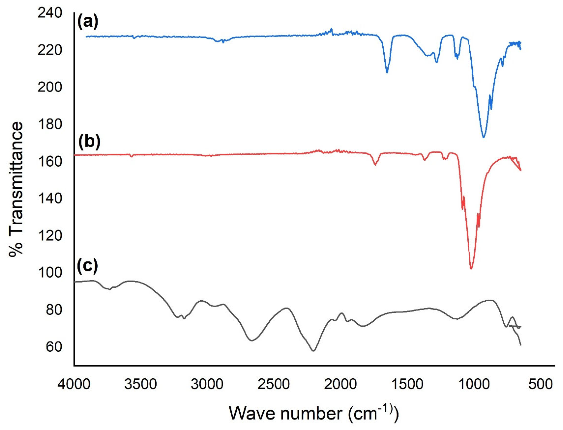
Figure 2.
Stacked X-ray diffraction patterns of samples (a) CAH, (b) wnHAp, and (c) cnHAp showing characteristic Miller indices (hkl) for identified crystallographic planes.
Figure 2.
Stacked X-ray diffraction patterns of samples (a) CAH, (b) wnHAp, and (c) cnHAp showing characteristic Miller indices (hkl) for identified crystallographic planes.
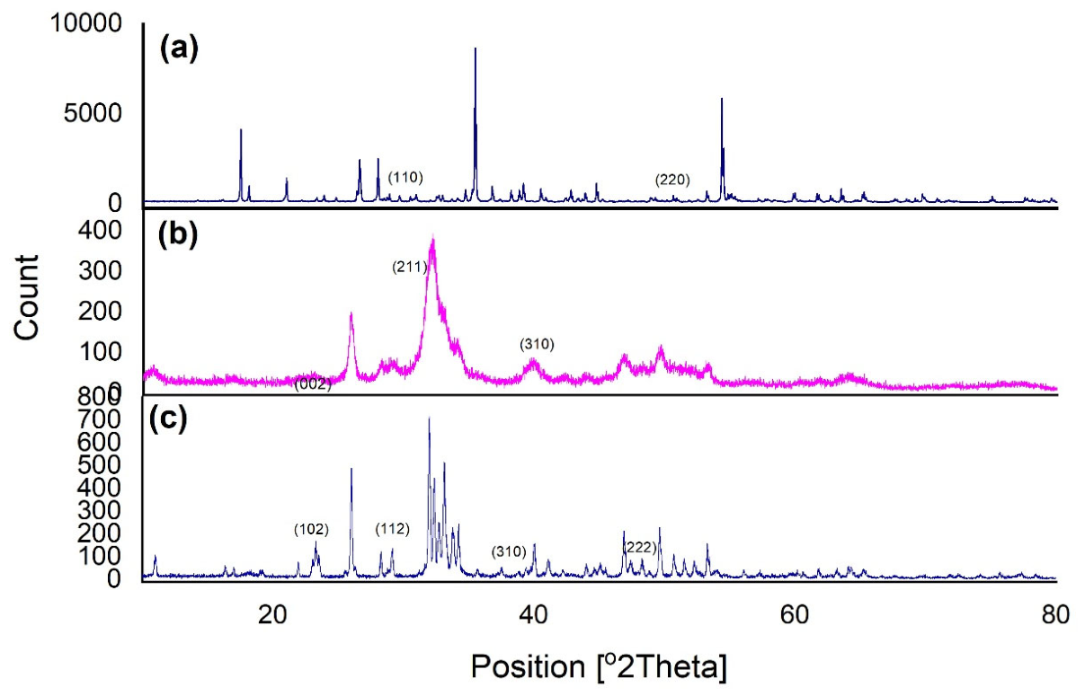
Figure 3.
SEM micrographs (10.00 kX magnification, 1 μm scale bar) showing the surface morphology of (a) wnHAp and (b) cnHAp, accompanied by their corresponding EDX spectra and elemental mapping distributions indicating homogeneous spatial dispersion of Ca, P, O, C, Na, and Mg.
Figure 3.
SEM micrographs (10.00 kX magnification, 1 μm scale bar) showing the surface morphology of (a) wnHAp and (b) cnHAp, accompanied by their corresponding EDX spectra and elemental mapping distributions indicating homogeneous spatial dispersion of Ca, P, O, C, Na, and Mg.
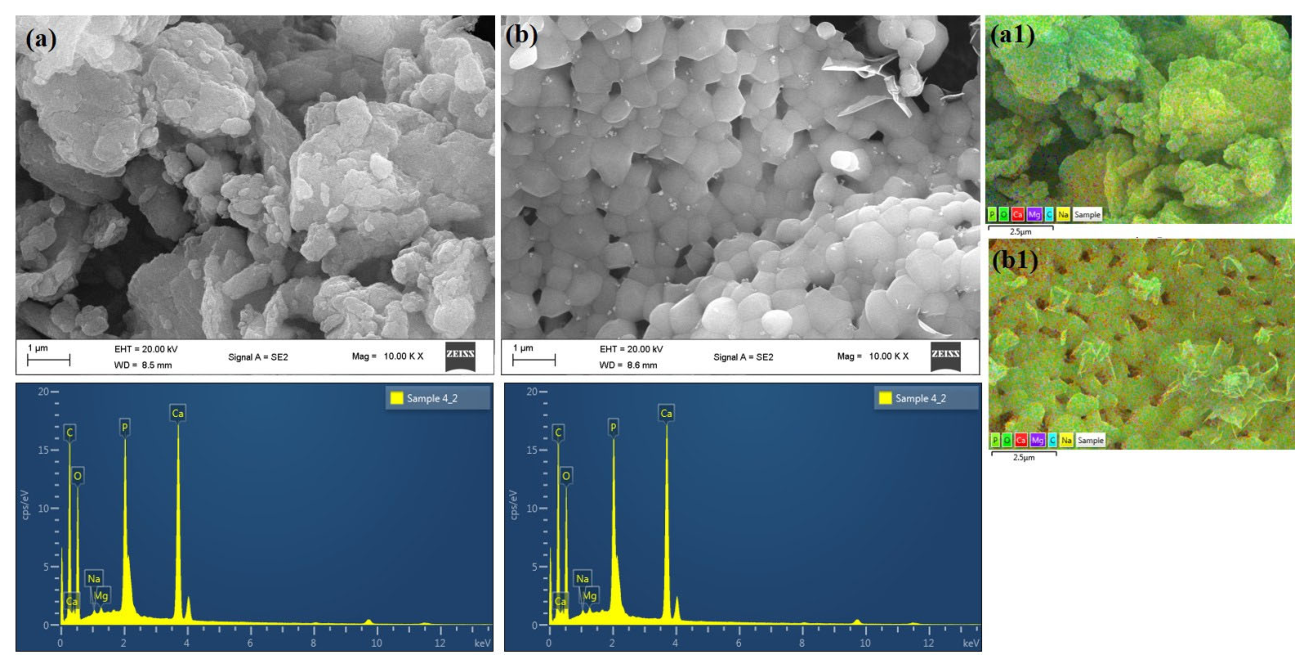
Figure 4.
HRTEM (a) wnHAp; (b) cnHAp.
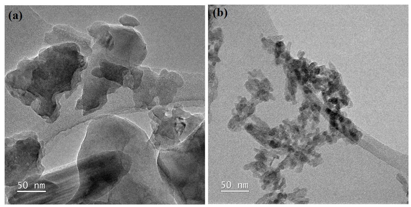
Figure 5.
Particle size distribution histograms with corresponding Gaussian fitting curves for (a) cnHAp (M = 42.41 ± 2.00 nm) and (b) WnHAp (M = 181.09 ± 3.64 nm).
Figure 5.
Particle size distribution histograms with corresponding Gaussian fitting curves for (a) cnHAp (M = 42.41 ± 2.00 nm) and (b) WnHAp (M = 181.09 ± 3.64 nm).
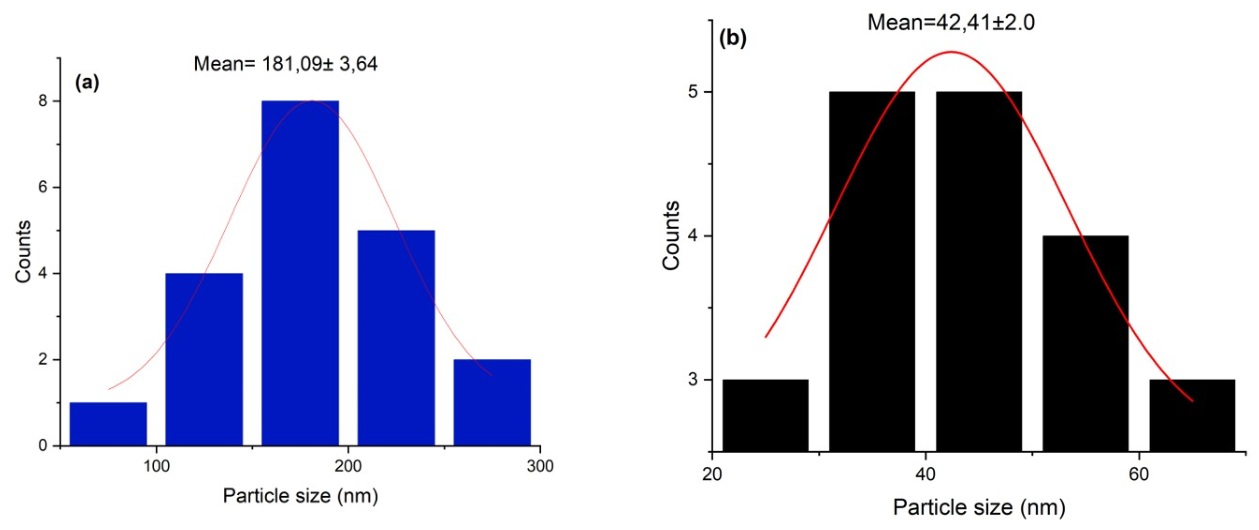
Figure 6.
Dissolution kinetics of calcium aluminate hydrate (CAH), calcined nano-hydroxyapatite (cnHAp), and water bath extracted nano-hydroxyapatite (wnHAp) over a 72-hour immersion period. Data points represent group means, and error bars denote 95% confidence intervals (CI). Non-overlapping error bars indicate statistically significant differences between material formulations and time points (p < 0.05).
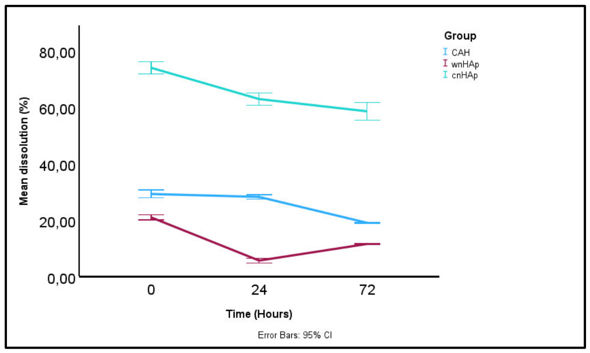

Table 2.
Elemental Composition and Ca/P Atomic Ratio of wnHAp and cnHAp Determined by EDX.
| Parameter | wnHAp | cnHAp |
| Calcium (Ca) | 22,45 wt% | 26,95 wt% |
| Phosphorus (P) | 11,02 wt% | 11,14 wt% |
| Carbon (C) | 27,97 wt% | 28,90 wt% |
| Magnesium (Mg) | 0,40 wt% | 0,36 wt% |
| Sodium (Na) | 0,36 wt% | 0,18 wt% |
| Ca/P Ratio | 1,57 | 1,87 |
| Primary Phase | Ca-Deficient HAp | Mixed HAp Phase |
Note. wnHAp = water-bath/ultrasonication-extracted nanohydroxyapatite; cnHAp = calcined nanohydroxyapatite. Elemental percentages were determined by energy-dispersive X-ray spectroscopy (EDX). Phase identification was based on X-ray diffraction (XRD), rather than EDX alone.
Table 3.
Calcium concentration across time by experimental group and results of the two-way mixed-design repeated-measures ANOVA.
Table 3.
Calcium concentration across time by experimental group and results of the two-way mixed-design repeated-measures ANOVA.
| Group | 0 h, M (SE) | 24 h, M (SE) | 72 h, M (SE) | Time effect, F(2, 12) | Group effect, F(2, 6) | Time × Group, F(4, 12) |
| CAH | 29.23 (0.36) | 28.20 (0.33) | 19.03 (0.41) | 1083.09*** | 11,182.85*** | 196.85*** |
| cnHAp | 73.87 (0.36) | 62.80 (0.33) | 58.47 (0.41) | |||
| wnHAp | 20.97 (0.36) | 5.69 (0.33) | 11.57 (0.41) | |||
| Overall test | 1083.09* | 11,182.85* | 196.85* | |||
| Partial η² | .995 | 1.000 | .985 |
Note. M = estimated marginal mean; SE = standard error; nHAp = nanohydroxyapatite. Time was the within-subjects factor (0, 24, and 72 h), whereas Group was the between-subjects factor (calcium phosphate, cnHAp, and wnHAp). Mauchly’s test indicated that the assumption of sphericity was not violated, W = .357, χ²(2) = 5.15, p = .076; therefore, sphericity-assumed results are reported. **p < .001.
Table 1.
Calibration Performance of Calcium Emission Wavelengths Evaluated by ICP-OES.
| Emission wavelength (nm) | R | LOD (3σ; mg/L) | LOQ (10σ; mg/L) | Assessment |
| 183,801 nm | 0,99948 | 0,6993 | 2,3309 | Good linearity; potential spectral interference |
| 220,861 nm | 0,99931 | 7,7807 | 25,9355 | Poor sensitivity; relatively high LOD/LOQ and replicate scatter |
| 315,887 nm | 0,99946 | 0,0450 | 0,1500 | Excellent sensitivity and low detection limit |
| 317,933 nm | 0,99945 | 0,0278 | 0,0928 | Excellent sensitivity and precision |
| 422,673 nm | 0,99967 | 0,0181 | 0,0602 | Selected: most favourable overall analytical performance |
Note. LOD = limit of detection; LOQ = limit of quantification; σ = standard deviation. The selected wavelength was determined from the combined performance of calibration linearity, detection limits, emission intensity, and precision.
Disclaimer/Publisher’s Note: The statements, opinions and data contained in all publications are solely those of the individual author(s) and contributor(s) and not of MDPI and/or the editor(s). MDPI and/or the editor(s) disclaim responsibility for any injury to people or property resulting from any ideas, methods, instructions or products referred to in the content. |
© 2026 by the authors. Licensee MDPI, Basel, Switzerland. This article is an open access article distributed under the terms and conditions of the Creative Commons Attribution (CC BY) license.
Copyright: This open access article is published under a Creative Commons CC BY 4.0 license, which permit the free download, distribution, and reuse, provided that the author and preprint are cited in any reuse.