

Submitted:
07 October 2026
Posted:
09 October 2026
You are already at the latest version
Abstract
Pigmentation and hair morphology are both among the earliest visible traits studied in the context of human heredity and genetics. Over the past 100+ years, both traits have been defined in various ways and their genetic underpinnings have been studied across major methodological approaches in the field to varying degrees of success. Here we review what can be learned from the divergent trajectories of genetic research on these two traits, particularly about how phenotypic definitions shape connections between genetic findings, biological mechanisms, and measured variation. We consider how a new era of functional genomics may integrate these bodies of knowledge, and to move forward our understanding of complex traits, for which pigmentation and hair morphology provide two illustrative models.
Keywords:
pigmentation
; hair morphology
; hair color
; iris color
; complex traits
1. From Shared Historical Origins to Different Genetic Understandings
Genetic research on human pigmentation and hair morphology (see Glossary) finds its origins in studies of heredity dating over a century ago (Figure Section 1). Despite this shared starting point, we find two bodies of genetic knowledge that have developed in different ways. Pigmentation research has linked variation in skin, iris, and hair color to quantitative and categorical phenotypes, and has established more direct links between genotype, phenotype and the mediating biological mechanisms than what is seen in efforts to connect genetic variation with the normal variation in what is commonly referred to as hair texture, or hair form. Unless otherwise specified, we use hair morphology to refer to the longitudinal curvature and cross-sectional size and shape of human scalp hair fibers.
Previous reviews give substantial accounts of the biology, genetics, and evolution of pigmentation and hair morphology. In pigmentation, Barsh [1] used discoveries in mouse and human genetics to examine the interacting genetic pathways and developmental networks underlying pigmentary phenotypes. Reviews of non-human pigmentation have addressed the genetic basis of phenotypic evolution in Drosophila [2] and connections between molecular mechanisms and adaptive variation across vertebrates [3]. Focusing on human pigmentation, Pavan and Sturm [4] synthesized evidence from albinism, comparative genetics, and genetic association studies. More recent reviews explore the contributions of transposable elements [5] and DNA and RNA methylation [6].
Animal models and inherited human disorders have also featured prominently in reviews of hair genetics. Shimomura and Christiano [7] drew on both in their account of the molecular basis of hair development and function, while Duverger and Morasso [8] focused on what genetic disorders reveal about follicular morphogenesis and hair structure. Earlier reviews had already synthesized experimental work on follicle development and highlighted its potential for genetic investigation [9]. The connection between this mechanistic knowledge and normal-range human hair morphology, however, has been less extensively developed than for pigmentation. Needle et al. [10] reviewed genetic association studies across a broader range of human hair traits, including curvature, hairiness, pigmentation, and graying.
Figure 1.
Timeline of pigmentation and hair genetics. Timeline of eras of research approaches. Representative studies are cited for each approach. (A) Familial heredity, classification, and eugenics, 1900s–1930s [11,12,13]. (B) Animal crosses and developmental experiments, 1900s–1970s [14,15,16,17]. (C) Human pedigrees and biochemical classification of disorders, 1950s–1970s [18,19,20]. (D) Hair-shaft structure, fiber proteins, and follicular gene action, 1970s–1990s [21,22,23]. (E) Molecular identification of experimental and clinical loci, 1980s–1990s [24,25,26]. (F) Human candidate-gene associations and population sequencing, 1990s–2000s [27,28]. (G) Population catalogs, differentiation, and selection analyses, 2000s onward [29]. (H) Genome-wide association studies, 2000s onward [30,31]. (I) Expanded population sampling and quantitative measurement, 2010s onward [32,33,34,35]. (J) Functional genomics and cell-state resolution, 2010s onward. Created with BioRender.
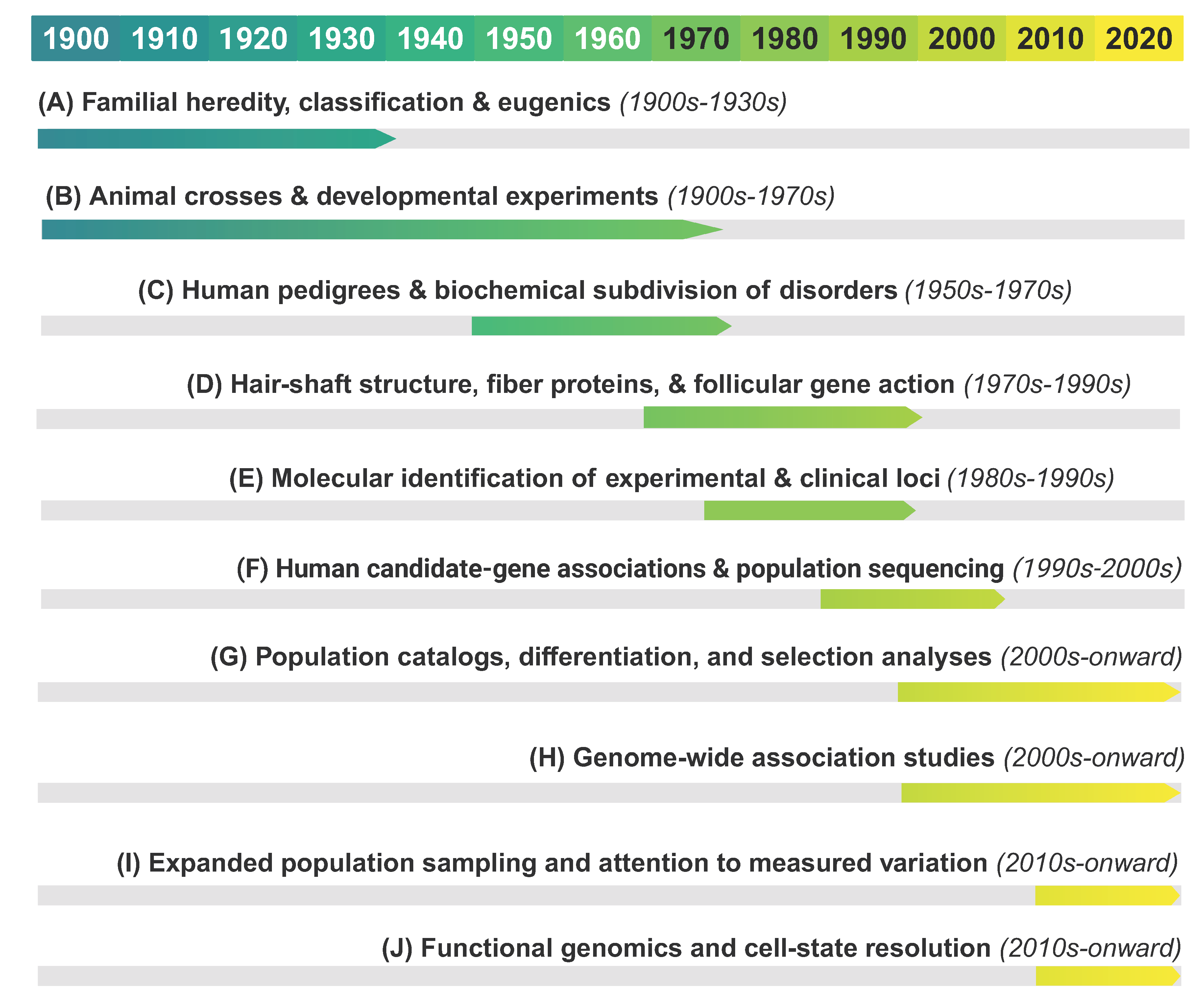
In this review, we present the histories of pigmentation and hair morphology side by side to consider how the construction of phenotypes and the genetic frameworks used to explain them have shaped one another. Definitions of traits determine how variation is represented, and by extension, which components of its genetic basis are made visible. Conversely, genetic findings and mechanistic evidence of biological processes can refine or challenge conventional phenotypic categorizations. This reciprocal process shapes both what genetic studies can discover and what is understood to constitute the trait itself.
We trace how early trait classifications, animal models, and inherited disorders shaped the questions and phenotypes subsequently investigated through association and selection studies. We then consider how functional genomics can provide a path forward for considering how phenotypes should be defined in order to maximize their relationship to underlying genetic variation.
Glossary
Coat (fur): the hair covering an animal’s body. Wool, fur and hair on human scalps, faces and other
areas of the body are all hair. We use coat for the whole-animal phenotypes described mainly in mice,
and hair is used for humans and more generally.
Cross-sectional size and shape: the size and shape of the hair fiber cut perpendicular to its length.
Size is sometimes measured as diameter, other times as area, often described as hair thickness. Shape
may be simply the ratio of maximum to minimum diameter (sometimes called ellipticity) or measures
derived from it, such as eccentricity, and other times shape is described in terms of circular, elliptical,
triangular or other “irregular” shapes.
Curvature: the longitudinal arc of a hair fiber, describing the variation of hair from straight to tightly
coiled. It may be measured explicitly through various methods. In this review, we refer to this longitudinal
dimension of hair morphology as “curvature” regardless of whether it is explicitly measured
or categorically described, to distinguish it from other aspects of hair “texture” or form (i.e., crosssectional
properties).
Follicular: relating to the hair follicle—the anatomical structure in the skin that produces the hair
shaft.
Hair morphology (hair form, hair shape, hair texture): we primarily use this to refer to the longitudinal
curvature and cross-sectional size and shape of scalp hair fibers (see above). In such cases, hair
form, hair shape, and hair texture mean the same thing. However, in certain cases, we use hair morphology
in the broad sense, meaning all non-pigmentation characteristics of hair (including presence
of facial and body hair, growth and density).
Hair shaft: the hair fiber as a distinct unit separate from the root or follicle. This definition is usually
relevant to the experimental or developmental papers reviewed. It includes all layers of the hair shaft
present: cuticle, cortex, medulla.
Iris: the anatomical structure around the pupil that may be pigmented. It contains multiple layers of
pigmentation and when “iris color” or “eye color” is discussed, this usually refers to the amount and
distribution of melanin in the anterior layers, which produces the variation from blue to brown eyes.
Normal variation: trait variation among people unrelated to a clinical or pathological condition, such
as albinism or monilethrix. This term may be used interchangeably with “normal range”, with
2. The Influence of Early Inheritance Work
2.1. Eugenics Origins
Visible variation in human pigmentation and hair morphology, together with observations of familial resemblance, made these traits a focal part of early human heredity research, including studies motivated by eugenic concerns. Gertrude and Charles Davenport, prominent figures in American eugenics, published on eye color (1907) [11], hair form (1908) [36], hair color (1909) [37] and skin pigmentation (1910) [38]. Such investigations of familial inheritance included studies of families whose ancestry researchers described in terms of racial “crosses,” as in the Davenports’ work on skin pigmentation (1910) [38] and Bean’s study of hair form (1911) [39]. Interest in these traits encompassed their use in racial classification [40] with subsequent work by Mildred Trotter examining hair form, fiber size, and color and the schemes used to classify them [41,42,43]. The broader ambitions attached to such research were explicit in some of these papers as well as works like Punnett’s Mendelism (1911) [12] which concludes with a discussion of human eye-color inheritance and a discussion of the regulation of reproduction for hereditary improvement (pp 177-185).
2.2. Phenotype Categories in Early Inheritance Studies
Even within these early studies, there were disagreements about trait classification, and these were often connected to discussions of inheritance patterns. Using family records, Davenport and Davenport (1907) [11] analyzed blue, gray, and brown eyes, arguing that blue was recessive to gray and gray to brown. In the case of a family with two blue-eyed parents where the offspring were recorded as having blue-gray or gray eyes, they attributed this to an inaccurate classification. Hurst (1908) [44] proposed a different classification in eye color based on the presence and absence of anterior iris pigment. This division cut across color labels, grouping clear blue and clear gray eyes together while separating them from all other eye colors that were deemed to contain anterior pigment (green, brown etc). Hurst’s interpretation of family distributions supported Mendelian inheritance of eye color with this classification schema, though he acknowledged that the inheritance of different shades and color patterns remained unresolved. Holmes and Loomis (1909) [45], who recorded blue, green, gray, hazel, brown, and black separately, remarked on the limitations of categories in general noting that the gradations between them suggested continuous variation. Punnett (1911, p 178) [12] endorsed Hurst’s interpretation of anterior iris pigmentation as a Mendelian character and used it to argue that eye-color classifications should reflect inherited factors rather than color intensity.
In studies of hair morphology, commonly called hair form in these papers, classifications likewise connected visible appearance to expectations about inheritance. Davenport and Davenport (1908) [36] drew on Deniker’s account of hair form, which compared the shape of hair cross-sections across racialized population groups, linking them to the appearance of straight to curly hair [40]. Cross-sectional shape was represented by a ratio between the maximum and minimum cross-sectional diameter allowing for the quantification of hair on a scale from round to more elliptical (or in this literature, “flattened”) cross sections. Bean’s students used these measurements to assign hair as straight, wavy, and curly in families that were described as “Chinese-Tagalog crosses” [39]. This cross-sectional classification was thus used as a stand-in for the longitudinal morphology of hair form. This is an assumption that was not rectified until many decades later with observations across and within populations (see [35,46]). Assumptions about populations also influenced which categories were used. For example, the Davenports’ describe their decision: “As our cards were distributed only among whites the term “woolly” was not used.” [36]. Interestingly, Mohr (1932) [13] describes a Norwegian family with hair he termed “woolly” in the context of racialized expectations, noting “The capillus [head hair] is very strikingly curled or frizzled, not unlike negro hair,” [13]. Mohr describes at length the family’s genealogy and perceived “Nordic” appearance, as well as the trait’s transmission across the pedigree, arguing that it had arisen through an independent mutation rather than any “racial” admixture.
Together, these studies show that defining the phenotype was itself part of investigating heredity. Agreement with Mendelian expectations could be treated as evidence supporting a proposed classification, with apparent exceptions explained away as errors in classification rather than challenges to the inheritance model. This illustrates a reciprocal relationship between defining a phenotype and elucidating its inheritance, and by extension, its genetic underpinnings. This sets the stage for the same dialectical relationship shaping our understanding in experimental and clinical settings, and subsequently molecular genetics.
3. Lessons from Animal Models and Inherited Disorders
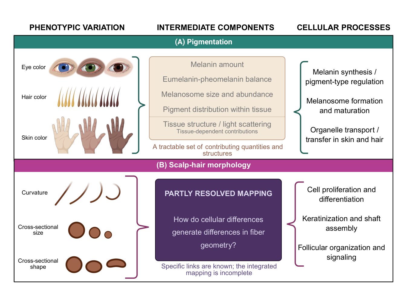
Animal experiments and studies of inherited human disorders helped identify genes and biological processes involved in pigmentation and hair formation. Explaining normal human variation required a further step: establishing how those genes and processes contribute to trait variation among humans. For pigmentation, differences in skin, iris, and hair color can be related to intermediate components such as melanin amount and type, melanosome properties, and pigment distribution. For scalp-hair morphology, curvature and cross-sectional size and shape define the variation to be explained, but the mapping from cellular and developmental processes to differences in fiber geometry remains less complete (Figure Section 3.2). We argue that the clearer mapping between the biology of pigmentation and its corresponding phenotypic variation provided a more direct route from animal and clinical discoveries to explanations of normal human pigmentation.
3.1. Pigment Mechanisms Identified in Animal Models
In pigmentation research, inherited coat-color differences became experimental tools for distinguishing the processes underlying visible traits. Rawles (1947) [16] transplanted mouse embryonic tissues into chick embryos to establish the neural-crest origin of pigment cells in skin and hair. Mouse skin-grafting experiments in the 1950s showed that the type of melanin produced by pigment cells entering grafted skin followed the agouti genotype of the graft’s hair follicles rather than the pigment cells’ own agouti genotype. This indicated that the agouti locus acts through the follicular environment (reviewed in [47]). Mintz (1967) [17] combined early embryos with different coat-color genotypes to produce chimeric mice, then used their coat-color patterns to infer how descendants of individual pigment-cell precursors spread through the skin. Mayer (1970) [48] compared albino, steel, and dominant-spotting mutants to distinguish defects involving pigment cells from those involving their tissue environment. These experiments allowed for the decomposition of similar outcomes into different biological causes: an absence of pigment could reflect differences in melanocyte development, the environment supporting those cells, or their capacity to synthesize melanin within specialized organelles called melanosomes.
Human studies of albinism likewise show how inheritance and biochemical evidence could change the definition of a phenotype or assumptions about its mechanism. Trevor-Roper (1952) [18] reported children without albinism born to two parents with albinism, an observation that challenged the assumption that albinism had the same recessive genetic cause across families. Biochemical investigations subsequently demonstrated that hair bulbs from some individuals with albinism produced pigment when incubated with tyrosine, whereas others did not [20]. Combining family and biochemical evidence, Witkop and colleagues (1970) [49] argued for genetic heterogeneity within oculocutaneous albinism. Therefore, appearance provided a clinical category while inheritance patterns suggested different etiologies on a genetic and biochemical level.
Molecular studies subsequently connected these experimentally and clinically defined phenotypes to particular genes. In mice, Jenkins and colleagues (1981) [24] found evidence that insertion of viral DNA into the mouse genome caused the dilute coat-color phenotype; the dilute locus was subsequently identified as Myo5a, which encodes the motor protein myosin VA [50]. Cross-species work also connected human tyrosinase (TYR) to the mouse albino locus (Tyr): a human tyrosinase complementary DNA (cDNA) probe mapped near or at this locus in mice [51]. Studying Japanese patients with tyrosinase-negative albinism Tomita and colleagues (1989) [52] identified a single-base insertion in the human tyrosinase gene (TYR) and showed, by expressing the mutant gene in mouse melanoma cells, that it produced an inactive enzyme. The gene now designated OCA2, originally called P, illustrates the complementary contributions of human pedigrees and mouse mutants. Ramsay and colleagues (1992) [53] mapped tyrosinase-positive oculocutaneous albinism to chromosome 15 and proposed a relationship with the mouse pink-eyed dilution locus, whose mutations reduce pigmentation of the eyes and coat. Rinchik and colleagues (1993) [25] subsequently identified the mouse gene and its human counterpart. In an individual with both type II albinism and Prader-Willi syndrome, both copies of the human gene were affected: one was lost in a large chromosome 15 deletion, while the other carried a partial deletion. These findings connected distinctions developed through earlier inheritance and biochemical studies to molecular causes, allowing human clinical phenotypes and experimentally characterized animal phenotypes to inform one another.
A particularly direct connection to normal-range human variation emerged from experiments on the regulation of pigment type. Before the underlying genes were identified molecularly, Tamate and Takeuchi (1984) [54] compared hair-bulb melanocytes from lethal yellow mice, carrying an agouti-locus mutation, and recessive yellow mice, carrying an extension-locus mutation. Melanocytes from lethal yellow mice produced eumelanin in response to alpha-melanocyte-stimulating hormone, whereas those from recessive yellow mice did not. This implicated the extension locus in melanocytes’ responsiveness to the hormone. Subsequent studies characterized the gene at the mouse agouti locus, encoding agouti signaling protein, and established the extension locus as the melanocortin 1 receptor (Mc1r; [55,56]). These findings connected inherited coat-color phenotypes to components regulating the production of brown/black eumelanin and red/yellow pheomelanin [1]. Valverde and colleagues (1995) [27] then associated variants in human MC1R with red hair and light skin. Here, knowledge of the mechanism underlying an experimentally studied phenotype supplied a candidate gene whose relevance to normal variation was supported by human association evidence.
3.2. Hair Mechanisms Discovered in Animal Models
In hair research, animal experiments connected differences in coat appearance to the development and composition of the hair shaft. Trigg (1972) [21] for example, examined mouse coat-texture mutants in which observed differences accompanied changes in shaft thickness, internal structure, and the general process of hair shaft formation. Studies of sheep wool characterized genes encoding the keratin-associated proteins B2A, B2C, and B2D, now designated KRTAP1-1, KRTAP1-3, and KRTAP1-4, and subsequently examined the expression of type II keratin genes corresponding to human KRT81, KRT83, and KRT86 during formation of the hair cortex [57,58]. In mice, Luetteke and colleagues (1993) [23] showed that disrupting Tgfa produced wavy fur and whiskers, and breeding experiments established that the previously described waved-1 mutation affected the same gene. Tanaka and colleagues (2007) [59] subsequently identified mutations in Krt25 in Rex mice.
Human pedigrees likewise connected various inherited differences in shaft structure to investigations of their genetic basis. Deraemaeker’s (1957) [19] investigation of a family with monilethrix, a condition characterized by repeated shaft constrictions (beading) and breakage, illustrates how inheritance informed the recognition of a phenotype. Two relatives whose hair appeared normal were found on closer examination to have small proportions of beaded hairs, which complicated the simplification of the phenotype as present or absent. Structural observations subsequently helped guide molecular investigation: abnormalities in the hair cortex suggested keratin genes as candidates, and Healy and colleagues (1995) [60] linked monilethrix to the type II keratin gene cluster in two families. Winter and colleagues (1997) [26] then identified mutations in the hair cortex keratin gene now called KRT86. Other clinical phenotypes implicated different keratins: Shimomura and colleagues (2010) [61] identified a KRT74 mutation in a Pakistani family with autosomal dominant “woolly hair.”
The relevance of these discoveries to normal-range human variation depended on whether the experimental and clinical phenotypes could be understood along the same dimensions. In pigmentation, findings concerning melanin production and pigment-type switching provided explanations relevant to variation in absolute pigment amount and relative balance of eumelanin and pheomelanin. In hair, the repeated constrictions characteristic of monilethrix, for example, are not simply an extreme of either overall shaft diameter or curvature. Explaining their genetic basis therefore does not necessarily explain why human scalp hair varies from straight to tightly coiled or from thin to thick. Likewise, describing animal coats and human hair as “curly”, “wavy,” or “woolly” does not establish that these labels capture equivalent dimensions of variation or that the same mechanisms explain those differences (Figure Section 3.2).
Figure 2.
Phenotypic dimensions and biological processes underlying pigmentation and hair morphology. (A) Variation in skin, hair, and eye color is influenced by the amount and type of melanin, as well as by melanosome properties and pigment distribution fileswfalse [4,62,63]. (B) Scalp-hair morphology is characterized by fiber curvature and cross-sectional size and shape. These features reflect follicle organization and signaling, cellular proliferation and differentiation, and hair-shaft keratinization fileswfalse [64,65,66]. How these processes contribute to normal variation in fiber geometry remains incompletely understood fileswfalse [10,35]. Created with BioRender.
4. Genetic Discovery in Human Variation
Around the turn of the 21st century, the sequencing of the human genome, population catalogs of genetic variation, and advances in genotyping transformed the scale at which human genetic differences could be investigated [67,68]. These new approaches provided new ways to validate the role of experimentally derived gene candidates in normal-range variation, and further, to ask questions about the way they might have been shaped by natural selection.
4.1. Biological Candidates and Population Variation
Before genome-wide scans became common, early molecular investigations into population-level questions came from exploring genes that had already been proven to be linked to phenotypic variation from the preceding era of research. For example, Harding and colleagues [28] compared MC1R sequence variation among populations, interpreting the absence of non-synonymous variants in their African samples as evidence of strong functional constraint and the greater variation in Europe and northern Asia as reflecting relaxed constraint. Rogers and colleagues [69] subsequently proposed an alternative evolutionary explanation involving positive selection. These papers extended the molecular understanding of MC1R’s role in pigmentation to questions about evolutionary history, but also raised questions about how much one could interpret from sequence variation at a single locus.
For hair morphology, EDAR provides an example in which developmental knowledge and population genetic differentiation helped identify a candidate before its association with normal variation was established. Fujimoto and colleagues [29] examined differentiation among populations at 170 genes implicated in hair morphogenesis, using data from the International HapMap Project. They prioritized the EDAR V370A variant, which was highly differentiated and had also been implicated in earlier selection analyses [29,70]. They then measured hair cross-sectional diameters and area in Indonesian and Thai-Mai participants, finding an association with greater shaft thickness that was subsequently replicated in Japanese participants [29,71]. Further candidate analysis identified an association between FGFR2 and hair thickness, although the authors described the association as much weaker than that of EDAR and called for replication [72]. These findings connected developmental candidate genes to normal variation in human hair thickness. But even with its association, the pleiotropic effects of EDAR made it difficult to understand what phenotype precisely was the target of selection on the V370A variant [73].
4.2. Genome-Wide Associations
Genome-wide association studies (GWAS) expanded discovery beyond genes nominated in advance while providing further tests of connections established through experimental research. Stokowski and colleagues [30] identified replicated associations at SLC24A5, SLC45A2, and TYR in a genome-wide study of skin reflectance in UK participants of South Asian ancestry, selected specifically from the lighter and darker ends of the measured distribution. These findings connected normal-range skin pigmentation to both the zebrafish golden gene, SLC24A5, and the albinism gene TYR [30,74]. In Icelandic and Dutch samples, Sulem and colleagues [75] investigated categorical differences in hair and eye color, sun sensitivity and freckling, recovering familiar pigmentation loci alongside additional associations. Larger studies subsequently expanded the associations with hair color and eye color, including contributions to red hair beyond MC1R and regions containing genes previously associated with iris structure [76,77,78]. Pigmentation research thus accumulated many connections between normal variation and established biology while also identifying components requiring additional explanation.
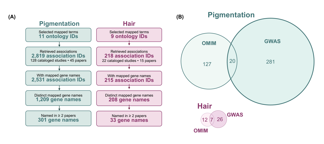
For hair morphology, Medland and colleagues [31] identified an association for hair curvature in three Australian cohorts of European ancestry near TCHH, which encodes trichohyalin–a protein expressed during inner root sheath development. Later studies implicated PADI3, a gene in which rare mutations cause “uncombable hair syndrome” [79,80]. In Latin American participants, Adhikari and colleagues [32] also identified associations with beard density, eyebrow thickness, and monobrow near EDAR, FOXL2, and PAX3, respectively. These findings extended connections with developmental and clinical research across several hair traits. Figure Section 4.2 compares compiled OMIM genes with GWAS Catalog mapped gene names supported by at least two distinct papers per trait group, pooling core and related traits, including facial and body hair traits in the hair group. Fourteen gene names occur in both compiled trait groups, including FRAS1 and PADI3 (see supplementary gene table).
Figure 3.
Pigmentation and hair genes in OMIM and the GWAS Catalog. (A) GWAS Catalog (https://www.ebi.ac.uk/gwas) mapped gene names from core (strict) and related (broad) traits, pooled within pigmentation and hair groups. Names are retained if supported by at least two distinct papers (unique PubMed IDs) within the same group; multiple records from one paper count once. No discovery sample-size cutoff is applied. (B) Exact-name overlap with OMIM (https://omim.org) genes. Areas share one gene-count scale. Fourteen gene names occur in both trait groups; their source memberships are listed in the supplementary gene table. Pigmentation: 170 OMIM IDs from D’Arcy et al. fileswfalse [81] and the hair color and graying entries of Needle et al. fileswfalse [10] map to 147 gene symbols. Hair, including facial and body hair: 27 OMIM IDs from Needle et al.’s other hair traits fileswfalse [10] map to 19 gene symbols. Abbreviations: GWAS, genome-wide association study; OMIM, Online Mendelian Inheritance in Man.
4.3. Population Sampling and Phenotyping Tools
The populations sampled and the phenotyping tools used define what variation can be quantified and related to genetic differences. Adhikari and colleagues [32] recorded hair shape as straight, wavy, curly, and frizzy, but excluded the last category, representing 2.4% of participants, from the hair-shape GWAS because it was so uncommon. Such statistically justifiable choices become necessary when a continuous trait is forced into discretization. Categories can obscure variation across and within groups and strongly affect the distribution of variation in a way that can affect the associations that can be made in a genetic association study (see [35]).
Pigmentation studies show what combining broader sampling with quantitative phenotyping can make possible. The landmark papers on African skin pigmentation genomics [33,34] would not have been possible without the use of reflectance spectrophotometry to obtain continuous representation of skin color, represented as Melanin Index. A categorization like the Fitzpatrick scale would have rendered all individuals within and across groups as members of two types in this classification, at best, which would not have allowed for the statistical power necessary to make the discoveries those studies did. Crawford and colleagues [33] identified novel loci (e.g., MFSD12) and novel variants in known loci that were relevant to African skin pigmentation. And in the Nama and Khomani San, Martin and colleagues found that gene sets assembled from earlier discoveries explained only a small estimated fraction of heritable pigmentation variation.
The populations sampled and phenotypes measured also shape predictions of appearance from DNA. HIrisPlex-S predicts eye, hair, and skin color categories, while a model developed by Pośpiech and colleagues predicts straight versus non-straight hair [82,83]. These predictions depend on the phenotype definitions and the genetic associations represented in the samples used to develop the methods. Applying them to ancient genomes raises a further problem of transfer (Box Section 4.3). Predictive accuracy cannot be assumed in the ancient genetic backgrounds, and the results cannot be checked against the appearance of the individuals they describe nor a representative population [84].
Together, these studies have expanded the genetic account of pigmentation and hair morphology, with applications including prediction of appearance from DNA [82,83]. Quantitative studies have broadened our understanding of pigmentation across populations, while studies of hair morphology have often left variation within conventional categories unresolved. The implication for further discovery is that larger genetic datasets need to be accompanied by phenotyping that preserves the variation we want to explain.
Box 1. Red hair in Neanderthals
The idea of a red-headed Neanderthal has become prominent in the public imagination regardless of it having outlived its evidentiary basis. Lalueza-Fox et al. fileswfalse [85] sequenced MC1R in two purported Neanderthal specimens from the Italian Monti Lessini and the Spanish El Sidrón site. In their manuscript, they describe the Monti Lessini specimen had a substitution in the gene which they argued was functionally analogous to a classic loss-of-function mutation in modern humans that confers lighter skin pigmentation and red hair fileswfalse [85]. At the time, this substitution was cross validated across three labs for the Monti Lessini sample, but only recovered in one lab for the El Sidrón Neanderthal. Subsequent studies have cast doubt on the original conclusions. Samples from the Monti Lessini site (albeit not the sample in question) were later directly radiocarbon dated by accelerator mass spectrometry to ~5,580 years before present, 20,000-30,000 years after Neanderthal extinction, and geometric morphometrics and mtDNA sequencing have also suggested that samples were of anatomically modern human origin fileswfalse [86]. Additionally, studies have failed to replicate the specific MC1R variant from the study, indicating that the variant may be a reflection of PCR-based contamination and that if it was present in Neanderthal populations, it was likely rare fileswfalse [87,88].
5. Functional Genomics of Pigmentation and Hair
Pigmentation and hair biology are built through highly polygenic systems. This polygenicity emphasizes the complex molecular architecture foregrounding challenges in mapping genotypes to phenotypic variations. Functional studies provide researchers with the important context information needed to adequately bridge the molecular, cellular, and developmental effects of genetic variants on pigmentation and hair traits.
5.1. Regulatory Variants and Gene Expression
Regulatory assays, expression studies and perturbation experiments allow researchers to test how associated variants affect transcription, RNA processing and chromatin states in pigmentation and hair biology (Table Section 5.1). Given the pleiotropic nature of some pigmentation variants along with the polygenic nature of both pigmentation and hair morphology, functional studies become strong sources of experimental evidence showcasing the coordinated molecular components underlying these genetic interactions. Functional investigation of pigmentation-associated variants near OCA2 underscored the pleiotropic nature of pigmentation, highlighting the need for detailed cellular and molecular network assessment. Reporter assays test how a DNA sequence affects expression of a measurable reporter gene. Combining reporter assays with analysis of chromatin states has identified that genetic variants at HERC2 (rs12913832) show opposite directional effects on transcription factor (TF) recruitment and gene expression of OCA2 [89]. This directionality was also noted in the resulting dark versus light pigmentation phenotypes [89]. Similarly, a reporter screen of 1,157 candidate loci highlighted 165 variants with differential allelic activity, including multiple enhancer-associated alleles (rs6497271, rs6510760, and rs7948623) acting on OCA2, MFSD12, and TMEM138 [90]. Following these reported assays with directed CRISPR/Cas9 mediated gene editing provides a more direct and controlled way of validating upstream results [90,91]. In addition to contextualizing the enhancer activity regulating OCA2 [89,90], functional studies have also validated the transcriptional effects of two variants near SOX2 and FOXD1, providing new insights into the molecular mechanisms regulating eyebrow thickness [91]. Upstream functional analyses, including various mapping techniques, can inform the prioritization of variants for validation by helping to disentangle the genomic and regulatory context surrounding them.
Table 1. Functional validation of genetic variants in pigmentation and hair phenotypes. a
| Approach | What it can show | Pigmentation Examples | Hair Examples | Refs |
| Expression association (eQTL, splicing QTL, colocalization, TWAS) | Genetic variants correlate with differences in gene expression or splicing | Dark pigmentation-associated alleles upstream of MFSD12 correlate with lower MFSD12 expression; alleles in a linked region within HERC2 correlate with higher OCA2 expression in melanocytes; an ASIP insertion correlates with 2.2-fold higher ASIP expression | Hairiness signals colocalize with TBX15 and LIMS1-AS1 eQTLs; follicle eQTLs nominate PADI3 at a hair-shape locus | [33,92,93,94] |
| Chromatin and 3D genome mapping | Which regulatory elements are active in which cell type, and which genes they interact with | The rs12913832 enhancer loops to the OCA2 promoter in melanocytes more strongly with the dark pigment-associated allele; NFIB directly binds Edn2 in the follicle stem-cell niche | Male-pattern hair-loss heritability is concentrated in dermal papilla open chromatin | [89,95,96] |
| Single-cell and tissue expression mapping | Where and when candidate genes are expressed | Expression of 107 of 158 screen hits tracks melanin content across donors | Cell-type atlases of the human follicle; 85 genes differ between curly and straight follicles, with candidates expressed in the inner root sheath | [97,98,99] |
| Reporter assays and MPRAs | Whether a variant changes enhancer activity | The rs12913832 T allele in a HERC2 enhancer regulating OCA2 produces approximately fivefold greater reporter activity than the C allele; 165 of 1,157 tested SNPs show allele-specific activity | A regulatory fragment near SOX2 containing rs1345417 increases reporter expression 3–4-fold; changing the G allele to C reduces activity | [89,90,91] |
| Editing variants or enhancers in cells | Whether a regulatory element controls a target gene in its native genomic context | Deleting an OCA2 enhancer lowers OCA2 and melanin by more than 50% | Editing eyebrow-thickness loci lowers SOX2, FOXD1 and LIMS1 expression | [90,91] |
| Gene perturbation in cells (knockout, knockdown, screens) | Whether loss or gain of a gene changes a cellular phenotype | A genome-wide CRISPR screen finds 169 melanin-promoting genes, 135 of them new; COMMD3 loss over-acidifies melanosomes; Mfsd12 knockdown raises melanin | The PRSS53 Q30R variant alters protein processing and secretion | [32,33,97] |
| Engineered animal models | Whether a gene affects the phenotype in a whole organism | Mfsd12 knockout mice have gray coats; dermal papilla -catenin sets yellow vs black coat partly through agouti and corin | Raising Edar changes follicle patterning and density; halving En1 yields more hair follicles and fewer sweat glands | [32,33,100,101,102] |
| Classical genetics and QTL mapping | Gene–phenotype links from naturally occurring or strain variation | The classical grizzled mouse carries an Mfsd12 deletion | One mouse locus containing En1 controls both hair-follicle and sweat-gland density | [33,102] |
| Computational prediction and modeling | Which variants, genes or pathways to test | Fine-mapping isolates the ASIP insertion as the likely causal variant; virtual knockouts in a pigmentation signaling network | Sequence models flag hair-loss and hair-color SNPs predicted to alter chromatin; variant scores prioritize a TRPS1 enhancer SNP for hair thickness | [92,96,103,104] |
| a Abbreviations: CRISPR, clustered regularly interspaced short palindromic repeats; eQTL, expression quantitative trait locus; MPRA, massively parallel reporter assay; QTL, quantitative trait locus; SNP, single-nucleotide polymorphism; TWAS, transcriptome-wide association study. | ||||
Functional annotation, fine-mapping, expression and methylation quantitative trait loci (eQTL, meQTL) analyses offer additional opportunities for researchers to untangle the genomic and regulatory context surrounding genetic variants. Application of functional annotation has framed descriptions of enhancer-related activity in conjunction with hair morphological traits, showcased in part by annotation of a TRPS1 allele found to influence hair thickness in Han Chinese individuals [104]. Functional annotation of GWAS hits has helped prioritize candidate variants for further downstream functional testing [33,91], as well as further computational analyses [93,105]. In silico, statistical framing has shaped fine-mapping studies outlining differences in RNA processing in relation to pigmentation phenotypes, including the regulatory effects of SVA retrotransposon on ASIP expression in skin [92]. Fine-mapping has also been used to resolve independent signals and rank candidate variants by statistical evidence of causality. This approach helped identify the causal signals attached to independent alleles at the TPCN2 locus, refining the potential effects of these variants on blond hair phenotypes [105]. Further assessment of regulatory effects can also be done through QTL analysis, providing expression-related associations for candidate single-nucleotide polymorphisms (SNPs) in multiple tissue-specific contexts. Application of QTL analyses has underlined enrichment of genes associated with hair growth represented within eQTLs [94] and when combined with colocalization analysis, meQTLs for blond hair color were noted in association with MC1R and IRF4 among others [105].
Regulation does not only occur at the gene level. RNA splicing, methylation and regulation by small RNAs can directly impact translation, and therefore protein bioavailability influencing expression of resulting phenotypes. RNA splicing can lead to variation in mRNA transcript viability, thus affecting downstream translation. Variation in mRNA splicing has previously been associated with differences in lab animal coloration due to multiple transposable element (TE) insertions [5]. TEs can impact RNAs in multiple ways, as seen in the effect of SINE-VNTR-Alu (SVA) family retrotransposons on human pigmentation phenotypes [92]. In Europeans, a 3.3 kb SVA F1 insertion was found to increase ASIP transcript abundance through a coordinated effort leading to productive splicing in ASIP and therefore decreasing pigmentation [92]. mRNA splicing can also be affected by RNA methylation, which, along with DNA methylation, has been shown to vary animal coloration in myriad ways [6]. Small RNAs like micro RNAs (miRNAs) also modulate gene transcript availability outcomes. Dicer is the enzyme responsible for miRNA biogenesis, and an inducible knockout of Dicer1 in the melanocyte lineage in mice, revealed that this protein may also play a role in hair graying phenotypes [106]. Together, these studies show that exploring multiple facets of gene transcript regulation is necessary in providing the support for interpreting genotype to phenotype transitions in pigmentation and hair biology.
In all the above cases, the functional outcomes of genetic variants were evaluated in ways that underscored the complex nature of gene activity regulation. Candidate gene and GWAS provide an important starting point in investigating the genetics behind pigmentation and hair phenotypes, but they require the context gained from exploring multiple facets of gene expression regulation [107]. These interacting cellular mechanisms accentuate the complex nature of the molecular architecture underlying genotype to phenotype translation in pigmentation and hair biology.
5.2. Cells, Compartments and Developmental Stages
Identifying the cells, compartments and developmental stages that produce a trait can guide phenotype definitions based on biological processes, which may provide a closer correspondence to biologically salient analytical units than a definition based purely on appearance. Pigmentation and hair morphology phenotypes are not simply regulated by differences in gene expression, but are also impacted by additional cellular processes and interactions. Research joining these different elements is done in part by mapping gene regulation and cellular signaling in a cell-type specific manner.
These analyses can be applied to investigate patterns in variation of regulation activity and abundance among different cell types under different phenotypic conditions. This was seen through the identification of varied activity and abundance of TFs across basal keratinocytes and dermal fibroblasts among others, as well as the depletion of hair follicle stem cells in conditions like alopecia areata compared to control [96]. Additionally, this level of cell-type-specific transcriptomic mapping has revealed differences in gene expression enrichment in hair follicle cell types compared to associated cells including melanocytes and immune cells [98]. These detailed assessments help improve the understanding of the gene and protein landscapes shaping different scalp and follicular cell types, allowing for advancements in future investigations [96,98]. Moreover, mapping the coordinated signaling mechanisms which underlie cellular differentiation and behavior in model systems including mice, has opened the door to understanding the role of factors like NFIB in melanocyte stem cell (McSCs) differentiation [95] and the ways in which Wnt signaling guides both McSC differentiation and hair pigmentation. In this way, mapping out the variety of cell types, states and mechanisms that form complex phenotypes is vital in understanding how they diverge.
Identifying the follicle compartments in which genes act can help connect molecular findings to pigmentation and hair morphology. The inner root sheath (IRS) surrounds the developing hair shaft, while the dermal papilla (DP) signals to the cells that form it. Morphological centered studies generating microarray data under different hair curvature conditions identified 85 genes showing differential expression [99]. Most of this expression variation localized to the IRS and included genes involved in oxidative stress, suggesting a more direct mechanism by which gene activity within follicular compartments impacts fiber morphology [99]. Chen et al. (2025) [108] identified a missense variant in KRT71, which encodes an inner root sheath keratin, that segregated with autosomal dominant “woolly hair” in a Chinese family. Characterizing the mechanisms controlling the eumelanin to pheomelanin switch and general melanin synthesis, are vital points of emphasis in mapping pigmentation phenotypes. Determining how networks within hair follicle (HF) compartment cells, including DP, modulate eumelanin-pheomelanin switches presents a key component of this general inquiry. Mouse models revealing coordinated signaling through the Wnt/-catenin pathway as well as the regulatory effects of Sox2 expression, both demonstrate DP-centric mechanisms of eumelanin to pheomelanin switch control [100,109]. Methods developed in effort to quantify melanin synthesis have generated detailed annotations of the genetic networks regulating this process. Bajpai and colleagues combined a genome-wide CRISPR screen with quantitative assessment of light scattering in melanocytes, identifying new pathways that coordinate melanosome function and maturation [97]. Exploring variation within the elements comprising a particular phenotype strengthens our understanding of the genotype to phenotype transition within complex traits, and provides substantial direction for future work.
Identifying the developmental stages at which traits emerge grounds phenotypic inquiry in the biological processes that govern complex phenotypes, rather than on appearance alone. The patterning of hair follicles provides insight into the timing of HF development through anatomical assembly. In cultured embryonic mouse skin cells, the tunable signaling of Edar expression and copy number, coordinated through different agents with the bone morphogenetic protein (BMP) pathway, was found to control follicle density [101]. This sets the stage for future research to further tease apart how variations within EDAR and BMP signaling shift the molecular network underlining the patterning of this phenotype. The relationship between EDAR and HF density was further described in humans as EDAR SNPs showed strong association with beard density [32]. Furthermore, the developmental trade-offs noted between the density of eccrine glands vs HFs demonstrates another aspect of the layering networks modulating HF patterning [102]. In mice, the transcriptional activity of En1 was found to regulate the concentration of eccrine glands to HFs [102]. These findings underscore the interconnectedness of multiple biological processes controlling complex phenotypes, leaving many questions concerning the specificity of various network-level interactions open for future research.
6. Concluding Remarks
The genetic basis of pigmentation has become relatively advanced due to connections between biological mechanisms and phenotypic variation that have remained less fully resolved for scalp-hair morphology. Studies of melanin production and pigment-type switching helped connect discoveries from animal experiments and inherited disorders to normal variation in human pigmentation. The gene sets compiled from OMIM and the GWAS Catalog are also larger for pigmentation, even when facial and body hair traits are included (Figure Section 4.2). Intermediate components of pigmentation such as melanin amount and type, melanosome properties, and pigment distribution helped make experimental findings interpretable in relation to human color variation. On the other hand, hair research has established substantial knowledge of follicle development, growth, and shaft formation, but how differences in these processes produce variation in curvature and cross-sectional size and shape remains less complete (Figure Section 3.2). Further progress therefore would benefit from connecting genetic discoveries and functional experiments to the same defined dimensions of phenotypic variation.
Despite the greater progress in pigmentation genetics, the spatial and temporal variation in hair within individuals offers an opportunity to investigate mechanisms that may also help explain differences in scalp-hair morphology among individuals. Fibers can differ across scalp regions and change over time, including after chemotherapy, allowing comparisons among follicles within a shared inherited genetic background. Such comparisons could help identify differences in gene expression and cellular state associated with variation in fiber geometry. Pairing these studies with quantitative measurements of curvature and cross-sectional size and shape would provide specific outcomes for functional experiments to test. Changes in hair pigmentation through childhood and graying offer related opportunities. For both traits, connecting measured variation to experimentally tested mechanisms would allow genetic findings and phenotype definitions to inform one another (see Outstanding Questions).
Code and compiled gene tables supporting Figure 3 are available at https://github.com/lasisilab/tig-omim-gwas.
Acknowledgments
This research did not receive any specific grant to support this work.
Conflicts of Interest
The authors declare no competing interests.
Use of Artificial Intelligence
During the preparation of this work, the authors used Undermind.ai and Claude Science (Anthropic) to search for additional literature sources. Codex (OpenAI) was used to write code for API queries compiling gene sources and for editorial suggestions to improve clarity of the text which was drafted by the authors. Claude Code (Anthropic) was used to format the manuscript in LaTeX. All sources were manually verified and all text in the manuscript is written by the authors who take full responsibility for the content of the published article.
References
- Barsh, G.S. The genetics of pigmentation: From fancy genes to complex traits. Trends Genet. 1996, 12, 299–305. [Google Scholar] [CrossRef] [PubMed]
- Wittkopp, P.J.; Carroll, S.B.; Kopp, A. Evolution in black and white: Genetic control of pigment patterns in Drosophila. Trends Genet. 2003, 19, 495–504. [Google Scholar] [CrossRef] [PubMed]
- Hubbard, J.K.; Uy, J.A.C.; Hauber, M.E.; Hoekstra, H.E.; Safran, R.J. Vertebrate pigmentation: From underlying genes to adaptive function. Trends Genet. 2010, 26, 231–239. [Google Scholar] [CrossRef] [PubMed]
- Pavan, W.J.; Sturm, R.A. The genetics of human skin and hair pigmentation. Annu. Rev. Genom. Hum. Genet. 2019, 20, 41–72. [Google Scholar] [CrossRef] [PubMed]
- Galbraith, J.D.; Hayward, A. The influence of transposable elements on animal colouration. Trends Genet. 2023, 39, 624–638. [Google Scholar] [CrossRef] [PubMed]
- Strowbridge, N.; Vieites, D.R.; Ritchie, M.G.; Elmer, K.R. Contributions of epigenomic and epitranscriptomic methylation to animal colouration. Trends Genet. 2025, 41, 1005–1016. [Google Scholar] [CrossRef] [PubMed]
- Shimomura, Y.; Christiano, A.M. Biology and genetics of hair. Annu. Rev. Genom. Hum. Genet. 2010, 11, 109–132. [Google Scholar] [CrossRef] [PubMed]
- Duverger, O.; Morasso, M.I. To grow or not to grow: Hair morphogenesis and human genetic hair disorders. Semin. Cell Dev. Biol. 2014, 25-26, 22–33. [Google Scholar] [CrossRef] [PubMed]
- Hardy, M.H. The secret life of the hair follicle. Trends Genet. 1992, 8, 55–61. [Google Scholar] [CrossRef] [PubMed]
- Needle, C.D.; Brinks, A.L.; Perez, O.D.; Shapiro, J.; Lo Sicco, K.I.; Tsirigos, A.; Petukhova, L. A comprehensive review of GWASs of human hair traits. J. Investig. Dermatol. 2025, 145, 2964–2972. [Google Scholar] [CrossRef] [PubMed]
- Davenport, G.C.; Davenport, C.B. Heredity of eye-color in man. Science 1907, 26, 589–592. [Google Scholar] [CrossRef]
- Punnett, R.C. Mendelism, third edition ed.; The Macmillan Company: New York, 1911. [Google Scholar]
- Mohr, O.L. Woolly hair a dominant mutant character in man. J. Hered. 1932, 23, 345–352. [Google Scholar] [CrossRef]
- Cuénot, L. La loi de Mendel et l’hérédité de la pigmentation chez les souris. Arch. De Zool. Expérimentale Et. Générale 3e Série 1902, 10, XXVII-XXX. [Google Scholar]
- Gayon, J.; Burian, R.M. France in the era of Mendelism (1900–1930). Comptes Rendus de l’Académie des Sciences-Series III-Sciences de la Vie 2000, 323, 1097–1106. [Google Scholar] [CrossRef] [PubMed]
- Rawles, M.E. Origin of pigment cells from the neural crest in the mouse embryo. Physiol. Zool. 1947, 20, 248–266. [Google Scholar] [CrossRef] [PubMed]
- Mintz, B. Gene control of mammalian pigmentary differentiation. I. Clonal origin of melanocytes. Proc. Natl. Acad. Sci. 1967, 58, 344–351. [Google Scholar] [CrossRef] [PubMed]
- Trevor-Roper, P.D. Marriage of two complete albinos with normally pigmented offspring. Br. J. Ophthalmol. 1952, 36, 107–108. [Google Scholar] [CrossRef] [PubMed]
- Deraemaeker, R. Monilethrix: Report of a family with special reference to some problems concerning inheritance. Am. J. Hum. Genet. 1957, 9, 195–199. [Google Scholar] [PubMed]
- Kugelman, T.P.; Van Scott, E.J. Tyrosinase activity in melanocytes of human albinos. J. Investig. Dermatol. 1961, 37, 73–76. [Google Scholar] [CrossRef]
- Trigg, M.J. Hair growth in mouse mutants affecting coat texture. J. Zool. 1972, 168, 165–198. [Google Scholar] [CrossRef]
- Steinert, P.M.; Rogers, G.E. Characterization of the proteins of guinea-pig hair and hair-follicle tissue. Biochem. J. 1973, 135, 759–771. [Google Scholar] [CrossRef] [PubMed]
- Luetteke, N.C.; Qiu, T.H.; Peiffer, R.L.; Oliver, P.; Smithies, O.; Lee, D.C. TGF alpha deficiency results in hair follicle and eye abnormalities in targeted and waved-1 mice. Cell 1993, 73, 263–278. [Google Scholar] [CrossRef] [PubMed]
- Jenkins, N.A.; Copeland, N.G.; Taylor, B.A.; Lee, B.K. Dilute (d) coat colour mutation of DBA/2J mice is associated with the site of integration of an ecotropic MuLV genome. Nature 1981, 293, 370–374. [Google Scholar] [CrossRef] [PubMed]
- Rinchik, E.M.; Bultman, S.J.; Horsthemke, B.; Lee, S.T.; Strunk, K.M.; Spritz, R.A.; Avidano, K.M.; Jong, M.T.C.; Nicholls, R.D. A gene for the mouse pink-eyed dilution locus and for human type II oculocutaneous albinism. Nature 1993, 361, 72–76. [Google Scholar] [CrossRef] [PubMed]
- Winter, H.; Rogers, M.A.; Langbein, L.; Stevens, H.P.; Leigh, I.M.; Labrèze, C.; Roul, S.; Taieb, A.; Krieg, T.; Schweizer, J. Mutations in the hair cortex keratin hHb6 cause the inherited hair disease monilethrix. Nat. Genet. 1997, 16, 372–374. [Google Scholar] [CrossRef] [PubMed]
- Valverde, P.; Healy, E.; Jackson, I.; Rees, J.L.; Thody, A.J. Variants of the melanocyte–stimulating hormone receptor gene are associated with red hair and fair skin in humans. Nat. Genet. 1995, 11, 328–330. [Google Scholar] [CrossRef] [PubMed]
- Harding, R.M.; Healy, E.; Ray, A.J.; Ellis, N.S.; Flanagan, N.; Todd, C.; Dixon, C.; Sajantila, A.; Jackson, I.J.; Birch-Machin, M.A.; et al. Evidence for variable selective pressures at MC1R. Am. J. Hum. Genet. 2000, 66, 1351–1361. [Google Scholar] [CrossRef] [PubMed]
- Fujimoto, A.; Kimura, R.; Ohashi, J.; Omi, K.; Yuliwulandari, R.; Batubara, L.; Mustofa, M.S.; Samakkarn, U.; Settheetham-Ishida, W.; Ishida, T.; et al. A scan for genetic determinants of human hair morphology: EDAR is associated with Asian hair thickness. Hum. Mol. Genet. 2008, 17, 835–843. [Google Scholar] [CrossRef] [PubMed]
- Stokowski, R.P.; Pant, P.V.K.; Dadd, T.; Fereday, A.; Hinds, D.A.; Jarman, C.; Filsell, W.; Ginger, R.S.; Green, M.R.; van der Ouderaa, F.J.; et al. A genomewide association study of skin pigmentation in a South Asian population. Am. J. Hum. Genet. 2007, 81, 1119–1132. [Google Scholar] [CrossRef] [PubMed]
- Medland, S.E.; Nyholt, D.R.; Painter, J.N.; McEvoy, B.P.; McRae, A.F.; Zhu, G.; Gordon, S.D.; Ferreira, M.A.R.; Wright, M.J.; Henders, A.K.; et al. Common variants in the trichohyalin gene are associated with straight hair in Europeans. Am. J. Hum. Genet. 2009, 85, 750–755. [Google Scholar] [CrossRef] [PubMed]
- Adhikari, K.; Fontanil, T.; Cal, S.; Mendoza-Revilla, J.; Fuentes-Guajardo, M.; Chacón-Duque, J.C.; Al-Saadi, F.; Johansson, J.A.; Quinto-Sanchez, M.; Acuña-Alonzo, V.; et al. A genome-wide association scan in admixed Latin Americans identifies loci influencing facial and scalp hair features. Nat. Commun. 2016, 7, 10815. [Google Scholar] [CrossRef] [PubMed]
- Crawford, N.G.; Kelly, D.E.; Hansen, M.E.B.; Beltrame, M.H.; Fan, S.; Bowman, S.L.; Jewett, E.; Ranciaro, A.; Thompson, S.; Lo, Y.; et al. Loci associated with skin pigmentation identified in African populations. Science 2017, 358, eaan8433. [Google Scholar] [CrossRef] [PubMed]
- Martin, A.R.; Lin, M.; Granka, J.M.; Myrick, J.W.; Liu, X.; Sockell, A.; Atkinson, E.G.; Werely, C.J.; Möller, M.; Sandhu, M.S.; et al. An unexpectedly complex architecture for skin pigmentation in Africans. Cell 2017, 171, 1340–1353.e14. [Google Scholar] [CrossRef] [PubMed]
- Lasisi, T.; Zaidi, A.A.; Webster, T.H.; Stephens, N.B.; Routch, K.; Jablonski, N.G.; Shriver, M.D. High-throughput phenotyping methods for quantifying hair fiber morphology. Sci. Rep. 2021, 11, 11535. [Google Scholar] [CrossRef] [PubMed]
- Davenport, G.C.; Davenport, C.B. Heredity of hair-form in man. Am. Nat. 1908, 42, 341–349. [Google Scholar] [CrossRef]
- Davenport, G.C.; Davenport, C.B. Heredity of hair color in man. Am. Nat. 1909, 43, 193–211. [Google Scholar] [CrossRef] [PubMed]
- Davenport, G.C.; Davenport, C.B. Heredity of skin pigmentation in man. Am. Nat. 1910, 44, 641–672. [Google Scholar] [CrossRef] [PubMed]
- Bean, R.B. Heredity of hair form among the Filipinos. Am. Nat. 1911, 45, 524–536. [Google Scholar] [CrossRef] [PubMed]
- Deniker, J. The Races of Man: An Outline of Anthropology and Ethnography; Scott, W., Ed.; Charles Scribner’s Sons: London and New York, 1900. [Google Scholar]
- Trotter, M. The form, size, and color of head hair in American whites. Am. J. Phys. Anthropol. 1930, 14, 433–445. [Google Scholar] [CrossRef]
- Trotter, M. A review of the classifications of hair. Am. J. Phys. Anthropol. 1938, 24, 105–126. [Google Scholar] [CrossRef]
- Trotter, M. Classifications of hair color. Am. J. Phys. Anthropol. 1939, 25, 237–260. [Google Scholar] [CrossRef]
- Hurst, C.C. On the inheritance of eye-colour in man. Proceedings of the Royal Society of London. Series B, Containing Papers of a Biological Character 1908, 80, 85–96. [Google Scholar] [CrossRef]
- Holmes, S.J.; Loomis, H.M. The heredity of eye color and hair color in man. Biol. Bull. 1909, 18, 50–65. [Google Scholar] [CrossRef]
- Hrdy, D. Quantitative hair form variation in seven populations. Am. J. Phys. Anthropol. 1973, 39, 7–17. [Google Scholar] [CrossRef] [PubMed]
- Silvers, W.K. The Coat Colors of Mice: A Model for Mammalian Gene Action and Interaction; Springer-Verlag: New York, 1979. [Google Scholar] [CrossRef]
- Mayer, T.C. A comparison of pigment cell development in albino, steel, and dominant-spotting mutant mouse embryos. Dev. Biol. 1970, 23, 297–309. [Google Scholar] [CrossRef] [PubMed]
- Witkop, C.J.; Nance, W.E.; Rawls, R.F.; White, J.G. Autosomal recessive oculocutaneous albinism in man: Evidence for genetic heterogeneity. Am. J. Hum. Genet. 1970, 22, 55–74. [Google Scholar] [PubMed]
- Mercer, J.A.; Seperack, P.K.; Strobel, M.C.; Copeland, N.G.; Jenkins, N.A. Novel myosin heavy chain encoded by murine dilute coat colour locus. Nature 1991, 349, 709–713. [Google Scholar] [CrossRef] [PubMed]
- Kwon, B.S.; Haq, A.K.; Pomerantz, S.H.; Halaban, R. Isolation and sequence of a cDNA clone for human tyrosinase that maps at the mouse c-albino locus. Proc. Natl. Acad. Sci. 1987, 84, 7473–7477. [Google Scholar] [CrossRef] [PubMed]
- Tomita, Y.; Takeda, A.; Okinaga, S.; Tagami, H.; Shibahara, S. Human oculocutaneous albinism caused by single base insertion in the tyrosinase gene. Biochem. Biophys. Res. Commun. 1989, 164, 990–996. [Google Scholar] [CrossRef] [PubMed]
- Ramsay, M.; Colman, M.A.; Stevens, G.; Zwane, E.; Kromberg, J.; Farrall, M.; Jenkins, T. The tyrosinase-positive oculocutaneous albinism locus maps to chromosome 15q11.2-q12. Am. J. Hum. Genet. 1992, 51, 879–884. [Google Scholar] [PubMed]
- Tamate, H.B.; Takeuchi, T. Action of the e locus of mice in the response of phaeomelanic hair follicles to alpha-melanocyte-stimulating hormone in vitro. Science 1984, 224, 1241–1242. [Google Scholar] [CrossRef] [PubMed]
- Bultman, S.J.; Michaud, E.J.; Woychik, R.P. Molecular characterization of the mouse agouti locus. Cell 1992, 71, 1195–1204. [Google Scholar] [CrossRef] [PubMed]
- Robbins, L.S.; Nadeau, J.H.; Johnson, K.R.; Kelly, M.A.; Roselli-Rehfuss, L.; Baack, E.; Mountjoy, K.G.; Cone, R.D. Pigmentation phenotypes of variant extension locus alleles result from point mutations that alter MSH receptor function. Cell 1993, 72, 827–834. [Google Scholar] [CrossRef] [PubMed]
- Powell, B.C.; Sleigh, M.J.; Ward, K.A.; Rogers, G.E. Mammalian keratin gene families: Organisation of genes coding for the B2 high-sulphur proteins of sheep wool. Nucleic Acids Res. 1983, 11, 5327–5346. [Google Scholar] [CrossRef] [PubMed]
- Powell, B.; Crocker, L.; Rogers, G. Hair follicle differentiation: Expression, structure and evolutionary conservation of the hair type II keratin intermediate filament gene family. Development 1992, 114, 417–433. [Google Scholar] [CrossRef] [PubMed]
- Tanaka, S.; Miura, I.; Yoshiki, A.; Kato, Y.; Yokoyama, H.; Shinogi, A.; Masuya, H.; Wakana, S.; Tamura, M.; Shiroishi, T. Mutations in the helix termination motif of mouse type I IRS keratin genes impair the assembly of keratin intermediate filament. Genomics 2007, 90, 703–711. [Google Scholar] [CrossRef] [PubMed]
- Healy, E.; Holmes, S.C.; Belgaid, C.E.; Stephenson, A.M.; McLean, W.H.I.; Rees, J.L.; Munro, C.S. A gene for monilethrix is closely linked to the type II keratin gene cluster at 12q13. Hum. Mol. Genet. 1995, 4, 2399–2402. [Google Scholar] [CrossRef] [PubMed]
- Shimomura, Y.; Wajid, M.; Petukhova, L.; Kurban, M.; Christiano, A.M. Autosomal-dominant woolly hair resulting from disruption of keratin 74 (KRT74), a potential determinant of human hair texture. Am. J. Hum. Genet. 2010, 86, 632–638. [Google Scholar] [CrossRef] [PubMed]
- Sturm, R.A.; Teasdale, R.D.; Box, N.F. Human pigmentation genes: Identification, structure and consequences of polymorphic variation. Gene 2001, 277, 49–62. [Google Scholar] [CrossRef] [PubMed]
- Rees, J.L. Genetics of hair and skin color. Annu. Rev. Genet. 2003, 37, 67–90. [Google Scholar] [CrossRef] [PubMed]
- Mou, C.; Thomason, H.A.; Willan, P.M.; Clowes, C.; Harris, W.E.; Drew, C.F.; Dixon, J.; Dixon, M.J.; Headon, D.J. Enhanced ectodysplasin-A receptor (EDAR) signaling alters multiple fiber characteristics to produce the East Asian hair form. Hum. Mutat. 2008, 29, 1405–1411. [Google Scholar] [CrossRef] [PubMed]
- Cloete, E.; Khumalo, N.P.; Ngoepe, M.N. The what, why and how of curly hair: A review. Proc. R. Soc. A Math. Phys. Eng. Sci. 2019, 475, 20190516. [Google Scholar] [CrossRef] [PubMed]
- Koch, S.L.; Tridico, S.R.; Bernard, B.A.; Shriver, M.D.; Jablonski, N.G. The biology of human hair: A multidisciplinary review. Am. J. Hum. Biol. 2020, 32, e23316. [Google Scholar] [CrossRef] [PubMed]
- Visscher, P.M.; McEVOY, B.; Yang, J. From Galton to GWAS: Quantitative genetics of human height. Genet. Res. 2010, 92, 371–379. [Google Scholar] [CrossRef] [PubMed]
- Claussnitzer, M.; Cho, J.H.; Collins, R.; Cox, N.J.; Dermitzakis, E.T.; Hurles, M.E.; Kathiresan, S.; Kenny, E.E.; Lindgren, C.M.; MacArthur, D.G.; et al. A brief history of human disease genetics. Nature 2020, 577, 179–189. [Google Scholar] [CrossRef] [PubMed]
- Rogers, A.R.; Iltis, D.; Wooding, S. Genetic variation at the MC1R locus and the time since loss of human body hair. Curr. Anthropol. 2004, 45, 105–108. [Google Scholar] [CrossRef]
- Sabeti, P.C.; Varilly, P.; Fry, B.; Lohmueller, J.; Hostetter, E.; Cotsapas, C.; Xie, X.; Byrne, E.H.; McCarroll, S.A.; Gaudet, R.; et al. Genome-wide detection and characterization of positive selection in human populations. Nature 2007, 449, 913–918. [Google Scholar] [CrossRef] [PubMed]
- Fujimoto, A.; Ohashi, J.; Nishida, N.; Miyagawa, T.; Morishita, Y.; Tsunoda, T.; Kimura, R.; Tokunaga, K. A replication study confirmed the EDAR gene to be a major contributor to population differentiation regarding head hair thickness in Asia. Hum. Genet. 2008, 124, 179–185. [Google Scholar] [CrossRef] [PubMed]
- Fujimoto, A.; Nishida, N.; Kimura, R.; Miyagawa, T.; Yuliwulandari, R.; Batubara, L.; Mustofa, M.S.; Samakkarn, U.; Settheetham-Ishida, W.; Ishida, T.; et al. FGFR2 is associated with hair thickness in Asian populations. J. Hum. Genet. 2009, 54, 461–465. [Google Scholar] [CrossRef] [PubMed]
- Bryk, J.; Hardouin, E.; Pugach, I.; Hughes, D.; Strotmann, R.; Stoneking, M.; Myles, S. Positive selection in East Asians for an EDAR allele that enhances NF-κB activation. PLoS ONE 2008, 3, e2209. [Google Scholar] [CrossRef] [PubMed]
- Lamason, R.L.; Mohideen, M.A.P.K.; Mest, J.R.; Wong, A.C.; Norton, H.L.; Aros, M.C.; Jurynec, M.J.; Mao, X.; Humphreville, V.R.; Humbert, J.E.; et al. SLC24A5, a putative cation exchanger, affects pigmentation in zebrafish and humans. Science 2005, 310, 1782–1786. [Google Scholar] [CrossRef] [PubMed]
- Sulem, P.; Gudbjartsson, D.F.; Stacey, S.N.; Helgason, A.; Rafnar, T.; Magnusson, K.P.; Manolescu, A.; Karason, A.; Palsson, A.; Thorleifsson, G.; et al. Genetic determinants of hair, eye and skin pigmentation in Europeans. Nat. Genet. 2007, 39, 1443–1452. [Google Scholar] [CrossRef] [PubMed]
- Hysi, P.G.; Valdes, A.M.; Liu, F.; Furlotte, N.A.; Evans, D.M.; Bataille, V.; Visconti, A.; Hemani, G.; McMahon, G.; Ring, S.M.; et al. Genome-wide association meta-analysis of individuals of European ancestry identifies new loci explaining a substantial fraction of hair color variation and heritability. Nat. Genet. 2018, 50, 652–656. [Google Scholar] [CrossRef] [PubMed]
- Morgan, M.D.; Pairo-Castineira, E.; Rawlik, K.; Canela-Xandri, O.; Rees, J.; Sims, D.; Tenesa, A.; Jackson, I.J. Genome-wide study of hair colour in UK Biobank explains most of the SNP heritability. Nat. Commun. 2018, 9, 5271. [Google Scholar] [CrossRef] [PubMed]
- Simcoe, M.; Valdes, A.; Liu, F.; Furlotte, N.A.; Evans, D.M.; Hemani, G.; Ring, S.M.; Smith, G.D.; Duffy, D.L.; Zhu, G.; et al. Genome-wide association study in almost 195,000 individuals identifies 50 previously unidentified genetic loci for eye color. Sci. Adv. 2021, 7, eabd1239. [Google Scholar] [CrossRef] [PubMed]
- Basmanav, F.B.; Cau, L.; Tafazzoli, A.; Méchin, M.C.; Wolf, S.; Romano, M.T.; Valentin, F.; Wiegmann, H.; Huchenq, A.; Kandil, R.; et al. Mutations in three genes encoding proteins involved in hair shaft formation cause uncombable hair syndrome. Am. J. Hum. Genet. 2016, 99, 1292–1304. [Google Scholar] [CrossRef] [PubMed]
- Liu, F.; Chen, Y.; Zhu, G.; Hysi, P.G.; Wu, S.; Adhikari, K.; Breslin, K.; Pośpiech, E.; Hamer, M.A.; Peng, F.; et al. Meta-analysis of genome-wide association studies identifies 8 novel loci involved in shape variation of human head hair. Hum. Mol. Genet. 2018, 27, 559–575. [Google Scholar] [CrossRef] [PubMed]
- D’Arcy, C.; Bass, O.; Junk, P.; Sevrin, T.; Oliviero, G.; Wynne, K.; Halasz, M.; Kiel, C. Disease-gene networks of skin pigmentation disorders and reconstruction of protein-protein interaction networks. Bioengineering 2023, 10, 13. [Google Scholar] [CrossRef] [PubMed]
- Chaitanya, L.; Breslin, K.; Zuñiga, S.; Wirken, L.; Pośpiech, E.; Kukla-Bartoszek, M.; Sijen, T.; Knijff, P.d.; Liu, F.; Branicki, W.; et al. The HIrisPlex-S system for eye, hair and skin colour prediction from DNA: Introduction and forensic developmental validation. Forensic Sci. Int. Genet. 2018, 35, 123–135. [Google Scholar] [CrossRef] [PubMed]
- Pośpiech, E.; Chen, Y.; Kukla-Bartoszek, M.; Breslin, K.; Aliferi, A.; Andersen, J.D.; Ballard, D.; Chaitanya, L.; Freire-Aradas, A.; van der Gaag, K.J.; et al. Towards broadening Forensic DNA Phenotyping beyond pigmentation: Improving the prediction of head hair shape from DNA. Forensic Sci. Int. Genet. 2018, 37, 241–251. [Google Scholar] [CrossRef] [PubMed]
- Mathieson, I. Polygenic prediction of human complex traits using ancient DNA. Curr. Opin. Genet. Dev. 2025, 95, 102396. [Google Scholar] [CrossRef] [PubMed]
- Lalueza-Fox, C.; Römpler, H.; Caramelli, D.; Stäubert, C.; Catalano, G.; Hughes, D.; Rohland, N.; Pilli, E.; Longo, L.; Condemi, S.; et al. A melanocortin 1 receptor allele suggests varying pigmentation among Neanderthals. Science 2007, 318, 1453–1455. [Google Scholar] [CrossRef] [PubMed]
- Talamo, S.; Hajdinjak, M.; Mannino, M.A.; Fasani, L.; Welker, F.; Martini, F.; Romagnoli, F.; Zorzin, R.; Meyer, M.; Hublin, J.J. Direct radiocarbon dating and genetic analyses on the purported Neanderthal mandible from the Monti Lessini (Italy). Sci. Rep. 2016, 6, 29144. [Google Scholar] [CrossRef] [PubMed]
- Cerqueira, C.C.S.; Paixão-Côrtes, V.R.; Zambra, F.M.B.; Salzano, F.M.; Hünemeier, T.; Bortolini, M. Predicting Homo pigmentation phenotype through genomic data: From Neanderthal to James Watson. Am. J. Hum. Biol. 2012, 24, 705–709. [Google Scholar] [CrossRef] [PubMed]
- Dannemann, M.; Kelso, J. The contribution of Neanderthals to phenotypic variation in modern humans. Am. J. Hum. Genet. 2017, 101, 578–589. [Google Scholar] [CrossRef] [PubMed]
- Visser, M.; Kayser, M.; Palstra, R.J. HERC2 rs12913832 modulates human pigmentation by attenuating chromatin-loop formation between a long-range enhancer and the OCA2 promoter. Genome Res. 2012, 22, 446–455. [Google Scholar] [CrossRef] [PubMed]
- Feng, Y.; Xie, N.; Inoue, F.; Fan, S.; Saskin, J.; Zhang, C.; Zhang, F.; Hansen, M.E.B.; Nyambo, T.; Mpoloka, S.W.; et al. Integrative functional genomic analyses identify genetic variants influencing skin pigmentation in Africans. Nat. Genet. 2024, 56, 258–272. [Google Scholar] [CrossRef] [PubMed]
- Wu, S.; Zhang, M.; Yang, X.; Peng, F.; Zhang, J.; Tan, J.; Yang, Y.; Wang, L.; Hu, Y.; Peng, Q.; et al. Genome-wide association studies and CRISPR/Cas9-mediated gene editing identify regulatory variants influencing eyebrow thickness in humans. PLoS Genet. 2018, 14, e1007640. [Google Scholar] [CrossRef] [PubMed]
- Kamitaki, N.; Hujoel, M.L.A.; Mukamel, R.E.; Gebara, E.; McCarroll, S.A.; Loh, P.R. A sequence of SVA retrotransposon insertions in ASIP shaped human pigmentation. Nat. Genet. 2024, 56, 1583–1591. [Google Scholar] [CrossRef] [PubMed]
- Endo, C.; Johnson, T.A.; Morino, R.; Nakazono, K.; Kamitsuji, S.; Akita, M.; Kawajiri, M.; Yamasaki, T.; Kami, A.; Hoshi, Y.; et al. Genome-wide association study in Japanese females identifies fifteen novel skin-related trait associations. Sci. Rep. 2018, 8, 8974. [Google Scholar] [CrossRef] [PubMed]
- Herrera-Rivero, M.; Hochfeld, L.M.; Sivalingam, S.; Nöthen, M.M.; Heilmann-Heimbach, S. Mapping of cis-acting expression quantitative trait loci in human scalp hair follicles. BMC Dermatol. 2020, 20, 16. [Google Scholar] [CrossRef] [PubMed]
- Chang, C.Y.; Pasolli, H.A.; Giannopoulou, E.G.; Guasch, G.; Gronostajski, R.M.; Elemento, O.; Fuchs, E. NFIB is a governor of epithelial–melanocyte stem cell behaviour in a shared niche. Nature 2013, 495, 98–102. [Google Scholar] [CrossRef] [PubMed]
- Ober-Reynolds, B.; Wang, C.; Ko, J.M.; Rios, E.J.; Aasi, S.Z.; Davis, M.M.; Oro, A.E.; Greenleaf, W.J. Integrated single-cell chromatin and transcriptomic analyses of human scalp identify gene-regulatory programs and critical cell types for hair and skin diseases. Nat. Genet. 2023, 55, 1288–1300. [Google Scholar] [CrossRef] [PubMed]
- Bajpai, V.K.; Swigut, T.; Mohammed, J.; Naqvi, S.; Arreola, M.; Tycko, J.; Kim, T.C.; Pritchard, J.K.; Bassik, M.C.; Wysocka, J. A genome-wide genetic screen uncovers determinants of human pigmentation. Science 2023, 381, eade6289. [Google Scholar] [CrossRef] [PubMed]
- Takahashi, R.; Grzenda, A.; Allison, T.F.; Rawnsley, J.; Balin, S.J.; Sabri, S.; Plath, K.; Lowry, W.E. Defining transcriptional signatures of human hair follicle cell states. J. Investig. Dermatol. 2020, 140, 764–773.e4. [Google Scholar] [CrossRef] [PubMed]
- Matamá, T.; Costa, C.; Fernandes, B.; Araújo, R.; Cruz, C.F.; Tortosa, F.; Sheeba, C.J.; Becker, J.D.; Gomes, A.; Cavaco-Paulo, A. Changing human hair fibre colour and shape from the follicle. J. Adv. Res. 2024, 64, 45–65. [Google Scholar] [CrossRef] [PubMed]
- Enshell-Seijffers, D.; Lindon, C.; Wu, E.; Taketo, M.M.; Morgan, B.A. β-Catenin activity in the dermal papilla of the hair follicle regulates pigment-type switching. Proc. Natl. Acad. Sci. 2010, 107, 21564–21569. [Google Scholar] [CrossRef] [PubMed]
- Mou, C.; Jackson, B.; Schneider, P.; Overbeek, P.A.; Headon, D.J. Generation of the primary hair follicle pattern. Proc. Natl. Acad. Sci. 2006, 103, 9075–9080. [Google Scholar] [CrossRef] [PubMed]
- Kamberov, Y.G.; Karlsson, E.K.; Kamberova, G.L.; Lieberman, D.E.; Sabeti, P.C.; Morgan, B.A.; Tabin, C.J. A genetic basis of variation in eccrine sweat gland and hair follicle density. Proc. Natl. Acad. Sci. 2015, 112, 9932–9937. [Google Scholar] [CrossRef] [PubMed]
- Raghunath, A.; Sambarey, A.; Sharma, N.; Mahadevan, U.; Chandra, N. A molecular systems approach to modelling human skin pigmentation: Identifying underlying pathways and critical components. BMC Res. Notes 2015, 8, 170. [Google Scholar] [CrossRef] [PubMed]
- Qian, Q.; Wu, S.; Luo, J.; Guan, Y.; Yang, Y.; Jin, L.; Zheng, W.; Wang, S. Enhancer of TRPS1 rs12549956 influences hair thickness in Chinese populations. J. Investig. Dermatol. 2025, 145, 1202–1205.e9. [Google Scholar] [CrossRef] [PubMed]
- Lona-Durazo, F.; Mendes, M.; Thakur, R.; Funderburk, K.; Zhang, T.; Kovacs, M.A.; Choi, J.; Brown, K.M.; Parra, E.J. A large Canadian cohort provides insights into the genetic architecture of human hair colour. Commun. Biol. 2021, 4, 1253. [Google Scholar] [CrossRef] [PubMed]
- Bertrand, J.U.; Petit, V.; Aktary, Z.; de la Grange, P.; Elkoshi, N.; Sohier, P.; Delmas, V.; Levy, C.; Larue, L. Loss of Dicer in newborn melanocytes leads to premature hair graying and changes in integrin expression. J. Investig. Dermatol. 2024, 144, 601–611. [Google Scholar] [CrossRef] [PubMed]
- Cano-Gamez, E.; Trynka, G. From GWAS to function: Using functional genomics to identify the mechanisms underlying complex diseases. Front. Genet. 2020, 11, 424. [Google Scholar] [CrossRef] [PubMed]
- Chen, X.; Wang, Z.; Li, X.; Liu, G. A missense variant within the helix termination motif of KRT71 causes autosomal dominant woolly hair/hypotrichosis in a Chinese family. J. Dermatol. 2025, 52, 1603–1607. [Google Scholar] [CrossRef] [PubMed]
- Ng, K.J.; Lim, J.; Tan, Y.N.; Quek, D.; Lim, Z.; Pantelireis, N.; Clavel, C. Sox2 in the dermal papilla regulates hair follicle pigmentation. Cell Rep. 2022, 40, 111100. [Google Scholar] [CrossRef] [PubMed]
Disclaimer/Publisher’s Note: The statements, opinions and data contained in all publications are solely those of the individual author(s) and contributor(s) and not of MDPI and/or the editor(s). MDPI and/or the editor(s) disclaim responsibility for any injury to people or property resulting from any ideas, methods, instructions or products referred to in the content. |
© 2026 by the authors. Licensee MDPI, Basel, Switzerland. This article is an open access article distributed under the terms and conditions of the Creative Commons Attribution (CC BY) license.
Copyright: This open access article is published under a Creative Commons CC BY 4.0 license, which permit the free download, distribution, and reuse, provided that the author and preprint are cited in any reuse.