

Submitted:
13 October 2026
Posted:
14 October 2026
You are already at the latest version
Abstract
Intestinal parasitic infections affect approximately 3.5 billion people worldwide, causing significant health and economic burdens, particularly in children. Major parasites include helminths and protozoa such as Entamoeba histolytica, Giardia lamblia, and Ascaris lumbricoides. Conventional diagnostic methods such as fecal smear microscopy, formol-ethyl acetate concentration, Kato–Katz thick smear, and cultivation are widely used but have limitations, including low sensitivity, especially in mild infections, dependence on fresh samples, prolonged analysis time, and risk of false negatives due to intermittent parasite excretion. Staining techniques such as Lugol’s iodine and Ziehl–Neelsen improve visualization but require skilled microscopists and are time-consuming. There is a pressing need for rapid, simple, and affordable field-deployable diagnostics. This study evaluates the efficacy of a novel multicolor staining reagent. The new Parasimax Multicolor technique demonstrated greater rapidity in parasite detection and superior image clarity, even in the presence of artifacts. Ova and larvae of parasites appeared in a bright yellowish coloration, allowing for clear distinction from fecal debris and other interfering elements. The mean detection time was 40 seconds, compared to the Lugol technique, which required 2 to 3 minutes. Furthermore, the Parasimax method is amenable to combination with fluorescence microscopy, and additionally permits the evaluation of parasite viability.
Keywords:
staining
; Parasimax multicolor
; intestinal parasites
1. Introduction
Intestinal parasitic infections affect approximately 3.5 billion people worldwide and pose a significant health and economic burden on both a personal and global scale. Helminths and protozoa represent a substantial portion of the parasitic infections that can impact the human gastrointestinal tract. These intestinal infections often result in malnutrition, severe diarrhea, nausea and vomiting, and pose a significant health risk, especially in developing children. According to the World Health Organization, soil transmitted helminth infections are estimated to affect 1.5 billion people globally, which accounts for approximately 24% of the world’s population. The main intestinal parasites that infect humans are Entamoeba histolytica, Balantidium coli, Giardia lamblia, Isospora belli, Cryptosporidium species, Taenia solium, Hymenolepis nana, Diphyllobothrium latum, Ascaris lumbricoides, Trichuris trichiura, Enterobius vermicularis, and Schistosoma mansoni [1,2,3].
Intestinal parasitic detection methods in routine practice are based on conventional methods, which comprise fecal simple smear microscopy, the formol-ethyl acetate concentration technique (FECT), fecal flotation, a Kato–Katz thick smear, and fecal cultivation. Fecal simple smear microscopy is the most convenient method and consumes the least time when compared to other methods. FECT helps to improve sensitivity for microscopic examination. The Kato–Katz thick fecal smear is one of the most common method recommended by the World Health Organization (WHO). However, this method has its limitations, especially its low sensitivity to mild infections in individuals and low sensitivity when using a single smear; it also has poor sensitivity for S. stercoralis infections. Moreover, these methods still have much lower sensitivity than cultivation techniques. Despite being considered the gold standard for detection, cultivation requires a fresh stool with viable parasites and needs more time for analysis than other detection methods. Additionally, the intermittent excretion of eggs/larvae, as well as the number of excreted eggs/larvae, have been shown to vary, depending on the species of the parasites. These issues result in low sensitivity of detection and might cause false negative results from microscopic examination [4].
Parasitological specimens may be stained with Lugol’s solution. The advantage of this method is the staining of the inner structure of worm eggs, while the disadvantage is that an excessive concentration of the Lugol’s iodine solution (higher than 1% solution) causes the death of larvae of the worms, the eggs are visible, but their structure is obscured. Although experienced technologists can often identify species accurately in wet mount fecal preparations, permanently stained smears are superior to saline or iodine stained wet mounts [5,6].
The need for the development of new laboratory diagnostic methods is a priority to accelerate the response to the demands of daily life in a hospital. Laboratory tests constitute one of the greatest requests for guidance in the process of discovering pathologies and their treatment. Ancient medicine based solely on observations without resorting to the scientific method continues to be a reality in some countries where the population does not have access to clinical diagnosis. This diagnostic gap highlights the critical need for affordable, rapid, simple, and field deployable diagnostic improvements that bridge the performance gap between insensitive microscopy and complex molecular assays. The main disadvantages of current staining methods are the difficulty in identifying artifacts in parasitological findings. Both the Lugol's iodine staining method, eosin staining, and the Ziehl-Neelsen method require experienced microscopists and take a long time for a complete observation. In contrast, the Parasimax multicolor method in previous studies has shown the ability to detect parasites in just a few seconds.
This study aims to evaluate the efficacy of a novel multicolor staining reagent, Parasimax multicolor for intestinal parasite detection.
2. Materials and Methods
2.1. Study Sites and Sample Collection
We obtained pre-diagnosed positive stool samples for common parasites from Bioanalitica laboratory, region of Cabo Verde. All samples were collected and processed in accordance with standard protocols.
2.2. Staining Procedures
The PARASIMAX® reagent, a multicolor stain for auxiliary diagnosis of parasites by light microscopy, was used. The product is registered and licensed by the Brazilian Health Regulatory Agency (ANVISA) under number 81649660014, as stated in the manufacturer’s instructions. For the Parasimax technique, 20 µL of staining material was added to the microscope slide, then a small portion of stool sample was mixed until completely homogenized. Then cover with coverslip and observed in the microscope (Figure 1). From the sample preparation to microscope observation takes approximately 30 seconds. The Lugol's method was applied following standard protocols.
3. Results
3.1. Observation of Helminth Eggs and Larvae
The comparison between the Lugol and Parasimax staining techniques revealed significant differences in the obtained microscopic images. In the Lugol method, the background appears brownish, while parasites and artifacts range in color from brown to amber. Visualization is optimal in less dense areas and at the periphery of the smear. Both internal structures and egg embryos stain brown. Epithelial cells exhibit a dark brown cytoplasm with pale staining nuclei. Nuclear structures of protozoa are clearly delineated and well defined. In contrast, with Parasimax staining, the background exhibits a blue to greenish color, depending on sample characteristics. Artifacts and dietary fibers are readily distinguishable, whereas parasite eggs and cysts appear pale to intense yellow. When a fluorescence filter was applied, no changes are observed in the Lugol-stained preparations. However, in Parasimax stained samples, parasites shows intense yellowish fluorescence (Figure 2).
Distinction between artifacts and true parasitological elements is markedly clearer with this method than with the Lugol's iodine technique, the latter rendering parasite visualization difficult in areas with debris. Viable larvae were not observable following Lugol's application, and larval detection in high density fecal zones remains problematic even under optimal conditions. The Lugol method may induce overstaining of eggs and promote the aggregation of chromatin and other structural components, thereby impairing visualization. Due to its toxic nature, larval motility cannot be assessed. In certain instances, artifacts may obscure the eggs, complicating their detection and potentially leading to false-negative results (Figure 2). Additionally, plant structures can be misidentified as eggs, yielding false-positive diagnoses.
When applied Parasimax multicolor, helminth eggs can be rapidly observed even using a 4× objectives lens due to the larger field of view, which reduces detection time. In unpreserved samples examined on the same day within a short time interval between 30 minutes and 3 hours after collection, under optimal temperature conditions, larval motion within the eggs can be observed, as well as hatching process.
Larvae can be kept viable for up to 2 hours in a humid chamber. Eggs of Hymenolepis nana, Ascaris lumbricoides, Trichuris trichiura, Taenia spp., Ancylostoma, Enterobius vermicularis, among others, are clearly visible. The staining of Trichuris trichiura is distinguished by its more opaque structure, which blocks light transmission. Rapid detection of Ascaris lumbricoides, Taenia, and Trichuris trichiura was verified (Figure 3). It was impressive to see the hatching process of the larvae, proving the versatility of the method. This technique allowed us to observe eggs of these helminths even among artifacts, which would be difficult with other methods.
In embryonated eggs, internal larval motility could be detected, and occasionally, the eclosion process can be witnessed. The comparatively large dimensions of helminth eggs relative to other parasitic elements such as protozoan cysts facilitate the identification. Their intense yellowish pigmentation renders them noticeable across all fields of the slide. Hymenolepis nana and Taenia spp. characteristically present well-defined nuclear hooks. Even in degenerated eggs, characteristic structural features remain visible. For a trained microscopist, the mean time required for egg detection per slide was between 30 and 60 seconds.
Whole parasites and their morphological structures could be examined following the application of Parasimax immediately, which enabled the visualization of segmental anatomy and the active movement of internal contents. In female larvae, structures such as lipid droplets and immature eggs were observed. This internal motility persisted even after mechanical crushing of the specimens (Figure 4). Conversely, the addition of Lugol's iodine resulted in immediate parasite death, thereby affecting the motion. The assessment of internal motility proved valuable for distinguishing parasitic organisms from artifacts, particularly given that intact structures are not consistently recoverable from the samples. Following staining, several parasitic were successfully identified, including Taenia spp., Enterobius vermicularis, Hymenolepis nana and Trichuris trichiura.
3.2. Observation of Protozoa
Protozoa could be observed under the 10× lens as minute punctate elements, exhibiting yellow to pale yellow color according to the microscopic field. Cysts are characterized by visible internal nuclei and a distinct double layered cyst wall (Figure 5c). Trophozoites with pseudopodia may be identified, and in recently collected specimens, characteristic motility may be observed. Flagella remain discernible even in non-motile forms. Additionally, chromatoid bodies within cysts are observable, presenting a pale staining tone. For better visualization, the amount of Parasimax should not be excessive. Cysts and trophozoites may be observed with the same color. Cysts, such as those of Entamoeba coli, could be observed even among the artifacts. The internal structures of protozoa could be better visualized by lowering the microscope condenser. Giardia cysts can be easily identified at 100× magnification, enabling diagnosis within seconds (Figure 5b). In some cases, motile trophozoites may be observed in freshly collected, unpreserved specimens. Optimal visualization of these protozoa is achieved at the periphery of the smear due to the monolayer arrangement which avoid the interference of background color. Smaller cysts, including Endolimax nana, follow the same pattern and could be easily identified. The typical flagella of Giardia and Chilomastix mesnili are also clearly distinguishable. Importantly, cysts maintain sufficient detectability to be recognized even when obscured by debris or partially degraded. Optimal visualization of protozoa was achieved by combining several important factors. In this regard, it is essential to lower the microscope condenser and adjust the light intensity accordingly. Lowering the condenser enhances the visibility of internal structures, such as the nucleus, intracellular inclusions, and internal filaments, including axonemes, the karyosome, and chromatoid bodies.
3.3. Observation of Sporozoans
We were able to observe sporozoans using the Parasimax multicolor. The staining appears yellow, similar to protozoa, showing sporocysts. Cystoisospora, Cryptosporidium, and Eimeria spp. were identified. These organisms exhibited a striking intense yellow coloration, which set them apart from other elements in the sample. Using Lugol's iodine staining, the identification of sporozoites was hindered by their morphological overlap with both artifacts and fungal elements present in the samples. In contrast, larger sporozoites, particularly those of Cystoisospora, were easily detected under lower magnification due to their intense yellow brightness following parasimax staining (Figure 8e). Additionally, their internal content revealed characteristic sporocysts.
3.4. Parasites Viability
The Parasimax multicolor allows the determination of viability through the bluish or yellow color of parasite eggs or cysts. If there is disintegration of the egg or cyst structure, we obtain structures with a bluish color, while viable structures stain yellowish (Figure 6). Samples preserved in formaldehyde and alcohol lose viability, turning bluish or reddish when stained by the Parasimax method at any time. Even in freshly collected samples due to the toxicity of these preservatives. Viability applying this technique was also observed in live parasite eggs of some species such as Hemynolepsis nana, Ancylostoma, Strongyloides, Toxocara canis and cysts of Entamoeba coli, Entamoeba histolytica, Balantidium coli, among others. Applying the Lugol's iodine method did not allow for the determination of viability, as the Lugol's solution penetrated directly into the internal structure. Viability could also be proved testing this method in the larvae after purposeful crushing, as dead specimens exhibit a greenish-blue color change.
3.5. Identification of Vegetables Fibers, Cells, Fats and Other Elements
The application of Parasimax multicolor allows the identification of vegetable fibers, starch granules, fat droplets, skeletal muscle fibers, amorphous matter, bile salts, leukocytes, erythrocytes, epithelial cells, as well as other miscellaneous components. Vegetable fibers showed variable staining, ranging from pale yellow and greenish to bluish tones. Starch granules stained blue, whereas muscle fibers presented a brownish color. Live bacteria are visualized as translucent structures, on another hands died bacteria changes to the blue color. The bile pigments and red blood cells exhibit a greenish color. When leukocytes are present, their characteristic cytoplasmic granules may be discerned. The differential staining properties of these methods enable improved identification of the various elements present in the sample, providing valuable information for assessing intestinal absorption status and other abnormalities, such as inflammation characterized by excessive epithelial cell as well as bleeding, infections, and other pathological conditions. The distinct detection of erythrocytes and leukocytes elevates the diagnostic potential to a higher level compared to classical methods, in which these elements are more difficult to visualize individually. Furthermore, the ability to observe bacteria without inducing their death allows for a more accurate assessment of intestinal flora imbalance (Figure 7).
3.6. Observation by Fluorescence Microscopy After Parasimax Staning
The application of fluorescence microscopy has been a fundamental tool for increasing detection sensitivity. In our study, it was also possible to adapt this microscopy technique after staining with the Parasimax multicolor. The use of the green filter allows the cultivation of artifacts, which allows for faster observation of the parasites. Eggs of helminths, as well as protozoa and sporozoa, could be observed even at lower magnifications. The application of microscopy further enhances the sensitivity and speed of parasitological findings, even when observed by 4× lens. The greenish background resulting from the filter effect obscures artifacts and other elements. However, the uniformity of this color prevents the visualization of other important structures, such as fat globules, vegetable fibers, and cells. In some cases, the epifluorescence technique may be employed, utilizing a high energy light source such as a mercury lamp, which allows for the practical suppression of over 90% of artifacts, since these elements do not emit fluorescence (Figure 8).
3.7. Detection Time
When comparing detection times between the Parasimax Multicolor and Lugol techniques, it was observed that parasites could be visualized even with a 4x lens using the Parasimax method, which was not possible with Lugol. At 100x magnification, the mean detection time was 40 seconds for Parasimax and 60 seconds for Lugol. At 400x magnification, parasite detection ranged from 40 to 60 seconds with Parasimax Multicolor, whereas the Lugol method required 2 minutes. Considering the standard observation of parasites under a 40x objective lens, processing 100 samples with Parasimax requires a total time of 5,000 seconds, which corresponds to 83.3 minutes or 1 hour and 23 minutes of continuous operation. In contrast, using Lugol's solution for the same batch of 100 samples increases the total processing time to 12,000 seconds, equivalent to 200 minutes, aproximatelly 3 hours and 20 minutes (Table 1).
4. Discussion
A study for the detection of intestinal protozoa in trichrome stained stool specimens using a deep convolutional neural network was able to detect intestinal parasites. However, in this method, the parasites could not be adequately differentiated from artifacts [7]. In a comparative study on the identification of Entamoeba histolytica in clinical samples, Eosin-Y proved as accurate as Wheatley's trichrome stain for visualizing nuclear chromatin and karyosome in trophozoites, and outperformed iodine staining. Eosin-Y stained preparations can be permanently preserved with DPX-sealed coverslips, although the stability period prior to drying requires further investigation. Importantly, trichrome staining is both laborious and prone to inducing morphological artifacts in parasites and eggs, reinforcing the potential utility of Eosin-Y as a simpler and more reliable diagnostic tool. A new study applying methylene blue and glycerol has shown promising results, as it prevents sample desiccation and highlights the structural features of eggs and protozoa. However, certain parasites cannot be stained by this method, one of which is Isospora. On the other hand, the technique yielded good results when applied to samples containing Ancylostoma spp. The distinction between parasites and artifacts remains challenging with the use of this method [8,9].
Another study applied Hibiscus extract for parasite staining, Ascaris lumbricoides absorbed the dye and were stained grey to pale green while the granular mass retained its yellow-brown color. This was in contrast to the conventional saline and iodine wet preparations which revealed A. lumbricoides as a yellow-brown oval shell with a central granular mass. The egg of Trichuris trichuria was evenly stained but not as intense as with lower concentrations of the dye [10]. A combination between Sirius Red, fast green and alcian blue was used to stain different parasites. It shows promising results. However, this process are laborious and requires training. So far this technique cannot be applied to detect live parasites due to application of organic solvent such as acetic acid [11]. Cell differentiation dye kits contain solutions that simultaneously identify live and dead cells. Study applying calcein, a highly lipophilic chemical which penetrates intact egg sheaths producing intense green fluorescence due to high esterase activity. Dead cells exhibit lower esterase activity and compromised membrane integrity, resulting in reduced green fluorescence. Propidium iodide, in contrast, enters only eggs with damaged sheaths and stains nucleic acids red. Both dyes are excited at 490 nm, enabling simultaneous monitoring of live and dead eggs under fluorescence microscopy. Despite its promising performance, the method remains expensive and technically demanding, requiring well-trained personnel for reliable execution [12].
The artefact is one of the biggest problems in the field of parasitology, it is rarely mentioned in the literature, and there are no articles that specifically address this topic. Research laboratories try at all costs to eliminate these interferents using special filtration techniques, but this is difficult due to the similar size of the parasitological findings. The main artifacts found in samples are plant fibers, fungi, plant parts, insect parts such as mites, starch grains, and epithelial cells. Artifacts constitute the main problem during trichrome staining and Lugol's staining according to the CDC [13].
The consequence is the reporting of false positive results, incorrect treatment, and patient injury. Identifying, introducing, and differentiating artifacts for laboratory personnel, especially inexperienced ones, is an important factor for accurate diagnosis. In the present study, the main diagnostic points for parasitic organisms and artifacts were categories [14,15].
For example, due to the presence of artifacts in stool samples, the diagnosis of Ascaris lumbricoidesis not always straightforward, especially in the case of fertilized and decorticated eggs [16]. The new Parasimax reagent also allows the observation of artifacts showing different colors, thus making it possible to distinguish them from other artifacts. This method greatly reduces false positives. Artifacts appear with a different color that varies from pink, red, brown, and colorless. Epithelial cells have a bluish appearance with a dense nucleus. Due to the variety of these colors, it is possible to identify parasites more easily due to their yellowish color. In this way, even if the artifact is present in large quantities in the sample, it is possible to distinguish it from parasites, even in very dense areas.
The staining of sporozoans using the Ziehl–Neelsen technique has been applied to fecal samples. However, it requires trained personnel, and specimen deformation may occur during processing. Several studies have employed this method for such purposes [17,18,19,20]. A study comparing fluorescence microscopy and conventional microscopy for the detection of Cryptosporidium demonstrated improved performance over the traditional method. However, this approach requires the use of a specialized kit, such as the direct fluorescent-antibody (DFA) assay employing monoclonal antibodies [21]. The detection of Cystoisospora is t important in veterinary medicine. The Lugol iodine method has proven ineffective for its detection. One study has shown that this sporozoan exhibits some degree of autofluorescence. However, the application of the Ziehl–Neelsen stain completely degrades these structures, making clearer visualization impossible. Autofluorescence is also emitted by other species, but it does not allow for the reduction of interference from artifacts. Cyclospora, for example, exhibits this phenomenon. An alternative method is the application of auramine staining. However, the use of acid during the procedures also deforms structures such as eggs and protozoa. Another study demonstrated that Kinyoun stained smears were difficult to interpret, whereas auramine stained smears were remarkably easy to read, thereby requiring less examination time. In addition, artifacts were readily recognizable in auramine preparations. However, the cost of auramine-phenol was substantially higher than that of Kinyoun's acid-fast stain [22,23,24]. In contrast, the Parasimax method does not require any special preparation procedures and allows for observation within just a few seconds.
5. Conclusion
The Parasimax multicolor staining method was successfully applied to fecal samples for the observation of intestinal parasites. Most parasites of medical importance could be observed with greater clarity when compared to Lugol's iodine staining. However, optimal visualization was achieved in unpreserved samples. Another major advantage is its adaptability to fluorescence microscopy techniques. Following the results obtained, further epidemiological studies may be conducted to determine the sensitivity and specificity of this method. Detection time was considerably reduced, from seconds to minutes even in lower magnification when applied parasimax multicolour technique.
Conflicts of Interest
The authors declare that they have no competing interests.
Abbreviations
| WHO | World Health Organization (WHO) |
| CDC | Center for disease control and prevention (CDC) |
References
- Grondin, J.A.; Jamal, A.; Mowna, S.; Seto, T.; Khan, W.I. Interaction between Intestinal Parasites and the Gut Microbiota: Implications for the Intestinal Immune Response and Host Defence. Pathogens 2024, 13, 608. [Google Scholar] [CrossRef]
- Al-Yousofi, A.; Yan, Y.; Al Mekhlafi, A.M.; Hezam, K.; Abouelnazar, F.A.; Al-Rateb, B.; Almamary, H.; Abdulwase, R. Prevalence of Intestinal Parasites among Immunocompromised Patients, Children, and Adults in Sana’a, Yemen. J. Trop. Med. 2022, 2022, 1–12. [Google Scholar] [CrossRef]
- Alqarni, A.S.; Wakid, M.H.; Gattan, H.S. Prevalence, type of infections and comparative analysis of detection techniques of intestinal parasites in the province of Belgarn, Saudi Arabia. PeerJ 2022, 10, e13889. [Google Scholar] [CrossRef]
- Srirungruang, S.; Mahajindawong, B.; Nimitpanya, P.; Bunkasem, U.; Ayuyoe, P.; Nuchprayoon, S.; Sanprasert, V. Comparative Study of DNA Extraction Methods for the PCR Detection of Intestinal Parasites in Human Stool Samples. Diagnostics 2022, 12, 2588. [Google Scholar] [CrossRef]
- Kaswi, N. Comparison of Eosin, Physiological Saline and Lugol Solutions by Using A Native Method for Detection of All Soil-Transmitted Helminths (STH). Hayyan J. 2025, 2, 5–11. [Google Scholar] [CrossRef]
- Yilmaz, M.Ö. Detection of intestinal parasites by different methods in our type 2 diabetic patients. Acta Medica Alanya 2022, 6, 64–71. [Google Scholar] [CrossRef]
- Kumar, S.; Arif, T.; Alotaibi, A.S.; Malik, M.B.; Manhas, J. Advances Towards Automatic Detection and Classification of Parasites Microscopic Images Using Deep Convolutional Neural Network: Methods, Models and Research Directions. Arch. Comput. Methods Eng. 2022, 30, 2013–2039. [Google Scholar] [CrossRef]
- Mohamed, Z. Identification of Entamoeba histolytica trophozoites in fresh stool sample: comparison of three staining techniques and study on the viability period of the trophozoites. Trop. Biomed. 2010. [Google Scholar]
- Khanna, V.; Tilak, K.; Rasheed, S.; Mukhopadhyay, C. Identification and Preservation of Intestinal Parasites Using Methylene Blue-Glycerol Mount: A New Approach to Stool Microscopy. J. Parasitol. Res. 2014, 2014, 1–4. [Google Scholar] [CrossRef]
- Okolie, N.J.C. Staining of Ova of intestinal parasites with extracts of Hibiscus sabdariffa and Azadirachta indica. Int. Sci. Res. J. 2008, 1(2), 116–119. [Google Scholar]
- González-Luna, J. D.; Gaytán, F. Application of RGB trichrome staining to the study of human parasites. bioRxiv 2022, 2022–09. [Google Scholar] [CrossRef]
- Włodarczyk, M.; Zdybel, J.; Próchniak, M.; Osiński, Z.; Karamon, J.; Kłapeć, T.; Cencek, T. Viability assessment of Ascaris suum eggs stained with fluorescent dyes using digital colorimetric analysis. Exp. Parasitol. 2017, 178, 7–13. [Google Scholar] [CrossRef]
- Ulaganeethi, R.; Rajkumari, N.; Ramachandrappa, V.K.S.; Dorairajan, G.; Saya, G.K. Are artefacts misleading the diagnosis of Ascaris lumbricoides in microscopy? Observations. J. Parasit. Dis. 2024, 48, 638–641. [Google Scholar] [CrossRef]
- Asadi, N.; Tappeh, K.H.; Yousefi, E.; Khademvatan, S. Differentiation of prevalent parasite from artifacts in parasitology laboratory. Iran. J. Med. Microbiol. 2019, 13, 89–101. [Google Scholar] [CrossRef]
- Colmer-Hamood, J.A. Fecal microscopy: artifacts mimicking ova and parasites. Lab. Med. 2001, 32(2), 80–84. [Google Scholar]
- Maurelli, M. P.; Alves, L. C.; Aggarwal, C. S.; Cociancic, P.; Levecke, B.; Cools, P.; Rinaldi, L. Ascaris lumbricoides eggs or artefacts? A diagnostic conundrum. Parasitology 2021, 148(13), 1554–1559. [Google Scholar]
- Henriksen, S.A.; Pohlenz, J.F.L. Staining of Cryptosporidia by a Modified Ziehl-Neelsen Technique. Acta Vet. Scand. 1981, 22, 594–596. [Google Scholar] [CrossRef]
- Linscott, A.J.; Sharp, S.E. Reagents, Stains, and Media: Parasitology; American Society for Microbiology, 2015; pp. 2310–2316. [Google Scholar] [CrossRef]
- Ortolani, E.L. Standardization of the modified Ziehl-Neelsen technique to stain oocysts of Cryptosporidium sp. Rev. Bras. De Parasitol. Veterinária 2000, 9(1), 29–31. [Google Scholar]
- Jimenez, J.A.; Rodriguez, S.; Moyano, L.M.; Castillo, Y.; García, H.H. Cysticercosis Working Group in Peru. Differentiating Taenia eggs found in human stools: Does Ziehl-Neelsen staining help? Trop. Med. Int. Health 2010, 15, 1077–1081. [Google Scholar] [CrossRef]
- Alles, A.J.; A Waldron, M.; Sierra, L.S.; Mattia, A.R. Prospective comparison of direct immunofluorescence and conventional staining methods for detection of Giardia and Cryptosporidium spp. in human fecal specimens. J. Clin. Microbiol. 1995, 33, 1632–4. [Google Scholar] [CrossRef]
- Pacheco, F.T.F.; Silva, R.K.N.R.; Martins, A.S.; Oliveira, R.R.; Alcântara-Neves, N.M.; Silva, M.P.; Soares, N.M.; Teixeira, M.C.A. Differences in the Detection ofCryptosporidiumandIsospora(Cystoisospora) Oocysts According to the Fecal Concentration or Staining Method Used in a Clinical Laboratory. J. Parasitol. 2013, 99, 1002–1008. [Google Scholar] [CrossRef]
- Ahmed, N.H.; Chowdhary, A. Comparison of Different Methods of Detection of Enteric Pathogenic Protozoa. 2013, 31, 154–60. [Google Scholar] [CrossRef]
- El-Naga, I.F.A.; Gaafar, M.R. Auramine-Phenol vs. Modified Kinyoun’s Acid-Fast Stains for Detection of Coccidia Parasites. Lab. Med. 2014, 45, 65–73. [Google Scholar] [CrossRef]
Figure 1.
Procedures for Parasimax multicolor staining.
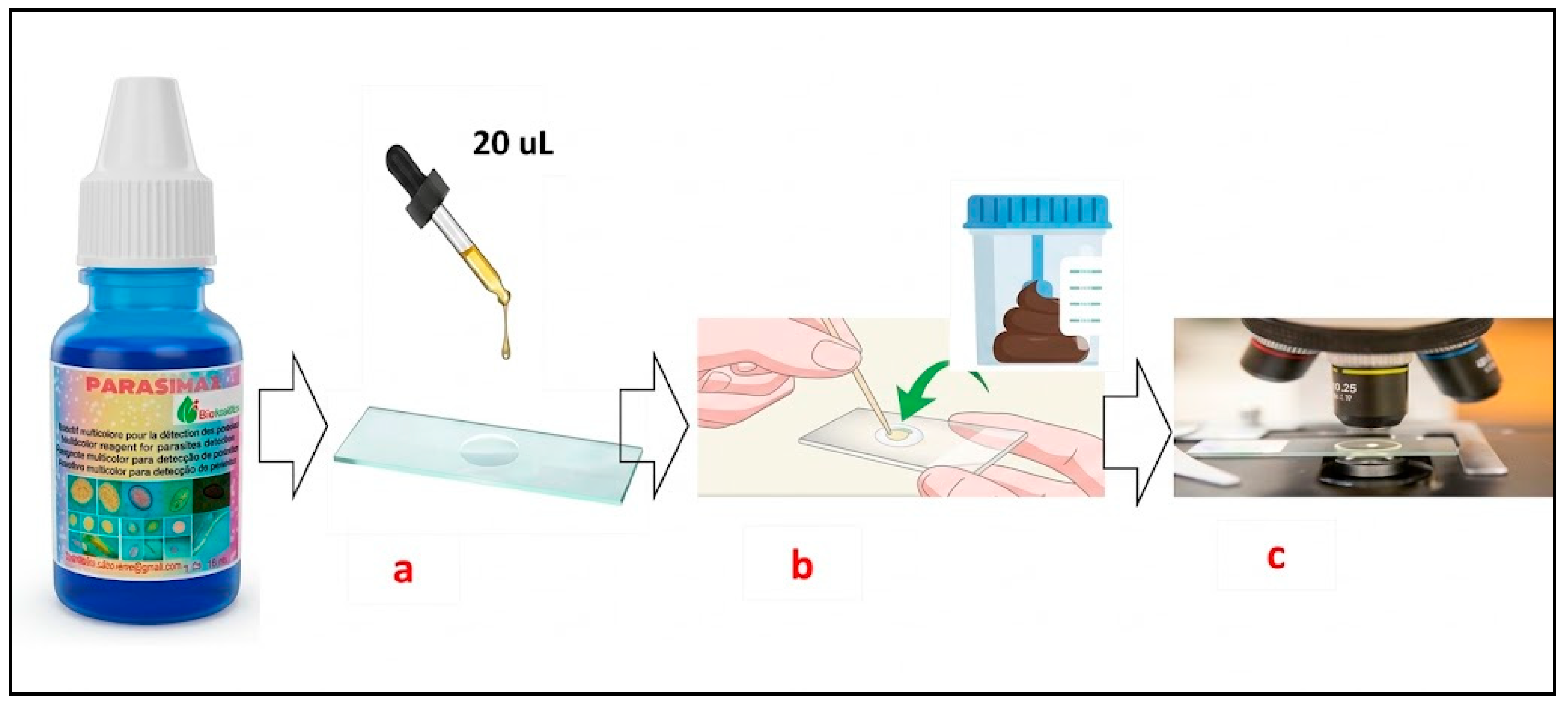
Figure 2.
Hymenolepis nana eggs between artifact observed with Parasimax multicolour (left) and Lugol (right).
Figure 2.
Hymenolepis nana eggs between artifact observed with Parasimax multicolour (left) and Lugol (right).
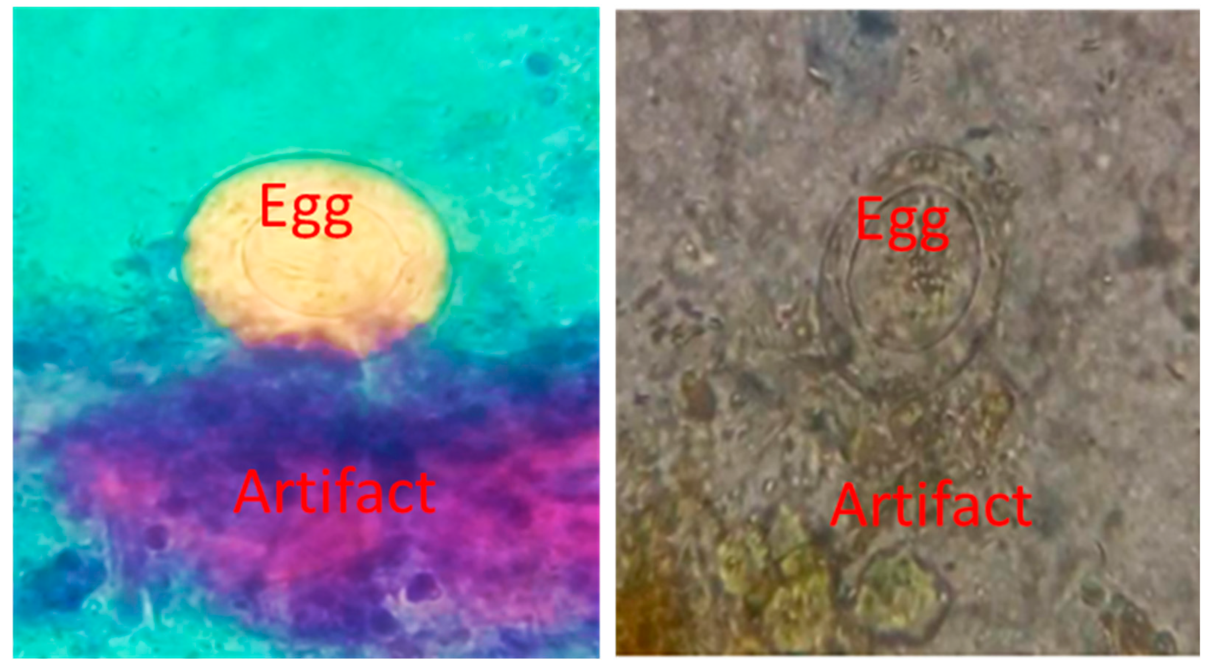
Figure 3.
Eggs of different helminths observed with Lugol (a1-d1) and Parasimax multicolor staining (a2-d2). a1, a2: Enterobius vermicularis; b1, b2: Hymenolepis nana; c1, c2: Taenia spp; d1, d2: Trichuris trichiura.
Figure 3.
Eggs of different helminths observed with Lugol (a1-d1) and Parasimax multicolor staining (a2-d2). a1, a2: Enterobius vermicularis; b1, b2: Hymenolepis nana; c1, c2: Taenia spp; d1, d2: Trichuris trichiura.
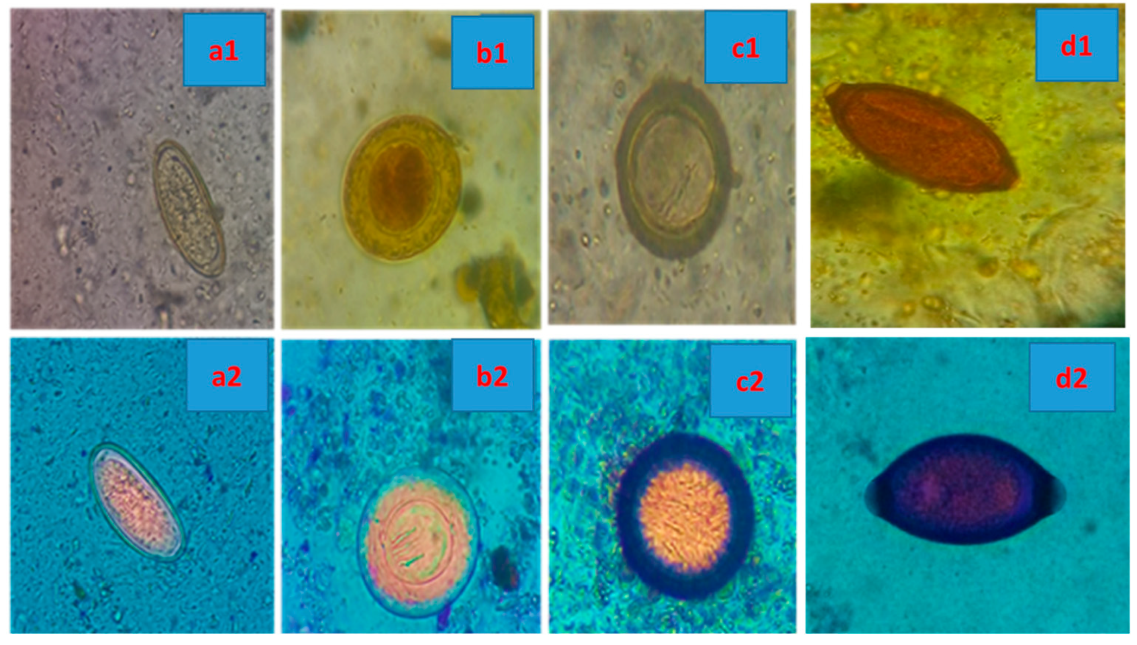
Figure 4.
Proglottids of Taenia spp. A). 40×. B) Observed with 100× lens .
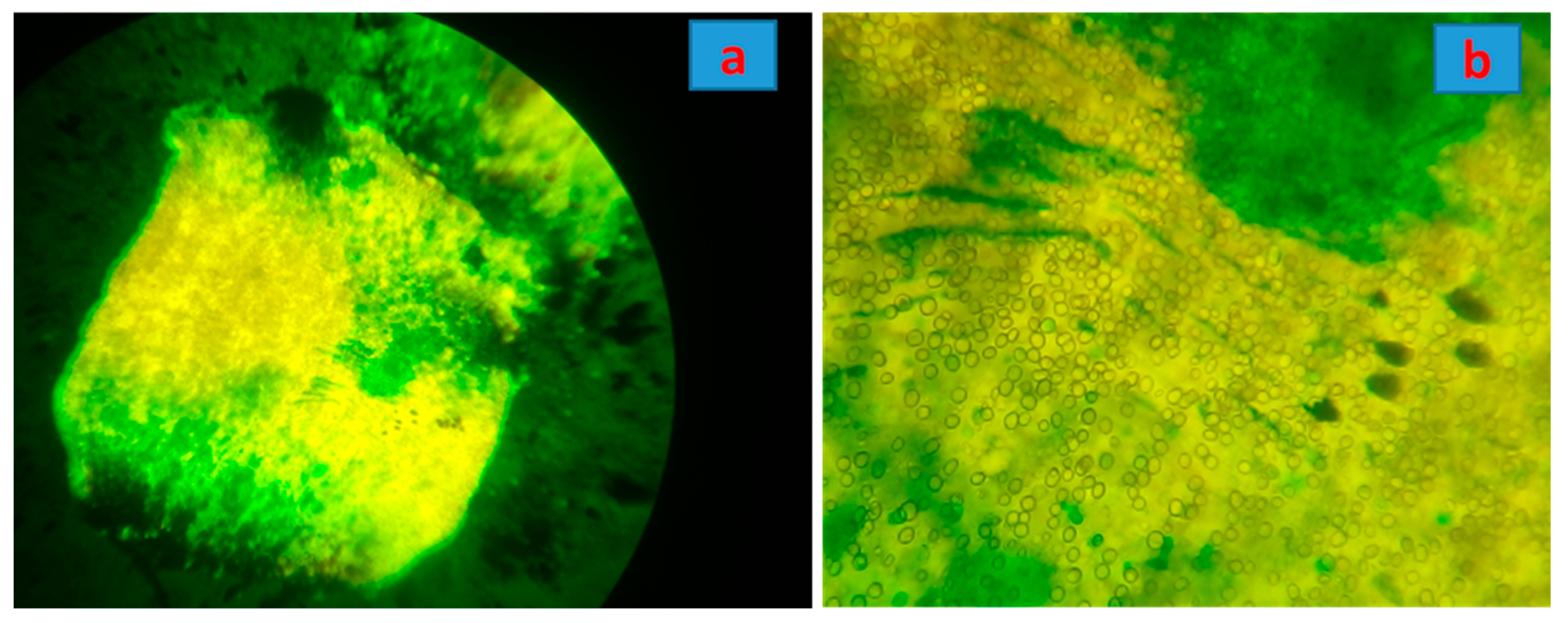
Figure 5.
Giardia lamblia cysts by Lugol (a) and Parasimax multucolor (b); Entamoeba coli cyst with typical nucleus (c).
Figure 5.
Giardia lamblia cysts by Lugol (a) and Parasimax multucolor (b); Entamoeba coli cyst with typical nucleus (c).
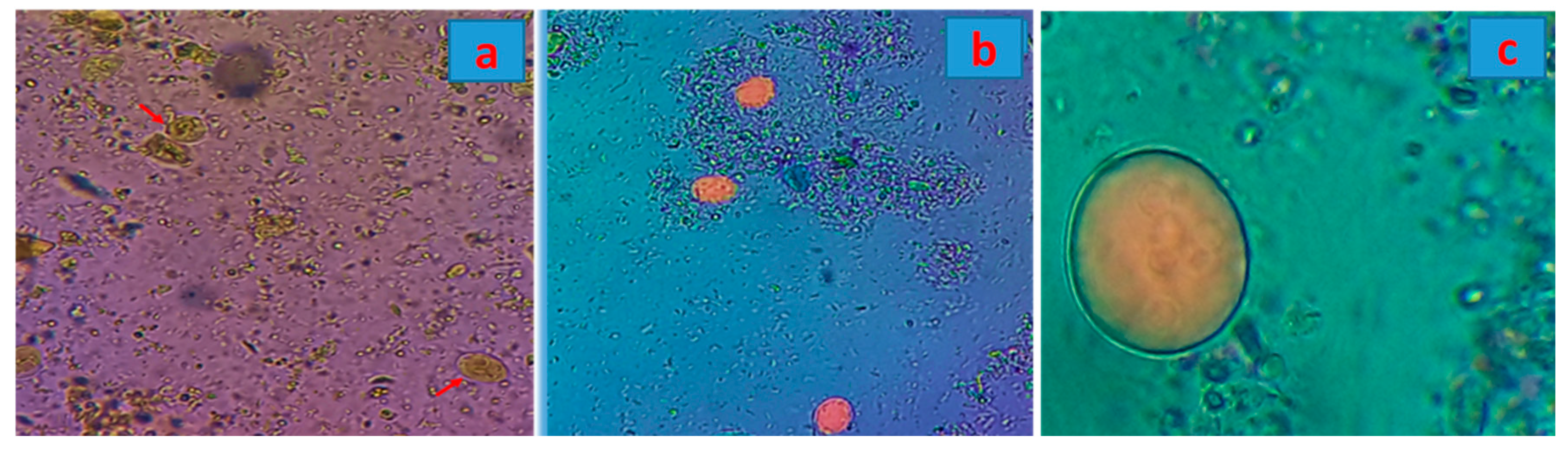
Figure 6.
Giardia lamblia cysts (A) and Enterobius vermicularis eggs (B). Live (yellowl) and dead (green).
Figure 6.
Giardia lamblia cysts (A) and Enterobius vermicularis eggs (B). Live (yellowl) and dead (green).
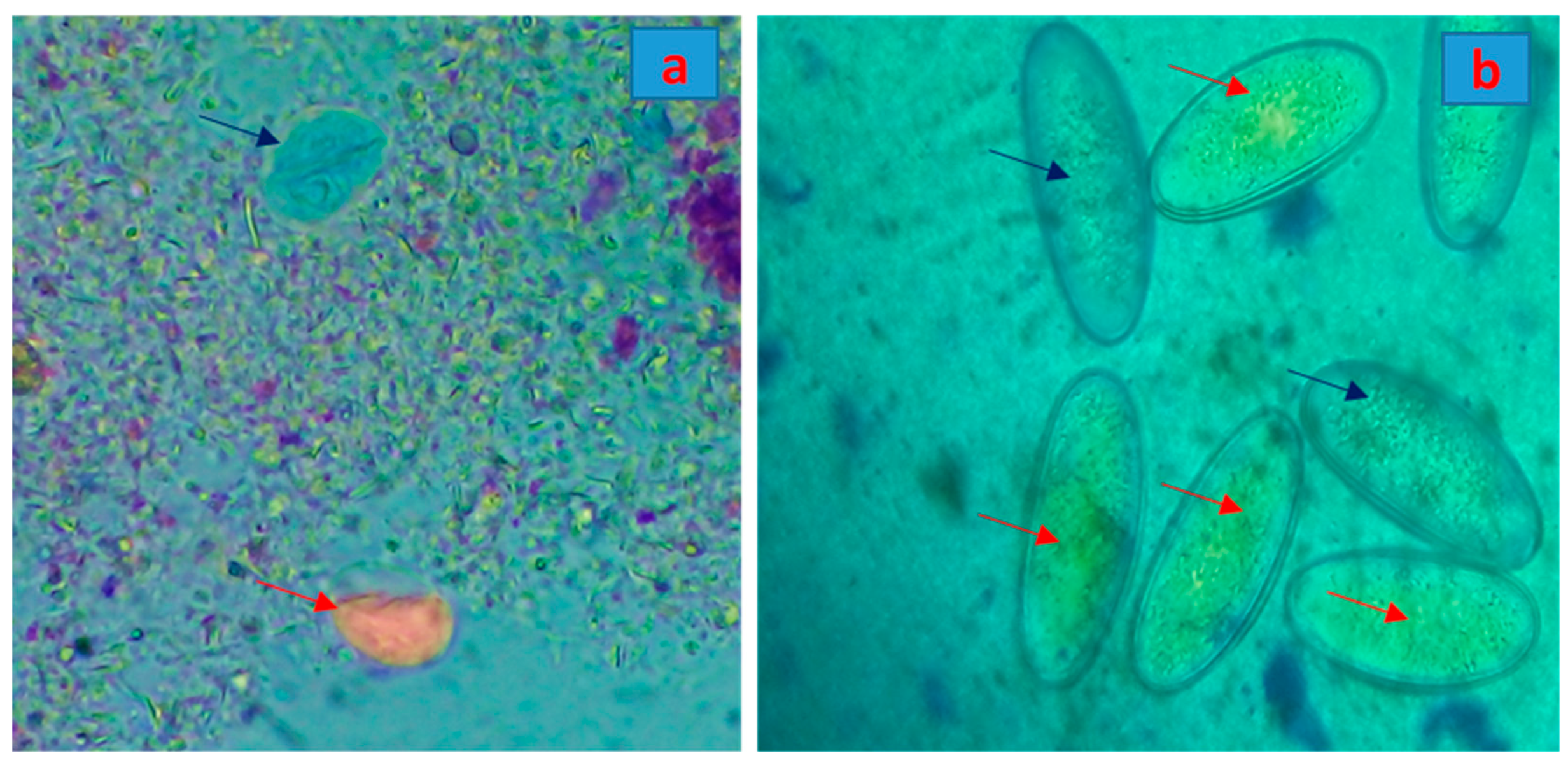
Figure 7.
Dietary fibers from plants stained by parasimax multicolor. 400×.
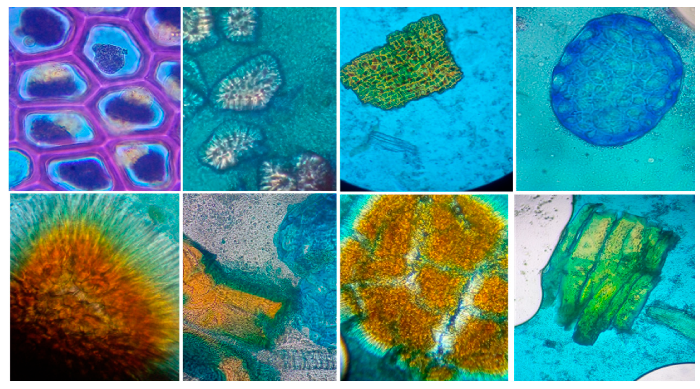
Figure 8.
: Eggs of different helminths, larvae and sporozoa stained by parasimax multicolor and observed by fluorescence microscopy. A) H. nana; B) Ancylostoma spp; C) Ascaris lumbricoides; D) Strongyloides stercoralis (larvae); E) Cystoisospora; F) Enterobius vermiculares (larvae); G) Trichuris trichiura; H) Taenia spp; I) Ascaris lumbricoides (larvae); J) Enterobius vermicularis.
Figure 8.
: Eggs of different helminths, larvae and sporozoa stained by parasimax multicolor and observed by fluorescence microscopy. A) H. nana; B) Ancylostoma spp; C) Ascaris lumbricoides; D) Strongyloides stercoralis (larvae); E) Cystoisospora; F) Enterobius vermiculares (larvae); G) Trichuris trichiura; H) Taenia spp; I) Ascaris lumbricoides (larvae); J) Enterobius vermicularis.
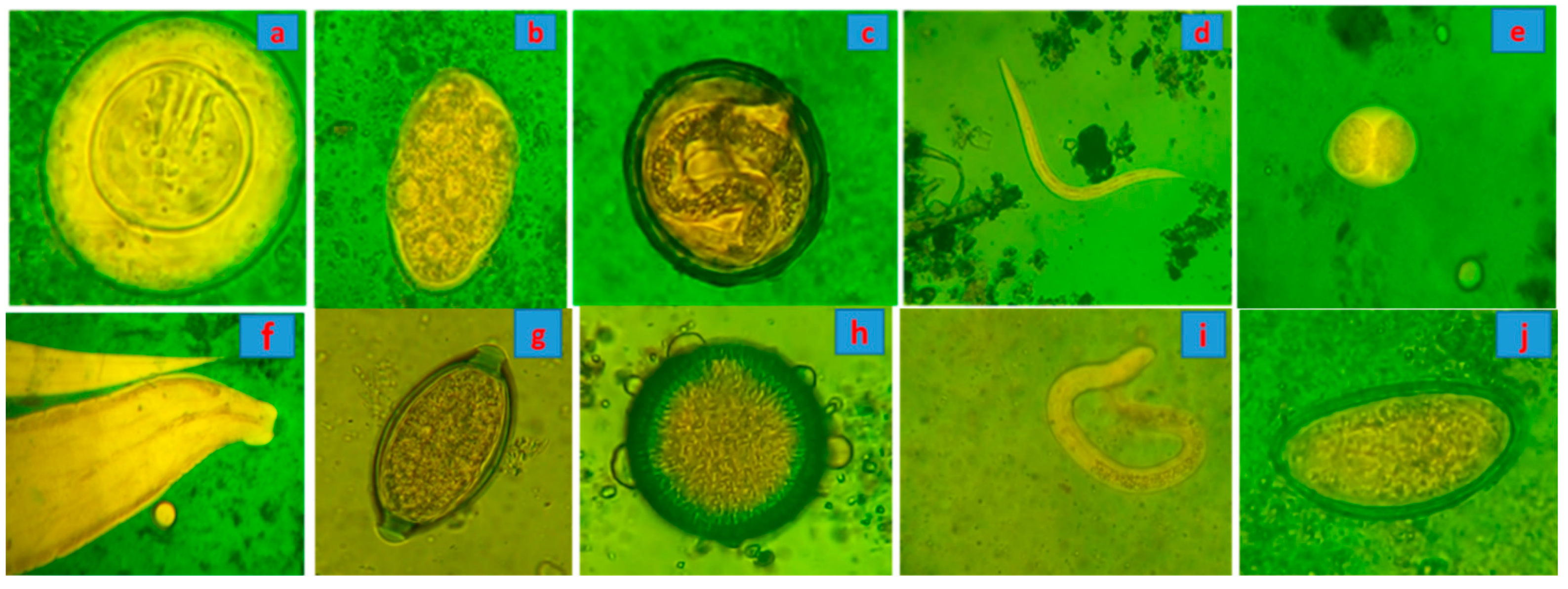

Table 1.
Detection time, comparison between Parasimax multicolur and Lugol by different magnification.
Table 1.
Detection time, comparison between Parasimax multicolur and Lugol by different magnification.
| 40x | 100x | 400x | |
|---|---|---|---|
| Parasimax multicolor | 20 seconds | 40 seconds | 60 seconds |
| Lugol | Not possible | 60 seconds | 2 minutes |
Disclaimer/Publisher’s Note: The statements, opinions and data contained in all publications are solely those of the individual author(s) and contributor(s) and not of MDPI and/or the editor(s). MDPI and/or the editor(s) disclaim responsibility for any injury to people or property resulting from any ideas, methods, instructions or products referred to in the content. |
© 2026 by the authors. Licensee MDPI, Basel, Switzerland. This article is an open access article distributed under the terms and conditions of the Creative Commons Attribution (CC BY) license (http://creativecommons.org/licenses/by/4.0/).
Copyright: This open access article is published under a Creative Commons CC BY 4.0 license, which permit the free download, distribution, and reuse, provided that the author and preprint are cited in any reuse.