

Submitted:
05 October 2026
Posted:
08 October 2026
You are already at the latest version
Abstract
Triple-ion substitution and femtosecond laser surface microstructuring are promising strategies for tailoring the physicochemical and biological properties of β-tricalcium phosphate (β-TCP) ceramics for bone tissue engineering. In this study, Mg (8-9mol%)-, Zn (3 mol%)-, and Sr(1 or 5 mol%) -substituted β-TCP ceramics were synthesized by a biomimetic precipitation route followed by calcination. The ceramic tablets were subsequently surface-textured using a femtosecond laser to generate hierarchical microstructures with different scanning geometries. The phase composition and surface characteristics were investigated by chemical analysis, powder X-ray diffraction, scanning electron microscopy, energy-dispersive X-ray spectroscopy, optical profilometry, and water contact angle measurements. Cytocompatibility was evaluated using HaCaT keratinocytes, while the antibacterial response was assessed against Escherichia coli K12 by colony-forming unit counting and scanning electron microscopy. The laser-textured surfaces exhibited modified roughness and substantially improved wettability compared with the untreated controls. The 45° intersecting-line pattern produced the highest surface roughness, particularly for the 5 mol% Sr composition, while the laser-treated Sr5 surfaces were immediately wetted by water, indicating highly hydrophilic behavior. Laser texturing also reduced bacterial adhesion and viable bacterial counts compared with the untreated controls, with the lowest bacterial counts observed for the surfaces patterned with parallel laser lines. At the same time, the laser-treated ceramics maintained their cytocompatibility, with no increase in cell death or adverse morphological changes observed in HaCaT keratinocytes after 24 and 48 h of direct contact. Overall, the combination of Mg, Zn, and Sr substitution with femtosecond laser surface texturing effectively tailored the surface morphology, roughness, wettability, and biological response of β-TCP ceramics.
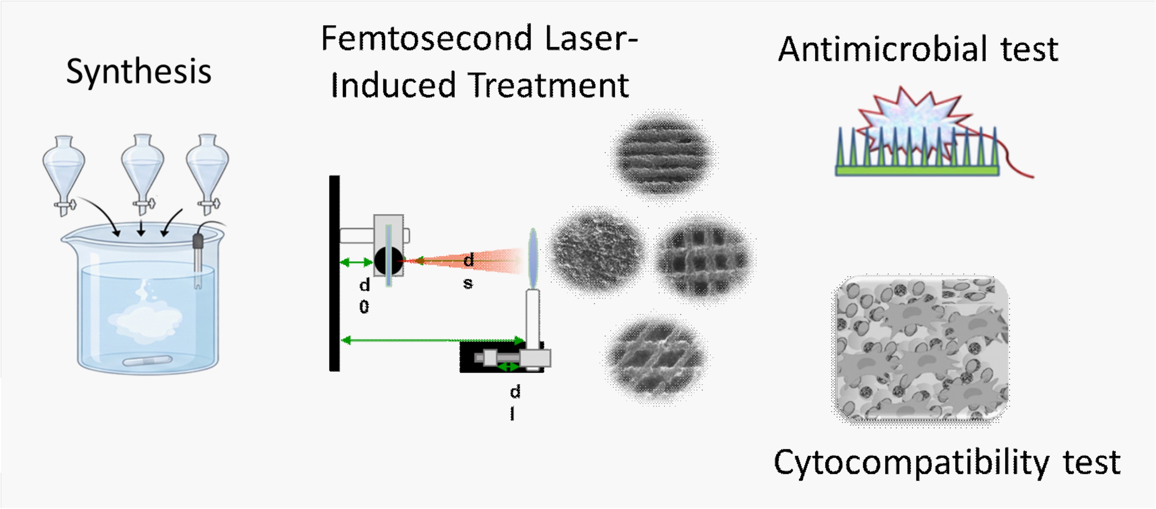
Keywords:
substituted β-TCP
; ceramics
; femtosecond laser treatment
; cytotoxicity
; antimicrobial test
1. Introduction
Ion-modified nanoscale calcium-deficient carbonate-phosphate apatite, called “biological apatite,” is the inorganic constituent found in mineralized biological structures. The key cationic inclusions in this so-called “biological apatite” are Na+, K+, Mg2+, Fe2+, Zn2+, and Sr2+, with their concentrations varying with the specific type of mineralized tissue, the individual's age, and personal characteristics [1,2]. Due to the chemical and structural resemblance of calcium orthophosphate compounds to “biological apatite”, they are well-suited for creating biomaterials for bone regeneration and reconstruction. The ideal synthetic biomaterial for implanting and reconstructing bone defects should be bioactive, bioresorbable, and biocompatible, but no synthetic material currently meets all three requirements.
Calcium phosphate ceramics are widely used in bone tissue engineering due to their excellent osteoconductivity and favorable biocompatibility [3]. Among them, β-tricalcium Phosphate (β-TCP) has attracted significant attention due to its bioresorbability [4,5]. However, pure β-TCP may have limited biological activity, motivating compositional modification through ionic substitutions [6,7,8,9]. Incorporating biologically relevant ions such as Mg²⁺, Zn²⁺, and Sr²⁺ into the β-TCP lattice has been shown to modify its physicochemical properties, including crystal lattice parameters, solubility, and ion-release behavior, thereby further enhancing its biological functionality [10,11,12]. Magnesium plays an important role in bone metabolism and improves cell adhesion and proliferation [13]. Zinc stimulates osteoblast activity and exhibits antimicrobial properties, reducing the risk of implant-associated infections [14]. Strontium ions stimulate osteoblast proliferation and differentiation, the cells responsible for new bone formation, while reducing osteoclast activity, which is involved in bone degradation [15]. When these ions are co-doped into the β-TCP structure, they could act synergistically to enhance both the physicochemical and biological properties of the material.
Researchers have widely investigated incorporating Mg²⁺, Zn²⁺, and Sr²⁺ ions, individually or in various combinations, into calcium phosphate ceramics [10,11,12,16,17,18]. However, information on triple modification remains scarce. Ressler et al. [19,20] synthesized composite scaffolds based on Mg-, Se-, Sr-, and Zn-substituted calcium phosphates to enhance bone tissue regeneration. The mono- and multi-substituted calcium phosphate powders were prepared with ionic substitution levels of 1 or 5 mol% for each ion (Sr²⁺, Mg²⁺, Zn²⁺, and SeO₃²⁻). Their findings, based on evaluations of bone cell growth and proliferation and the expression of osteogenic markers such as alkaline phosphatase, bone sialoprotein, and dentin matrix protein, indicate that the multi-substituted materials with 5 mol% substitution perform best due to the synergistic effect of the incorporated trace elements.
Although substituted calcium phosphate ceramics have been widely developed as bone graft materials, their full regenerative potential is often limited by poor surface characteristics. Surface properties, such as topography, roughness, wettability, etc., are important for cell attachment and differentiation [19]. This highlights the need for additional surface engineering strategies to better control cell-material interactions. In this context, laser surface modification is a promising approach, as it enables precise, controllable tailoring of surface morphology without altering the material's bulk composition [20,21]. Therefore, combining the preparation of ion-substituted calcium phosphate ceramics with laser surface modification offers a powerful strategy to optimize both bulk and surface properties, ultimately improving their performance as bone graft materials.
In this article, we extend our previous studies on the synthesis and characterization of doubly doped β-TCP with Zn and Mg [18,21] by incorporating a third bioactive element, Sr, at concentrations of 1 and 5 mol%. The surfaces of the resulting ceramic tablets were textured with a femtosecond laser to evaluate different processing regimes. Furthermore, the cytocompatibility of laser-textured calcium phosphate ceramic tablets was evaluated using a direct-contact assay with HaCaT keratinocytes, enabling both qualitative and quantitative assessment of cell responses, including morphology and viability. In parallel, a preliminary evaluation of Escherichia coli viability and morphology was conducted on selected samples, demonstrating that laser-induced surface texturing—combined with metal ion doping—can generate nano-, micro-, or hierarchical surface structures with potential antimicrobial effects.
2. Materials and Methods
2.1. Preparation and Characterization of Triple-Doped (Mg, Zn, Sr)-β-TCP Ceramic Tablets
A three-step method was used to prepare ceramic tablets that includes: (i) biomimetic synthesis of amorphous (Sr, Mg, Zn)-modified calcium phosphate; (ii) tablet preparation; and (iii) calcination of the tablets.
A continuous precipitation method was used to synthesize amorphous (Sr, Mg, Zn)-modified calcium phosphate. The biomimetic approach uses a simulated body fluid (SBF) as the solvent for the starting materials. Conventional SBF (SBFc) proposed by Kokubo [22] was used as a base. Calcium-free conventional simulated body fluid (SBFc) was used as a solvent for K2HPO4 (Merck, A.R., Darmstadt, Germany), while phosphorus-free SBFc was used for dissolution of CaCl2.2H2O (Sigma-Aldrich, A.R., St. Louis,MO, USA), MgCl2.6H2O (Merck, A.R., Darmstadt, Germany), SrCl2.6H2O (Riedel de Haen, A.R., Seelze, Germany), and ZnCl2.2H2O (Merck, A.R., Darmstadt, Germany) (Table 1).
Concentrations of Mg2+, Zn2+, Ca2+, Sr2+ and PO43- ions were calculated to produce calcium phosphate precursors with ratios:
(Ca2+ + Mg2+ + Zn2+ + Sr2+)/Р = 1.67
Mg2+/(Ca2+ + Mg2+ + Zn2+ + Sr2+) = 0.07
Zn2+/(Ca2+ + Mg2+ + Zn2+ + Sr2+) = 0.03
Sr/(Ca2+ + Mg2+ + Zn2+ + Sr2+) = 0.01 (called hereafter Sr1) or
Sr/(Ca2+ + Mg2+ + Zn2+ + Sr2+) = 0.05 (called hereafter Sr5)
In our previous studies, we found that about half of the Mg2+ ions introduced with the solution are incorporated into the solid phase; therefore, the Mg2+ concentration is doubled compared to the calculated value [23]. The concentrations of Mg2+ and Zn2+ ions were selected on the basis of our previous studies on the biocompatibility of Mg- or Zn-modified tri-calcium phosphates [24].
A combined apparatus for automatic titration and controlled synthesis (Titrando 907, Metrohm AG, Switzerland) was used. Equal volumes of Solution 1, Solution 2, and Solution 3 (Table 1) were added to the same volume of glycine buffer solution [25] at room temperature with continuous stirring at a rate of 3 mL min-1, keeping the pH at 8.0–8.2 using 2 M KOH. The pH is chosen to be close enough to physiological but not to allow the formation of acid calcium salts. The pH of Solution 3 (Table 1) was not corrected to avoid Zn-hydrolysis products. After precipitation, the suspension was matured in the mother liquor for 1 h under continuous stirring at room temperature and then washed with water (to remove chlorides) and lyophilized.
The lyophilized powder was milled in a ball mill (Fritsch 6) for 0.5 h at 500 rpm, then passed through a 0.1mm sieve. An amount of 0.65 g of the sieved powder was tableted at a pressure of 6.5 tons on a SPECAC GS15011 press (Orpington, GB). Prepared tablets with a diameter of 13.1 mm and thickness of 2.65 mm were calcined at 400, 600, and 1000 ℃ and atmospheric pressure in a high-temperature furnace (type VP 04/17, LAC Ltd. Company, Rajhrad, Czech Republic). The working regime was heating at a rate of 5 ℃ .min-1 until the desired temperature was reached and then keeping it constant for 1 h.
The concentrations of Ca2+, Zn2+, Mg2+ and Sr2+ ions in the solid samples were determined by Inductively Coupled Plasma–Optical Emission Spectrometry (ICP-OES) (PRODIGY 7, Teledyne, Leeman Labs, USA). P-PO43- and Cl- ions were analyzed spectrophotometrically by NOVA 60 equipment with Merck Spectroquant test kits. Ten parallel independent measurements were performed for each assay. The statistical analysis of the data was carried out using Microsoft Excel 2016. The standard deviation value expresses the accuracy of the results. The accuracy of the (Ca + Mg + Zn)/P ratio was calculated using Equation (1), and the accuracy of the M/(Ca + Mg + Zn+Sr) (M = Mg, Zn, Sr) ratio - using Equation (2):
where SD—standard deviation; CCa, CMg, CZn, CSr and CP – average value of measurements of concentration of Ca2+, Mg2+, Zn2+, Sr2+ and, P-PO43− ions, sCa, sMg, sZn, sSr and sP—standard deviation of measurements of Ca2+, Mg2+, Zn2+, Sr2+ and, P-PO43− ions, M = Mg2+, Zn2+, Sr2+, D = CCa+CMg+CZn+CSr, s2 = s2Ca+s2Mg+s2Zn+s2Sr
Powder X-ray diffraction (PXRD) was performed using a Bruker D8 Advance diffractometer with Cu Kα radiation and a LynxEye detector (Bruker AXS Advanced X-ray Solutions GmbH, Billerica, MA, USA). For the primary phase identification, the data were collected in the range of 10 to 90° 2θ with step 0.03° 2θ, with counting time 57 s/step.
2.2. Processing with a Femtosecond Laser and Characterization
Surface texturing of the sintered at 1000 °C triple-doped (Mg, Zn, Sr)-β-TCP ceramic tablets was performed using an ultrashort-pulse Ti laser system (Solstice ACE, MKS Spectra-Physics, USA). The laser operated at a wavelength of 800 nm with a pulse duration of 70 fs and a repetition frequency of 1 kHz. The maximum available output power was 6 W. Beam delivery was achieved through a galvanometric scanning head (SCANLAB, Germany) integrated with a motorized Z-axis positioning module, enabling accurate fabrication of complex microstructured surface architectures. The laser beam was focused normally to the tablet surface using an f-theta lens with a focal length of 160 mm, resulting in a focal spot diameter of approximately 50 μm. This optical configuration enabled reproducible generation of hierarchical surface patterns with controlled geometrical features.
Laser structuring was carried out by raster scanning while systematically adjusting the processing parameters, including laser power (P), scanning velocity (v) and hatch distance (dx). Parameter selection was based on previous optimization studies aimed at obtaining well-defined surface morphologies while minimizing thermal damage, micro cracking, or material degradation. Furthermore, the selected processing conditions were chosen to produce topographical characteristics suitable for biological interactions and antimicrobial performance. For both tablet compositions (Sr1 and Sr5), several laser-patterning strategies were initially evaluated at a constant laser power of 50 mW, with varying scanning velocities (v=0.1-1.5mm/s) and line orientations (90° and 45°). Based on the obtained surface characteristics and subsequent biological assessment, six representative laser-treated groups were selected for detailed investigation:
- Sr1-lines – Sr1 tablets processed at P = 50 mW and v = 1.0 mm/s using parallel scan lines, dx=50µm.
- Sr1-90° – Sr1 tablets processed at P = 50 mW and v = 1.0 mm/s using intersecting scan lines oriented at 90°, dx=100µm.
- Sr1-45° – Sr1 tablets processed at P = 50 mW and v = 1.0 mm/s using intersecting scan lines oriented at 45°, dx=100µm.
- Sr5-lines – Sr5 tablets processed at P = 50 mW and v = 1.0 mm/s using parallel scan lines, dx=50µm.
- Sr5-90° – Sr5 tablets processed at P = 50 mW and v = 1.0 mm/s using intersecting scan lines oriented at 90°, dx=100µm.
- Sr5-45° – Sr5 tablets processed at P = 50 mW and v = 1.0 mm/s using intersecting scan lines oriented at 45°, dx=100µm.
After laser treatment, all ceramic tablets were ultrasonically cleaned in 70% ethanol to remove residual particles generated during ablation. The samples were then dried under ambient laboratory conditions before further characterization and biological testing. Untreated tablets of identical composition served as control specimens (cS1 and cS5).
The morphology of laser-textured ceramic tablets was investigated using a field-emission scanning electron microscope (SU8020, Hitachi High-Technologies, Japan). Before imaging, the samples were coated with a conductive carbon layer approximately 20 nm thick to suppress charging effects during electron beam exposure. Micrographs were recorded at three magnifications as follows: 500×, 2500× and 10 000× in order to evaluate both microscale and submicron surface features generated by laser processing.EDX spectra
Three-dimensional surface topography was assessed using a Zeta-20 optical profilometer (KLA Corporation, USA). The instrument combines optical microscopy and vertical scanning interferometry, providing high-resolution surface reconstruction and quantitative characterization of textured regions. Surface roughness parameters were extracted from the reconstructed 3D maps (taken at 20× magnification). The arithmetic mean roughness (Ra) and areal roughness (Sa) were calculated to quantify the influence of laser processing on surface development. For each sample type, five independent measurements were performed at different locations, and the reported values correspond to the mean value. Additional data processing and visualization were carried out using ProfilmOnline 2.0.0 software (Filmetrics, KLA Corporation, USA).
The wetting behavior of the untreated and laser-structured ceramic surfaces was determined using a DSA100 contact-angle measurement system (KRÜSS GmbH, Germany). Static and dynamic water contact angle measurements were conducted at room temperature using 2 μL droplets of deionized water deposited on the tablet surfaces. The droplet profile evolution was monitored for 3 min using a high-resolution video acquisition system. Contact angle values were calculated automatically with ADVANCE software (KRÜSS) employing Young–Laplace fitting of the droplet contour. Measurements were carried out at three different positions on each sample, with five independent specimens analyzed for every experimental group. Average water contact angle values (WCA, [°]) were calculated and compared with those obtained for the corresponding untreated control tablets to evaluate the influence of femtosecond laser texturing on surface wettability.
2.3. Cytocompatibility Test Through Direct Contact Assay
HaCaT keratinocytes (ATCC, USA) were seeded in 12-well plates at a density of 1 × 10⁵ cells per well and cultured until reaching approximately 70% confluency. The test sample specimens were then carefully placed on the cell layer at the center of each vessel. After 24 h, phase-contrast images (10× objective, Zeiss AxioVert 200 M, Zeiss, Germany) were acquired, and cells were trypsinized. Half of the cells were pelleted (500 × g, 5 min), while the remaining cells were further incubated with the tablets for an additional 24 h. Cells present in the culture medium were also collected. Following a single Phosphate-buffered saline (PBS) wash, cells were resuspended in 10 µg/mL propidium iodide (PI) in PBS and immediately analysed by flow cytometry (BD FACSCalibur). The PI viability assay is based on propidium iodide selectively penetrating cells with compromised membrane integrity, allowing rapid and accurate quantification of viable and non-viable cells by flow cytometry. Three independent experiments were performed. 30000 cells were counted per condition.
2.4. Antimicrobial Test
2.4.1. In Vitro Determination of Antimicrobial Activity by Plate Count
E. coli K12 strain was used. The strain was inoculated in Tryptic Soy Broth (TSB, Sigma-Aldrich, Berlin, Germany) and cultivated for 18 h at 37°C under static conditions. The ceramic tablets were sterilized by dry heat at 200°C for 30 min and placed in a 12-well plate. The 18-hour inoculum was distributed in the presence of the laser-treated tablets. Cultivation was then carried out for 24 h at 37°C. Cell viability was assessed using the drop plate method. Bacterial cells were collected from the surface of the tablets using a sterile swab and serially diluted in sterile physiological saline. The number of surviving bacterial cells was determined by plating 100 µL of each dilution onto TSA Tryptic Soy Agar plates (TSA, Sigma-Aldrich, Berlin, Germany), followed by surface spreading. The inoculated plates were incubated at 37°C for 24 h, after which bacterial colonies (CFU) were counted. The analysis was performed in triplicate.
2.4.2. Evaluation of E. coli Cell Morphology by Scanning Electron Microscopy (SEM)
To evaluate the changes in bacterial morphology after cultivation in the presence of laser-textured ceramic tablets, scanning electron microscopy was performed. The bacterial strain was prepared as described above. Each sample was prepared in duplicate, and SEM processing followed Daskalova et al. [21].
3. Results
3.1. Preparation and Characterization of Triple-Doped (Mg, Zn, Sr)-β-TCP Ceramic Tablets
Experimental studies showed that X-ray amorphous calcium phosphate (Figure 1) with (Ca+Zn+Mg+Sr)/P molar ratio lower than that of hydroxyapatite (Ca/P = 1,67) but close to that of β-TCP (Ca/P = 1.5) was obtained in both series independently of Sr concentration (Table 2).
The main crystallographic peaks correspond to the characteristic peaks of whitlockite (ICSD database, Collection Code 6190). Increasing the calcination temperature increases peak intensity and narrows the diffraction peaks, indicating a higher degree of crystallinity. The magnified section of the diffractogram clearly shows a shift in the diffraction maxima of the synthesized samples relative to the standard. This indicates a change in the crystal lattice parameters resulting from the isomorphous incorporation of doping ions into the structure.
Based on our previous studies on doubly doped calcium phosphates [23,26], we expect an amorphous precursor to form and subsequently transform into β-TCP upon heating. Under these synthesis conditions, kinetic factors governing amorphous calcium phosphate precipitation dominate, and divalent modifying ions such as Zn, Mg, and Sr slow the transformation to the thermodynamically most stable hydroxyapatite during synthesis, maturation, and even heating up to 400 °C. It has also been confirmed that when the total concentration of substitution ions exceeds 5 mol %, the b-TCP structure is stabilized and remains intact even at 600 °C without any hydroxyapatite admixture [23]. The chemical analysis results (Table 2) show that the Zn and Sr concentrations match those introduced into the starting solutions, and that the Mg concentration, as expected, is half that introduced. This is because the energy barrier must be overcome to destroy the strong magnesium hexa-aqua octahedral complexes forming in solution [23,26].
3.2. Femtosecond Laser-Induced Microstructure Formation and Wettability Evaluation
The laser-generated patterns increased the surface complexity and introduced one- or bidirectional hierarchical microtopographical features compared with the relatively smooth, untreated control surfaces (Figure 2).
Femtosecond laser processing successfully generated distinct microstructured surface architectures on both Sr1 and Sr5 β-TCP ceramic tablets. Figure 3 shows representative SEM images. The untreated control surfaces exhibited relatively smooth morphologies with granular composition (Figure 2), whereas laser irradiation produced well-defined porous microgrooves and hierarchical microstructures with no evidence of extensive melting, cracking, or thermal degradation (Figure 3).
Depending on the scanning strategy, either parallel grooves (Sr1-lines and Sr5-lines) or cross-hatched architectures formed by intersecting lines at 90° (Sr1-90° and Sr5-90°) and 45° (Sr1-45° and Sr5-45°) were obtained, and ejection of the material above the sample's baseline at the site of interaction was monitored. As a result, we observed the formation of porous-granular foam, clearly visible in the SEM images at higher magnification (2000× and 10,000×) of all laser-patterned Sr1 and Sr5 samples (Figure 3).
A representative compilation of 2D transverse profile and 3D real-colour reconstruction images of the laser-structured tablets is given in Figure 4.
Quantitative topographical analysis from optical profilometry complements the SEM results and shows a strong dependence of surface roughness on laser-pattern geometry. The untreated Sr1 control exhibits an areal roughness (Sa) of 3.02 μm and a linear roughness (Ra) of 1.25 μm (Figure 4(A)). Laser processing increased the Ra values for all investigated surface designs, indicating the formation of pronounced microrelief porous structures. Among the Sr1 samples, the highest roughness values were measured for the Sr1-45° group (Sa = 3.31 μm, Ra = 2.09 μm), followed by Sr1-lines (Sa = 2.31 μm, Ra = 1.45 μm) and Sr1-90° (Sa = 2.15 μm, Ra = 1.26μm). Notably, the cross-hatched pattern generated at 45° (Figure 4(D)) produced a substantially rougher surface than the corresponding 90° architecture (Figure 4(C)). This behavior can be attributed to the scanning geometry, as the oblique intersecting lines (45°) create a higher effective overlap of adjacent laser tracks and a denser network of intersections than the orthogonal pattern (90°), which is clearly seen in the SEM micrographs presented in Figure 3(B and C). Consequently, repeated laser irradiation over a larger fraction of the surface promotes more extensive material removal and redistribution, resulting in deeper valleys, higher peaks, and more pronounced hierarchical surface topography, as reflected in the higher Sa and Ra values (Figure 4).
A similar trend was observed for the Sr5 series (Figure 4 (E-H)). The untreated Sr5 control sample displays lower initial roughness values (Sa = 2.45 μm, Ra = 0.7 μm) than the Sr1 control sample. After laser treatment, both roughness parameters increased significantly. The Sr5-45° surface exhibited the highest roughness among all investigated groups, reaching Sa = 4.95 μm and Ra = 2.26 μm. The Sr5-lines and Sr5-90° groups showed intermediate values of Sa = 3.69 μm and 2.94 μm, and Ra = 2.82 μm and 1.92 μm, respectively. These results indicate that the 45° intersecting-line configuration promotes more pronounced three-dimensional surface morphology above and below the laser-structured tablet's surface baseline, regardless of Sr concentration.
Comparative roughness analysis further showed that incorporating 5 mol% Sr enhanced the effect of laser texturing. For identical laser-processing conditions, Sr5 samples consistently exhibited higher Sa values than their Sr1 analogs. This effect was particularly evident in the 45° pattern, where the Sa parameter increased from 3.31 μm for Sr1-45° to 4.95 μm for Sr5-45°, representing an increase of approximately 50%. These differences suggest that ceramic composition influences laser–material interaction and, consequently, the resulting surface topography. The increased Sr content facilitates the formation of a more developed three-dimensional surface morphology during femtosecond laser processing, although the underlying mechanism requires further investigation.
Figure 5 demonstrates that femtosecond laser texturing substantially modified the wetting behavior of the triple-doped β-TCP ceramics. The untreated Sr1 and Sr5 control samples exhibited similar mean water contact angles of 83.05° and 84.59°, respectively, indicating moderately hydrophilic surfaces (Table 3). After laser processing, all Sr1 surface architectures showed a pronounced reduction in WCA, with values between 34.19° and 40.92°, corresponding to an approximately 50–60% decrease compared with the untreated surface. Among the laser-treated Sr1 groups, the 45° intersecting-line pattern showed the lowest contact angle, consistent with its highest surface roughness (Sa = 3.31 μm, Ra = 2.09 μm), suggesting that the more developed hierarchical microtopography promotes more efficient water spreading.
An even stronger wettability enhancement was observed for the Sr5 series. Although the untreated Sr5 tablets displayed a contact angle comparable to that of the Sr1 control, all laser-textured Sr5 surfaces became completely wetted immediately after droplet deposition, preventing reliable contact-angle measurements. This behavior is consistent with the significantly higher roughness values monitored for the Sr5 laser-treated samples and more developed hierarchical microstructures produced by femtosecond laser texturing, particularly the Sr5-45° surface (Sa = 4.95 μm), and indicates a transition to superhydrophilic behavior.
3.3. Effects on Bacterial Viability and Cell Morphology
The analysis of the colony count results revealed clear differences among the tested samples. Compared with the control samples, the laser-treated samples showed lower CFU values (Figure 6).
The laser-processed Sr1/Sr5-line sample showed the lowest CFU values, indicating suppressed bacterial growth and a potential bactericidal effect compared with laser-treated tablets using intersecting scan lines oriented at 45° or 90°. In the sample treated with intersecting scan lines oriented at 90°, the number of bacterial colonies was approximately twice that of Sr1/Sr5-line but still less than half of those observed with intersecting scan lines oriented at 45°, suggesting a certain level of bacterial sensitivity. In this experiment, the laser treatment of the ceramic tablets and the ingredient ratios in the tablets were additional factors influencing the degree of bacterial adhesion, which in turn affected the total number of viable cells. This observation is consistent with our previous findings, which showed that laser-induced surface modifications alter bacterial attachment and survival [21].
Analyzing the SEM results, we can also conclude that in the laser-treated samples the number of adherent bacteria was significantly reduced compared to the two control samples for both the Sr1 and Sr5 series (Figure 7).
It is important to note that the Sr5 tablet (Figure 7 B, D, F, H) significantly reduced the adhesion ability of bacterial cells compared to Sr1 (Figure 7 A, C, I, G). This reduction in bacterial colonization correlated with the viability assessment based on colony-forming unit counts. In the S1-line and S5-line samples, we observed the sparsest distribution of bacterial cells along the laser-generated surface architecture for both tablet compositions (Sr1 and Sr5). The other laser-treated samples (Sr1-45; Sr1-90; Sr5-45 and Sr5-90) similarly showed uneven distribution, lack of three-dimensional structure, and reduced cell density, which are usually characteristic of mature biofilm communities. The cells were located singly in separate areas and were not grouped in small aggregates, as observed in the two control groups. The Sr5 tablet composition reduced adhered cells more significantly. In line with our findings, Eghbali et al. (2021) [27] reported suppressed adhesion of E. coli on modified Ti-6Al-4V alloy surfaces. Our research also found no morphological abnormalities in either the control or treated groups, no cell wall deformations (such as dents, bulges, or tears), and no changes in shape or cell division patterns. Among the laser-treated samples, those of Sr1-line and Sr5-line showed the most pronounced reduction in bacterial colonization, indicating a potential antimicrobial effect compared to the control groups. Wu et al. (2018) [28] and Jin et al. (2015) [29] reported similar observations on antibacterial microtopographies.
3.3. Cytocompatibility Tests
The cytocompatibility of the calcium phosphate ceramic tablets after laser-induced surface texturing was evaluated using a direct contact assay. The direct contact assay allowed both qualitative (cell morphology) and quantitative (number of live cells) evaluation of cytotoxicity by placing the test specimens directly onto a subconfluent cell layer and assessing cell response after incubation. The approach enabled assessment of both direct contact effects and potential influences from soluble components released into the culture medium.
HaCaT keratinocytes were selected as a representative human skin cell line frequently used for biomaterial testing due to their robustness and relevance for studying surface–cell interactions. Cellular responses were monitored for up to 48 hours. No increase in cell death was detected for any of the tested tablet variants compared with untreated control cells (Figure 8).
Moreover, phase-contrast microscopy showed no morphological alterations, confirming that neither the base ceramic nor the laser-induced surface modification exhibited cytotoxic or morphologically adverse effects on HaCaT cells. These findings indicate that the laser-treated calcium phosphate ceramics maintain high biocompatibility under direct contact conditions.
4. Discussion
Ion-modified β-TCP ceramics represent a promising class of bioactive materials for orthopedic and dental applications, particularly when combined with advanced surface modification techniques to further improve cellular response and antimicrobial performance.
In this study, triple-doped (Mg, Zn, Sr)-β-TCP ceramic tablets were prepared via biomimetic precipitation of the precursor, followed by calcination. In the syntheses, the concentrations of Mg and Zn were kept constant at Mg/(Mg+Zn+Sr+Ca) = 0.07 and Zn/(Mg+Zn+Sr+Ca) = 0.03, while Sr/(Mg+Zn+Sr+Ca) was varied to 0.01 or 0.05. Chemical analysis confirmed that the Zn and Sr concentrations incorporated into the solid phase were similar to the target values, whereas approximately half of the Mg introduced into the synthesis solution was incorporated into the precipitate (Table 1 and Table 2). This behavior is consistent with the previously observed lower incorporation of Mg into calcium phosphate phases and can be related to the strong hydration of Mg²⁺ ions in aqueous solution [23,26]. The biomimetic medium ensures the incorporation of Na+, K+, and Cl− ions, which are also characteristic of biological apatite [23]. The resulting (Ca+Mg+Zn+Sr)/P ratios of 1.55 and 1.51 for the Sr1 and Sr5 series, respectively, were close to the stoichiometric ratio of β-TCP (1.50). PXRD analysis (Figure 2) further confirmed the formation of a whitlockite-type β-TCP phase after calcination. The shift of the diffraction maxima relative to the reference material indicates changes in the crystal lattice associated with incorporating the modifying ions. Thus, the biomimetic synthesis route enabled preparation of triple-doped β-TCP ceramics with controlled Sr content while maintaining the β-TCP-type crystalline structure.
A major finding of the present work is that femtosecond laser processing produced well-defined hierarchical surface structures on both compositions. The untreated ceramic surfaces were comparatively smooth and granular, whereas laser irradiation generated parallel grooves and intersecting architectures with porous and granular features at the microscale and submicroscale. Importantly, no extensive melting, cracking, or obvious thermal degradation was observed. This confirms that the selected ultrashort-pulse laser conditions enabled localized material removal and redistribution while preserving the overall integrity of the ceramic surface (Figure 2 and Figure 3).
Surface topography strongly depended on the laser scanning geometry. For both Sr1 and Sr5 compositions, the 45° intersecting-line pattern produced the highest roughness values (Figure 4), which can be attributed to increased overlap of laser tracks and the greater number of intersections generated during scanning. Consequently, a greater fraction of the surface is subjected to repeated laser irradiation, resulting in more pronounced valleys and elevated regions. These findings show that the surface architecture can be controlled independently of the general chemical composition by adjusting the laser scanning strategy.
These changes in surface morphology were accompanied by a substantial change in wettability (Figure 5). This correlation suggests that hierarchical surface structures facilitate water spreading. The effect was even more pronounced for the Sr5 samples. All laser-treated Sr5 surfaces wetted immediately after droplet deposition, preventing reliable contact-angle determination and indicating a highly hydrophilic, potentially superhydrophilic, surface state.
The transient fluctuations in the water contact angle during the first minute of measurement are attributed to heterogeneous wetting on the laser-textured surfaces and are consistent with the Wenzel wetting model [30]. The femtosecond laser-generated micro- and submicrometer-scale topography creates a complex rough surface consisting of ridges, valleys, and partially interconnected pores. Immediately after droplet deposition, water progressively penetrates these surface features through capillary action while simultaneously spreading over the asperities. This dynamic wetting process causes temporary changes in the apparent contact angle until a stable equilibrium is reached. The fluctuations are more pronounced for the laser-treated samples because their higher surface roughness provides more capillary pathways and local pinning sites for the three-phase contact line. In contrast, the untreated, smoother surfaces show more stable contact-angle evolution because they lack pronounced surface irregularities. The greater oscillations observed for the Sr5 laser-treated samples are consistent with their higher Sa and Ra values. The increased roughness enhances both capillary-driven liquid penetration and contact-line pinning, resulting in a more dynamic wetting process before complete spreading occurs. This behavior culminates in the rapid transition to complete wetting observed for all laser-treated Sr5 specimens monitored at the moment of the first contact with the water droplet.
Overall, the results reveal a clear correlation between laser-induced surface texturing, increased surface roughness, and enhanced wettability. The combination of higher Sr content and femtosecond laser structuring produced the most hydrophilic surfaces, which are expected to favor initial protein adsorption, subsequent cell attachment, and tissue integration [31,32]. The enhanced wettability, together with the laser-induced surface topography, may also contribute to the reduced bacterial adhesion observed in the biological experiments presented herein [33].
The biological results demonstrate that the laser-generated surface architecture influenced bacterial interaction with the ceramic. The colony-count measurements (Figure 6) showed fewer viable E. coli cells on the laser-treated samples than on the untreated controls. The SEM observations (Figure 7) supported these results, showing a lower density of adherent bacterial cells on the laser-treated surfaces, particularly for the Sr5 composition. In contrast, untreated surfaces showed more attached cells and a stronger tendency toward cell aggregation.
The reduced bacterial attachment can be related to the combined effects of surface topography and chemical composition. Surface modification at the micro- and submicrometer scale can alter the available area for bacterial attachment and interfere with the formation of organized bacterial communities. At the same time, Zn and Sr may contribute to the chemical environment at the ceramic surface. The stronger reduction in bacterial adhesion observed for the Sr5 samples suggests that the higher Sr content may also contribute to the observed response. Consistent with our findings, evidence suggests that increased release of Sr²⁺ ions can affect bacterial adhesion mechanisms and inhibit initial attachment [34]. However, because the present experiments simultaneously varied surface structure and composition, we cannot quantitatively separate the individual contribution of Sr²⁺ release from the effect of surface topography.
The cytocompatibility results provide additional evidence that the laser-induced surface modification did not adversely affect the biological compatibility of the ceramics under the conditions investigated. Direct contact between the untreated and laser-textured tablets and HaCaT keratinocytes did not increase cell death after either 24 or 48 h of incubation (Figure 8). In addition, phase-contrast microscopy revealed no evident morphological alterations. These findings indicate that neither triple ion substitution nor femtosecond laser treatment introduced detectable cytotoxic effects under the experimental conditions used. The preservation of cell viability despite the substantial changes in surface roughness and wettability is particularly relevant because it demonstrates that the laser treatment can modify the surface properties without compromising the cellular response measured in this study.
5. Conclusions
Triple-doped (Mg, Zn, Sr)-β-TCP ceramic tablets were successfully prepared by biomimetic precipitation followed by calcination, resulting in a β-TCP/whitlockite-type crystalline phase with controlled incorporation of Mg, Zn, and Sr. Variation of the Sr content from 1 to 5 mol% did not change the main crystalline phase, while influencing the response of the ceramics to femtosecond laser processing. Laser texturing generated well-defined parallel and intersecting microgroove architectures with hierarchical micro- and submicrometre features, with no evidence of extensive melting, cracking, or thermal degradation. The 45° intersecting-line pattern produced the most pronounced surface roughness, which was particularly evident for the 5 mol% Sr composition.
Femtosecond laser treatment substantially changed the ceramics' wettability. The water contact angle of the Sr1 samples decreased from approximately 83° for the untreated surface to 34–41° after laser treatment, while the laser-treated Sr5 surfaces were immediately wetted by water. These changes demonstrate that laser-induced hierarchical surface structuring can effectively increase the hydrophilicity of triple-doped β-TCP ceramics. The laser-treated surfaces also showed reduced E. coli adhesion and lower viable bacterial counts than the untreated controls, with the most pronounced reduction on surfaces produced using parallel laser lines. The results indicate that the antibacterial response is associated with the combined effects of surface topography and ceramic composition.
At the same time, the investigated laser-textured ceramics remained cytocompatible. No increase in cell death or adverse morphological changes was observed for HaCaT keratinocytes after 24 and 48 h of direct contact with the ceramic tablets. Overall, combining Mg, Zn, and Sr incorporation with femtosecond laser surface texturing provides an effective approach to tailoring the composition, morphology, wettability, and biological response of β-TCP ceramics. The obtained results demonstrate the potential of this combined strategy for developing calcium phosphate biomaterials with controlled surface properties, reduced bacterial adhesion, and preserved cytocompatibility.
Author Contributions
Conceptualization, D.R. and A.D.; methodology, D.R., K.S., A.D., T.P-K and S.Y-M.; synthesis and characterization, K.S. and R.G.; laser treatment and characterization L.A. and A.D.; Cytocompatibility test, (S.Y-M) and (I.U.); Antimicrobial test, (T.P-K) and (D.B.); writing—original draft preparation, D.R., K.S., A.D., L.A., T.P-K and S.Y-M.; writing—review and editing, D.R., K.S., A.D., L.A., T.P-K and S.Y-M. All authors have read and agreed to the published version of the manuscript.
Funding
The Bulgarian Ministry of Education and Science supported the study under the projects: KP-06-N99/7/2025 for the synthesis and physicochemical characterization, and KP06-N98/1/2025 for processing with a femtosecond laser and characterization.
Institutional Review Board Statement
Not applicable.
Informed Consent Statement
Not applicable.
Data Availability Statement
All data are included in the article.
Acknowledgments
The authors thank the Bulgarian Ministry of Education and Science for financial support under the projects: KP-06-N99/7/2025 for the synthesis and physicochemical characterization, and KP06-N98/1/2025 for processing with a femtosecond laser and characterization.
Conflicts of Interest
The authors declare no conflicts of interest.
Abbreviations
The following abbreviations are used in this manuscript:
| β-TCP | Beta-tricalcium phosphate |
| SBF | Simulated body fluid |
| ICP-OES | Inductively Coupled Plasma–Optical Emission Spectrometry |
| PXRD | Powder X-ray diffraction |
| CFU | Colony-Forming Units |
| SEM | Scanning Electron Microscopy |
| ICSD | Inorganic Crystal Structure Database |
| Sa | Areal roughness |
| Ra | Linear roughness |
| WCA | Water Contact Angle |
References
- Boanini, E.; Gazzano, M.; Bigi, A. Ionic substitutions in calcium phosphates synthesized at low temperature. Acta Biomater. 2010, 6, 1882–1894. [Google Scholar] [CrossRef] [PubMed]
- Liu, Q.; Huang, S.; Matinlinna, J.P.; Chen, Z.; Pan, H. Insight into biological apatite: Physiochemical properties and preparation approaches. BioMed Res. Int. 2013, 2013, 929748. [Google Scholar] [CrossRef] [PubMed]
- Samavedi, S.; Whittington, A.R.; Goldstein, A.S. Calcium phosphate ceramics in bone tissue engineering: A review of properties and their influence on cell behavior. Acta Biomater. 2013, 9, 8037–8045. [Google Scholar] [CrossRef] [PubMed]
- Tran, D.L.; Ta, Q.T.H.; Tran, M.H.; Nguyen, T.M.H.; Le, N.T.T.; Nguyen Hong, A.P.; Park, H.J.; Park, K.D.; Nguyen, D.H. Optimized synthesis of biphasic calcium phosphate: Enhancing bone regeneration with a tailored β-tricalcium phosphate/hydroxyapatite ratio. Biomater. Sci. 2025, 13, 969–979. [Google Scholar] [CrossRef] [PubMed]
- Gethin, Rh.; Owen, G.R.; Dard, M.; Larjava, H. Hydoxyapatite/beta-tricalcium phosphate biphasic ceramics as regenerative material for the repair of complex bone defects. J. Biomed. Mater. Res. B Appl. Biomater. 2018, 106, 2493–2512. [Google Scholar] [CrossRef] [PubMed]
- Bohner, M.; Le Gars Santoni, B.; Döbelin, N. β-Tricalcium phosphate for bone substitution: Synthesis and properties. Acta Biomater. 2020, 113, 23–41. [Google Scholar] [CrossRef] [PubMed]
- Kim, S.-M.; Yoo, K.-H.; Kim, H.; Kim, Y.-I.; Yoon, S.-Y. Simultaneous substitution of Fe and Sr in beta-tricalcium phosphate: Synthesis, structural, magnetic, degradation, and cell adhesion properties. Materials 2022, 15, 4702. [Google Scholar] [CrossRef] [PubMed]
- Sinusaite, L.; Popov, A.; Antuzevics, A.; Mazeika, K.; Baltrunas, D.; Yang, J.-C.; Horng, J.L.; Shi, S.; Sekino, T.; Ishikawa, K.; et al. Fe and Zn co-substituted beta-tricalcium phosphate (β-TCP): Synthesis, structural, magnetic, mechanical and biological properties. Mater. Sci. Eng. C 2020, 112, 110918. [Google Scholar] [CrossRef] [PubMed]
- Le Gars Santoni, B.; Döbelin, N.; Dolder, S.; Heuberger, R.; Maazouz, Y.; Stähli, C.; Bowen, P.; Hofstetter, W.; Bohner, M. Copper-doped β-tricalcium phosphate: Synthesis, characterization, and in vitro osteoclastic resorption. Mater. Today Bio 2026, 37, 103019. [Google Scholar] [CrossRef] [PubMed]
- Wang, J.; Qian, J.; Xu, W.; Wang, Y.; Hou, G.; Sun, T.; Luo, L. Effects of Sr²⁺/Zn²⁺ co-substitution on crystal structure and properties of nano-β-tricalcium phosphate. Ceram. Int. 2018, 44, 6096–6103. [Google Scholar] [CrossRef]
- Alshemary, A.Z.; Qasim, H.B.; Saleh, A.T. Microstructural analysis and antibacterial response of Zn²⁺/Mg²⁺ dual doped β-tricalcium phosphate bioceramics. Indones. J. Chem. 2022, 22, 539–547. [Google Scholar] [CrossRef]
- Bussola Tovani, C.; Gloter, A.; Azaïs, T.; Selmane, M.; Ramos, A.P.; Nassif, N. Formation of stable strontium-rich amorphous calcium phosphate: Possible effects on bone mineral. Acta Biomater. 2019, 92, 315–324. [Google Scholar] [CrossRef] [PubMed]
- Zhu, Y.; Su, J.; Qi, T.; Zhang, G.; Liu, P.; Qin, H.; Yang, Q.; Yao, S.; Zheng, Y.; Weng, J.; Zeng, H.; Yu, F. Research progress on the role and mechanism of magnesium-containing materials in bone repair. Biomater. Transl. 2025, 6, 114–126. [Google Scholar] [CrossRef] [PubMed]
- Maleki-Ghaleh, H.; Siadati, M.H.; Fallah, A.; Koc, B.; Kavanlouei, M.; Khademi-Azandehi, P.; Moradpur-Tari, E.; Omidi, Y.; Barar, J.; Beygi-Khosrowshahi, Y.; et al. Antibacterial and cellular behaviors of novel zinc-doped hydroxyapatite/graphene nanocomposite for bone tissue engineering. Int. J. Mol. Sci. 2021, 22, 9564. [Google Scholar] [CrossRef] [PubMed]
- Borciani, G.; Ciapetti, G.; Vitale-Brovarone, C.; Baldini, N. Strontium functionalization of biomaterials for bone tissue engineering purposes: A biological point of view. Materials 2022, 15, 1724. [Google Scholar] [CrossRef] [PubMed]
- Kannan, S.; Goetz-Neunhoeffer, F.; Neubauer, J.; Pina, S.; Torres, P.M.C.; Ferreira, J.M.F. Synthesis and structural characterization of strontium- and magnesium-co-substituted β-tricalcium phosphate. Acta Biomater. 2010, 6, 571–576. [Google Scholar] [CrossRef] [PubMed]
- Fadeeva, I.V.; Deyneko, D.V.; Forysenkova, A.A.; Morozov, V.A.; Akhmedova, S.A.; Kirsanova, V.A.; Sviridova, I.K.; Sergeeva, N.S.; Rodionov, S.A.; Udyanskaya, I.L.; et al. Strontium substituted β-tricalcium phosphate ceramics: Physicochemical properties and cytocompatibility. Molecules 2022, 27, 6085. [Google Scholar] [CrossRef] [PubMed]
- Rabadjieva, D.; Gergulova, R.; Sezanova, K.; Kovacheva, D.; Titorenkova, R. Mg, Zn substituted calcium phosphates—Thermodynamic modeling, biomimetic synthesis in the presence of low-weight amino acids and high temperature properties. Materials 2023, 16, 6638. [Google Scholar] [CrossRef] [PubMed]
- Hamraoui, A. Cell adhesion and surface interactions: A comprehensive review of surface energy, wettability, and topography effects. AIP Adv. 2025, 15, 090702. [Google Scholar] [CrossRef]
- Soares, Í.; Sotelo, L.; Erceg, I.; Jean, F.; Lasgorceix, M.; Leriche, A.; Sikirić, M.D.; Marušić, K.; Christiansen, S.; Daskalova, A. Improvement of metal-doped β-TCP scaffolds for active bone substitutes via ultra-short laser structuring. Bioengineering 2023, 10, 1392. [Google Scholar] [CrossRef] [PubMed]
- Daskalova, A.; Sezanova, K.; Angelova, L.; Paunova-Krasteva, T.; Gergulova, R.; Kovacheva, D.; Rabadjieva, D. Ultra-short laser-assisted micro-structure formations on Mg/Zn double-doped calcium phosphate ceramics for enhanced antimicrobial activity. Materials 2023, 16, 6626. [Google Scholar] [CrossRef] [PubMed]
- Kokubo, T. Surface chemistry of bioactive glass-ceramics. J. Non-Cryst. Solids 1990, 120, 138–151. [Google Scholar] [CrossRef]
- Rabadjieva, D.; Tepavitcharova, S.; Gergulova, R.; Sezanova, K.; Titorenkova, R.; Petrov, O.; Dyulgerova, E. Mg- and Zn-modified calcium phosphates prepared by biomimetic precipitation and subsequent treatment at high temperature. J. Mater. Sci. Mater. Med. 2011, 22, 2187–2196. [Google Scholar] [CrossRef] [PubMed]
- Andonova-Lilova, B.; Alexandrova, R.; Rabadjieva, D.; Tepavitcharova, S. Comptes Rendus De l’Académie Bulg. Des. Sci. 2012, 65, 1099.
- Sykora, V. Chemicko Analyticke Tabulky; Praha, Czech Republic, 1976. [Google Scholar]
- Sezanova, K.; Gergulova, R.; Shestakova, P.; Rabadjieva, D. Thermodynamic and kinetic studies of the precipitation of double-doped amorphous calcium phosphate and its behaviour in artificial saliva. Biomimetics 2024, 9, 455. [Google Scholar] [CrossRef] [PubMed]
- Eghbali, N.; Naffakh-Moosavy, H.; Sadeghi Mohammadi, S.; Naderi-Manesh, H. The influence of laser frequency and groove distance on cell adhesion, cell viability, and antibacterial characteristics of Ti-6Al-4V dental implants treated by modern fiber engraving laser. Dent. Mater. 2021, 37, 547–558. [Google Scholar] [CrossRef] [PubMed]
- Wu, S.; Zhang, B.; Liu, Y.; Suo, X.; Li, H. Influence of surface topography on bacterial adhesion: A review. Biointerphases 2018, 13, 060801. [Google Scholar] [CrossRef]
- Jin, L.; Guo, W.; Xue, P.; Gao, H.; Zhao, M.; Zheng, C.; Zhang, Y.; Han, D. Quantitative assay for the colonization ability of heterogeneous bacteria on controlled nanopillar structures. Nanotechnology 2015, 26, 055702. [Google Scholar] [CrossRef] [PubMed]
- Wenzel, R.N. Resistance of solid surfaces to wetting by water. Ind. Eng. Chem. 1936, 28, 988–994. [Google Scholar] [CrossRef]
- Anselme, K. Osteoblast adhesion on biomaterials. Biomaterials 2000, 21, 667–681. [Google Scholar] [CrossRef] [PubMed]
- Gittens, R.A.; Scheideler, L.; Rupp, F.; Hyzy, S.L.; Geis-Gerstorfer, J.; Schwartz, Z.; Boyan, B.D. A review on the wettability of dental implant surfaces II: Biological and clinical aspects. Acta Biomater. 2014, 10, 2907–2918. [Google Scholar] [CrossRef] [PubMed]
- Bazaka, K.; Jacob, M.V.; Crawford, R.J.; Ivanova, E.P. Efficient surface modification of biomaterial to prevent biofilm formation and the attachment of microorganisms. Appl. Microbiol. Biotechnol. 2012, 95, 299–311. [Google Scholar] [CrossRef] [PubMed]
- Ghezzi, D.; Graziani, G.; Cappelletti, M.; Fadeeva, I.V.; Montesissa, M.; Sassoni, E.; Borciani, G.; Barbaro, K.; Boi, M.; Baldini, N.; Rau, J.V. New strontium-based coatings show activity against pathogenic bacteria in spine infection. Front. Bioeng. Biotechnol. 2024, 12, 1347811. [Google Scholar] [CrossRef] [PubMed]
Figure 1.
PXRD powder pattern of the Sr1 (a) and Sr5 (b) samples. line is reference data according to the ICSD database CC 6190.
Figure 1.
PXRD powder pattern of the Sr1 (a) and Sr5 (b) samples. line is reference data according to the ICSD database CC 6190.
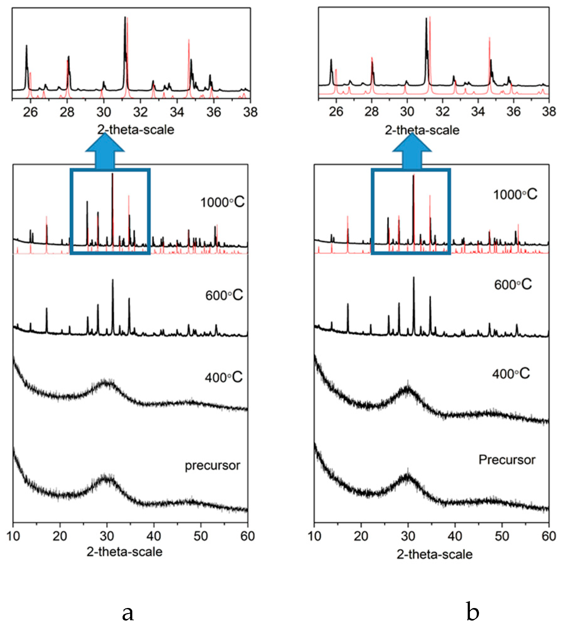
Figure 2.
Representative SEM micrographs of untreated Sr1 (A) and Sr5 (B) β-TCP tablets taken at 2000×, and 10,000× magnification.
Figure 2.
Representative SEM micrographs of untreated Sr1 (A) and Sr5 (B) β-TCP tablets taken at 2000×, and 10,000× magnification.
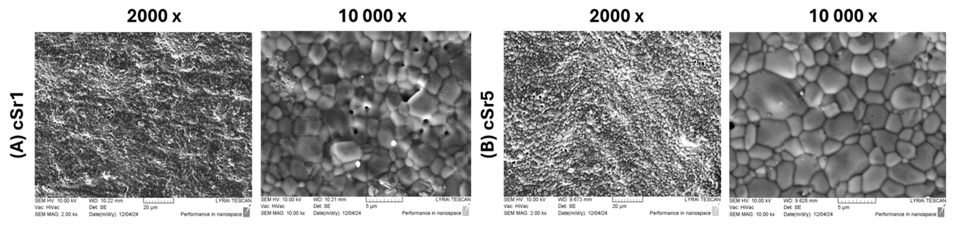
Figure 3.
Representative SEM micrographs of laser-structured Sr1 and Sr5 β-TCP tablets at different magnifications (500×, 2000×, and 10,000×), illustrating the formation of parallel and intersecting microgroove architectures after femtosecond laser processing: (A) Sr1-lines; (B) Sr1-90°; (C) Sr1-45°; (D) Sr5-lines; (E) Sr5-90°; (F) Sr5-45°.
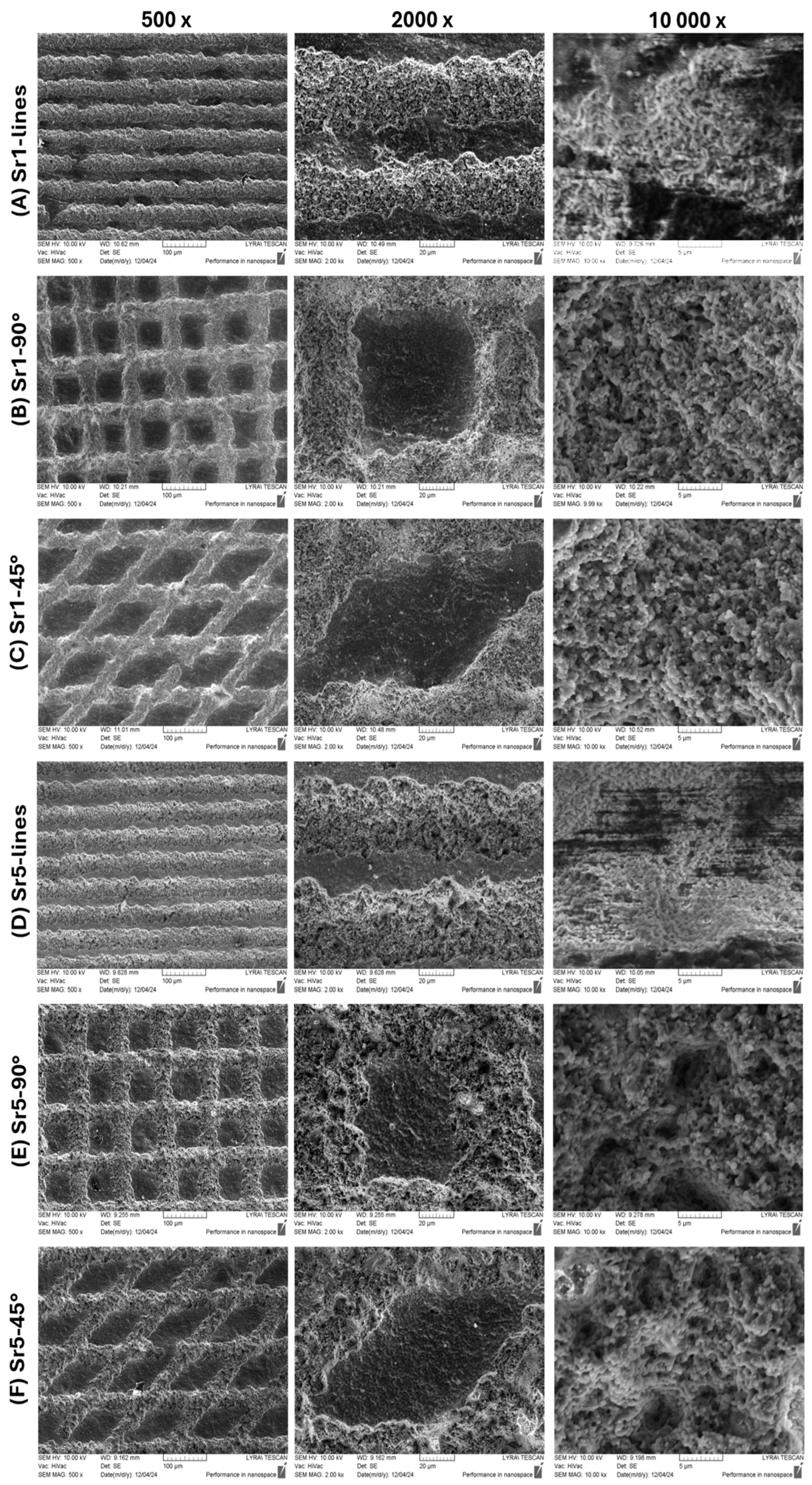
Figure 4.
Representative 2D and 3D real-color reconstruction images and linear roughness profiles of control and laser-structured Sr1 and Sr5 tablets: (A) Sr1-control; (B) Sr1-lines; (C) Sr1-90°; (D) Sr1-45°; (E) Sr1-control; (F) Sr5-lines; (G) Sr5-90°; (H) Sr5-45°.
Figure 4.
Representative 2D and 3D real-color reconstruction images and linear roughness profiles of control and laser-structured Sr1 and Sr5 tablets: (A) Sr1-control; (B) Sr1-lines; (C) Sr1-90°; (D) Sr1-45°; (E) Sr1-control; (F) Sr5-lines; (G) Sr5-90°; (H) Sr5-45°.
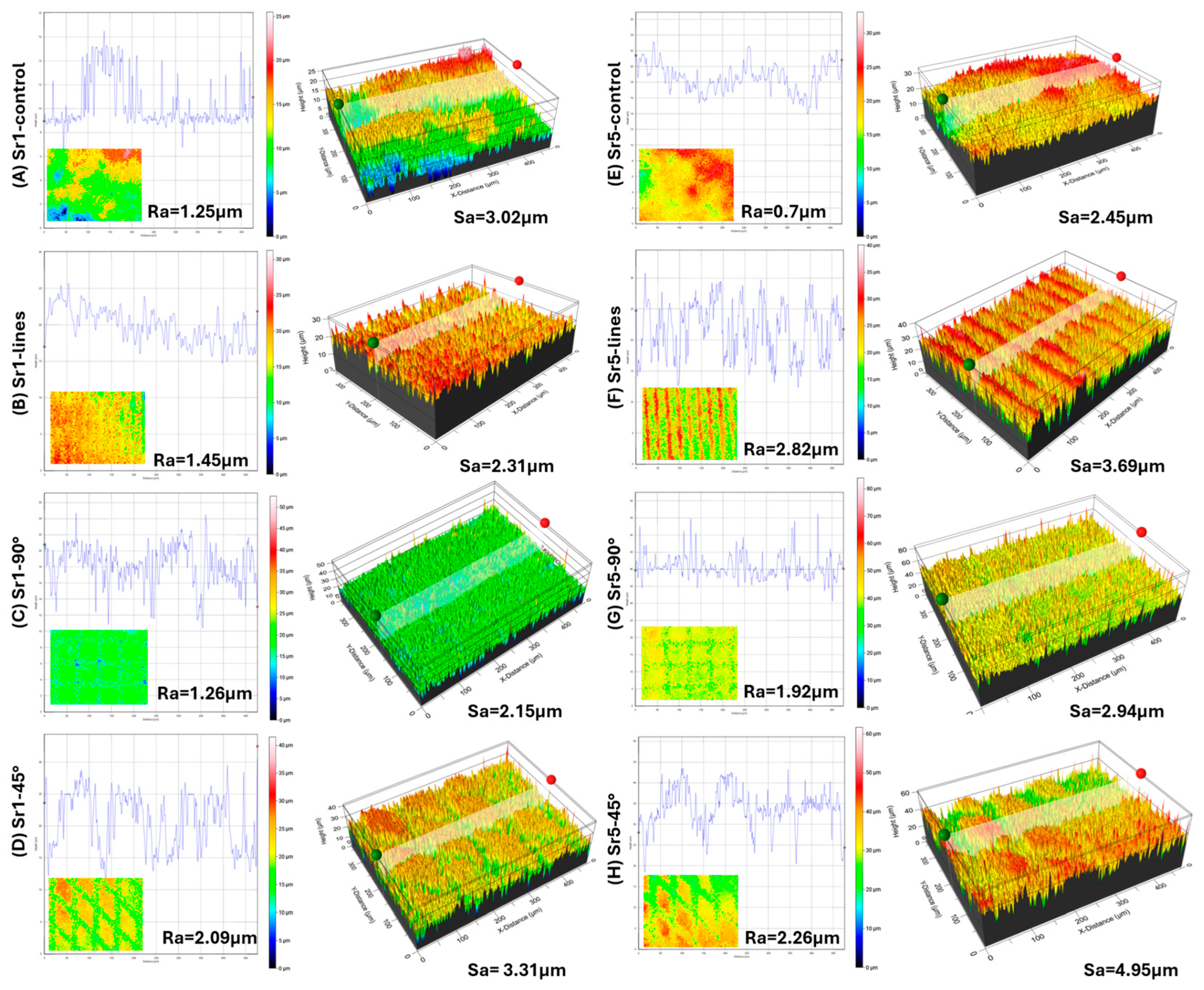
Figure 5.
WCA evaluation of (A) cSr1, Sr1-45°, and Sr1-90°; (B) cSr1 and cSr5 ceramic tablets.
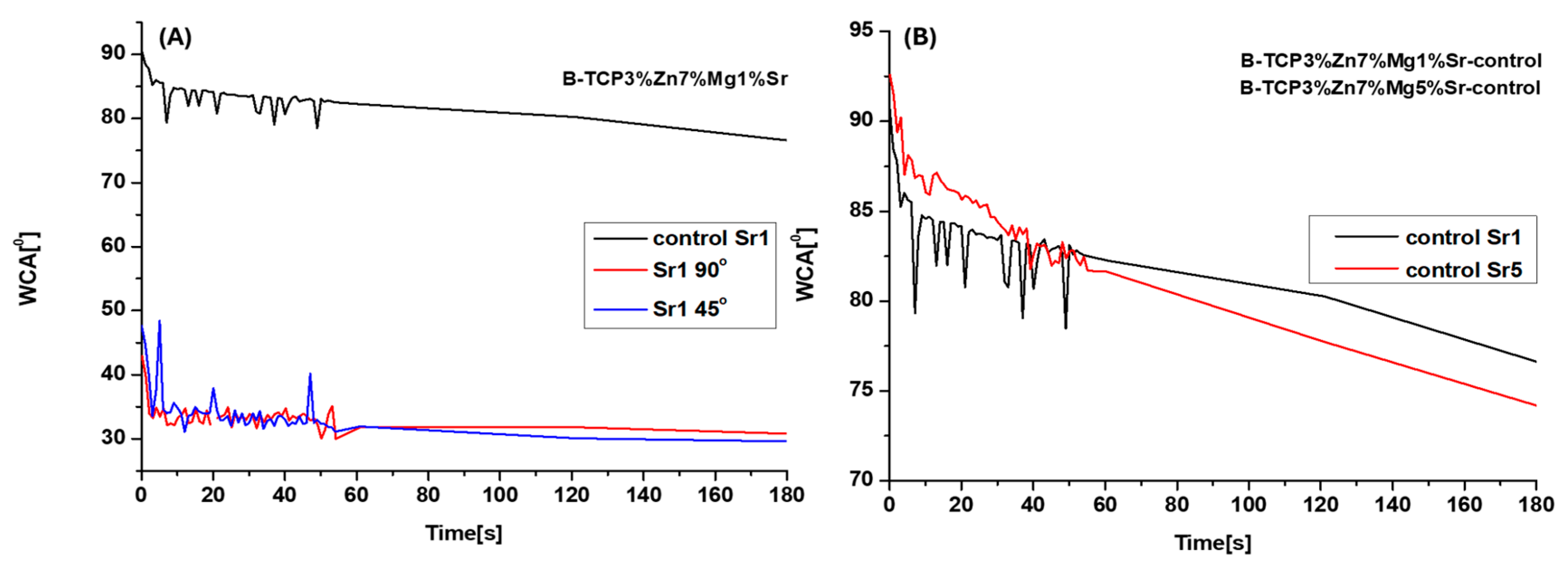
Figure 6.
Evaluation of antimicrobial activity after co-cultivation with ceramic tablets. Legend: Sparse pattern - Sr1 sample; Cross sparse pattern - Sr5 sample.
Figure 6.
Evaluation of antimicrobial activity after co-cultivation with ceramic tablets. Legend: Sparse pattern - Sr1 sample; Cross sparse pattern - Sr5 sample.
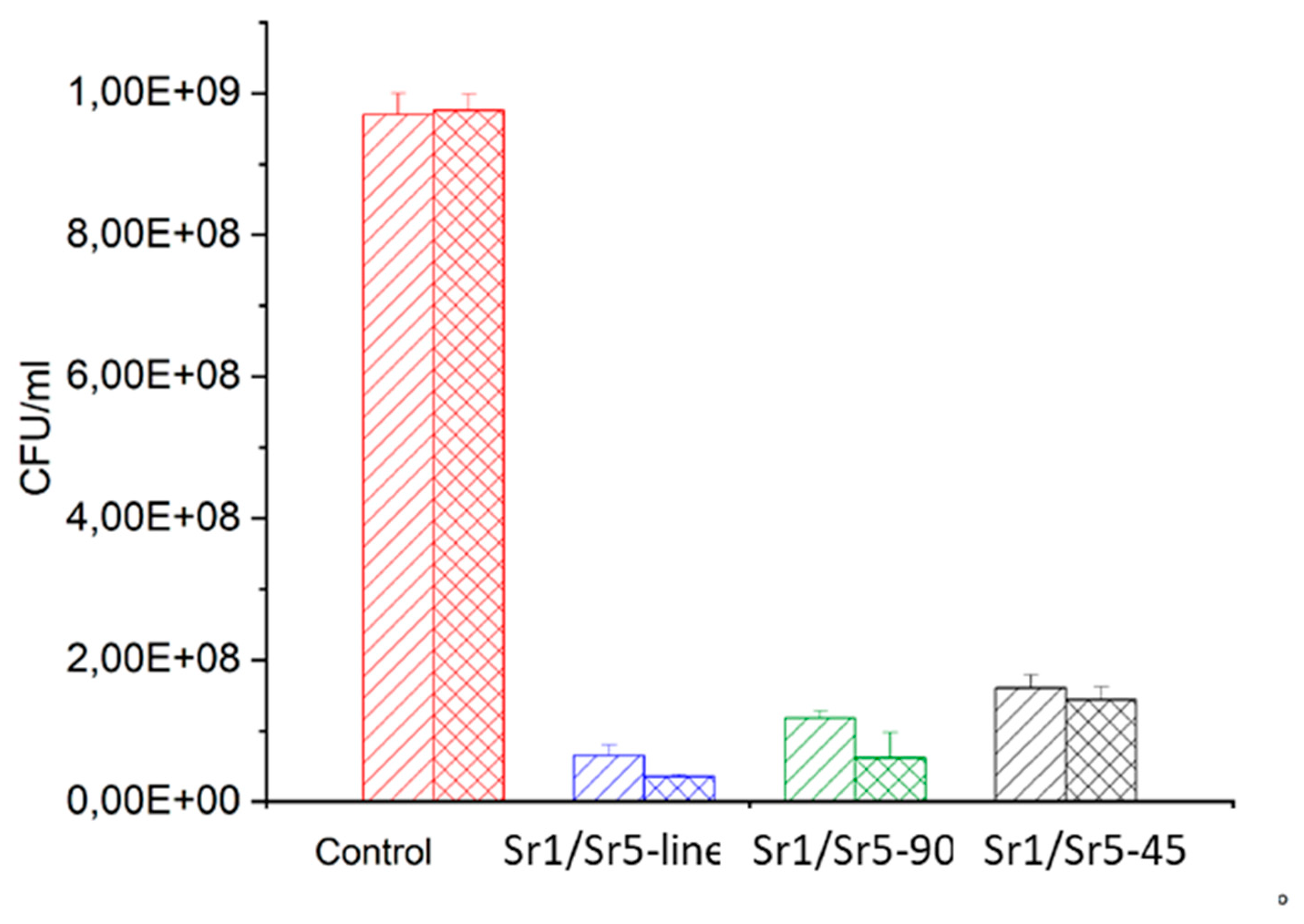
Figure 7.
Scanning electron microscopy of E. coli K12, co-cultivated with laser-treated ceramic tablets. Control group untreated tablets (A) Sr1 and (B) Sr5 ; Laser-treated: (C) Sr1-line; (D) Sr5-line; Sr1-90° (I); (F) Sr5-90; 45° (G) Sr1-45 and (H) Sr5-45 . Bars = 5 µm.
Figure 7.
Scanning electron microscopy of E. coli K12, co-cultivated with laser-treated ceramic tablets. Control group untreated tablets (A) Sr1 and (B) Sr5 ; Laser-treated: (C) Sr1-line; (D) Sr5-line; Sr1-90° (I); (F) Sr5-90; 45° (G) Sr1-45 and (H) Sr5-45 . Bars = 5 µm.
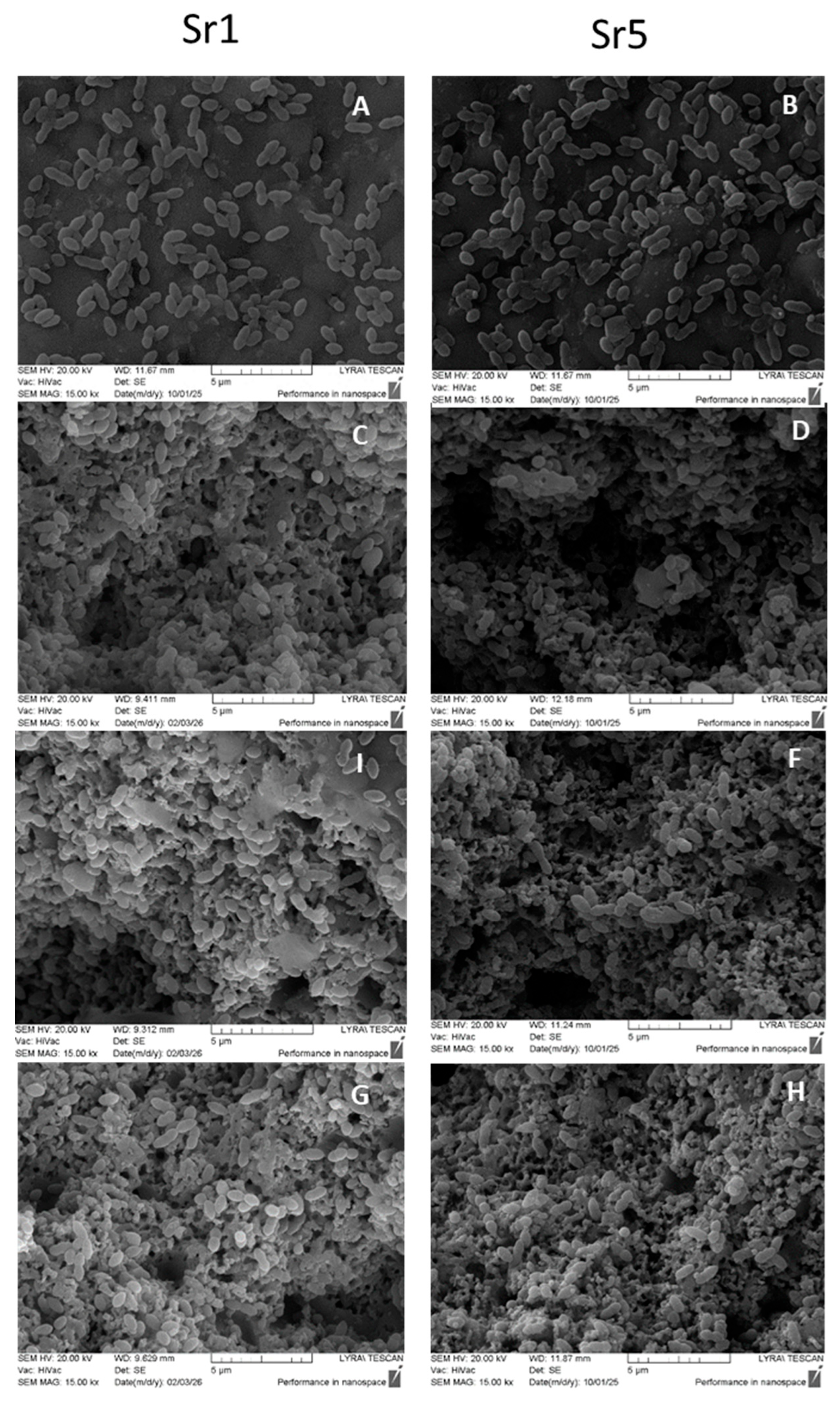
Figure 8.
Cell viability after incubation with calcium phosphate ceramic tablets subjected to laser-induced surface texturing. (A) Viable cell numbers normalized to the untreated control after 24 h and 48 h of incubation with the tablets. Data represent means ± SEM from three independent experiments. A total of 30,000 cells were analyzed per condition. (B) Phase-contrast microscopy images of untreated and treated cells showing no morphological changes associated with the treatment. Scale bar: 20 µm.
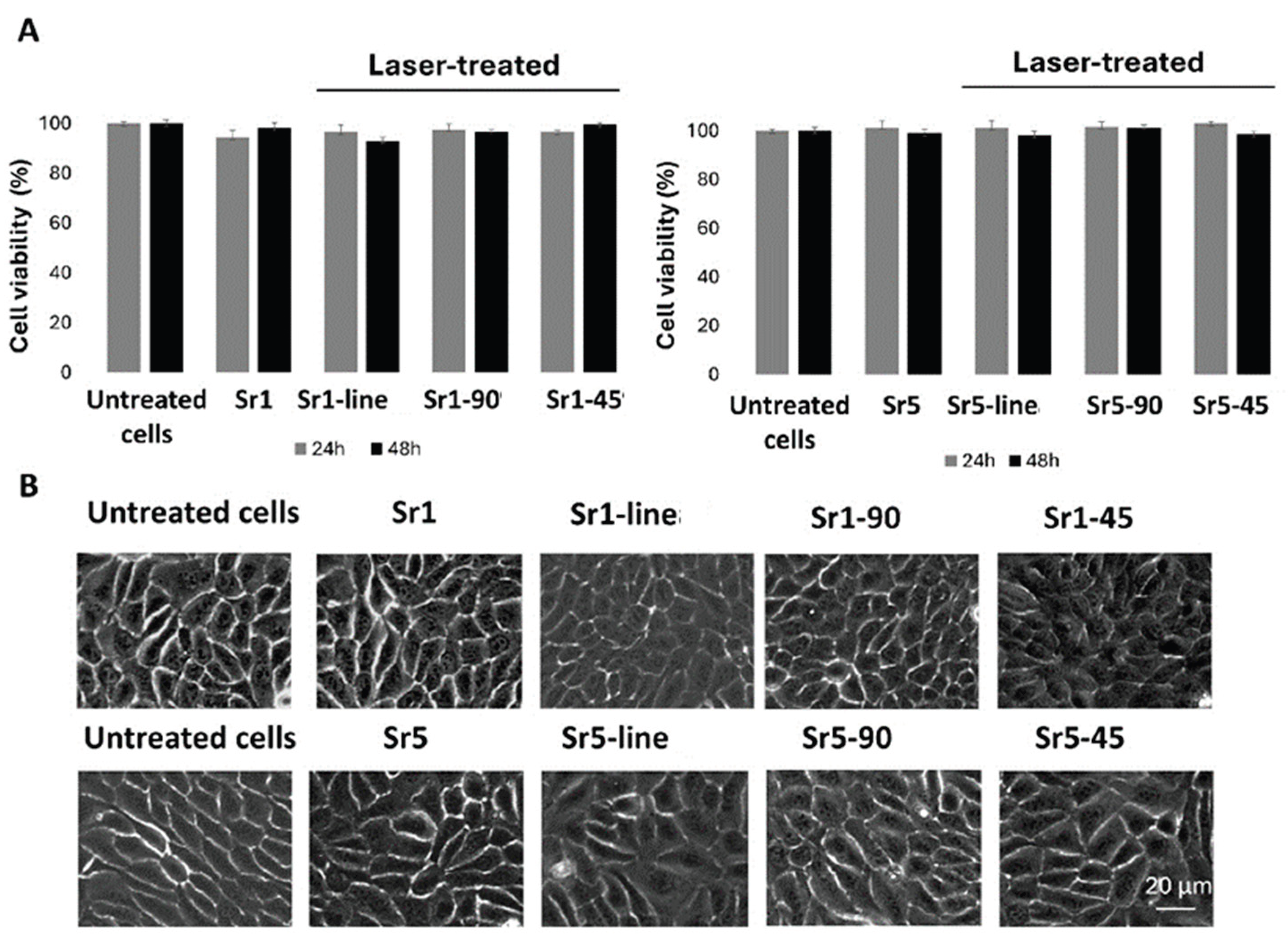

Table 1.
Composition (mmol.L-1) of the modified body fluids used for the synthesis.
| Components | Solution 1 | Solution 2 | Solution 3 |
|---|---|---|---|
| Na+ | 141.9 | 141.9 | 141.9 |
| K+ | 501 | 3 | 3 |
| Ca2+ in experiments Sr1 Ca2+ in experiments Sr5 |
0 | 361 344 |
2.5 2.5 |
| Mg2+ | 0 | 29.15 (calc.) 58.3 (in exp.) |
1.5 |
| Zn2+ | 0 | 0 | 12.2 |
| Sr2+ in experiments Sr1 Sr2+ in experiments Sr5 |
0 | 4.16 20.8 |
00 |
| Cl- | 143 | 949.5 | 167 |
| SO42- | 0.5 | 0.5 | 0.5 |
| HCO3- | 4.2 | 4.2 | 4.2 |
| HPO42- | 243 | 0 | 0 |
| pH | 8.0-8.2 | 8.0-8.2 | 6.5 |
Table 2.
Chemical composition of the tablets from both series.
| Ions. mmol/g | Series | |
|---|---|---|
| Sr1 | Sr5 | |
| Ca2+ | 9.46 ± 0.02 | 8.38 ± 0.02 |
| Mg2+ | 0.85 ± 0.01 | 0.91 ± 0.01 |
| Zn2+ | 0.31 ± 0.01 | 0.33 ± 0.01 |
| Sr2+ | 0.11 ± 0.01 | 0.55 ± 0.01 |
| P-PO43- | 6.90 ± 0.02 | 6.73 ± 0.02 |
| Cl- | <0.05 ± 0.01 | <0.05 ± 0.01 |
| K+ | 0.04 ± 0.01 | 0.03 ± 0.01 |
| Na+ | 0.07 ± 0.01 | 0.08 ± 0.01 |
| Sr/(Mg+Zn+Sr+Ca) | 0.01 ± 0.001 | 0.05 ± 0.001 |
| Mg/(Mg+Zn+Sr+Ca) | 0.08 ± 0.001 | 0.09 ± 0.001 |
| Zn/(Mg+Zn+Sr+Ca) | 0.03 ± 0.001 | 0.03 ± 0.001 |
| (Mg+Zn+Sr+Ca)/P | 1.55 ± 0.01 | 1.51 ± 0.01 |
Table 3.
Mean WCA [o] of control and femtosecond laser-structured ceramic samples.
| Samples | Mean WCA [o] |
|---|---|
| cSr1 | 83.05° |
| Sr1-lines | 35.26 |
| Sr1-45° | 34.19° |
| Sr1-90° | 40.92° |
| cSr5 | 84.59° |
| Sr5-lines | - |
| Sr5-45° | - |
| Sr5-90° | - |
Disclaimer/Publisher’s Note: The statements, opinions and data contained in all publications are solely those of the individual author(s) and contributor(s) and not of MDPI and/or the editor(s). MDPI and/or the editor(s) disclaim responsibility for any injury to people or property resulting from any ideas, methods, instructions or products referred to in the content. |
© 2026 by the authors. Licensee MDPI, Basel, Switzerland. This article is an open access article distributed under the terms and conditions of the Creative Commons Attribution (CC BY) license (http://creativecommons.org/licenses/by/4.0/).
Copyright: This open access article is published under a Creative Commons CC BY 4.0 license, which permit the free download, distribution, and reuse, provided that the author and preprint are cited in any reuse.