

Submitted:
05 October 2026
Posted:
08 October 2026
You are already at the latest version
Abstract
Microenergy acoustic pulse (MAP) therapy has been associated with functional and structural improvement after childbirth-related urethral injury, but its multicellular ef-fects remain incompletely defined. We explored treatment- and injury-associated dif-ferences in the urethral microenvironment using single-cell RNA sequencing. Eight female Sprague–Dawley rats were assigned to sham, vaginal balloon dilation plus ovariectomy (VBDO), VBDO plus beta-aminopropionitrile (BAPN), or VBDO plus BAPN and MAP (MAP) groups, with one 24-week-old and one 48-week-old rat per group. Urethral tissues were analyzed for cell composition, differential gene expression, Gene Ontology enrichment, stem/progenitor features, senescence-associated secretory phe-notype activity, and CellChat-inferred intercellular communication. A balanced sin-gle-cell dataset comprising 23,128 cells was used for downstream comparative analyses and resolved 19 annotated cell-type/state categories. Across the injury- and age-associated experimental conditions, epithelial, vascular, stromal, immune, and muscle compart-ments showed distinct transcriptional patterns, whereas MAP-treated samples showed a distinct cellular distribution; a separate histological cohort showed higher urethral muscle content. MAP-treated samples also showed differences in CellChat-inferred signaling activity in selected extracellular-matrix, adhesion, vascular, developmental, and neu-ral/neuromuscular pathways, including LAMININ, FN1, COLLAGEN, ANGPT, VEGF, WNT, NOTCH, RELN, SLIT, NCAM, and NRXN. Exploratory enrichment analysis identified treatment-associated differences in translation, oxidative metabolism, stress adaptation, synaptic-organization processes, progenitor-associated transcriptional states, and senescence-related programs. Overall, the findings are descriptive and hypothe-sis-generating and require validation in independently replicated cohorts.
Keywords:
stress urinary incontinence
; microenergy acoustic pulse
; single-cell RNA sequencing
; urethral repair
; aging
; cell-cell communication
1. Introduction
Stress urinary incontinence (SUI) is defined as involuntary urine leakage during physical activity, exertion, coughing, or sneezing [1,2]. Vaginal delivery and aging are major risk factors for SUI, and the combination of childbirth-related tissue injury, estrogen deprivation, and age-related loss of regenerative capacity may contribute to persistent urethral dysfunction [3,4,5,6]. Current treatment is centered on pelvic floor muscle training, urethral bulking, and surgery; however, therapies that directly restore the damaged urethral microenvironment remain limited [7,8] .Urethral continence depends on coordinated interactions among smooth muscle, the external striated urethral sphincter, connective tissue, vasculature, peripheral nerves, immune cells, and the urothelial barrier [9,10,11]. Vaginal distension models show that simulated childbirth injury can disrupt sphincter structure and extracellular matrix integrity, whereas ovariectomy and inhibition of collagen crosslinking can further aggravate tissue damage [12,13]. Our previous single-cell study further suggested that aging redirects urethral repair from coordinated regeneration toward muscle loss, denervation-related transcriptional changes, fibroblast activation, inflammation, extracellular matrix deposition, and urothelial senescence.
Microenergy acoustic pulse (MAP) is a low-intensity mechanical stimulation therapy designed to activate endogenous tissue repair without thermal ablation. In animal models of SUI, MAP improves leak point pressure and restores pelvic floor and urethral muscle structure, accompanied by activation of resident progenitor cells and enhancement of repair-related signaling [14]. However, the effects of MAP on individual urethral cell populations and multicellular communication networks remain unclear.
Single-cell RNA sequencing (scRNA-seq) can resolve rare cell states in complex tissues and infer ligand-receptor communication within the urethral microenvironment [15,16,17,18]. This exploratory study integrated single-cell data from eight animals representing aging, injury, fibrosis-prone injury, and MAP treatment states. We examined treatment-associated differences in cellular community structure, inferred intercellular communication networks, and regenerative, senescence, and stress-associated transcriptional states. Given the limited biological replication, these analyses were considered descriptive and hypothesis-generating rather than definitive evidence of treatment mechanism.
2. Results
2.1. The Single-Cell Atlas Resolves a Complex Multicellular Urethral Ecosystem
UMAP visualization showed broad integration across individual samples, treatment groups, and ages while preserving biologically distinct cell populations. Integrated analysis identified 19 annotated cell-type/state categories spanning ciliated, luminal, secretory, glandular, squamous, basal, tuft/chemosensory, neuroendocrine, and epithelial/neural-like states, together with endothelial, lymphatic endothelial, fibroblast, stromal/vascular-associated, immune, smooth muscle, striated muscle, and high-mitochondrial/stressed populations. Cell-type identities were assigned using representative canonical marker genes as described in the Methods. To reduce unequal representation caused by differences in cell recovery, a balanced subset of 23,128 cells was retained after sample-level downsampling for comparative downstream analyses (Table S1; Methods). These findings establish the urethra as a multicellular tissue ecosystem rather than an isolated muscular conduit (Figure 1A–D).
2.2. Transcriptional Differences Across Injury- and Age-Associated Experimental Conditions
Comparison of age and injury conditions indicated a shift from coordinated homeostasis toward a maladaptive repair state. Muscle populations showed reduced regenerative and neuron-projection-associated programs, consistent with impaired neuromuscular integrity. Fibroblast and stromal populations were enriched for extracellular-structure organization, growth-factor responses, and wound-remodeling programs, whereas immune populations showed inflammatory activation. Differentiated urothelial features were reduced and senescence-associated activity increased in selected epithelial states. Collectively, the experimental conditions representing childbirth-like injury, estrogen deprivation, fibrosis-prone remodeling, and different ages were associated with coordinated differences involving muscle, matrix, vasculature, immunity, and epithelial barrier function (Figure 1D). Given the limited biological replication, the independent contributions of age and treatment cannot be resolved.
2.3. MAP Is Associated with Differences in Cellular Composition and Urethral Muscle Content
Cell-type composition differed across sham, VBDO, BAPN, and MAP tissues. VBDO and BAPN were associated with differences in relative abundance of epithelial, endothelial, lymphatic endothelial, fibroblast, immune, stromal/vascular-associated, smooth-muscle, and striated-muscle states. MAP-treated tissues showed a different cellular configuration rather than simply reproducing the sham distribution (Figure 2A,B). Histologic examination in a separate, independently analyzed cohort showed marked disruption and loss of urethral muscle after VBDO and BAPN, whereas MAP-treated tissues displayed greater circumferential organization and higher pha-positive striated muscle and α-SMA-positive smooth muscle content. Quantification confirmed higher muscle content in the MAP group relative to the injured groups, although restoration was incomplete compared with sham (Figure 2C,D).
2.4. MAP Treatment-Associated Differences in Inferred Signaling and Biological Programs
CellChat-based information-flow comparisons showed differences in inferred signaling profiles between MAP-treated samples and sham, VBDO, and BAPN samples (Figure 3A). Relatively higher inferred activity in MAP-treated samples involved extracellular-matrix and basement-membrane pathways, cell adhesion and junctional signaling, vascular remodeling, developmental regulation, and neural communication. Representative pathways included COLLAGEN, LAMININ, FN1, ANGPT, VEGF, PECAM1, CDH5, CLDN, NECTIN, JAM, WNT/ncWNT, TGFβ, BMP, RELN, SLIT, NCAM, NRXN, MPZ, and semaphorin signaling. Treatment-specific and lineage-specific expression of selected repair-associated candidate genes is shown in Supplementary Figures S2–S4. The cell-type distribution of representative marker-gene and pathway-associated genes is shown in Figures S5 and S6. In the MAP-versus-BAPN comparison, LAMININ- and FN1-associated information flow was particularly prominent, consistent with relatively greater inferred cell-matrix and basement-membrane communication (Figure 3B). Exploratory Gene Ontology analysis of the cell-level comparison showed that genes with higher expression in MAP-treated samples were associated with cytoplasmic translation, oxidative energy metabolism, cellular respiration, Golgi-mediated transport, reactive oxygen species handling, postsynaptic organization, synapse regulation, and apoptotic-signal regulation. Conversely, genes with lower expression in MAP-treated samples were associated with RNA processing, steroid-hormone responses, mesenchymal development, developmental growth, and persistent wound-remodeling programs. These enrichment results are descriptive and hypothesis-generating because adjusted P values derive from cell-level exploratory analyses rather than independent animals. Together, these descriptive differences involved transcriptional features related to prolonged-injury signaling, metabolic competence, stress adaptation, and structural organization (Figure 3C).
2.5. MAP Treatment-Associated Differences in Epithelial-Stromal-Vascular-Muscle Communication
Differential CellChat networks comparing MAP with BAPN showed broad differences in both the number and aggregate strength of predicted ligand-receptor interactions (Figure 4A). The largest network differences involved endothelial and lymphatic endothelial cells, fibroblasts, immune cells, luminal and secretory epithelial populations, and smooth and striated muscle. Incoming-signaling heatmaps showed differences in receptor-associated signals received by multiple cell populations, including muscle compartments, whereas outgoing-signaling heatmaps showed parallel differences in ligand-associated signals transmitted to surrounding vascular, immune, epithelial, and matrix-associated cells (Figure 4B,C). The predicted network differences were distributed across multiple sender and receiver populations rather than being dominated by a single cell type or signaling pathway, indicating broad treatment-associated differences in bidirectional communication across the urethral microenvironment.
2.6. MAP-Associated Shifts in Progenitor-Associated Transcriptional States
Stem/progenitor-associated markers displayed lineage-restricted expression across the urethral atlas (Figure 5A). Epithelial progenitor-like populations expressed markers including Krt15 and Sox9, muscle-associated cells expressed Itga7, and vascular/stromal repair populations expressed Cd34 and Met. Descriptive treatment-split marker visualization showed treatment-associated expression differences in selected repair-associated genes, particularly Krt15 and Met (Figure 5B). Treatment-split UMAPs and lineage-specific violin/dot plots for Krt15, Met, Sox9, Itga7, and Cd34 are provided in Supplementary Figures S2–S4. These observations are consistent with differences in progenitor-associated transcriptional states but do not establish lineage expansion, progenitor reactivation, or regeneration.
2.7. MAP Treatment-Associated Differences in SASP Activity Across Urethral Lineages
SASP module scores varied substantially by cell type and treatment (Figure 5C). Relatively high activity occurred in selected squamous/basal epithelial, fibroblast, epithelial/neural-like, neuroendocrine/chemosensory, and muscle-associated populations, whereas several endothelial, immune, and other epithelial states showed lower scores. MAP was associated with lower SASP-associated module scores in selected vascular, immune, smooth-muscle, and striated-muscle populations, whereas epithelial and stromal populations showed a different pattern. The schematic model summarizes this lineage-specific balance between limiting chronic injury signaling and maintaining repair-associated communication (Figure 5D).
3. Discussion
3.1. MAP Treatment-Associated Remodeling of the Urethral Ecosystem
For decades, stress urinary incontinence (SUI) has often been viewed as a relatively simple mechanical failure, primarily attributed to lax urethral support or insufficient sphincter contraction, with treatment therefore focused on tightening, supporting, or repairing damaged tissues [2]. The urethra is not a passive conduit composed of a single muscular structure; rather, it is a dynamic tissue ecosystem maintained by epithelial, vascular, immune, stromal, neural, and muscle cells. SUI, particularly when associated with aging and childbirth injury, may therefore represent widespread dysfunction of this multicellular ecosystem rather than an isolated mechanical failure of muscle.
In this study, scRNA-seq captured active transcriptional programs in individual cells, and UMAP grouped cells with similar expression profiles into distinct clusters, enabling construction of a high-resolution cellular atlas of the rat urethra. The integrated analysis identified transcriptionally distinct populations spanning epithelial, endothelial, lymphatic endothelial, immune, fibroblast, stromal/vascular-associated, smooth muscle, striated muscle, neuroendocrine, and stressed cell states.
The present data suggest several levels at which MAP-treated samples differ from injured comparators. First, cell-type composition showed treatment-associated redistribution. Second, CellChat analysis identified broad differences in predicted incoming and outgoing signaling across epithelial, endothelial, lymphatic endothelial, immune, fibroblast, and muscle populations; these predictions do not establish functional communication. Third, progenitor-associated and SASP-associated transcriptional states varied among treatment groups in a cell-type-dependent manner. These observations are descriptive and should be regarded as hypotheses for future studies with independent biological replication and orthogonal validation.
The present study supports a broader framework for examining the urethra as an interdependent multicellular tissue rather than an isolated muscular structure. The treatment-associated differences observed here involve cellular composition, inferred intercellular communication, and intrinsic transcriptional states. Because the principal single-cell comparisons are based on limited biological replication, they do not establish complete biological recovery or a definitive MAP-specific mechanism. Rather, they provide testable directions for future functional, histologic, and mechanistic validation.
3.2. Potential Multicellular Mechanisms and Translational Implications
This exploratory single-cell study extends SUI from a disorder of the sphincter alone to a failure of multicellular tissue repair [18]. MAP-treated samples showed differences in cellular composition and predicted communication pathways related to ECM and basement membrane organization, vascular support, cell adhesion, neural guidance, and synaptic structure, together with differences in progenitor-associated transcriptional states. These observations provide testable hypotheses that may help explain the functional and structural improvements previously observed after MAP treatment in animal models of SUI [14].
Urethral sphincter function depends on both muscle integrity and coordinated neuromuscular activation. Age-associated reduction in neuron-projection programs and neuromuscular junctions may contribute to persistent SUI. In the present exploratory analysis, MAP-treated samples showed greater representation of RELN, SLIT, semaphorin, NCAM, NRXN, MPZ, glutamate-related signaling and postsynaptic-organization programs, together with differences in ECM and cell-adhesion signaling. Increased collagen and laminin information flow should not be interpreted simply as promotion of fibrosis. Notably, GO analysis showed reduced persistent wound-healing and mesenchymal-development programs together with enhanced oxidative metabolism and stress adaptation, a combination more consistent with maturation of repair than with uncontrolled fibrosis.
Vascular signaling may represent another important mechanism. ANGPT, VEGF, PECAM1, and CDH5-associated signaling showed greater representation in selected MAP comparisons, and endothelial and lymphatic endothelial cells occupied prominent positions in differential communication networks. Adequate perfusion is essential for progenitor activation, muscle metabolism, immune resolution, and matrix turnover. These observations raise the hypothesis that vascular-niche remodeling may contribute to MAP-associated urethral repair, which will require direct experimental validation.
Progenitor-associated transcriptional states provide a potential link to previous observations that MAP can influence resident muscle progenitor biology [9,14]. Krt15, Met, Sox9, Itga7, and Cd34 marked distinct epithelial, muscle, and stromal/vascular repair populations. The overall progenitor-associated signature in MAP-treated samples differed from BAPN and remained distinct from sham, consistent with treatment-associated differences in progenitor-related transcriptional states rather than demonstrated progenitor reactivation. Tissue repair depends not only on progenitor abundance but also on microenvironmental signals that regulate survival, differentiation, and integration. MAP-treated samples showed lower stress-related transcriptional activity in selected vascular and striated-muscle populations while retaining repair-associated remodeling programs in epithelial and stromal compartments [18]. The present data show cell-type-specific differences in a predefined 15-gene SASP-associated module across treatment groups, but they do not establish beneficial suppression or reprogramming.
Other energy-based approaches, including vaginal laser therapy, have also been investigated for SUI, with randomized sham-controlled trials evaluating their clinical efficacy [19]. In contrast, MAP represents a nonthermal mechanical stimulation strategy that may influence tissue remodeling and regenerative responses through distinct mechanisms; however, direct comparative studies are required to determine potential differences in efficacy, durability, cellular targets, and safety.
Future clinical studies of MAP should incorporate validated patient-reported outcome measures in addition to objective functional and anatomical endpoints. Instruments such as the King’s Health Questionnaire (KHQ) and the International Consultation on Incontinence Questionnaire-Short Form (ICIQ-SF) can provide complementary assessment of urinary-incontinence symptom burden, quality-of-life impact, and patient-perceived treatment benefit [20].
This study has important limitations. First, the transcriptomic cohort included only eight animals, with one animal in each age-by-treatment condition. Thus, the two animals within each treatment group differed in age, and the independent contributions of age and treatment cannot be resolved. The large number of cells obtained from each animal does not constitute independent biological replication, and cell-level statistical results should not be interpreted as treatment-level inference. Second, CellChat infers communication from transcript abundance and curated ligand-receptor databases and does not demonstrate protein-level signaling or physical interaction. Third, stem/progenitor and SASP classifications depend on prespecified gene-set definitions and should be regarded as operational transcriptional signatures rather than definitive lineage identities; lineage tracing and orthogonal validation are required. Fourth, the separate histological cohort provides tissue-level support for muscle-content differences, but the principal transcriptomic findings have not been independently validated by targeted qPCR, Western blotting, or immunohistochemistry. Finally, balanced downsampling may improve comparability but reduce detection of rare populations, and some sample-level preprocessing metrics were not available in sufficient detail for retrospective reconstruction. Accordingly, the transcriptomic results are presented as descriptive, hypothesis-generating observations to guide future studies with independent biological replication.
4. Materials and Methods
4.1. Animals and Experimental Design
All animal procedures were conducted in accordance with institutional guidelines and were approved by the Institutional Animal Care and Use Committee of the University of California, San Francisco. This scRNA-seq study included eight female Sprague-Dawley rats. Animals were randomly assigned to four groups: sham control, vaginal balloon dilation plus ovariectomy (VBDO), VBDO plus beta-aminopropionitrile (BAPN), and VBDO plus BAPN and MAP (MAP). Each group contained one 24-week-old and one 48-week-old animal, yielding a 4-treatment × 2-age experimental design (Table 1).
A separate histological cohort consisting of 16 additional 24-week-old female Sprague–Dawley rats was used for the immunofluorescence and muscle-tissue quantification. These animals were randomly assigned to the same four experimental groups (Sham, VBDO, BAPN, and MAP) and were not included in the scRNA-seq cohort. The number of animals in each histological group was Sham, n = 4; VBDO, n = 4; BAPN, n = 4; and MAP, n = 4 (total n = 16).
Sham animals underwent anesthesia, vaginal catheter insertion, and surgical preparation without balloon dilation or ovariectomy. Simulated childbirth injury was induced using an 18-Fr Foley balloon filled with 4 mL of water and maintained under a 130-g load for 4 h; bilateral ovariectomy (OVX) was performed 1 week later [13,14]. Beginning at Week 2, animals in the BAPN group received BAPN (300 mg/kg, intraperitoneally, twice weekly) for 4 weeks. Animals in the MAP group received the same BAPN regimen together with MAP treatment applied to the pelvic floor/urethral region at an energy flux density of 0.033 mJ/mm2, 3 Hz, 500 pulses per session, twice weekly for 4 weeks [14]. After completion of the 4-week treatment period, animals underwent a 1-week washout. All experimental groups were followed to the same predefined Week 7 endpoint, at which urethral tissues were collected. Thus, the BAPN and MAP groups were time-matched for tissue collection. A detailed experimental timeline is provided in Figure 1A and Supplementary Figure S1.
4.2. Single-Cell RNA Sequencing and Data Preprocessing
Fresh urethral tissue was enzymatically dissociated to generate single-cell suspensions. Libraries were constructed using the 10x Genomics Chromium Single Cell 3′ platform and sequenced on an Illumina NovaSeq instrument. Raw reads were aligned to the Rattus norvegicus reference genome using Cell Ranger. Cells with 500-9,000 detected genes, fewer than 25,000 unique molecular identifiers, and less than 20% mitochondrial transcripts were retained. Doublets, low-quality droplets, and cells without reliable annotation were removed. Sample-level cell recovery and sequencing quality-control metrics are provided in Table S1.
Following quality control, 161,561 cells were available across the eight samples. To reduce disproportionate representation of samples with higher cell recovery, sample-level downsampling was subsequently performed, retaining 23,128 cells for balanced comparative downstream analyses. This balancing step was performed after QC and was not part of the QC exclusion process; it also did not increase the number of biological replicates. Subsequent analyses were performed in R 4.6.0 using Seurat 5.5.0 and SeuratObject 5.4.0. Seurat was used for normalization, variable-feature selection, integration, principal-component analysis, neighborhood construction, clustering, and UMAP visualization. Additional analyses used CellChat 2.2.0.9001 for inferred cell-cell communication and clusterProfiler 4.20.0 for functional enrichment. Full software and package-version information is provided in the Supplementary Materials.
4.3. Cell-Type Annotation and Differential Expression Analysis
Clusters were annotated using canonical markers and differentially expressed marker genes as described below. Reanalysis identified 19 annotated cell-type/state categories: ciliated epithelial, luminal urothelial, secretory epithelial, secretory/glandular epithelial, squamous epithelial, squamous/basal epithelial, tuft/chemosensory epithelial, neuroendocrine/chemosensory, epithelial/neural-like, neural/epithelial-like, endothelial, endothelial subtype, lymphatic endothelial, fibroblast, stromal/vascular-associated, immune, smooth muscle, striated muscle, and high-mitochondrial/stressed populations. For exploratory candidate-gene identification, cell-level expression differences were assessed using the Wilcoxon rank-sum test in Seurat. GO enrichment analysis was then performed using genes showing higher or lower expression in MAP-treated samples relative to BAPN samples. Because each age-by-treatment condition was represented by one animal, these cell-level adjusted P values and downstream enrichment results were used only for hypothesis generation.
4.4. Stem/Progenitor and SASP Analyses
Progenitor-associated transcriptional states were assessed using prespecified lineage-specific multigene panels: epithelial (Krt5, Krt14, Krt15, Krt17, Krt19, Trp63, Sox2, Klf5, Itga6, and Itgb4), muscle (Pax7, Myf5, Itga7, Vcam1, Sdc4, Spry1, Met, and Cdh15), and stromal/vascular (Pdgfra, Cd34, Pi16, Dpp4, Col15a1, Ly6a, Pdgfrb, Cspg4, Mcam, and Rgs5). Single-cell module scores were calculated using Seurat AddModuleScore based on normalized RNA expression and expression-matched control genes. Continuous module scores were used for group-level descriptive comparisons and were interpreted as progenitor-associated transcriptional states rather than definitive lineage identities. Met was retained in the prespecified muscle-associated module used for scoring; Supplementary Figure S4 visualizes its detectable expression within stromal/vascular populations as a separate descriptive analysis.
A predefined 15-gene SASP-associated panel comprising Il6, Il1a, Il1b, Ccl2, Cxcl1, Cxcl2, Cxcl3, Csf2, Csf3, Mmp3, Mmp9, Serpine1, Timp1, Tgfb1, and Icam1 was used. All 15 genes were detected in the scRNA-seq dataset. Single-cell SASP-associated module scores were calculated using Seurat AddModuleScore based on normalized RNA expression and expression-matched control genes. Module scores were subsequently averaged within each annotated cell type and treatment group for visualization. Because SASP programs are context and lineage dependent, these scores were interpreted as relative transcriptional signatures rather than as evidence of a uniform beneficial or detrimental senescence response.
4.5. Cell-Cell Communication Analysis
CellChat was used to infer ligand-receptor communication among major urethral cell populations and to compare MAP with sham, VBDO, and BAPN. Outcomes included the number and aggregate strength of predicted interactions, incoming and outgoing signaling patterns, and relative information flow across signaling families. Pathways of particular interest included ECM and basement membrane signaling (COLLAGEN, LAMININ, FN1), angiogenic signaling (ANGPT, VEGF), adhesion and junctional signaling (CDH, JAM, NECTIN, CLDN, PECAM1), developmental and regenerative signaling (WNT, ncWNT, NOTCH, EGF, FGF, TGFβ), and neural/neuromuscular communication (AGRN, NCAM, NRXN, RELN, SLIT, semaphorins, Netrin, MPZ, and glutamate-related signaling).
4.6. Immunofluorescence Staining
Tissues were fixed in cold 2% formaldehyde containing 0.002% saturated picric acid in 0.1 M phosphate buffer (pH 8.0) for 4 h, cryoprotected overnight in 30% sucrose, embedded in OCT compound, and stored at −70 °C. Cryosections (10 μm) were mounted on SuperFrost-Plus slides, treated with 0.3% H2O2 in methanol for 10 min, washed with PBS, and blocked for 30 min in 3% horse serum containing 0.3% Triton X-100. For smooth muscle staining, sections were incubated overnight at 4 °C with an anti-smooth muscle actin antibody (SMA; 1:1000, Abcam), followed by an Alexa Fluor 594-conjugated secondary antibody (1:500, Invitrogen). For F-actin visualization, cryosections were washed with PBS, permeabilized with 0.1% Triton X-100 for 5–10 min, and incubated with Alexa Fluor 488-conjugated phalloidin in PBS for 30–60 min at room temperature in the dark. Sections were then washed with PBS, counterstained with DAPI, mounted with antifade mounting medium, and imaged by fluorescence microscopy. Histological quantification was performed by an investigator blinded to treatment group. Microscopic fields/sections were selected according to predefined criteria, and the same number of fields/sections was analyzed for all animals, with five fields/sections evaluated per animal. Measurements from these technical measurements were averaged within each animal to generate a single animal-level value for statistical analysis.
4.7. Statistical Analysis
The animal was considered the biological experimental unit. This was an exploratory single-cell study with one animal in each age-by-treatment condition. Cell-level tests were used to prioritize candidate genes and pathways rather than to infer population-level treatment efficacy. Accordingly, individual cells were not treated as independent biological replicates, and formal age-by-treatment interaction testing was not performed. For the independent histological cohort, each animal contributed one summarized value to the statistical analysis; sections and microscopic fields were treated as technical measurements. Descriptive values, adjusted P values from transcriptomic analyses, and sample-level data points are reported where available. Conclusions are expressed using association-based language and require validation in larger independent cohorts. Specifically, adjusted P values from Seurat/Wilcoxon transcriptomic comparisons reflect exploratory cell-level testing and should not be interpreted as population-level evidence of treatment efficacy.
5. Conclusions
MAP treatment was associated with transcriptomic differences in the urethral microenvironment following simulated childbirth injury. The exploratory analyses identified treatment-associated differences in epithelial, vascular, immune, stromal, and muscle cell representation; CellChat-inferred matrix-, adhesion-, angiogenic-, neural-, and neuromuscular communication patterns; and progenitor-associated, SASP-associated, and injury/stress-related transcriptional states. A separate histological cohort showed higher urethral muscle content in MAP-treated animals than in injured comparators. These findings are hypothesis-generating and do not establish treatment efficacy or causal mechanisms. Independent biological replication, prospectively documented experimental procedures, functional testing, orthogonal molecular validation, and targeted mechanistic studies are required to confirm the observations and determine their biological significance.
Supplementary Materials
The following supporting information can be downloaded at the website of this paper posted on Preprints.org. Additional supporting data are provided in the Supplementary Materials, including supplementary figures and tables that further support the experimental design, single-cell RNA-sequencing analyses, and candidate-gene expression results.
Author Contributions
Study conception and design: L.Z., G.L., and T.F.L.; data acquisition: L.Z., G.L., E.X., T.B., H.L., Y.T., T.C., G.W., Y.T., and T.F.L.; bioinformatic analysis and interpretation: L.Z., G.L., and T.F.L.; drafting of the manuscript: L.Z. and G.L.; critical revision for important intellectual content: all authors; supervision: G.L. and T.F.L. All authors approved the final manuscript.
Funding
This work was supported by the National Institute of Diabetes and Digestive and Kidney Diseases (1R01DK105097).
Institutional Review Board Statement
All procedures were approved by the Institutional Animal Care and Use Committee of the University of California, San Francisco (AN202745-00B, date of approval 02/23/2024).
Informed Consent Statement
Not applicable.
Data Availability Statement
The single-cell RNA-sequencing data supporting this study are available in the NCBI Gene Expression Omnibus under accession number GSE348358. A secure reviewer token (cnwdyiwclpifteb) has been generated to allow confidential access during peer review. The GEO record is currently in private status and is scheduled for public release on January 6, 2027.
Acknowledgments
The authors thank the members of the Knuppe Molecular Urology Laboratory for technical support.
Conflicts of Interest
The authors declare no conflicts of interest.
References
- Vaughan, C. P.; Markland, A. D. Urinary Incontinence in Women. Ann. Intern Med. 2020, 172(3), ITC17–ITC32. [Google Scholar] [CrossRef]
- Malmur, M.; Mlodawski, J.; Mlodawska, M.; Misiek, M.; Adamczyk-Gruszka, O.; Niziurski, P.; Gluszek, S.; Rokita, W. Could pelvic floor sonography be a standalone method for excluding genuine stress urinary incontinence in women? Ginekol. Pol. 2023. [Google Scholar] [CrossRef]
- Moris, L.; Heesakkers, J.; Nitti, V.; O’Connell, H. E.; Peyronnet, B.; Serati, M.; Omar, M. I.; Harding, C. Prevalence, Diagnosis, and Management of Stress Urinary Incontinence in Women: A Collaborative Review. Eur. Urol. 2025, 87(3), 292–301. [Google Scholar] [CrossRef]
- Tahtinen, R. M.; Cartwright, R.; Tsui, J. F.; Aaltonen, R. L.; Aoki, Y.; Cardenas, J. L.; El Dib, R.; Joronen, K. M.; Al Juaid, S.; Kalantan, S.; Kochana, M.; Kopec, M.; Lopes, L. C.; Mirza, E.; Oksjoki, S. M.; Pesonen, J. S.; Valpas, A.; Wang, L.; Zhang, Y.; Heels-Ansdell, D.; Guyatt, G. H.; Tikkinen, K. A. O. Long-term Impact of Mode of Delivery on Stress Urinary Incontinence and Urgency Urinary Incontinence: A Systematic Review and Meta-analysis. Eur. Urol. 2016, 70(1), 148–158. [Google Scholar] [CrossRef]
- Blomquist, J. L.; Munoz, A.; Carroll, M.; Handa, V. L. Association of Delivery Mode With Pelvic Floor Disorders After Childbirth. JAMA 2018, 320(23), 2438–2447. [Google Scholar] [CrossRef]
- Wang, C.; Wei, W.; Ma, D.; Yu, H.; Yu, L. Prevalence and Determinants of Stress Urinary Incontinence in Middle-Aged and Older Women: A Systematic Review and Meta-Analysis. Arch. Esp. Urol. 2025, 78(1), 46–55. [Google Scholar] [CrossRef]
- Qaseem, A.; Dallas, P.; Forciea, M. A.; Starkey, M.; Denberg, T. D.; Shekelle, P. Clinical Guidelines Committee of the American College of, P., Nonsurgical management of urinary incontinence in women: a clinical practice guideline from the American College of Physicians. Ann. Intern Med. 2014, 161(6), 429–40. [Google Scholar] [CrossRef]
- Hu, J. S.; Pierre, E. F. Urinary Incontinence in Women: Evaluation and Management. Am. Fam. Physician 2019, 100(6), 339–348. [Google Scholar]
- Kang, N.; Peng, D.; Wang, B.; Ruan, Y.; Zhou, J.; Reed-Maldonado, A. B.; Banie, L.; Wang, G.; Xing, N.; Tang, Y.; Lin, G.; Lue, T. F. The effects of microenergy acoustic pulses on animal model of obesity-associated stress urinary incontinence. Part 2: In situ activation of pelvic floor and urethral striated muscle progenitor cells. Neurourol. Urodyn. 2019, 38(8), 2140–2150. [Google Scholar] [CrossRef]
- Akimoto, T.; Ashikari, A.; Kurobe, M.; Takamatsu, G.; Akiyama, Y.; Kume, H.; Miyazato, M. Low-Intensity Extracorporeal Shock Wave Therapy Restores Passive Urethral Closure Mechanism in Vaginal Distension Rat Model. Int. Urogynecol J. 2026. [Google Scholar] [CrossRef]
- Yang, X.; Wang, X.; Gao, Z.; Li, L.; Lin, H.; Wang, H.; Zhou, H.; Tian, D.; Zhang, Q.; Shen, J. The Anatomical Pathogenesis of Stress Urinary Incontinence in Women. Medicina 2022, 59(1). [Google Scholar] [CrossRef]
- Tsumori, T.; Tsumiyama, W. Birth trauma simulated by vaginal distention induced possible irreversible changes in the composition of the fiber types in the external urethral sphincter of rats. Anat. Rec. 2024, 307(2), 385–394. [Google Scholar] [CrossRef]
- Wang, G.; Lin, G.; Zhang, H.; Qiu, X.; Ning, H.; Banie, L.; Fandel, T.; Albersen, M.; Lue, T. F.; Lin, C. S. Effects of prolonged vaginal distension and beta-aminopropionitrile on urinary continence and urethral structure. Urology 2011, 78(4), 968 e13–9. [Google Scholar] [CrossRef]
- Lin, G.; Van Kuiken, M.; Wang, G.; Banie, L.; Tan, Y.; Zhou, F.; Wang, Z.; Chen, Y.; Zhang, Y.; Lue, T. F. Microenergy acoustic pulse therapy restores function and structure of pelvic floor muscles after simulated birth injury. Transl. Androl. Urol. 2022, 11(5), 595–606. [Google Scholar] [CrossRef]
- Tang, F.; Barbacioru, C.; Wang, Y.; Nordman, E.; Lee, C.; Xu, N.; Wang, X.; Bodeau, J.; Tuch, B. B.; Siddiqui, A.; Lao, K.; Surani, M. A. mRNA-Seq whole-transcriptome analysis of a single cell. Nat. Methods 2009, 6(5), 377–82. [Google Scholar] [CrossRef]
- Jasmine; Samtiya, M.; Rodrigues, R.; Mandal, A.; Kavya, T. T.; Anushree, A.; Lafin, J. T.; Vezina, C. M.; Strand, D. W.; Binoy Joseph, D. Single cell map of the adult female mouse urethra reveals epithelial and stromal macrophages with distinct functional identities. Mucosal Immunol. 2025, 18(6), 1325–1340. [Google Scholar] [CrossRef]
- Henry, G. H.; Malewska, A.; Joseph, D. B.; Malladi, V. S.; Lee, J.; Torrealba, J.; Mauck, R. J.; Gahan, J. C.; Raj, G. V.; Roehrborn, C. G.; Hon, G. C.; MacConmara, M. P.; Reese, J. C.; Hutchinson, R. C.; Vezina, C. M.; Strand, D. W. A Cellular Anatomy of the Normal Adult Human Prostate and Prostatic Urethra. Cell Rep. 2018, 25(12), 3530–3542 e5. [Google Scholar] [CrossRef]
- Zhang, J.; Zhang, L.; Wang, W.; Wang, L.; Liang, X.; Wei, L.; Hao, Q.; Wang, L.; Liu, X. Heterogeneity in extracellular matrix and immune microenvironment of anterior vaginal wall revealed by single-cell sequencing in women with stress urinary incontinence. Exp. Cell Res. 2024, 442(2), 114280. [Google Scholar] [CrossRef]
- O’Reilly, B. A.; Viereck, V.; Phillips, C.; Toozs-Hobson, P.; Kuhn, A.; Athanasiou, S.; Lukanovic, A.; Palmer, B.; Dahly, D.; Daykan, Y.; Cardozo, L. Vaginal erbium laser treatment for stress urinary incontinence: A multicenter randomized sham-controlled clinical trial. Int. J. Gynaecol. Obstet. 2024, 164(3), 1184–1194. [Google Scholar] [CrossRef]
- Kieres, P.; Skorupska, K.; Mlodawski, J.; Misiek, M.; Rokitaⴕ, W.; Rechberger, T. Reliability of The King’s Health Questionnaire and the International Consultation on Incontinence Modular Questionnaire (ICIQ-SF) Short Form in assessing urinary incontinence effects in Polish women. Ginekol. Pol. 2021, 92(12), 850–855. [Google Scholar] [CrossRef]
Figure 1.
Experimental design, single-cell RNA-sequencing workflow, and integrated urethral cell atlas. (a) Study design and experimental timeline. Female Sprague–Dawley rats at 24 and 48 weeks of age were assigned to Sham, VBDO, VBDO+BAPN, and VBDO+BAPN+MAP groups, with one animal per age-by-treatment condition (total n = 8). VBD was performed at Week 0, bilateral ovariectomy at Week 1, and BAPN treatment was administered twice weekly for 4 weeks beginning at Week 2. MAP treatment was given concurrently with BAPN in the MAP group. After a 1-week washout, urethral tissues were collected at Week 7. (b) Single-cell RNA-sequencing workflow from urethral tissue dissociation and 10x Genomics sequencing to computational processing, integration, and UMAP clustering. (c) Annotated UMAP of the integrated urethral single-cell atlas showing the major transcriptionally defined cell populations. (d) Integrated UMAPs colored by individual sample, age, and treatment group to show the contribution of each experimental condition to the atlas. A detailed week-by-week experimental timeline is provided in Supplementary Figure S1.
Figure 1.
Experimental design, single-cell RNA-sequencing workflow, and integrated urethral cell atlas. (a) Study design and experimental timeline. Female Sprague–Dawley rats at 24 and 48 weeks of age were assigned to Sham, VBDO, VBDO+BAPN, and VBDO+BAPN+MAP groups, with one animal per age-by-treatment condition (total n = 8). VBD was performed at Week 0, bilateral ovariectomy at Week 1, and BAPN treatment was administered twice weekly for 4 weeks beginning at Week 2. MAP treatment was given concurrently with BAPN in the MAP group. After a 1-week washout, urethral tissues were collected at Week 7. (b) Single-cell RNA-sequencing workflow from urethral tissue dissociation and 10x Genomics sequencing to computational processing, integration, and UMAP clustering. (c) Annotated UMAP of the integrated urethral single-cell atlas showing the major transcriptionally defined cell populations. (d) Integrated UMAPs colored by individual sample, age, and treatment group to show the contribution of each experimental condition to the atlas. A detailed week-by-week experimental timeline is provided in Supplementary Figure S1.
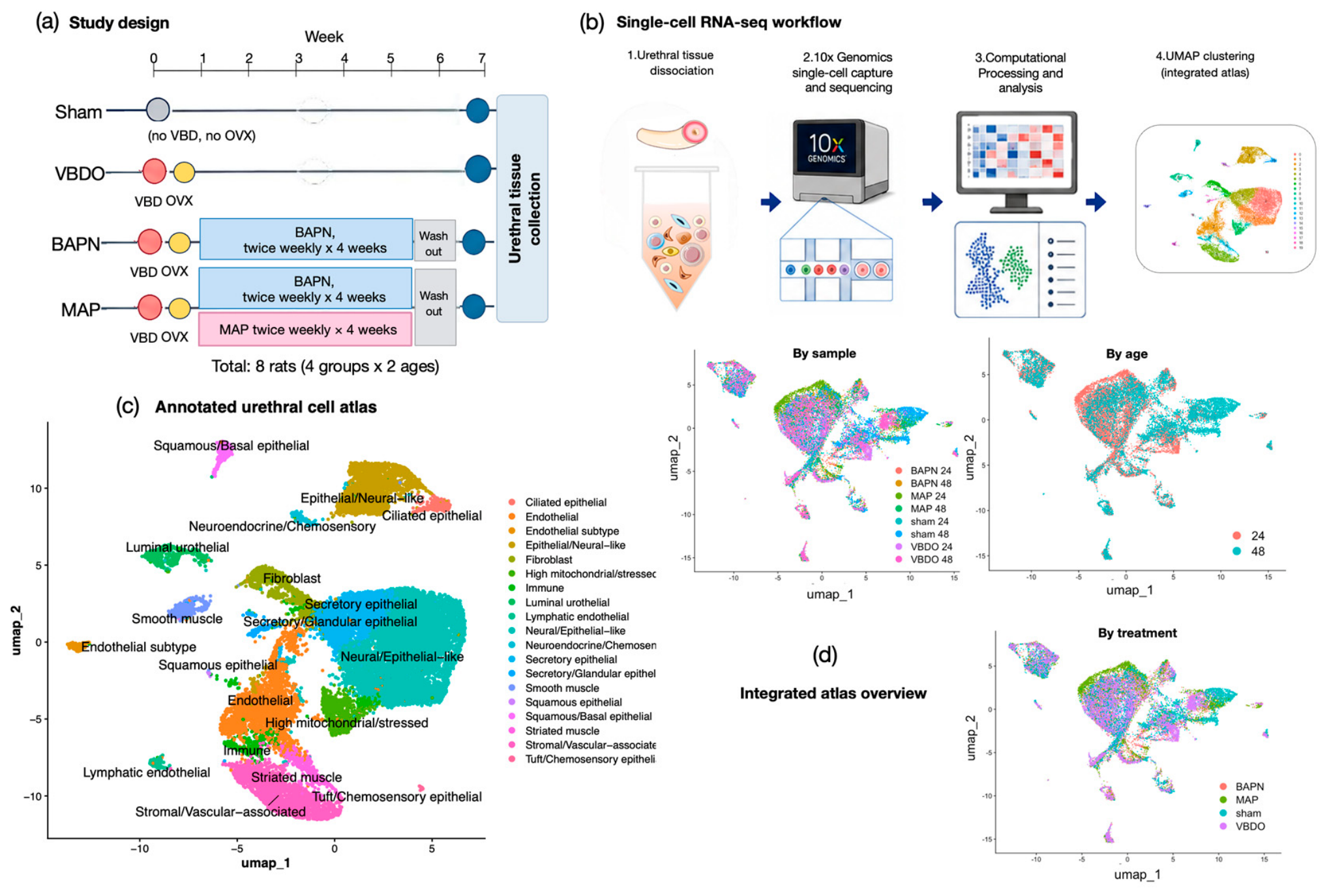
Figure 2.
Treatment-associated differences in the injured urethral microenvironment and urethral muscle content in a separate histological cohort. (a) Relative proportions of major urethral cell populations identified by single-cell RNA sequencing in the Sham, VBDO, BAPN, and MAP groups. Relative cell-type distributions differed descriptively among groups; MAP-treated samples differed from BAPN and were not identical to the Sham profile. (b) Schematic summary of injury/aging-associated features and MAP-associated differences in the urethral ecosystem. (c) Representative transverse urethral sections stained with Pha (green), α-smooth muscle actin (α-SMA; red), and DAPI (blue) in the indicated groups. This histological cohort consisted of 16 additional 24-week-old female Sprague–Dawley rats that were not included in the scRNA-seq cohort. Animals were assigned to Sham (n = 4), VBDO (n = 4), BAPN (n = 4), and MAP (n = 4) groups. (d) Quantification of urethral muscle content. Each dot represents one independent animal (n = 4 per group), and bars show mean ± SEM. Statistical analysis was performed using one-way ANOVA followed by Tukey’s multiple-comparisons test. Adjusted P values are indicated for the specified pairwise comparisons; ****P < 0.0001; ns, not significant.
Figure 2.
Treatment-associated differences in the injured urethral microenvironment and urethral muscle content in a separate histological cohort. (a) Relative proportions of major urethral cell populations identified by single-cell RNA sequencing in the Sham, VBDO, BAPN, and MAP groups. Relative cell-type distributions differed descriptively among groups; MAP-treated samples differed from BAPN and were not identical to the Sham profile. (b) Schematic summary of injury/aging-associated features and MAP-associated differences in the urethral ecosystem. (c) Representative transverse urethral sections stained with Pha (green), α-smooth muscle actin (α-SMA; red), and DAPI (blue) in the indicated groups. This histological cohort consisted of 16 additional 24-week-old female Sprague–Dawley rats that were not included in the scRNA-seq cohort. Animals were assigned to Sham (n = 4), VBDO (n = 4), BAPN (n = 4), and MAP (n = 4) groups. (d) Quantification of urethral muscle content. Each dot represents one independent animal (n = 4 per group), and bars show mean ± SEM. Statistical analysis was performed using one-way ANOVA followed by Tukey’s multiple-comparisons test. Adjusted P values are indicated for the specified pairwise comparisons; ****P < 0.0001; ns, not significant.
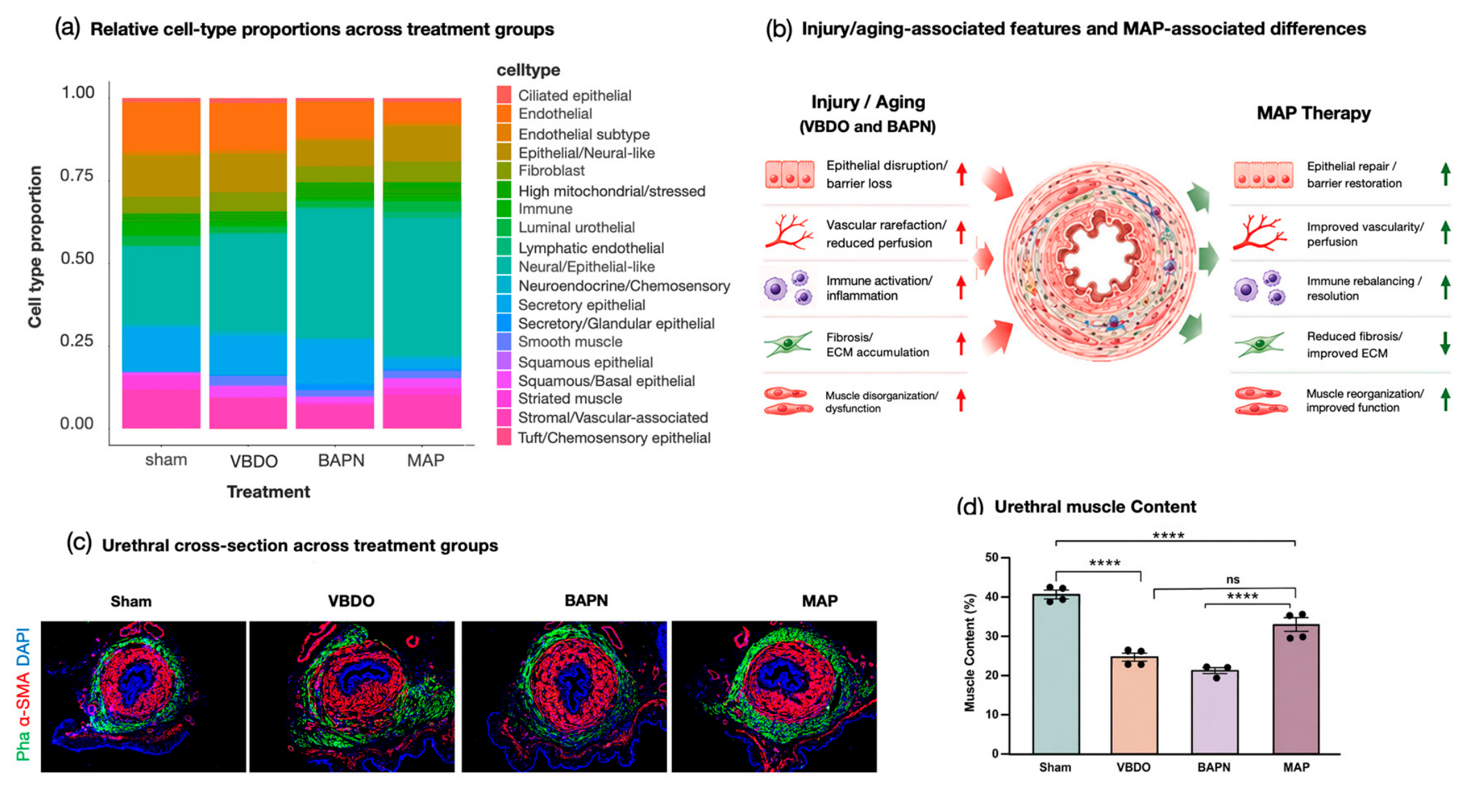
Figure 3.
MAP treatment-associated differences in CellChat-inferred intercellular communication and biological processes in urethral tissues. (a) CellChat-based comparison of relative information flow between MAP and the sham, VBDO, and BAPN groups. Each horizontal stacked bar represents a signaling pathway, with the relative contribution of MAP and the corresponding comparator normalized to a total of 1. Pathways with a greater MAP-associated fraction indicate relatively higher inferred signaling activity in MAP-treated samples, whereas pathways dominated by the comparator indicate relatively lower inferred activity in MAP-treated samples. (b) Relative information flow of representative signaling pathways in the MAP and BAPN groups. (c) Exploratory Gene Ontology biological-process enrichment analysis based on the cell-level MAP-versus-BAPN comparison. Blue bars show processes associated with genes showing lower expression in MAP-treated samples, whereas red bars show processes associated with genes showing higher expression in MAP-treated samples. Bar length represents −log10-adjusted P value from the exploratory cell-level analysis and does not represent treatment-group inference across independent animals.
Figure 3.
MAP treatment-associated differences in CellChat-inferred intercellular communication and biological processes in urethral tissues. (a) CellChat-based comparison of relative information flow between MAP and the sham, VBDO, and BAPN groups. Each horizontal stacked bar represents a signaling pathway, with the relative contribution of MAP and the corresponding comparator normalized to a total of 1. Pathways with a greater MAP-associated fraction indicate relatively higher inferred signaling activity in MAP-treated samples, whereas pathways dominated by the comparator indicate relatively lower inferred activity in MAP-treated samples. (b) Relative information flow of representative signaling pathways in the MAP and BAPN groups. (c) Exploratory Gene Ontology biological-process enrichment analysis based on the cell-level MAP-versus-BAPN comparison. Blue bars show processes associated with genes showing lower expression in MAP-treated samples, whereas red bars show processes associated with genes showing higher expression in MAP-treated samples. Bar length represents −log10-adjusted P value from the exploratory cell-level analysis and does not represent treatment-group inference across independent animals.
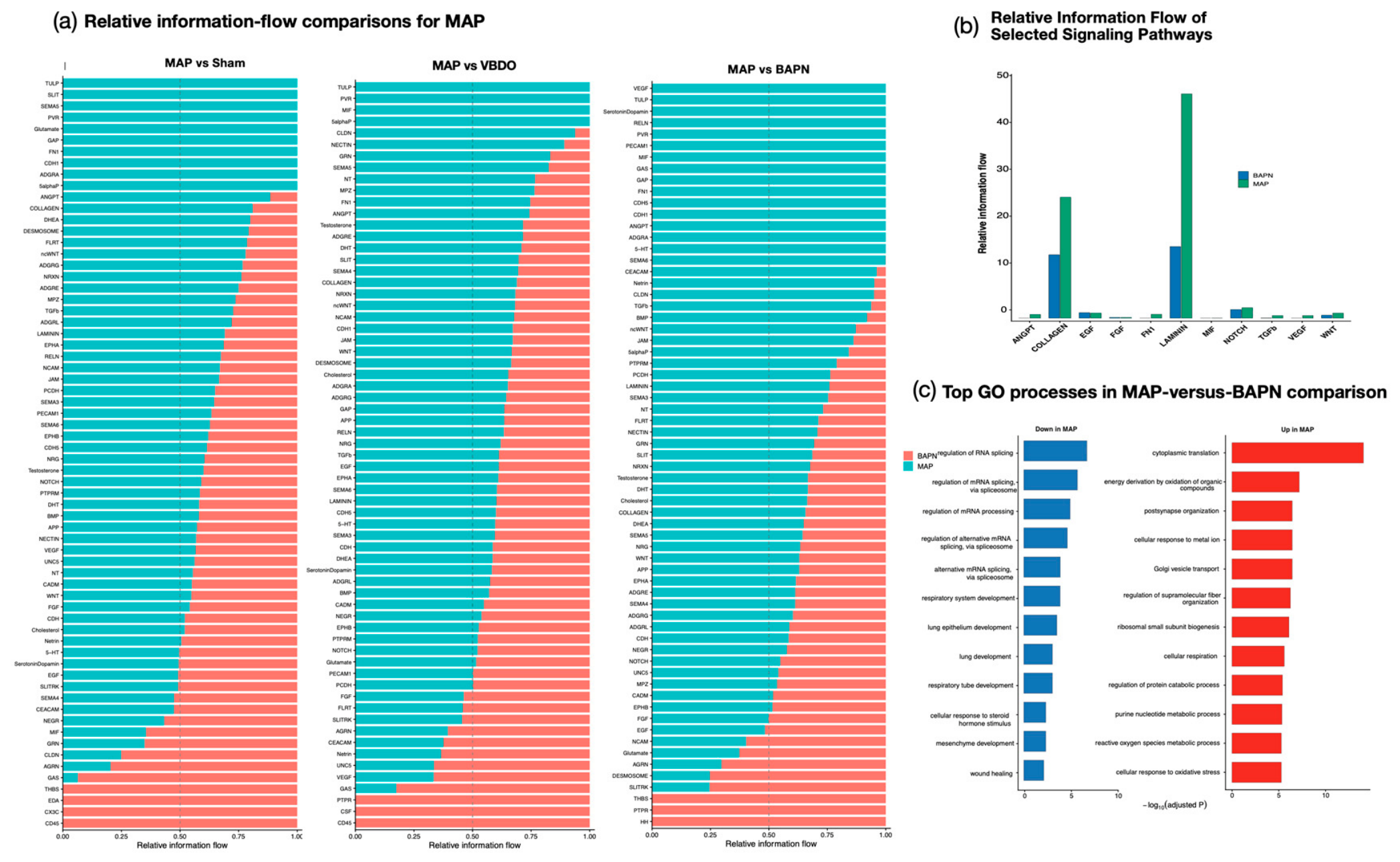
Figure 4.
CellChat analysis identifies treatment-associated differences in predicted intercellular communication between MAP and BAPN groups. (a) Differential cell–cell interaction networks comparing the MAP and BAPN groups. The upper circle plot shows differences in the number of predicted ligand–receptor interactions MAP-versus-BAPN comparison, whereas the lower plot shows differences in overall interaction strength MAP-versus-BAPN comparison. Nodes represent major urethral cell populations, and node colors indicate cell identity. Red edges indicate interactions with greater inferred activity in MAP-treated samples, blue edges indicate interactions with lower inferred activity in MAP-treated samples, and gray edges indicate relatively unchanged inferred activity compared with BAPN samples. Edge thickness is proportional to the magnitude of the difference. (b) Heatmaps of incoming signaling activity, reflecting receptor-associated signaling received by each cell population in the BAPN and MAP groups. Rows represent signaling pathways and columns represent recipient cell types. Darker green indicates greater relative incoming signaling activity. (c) Heatmaps of outgoing signaling activity, reflecting ligand-associated signaling transmitted by each cell population in the BAPN and MAP groups. Rows represent signaling pathways and columns represent sender cell types. Darker green indicates greater relative outgoing signaling activity.
Figure 4.
CellChat analysis identifies treatment-associated differences in predicted intercellular communication between MAP and BAPN groups. (a) Differential cell–cell interaction networks comparing the MAP and BAPN groups. The upper circle plot shows differences in the number of predicted ligand–receptor interactions MAP-versus-BAPN comparison, whereas the lower plot shows differences in overall interaction strength MAP-versus-BAPN comparison. Nodes represent major urethral cell populations, and node colors indicate cell identity. Red edges indicate interactions with greater inferred activity in MAP-treated samples, blue edges indicate interactions with lower inferred activity in MAP-treated samples, and gray edges indicate relatively unchanged inferred activity compared with BAPN samples. Edge thickness is proportional to the magnitude of the difference. (b) Heatmaps of incoming signaling activity, reflecting receptor-associated signaling received by each cell population in the BAPN and MAP groups. Rows represent signaling pathways and columns represent recipient cell types. Darker green indicates greater relative incoming signaling activity. (c) Heatmaps of outgoing signaling activity, reflecting ligand-associated signaling transmitted by each cell population in the BAPN and MAP groups. Rows represent signaling pathways and columns represent sender cell types. Darker green indicates greater relative outgoing signaling activity.
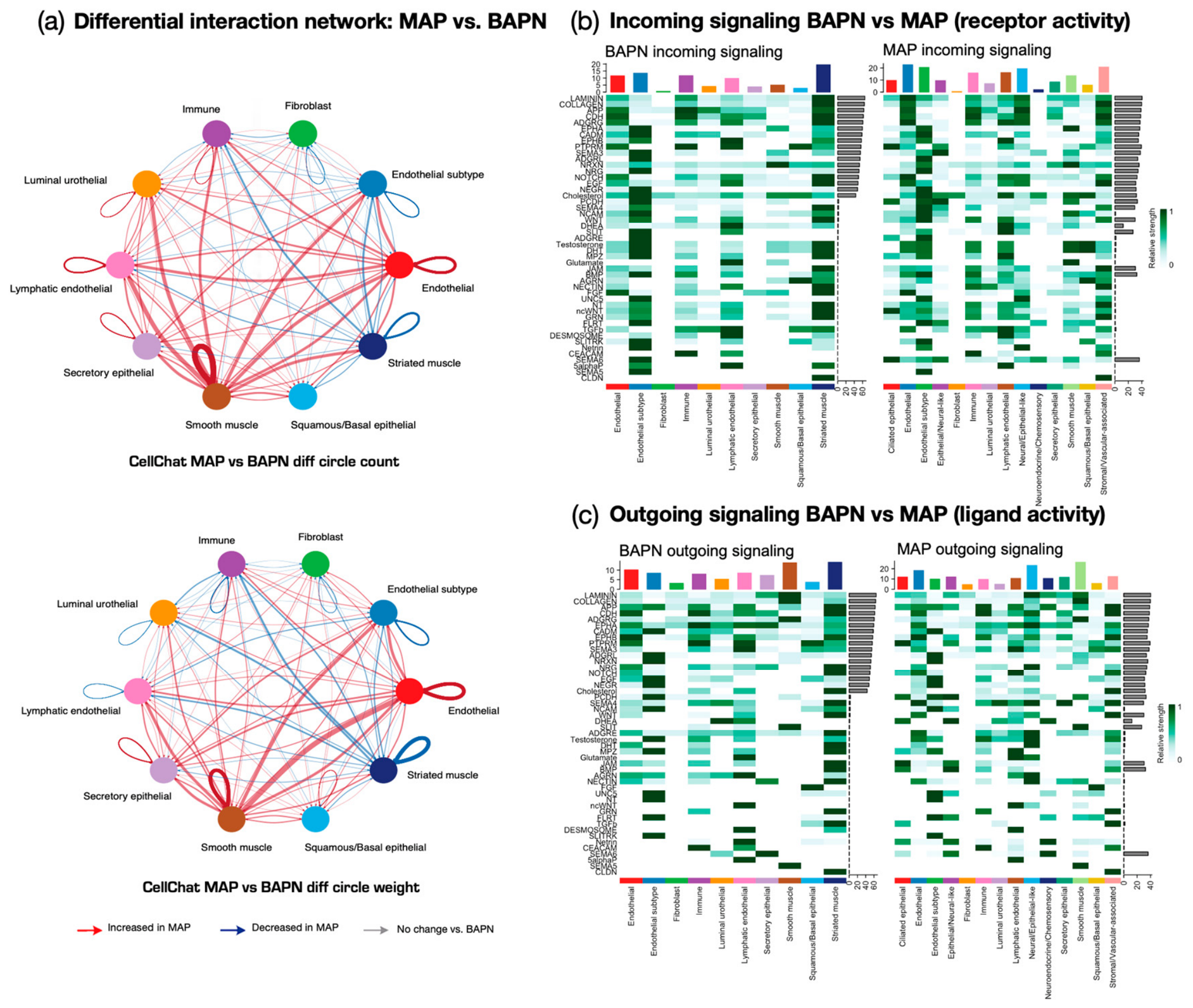
Figure 5.
Stem/progenitor cell signatures and selective modulation of senescence-associated secretory phenotype activity following MAP treatment. (a) Dot plot showing the expression of representative stem/progenitor-associated markers across major urethral cell populations. Dot size indicates the percentage of cells expressing each gene, and color intensity represents the average scaled expression level. (b) Dot plot comparing stem/progenitor marker expression among treatment groups. Dot size represents the percentage of expressing cells, whereas color intensity indicates average expression. (c) Heatmap of senescence-associated secretory phenotype (SASP) module scores across cell types and treatment groups. Red indicates relatively increased SASP activity, whereas blue indicates reduced SASP activity. MAP-treated samples showed a cell type–selective pattern rather than a uniform suppression of SASP signaling. (d) Schematic summary of MAP-associated selective SASP modulation within the urethral tissue microenvironment.
Figure 5.
Stem/progenitor cell signatures and selective modulation of senescence-associated secretory phenotype activity following MAP treatment. (a) Dot plot showing the expression of representative stem/progenitor-associated markers across major urethral cell populations. Dot size indicates the percentage of cells expressing each gene, and color intensity represents the average scaled expression level. (b) Dot plot comparing stem/progenitor marker expression among treatment groups. Dot size represents the percentage of expressing cells, whereas color intensity indicates average expression. (c) Heatmap of senescence-associated secretory phenotype (SASP) module scores across cell types and treatment groups. Red indicates relatively increased SASP activity, whereas blue indicates reduced SASP activity. MAP-treated samples showed a cell type–selective pattern rather than a uniform suppression of SASP signaling. (d) Schematic summary of MAP-associated selective SASP modulation within the urethral tissue microenvironment.
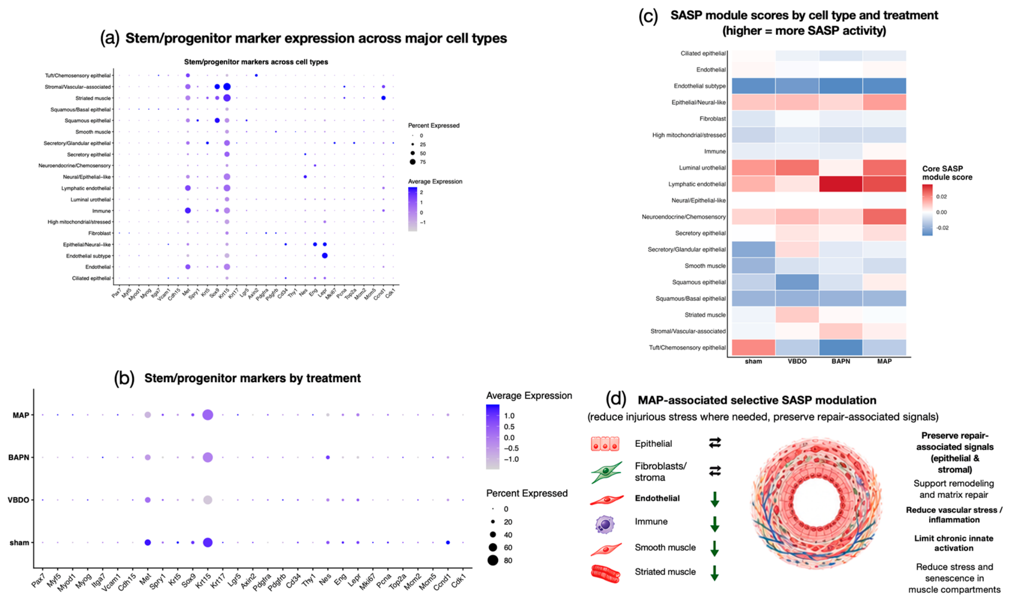

Table 1.
Experimental design of the single-cell RNA-sequencing cohort (n=8).
| Treatment group | 24 weeks | 48 weeks | Total |
|---|---|---|---|
| Sham | 1 | 1 | 2 |
| VBDO | 1 | 1 | 2 |
| BAPN | 1 | 1 | 2 |
| MAP | 1 | 1 | 2 |
| Total | 4 | 4 | 8 |
Disclaimer/Publisher’s Note: The statements, opinions and data contained in all publications are solely those of the individual author(s) and contributor(s) and not of MDPI and/or the editor(s). MDPI and/or the editor(s) disclaim responsibility for any injury to people or property resulting from any ideas, methods, instructions or products referred to in the content. |
© 2026 by the authors. Licensee MDPI, Basel, Switzerland. This article is an open access article distributed under the terms and conditions of the Creative Commons Attribution (CC BY) license.
Copyright: This open access article is published under a Creative Commons CC BY 4.0 license, which permit the free download, distribution, and reuse, provided that the author and preprint are cited in any reuse.