

Submitted:
30 September 2026
Posted:
02 October 2026
You are already at the latest version
Abstract
Background/Objectives: Cisplatin induced hearing loss (CIHL) is a major global health sequela of chemotherapy, affecting over half a million cancer patients annually. We previously reported that honokiol, a multifunctional molecule compatible with cisplatin, protects against CIHL and upregulates sirtuin 3. Sirtuin family is composed of seven highly conserved while differentially expressed members which are critical enzymes for oxidative stress regulation in different cellular compartments. Therefore, this study aims to further verify the expression and upregulation of the entire sirtuin family during honokiol-mediated hearing protection against CIHL in a tumor-bearing mouse model. Methods: Adult female mice carrying mouse mammary tumor virus polyomavirus middle T agent oncogene were used. These mice developed palpable tumors automatically at the age of 70.8±10.3 days. CIHL was induced using a three-cycle (14 days/cycle) low-dose (4 mg/kg/day for 4 days per cycle, intraperitoneal injection) cisplatin treatment mimicking chemotherapy regimens. Honokiol (5-20 mg/kg) was administered one hour before cisplatin. Cochlear function was evaluated by auditory brainstem response threshold shifts and synaptic changes. Immunostaining and RT-qPCR were performed to verify the upregulation of sirtuin family members (1, 2, 3, 5, and 7) during these processes. Results: CIHL was observed at high-frequency range (24-32 kHz), which was mitigated by honokiol treatment. The optimal dose of honokiol for oto-protection and animal survival was 10 mg/kg. Fluorescence signals of sirtuins 1, 2, 3, 5, and 7 increased in cochleae from honokiol-treated mice. RT-qPCR confirmed a significant upregulation of sirtuin 3 mRNA with honokiol treatment. Conclusions: Sirtuin family upregulation was involved in honokiol-mediated hearing protection against CIHL. The dose-response curve of honokiol lays the foundation for potential translational applications for human studies.
Keywords:
cisplatin-induced hearing loss
; tumor-bearing mice
; chemotherapy
; auditory brainstem response
; RT-qPCR
; cancer treatment
1. Introduction
Cisplatin is a potent, widely used platinum-based chemotherapeutic agent [1]. However, it is also a well-recognized ototoxic compound, with an incidence of hearing loss close to or over 50% in treated patients, varying with age, dose, and treatment regimen [2,3,4]. In recent review studies, cisplatin-induced hearing loss (CIHL) is identified as the predominant cause of hearing loss during chemotherapy [5], leading to an estimated half a million new cases globally each year [6]. CIHL causes cellular and structural damage throughout the cochlea, with outer hair cells (OHCs) being particularly vulnerable [7,8]. The underlying mechanisms are primarily attributed to excessive generation of reactive oxygen species (ROS) [9,10,11], which causes the depletion of intrinsic antioxidants (e.g., glutathione) and activation of multiple cell death pathways [12,13]. Conventional protective strategies, such as the use of exogenous antioxidants as free radical scavengers [14,15,16], raise concerns of deactivating cisplatin [17,18,19] and protecting tumor cells [20]. Currently, sodium thiosulfate is the only approved clinical intervention for CIHL but its use is limited to pediatric patients, with the known risk of reducing cisplatin’s antitumor effect [21].
Sirtuins represent a highly conserved family of NAD+-dependent deacetylases critical for metabolic regulation and oxidative stress defense [22,23,24]. In mammals, seven sirtuin family members (Sirt1-7) are differentially expressed in the cytoplasm (Sirt1 and Sirt2), mitochondria (Sirt3-5), and nucleus (Sirt1, 2, 6, 7) [23,25,26,27], forming a network for precise control of diverse physiological processes. Through modulating ROS, sirtuins play critical roles in various biological processes, including metabolism [28], longevity [25,29], and carcinogenesis [29]. These versatile activities highlighted the broader therapeutic potential of sirtuins as targets for intervention in various disease models. For example, pharmacological upregulation of Sirt1 attenuates cisplatin-induced pulmonary [30] and vascular [31] injury through the activation of nuclear factor erythroid 2-related factor 2 (Nrf2) pathway. Similarly, activation of Sirt3 is critical for maintaining mitochondrial redox homeostasis in cisplatin-induced hair cell loss in zebrafish [32] and cardiotoxicity in rats [33]. In addition, sirtuins are also therapeutic targets for cancer [34] and other diseases as summarized in a recent review [35].
Sirtuins are also expressed in cochlear cells [36]. However, the roles of sirtuins in hearing protection remain poorly understood despite their critical regulatory functions [37]. We previously showed that Sirt3 was upregulated during hearing protection against cisplatin ototoxicity using honokiol, a multifunctional natural compound and dietary supplement [38]. As a reported Sirt3 activator, honokiol shows protective effects against oxidative damage in multiple tissues [39] and enhanced tumor suppression effect that is compatible with cisplatin [40]. These studies suggest that honokiol is a promising candidate for treating CIHL, and Sirt3 might be involved in the mechanism of this honokiol-mediated protection. We have also shown that Sirt5 is upregulated by honokiol in an early-onset hearing loss mouse model due to Sirt3 knockout [41]. Given the high conservation in sequence and function among the Sirtuin family members, it is logical to hypothesize that upregulation by honokiol is not limited to Sirt3 and Sirt5 but rather involves the broader sirtuin network in different cellular compartments. In this study, we thoroughly investigated this honokiol-mediated hearing protection against CIHL in a tumor-bearing mouse model, further examined the expression of multiple sirtuin family members in this process, and explored potential functional pathways involving sirtuins during this process.
2. Results
2.1. Honokiol Ameliorates CIHL in a Tumor-Bearing Mouse Model
2.1.1. Significant CIHL Was Induced by a Three-Cycle Cisplatin Treatment Regimen
Transgenic mice carrying mouse mammary tumor virus polyomavirus middle T agent oncogene (MMTV-PyMT, Jax Stock #: 002374, FVB background) [42] were used as the cancer mouse model for this study. Female MMTV-PyMT carriers developed palpable mammary tumors reaching a volume of 500 mm3 at 70.8 ± 10.3 days of life (n = 84), which was set as the starting point for therapeutic interventions. To mimic clinical chemotherapy paradigms, mice received a three-cycle (14 days/cycle) low-dose cisplatin regimen consisting of 4 mg/kg /day for four consecutive days per cycle (Figure 1) [38,43,44], delivered through intraperitoneal (i.p.) injection. Hearing was assessed by auditory brainstem response (ABR) thresholds at frequencies from 4 to 32 kHz (Figure 2). Baseline (Day 0) ABR thresholds showed no significant difference among experimental groups (Figure 2A). After three cycles of cisplatin treatment, mice exhibited clear ABR threshold elevations across all frequencies (Figure 2B, Cis-Only, the blue curve). Threshold shifts reached statistical significance at 32 kHz (p < 0.05), compared to the baseline of the same group. This is consistent with our previous study [38] and comparable with the results from other studies showing that CIHL primarily affects the high frequency range [44,45].
2.1.2. CIHL Was Ameliorated by Parallel Honokiol Treatment
To verify the protective effect of honokiol against CIHL, three different doses (5, 10, and 20 mg/kg) were adopted and were given through i.p. injection one hour before each cisplatin dose. As shown in Figure 2B, CIHL was largely ameliorated by honokiol application in all three dosage groups (marked as HNK5+Cis, HNK10+Cis, and HNK20+Cis, respectively). No statistically significant ABR threshold shift was observed in any frequency in honokiol treated groups.
It should be noted that not all animals have completed the full treatment course. Animals with tumor size ≥ 1500 mm3, weight loss ≥ 25%, or severe health deterioration were removed from the study. Figure 2B includes all the mice survived until the end of the three treatment cycles.
2.1.3. Cumulative Percentage Plots Delineated Threshold Shifts in Different Groups
ABR thresholds were also plotted as a cumulative percentage to better capture data variability and to address missing data points in case of animal loss or no response to acoustic stimuli [46] (Figure 2C-F). As an example, baseline ABR thresholds at 32 kHz of all treatment groups were shown in Figure 2C. Similar distribution of the ABR thresholds were indicated by the curves, which were fitted to a logistic function (refer to Materials and Methods). The xhalf, or the average ABR threshold depicted by the curve fitting, was 37.4 ± 16.4 dB SPL (Figure 2C, the dashed curve). After three treatment cycles (Day 42, Figure 2D), the plot shifted to the right for the animals treated with cisplatin alone (Cis-Only group, the blue curve), with an xhalf of 61.0 ± 17.5 dB SPL. Expressed as threshold elevations at 32 kHz: The CIHL after the third treatment cycle (Day 42) was 23.6 ± 1.4 dB, whereas no significant elevation occurred after the first (Day 14, -1.8 ± 2.2 dB) or second (Day 28, 5.5 ± 2 dB) treatment cycles (Figure 2E). Combined cisplatin and honokiol treatment attenuated this threshold shift, with xhalfs of 47.7±14.8, 47.9±21, and 42.3±12.9, for honokiol at 5, 10, and 20 mg/kg, respectively (Figure 2D, HNK5+Cis, HNK10+Cis, and HNK20+Cis groups). Compared to the significant cisplatin-induced threshold shifts (Figure 1E), a minor and non-significant threshold shift occurred in cycle 3 (8.0±2.0 dB, p>0.05) in the HNK10+Cis group (Figure 2F).
ABR threshold data measured in all three cycles during the chemotherapy regimen was plotted in Figure S1. Cumulative percentage plots of ABR threshold changes at 24 kHz can be found in Figure S2, and the rest in the folder for the raw data shared via the link provided at the end of the paper. A significant threshold change was also observed at 24 kHz on cycle 3 (Day 42) between Cis-Only and HNK5+Cis (p<0.05) and between Cis-Only and HNK20+Cis (p<0.05) groups. However, no significant changes were detected within the Cis-Only group in different cycles at 24 kHz. In addition, no significant threshold shift was observed in any other frequencies of all groups. Overall, the results showed that honokiol treatment significantly mitigated CIHL, which was presented at later stages of treatment at high frequencies.
Since the mice in the control and honokiol alone-treated groups did not survive till cycle 3 due to uncontrollable tumor growth, proper control groups were missing with these MMTV-PyMT mice. Therefore, a group of female FVB wild-type mice treated with honokiol with a dose of 20 mg/kg/day (n=8) or vehicles (n=6) were used as the healthy control, which was also included in Figure S1. Honokiol treatment alone did not affect ABR threshold during the three-cycle regimen.
2.2. Honokiol mitigates cisplatin-induced systemic toxicity while preserving anti-tumor efficacy
2.2.1. Honokiol improves survival rate and prevents weight loss in animals undergoing cisplatin chemotherapy
Cisplatin treatment could induce severe weight loss, which can be up to 40% in some studies [47]. In our study, animals that lost more than 25% of weight or reaching moribund conditions were removed from the study and euthanized for humane reasons. This systemic toxicity, combined with tumor oversize criteria, resulted in a low overall survival rate, with only 57.1% (12 of 21) of the animals completing the third treatment cycle in Cis-only group (Figure 3A). Among the nine prematurely terminated cases, the majority (six) were due to weight loss or moribund, while three were due to oversized tumors. Furthermore, the surviving animals in Cis-Only group displayed a cycle-by cycle physical deterioration, with the average body weight dropping to 81.0 ± 14.9% of their pre-treatment baseline by the end of the third cycle (Figure 3B).
Co-treatment with honokiol largely prevented this cisplatin-induced physical decline. By the end of the third cycle, survival rates for the honokiol co-treated groups were 72.7% (8 of 11), 83.3% (10 of 12), and 50% (6 of 12) for honokiol 5, 10, and 20 mg/kg, respectively (Figure 3A). The survival rates of all the groups by the end of each cycle was summarized in Table S1. The data indicated that 10 mg/kg dosage of honokiol provided the optimal protection for animal survival. The loss of body mass was also alleviated in survival animals, with average weights at the conclusion of the study maintained at 101.8 ± 8.3%, 97.0 ± 15.0%, and 101.0 ± 5.7%. Since the group sizes were relatively small and unequal, a more conservative Welch’s ANOVA was performed (F(3,12.43) = 4.05, P = 0.0323). A following Games–Howell post-hoc testing showed that the weight losses were significantly lower in honokiol co-treated groups (p = 0.024, 0.254, and 0.028 for honokiol 5, 10, and 20 mg/kg, respectively, compared to Cis-Only group) (Figure 3B). The values of animal weight during the treatment were reported in Figure S3. No significant difference in pre-treatment baseline weight was detected in different treatment groups.
Kaplan–Meier survival analysis [48] was not performed because the exact timing of individual animal losses was not recorded consistently throughout the study. Instead, survival of the four cisplatin-treated groups at the end of Cycle 3 was compared using a 4-group contingency analysis. Although the proportion of surviving animals was numerically higher in the honokiol co-treatment groups than in the Cis-Only group, the overall difference among groups was not statistically significant (χ²(3) = 1.36, P = 0.715).
2.2.2. Honokiol is Compatible with Cisplatin in Tumor Suppression
Interference of the therapeutic effects of cisplatin is one of the biggest concerns for CIHL treatment during chemotherapy. We have reported in a previous publication that co-treatment of honokiol and cisplatin exert further tumor suppression based on limited data [38]. Adding to the results, further studies on tumor size measurement were also performed on a subset of the animals as shown in Figure S4. In the control group, tumors grew rapidly from 500 mm3 to the 1500 mm3 euthanasia threshold within 6.5 ± 1.2 days (n=4, the black traces). Honokiol alone delayed this progression by doubling the time required (13.3±3.2 days, n=4, the green traces) to reach the maximum tumor burden, indicating that honokiol alone has antitumor effect.
As expected, cisplatin alone had a prevailing effect on tumor growth (n=6, the blue traces). Although cisplatin did not fully cure the tumor, it reduced the tumor size during the treatment period of each cycle. However, tumor growth restarted during recovery period, and tumor size increased significantly by the end of cycle 2 (Day 28) and cycle 3 (Day 42) to about 150% of the original size.
Co-treatment of honokiol (10 mg/kg) and cisplatin further suppressed tumor growth (n=7, the red traces), as the total tumor size was always smaller than that of the Cis-Only group in each given date during the study. At the end of each cycle, the tumor size was 50%, 110%, and 110%, respectively. Further tumor suppression was also shown in animals co-treated with honokiol with the dosages of 5 (n=2) and 20 mg/kg (n=4), although the full record of tumor growth was lost with these animals. These results indicated an additional antitumor effect introduced by honokiol, since the co-treatment exert further tumor suppression power. This enhancement from honokiol has also been proven frequently in previous studies with different in vitro and in vivo models and different cancer types [49,50].
2.3. Honokiol protects IHC Ribbon Synapses During Cisplatin Treatment
2.3.1. Cisplatin Induces the Degeneration of Ribbon Synapses
OHC loss is the most frequently observed changes in CIHL, which is well-documented and have also been reported in our previous study [51]. In the current study, we further investigated the impact of the three-cycle cisplatin regimen on IHC ribbon synapses. Synaptic integrity was evaluated using immunostaining combined with 3D confocal microscopy on harvested cochlear samples. Figure 4 shows the representative confocal images of cochlear samples from different groups following three cycles of treatments. Puncta of C-terminal binding protein 2, (CtBP2, pre-synaptic component, shown in red) and glutamate receptor 2 (GluR2, post-synaptic component, green) were identifiable at the bottom of the IHCs. Most CtBP2 and GluR2 puncta were colocalized in the untreated mice (Figure 4A). Cisplatin treatment caused a decrease in both the numbers and size of the puncta (Figure 4B). For quantitative studies, IHCs and their synapses were digitally separated (Figure4, A’, B’, C, and D). All samples in Figure 4 were harvested from the animals treated in parallel, from the same cochlear segment (frequency range: 9.5-19.1 kHz [51]), and processed in parallel during immunostaining and confocal imaging. An enlarged area at the basal end (around 15-19 kHz) of each sample was shown in the inset of each panel.
Synapse counting and statistics were performed on two cochlear regions, segment 2 and 3 (S2 and S3), which equal to frequency ranges 9.5-19.1 and 19.1-36.5 kHz, respectively (Figure 5) [51]. CtBP2 and GluR2 colocalized puncta at the bottom region of IHCs were considered intact synapses (Figure 4, the insets showing the enlarged IHCs), and each dot in Figure 5 represents the average number of intact synapses per IHC from one sample. Mice in the control group contained an average of 14.94 ± 0.78 and 15.58 ± 0.54 per IHC in the S2 and S3 region, respectively (Figure 4A’ and Figure 5, Ctr). After three cycles of cisplatin treatment, a significant decrease in synaptic counting was observed (11.90 ± 2.10 and 12.19 ± 2.18, representatively. P = 0.00023, compared with controls) (Figure 4B’ and Figure 5, Cis).
2.3.2. Honokiol Prevents the Loss of Ribbon Synapses Induced by Cisplatin
Honokiol treatment alone did not alter the number of synapse (S2: 15.06 ± 1.28; S3: 14.80 ± 1.20. P = 0.73), indicating that honokiol itself had no detectable effect on normal cochlear synaptic integrity (Figure 4C and 5, HNK). However, co-administration of honokiol substantially preserved ribbon synapses following cisplatin treatment. Synapse counts increased to 15.49 ± 0.50 and 15.25 ± 0.75, respectively in S2 and S3 (P = 9.4 × 10⁻⁵, compared with the Cis-Only group) (Figure 4D and 5 H+C). The numbers were comparable to untreated controls or the honokiol-only treated group (P = 0.31). These results suggested that honokiol largely prevented cisplatin-induced cochlear synaptopathy.
2.3.3. Cisplatin Affects Both Pre- and Post-Synaptic Components, Which Were Both Rescued by Honokiol
To determine whether pre- and post-synaptic components could be affected by cisplatin and honokiol treatments differently, we counted the puncta for CtBP2 and GluR2 separately in partial of the samples. As plotted in Figure S5, both pre- (CtBP2, shown in red) and post-synaptic components (GluR2, shown in green) puncta decreased by around 20% due to cisplatin treatment, with the changes on CtBP2 significant (p=0.029) but not GluR2. This is potentially due to the high variation in GluR2 puncta counting. Honokiol co-treatment rescued both pre- and post-synaptic component loss.
2.4. Sirtuin Expression in the Cochlea Is Affected by Honokiol and Cisplatin Treatment
Our recent studies demonstrated that honokiol treatment upregulates both Sirt3 and Sirt5 in the cochlea in wild type littermates of a Sirt3 knockout mouse model [41]. Given the high sequence conservation among sirtuin family members, we sought to investigate the broader expression profile of sirtuin family members in the cochlear and the influence of cisplatin and honokiol using immunostaining. To maximize the observation of potential therapeutic effects, cochleae were harvested on Day 5, following 4 doses of treatment in cycle 1, from four treatment groups: Control, Cis-Only, HNK10-Only, and HNK10+Cis.
2.4.1. Upregulation of Mitochondrial Sirtuins by Honokiol
We first verified the expression of the two primary mitochondrial sirtuins, Sirt3 and Sirt5, in the cochlea (Figure 6). In control samples, fluorescence signals for Sirt3 (red) and Sirt5 (green) and their colocalization (yellow) were observed (Figure 6, A-A’’’). These fluorescence signals were markedly increased following honokiol treatment both alone (10 mg/kg) (Figure 6, B-B’’’) and in combination with cisplatin (Figure 6, D-D’’’). Cisplatin treatment alone did not affect the fluorescence signal of either Sirt3 or Sirt5 (Figure 6, C-C’’’). Despite their coordinated upregulation, the expression patterns of Sirt3 and Sirt5 exhibited subtle differences. While increased Sirt3 fluorescence signal was observed also in supporting cells (SCs), increased Sirt5 signal was mostly observed in hair cells, as shown in the xz orthogonal views in the panels for Sirt3 and Sirt5. Additionally, bright puncta of both Sirt3 and Sirt5 were consistently observed at the basal part of the IHCs as indicated by the white arrows.
2.4.2. Upregulation of Nuclear Sirtuin by Both Cisplatin and Honokiol
Figure 7 shows the expression of Sirt7, one of the nuclear sirtuins, in the cochlea, using Sirt3 as a positive control for confirming honokiol efficacy. Consistent with the previous results, the Sirt3 signal (green) was upregulated by honokiol, as evident with the 3D views at the left panels in Figure 7, A-D. Bright puncta of Sirt3 were again observed in specific locations close to the bottom of the IHCs, as indicated by the white arrows. Conversely, the Sirt7 signal (red) exhibited a distinct granular pattern in the nuclei. The right panels in Figure 7 showed Sirt7 expression in the nuclei of OHCs, IHCs and SCs, which was notably upregulated by both honokiol and cisplatin treatment. Comprehensive expression pattern and colocalization of different fluorescence signals were shown in Figure S6. Pronounced Sirt7 expression was also detected in pillar cells, especially after cisplatin and honokiol treatment (Figure S6, the yz orthogonal views).
2.4.3. Upregulation of Cytoplasmic Sirtuins by Honokiol
Sirt1 and Sirt2 are the two cytoplasmic sirtuins, the expression of Sirt1 and Sirt2 in the cochlea were also studied. Given that bright puncta expression at the basal pole of the IHCs close to the ribbon synapse region was a recurring observation across multiple sirtuins (Sirt1, 2, 3, and 5) in our pilot studies and as shown in Figure 6 and Figure 7, GluR2 (green) was employed as a postsynaptic marker to investigate the potential relationship between these sirtuin aggregations and ribbon synapses. Comprehensive confocal imaging of Sirt1 expression was shown in Figure S7. Sirt1 fluorescence signal (red) was observed in cytoplasm of the hair cells (Figure S7A), which was upregulated by honokiol (Figure S7, B & D) while potentially suppressed by cisplatin (Figure S7C). No obvious upregulation was observed in the SCs, no matter whether the samples were treated by honokiol or cisplatin. Furthermore, bright Sirt1 puncta were observed at the bottom of IHCs and was partially co-localized with GluR2 as indicated by the white arrows and the 3D projection plots in Figure S8. In addition, Sirt2 expression shared a highly similar pattern as Sirt1 in one batch of immunostaining, which was shown in Figure S9.
Given that Sirt1 and Sirt2 have been reported to exhibit nuclear localization in mammals, we conducted a close examination of Sirt1 expression in the nuclei of the hair cells and SCs. As shown in Figure 8, no definitive Sirt1 fluorescence signal was detected within the nuclei of either IHCs, OHCs, or SCs. In contrast, robust Sirt1 signal was observed in peri-nuclear area of the OHCs after honokiol treatment (Figure 8B and 8D, the central panels). Again, puncta of Sirt1 were clearly observed in vicinity of IHC nuclear area, partially colocalized with GluR2 (Figure 8, left panels; also refer to Figure S7, the surface and orthogonal views focusing on synapses and Figure S8). The exact component and function of these puncta are not clear at this point, although it is highly possible that they are associated with ribbon synapses as indicated by the co-localization with GluR2.
2.5. Transcriptional Upregulation of Sirt3 by Honokiol
To further corroborate honokiol-induced sirtuin upregulation observed at the protein level, RT-qPCR was performed to assess the relative mRNA expression of sirtuins and associated genes in the cochlea. Mice in HNK10-Only and Ctr groups were used, and cochleae were also harvested on Day 5 after four doses of treatments. Figure 9 shows the transcriptional profile in the form of relative expression changes calculated using the 2^-(∆∆Ct) method. Within the sirtuin family, honokiol treatment induced a significant upregulation of Sirt3 mRNA expression (1.32 ± 0.08 -fold change relative to control, p = 0.0007). The transcript levels of the other sirtuins remained unaffected by the treatment.
To map the molecular pathways driving and executing sirtuin upregulation, we evaluated a panel of genes spanning the sirtuin regulatory network. This included two critical upstream regulators: peroxisome proliferator-activated receptor gamma coactivator 1-alpha (PGC-1α), the master regulator of mitochondrial biogenesis targeting Sirt3 [37,54,55,56], and 8-oxoguanine DNA glycosylase (OGG1), an enzyme essential for transcriptional upregulation of Sirt1 [57]. Additionally, eight downstream targets related to sirtuin signaling were investigated to assess pathways involved in apoptosis (Cytochrome c, Cytc; Forkhead box O1 and O3, Foxo1 and Foxo3), as well as redox homeostasis and stress response (Glyceraldehyde 3-phosphate dehydrogenase, Gapdh; Glutathione peroxidase 4, Gpx4; Hypoxia-inducible factor 1-alpha, Hif1α; Isocitrate dehydrogenase, Idh2; Nrf2). Notably, among these associated pathways, only the transcript level of Cytc was significantly downregulated (0.70 ± 0.15 -fold change, p = 0.0004) following honokiol administration. These results suggest that, while modest changes were observed in mRNA expression of Sirt3 and Cytc, honokiol-mediated protection of cochlear function is potentially mediated through post-translational deacetylation or other transcriptional regulations.
3. Discussion
This study verified the hearing protective effects of honokiol in a three-cycle cisplatin treatment regimen simulating chemotherapy in a tumor-bearing mouse model. Pre-treatment with honokiol prevents cisplatin-induced ABR threshold shift and synaptic loss while protecting the general health of the animals during cisplatin treatment. Meanwhile, sirtuin family members in the mitochondria (Sirt3 and Sirt5), cytoplasm (Sirt1 and Sirt2), and nuclei (Sirt7) were all upregulated by honokiol treatment in protein level. Among them, Sirt3 upregulation was confirmed in the mRNA level. These results strongly support our hypothesis that sirtuins are upregulated during honokiol-mediated hearing protection against cisplatin ototoxicity, although sirtuin-associated regulatory transcripts tested in this study largely remained stable in honokiol treatment.
CIHL is a severe sequela in chemotherapy which is notoriously difficult to address due to various challenges. Interference with the therapeutic effects of cisplatin is the biggest concern, as hearing protective candidates may deactivate cisplatin [17,18,19] or protect cancer cells [20]. Local [18,58,59,60] or delayed [61,62] drug administration is often required for these candidates. To date, sodium thiosulfate is the only FDA approved treatment for CIHL, with restricted application time and limited use in pediatric patients [21,63].
Honokiol is a plant-originated multifunction polyphenol extracted from the root and bark of Magnolia grandiflora [64]. It has tumor suppression functions while protective effects against various stress or toxicity in multiple tissues and organs, as reviewed in our other publications [37,38]. More importantly, honokiol can enhance conventional anticancer agents, and therefore is potentially a viable adjunct in combination therapies with cisplatin [40]. It has been shown that honokiol is synergistic with cisplatin in multiple cancer models, including colon cancer [65], ovarian cancer [66], and oral squamous cell carcinoma [67]. Mechanically, Sirt3 activation seems to play critical roles [39,68,69], and honokiol can also sensitize cisplatin-resistant phenotype through modulating IL-6/Stat3 signaling [67], a critical pathway involved in tumorigenesis and development [70]. It is also shown that honokiol mitigates severe systemic toxicities, such as acute kidney injury, induced by cisplatin [71,72,73]. A recent review has summarized the application of honokiol in combination therapy for cancer treatment [40].
Our group further demonstrated that honokiol protects against CIHL in cell cultures, and in wild type and tumor bearing mice [38] (Figure 2). This honokiol-mediated hearing protection is shown in ABR, hair cell, and synaptic levels [38] (Figure 4), as well as in an early-onset hearing loss model [41]. Furthermore, honokiol brings additional benefits such as enhanced tumor suppression, improved survival rate, and mitigated weight loss. While the high dose of honokiol (20 mg/kg/day) shows a slightly stronger hearing protective effect than the other two doses (Figure 2D), the middle dose (10 mg/kg/day) has the best overall effect in both protecting the hearing and improving the general health of the animals (Figure 3). Mechanistically, honokiol reverses cisplatin-induced global transcriptomic downregulation in OHCs. Genes in multiple signaling pathways are involved, especially the ones affecting mitochondrial functions [48]. These results strongly support that honokiol is a promising candidate for treating CIHL.
In our study, the cisplatin-induced ABR threshold shift exhibited a more attenuated profile compared to those reported in previous studies using the same three-cycle regimen. While prior studies documented severe hearing loss spanning almost the entire tested frequency range (4-32 kHz), significant hearing loss was only observed at 32 kHz following the completion of the three cycles in our study. This discrepancy can potentially be attributed to two reasons. First, the criteria of the humane endpoint implemented in our protocol might be different from the previous studies. In our study, mice showed body weight loss exceeding 25% would be excluded from further investigation and analysis, a threshold considerably more restrictive than close to 40% weight loss permitted in other studies [47]. More severe weight loss might have reflected more severe systemic toxicity, which could further exacerbate cochlear function. Because these animals did not survive to the end of the third cycle, our reported ABR thresholds may have introduced a survival bias which underestimated the true severity of cisplatin ototoxicity. Second, premature termination of treatment regimen due to oversized tumor growth (1500 mm3) might have also introduced confounding variables when evaluating the final cumulative ototoxicity. Consequently, the overall magnitude of CIHL observed in our study was relatively subtle, which inherently limits the reliability in determining honokiol-mediated hearing protection, in terms of therapeutic magnitude and frequency range.
Besides traditional antioxidants, candidates including factors/cytokines in metabolic [74,75], inflammatory [76,77], or apoptosis [45,78,79] signaling pathways were also tested more recently with promising results. Polyphenols, a diverse group of natural compounds abundant in plants with antioxidant and anti-inflammatory functions, present a novel type of candidate for hearing protection against CIHL, which might bring additional benefits (for review, see [80,81]). For example, luteolin, a flavone derived from celery, green pepper, and chamomile, has anti-inflammatory and anti-carcinogenic properties and alleviates ferroptosis in CIHL [82]. Resveratrol, a gradient abundant in grapes and red wine with various protective effects, protects against CIHL in guinea pigs [83] and rats [84]. Curcumin, a food supplement with chemoprotective, antioxidant, and anti-inflammatory activities, also protects against CIHL in mice and rats [85,86]. Nevertheless, whether these polyphenols interfere with the therapeutic effects of cisplatin is yet to be determined. In addition, bioavailability might be another concern, since most of these polyphenols are water insoluble, including honokiol. Special formulations improving the solubility, such as liposome and amorphous solid dispersion, might help for their clinical applications.
ROS plays a central role in triggering the inflammatory and apoptotic pathways in CIHL [87] as well as in other types of hearing impairment [e.g., noise-induced hearing loss (NIHL) [88,89]; drug-induced hearing loss [11,90,91]; age-related hearing loss (ARHL) [92,93,94]; for a recent review, see [95]]. Following cisplatin exposure, ROS are generated at multiple sites along interconnected signaling pathways, such as inflammation and apoptosis, thereby causing damages across different cellular compartments [17,96,97]. Consequently, targeting a single site or pathway of ROS generation might be insufficient to achieve comprehensive protection against CIHL.
The sirtuin family comprises seven highly conserved members [22,23,24] that are distributed across different cellular compartments, and may therefore form an intrinsic anti-ROS network through regulating distinct downstream substrates [23,25,26,27]. Sirtuins play critical roles in metabolism [28], longevity [25,29], and carcinogenesis [29] by governing multiple cellular processes including homeostasis [98], responses to stress [99], DNA damage repairing [99], inflammation [100,101], and apoptosis [100]. However, the functions of individual sirtuins and their mechanisms in hearing protection remain largely unknown. A previous study examined cochlear sirtuin expression and age-related changes in cochlear sections [36]. However, the results require further verification due to limitations in immunostaining quality and unclear pattern of sirtuin expression in different cellular compartments (such as cytoplasm versus nuclei). In the present study, we found that multiple sirtuins were upregulated in honokiol-mediated hearing protection against CIHL, suggesting their potential roles in this process. In addition, this upregulation was more evident at the protein level than that at the mRNA level.
Individual sirtuins may have different functions in different processes. In hearing protection, studies have indicated the significance of Sirt1-3 in hearing loss and protection. Sirt1 has been shown to protect cochlear hair cells [102] and slows ARHL [103,104], associated with P53 [102], through regulating cellular processes such as autophagy [104]. In contrast, hemizygous Sirt1 knockout mice with a deficiency of Sirt1 prevents ARHL normally occurred in wild-type animals [105]. Noise exposure upregulates Sirt2 expression, and the treatment with a Sirt2 inhibitor reduced NIHL [106], indicating a negative role of Sirt2 in the protection of cochlea function. Nevertheless, Sirt2 upregulation is a protective mechanism in other tissues [107,108]. Sirt3 is involved in multiple intra-mitochondrial metabolic processes, including energy production, ROS generation, and detoxification [109]. The involvement of Sirt3 in hearing protection against different insults, including noise [110,111], drugs [112,113], and age [92,93] has also been studied. Glycolytic metabolism seems to play critical roles in Sirt3-dependent hearing protection, as demonstrated in an acute CIHL model [113]. Various compounds and treatments have been tested for hearing protection through Sirt3 activation in these studies, although none of the tested candidates are suitable for clinical application against CIHL [51]. In our current study, although Sirt5 and Sirt 7 were also upregulated during honokiol-mediated hearing protection, their function and influence are yet to be studied.
As a reported Sirt3 activator, the underlying mechanisms of honokiol are believed to be largely associated with Sirt3 [39], as shown in the heart [114,115], brain [116,117], and kidney [118]. Our recent transcriptomic study revealed a broad upregulation of antioxidant genes by honokiol [48]. Results in the current study further confirmed that honokiol upregulated Sirt3 in the mRNA level and suggested that other sirtuins were also upregulated in protein expression level. To gain a comprehensive understanding of honokiol-mediated cochlear protection, the upstream and downstream pathways involved in the upregulation of sirtuin family were further investigated using RT-qPCR. Two upstream regulators for sirtuins in mitochondria (PGC-1α, for Sirt3) and cytoplasm (OGG1, for Sirt1) that are essential for mitochondrial function and cisplatin damage were tested, although none are significantly altered. Other tested downstream effectors are all critically involved in multiple cellular processes as abovementioned. These include direct targets of Sirt3 (FoxO3 and Idh2) and Sirt1 (Nrf2) that upregulate intrinsic antioxidant defense. Unfortunately, the only observed significant change was the decrease of downstream gene Cytc, an indicator of mitochondrial-dependent apoptosis pathway, after honokiol treatment. On the other hand, Hif-1α, a stress-responsive transcription factor that is suppressed by Sirt3-mediated ROS reduction, is unchanged with honokiol treatment. Although more direct evidence for the association between sirtuin upregulation and hearing protection at molecular level is missing, our studies provided new insight into the strategy for treating CIHL. That is, comprehensive activation of intracellular anti-ROS system from different cellular compartments. Future works such as transcriptomic studies and further functional verifications will help to confirm the roles of individual sirtuins in this process.
An intriguing finding of the present study was the detection of high punctate sirtuin immunoreactivity (Sirt1, 2, 3, and 5) within the IHC ribbon synaptic region partially colocalized with postsynaptic GluR2, which is also potentially upregulated by honokiol. This is the first time that sirtuin expression in ribbon synaptic region is reported. Although the specific localization and functions of these sirtuins remain largely unknown, evidence from other neuronal systems might give some insights. Sirt3 is found in the mitochondria of presynaptic terminals, where it supports ATP production and thereby sustains synaptic transmission [119]. Sirt2 is enriched at the synapses of hippocampal neurons, regulating the trafficking, stability, transmission, and plasticity of GluA1, another subunit of the same AMPA receptor family as GluR2, through deacetylation [120]. Other studies have also shown potential protection of ribbon synapses by Sirt1 [121] and Sirt3 [122,123] while missing solid data on their synaptic localization. Although further studies are needed to confirm the roles of sirtuins in ribbon synapses and sirtuin-dependent or independent mechanisms underlying honokiol protection, these observations strongly suggest that sirtuins might be important for maintaining and protecting synaptic functions.
Despite the promising findings of the current study, there are several limitations. First, the severity of CIHL observed in our tumor-bearing mouse model was relatively milder as above discussed, which inherently limits the reliability of honokiol-mediated hearing protection. Second, although clear correlation between honokiol administration and the upregulation of various sirtuin family members were shown, direct causal connection between sirtuin upregulation and hearing protection is currently missing. Follow-up studies, such as sirtuin-specific knockouts or pharmacological inhibition, are necessary to definitively confirm the mechanistic roles of individual sirtuins in CIHL. Third, immunofluorescence intensity can be affected by various factors such as antibody affinity, tissue penetration, and photobleaching. Therefore, sirtuin expression and upregulation evaluated with fluorescence intensity should only be considered quasi-quantitative and interpreted as relative changes rather than quantitative changes in protein concentration. Upregulation in sirtuin protein levels does not necessarily indicate increased enzymatic activity, either. Furthermore, more tumor models are necessary to verify honokiol-mediated hearing protection against CIHL due to diverse oncology of different tumor models.
4. Materials and Methods
4.1. Ethics Statement
All procedures were carried out following the NIH Guide for the Care and Use of Laboratory Animals and approved by the Institutional Animal Care and Use Committee at Northwestern University, USA (protocol number IS00018749).
4.2. Animal Groups and Treatments
MMTV-PyMT mice (Jackson Laboratory, Bar Harbor, ME. Jax Stock #: 002374, FVB background) were used in this study as a breast tumor model. The MMTV-PyMT is an oncogenic RNA virus of the Retroviridae family, which drives breast tumorigenesis once activated. Tumor growth was monitored using a caliper when tumors were palpable. Tumor size (volume) was calculated using the formula [51,124]: (length × width2)/2. If multiple tumors were developed, their volumes were added to the total tumor size of the animal. Female MMTV-PyMT carriers develop palpable tumors reaching a total size of 500 mm3, the criteria of intervention, at 70.8±10.3 days of age (n=90).
Once the total tumor size reached 500 mm³, mice were randomly assigned to one of seven treatment groups detailed below (Table 1). A chemotherapy regimen was then performed consisting of three cycles [43,45,51]. Each cycle included four consecutive days of drug administration, followed by a 10-day recovery interval (Figure 1) [38,44]. Therefore, each cycle consisted of 14 days, and the entire regimen lasted for 42 days. Honokiol and cisplatin were both purchased from MilliporeSigma (Burlington, MA) or Tocris Bioscience (Minneapolis, MN). Honokiol (98% purity) was dissolved in corn oil (MilliporeSigma) and cisplatin in saline. Both were administered via intraperitoneal (i.p.) injection, with honokiol (or corn oil) given one hour prior to cisplatin (or saline). Hydration with Ringer’s Lactated Solution (RLS) was provided daily at 25 µl/g the first seven days of each cycle.
Honokiol [73,114,125] and cisplatin [38,44,45] doses were determined based on published studies. Following systemic administration, honokiol reaches peak plasma concentration at approximately 30 minutes [126]. One hour interval was adopted to ensure the optimal cochlear tissue saturation and activation of intracellular signaling pathways. Human equivalent dose of honokiol, calculated using the standard body-surface-area conversion described in FDA guidance, was approximately 0.41-1.62 mg/kg for the doses used in this study (5-20 mg/kg).
Animals were monitored daily for side effects of cisplatin treatment throughout the study. Treatment for pain will be performed when the animals showed the signs such as reduced grooming, reduced level of spontaneous activity, piloerection, and hunched posture. The tumor growth was measured every 2 days, and blood creatinine level was measured once per week. The animals meeting any of the following criteria were removed from the study and euthanized: weight loss > 25%, total tumor size > 1500 mm3, tumor diameter over 20 mm along any axis (IACUC predetermined limit), tumor burden visibly affecting the host, creatinine blood level above 2.72 mg/dl, or other physical ailments detrimental to long term wellbeing. Among them, oversized tumor burden mainly occurred in non-cisplatin treated groups, and animal loss or excessive weight loss was mainly observed in cisplatin treated groups. After the third cycle, all surviving animals were euthanized for histology.
In addition, Female wild-type FVB mice (Jackson Laboratory, Jax Stock #: 001800) treated with honokiol with a dose of 20 mg/kg/day (n=8) or vehicles (n=6) were used as the healthy control. For showing sirtuin expression, another 20 animals were randomly assigned into the four groups (5 in each group) but were treated for only four doses and euthanized on Day 5. These mice are also listed in Table 1.
4.3. ABR Measurement and Data Processing
The cochlear function was evaluated by ABRs, which were recorded before (Day 0) and after the treatment was initiated (Days 14, 28, and 42). The detailed method for ABR measurement has been described in our previous publications [51,127]. Briefly, animals were anesthetized by a mixture of ketamine (80-100 mg/kg) and xylazine (2-5 mg/kg), diluted at 1:20 in RLS. The level of anesthesia was monitored every 15 minutes with paw withdrawal reflex and maintained with ketamine dilution (1:20 in RLS). The animals' body temperature was maintained at 37°C using a water-filled heating pad and monitored every 15 minutes along with other vital signs (O2 saturation, respiratory, and pulse rates).
A customized and a Tucker-Davis Technologies (TDT) systems were used for ABR recordings. For the customized system, signal generation and data acquisition were controlled by custom-written software in Testpoint (SuperLogics, Inc. Massachusetts, MA). The sampling rate was set at 250 kHz. Pure tone bursts (36, 32, 24, 16, and 8 kHz) were generated with a computer I/O board (KCPI 3110, Keithley, Cleveland, OH), attenuated in 5 dB steps, amplified, and delivered via a Beyer DT770Pro speaker pre-calibrated with a Brüel & Kjær 1/8-inch microphone. Signals were delivered via a speculum positioned in the ear canal. Tone pips with a 5 ms duration, 1 ms rise/fall time, and a maximal sound level of ≤ 107 dB SPL (sound pressure level re 20 µPa) were adopted. To elicit ABR signals, three hypodermic needles were placed at the ipsilateral mastoid (high), vertex (low), and the body (common ground). The electrodes were connected to an ISO-80 differential amplifier (World Precision Instruments, Sarasota, FL) with an 80 dB amplification and a band filter set between 300-3k Hz (slope: -12 dB/octave). A frequency devices filter (Hewlett Packard, Palo Alto, CA) with a slope of -48 dB/octave and a gain of 20 dB was used for further filtering and amplification. Stored signals for all frequencies and sound levels were averaged 256 or until the signal/noise ratio ≥4.
For the TDT system, an RZ6 processor was used for generating pure tone pips, and a Medusa4Z were used to drive a pre-calibrated speaker. ABR signals were recorded using WS4 Computer Workstation. BioSigRZ software was used to display and acquire data. The attenuation was 21 dB, and the gain was 0 dB. The sound level was set between 85-20 dB SPL, with a step of 5 dB. The rest of the parameters and the recording electrode placement were kept consistent with the customized system.
ABR threshold was defined as the minimum level required for an ABR response with a root mean square (RMS) 1.5 times above the noise floor. Cumulative plots were generated by first rank ordering the ABR thresholds for each frequency and each group and then binning the thresholds with a bin size of 5 dB (Figure 2 and Figure S2) [46].
4.4. Animal Euthanasia, Cochleae Harvest, and Cochlear Dissection
At the end of the treatment, the animals were euthanized by Euthasol© injection (0.1 ml, i.p.), followed by cardiac perfusion with 4% paraformaldehyde (PFA). Cochleae were then harvested, followed by in-cochlea PFA injection, and left in PFA for 2 hours for post-fixation. Decalcification was then performed using 10% EDTA (Sigma Aldrich, St. Louis, MO) in 1X PBS (diluted from 10X PBS, Invitrogen) for 2-3 days. The full-length cochlear coil was then dissected out in 1X PBS and was cut at specific locations into four segments (S1-S4). Since the apical and basal parts tend to gain artificial damages during the processing of the samples, only the middle turn of the cochlea was used for immunostaining (S2 and S3, frequency range: ~9.5-19.1, and 19.1-36.5, respectively) for better immunostaining and mounting. The frequency range was determined previously by measuring the full length of the cochlea, which was correlated with the published frequency map in CBA/J mice [51,128].
4.5. Immunostaining and Confocal Imaging
Standard immunostaining protocols as described in our previous publications were applied [51,111]. Briefly, cochlear samples were first soaked in 30% sucrose, frozen on dry ice, and thawed at room temperature. After washing three times with 1X PBS and rinsed for 20 minutes between a shaker, the samples were blocked with 5% serum + 1% Triton. Primary antibody mixes were then added, and the samples were incubated at 37 oC overnight. Depending on the purpose, the primary antibody mix contained different primary antibodies. For synapses, it had rabbit anti-Myosin VIIa (for hair cells, Proteus Biosciences, 1:200), mouse (IgG1) anti-CtBP2 (for pre-synaptic component, BD Transduction Labs, 1:200), and mouse (IgG2a) anti-GluR2 (for post-synaptic component, Millipore, 1:500). For sirtuin family members, the antibodies for CtBP2 and GluR2 were replaced by 1-2 sirtuin family members, including Sirt1, 2, 3, and 5 (from Santa Cruz Biotechnology or Novus Biologicals). After being rinsed three times in PBS (10 minutes each), the secondary antibody mix consisting of duck anti-rabbit (IgGH-L) AF647 (Jackson Immuno Research, 1:200), goat anti-mouse (IgG1) AF568 (Thermo Fisher, 1:200), goat anti-mouse (IgG2a) AF488 (Thermo Fisher, 1:200), and Hoechst (Thermo Fisher, 1:1000), was added and incubated in the dark for 2 hours at 37 oC. Primary antibodies from Santa Crus Biotechnology are conjugated with fluorophores (Alexa Flour 488, 546, or 594); therefore, no secondary antibody was needed for these primary antibodies. The samples will then be washed three more times with 1X PBS and mounted on slides. Three or more replicates of immunostaining were performed for all the images included in the manuscript except Sirt2.
Confocal imaging was performed on a Nikon W1 Dual CAM Spinning Disk confocal microscopy at the Center for Advanced Microscopy/Nikon Imaging Center of Northwestern University. Confocal images were processed using FIJI (NIH Image) and Imaris (Oxford Instruments, Abingdon, Oxfordshire, England) afterward. Parameters for excitation, emission, and image processing settings were fixed for all the panels in the same images presented in the paper. As an example, fluorescence intensity analysis as shown in Figure 6 was performed in FIJI by drawing a line across desired location using the “Straight” tool, and doing “Profile Plot” under the dropoff menu “Analysis”.
4.6. Two Step RT-qPCR
Mice were euthanized and cochleae extracted after four doses of honokiol or vehicle treatment. The tissues below the round and oval window, the incus, stapes, and nerve fibers were removed, leaving only the cochlea and cochlear bones. Kimble pestles were used to homogenize the tissues in RNA shield at ambient temperature. The tissues were immediately centrifuged, and RNA extracted using Quick-RNA MiniPrep Plus Kit (Fisher Scientific, A46012). DNase I treatment step was performed during RNA extraction to ensure RNA purity. Extracted RNA was eluted with 50 ul nuclease free water. Nanodrop Lite Plus was used to measure RNA concentration. Nanodrop was blanked with nuclease free water and 1 ul of RNA was placed. The purity of RNA was measured by calculating A260/A280 ratio, with values between 1.8 to 2.2 considered pure. SuperScript IV VILO was used for cDNA conversion, in which 300 ng of RNA was used. A previous optimization showed that 100, 300, 1000 ng of RNA were all appropriate for the downstream qPCR. SYBR based RT-qPCR was carried out using QuantStudio 7 Flex Real-Time PCR system. The reaction involved 2 ul primers at 200 nM concentration, 1 ul of cDNA, 5 ul of SYBR green master mix, and 2 ul of nuclease free water. PCR ran for 2 minutes at 95 °C for enzyme activation, and 40 cycles of denaturation at 95 °C for 15 seconds, annealing at 60 °C for 1 minute. The reaction condition was identical for all primers used. There was a dissociation step in which the PCR was run at 95 °C for 15 seconds, 60 °C for 1 minute, and 95 °C for 15 seconds. Custom primers were purchased from Sigma Aldritch. The selection of primers was based on our recent and other publications involving Sirt3 and proteins which were involved in Sirt3-mediated mitochondrial antioxidant pathway (e.g., [31,93,129,130]). Biological and technical replicates (4 HNK treated mice, 3 control mice) were used. Three controls were used: the absence of RNA, reverse transcriptase, and cDNA. Three technical replicates were averaged to calculate the concentration (Ct values). ∆Ct was then calculated using the formula:
∆Ct = Ct (gene x) – CT (housekeeping gene)
∆∆Ct was calculated:
∆∆Ct = ∆Ct (HNK treated) – average ∆Ct (control)
Based on these calculations, 2-(∆∆Ct) values were used to describe the difference in gene expression. Actb was used as a housekeeping gene.
4.7. Data Analysis
ABR recordings were uniformly formatted and processed using MatLab (The Mathworks, Inc., Natik, MA), and averages and standard errors of the mean (SEM) of the thresholds were calculated and presented. One-way ANOVA (two tails), Rank Sum, and test for normality were performed in Matlab for all the ABR threshold and synapse counting data. If the ANOVA indicated differences among the means, the Tukey’s Honestly Significant Difference post-hoc test was followed. The finalized data was plotted using IGOR Pro (Wavemetrics, Lake Oswego, OR). The tests were part of a statistical package provided by IGOR Pro. Statistical decisions were made for a probability of 0.05. ABR thresholds were also analyzed using a linear mixed-effects mode (Table 2). The model evaluates the fixed effects of cisplatin, treatment cycle, honokiol, and frequency (linear and quadratic components), along with their interactions. Mouse ID was included as a random intercept to account for repeated measures.
Synapse counting was initially performed in Imaris using the function of surface plot. Only the dots surrounding the bottom area of the IHCs were included with a size range of 0.25 ± 0.3 µm, and the colocalization was recognized by the software automatically. The number of synapses was normalized by the count of IHCs in the area. Since the size of the synapses was negatively affected by cisplatin treatment, the results were verified by manual counting from three different authors based on unprocessed synaptic staining (CtPB2, GluR2, and colocalization). The reported values were based on the manual counting.
5. Conclusions
This study demonstrated that honokiol mitigates CIHL in a tumor-bearing mouse model without compromising therapeutic efficacy of cisplatin. Pretreatment with honokiol significantly attenuated ABR threshold shifts, loss of ribbon synapse, and health deterioration induced by cisplatin treatment. This honokiol-mediated protection is accompanied by the upregulation of multiple sirtuin family members expressed in different cellular compartments, including Sirt1 & 2 (cytoplasm), Sirt3 & 5 (mitochondria), and Sirt7 (nucleus). Puncta of multiple sirtuins (Sirt1, 2, 3, and 5) were also observed in the ribbon synapse region. RT-qPCR confirmed the upregulation of Sirt3 but not other sirtuins at mRNA level. These findings highlight the potential of honokiol as a promising candidate for treating CIHL. Furthermore, sirtuins in different compartments form a cellular network for oxidative stress regulation. Sirtuin upregulation suggests their potential roles in honokiol-mediated hearing protection, which requires further investigation.
Supplementary Materials
The following supporting information can be downloaded at the website of this paper posted on Preprints.org. Figure S1: ABR threshold data measured in all three cycles during the chemotherapy regimen. Figure S2: Cumulative percentage plots of all the groups at 24 kHz. Figure S3: Changes of Animal body weight of the different groups during the treatments. Figure S4: Tumor growth curves of the different groups during the treatments. Figure S5: Pre- (CtBP2) and post- (GluR2) synaptic changes after three cycles of treatments. Figure S6: Sirt3 and Sirt7 expressions in the cochlea (9.5-19.1 kHz) after different treatments. Figure S7: Cytoplasmic Sirt1 (red) and post-synaptic GluR2 (green) expression in the cochlea (frequency range: 19.1-36.5 kHz) after different treatments. Figure S8: Projection of confocal image stacks for Sirt1 vs GluR2 expression in the cochlea. Figure S9: Cytoplasmic Sirt2 and post-synaptic GluR2 expression in the cochlea (frequency range: 9.5-19.1 kHz) after different treatments.
Author Contributions
Conceptualization, X.T. and C.P.R.; methodology: X.T., C.P.R., D.T., Y.Z., P.P., and C.K.; software (Matlab program), C.P.R. and D.T.; validation, C.P.R., X.T., and D.T.; formal analysis, C.P.R. and D.T.; investigation, Y.Z., X.T., D.T., L.K., P.P., A.R., R.F., and C.K.; resources, C.P.R. and X.T.; data curation, D.T., C.P.R., and X.T.; writing—original draft preparation, X.T., D.T. and C.P.R.; writing—review and editing, all authors; visualization: X.T., C.P.R., and D.T.; supervision, X.T. and C.P.R.; Project administration, X.T., D.T., and R.F.; funding acquisition, X.T. and C.P.R. All authors have read and agreed to the published version of the manuscript.
Funding
This research was funded by Hearing Health Foundation 2017 Emerging Research Grants (X.T.), American Hearing Research Foundation 2022 Emerging Research Grants (X.T.), startup funds from the Department of Otolaryngology, Feinberg School of Medicine at Northwestern University (X.T.), and the National Institutes of Health grant R01DC019434 (X.T. and C.P.R.).
Institutional Review Board Statement
The study was conducted in accordance with the NIH Guide for the Care and Use of Laboratory Animals. The animal study protocols were approved by the Institutional Animal Care and Use Committee at Northwestern University, USA (protocol code / date of approval: IS00005834/May 15, 2017; IS00013632/June 17, 2020; IS00018749/September 2, 2021; IS00020409/March 2, 2022).
Data Availability Statement
The processed and analyzed data supporting the conclusions of this article are included in the manuscript and the supplementary materials. Raw data, including the original ABR recordings, confocal images, and relevant data analysis files and codes, are securely stored on Northwestern University server and will be accessible upon request.
Acknowledgments
We thank Dr. Frederic Depreux and Dr. Lyubov Czech for the assistance in immunostaining, and Ruiyang Tan for language editing. We also thank Dr. Shaozhong He for the introduction of HNK, which inspired the project. Imaging work was performed at the Northwestern University Center for Advanced Microscopy (RRID: SCR_020996) generously supported by CCSG P30 CA060553 awarded to the Robert H Lurie Comprehensive Cancer Center. RT-qPCR was supported by the Northwestern University NUSeq Core Facility.
Conflicts of Interest
Dr. Tan and Dr. Richter are inventors of a patent assigned to Northwestern University, USA that is related to the work reported in this manuscript (US20240350536A1). The authors declare no other conflict of interest. The funders of the study had no role in the design of the study; in the collection, analyses, or interpretation of data; in the writing of the manuscript; or in the decision to publish the results.
Abbreviations
The following abbreviations are used in this manuscript:
| ABR | Auditory brainstem response |
| ARHL | Age-related hearing loss |
| CIHL | Cisplatin-induced hearing loss |
| Cytc | Cytochrome c |
| CIHL | Cisplatin-induced hearing loss |
| Foxo1 | Forkhead box O1 |
| Foxo3 | Forkhead box O3 |
| Gapdh | Glyceraldehyde 3-phosphate dehydrogenase |
| GluR2 | Glutamate receptor 2 |
| Gpx4 | Glutathione peroxidase 4 |
| Hif1α | Hypoxia-inducible factor 1-alpha |
| Idh2 | Isocitrate dehydrogenase |
| IHC | Inner hair cells |
| i.p. | Intraperitoneal |
| MMTV-PyMT | Mouse mammary tumor virus polyomavirus middle T agent oncogene |
| NIHL | Noise-induced hearing loss |
| Nrf2 | Nuclear factor erythroid 2-related factor 2 |
| OGG1 | 8-oxoguanine DNA glycosylase |
| OHC | Outer hair cell |
| PGC-1α | Peroxisome proliferator-activated receptor gamma coactivator 1-alpha |
| ROS | Reactive oxygen species |
| RT-qPCR | Reverse transcription quantitative polymerase chain reaction |
| SC | Supporting cells |
| SD | Standard deviations |
| SE | Standard error |
| Sirt | Sirtuin family members (e.g., Sirt1, Sirt3) |
References
- Johnstone, T.C.; Park, G.Y.; Lippard, S.J. Understanding and improving platinum anticancer drugs--phenanthriplatin. Anticancer Res. 2014, 34, 471–476. [Google Scholar] [PubMed]
- Benedetti Panici, P.; Greggi, S.; Scambia, G.; Baiocchi, G.; Lomonaco, M.; Conti, G.; Mancuso, S. Efficacy and toxicity of very high-dose cisplatin in advanced ovarian carcinoma: 4-year survival analysis and neurological follow-up. Int. J. Gynecol. Cancer 1993, 3, 44–53. [Google Scholar] [CrossRef] [PubMed]
- Bertolini, P.; Lassalle, M.; Mercier, G.; Raquin, M.A.; Izzi, G.; Corradini, N.; Hartmann, O. Platinum Compound-Related Ototoxicity in Children: Long-Term Follow-Up Reveals Continuous Worsening of Hearing Loss. J. Pediatr. Hematol. Oncol. 2004, 26, 649–655. [Google Scholar] [CrossRef] [PubMed]
- Knight, K.R.; Kraemer, D.F.; Neuwelt, E.A. Ototoxicity in children receiving platinum chemotherapy: underestimating a commonly occurring toxicity that may influence academic and social development. J. Clin. Oncol. 2005, 23, 8588–8596. [Google Scholar] [CrossRef] [PubMed]
- Kessler, L.; Koo, C.; Richter, C.P.; Tan, X. Hearing loss during chemotherapy: prevalence, mechanisms, and protection. Am. J. Cancer Res. 2024, 14, 4597–4632. [Google Scholar] [CrossRef] [PubMed]
- Dillard, L.K.; Lopez-Perez, L.; Martinez, R.X.; Fullerton, A.M.; Chadha, S.; McMahon, C.M. Global burden of ototoxic hearing loss associated with platinum-based cancer treatment: A systematic review and meta-analysis. Cancer Epidemiol. 2022, 79, 102203. [Google Scholar] [CrossRef] [PubMed]
- van Ruijven, M.W.; de Groot, J.C.; Smoorenburg, G.F. Time sequence of degeneration pattern in the guinea pig cochlea during cisplatin administration. A quantitative histological study. Hear Res. 2004, 197, 44–54. [Google Scholar] [CrossRef] [PubMed]
- van Ruijven, M.W.; de Groot, J.C.; Klis, S.F.; Smoorenburg, G.F. The cochlear targets of cisplatin: an electrophysiological and morphological time-sequence study. Hear Res. 2005, 205, 241–248. [Google Scholar] [CrossRef] [PubMed]
- Bragado, P.; Armesilla, A.; Silva, A.; Porras, A. Apoptosis by cisplatin requires p53 mediated p38alpha MAPK activation through ROS generation. Apoptosis 2007, 12, 1733–1742. [Google Scholar] [CrossRef] [PubMed]
- Choi, Y.M.; Kim, H.K.; Shim, W.; Anwar, M.A.; Kwon, J.W.; Kwon, H.K.; Kim, H.J.; Jeong, H.; Kim, H.M.; Hwang, D.; et al. Mechanism of Cisplatin-Induced Cytotoxicity Is Correlated to Impaired Metabolism Due to Mitochondrial ROS Generation. PLoS ONE 2015, 10, e0135083. [Google Scholar] [CrossRef] [PubMed]
- Schacht, J.; Talaska, A.E.; Rybak, L.P. Cisplatin and aminoglycoside antibiotics: hearing loss and its prevention. Anat. Rec. 2012, 295, 1837–1850. [Google Scholar] [CrossRef] [PubMed]
- Ravi, R.; Somani, S.M.; Rybak, L.P. Mechanism of cisplatin ototoxicity: antioxidant system. Pharmacol. Toxicol. 1995, 76, 386–394. [Google Scholar] [CrossRef] [PubMed]
- Sheth, S.; Mukherjea, D.; Rybak, L.P.; Ramkumar, V. Mechanisms of Cisplatin-Induced Ototoxicity and Otoprotection. Front Cell Neurosci. 2017, 11, 338. [Google Scholar] [CrossRef] [PubMed]
- Marina, N.; Chang, K.W.; Malogolowkin, M.; London, W.B.; Frazier, A.L.; Womer, R.B.; Rescorla, F.; Billmire, D.F.; Davis, M.M.; Perlman, E.J.; et al. Amifostine does not protect against the ototoxicity of high-dose cisplatin combined with etoposide and bleomycin in pediatric germ-cell tumors: a Children's Oncology Group study. Cancer 2005, 104, 841–847. [Google Scholar] [CrossRef] [PubMed]
- Fouladi, M.; Chintagumpala, M.; Ashley, D.; Kellie, S.; Gururangan, S.; Hassall, T.; Gronewold, L.; Stewart, C.F.; Wallace, D.; Broniscer, A.; et al. Amifostine protects against cisplatin-induced ototoxicity in children with average-risk medulloblastoma. J. Clin. Oncol. 2008, 26, 3749–3755. [Google Scholar] [CrossRef] [PubMed]
- Marshak, T.; Steiner, M.; Kaminer, M.; Levy, L.; Shupak, A. Prevention of Cisplatin-Induced Hearing Loss by Intratympanic Dexamethasone: A Randomized Controlled Study. Otolaryngol. Head Neck Surg. 2014, 150, 983–990. [Google Scholar] [CrossRef] [PubMed]
- Hazlitt, R.A.; Min, J.; Zuo, J. Progress in the Development of Preventative Drugs for Cisplatin-Induced Hearing Loss. J. Med. Chem. 2018, 61, 5512–5524. [Google Scholar] [CrossRef] [PubMed]
- Videhult, P.; Laurell, G.; Wallin, I.; Ehrsson, H. Kinetics of Cisplatin and its monohydrated complex with sulfur-containing compounds designed for local otoprotective administration. Exp. Biol. Med. 2006, 231, 1638–1645. [Google Scholar] [CrossRef] [PubMed]
- Sooriyaarachchi, M.; George, G.N.; Pickering, I.J.; Narendran, A.; Gailer, J. Tuning the metabolism of the anticancer drug cisplatin with chemoprotective agents to improve its safety and efficacy. Metallomics 2016, 8, 1170–1176. [Google Scholar] [CrossRef] [PubMed]
- Herr, I.; Ucur, E.; Herzer, K.; Okouoyo, S.; Ridder, R.; Krammer, P.H.; von Knebel Doeberitz, M.; Debatin, K.M. Glucocorticoid cotreatment induces apoptosis resistance toward cancer therapy in carcinomas. Cancer Res. 2003, 63, 3112–3120. [Google Scholar] [PubMed]
- Meijer, A.J.M.; Diepstraten, F.A.; van den Heuvel-Eibrink, M.M.; Bleyer, A. Prevention of cisplatin-induced hearing loss in children: achievements and challenges for evidence-based implementation of sodium Thiosulfate. Front Oncol. 2024, 14, 1336714. [Google Scholar] [CrossRef] [PubMed]
- Greiss, S.; Gartner, A. Sirtuin/Sir2 phylogeny, evolutionary considerations and structural conservation. Mol. Cells 2009, 28, 407–415. [Google Scholar] [CrossRef] [PubMed]
- Vassilopoulos, A.; Fritz, K.S.; Petersen, D.R.; Gius, D. The human sirtuin family: evolutionary divergences and functions. Hum. Genom. 2011, 5, 485–496. [Google Scholar] [CrossRef] [PubMed]
- Feldman, J.L.; Dittenhafer-Reed, K.E.; Denu, J.M. Sirtuin catalysis and regulation. J. Biol. Chem. 2012, 287, 42419–42427. [Google Scholar] [CrossRef] [PubMed]
- Hall, J.A.; Dominy, J.E.; Lee, Y.; Puigserver, P. The sirtuin family's role in aging and age-associated pathologies. J. Clin. Invest 2013, 123, 973–979. [Google Scholar] [CrossRef] [PubMed]
- Kelly, G. A review of the sirtuin system, its clinical implications, and the potential role of dietary activators like resveratrol: part 1. Altern. Med. Rev. 2010, 15, 245–263. [Google Scholar] [PubMed]
- Kelly, G.S. A review of the sirtuin system, its clinical implications, and the potential role of dietary activators like resveratrol: part 2. Altern. Med. Rev. 2010, 15, 313–328. [Google Scholar] [PubMed]
- Chang, H.C.; Guarente, L. SIRT1 and other sirtuins in metabolism. Trends Endocrinol. Metab. 2014, 25, 138–145. [Google Scholar] [CrossRef] [PubMed]
- Saunders, L.R.; Verdin, E. Sirtuins: critical regulators at the crossroads between cancer and aging. Oncogene 2007, 26, 5489–5504. [Google Scholar] [CrossRef] [PubMed]
- Hassanein, E.H.M.; Sayed, G.A.; Alzoghaibi, A.M.; Alammar, A.S.; Abdel-Wahab, B.A.; Abd El-Ghafar, O.A.M.; Mahdi, S.E.; Atwa, A.M.; Alzoghaibi, M.A.; Mahmoud, A.M. Azithromycin Mitigates Cisplatin-Induced Lung Oxidative Stress, Inflammation and Necroptosis by Upregulating SIRT1, PPARgamma, and Nrf2/HO-1 Signaling. Pharmaceuticals 2022, 16. [Google Scholar] [CrossRef] [PubMed]
- Mahmoud, A.M.; Alnasser, S.M.; Abd El-Ghafar, O.A.M.; Alruhaimi, R.S.; Althagafy, H.S.; Atwa, A.M.; Hassanein, E.H.M. Candesartan preserves aortic structure and function in cisplatin-treated rats by upregulating SIRT1/Nrf2/HO-1 signaling and suppressing oxidative stress, TLR-4/NF-kappaB signaling, and necroptosis. Front Pharmacol. 2025, 16, 1678921. [Google Scholar] [CrossRef] [PubMed]
- Kong, S.; Xiao, Y.; Chen, L.; Jin, Y.; Qiao, R.; Xu, K.; Xu, L.; Wang, H. Apigenin attenuates cisplatin-induced hair cell damage in the zebrafish lateral line. Food Chem. Toxicol. 2024, 194, 115099. [Google Scholar] [CrossRef] [PubMed]
- Sumneang, N.; Intakhad, J.; Boonhoh, W.; Pudgerd, A.; Wongmekiat, O.; Kobroob, A. Apigenin Protects Against Cisplatin-Induced Cardiotoxicity: Potential Involvement of CD38-Sirt3 Signaling in Rats. Molecules 2026, 31, 2300. [Google Scholar] [CrossRef] [PubMed]
- Besli, N.; Ercin, N.; Tokocin, M.; Boluk, S.E.; Cakmak, R.K.; Ozdogan, K.; Aktokmakyan, T.V.; Toprak, M.; Ercan, G.; Beker, M.; et al. Carvacrol Selectively Induces Mitochondria-Related Apoptotic Signaling in Primary Breast Cancer-Associated Fibroblasts. Pharmaceuticals 2026, 19. [Google Scholar] [CrossRef] [PubMed]
- Akantibila, M.; Carabetta, V.J. Sirtuins as Therapeutic Targets for Treating Cancer, Metabolic Diseases, and Neurodegenerative Diseases. Pharmaceuticals 2025, 18. [Google Scholar] [CrossRef] [PubMed]
- Takumida, M.; Takumida, H.; Katagiri, Y.; Anniko, M. Localization of sirtuins (SIRT1-7) in the aged mouse inner ear. Acta Otolaryngol. 2016, 136, 120–131. [Google Scholar] [CrossRef] [PubMed]
- Koo, C.; Richter, C.P.; Tan, X. Roles of Sirtuins in Hearing Protection. Pharmaceuticals 2024, 17. [Google Scholar] [CrossRef] [PubMed]
- Tan, X.; Zhou, Y.; Agarwal, A.; Lim, M.; Xu, Y.; Zhu, Y.; O'Brien, J.; Tran, E.; Zheng, J.; Gius, D.; et al. Systemic application of honokiol prevents cisplatin ototoxicity without compromising its antitumor effect. Am. J. Cancer Res. 2020, 10, 4416–4434. [Google Scholar] [PubMed]
- Xiang, F.; Zhang, Z.; Li, Y.; Xia, B.; Liao, Y.; Yang, C.; He, Z.; Lin, L.; Xie, J. Honokiol Targeting SIRT3: From Molecular Mechanisms to Therapeutic Opportunities. FASEB J. 2025, 39, e70798. [Google Scholar] [CrossRef] [PubMed]
- Rawat, L.; Solanki, R.; Kumar, R.; Pal, S.; Sabarwal, A. Honokiol in cancer: Roles in enhancing combination therapy efficacy and preventing post-transplant malignancies. Tumor Discov. 2025, 4, 42–54. [Google Scholar] [CrossRef] [PubMed]
- Koo, C.; Zhou, Y.; Thomas, D.; Fuentes, R.; Li, J.; Kim, M.; Richter, C.P.; Tan, X. Sex-specific hearing loss in Sirt3 knockout mice and attenuation by honokiol. Hear Res. 2026, 470, 109502. [Google Scholar] [CrossRef] [PubMed]
- Guy, C.T.; Cardiff, R.D.; Muller, W.J. Induction of mammary tumors by expression of polyomavirus middle T oncogene: a transgenic mouse model for metastatic disease. Mol. Cell Biol. 1992, 12, 954–961. [Google Scholar] [CrossRef]
- Roy, S.; Ryals, M.M.; Van den Bruele, A.B.; Fitzgerald, T.S.; Cunningham, L.L. Sound preconditioning therapy inhibits ototoxic hearing loss in mice. J. Clin. Invest 2013, 123, 4945–4949. [Google Scholar] [CrossRef] [PubMed]
- Breglio, A.M.; Rusheen, A.E.; Shide, E.D.; Fernandez, K.A.; Spielbauer, K.K.; McLachlin, K.M.; Hall, M.D.; Amable, L.; Cunningham, L.L. Cisplatin is retained in the cochlea indefinitely following chemotherapy. Nat. Commun. 2017, 8, 1654. [Google Scholar] [CrossRef] [PubMed]
- Teitz, T.; Fang, J.; Goktug, A.N.; Bonga, J.D.; Diao, S.; Hazlitt, R.A.; Iconaru, L.; Morfouace, M.; Currier, D.; Zhou, Y.; et al. CDK2 inhibitors as candidate therapeutics for cisplatin- and noise-induced hearing loss. J. Exp. Med. 2018, 215, 1187–1203. [Google Scholar] [CrossRef] [PubMed]
- Richter, C.P.; Young, H.; Richter, S.V.; Smith-Bronstein, V.; Stock, S.R.; Xiao, X.; Soriano, C.; Whitlon, D.S. Fluvastatin protects cochleae from damage by high-level noise. Sci. Rep. 2018, 8, 3033. [Google Scholar] [CrossRef] [PubMed]
- Sung, C.Y.W.; Hayase, N.; Yuen, P.S.T.; Lee, J.; Fernandez, K.; Hu, X.; Cheng, H.; Star, R.A.; Warchol, M.E.; Cunningham, L.L. Macrophage depletion protects against cisplatin-induced ototoxicity and nephrotoxicity. Sci. Adv. 2024, 10, eadk9878. [Google Scholar] [CrossRef] [PubMed]
- Koo, C.; Fuentes, R.; Pandya, P.; Takahashi, S.; Thomas, D.; Zhou, Y.; Homma, K.; Li, J.; Richter, C.P.; Tan, X. Honokiol-mediated reversal of cisplatin-induced transcriptomic changes in outer hair cells. BioMed Pharmacother. 2026, 203, 119857. [Google Scholar] [CrossRef] [PubMed]
- Jiang, Q.Q.; Fan, L.Y.; Yang, G.L.; Guo, W.H.; Hou, W.L.; Chen, L.J.; Wei, Y.Q. Improved therapeutic effectiveness by combining liposomal honokiol with cisplatin in lung cancer model. BMC Cancer 2008, 8, 242. [Google Scholar] [CrossRef] [PubMed]
- Luo, H.; Zhong, Q.; Chen, L.J.; Qi, X.R.; Fu, A.F.; Yang, H.S.; Yang, F.; Lin, H.G.; Wei, Y.Q.; Zhao, X. Liposomal honokiol, a promising agent for treatment of cisplatin-resistant human ovarian cancer. J. Cancer Res. Clin. Oncol. 2008, 134, 937–945. [Google Scholar] [CrossRef] [PubMed]
- Tan, X.; Zhou, Y.; Agarwal, A.; Lim, M.; Xu, Y.; Zhu, Y.; O'Brien, J.; Tran, E.; Zheng, J.; Gius, D.; et al. Systemic Application of Honokiol Prevents Cisplatin Ototoxicity Without Compromising its Antitumor Effect. Am. J. Cancer Res. 2020, Accepted. [Google Scholar]
- Jiang, H.; Sha, S.H.; Forge, A.; Schacht, J. Caspase-independent pathways of hair cell death induced by kanamycin in vivo. Cell Death Differ. 2006, 13, 20–30. [Google Scholar] [CrossRef] [PubMed]
- Wang, X.; Zhu, Y.; Long, H.; Pan, S.; Xiong, H.; Fang, Q.; Hill, K.; Lai, R.; Yuan, H.; Sha, S.H. Mitochondrial Calcium Transporters Mediate Sensitivity to Noise-Induced Losses of Hair Cells and Cochlear Synapses. Front Mol. Neurosci. 2018, 11, 469. [Google Scholar] [CrossRef] [PubMed]
- Han, H.; Dong, Y.; Ma, X. Dihydromyricetin Protects Against Gentamicin-Induced Ototoxicity via PGC-1alpha/SIRT3 Signaling in vitro. Front Cell Dev. Biol. 2020, 8, 702. [Google Scholar] [CrossRef] [PubMed]
- Matouk, A.I.; Awad, E.M.; Mousa, A.A.K.; Abdelhafez, S.M.N.; Fahmy, U.A.; El-Moselhy, M.A.; Abdel-Naim, A.B.; Anter, A. Dihydromyricetin protects against gentamicin-induced nephrotoxicity via upregulation of renal SIRT3 and PAX2. Life Sci. 2024, 336, 122318. [Google Scholar] [CrossRef] [PubMed]
- Kong, X.; Wang, R.; Xue, Y.; Liu, X.; Zhang, H.; Chen, Y.; Fang, F.; Chang, Y. Sirtuin 3, a new target of PGC-1alpha, plays an important role in the suppression of ROS and mitochondrial biogenesis. PLoS ONE 2010, 5, e11707. [Google Scholar] [CrossRef] [PubMed]
- Antoniali, G.; Lirussi, L.; D'Ambrosio, C.; Dal Piaz, F.; Vascotto, C.; Casarano, E.; Marasco, D.; Scaloni, A.; Fogolari, F.; Tell, G. SIRT1 gene expression upon genotoxic damage is regulated by APE1 through nCaRE-promoter elements. Mol. Biol. Cell 2014, 25, 532–547. [Google Scholar] [CrossRef] [PubMed]
- Wang, J.; Lloyd Faulconbridge, R.V.; Fetoni, A.; Guitton, M.J.; Pujol, R.; Puel, J.L. Local application of sodium thiosulfate prevents cisplatin-induced hearing loss in the guinea pig. Neuropharmacology 2003, 45, 380–393. [Google Scholar] [CrossRef] [PubMed]
- Hill, G.W.; Morest, D.K.; Parham, K. Cisplatin-induced ototoxicity: effect of intratympanic dexamethasone injections. Otol. Neurotol. 2008, 29, 1005–1011. [Google Scholar] [CrossRef] [PubMed]
- Calli, C.; Pinar, E.; Oncel, S.; Alper Bagriyanik, H.; Umut Sakarya, E. Recovery of hearing in Cisplatin-induced ototoxicity in the Guinea pig with intratympanic dexamethasone. Indian J. Otolaryngol. Head Neck Surg. 2012, 64, 46–50. [Google Scholar] [CrossRef] [PubMed]
- Brock, P.R.; Maibach, R.; Childs, M.; Rajput, K.; Roebuck, D.; Sullivan, M.J.; Laithier, V.; Ronghe, M.; Dall'Igna, P.; Hiyama, E.; et al. Sodium Thiosulfate for Protection from Cisplatin-Induced Hearing Loss. N Engl. J. Med. 2018, 378, 2376–2385. [Google Scholar] [CrossRef] [PubMed]
- Freyer, D.R.; Chen, L.; Krailo, M.D.; Knight, K.; Villaluna, D.; Bliss, B.; Pollock, B.H.; Ramdas, J.; Lange, B.; Van Hoff, D.; et al. Effects of sodium thiosulfate versus observation on development of cisplatin-induced hearing loss in children with cancer (ACCL0431): a multicentre, randomised, controlled, open-label, phase 3 trial. Lancet Oncol. 2017, 18, 63–74. [Google Scholar] [CrossRef] [PubMed]
- Dhillon, S. Sodium Thiosulfate: Pediatric First Approval. Paediatr. Drugs 2023, 25, 239–244. [Google Scholar] [CrossRef] [PubMed]
- Lo, Y.C.; Teng, C.M.; Chen, C.F.; Chen, C.C.; Hong, C.Y. Magnolol and honokiol isolated from Magnolia officinalis protect rat heart mitochondria against lipid peroxidation. Biochem Pharmacol. 1994, 47, 549–553. [Google Scholar] [CrossRef] [PubMed]
- Cheng, N.; Xia, T.; Han, Y.; He, Q.J.; Zhao, R.; Ma, J.R. Synergistic antitumor effects of liposomal honokiol combined with cisplatin in colon cancer models. Oncol. Lett. 2011, 2, 957–962. [Google Scholar] [CrossRef] [PubMed]
- Liu, Y.; Chen, L.; He, X.; Fan, L.; Yang, G.; Chen, X.; Lin, X.; Du, L.; Li, Z.; Ye, H.; et al. Enhancement of therapeutic effectiveness by combining liposomal honokiol with cisplatin in ovarian carcinoma. Int. J. Gynecol. Cancer 2008, 18, 652–659. [Google Scholar] [CrossRef] [PubMed]
- Chang, M.T.; Lee, S.P.; Fang, C.Y.; Hsieh, P.L.; Liao, Y.W.; Lu, M.Y.; Tsai, L.L.; Yu, C.C.; Liu, C.M. Chemosensitizing effect of honokiol in oral carcinoma stem cells via regulation of IL-6/Stat3 signaling. Environ. Toxicol. 2018, 33, 1105–1112. [Google Scholar] [CrossRef] [PubMed]
- Chen, Y.; Fu, L.L.; Wen, X.; Wang, X.Y.; Liu, J.; Cheng, Y.; Huang, J. Sirtuin-3 (SIRT3), a therapeutic target with oncogenic and tumor-suppressive function in cancer. Cell Death Dis. 2014, 5, e1047. [Google Scholar] [CrossRef] [PubMed]
- Schumacker, P.T. SIRT3 controls cancer metabolic reprogramming by regulating ROS and HIF. Cancer Cell 2011, 19, 299–300. [Google Scholar] [CrossRef] [PubMed]
- Huang, B.; Lang, X.; Li, X. The role of IL-6/JAK2/STAT3 signaling pathway in cancers. Front Oncol. 2022, 12, 1023177. [Google Scholar] [CrossRef] [PubMed]
- Mao, R.W.; He, S.P.; Lan, J.G.; Zhu, W.Z. Honokiol ameliorates cisplatin-induced acute kidney injury via inhibition of mitochondrial fission. Br. J. Pharmacol. 2022, 179, 3886–3904. [Google Scholar] [CrossRef] [PubMed]
- Wang, T.J.; Liu, H.T.; Lai, Y.H.; Jan, T.R.; Nomura, N.; Chang, H.W.; Chou, C.C.; Lee, Y.J.; Tsai, P.J. Honokiol, a Polyphenol Natural Compound, Attenuates Cisplatin-Induced Acute Cytotoxicity in Renal Epithelial Cells Through Cellular Oxidative Stress and Cytoskeleton Modulations. Front Pharmacol. 2018, 9, 357. [Google Scholar] [CrossRef] [PubMed]
- Li, M.; Li, C.M.; Ye, Z.C.; Huang, J.; Li, Y.; Lai, W.; Peng, H.; Lou, T.Q. Sirt3 modulates fatty acid oxidation and attenuates cisplatin-induced AKI in mice. J. Cell Mol. Med. 2020, 24, 5109–5121. [Google Scholar] [CrossRef] [PubMed]
- Astolfi, L.; Simoni, E.; Valente, F.; Ghiselli, S.; Hatzopoulos, S.; Chicca, M.; Martini, A. Coenzyme Q10 plus Multivitamin Treatment Prevents Cisplatin Ototoxicity in Rats. PLoS ONE 2016, 11, e0162106. [Google Scholar] [CrossRef] [PubMed]
- Scasso, F.; Sprio, A.E.; Canobbio, L.; Scanarotti, C.; Manini, G.; Berta, G.N.; Bassi, A.M. Dietary supplementation of coenzyme Q10 plus multivitamins to hamper the ROS mediated cisplatin ototoxicity in humans: A pilot study. Heliyon 2017, 3, e00251. [Google Scholar] [CrossRef] [PubMed]
- Fernandez, K.; Spielbauer, K.K.; Rusheen, A.; Wang, L.; Baker, T.G.; Eyles, S.; Cunningham, L.L. Lovastatin protects against cisplatin-induced hearing loss in mice. Hear Res. 2020, 389, 107905. [Google Scholar] [CrossRef] [PubMed]
- Fernandez, K.A.; Allen, P.; Campbell, M.; Page, B.; Townes, T.; Li, C.M.; Cheng, H.; Garrett, J.; Mulquin, M.; Clements, A.; et al. Atorvastatin is associated with reduced cisplatin-induced hearing loss. J. Clin. Invest 2021, 131. [Google Scholar] [CrossRef] [PubMed]
- Hazlitt, R.A.; Teitz, T.; Bonga, J.D.; Fang, J.; Diao, S.; Iconaru, L.; Yang, L.; Goktug, A.N.; Currier, D.G.; Chen, T.; et al. Development of Second-Generation CDK2 Inhibitors for the Prevention of Cisplatin-Induced Hearing Loss. J. Med. Chem. 2018, 61, 7700–7709. [Google Scholar] [CrossRef] [PubMed]
- Ingersoll, M.A.; Malloy, E.A.; Caster, L.E.; Holland, E.M.; Xu, Z.; Zallocchi, M.; Currier, D.; Liu, H.; He, D.Z.Z.; Min, J.; et al. BRAF inhibition protects against hearing loss in mice. Sci. Adv. 2020, 6. [Google Scholar] [CrossRef] [PubMed]
- Osakabe, N.; Modafferi, S.; Ontario, M.L.; Rampulla, F.; Zimbone, V.; Migliore, M.R.; Fritsch, T.; Abdelhameed, A.S.; Maiolino, L.; Lupo, G.; et al. Polyphenols in Inner Ear Neurobiology, Health and Disease: From Bench to Clinics. Medicina 2023, 59. [Google Scholar] [CrossRef] [PubMed]
- Tan, W.J.T.; Vlajkovic, S.M. Molecular Characteristics of Cisplatin-Induced Ototoxicity and Therapeutic Interventions. Int. J. Mol. Sci. 2023, 24. [Google Scholar] [CrossRef] [PubMed]
- Liu, Z.; Zhang, H.; Hong, G.; Bi, X.; Hu, J.; Zhang, T.; An, Y.; Guo, N.; Dong, F.; Xiao, Y.; et al. Inhibition of Gpx4-mediated ferroptosis alleviates cisplatin-induced hearing loss in C57BL/6 mice. Mol. Ther. 2024. [Google Scholar] [CrossRef] [PubMed]
- Yumusakhuylu, A.C.; Yazici, M.; Sari, M.; Binnetoglu, A.; Kosemihal, E.; Akdas, F.; Sirvanci, S.; Yuksel, M.; Uneri, C.; Tutkun, A. Protective role of resveratrol against cisplatin induced ototoxicity in guinea pigs. Int. J. Pediatr. Otorhinolaryngol. 2012, 76, 404–408. [Google Scholar] [CrossRef] [PubMed]
- Olgun, Y.; Kirkim, G.; Kolatan, E.; Kiray, M.; Bagriyanik, A.; Olgun, A.; Kizmazoglu, D.C.; Ellidokuz, H.; Serbetcioglu, B.; Altun, Z.; et al. Friend or foe? Effect of oral resveratrol on cisplatin ototoxicity. Laryngoscope 2014, 124, 760–766. [Google Scholar] [CrossRef] [PubMed]
- Fetoni, A.R.; Eramo, S.L.; Paciello, F.; Rolesi, R.; Podda, M.V.; Troiani, D.; Paludetti, G. Curcuma longa (curcumin) decreases in vivo cisplatin-induced ototoxicity through heme oxygenase-1 induction. Otol. Neurotol. 2014, 35, e169-177. [Google Scholar] [CrossRef] [PubMed]
- Soyalic, H.; Gevrek, F.; Koc, S.; Avcu, M.; Metin, M.; Aladag, I. Intraperitoneal curcumin and vitamin E combination for the treatment of cisplatin-induced ototoxicity in rats. Int. J. Pediatr. Otorhinolaryngol. 2016, 89, 173–178. [Google Scholar] [CrossRef] [PubMed]
- Borse, V.; Al Aameri, R.F.H.; Sheehan, K.; Sheth, S.; Kaur, T.; Mukherjea, D.; Tupal, S.; Lowy, M.; Ghosh, S.; Dhukhwa, A.; et al. Epigallocatechin-3-gallate, a prototypic chemopreventative agent for protection against cisplatin-based ototoxicity. Cell Death Dis. 2017, 8, e2921. [Google Scholar] [CrossRef] [PubMed]
- Hu, B.H.; Henderson, D.; Yang, W.P. The impact of mitochondrial energetic dysfunction on apoptosis in outer hair cells of the cochlea following exposure to intense noise. Hear Res. 2008, 236, 11–21. [Google Scholar] [CrossRef] [PubMed]
- Ohlemiller, K.K.; Wright, J.S.; Dugan, L.L. Early elevation of cochlear reactive oxygen species following noise exposure. Audiol. Neurootol. 1999, 4, 229–236. [Google Scholar] [PubMed]
- Huth, M.E.; Ricci, A.J.; Cheng, A.G. Mechanisms of aminoglycoside ototoxicity and targets of hair cell protection. Int. J. Otolaryngol. 2011, 2011, 937861. [Google Scholar] [CrossRef] [PubMed]
- Alam, S.A.; Ikeda, K.; Oshima, T.; Suzuki, M.; Kawase, T.; Kikuchi, T.; Takasaka, T. Cisplatin-induced apoptotic cell death in Mongolian gerbil cochlea. Hear Res. 2000, 141, 28–38. [Google Scholar] [CrossRef] [PubMed]
- Someya, S.; Prolla, T.A. Mitochondrial oxidative damage and apoptosis in age-related hearing loss. Mech. Ageing Dev. 2010, 131, 480–486. [Google Scholar] [CrossRef] [PubMed]
- Someya, S.; Yu, W.; Hallows, W.C.; Xu, J.; Vann, J.M.; Leeuwenburgh, C.; Tanokura, M.; Denu, J.M.; Prolla, T.A. Sirt3 mediates reduction of oxidative damage and prevention of age-related hearing loss under caloric restriction. Cell 2010, 143, 802–812. [Google Scholar] [CrossRef] [PubMed]
- Someya, S.; Xu, J.; Kondo, K.; Ding, D.; Salvi, R.J.; Yamasoba, T.; Rabinovitch, P.S.; Weindruch, R.; Leeuwenburgh, C.; Tanokura, M.; et al. Age-related hearing loss in C57BL/6J mice is mediated by Bak-dependent mitochondrial apoptosis. Proc. Natl. Acad. Sci. U S A 2009, 106, 19432–19437. [Google Scholar] [CrossRef] [PubMed]
- Kamogashira, T.; Fujimoto, C.; Yamasoba, T. Reactive oxygen species, apoptosis, and mitochondrial dysfunction in hearing loss. BioMed Res. Int. 2015, 2015, 617207. [Google Scholar] [CrossRef] [PubMed]
- So, H.; Kim, H.; Lee, J.H.; Park, C.; Kim, Y.; Kim, E.; Kim, J.K.; Yun, K.J.; Lee, K.M.; Lee, H.Y.; et al. Cisplatin cytotoxicity of auditory cells requires secretions of proinflammatory cytokines via activation of ERK and NF-kappaB. J. Assoc. Res. Otolaryngol. 2007, 8, 338–355. [Google Scholar] [CrossRef] [PubMed]
- So, H.; Kim, H.; Kim, Y.; Kim, E.; Pae, H.O.; Chung, H.T.; Kim, H.J.; Kwon, K.B.; Lee, K.M.; Lee, H.Y.; et al. Evidence that cisplatin-induced auditory damage is attenuated by downregulation of pro-inflammatory cytokines via Nrf2/HO-1. J. Assoc. Res. Otolaryngol. 2008, 9, 290–306. [Google Scholar] [CrossRef] [PubMed]
- Kupis, W.; Palyga, J.; Tomal, E.; Niewiadomska, E. The role of sirtuins in cellular homeostasis. J. Physiol. Biochem 2016, 72, 371–380. [Google Scholar] [CrossRef] [PubMed]
- Bosch-Presegue, L.; Vaquero, A. Sirtuins in stress response: guardians of the genome. Oncogene 2014, 33, 3764–3775. [Google Scholar] [CrossRef] [PubMed]
- Kim, D.; Park, W.; Lee, S.; Kim, W.; Park, S.K.; Kang, K.P. Absence of Sirt3 aggravates cisplatin nephrotoxicity via enhanced renal tubular apoptosis and inflammation. Mol. Med. Rep. 2018, 18, 3665–3672. [Google Scholar] [CrossRef] [PubMed]
- Barcena de Arellano, M.L.; Pozdniakova, S.; Kuhl, A.A.; Baczko, I.; Ladilov, Y.; Regitz-Zagrosek, V. Sex differences in the aging human heart: decreased sirtuins, pro-inflammatory shift and reduced anti-oxidative defense. Aging 2019, 11, 1918–1933. [Google Scholar] [CrossRef] [PubMed]
- Xiong, H.; Pang, J.; Yang, H.; Dai, M.; Liu, Y.; Ou, Y.; Huang, Q.; Chen, S.; Zhang, Z.; Xu, Y.; et al. Activation of miR-34a/SIRT1/p53 signaling contributes to cochlear hair cell apoptosis: implications for age-related hearing loss. Neurobiol. Aging 2015, 36, 1692–1701. [Google Scholar] [CrossRef] [PubMed]
- Xiong, H.; Dai, M.; Ou, Y.; Pang, J.; Yang, H.; Huang, Q.; Chen, S.; Zhang, Z.; Xu, Y.; Cai, Y.; et al. SIRT1 expression in the cochlea and auditory cortex of a mouse model of age-related hearing loss. Exp. Gerontol. 2014, 51, 8–14. [Google Scholar] [CrossRef] [PubMed]
- Pang, J.; Xiong, H.; Ou, Y.; Yang, H.; Xu, Y.; Chen, S.; Lai, L.; Ye, Y.; Su, Z.; Lin, H.; et al. SIRT1 protects cochlear hair cell and delays age-related hearing loss via autophagy. Neurobiol. Aging 2019, 80, 127–137. [Google Scholar] [CrossRef] [PubMed]
- Han, C.; Linser, P.; Park, H.J.; Kim, M.J.; White, K.; Vann, J.M.; Ding, D.; Prolla, T.A.; Someya, S. Sirt1 deficiency protects cochlear cells and delays the early onset of age-related hearing loss in C57BL/6 mice. Neurobiol. Aging 2016, 43, 58–71. [Google Scholar] [CrossRef] [PubMed]
- Liu, Y.; Ao, L.; Li, Y.; Zhao, Y.; Wen, Y.; Ding, H. The SIRT2 inhibitor AK-7 decreases cochlear cell apoptosis and attenuates noise-induced hearing loss. Biochem Biophys. Res. Commun. 2019, 509, 641–646. [Google Scholar] [CrossRef] [PubMed]
- Lemos, V.; de Oliveira, R.M.; Naia, L.; Szego, E.; Ramos, E.; Pinho, S.; Magro, F.; Cavadas, C.; Rego, A.C.; Costa, V.; et al. The NAD+-dependent deacetylase SIRT2 attenuates oxidative stress and mitochondrial dysfunction and improves insulin sensitivity in hepatocytes. Hum. Mol. Genet 2017, 26, 4105–4117. [Google Scholar] [CrossRef] [PubMed]
- Singh, P.; Hanson, P.S.; Morris, C.M. Sirtuin-2 Protects Neural Cells from Oxidative Stress and Is Elevated in Neurodegeneration. Park. Dis. 2017, 2017, 2643587. [Google Scholar] [CrossRef] [PubMed]
- Giralt, A.; Villarroya, F. SIRT3, a pivotal actor in mitochondrial functions: metabolism, cell death and aging. Biochem J. 2012, 444, 1–10. [Google Scholar] [CrossRef] [PubMed]
- Brown, K.D.; Maqsood, S.; Huang, J.Y.; Pan, Y.; Harkcom, W.; Li, W.; Sauve, A.; Verdin, E.; Jaffrey, S.R. Activation of SIRT3 by the NAD(+) precursor nicotinamide riboside protects from noise-induced hearing loss. Cell Metab. 2014, 20, 1059–1068. [Google Scholar] [CrossRef] [PubMed]
- Patel, S.; Shah, L.; Dang, N.; Tan, X.; Almudevar, A.; White, P.M. SIRT3 promotes auditory function in young adult FVB/nJ mice but is dispensable for hearing recovery after noise exposure. PLoS ONE 2020, 15, e0235491. [Google Scholar] [CrossRef] [PubMed]
- Quan, Y.; Xia, L.; Shao, J.; Yin, S.; Cheng, C.Y.; Xia, W.; Gao, W.Q. Adjudin protects rodent cochlear hair cells against gentamicin ototoxicity via the SIRT3-ROS pathway. Sci. Rep. 2015, 5, 8181. [Google Scholar] [CrossRef] [PubMed]
- Tang, W.; Zong, S.; Du, P.; Yu, X.; Li, T.; Liu, B.; Zhong, Y.; Lei, W.; Yu, W.; Xiao, H. SIRT3 regulates PFKFB3-mediated glycolysis to attenuate cisplatin-induced ototoxicity both in vivo and in vitro. Arch. Toxicol. 2025, 99, 2143–2156. [Google Scholar] [CrossRef] [PubMed]
- Pillai, V.B.; Samant, S.; Sundaresan, N.R.; Raghuraman, H.; Kim, G.; Bonner, M.Y.; Arbiser, J.L.; Walker, D.I.; Jones, D.P.; Gius, D.; et al. Honokiol blocks and reverses cardiac hypertrophy in mice by activating mitochondrial Sirt3. Nat. Commun. 2015, 6, 6656. [Google Scholar] [CrossRef] [PubMed]
- Pillai, V.B.; Kanwal, A.; Fang, Y.H.; Sharp, W.W.; Samant, S.; Arbiser, J.; Gupta, M.P. Honokiol, an activator of Sirtuin-3 (SIRT3) preserves mitochondria and protects the heart from doxorubicin-induced cardiomyopathy in mice. Oncotarget 2017, 8, 34082–34098. [Google Scholar] [CrossRef] [PubMed]
- Wang, J.; Nisar, M.; Huang, C.; Pan, X.; Lin, D.; Zheng, G.; Jin, H.; Chen, D.; Tian, N.; Huang, Q.; et al. Small molecule natural compound agonist of SIRT3 as a therapeutic target for the treatment of intervertebral disc degeneration. Exp. Mol. Med. 2018, 50, 1–14. [Google Scholar] [CrossRef] [PubMed]
- Zheng, J.; Shi, L.; Liang, F.; Xu, W.; Li, T.; Gao, L.; Sun, Z.; Yu, J.; Zhang, J. Sirt3 Ameliorates Oxidative Stress and Mitochondrial Dysfunction After Intracerebral Hemorrhage in Diabetic Rats. Front Neurosci. 2018, 12, 414. [Google Scholar] [CrossRef] [PubMed]
- Quan, Y.; Park, W.; Jin, J.; Kim, W.; Park, S.K.; Kang, K.P. Sirtuin 3 Activation by Honokiol Decreases Unilateral Ureteral Obstruction-Induced Renal Inflammation and Fibrosis via Regulation of Mitochondrial Dynamics and the Renal NF-kappaBTGF-beta1/Smad Signaling Pathway. Int. J. Mol. Sci. 2020, 21. [Google Scholar] [CrossRef] [PubMed]
- Tiwari, A.; Hashemiaghdam, A.; Laramie, M.A.; Maschi, D.; Haddad, T.; Stunault, M.I.; Bergom, C.; Javaheri, A.; Klyachko, V.; Ashrafi, G. Sirtuin3 ensures the metabolic plasticity of neurotransmission during glucose deprivation. J. Cell Biol. 2024, 223. [Google Scholar] [CrossRef] [PubMed]
- Wang, G.; Li, S.; Gilbert, J.; Gritton, H.J.; Wang, Z.; Li, Z.; Han, X.; Selkoe, D.J.; Man, H.Y. Crucial Roles for SIRT2 and AMPA Receptor Acetylation in Synaptic Plasticity and Memory. Cell Rep. 2017, 20, 1335–1347. [Google Scholar] [CrossRef] [PubMed]
- Luo, Y.; Wu, H.; Min, X.; Chen, Y.; Deng, W.; Chen, M.; Yang, C.; Xiong, H. SIRT1 prevents noise-induced hearing loss by enhancing cochlear mitochondrial function. Cell Commun. Signal 2025, 23, 160. [Google Scholar] [CrossRef] [PubMed]
- Zhao, C.; Liang, W.; Yang, Z.; Chen, Z.; Du, Z.; Gong, S. SIRT3-mediated deacetylation protects inner hair cell synapses in a H(2)O(2)-induced oxidative stress model in vitro. Exp. Cell Res. 2022, 418, 113280. [Google Scholar] [CrossRef] [PubMed]
- Zhang, L.; Du, Z.; He, L.; Liang, W.; Liu, K.; Gong, S. ROS-Induced Oxidative Damage and Mitochondrial Dysfunction Mediated by Inhibition of SIRT3 in Cultured Cochlear Cells. Neural Plast. 2022, 2022, 5567174. [Google Scholar] [CrossRef] [PubMed]
- Park, J.; Morley, T.S.; Scherer, P.E. Inhibition of endotrophin, a cleavage product of collagen VI, confers cisplatin sensitivity to tumours. EMBO Mol. Med. 2013, 5, 935–948. [Google Scholar] [CrossRef] [PubMed]
- Liu, J.; Tang, M.; Li, T.; Su, Z.; Zhu, Z.; Dou, C.; Liu, Y.; Pei, H.; Yang, J.; Ye, H.; et al. Honokiol Ameliorates Post-Myocardial Infarction Heart Failure Through Ucp3-Mediated Reactive Oxygen Species Inhibition. Front Pharmacol. 2022, 13, 811682. [Google Scholar] [CrossRef] [PubMed]
- Chen, F.; Wang, T.; Wu, Y.F.; Gu, Y.; Xu, X.L.; Zheng, S.; Hu, X. Honokiol: a potent chemotherapy candidate for human colorectal carcinoma. World J. Gastroenterol. 2004, 10, 3459–3463. [Google Scholar] [CrossRef] [PubMed]
- Tan, X.; Jahan, I.; Xu, Y.; Stock, S.; Kwan, C.C.; Soriano, C.; Xiao, X.; Garcia-Anoveros, J.; Fritzsch, B.; Richter, C.P. Auditory Neural Activity in Congenitally Deaf Mice Induced by Infrared Neural Stimulation. Sci. Rep. 2018, 8, 388. [Google Scholar] [CrossRef] [PubMed]
- Muller, M.; von Hunerbein, K.; Hoidis, S.; Smolders, J.W. A physiological place-frequency map of the cochlea in the CBA/J mouse. Hear Res. 2005, 202, 63–73. [Google Scholar] [CrossRef] [PubMed]
- Zhang, B.; Cui, S.; Bai, X.; Zhuo, L.; Sun, X.; Hong, Q.; Fu, B.; Wang, J.; Chen, X.; Cai, G. SIRT3 overexpression antagonizes high glucose accelerated cellular senescence in human diploid fibroblasts via the SIRT3-FOXO1 signaling pathway. Age (Dordr) 2013, 35, 2237–2253. [Google Scholar] [CrossRef] [PubMed]
- Finley, L.W.; Carracedo, A.; Lee, J.; Souza, A.; Egia, A.; Zhang, J.; Teruya-Feldstein, J.; Moreira, P.I.; Cardoso, S.M.; Clish, C.B.; et al. SIRT3 opposes reprogramming of cancer cell metabolism through HIF1alpha destabilization. Cancer Cell 2011, 19, 416–428. [Google Scholar] [CrossRef] [PubMed]
Figure 1.
Schematic image showing the three-cycle low-dose chemotherapy regimen. Treatments commenced when the total tumor volume reached 500 mm³. The dose of cisplatin was 4 mg/kg/day, or 16 mg/kg/cycle, with a total amount of 48 mg/kg after three cycles. Honokiol was administered one hour prior to cisplatin, with three different daily doses (5, 10, and 20 mg/kg). Both cisplatin and honokiol were delivered through i.p. injections, and vehicles (saline or corn oil) were given as controls whenever applicable.
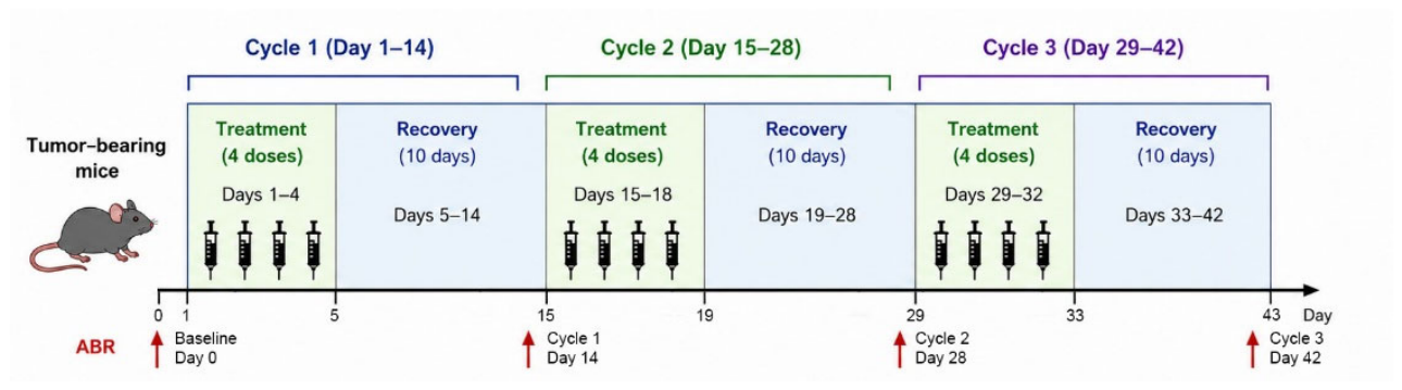
Figure 2.
ABR threshold measured before, during, and after the three-cycle chemotherapy regimen. (A) Baseline (Day 0) of all the groups measured before the treatment. (B) End of the study (Day 42) of all surviving animals. Note that the rest of the animals were removed from the study due to oversized tumor growth or health issues. (C) Cumulative percentage plots of the data from all the groups at 32 kHz on Day 0. A logistic curve fitting (dashed curve) was performed, from which an xhalf (average threshold) of 37.4±2.4 dB SPL was obtained. (D) Cumulative percentage plots and their curve fittings of the survived animals in the four treatment groups at 32 kHz on Day 42. (E) Progression of the threshold shift over each cycle of the Cis-Only group at 32 kHz. (F) Progression of the threshold shifts over each cycle of the HNK10+Cis group at 32 kHz. Ctr: Control; HNK: honokiol; Cis: cisplatin. The doses of honokiol (5, 10, 20 mg/kg) were indicated by the names of the treatment groups. Data presented in (A) and (B): mean ± standard error.
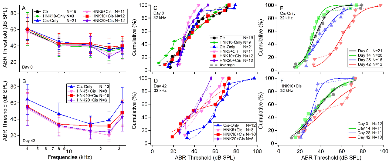
Figure 3.
Impact of cisplatin and honokiol co-treatment on animal survival and weight change. (A) Animal survival rates recorded at the end of each treatment cycle. (B) Average animal weight changes in each cycle, normalized to baseline values measured immediately prior to treatment (Day 0). Data presented in (B): mean ± standard deviations (SD).
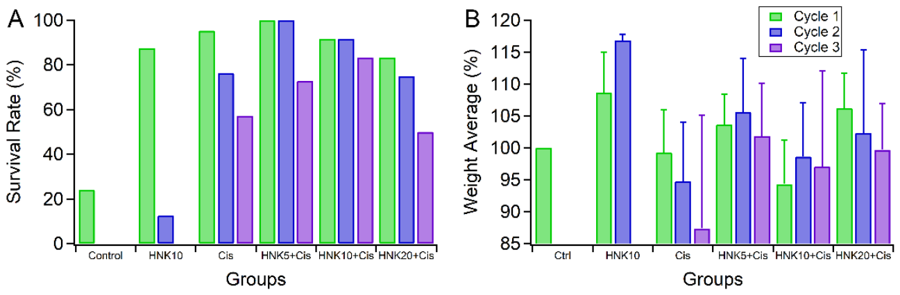
Figure 4.
Confocal images showing the changes of ribbon synapses after different treatments. (A) and (B) Representative confocal image stacks of the Ctr and Cis-Only groups; (A’), (B’), (C), and (D) Processed images showing only the hair cells and synaptic puncta at the bottom region of the IHCs. All images were taken from cochlear samples at the frequency range of 9.5-19.1 kHz. Ctr: control; Cis: cisplatin 4 mg/kg; HNK: honokiol 10 mg/kg. The insets are enlarged from the rectangular area in each panel (estimated frequency range: 15-19 kHz). Scale bars: 10 µm.
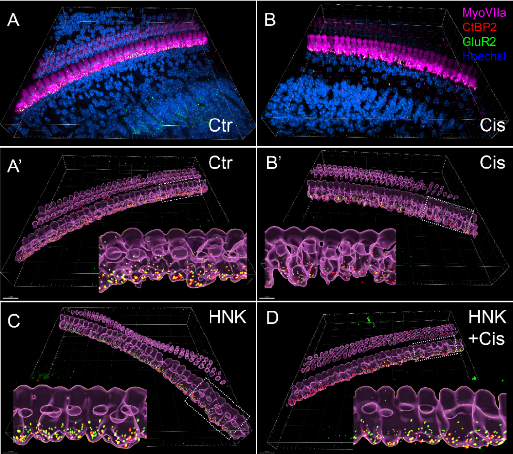
Figure 5.
Box-and-whisker plots showing the distribution of ribbon synapse counts per IHC in control (Ctr), honokiol-treated (HNK), cisplatin-treated (Cis), and honokiol plus cisplatin-treated (H+C) groups after three cycles of treatments. Synapse counts were quantified in two cochlear frequency regions: S2 (9.5–19.1 kHz; red symbols) and S3 (19.1–36.5 kHz; blue symbols). Each dot represents the average counts from one sample. Box plots indicate the median (center line), interquartile range (box), and minimum and maximum values (whiskers). Mean ± SDs were also plotted. Cisplatin treatment significantly reduced synapse counts compared with control animals, whereas honokiol co-treatment preserved synapse counts and prevented cisplatin-induced synaptopathy. Statistical comparisons were performed between the pooled control and cisplatin groups and between the pooled cisplatin and honokiol plus cisplatin groups.
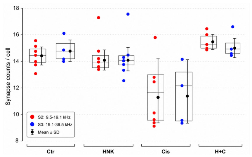
Figure 6.
Confocal images showing Sirt3 and Sirt5 expression in the cochlea (frequency range: 19.1-36.5 kHz) after different treatments. (A) Control; (B) HNK10-Only; (C) Cis-Only; and (D) HNK10+Cis. Surface (xy plane) and orthogonal views (right: yz plane; bottom: xz plane) are presented. Surface plots are focused on the cell body of the OHCs and IHCs. Xz orthogonal views cross over three OHCs (white triangles) and one IHC (white stars). Sirt3 and Sirt5 were both expressed and upregulated by honokiol and colocalized (showing yellow) in the OHCs and IHCs in (B) and (D). White arrows show bright puncta of Sirt3 and Sirt5 at IHC bottom regions. Insets in the last panel of each row show the fluorescence intensity of Sirt3 (red) and Sirt5 (green) at the lines across OHC and IHC regions in the surface plot and the xz orthogonal view. All images were taken from cochlear samples at frequency range of 19-36 kHz. Note that fluorescence intensity is the original value obtained from unprocessed confocal images, and that baseline fluorescence signal is identical in all four samples.
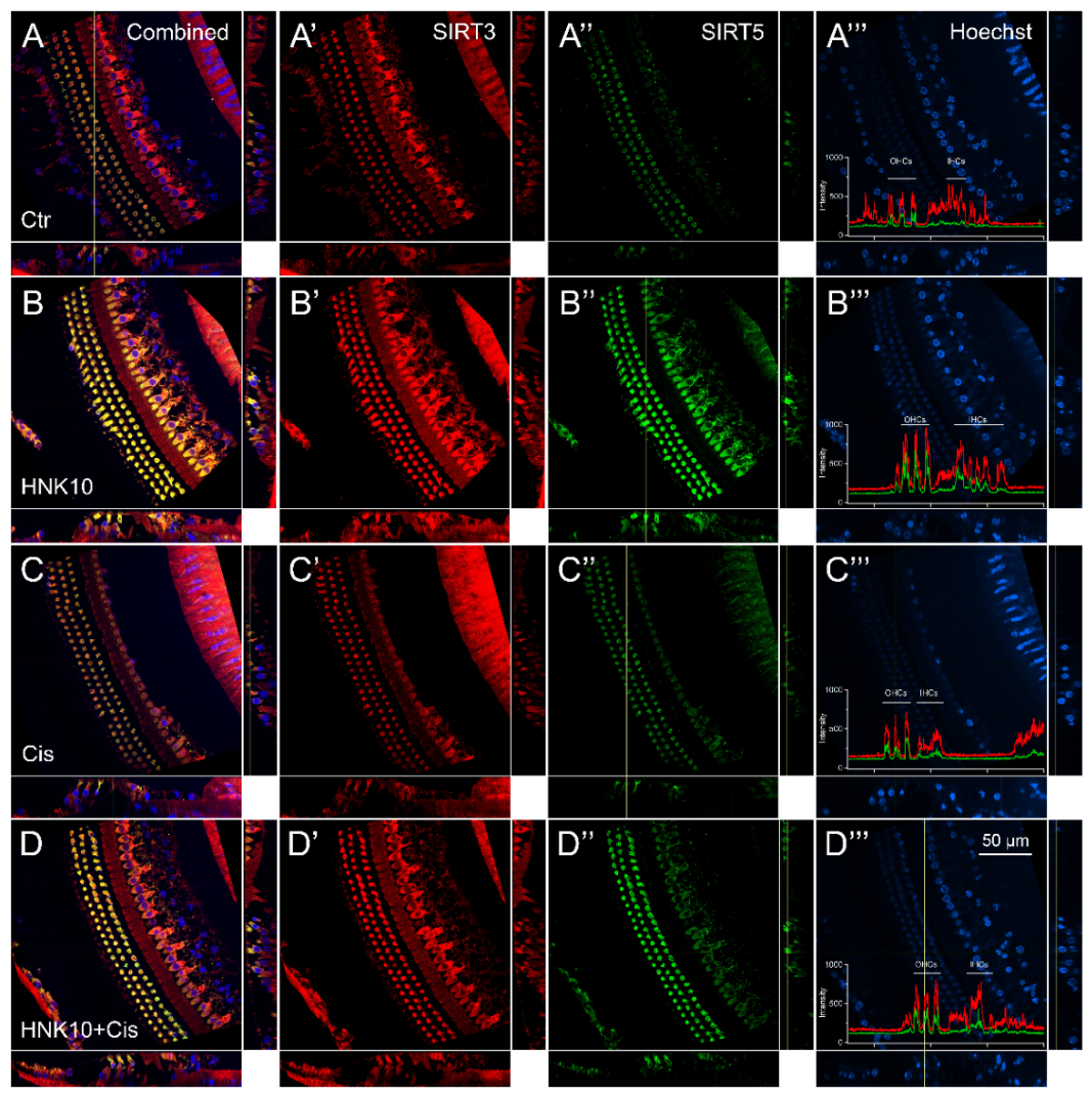
Figure 7.
Nuclear Sirt7 expression and colocalization with Sirt3 in the cochlea (frequency range: 9.5-19.1 kHz) after different treatments. (A) Control; (B) HNK10-Only; (C) Cis-Only; and (D) HNK10+Cis. The left panels show the comprehensive expression patterns of Sirt3 and Sirt7 in the 3D views of confocal image stacks. The right panels show Sirt7 expression focusing on the nuclei of OHCs, IHCs and SCs. Sirt3 was used to confirm the expression pattern in the cochlea and upregulation by honokiol (the left panels). Sirt7 signal was observed in the nuclei in a granular pattern and was upregulated by both honokiol and cisplatin (the right panels). White arrows show puncta of Sirt3 at the bottom regions of IHCs. Note that Sirt3 was expressed mostly in the soma, and therefore the signal in nuclear region on the right panels was relatively weak. Scale bars: 50 µm.
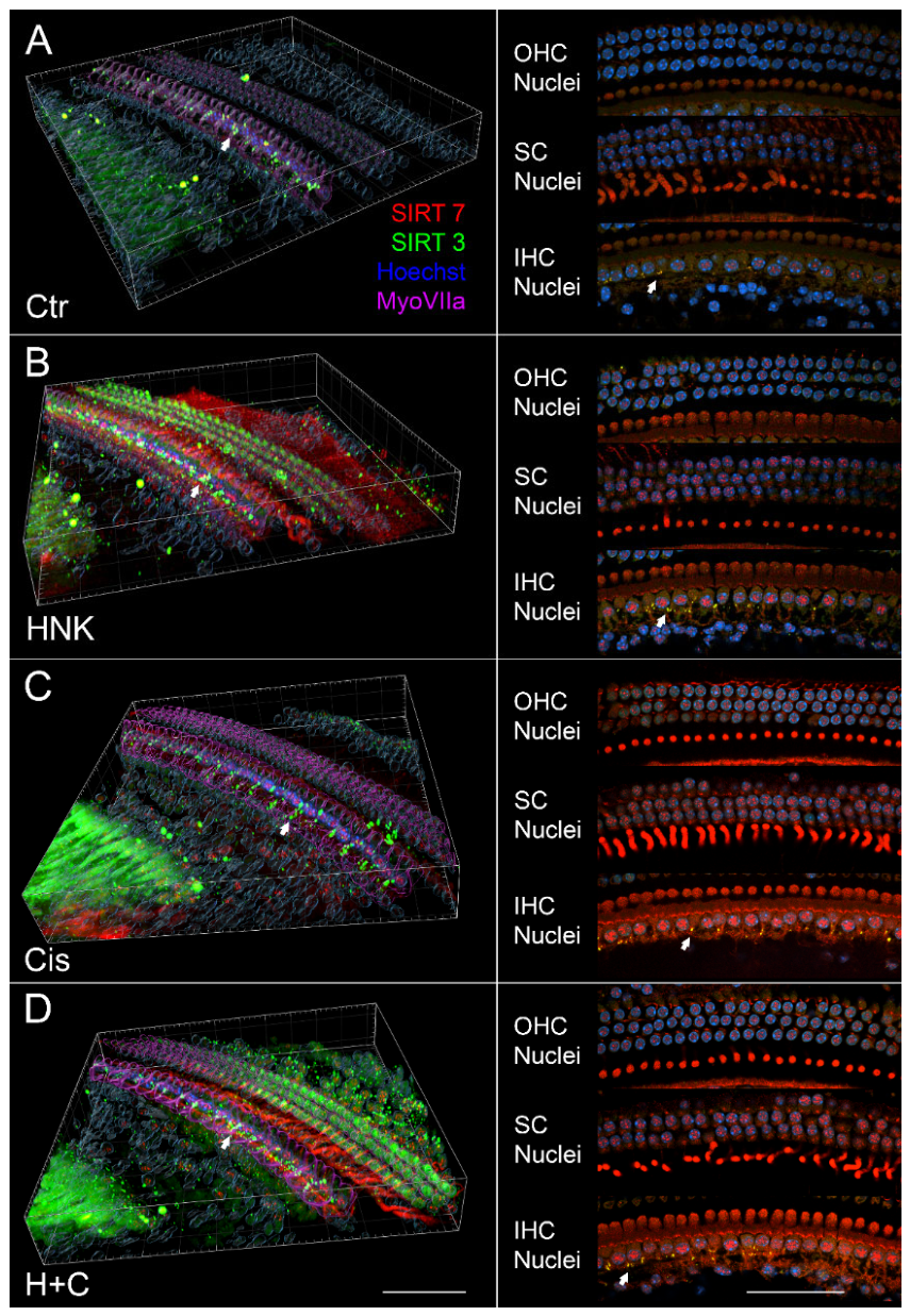
Figure 8.
Cytoplasmic Sirt1 and postsynaptic GluR2 expression in the nuclear regions of the cochlea (frequency range: 19.1-36.5 kHz) after different treatments. (A) Control; (B) HNK10-Only; (C) Cis-Only; and (D) HNK10+Cis. The left, central, and right panels focused on the nuclei of IHCs, OHCs, and SCs, respectively. Sirt1 (red) expression was detected in hair cells and upregulated following honokiol treatment (B, D). Note that no nuclear localization was observe for Sirt1, although upregulated expression in peri-nuclear area was observed after honokiol treatment. White arrows indicate Sirt1 puncta observed at IHC synaptic regions, which was partially colocalized with the postsynaptic marker GluR2 (green).
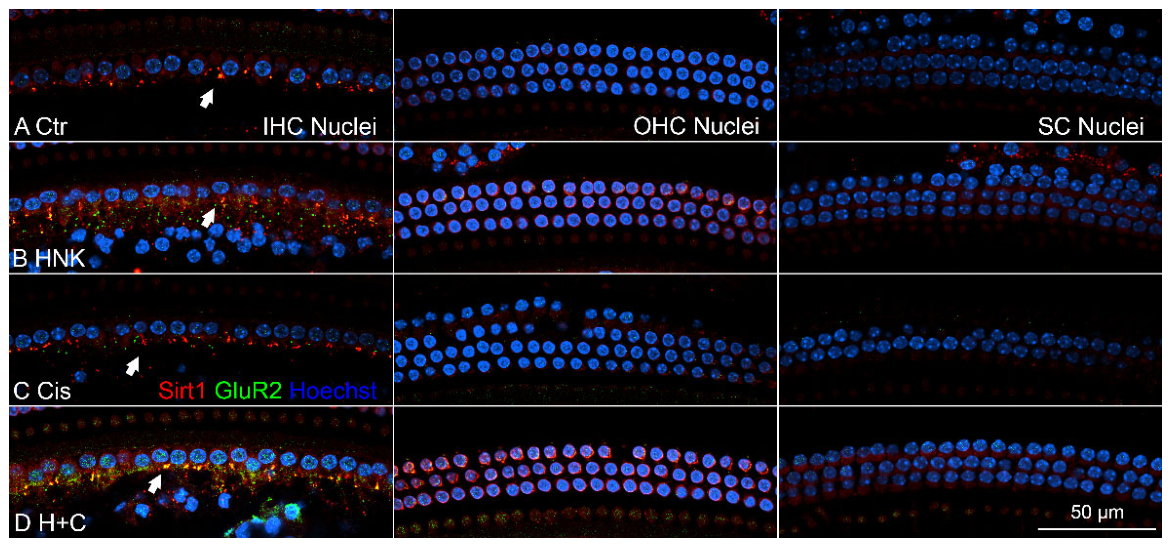
Figure 9.
RT-qPCR showing gene expression profile in the cochleae of honokiol-treated mice. Relative expression (Y-axis) was calculated using 2^-(∆∆Ct) method, normalized to Actb. Bars indicate fold changes in gene expression in the honokiol-treated group versus vehicle controls. Statistical significance was calculated for each gene by an unpaired two-tailed Student’s t-test comparing HNK10-Only group (n = 4) versus Control group (n = 3). Error bars represent standard deviation. ** p < 0.01.
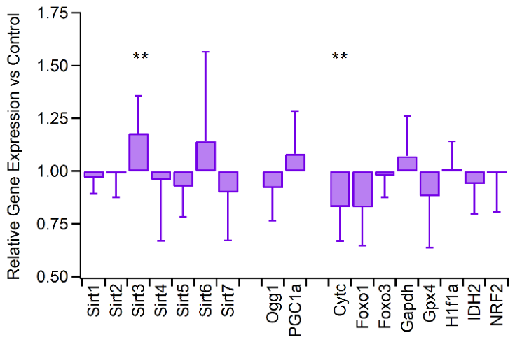

Table 1.
Animal groups and treatments.
| Strains | Group # | Group name | Number | Treatment |
| MMTV-PyMT female carrier |
1 | Control | n=19 | corn oil + saline (vehicle only) |
| 2 | Cis-Only | n=21 | corn oil + cisplatin (4 mg/kg/day) | |
| 3 | HNK10-Only | n=9 | honokiol (10 mg/kg/day) + saline | |
| 4 | HNK20-Only | n=8 | honokiol (20 mg/kg/day) + saline | |
| 5 | HNK5+Cis | n=11 | honokiol (5 mg/kg/day) + cisplatin (4 mg/kg/day) | |
| 6 | HNK10+Cis | n=12 | honokiol (10 mg/kg/day) + cisplatin (4 mg/kg/day) | |
| 7 | HNK20+Cis | n=12 | honokiol (20 mg/kg/day) + cisplatin (4 mg/kg/day) | |
| Female FVB wild type * |
1 | Control | n=6 | corn oil + saline (vehicle only) |
| 2 | HNK20-Only | n=8 | honokiol (20 mg/kg/day) + saline | |
| MMTV-PyMT female carrier ** |
1 | Control | n=5 | corn oil + saline (vehicle only) |
| 2 | Cis-Only | n=5 | corn oil + cisplatin (4 mg/kg/day) | |
| 3 | HNK10-Only | n=5 | honokiol (10 mg/kg/day) + saline | |
| 4 | HNK10+Cis | n=5 | honokiol (10 mg/kg/day) + cisplatin (4 mg/kg/day) |
* For ABR controls. ** For immunostaining, treated for one cycle only and euthanized on Day 5.
Table 2.
Linear mixed-effects model analysis of ABRs.
| Fixed Effect Term | Estimate | SE | t-value | Pval |
| (Intercept) | 3.73815 | 0.053517 | 69.85 | < 2.2e-16 *** |
| Cisplatin | 0.013398 | 0.00847 | 1.58 | 0.1137 |
| Cycle | 0.00222 | 0.001364 | 1.63 | 0.1035 |
| Honokiol | -0.005527 | 0.003584 | -1.54 | 0.1231 |
| poly(Frequency, 2)1 (linear) | 3.328328 | 0.180299 | 18.46 | < 2.2e-16 *** |
| poly(Frequency, 2)2 (quadratic) | 1.488667 | 0.179373 | 8.3 | < 2.2e-16 *** |
| Cycle:Honokiol | -0.000185 | 0.000072 | -2.56 | 0.0105 * |
| Honokiol:poly(Fr, 2)1 | -0.038165 | 0.014264 | -2.68 | 0.0074 ** |
| Honokiol:poly(Fr, 2)2 | 0.018241 | 0.014312 | 1.27 | 0.2025 |
* P<0.05; ** P<0.01; *** P<0.001.
Disclaimer/Publisher’s Note: The statements, opinions and data contained in all publications are solely those of the individual author(s) and contributor(s) and not of MDPI and/or the editor(s). MDPI and/or the editor(s) disclaim responsibility for any injury to people or property resulting from any ideas, methods, instructions or products referred to in the content. |
© 2026 by the authors. Licensee MDPI, Basel, Switzerland. This article is an open access article distributed under the terms and conditions of the Creative Commons Attribution (CC BY) license (http://creativecommons.org/licenses/by/4.0/).
Copyright: This open access article is published under a Creative Commons CC BY 4.0 license, which permit the free download, distribution, and reuse, provided that the author and preprint are cited in any reuse.