

Submitted:
01 October 2026
Posted:
02 October 2026
You are already at the latest version
Abstract
Fungal stress responses are frequently discussed in relation to virulence and antifungal resistance, yet most underlying systems have deeper evolutionary histories and function across diverse ecological contexts. This review integrates evolutionary, ecological, and molecular perspectives on stress adaptation in major human-pathogenic fungi. Environmental pressures, including temperature fluctuations, water and nutrient limitation, hypoxia, microbial competition, predation, and antifungal exposure, can shape stress-response capacities before or independently of human infection. We examine major molecular systems involved in fungal stress adaptation, including chaperone networks, HOG and cell-wall-integrity signaling, pH and hypoxia responses, the unfolded protein response, autophagy, redox signaling, and mitochondrial retrograde signaling. These systems interact through shared regulators and physiological outputs to maintain cellular homeostasis. Although core components are often conserved, their regulatory connections and phenotypic outputs can differ substantially among fungal lineages. During host association, these evolutionarily shaped and subsequently modified networks can support adaptation to immune, metabolic, physicochemical, and therapeutic stresses. Their contributions to antifungal tolerance and persistence should be distinguished from stable resistance. We propose that fungal pathogenicity represents one ecological context in which pre-existing and subsequently modified stress-response capacities are deployed, rather than assuming that infection-associated functions necessarily reveal their evolutionary origins.
Keywords:
fungal stress response
; fungal evolution
; ecological niche
; HOG pathway
; cell wall integrity
; Hsp90
; environmental adaptation
; fungal pathogenesis
; antifungal tolerance
1. Introduction
Invasive fungal diseases are a major global health burden. A 2024 analysis estimated approximately 6.5 million cases annually, with 3.8 million associated deaths, including about 2.5 million directly attributable to fungal disease [1]. Survival under changing environmental and host conditions is a prerequisite for many forms of fungal pathogenesis.
Fungi respond to stress through coordinated transcriptional and physiological changes. Shared effectors and regulatory hubs connect responses to different insults, allowing cells to adjust growth, metabolism, and resource allocation. This review focuses on stress networks in human-pathogenic fungi and uses model organisms to explain their underlying architecture. Microsporidia are considered briefly in the evolutionary discussion, but their highly reduced cellular machinery places a detailed mechanistic treatment outside the scope of this review. Our aim is to connect molecular mechanisms with evolutionary and ecological context rather than provide an exhaustive account of every pathway.
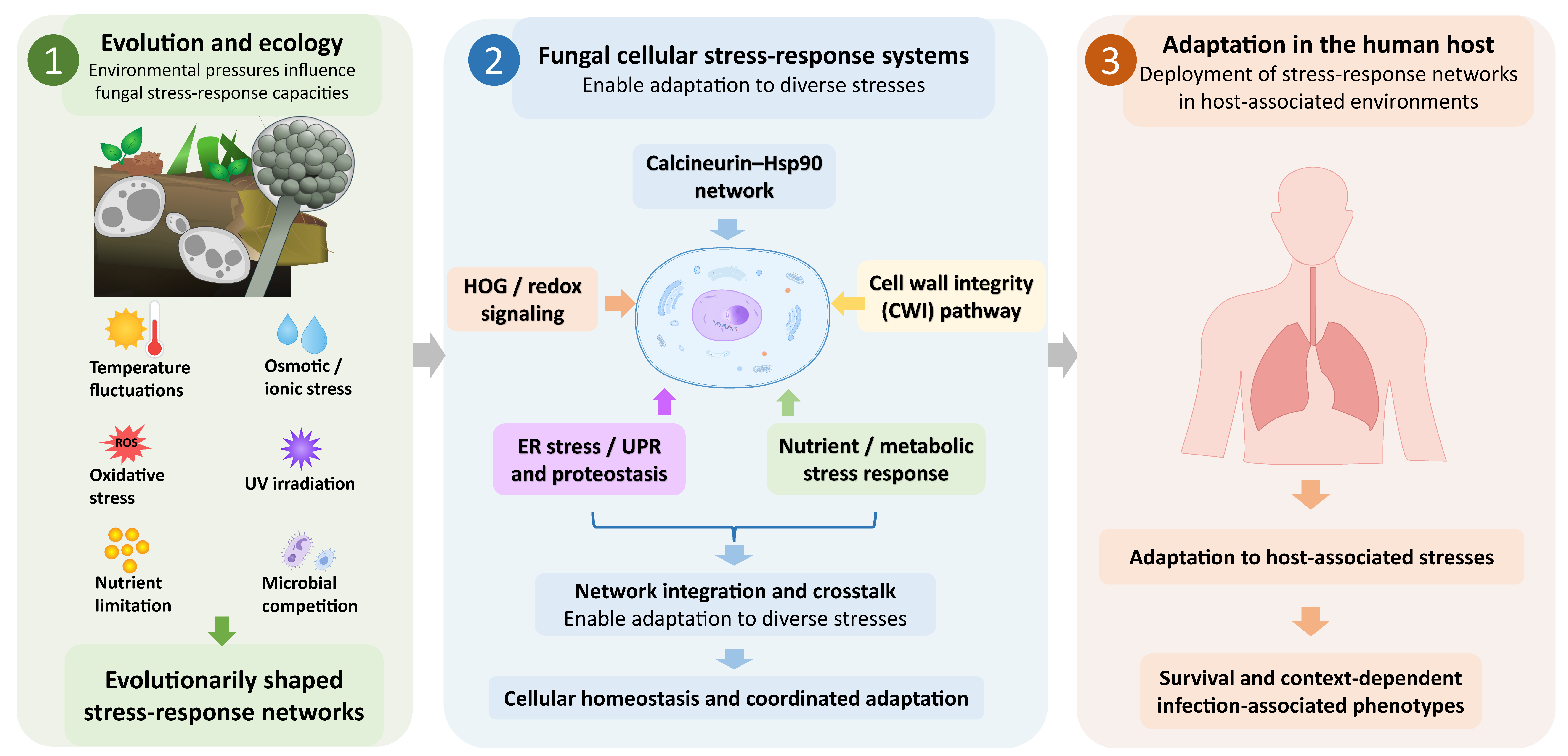
Here, we review stress-response systems in major human-pathogenic fungi and use an evolutionary-ecological framework to distinguish infection-associated function from evolutionary origin. A pathway that promotes survival in a mammalian host should not automatically be interpreted as having evolved specifically for mammalian infection; conserved stress modules often have demonstrable or plausible functions in soil, plant-associated habitats, aquatic environments, microbial communities, or interactions with environmental predators [2,3]. We therefore use the terms pathogenicity and virulence cautiously and treat environmental selection, exaptation, and pleiotropy as hypotheses to be evaluated rather than default explanations [4,5]. The evolutionary–ecological framework linking environmental stress adaptation to deployment of stress-response networks in the human host is summarized in Figure 1.
2. Evolutionary Background: Stress Adaptation Before Human Pathogenesis
2.1. Deep Evolutionary Origins of Fungal Stress Adaptation
Fungi are an ancient eukaryotic lineage. Fossils interpreted as fungi date to approximately 1.0–0.9 billion years ago, whereas molecular estimates place fungal origins potentially around 1.5 billion years ago [6,7]. Early fungal evolution probably included aquatic habitats followed by transitions to terrestrial environments, with some lineages subsequently returning to aquatic lifestyle [8]. These transitions exposed fungi to increasingly diverse physicochemical and biological challenges. Phylogenomic evidence also links early fungal diversification to the capacity to digest cell-wall components of algal relatives of land plants, although this does not establish when adaptations relevant to host immunity arose [9]. Microsporidia illustrate a distinct evolutionary trajectory characterized by extensive genomic reduction associated with obligate intracellular parasitism [10].
Terrestrial habitats expose fungi to desiccation, temperature fluctuations, radiation, and heterogeneous nutrient availability, while interactions with plants, invertebrates, microbial competitors, and fungal predators impose additional selective pressures [11]. Stress-response systems can therefore support survival across ecological settings that differ substantially from the human host [4]. Selective histories also vary among fungal lineages; for example, entomopathogenic fungi are primarily adapted to insect infection, whereas other fungi occupy broader ecological ranges [12]. Proposed roles for fungal disease in vertebrate evolutionary history, including hypotheses concerning dinosaur susceptibility, remain speculative [13].
Human pathogenicity has emerged independently in multiple fungal lineages. Many opportunistic pathogens retain environmental lifestyles, whereas organisms such as Pneumocystis jirovecii show pronounced host specialization [5]. Microsporidia likewise infect diverse hosts and should not be regarded as a lineage specialized for humans [14]. In environmental fungi, traits that promote survival during encounters with amoebae may also provide protection against mammalian phagocytes [15,16]. Such functional overlap is consistent with exaptation but does not establish the historical selective pressures that originally shaped a particular trait.
Terminology is important in this context. Pathogenicity generally denotes the capacity to cause disease, whereas definitions of virulence vary across disciplines [17,18]. In evolutionary studies, virulence is often evaluated through infection-associated reductions in host fitness [18]. We therefore interpret infection-associated phenotypes in relation to the host, fungal strain, and experimental conditions. A trait can contribute to disease severity without having evolved specifically to cause host damage, and an in vitro phenotype alone does not establish its contribution to disease in vivo.
2.2. Repeated Origins of Pathogenicity and Environmental Exaptation
The repeated emergence of human pathogenicity across fungal lineages is consistent with the recruitment and modification of older stress-response systems [4,5]. The fungal infection–mammalian selection hypothesis proposes that mammalian endothermy provided protection from fungal disease and may have influenced vertebrate evolution, although the reciprocal evolutionary effects remain uncertain [19]. Environmental predation provides a more experimentally tractable model for examining traits that can function in both environmental survival and mammalian infection [20]. Exaptive virulence is therefore a useful hypothesis for environmental opportunists, while host-adapted fungi may have experienced different selective histories [21]. Importantly, demonstrating that a gene contributes to disease severity establishes its function during infection, but not the evolutionary origin of that function.
3. Ecological Niches as Selective Environments for Stress Responses
The selective value of a stress-response system depends on the environments in which a fungus normally grows, disperses, persists, or reproduces. Because many fungal stress-response systems predate human infection, their infection-associated functions should be interpreted alongside their ecological roles rather than assumed to have evolved specifically in the mammalian host [4,5].
3.1. Natural Reservoirs and Environmental Heterogeneity
Clinically relevant fungi occupy heterogeneous environmental reservoirs rather than a single characteristic niche. For example, clinically relevant Mucorales occur in soil, decaying plant material, compost, and other organic substrates, illustrating how opportunistic pathogens can persist outside mammalian hosts [22]. More broadly, fungal diversity does not follow a single simple latitudinal pattern, and geological and climatic factors influence fungal guilds differently [23,24,25]. For example, saprotrophic fungal diversity has been associated with temperature, precipitation, and seasonal temperature variability [25]. These observations support a lineage- and habitat-specific view of ecological selection on fungal stress responses rather than attributing pathogenic potential to any single environmental variable.
3.2. Thermotolerance and Climate-Associated Emergence Hypotheses
Thermotolerance provides a clear example of an environmental adaptation with potential relevance to mammalian infection. Saprotrophic fungi such as Aspergillus fumigatus and some Mucorales can grow at mammalian body temperatures [22,26], while heat generated during composting provides an environmental setting in which thermotolerance may be advantageous independently of infection [27]. The emergence of Candidozyma auris (formerly Candida auris) further illustrates the distinction between ecological plausibility and demonstrated evolutionary causation. C. auris . Rising environmental temperatures could favor heat tolerance and narrow the thermal barrier that excludes many fungi from mammals [28], but climate-associated selection remains one hypothesis within a multifactorial explanation of emergence. Variation in thermotolerance and pathogenicity among C. auris lineages [29,30], together with observations of thermotolerant opportunistic fungi from cold environments [31], indicates that environmental temperature alone does not reliably predict growth at mammalian body temperature.
3.3. Osmotic and Desiccation Stress in Natural Habitats
Osmotic adaptation supports fungal growth in estuarine and marine habitats, saline soils, and sugar-rich substrates such as fruit In drying soils, increasing solute concentrations coincide with reduced diffusion of extracellular enzymes and their substrates[33]. Fluctuating water availability can therefore impose recurrent osmotic and desiccation stresses that favor rapid physiological adjustment in soil-associated fungi [34].
3.4. Persistence in Built Environments and Transmission
Stress-response capacity can also influence persistence on abiotic surfaces and transmission between hosts [35]. C. auris, for example, can persist in healthcare environments, making desiccation tolerance, surface survival, and susceptibility to disinfectants relevant to transmission, although the contribution of individual signaling pathways must be established for each phenotype [36].
Ecological interactions within host-associated microbial communities provide another form of environmental complexity. Farnesol, bacterial short-chain fatty acids, and secondary bile acids can influence Candida albicans growth and morphogenesis in concentration- and condition-dependent ways [37,38,39]. while antibiotic-associated reductions in microbial short-chain fatty acids correlate with increased gastrointestinal colonization [40]. Colonization, morphogenesis, and tissue invasion should nevertheless be regarded as distinct phenotypes. Species also differ in their responses: Candida tropicalis, for example, retains a stronger morphogenetic response to deoxycholate than C. albicans [41] and occurs in both host-associated and environmental settings [42]. Thus, stress adaptation occurs not only in physical environments but also within complex biotic environments whose chemical and microbial composition can vary substantially.
3.5. Environmental Exposure to Antifungals and Development of Antifungal Resistance
Environmental antifungal exposure can select acquired resistance, particularly in A. fumigatus, although its contribution varies among species, drugs, and ecological settings. Agricultural demethylation-inhibitor fungicides and medical azoles share CYP51 as a target, creating opportunities for cross-resistance. Experimental exposure of A. fumigatus to agricultural triazoles supports this route [43], while related resistant genotypes recovered from environmental and clinical samples support environmental acquisition of resistant infections [44]. However, recovery of a resistant isolate from the environment does not establish where resistance arose or which exposure selected it. Resistant environmental isolates have also been reported for C. albicans, C. auris, and Rhodotorula mucilaginosa [30,31,45] but their isolation sites alone cannot establish the origin of resistance.
Intrinsic reduced susceptibility must also be distinguished from acquired resistance. Aspergillus calidoustus, for example, has intrinsically low susceptibility to several azoles [46]. Similarly, the poor activity of fluconazole against A. fumigatus is influenced by intrinsic CYP51A sequence features, with experimental studies identifying Thr289 and Ile301 as determinants of susceptibility [47,48]. Thus, antifungal exposure can select among pre-existing susceptibility phenotypes without necessarily having generated them.
Agricultural antifungal use should therefore be considered an anthropogenic selective pressure acting on pre-existing fungal diversity. Interpretation requires distinguishing intrinsic reduced susceptibility from acquired resistance, and the origin of resistance from its subsequent selection and dispersal, particularly in species lacking well-defined wild-type susceptibility and epidemiological cutoff values [49].
4. Molecular Mechanisms of Fungal Stress Responses
Fungal cells use membrane sensors, protein kinases, phosphatases, transcription factors, chaperones, metabolic regulators, and organelle-quality-control systems to detect and adapt to environmental change. Much of the mechanistic framework derives from Saccharomyces cerevisiae, a widely used model yeast that can also cause opportunistic infection. However, conservation of individual proteins does not necessarily imply conservation of upstream inputs, downstream targets, regulatory connections, or phenotypic outputs. The subsections below therefore use conserved or well-characterized architectures as reference frameworks while highlighting lineage-specific rewiring in human-pathogenic fungi [4,50]. Table 1 provides an overview of the stress-response systems discussed in this section.
The major stress-response systems and their relationships to environmental, host-associated, and therapeutic stresses are summarized in Figure 2.
4.1. the Heat Shock Chaperone–co-Chaperone Network: Hsf1, Hsp110, Hsp70, and Hsp90
Elevated temperature can destabilize proteins and promote aggregation, increasing the demand for protein refolding and disaggregation [51]. Heat-shock transcription factors and molecular chaperones coordinate this response. Although broadly conserved, their regulation and effects on growth, morphogenesis, and stress adaptation vary among fungal lineages.
4.1.1. Heat Shock Factor 1 (Hsf1)
In S. cerevisiae, Hsf1 supports basal expression of essential proteostasis genes and increases transcription during heat stress [52,53]. Hsf1 can occupy target promoters as a trimer under basal conditions. During heat stress, unfolded proteins titrate Hsp70 away from Hsf1, relieving repression and increasing heat-shock gene expression; newly synthesized Hsp70 subsequently restores negative feedback. Hsf1 phosphorylation modulates transcriptional output rather than acting as a simple obligatory activation switch [54].
4.1.2. Hsp110
The S. cerevisiae Hsp110-family chaperone Sse1 supports growth and proteostasis and functions principally as a nucleotide-exchange factor for Hsp70 [55,56]. By promoting nucleotide exchange, Hsp110 supports repeated Hsp70 substrate-binding cycles and cooperates with Hsp104 in protein refolding and disaggregation [51,55]. Hsp110 can also exhibit substrate-binding and holdase activities, although their cellular contributions depend on experimental context [57].
4.1.3. Hsp90 Client Proteins and Co-Chaperones
Hsp90 is an essential, highly conserved chaperone that supports the maturation and function of many client proteins, including kinases and transcriptional regulators [58]. Its co-chaperones regulate ATPase cycling and client selection. In S. cerevisiae, Cdc37 protects newly synthesized kinases from degradation and promotes their maturation [59], Sgt1 links Hsp90 to Skp1-associated machinery and contributes to kinetochore assembly and responses to replication stress [60,61,62]. It also interacts with the adenylyl cyclase Cyr1 and supports cAMP signaling [63]. In C. albicans, Hsp90 and Sgt1 connect temperature responses to Ras1–cAMP–PKA-dependent morphogenesis; compromising Hsp90 function can release repression of filamentation [64,65].
Sti1, the fungal counterpart of mammalian Hop, uses tetratricopeptide-repeat domains to coordinate interactions between Hsp70 and Hsp90 [66,67]. This dynamic organization supports client transfer and regulates the Hsp90 chaperone cycle [68]. Aha1 stimulates Hsp90 ATPase activity [69], whereas its yeast-specific orthologue Hch1 has distinct regulatory effects that cannot be described simply as equivalent ATPase stimulation [70,71]. Sba1, the yeast counterpart of p23, stabilizes the closed, nucleotide-bound Hsp90 conformation and couples ATPase cycling to client maturation [72,73].
4.1.4. Hsp90-Dependent Crosstalk and Comparative Features in Human-Pathogenic Fungi
Hsp90 supports calcineurin stability and function, linking proteostasis to calcium-dependent stress signaling [74]. Additional Hsp90-dependent connections vary among fungal species and are considered below. Hsf1 contributes to temperature responses across fungi, but its regulatory interactions and downstream outputs are not identical among species [52]. In C. albicans, Mkc1 and Hog1 also depend on Hsp90 for normal activity or stability [75,76], and Hsp90 interaction networks include regulators of morphogenesis and sterol homeostasis, such as Tec1, Tup1, and Upc2 [77]. Elevated CO2 provides another input: Ptc2 can form condensates that alter Hsp90 phosphorylation and client interactions, connecting a host-associated environmental cue to chaperone regulation [78]. Together, these findings illustrate how Hsp90-centered proteostasis can intersect with calcium signaling, MAPK pathways, morphogenesis, and environmental sensing.
Comparable chaperone dependence produces distinct outputs in other pathogens. In C. auris, Hsp90 depletion induces filamentous growth and increases azole susceptibility, while Cdr1 makes a separate contribution to high-level azole resistance [79]. In A. fumigatus, Hsp90 repression impairs conidiation and cell-wall integrity and enhances caspofungin activity, without reproducing the same voriconazole sensitization [80]. In C. neoformans, Hsp90 depletion compromises growth at host temperature, stress tolerance, melanization, and virulence in a mouse inhalation model [81]. Thus, conserved proteostasis machinery can participate in distinct developmental, stress-response, and drug-response programs across fungal lineages.
4.2. the High-Osmolarity Glycerol (Hog) Mapk Pathway
The high-osmolarity glycerol (HOG) pathway coordinates adaptation to osmotic changes. Its additional contributions to oxidative stress, heavy-metal exposure, morphogenesis, and other phenotypes depend on fungal lineage and experimental conditions.
4.2.1. Upstream Sensing Architecture
In S. cerevisiae, two upstream signaling branches converge on the MAPK kinase Pbs2. The Sln1 branch uses a multistep phosphorelay involving Sln1, Ypd1, and Ssk1 [82]. Hyperosmotic stress reduces this phosphorelay, allowing Ssk1 to activate the MAPK kinase kinases Ssk2 and Ssk22, which signal through Pbs2 to Hog1 [83]. The Sho1 branch instead uses Sho1 as a membrane-associated adaptor and signaling organizer [84]. The membrane mucins Hkr1 and Msb2, together with Opy2 and associated signaling proteins, connect osmotic inputs to the Ste11 MAPK kinase kinase [85]. Additional polarity and adaptor proteins, including Bem1, contribute to this branch [86]. Ste11 then signals through Pbs2 to activate Hog1 [87].
4.2.2. Core Phosphorylation Cascade and Nuclear Targets
Activated Hog1 accumulates in the nucleus and regulates stress-responsive transcription through factors including Hot1, Sko1, Smp1, and the broader Msn regulatory network in S. cerevisiae [88]. A major physiological output is glycerol accumulation. HOG signaling promotes glycerol production and uptake through metabolic regulation and factors including Gpd1, Gpp1/Gpp2, Pfk26, and Stl1 [89,90,91,92]. Hog1-dependent phosphorylation of the Fps1 regulator Rgc2 limits glycerol efflux through the Fps1 channel [93]. Increased intracellular glycerol helps restore osmotic balance [94].
HOG signaling can also contribute to oxidative protection. In S. cerevisiae and the halotolerant yeast Debaryomyces hansenii, it contributes to catalase regulation under osmotic or hydrogen-peroxide stress [95,96]. HOG signaling further intersects with other stress pathways: cAMP-dependent PKA can oppose parts of the Hog1/Msn2/Msn4 program [97], whereas Hog1-dependent regulation of Ubp3 can suppress Ras-dependent PKA activity [98]. Hog1 activation during later stages of ER stress provides an additional connection to ER homeostasis [99].
4.2.3. Comparative Features in Human-Pathogenic Fungi
Although the HOG pathway is broadly conserved, its upstream sensing architecture differs substantially among fungi. C. albicans has several hybrid histidine kinases, including Sln1, Nik1, and Chk1, with overlapping but distinct roles in stress responses and morphogenesis [100]. Chk1-associated phenotypes also differ between C. albicans and Meyerozyma guilliermondii, illustrating why orthology alone does not establish functional equivalence [101]. A. fumigatus has a larger histidine-kinase repertoire, including TcsC, with different sensors implicated in osmotic, oxidative, light, and temperature responses [102].
Histidine-kinase inventories also vary among thermally dimorphic fungi, including Paracoccidioides brasiliensis, Blastomyces dermatitidis, H. capsulatum, and Coccidioides immitis, and among basidiomycetes such as C. neoformans and Trichosporon asahii [103]. Mucorales, including Rhizopus delemar, Mucor circinelloides, and Lichtheimia corymbifera, possess lineage-specific repertoires, whereas annotated hybrid histidine kinases are absent from the examined genomes of the obligate parasites P. jirovecii and Enterocytozoon bieneusi [104].
Downstream architecture also varies. A. fumigatus has two Hog1-related MAPKs, SakA and MpkC [105]. In some C. neoformans strain backgrounds, Hog1 is phosphorylated under basal conditions and becomes dephosphorylated after particular stresses, demonstrating that activation-state dynamics cannot simply be extrapolated from S. cerevisiae [106,107]. Sho1-associated signaling also differs across species. The canonical S. cerevisiae Ste11–Pbs2 connection is not conserved as the same HOG input in C. albicans or C. neoformans [106,108]. This difference in pathway coupling does not imply the absence of other Ste11-related signaling in cryptococci.
Hog1 outputs have also diverged. In C. albicans, Hog1 contributes to osmotic and heavy-metal responses and restrains inappropriate filamentation, whereas Cap1 has a prominent role in oxidative-stress transcription [109]. C. neoformans uses a broader regulatory network involving Atf1, Mbs1, Hrk1, and Sch9, with distinct contributions to osmotic and oxidative adaptation [106]. In Aspergillus nidulans, SakA and AtfA regulate general stress responses, development, and spore functions [110]. Thus, conservation of the HOG pathway does not imply conservation of its sensors, activation dynamics, regulatory connections, or phenotypic outputs.
4.3. the Cell Wall Integrity (Cwi)/pkc Pathway
The Cell Wall Integrity (CWI) pathway monitors cell envelope dynamics during growth, morphogenesis, and exposure to cell-wall-damaging agents.
4.3.1. Sensor Activation and the Rho1 Gtpase Switch
In S. cerevisiae, cell-wall stress sensing involves the Wsc-family proteins Wsc1–Wsc3 and the Mid-family proteins Mid2 and Mtl1. Their extracellular, O-mannosylated regions contribute to the mechanical properties of these sensors [111]. Signaling through the guanine nucleotide-exchange factors Rom1 and Rom2 promotes the active, GTP-bound state of Rho1. Rho1 then coordinates several outputs, including Pkc1 signaling, β-(1,3)-D-glucan (βDG) synthesis, actin organization, and secretion [112].
4.3.2. the Protein Kinase C Cascade
Rho1-GTP activates Pkc1, which initiates a kinase cascade comprising Bck1, Mkk1/Mkk2, and the MAPK Slt2 [113]. Activated Slt2 regulates the transcription factor Rlm1 and the Swi4/Swi6 complex [114,115]. Rlm1-dependent transcription promotes cell-wall construction and remodeling through genes including FKS1, BGL2, and CRH1 [116]. Recruitment of the SWI/SNF chromatin-remodeling and SAGA acetyltransferase complexes supports transcription at responsive promoters [117], while Rlm1-dependent induction of SLT2 provides positive feedback [116,118].
Swi4 and Swi6 form the SCB-binding factor (SBF), which recognizes cell-cycle regulatory elements [119]. Interaction between Slt2 and SBF further connects cell-wall stress signaling with transcriptional and cell-cycle-associated programs [120]. This Slt2–SBF interaction is distinct from the Rlm1 branch of the response.
4.3.3. Mck1 and Gsk-3-Family Stress Signaling
Mck1 is a glycogen synthase kinase 3 (GSK-3)-family kinase rather than a MAPK and contributes to stress signaling, metabolism, and cellular organization [121]. During glucose starvation in S. cerevisiae, Mck1 supports Msn2/Msn4-dependent stress responses and Snf1-associated metabolic reprogramming involving Cat8 and Adr1. These processes promote UDP-glucose accumulation, providing substrate for βDG synthesis, while Mck1 cooperates with Slt2 to support cell-wall thickening [122].
4.3.4. Comparative Features in Human-Pathogenic Fungi
The broad architecture of CWI signaling is conserved, but paralog numbers and regulatory connections vary. For example, S. cerevisiae uses the partially redundant MAPK kinases Mkk1 and Mkk2, whereas C. neoformans has a different complement of pathway components [123]. In C. albicans, Pkc1-dependent signaling connects to the Slt2-related MAPK Mkc1 and to chromatin-remodeling functions involving the RSC ATPase Sth1 [124]. These relationships link cell-wall adaptation to genome maintenance without implying that Pkc1 directly phosphorylates the terminal MAPK. In N. glabratus, loss of Slt2 reduces tolerance to elevated temperature and cell-wall-damaging agents, while increased Rlm1 expression enhances micafungin tolerance [125]. In A. fumigatus, MpkA contributes to CWI signaling and oxidative-stress responses, linking envelope maintenance to additional environmental challenges [126]. Thus, conservation of the core CWI architecture does not imply identical pathway composition, regulatory connections, or stress-response outputs across fungal lineages.
4.4. Ambient Ph Sensing and Rim101/pacc Signaling
The Rim101 pathway mediates adaptation to alkaline ambient pH. In S. cerevisiae, the membrane proteins Rim21, Rim9, and Dfg16 participate in sensing, followed by recruitment of Rim8 and downstream machinery that includes ESCRT components [127]. Rim20 and the protease Rim13 support processing of the inhibitory C-terminal region of Rim101 [128]. Processed Rim101 acts principally as a transcriptional repressor, including repression of NRG1, which helps relieve inhibition of alkaline-response genes such as the sodium-pump gene ENA1 [129]. Another target, SMP1, links this pathway to transcriptional programs associated with osmotic stress [130]. Additional outputs include regulation of ion and iron acquisition; ARN4 encodes a siderophore transporter rather than a siderophore itself [131,132].
The transcriptional effects of Rim101/PacC differ among fungi. In A. nidulans, PacC can activate alkaline-expressed genes as well as repress acid-expressed genes [133]. C. albicans Rim101 can likewise activate or repress transcription, depending on the target [134]. Its outputs include regulation of Fet-family multicopper oxidases. In defined gray-cell conditions with exogenous L-DOPA, Rim101-upregulated Fets promote dark pigment formation [135]. This cell-state- and substrate-dependent phenotype should not be generalized to constitutive melanization of C. albicans [136]. Fet34 also functions as a ferroxidase in high-affinity iron uptake [137].
4.5. Hypoxia and Sterol Adaptation
Fungi respond to oxygen limitation through mechanisms that differ from the mammalian HIF system [138]. Oxygen is required for sterol and heme biosynthesis, allowing changes in these metabolites to signal oxygen availability. In S. cerevisiae, heme promotes the activity of Hap1, which induces the transcriptional repressor ROX1 under aerobic conditions [139]. Reduced heme availability during hypoxia lowers this repression and permits expression of hypoxia-associated genes. Hap1 can also act directly as a repressor at selected promoters under hypoxic conditions [140].
Sterol depletion provides a second input. Ergosterol binding to the C-terminal regulatory region of Upc2 restrains its activity [141]. When sterol availability falls, Upc2 activates genes involved in sterol homeostasis, connecting oxygen-dependent metabolism to compensatory transcription [138].
In C. albicans, the Rox1-related factor Rfg1 primarily regulates filamentous growth and virulence-associated phenotypes rather than mirroring the S. cerevisiae hypoxic transcriptional program [142]. In A. fumigatus, the sterol regulatory element-binding protein SrbA coordinates hypoxic adaptation, while the globin protein fungoglobin also contributes to growth at low oxygen [143,144,145]. These examples illustrate how fungi can meet similar physiological demands under oxygen limitation through distinct regulatory architectures.
4.6. Endoplasmic Reticulum (Er) Stress and the Unfolded Protein Response (Upr)
ER stress develops when protein-folding demands or membrane perturbations exceed the capacity of the ER. The fungal unfolded protein response (UPR) is centered on the conserved sensor Ire1, although its downstream outputs vary among lineages [146]. Ire1 spans the ER membrane, with a luminal stress-sensing region and cytosolic kinase and endoribonuclease domains [147]. In S. cerevisiae, interactions with the ER chaperone Kar2/BiP and unfolded substrates regulate Ire1 clustering and activation [148]. Activated Ire1 removes an unconventional intron from HAC1 mRNA, enabling production of the bZIP transcription factor Hac1, which induces UPR target genes [147]. Gcn4 further connects the UPR with amino-acid control through regulation of HAC1 and shared transcriptional targets [149].
In A. fumigatus, the Ire1 ortholog IreA supports thermal and cell-wall stress responses through both HacA-dependent and HacA-independent functions [150]. In C. neoformans, Ire1 instead acts through the divergent bZIP factor Hxl1. Stress-dependent splicing of HXL1 mRNA parallels the unconventional splicing of HAC1, despite limited sequence similarity between the encoded transcription factors [151].
4.7. the Stripak Complex
Striatin-interacting phosphatase and kinase (STRIPAK) complexes combine protein phosphatase 2A (PP2A) with striatin-family scaffolds and associated regulatory proteins. In fungi, these complexes contribute to development, signaling, and secondary metabolism [152]. In S. cerevisiae, the related Far complex contains Far3, Far7, Far8, Far9, Far10, and Far11 and associates with PP2A machinery. Its association with the mitophagy receptor Atg32 provides a mechanism through which the Far complex can restrain mitophagy [153,154]. These findings illustrate how STRIPAK-related complexes can couple phosphatase organization with broader cellular regulatory processes rather than producing a single universal output.
In A. nidulans, STRIPAK includes the striatin scaffold StrA and associated Sip proteins, and disruption of several components affects oxidative and cell-wall stress tolerance as well as development [152]. Studies in the plant pathogen Fusarium graminearum further link STRIPAK to CWI signaling [155]. In C. neoformans, PP2A-associated and Far/Mob components have distinct effects on genome stability, sexual development, and infection-associated traits. Loss of Mob3 can increase thermotolerance and virulence-related phenotypes, contrasting with defects caused by disruption of other components [156]. Consequently, loss of a STRIPAK subunit should not be assumed to phenocopy inhibition of the entire complex.
4.8. Melanin Biosynthesis and Stress Protection
Melanins are chemically diverse pigments with overlapping protective functions. S. cerevisiae is not able to produce melanin, but there are two major fungal routes to produce polyketide-derived 1,8-dihydroxynaphthalene (DHN) melanin and pigment derived from L-3,4-dihydroxyphenylalanine (L-DOPA) or related phenolic substrates [157]. Other pigments include A. fumigatus pyomelanin and the non-canonical Asp-melanin of Aspergillus terreus [157,158,159]. Depending on the pigment and organism, melanization can alter radiation tolerance, redox buffering, metal binding, and cell-wall resistance [160]. Regulation is similarly diverse: in C. neoformans, cAMP/PKA signaling is important, but additional transcriptional circuits regulate melanization [161].
4.8.1. Dhn-Melanin Synthesis in Dematiaceous Fungi
The constitutive brown-to-black pigmentation of dematiaceous fungi is usually associated with cell-wall deposition of 1,8-dihydroxynaphthalene (DHN) melanin. In Exophiala dermatitidis, the pathway is initiated by the polyketide synthase WdPks1. Acetyl- and malonyl-CoA-derived polyketide intermediates are processed through tetrahydroxynaphthalene/scytalone and vermelone intermediates to 1,8-DHN, which is then oxidatively polymerized in the cell wall. A later biochemical analysis identified 2-acetyl-1,3,6,8-tetrahydroxynaphthalene as an upstream precursor in this pathway [162,163].
E. dermatitidis also displays substrate-dependent melanization. WdPKS1-deficient cells can produce a DOPA-derived pigment when exogenous L-DOPA is available in vitro, without establishing DOPA melanin as the normal replacement for constitutive DHN melanin [168]. In an ex vivo skin model, loss of PKS1 alters cell-wall and metabolic responses and impairs invasion, supporting a role for melanization in this specific tissue context [164].
Comparable DHN-melanin machinery is found across several clinically relevant melanized ascomycetes. In Cladosporium sphaerospermum, tricyclazole inhibition and genomic analysis support a DHN pathway and identify a non-reducing polyketide synthase predicted to participate in pigment synthesis [165]. The genome of the neurotropic dematiaceous fungus Cladophialophora bantiana encodes DHN-melanin synthesis machinery, although pathway activity and regulation require functional validation [166]. DHN melanin is also characteristic of agents of chromoblastomycosis and phaeohyphomycosis such as Fonsecaea pedrosoi, Cladophialophora carrionii, Exophiala jeanselmei, Phialophora verrucosa and related taxa [167].
4.8.2. Stress Protection and Infection-Associated Functions
In E. dermatitidis, disruption of WdPKS1 increases susceptibility to neutrophil killing and reduces disease severity in a mouse model [162,163]. Melanization also protects E. dermatitidis against temperature extremes and enzymatic cell-wall damage, with effects on antifungal killing that depend on the compound and assay [168]. In F. pedrosoi, experiments with melanized and tricyclazole-treated cells support protection against hydrogen peroxide and nitric-oxide-associated stress. Melanin can buffer reactive species, and reduced detectable nitrite need not mean suppression of macrophage nitric oxide synthase [169].
Radiation-associated changes in melanin properties and growth have been reported in Cladosporium sphaerospermum and other melanized fungi [170]. These observations do not establish a complete pathway that converts ionizing radiation into metabolically usable energy. Proposed radiosynthesis should therefore remain a hypothesis. More broadly, melanization is a multifunctional ecological trait whose contribution to infection or drug susceptibility must be demonstrated in the relevant species and conditions.
4.9. Stress Granules and Translational Control
Stress can inhibit translation initiation, redistribute non-translating mRNAs, and promote formation of RNA–protein condensates known as stress granules (SGs) [171,172]. Their composition and assembly requirements vary with the organism and stress. In yeast, implicated proteins include the RNA-binding proteins Pub1, Pab1, and Pbp1, together with translation-associated factors and additional regulators [173]. SG assembly can occur without eIF2α phosphorylation under some conditions, indicating that eIF2α phosphorylation is not universally required for fungal SG formation. Glucose deprivation, temperature shifts, sodium azide, ethanol, and other chemical stresses can produce distinct granule responses [174].
Granule-associated proteins also contribute to fungal biology beyond S. cerevisiae. In A. nidulans, a Pbp1-related protein supports sexual development and secondary-metabolite production [175]. In C. neoformans, calcineurin colocalizes with processing bodies and stress granules during thermal stress [176]. In C. albicans, Hsp90 influences stress-dependent modification of Rvb1 and other proteins associated with RNA granules or processing bodies [177]. These observations connect RNA–protein condensates with broader fungal stress networks, although they do not establish that every associated phenotype results directly from SG assembly.
4.10. Autophagy and Stress-Dependent Cellular Recycling
Autophagy supports nutrient recycling and clearance of damaged cellular material. Autophagy-related (ATG) genes encode core machinery used in both basal homeostasis and stress-induced turnover [178]. Increased autophagic activity can promote survival during nutrient limitation or organelle damage [179]. Nonselective autophagy delivers portions of the cytoplasm to the vacuole, releasing metabolites that can sustain cellular functions during starvation [180].
Selective pathways recognize particular cargoes. The cytoplasm-to-vacuole targeting (Cvt) pathway uses autophagy machinery for biosynthetic delivery of vacuolar enzymes in some yeasts and should be distinguished from starvation-induced degradation [181]. Aggrephagy targets protein aggregates, whereas mitophagy, pexophagy, and ER-phagy target mitochondria, peroxisomes, and ER material, respectively [182,183]. Nutrient-sensitive target of rapamycin complex 1 (TORC1) signaling normally restrains autophagy through regulation of the Atg machinery and its transcriptional control. Nutrient limitation relieves this inhibition, although TORC1 activity can be partially restored during prolonged starvation [184].
The consequences of autophagy perturbation vary across pathogens and experimental models. In A. fumigatus, deletion of ATG1 impaired sporulation but did not reduce virulence in the tested murine aspergillosis model [185]. In C. neoformans, ATG-gene deletion disrupts amino-acid homeostasis during nitrogen starvation [186]. In C. albicans, some ATG genes contribute to biofilm formation, but these effects may involve autophagy independent functions [187].
4.11. Ros Sensing and Thioredoxin-Dependent Redox Signaling
Reactive oxygen species (ROS) arise from aerobic metabolism and from environmental or host-derived oxidants. Fungal oxidative-stress responses therefore combine ROS detoxification with redox-sensitive signaling. A well-defined peroxide-sensing module couples the glutathione-peroxidase-like protein Gpx3/Orp1 to the AP-1-like transcription factor Yap1 (Cap1 in C. albicans). Skn7 provides a parallel transcriptional arm, while the thioredoxin system supplies reducing power and resets oxidized signaling proteins [188,189,190].
4.11.1. the Gpx3–yap1/cap1 Peroxide-Sensing Relay
In S. cerevisiae, Yap1 is not efficiently oxidized by H2O2 directly. Instead, H2O2 oxidizes the peroxidatic cysteine of Gpx3, which forms a transient intermolecular disulfide with Yap1. Resolution of this intermediate produces intramolecular disulfide bonds within Yap1, masking its C-terminal nuclear-export signal and preventing Crm1-dependent export; Yap1 consequently accumulates in the nucleus and activates antioxidant genes [188]. In C. albicans, the homologous Gpx3–Ybp1 relay promotes H2O2-induced oxidation and nuclear accumulation of Cap1. Ybp1 is required for efficient Cap1 oxidation and also stabilizes Cap1, linking peroxide sensing to antioxidant gene expression and survival after phagocytosis [191].
4.11.2. Skn7-Dependent Oxidative-Stress Signaling
Skn7 is better regarded as a redox-responsive transcriptional integrator than as a proven direct ROS receptor. In S. cerevisiae, Skn7 is a constitutively nuclear response regulator whose receiver domain participates in the Sln1–Ypd1 phosphorelay for some outputs, particularly cell-wall signaling. By contrast, its oxidative-stress function is largely independent of receiver-domain aspartyl phosphorylation [190]. Yap1 and Skn7 cooperate on a subset of peroxide-responsive promoters, including genes encoding thioredoxin and thioredoxin reductase, while each factor also controls distinct targets [189,190]. Together, these activities integrate peroxide sensing with a broader oxidative-stress transcriptional response.
4.11.3. Thioredoxin-Dependent Signal Reset and Crosstalk
The NADPH–thioredoxin reductase–thioredoxin system is both an antioxidant pathway and a regulator of signaling duration. Reduced thioredoxin restores oxidized cysteine residues in redox-sensitive proteins and regenerates peroxiredoxins; in the Yap1 system it also reduces oxidized Gpx3 and Yap1, terminating the peroxide signal [188]. In C. albicans, Trx1 reverses H2O2-induced Cap1 oxidation and is also required for full H2O2-induced phosphorylation of the Hog1 stress-activated protein kinase. Trx1 additionally influences Rad53-dependent morphogenetic responses, placing thioredoxin at a point of crosstalk between direct redox sensing, MAPK signaling, and checkpoint control [192].
4.11.4. Comparative Features in Human-Pathogenic Fungi
The relative contribution of these modules varies among pathogenic fungi. In C. albicans, the Gpx3–Ybp1–Cap1 relay is experimentally established, whereas Skn7 also contributes to peroxide resistance but its direct cooperation with Cap1 is less clearly defined than the Yap1–Skn7 interaction in S. cerevisiae [191,193]. In C. neoformans, Skn7 contributes to ROS resistance and oxidative-stress induction of thioredoxin reductase, and loss of Skn7 attenuates virulence in an inhalational model [194]. A Yap1 homologue also contributes to oxidative-stress and fluconazole resistance, indicating that the AP-1-like arm is retained but functionally rewired [195]. In A. fumigatus, both AfYap1 and AfSkn7 contribute to peroxide tolerance in vitro; however, deletion of either regulator did not consistently attenuate virulence in commonly used immunosuppressed mouse models, indicating that in vitro oxidative-stress phenotypes do not necessarily predict their contribution during infection [196,197].
4.12. Mitochondrial Retrograde Signaling
Mitochondrial retrograde signaling adjusts nuclear gene expression in response to mitochondrial functional status. In S. cerevisiae, the Rtg1–Rtg3 transcription-factor complex coordinates a compensatory metabolic response. Respiratory dysfunction promotes Rtg2-dependent partial dephosphorylation of Rtg3 and nuclear accumulation of the complex, inducing targets such as CIT2, which encodes peroxisomal citrate synthase [261]. Rtg2 antagonizes the negative regulator Mks1, while nutrient-responsive TOR signaling restrains RTG-dependent transcription by inhibiting Rtg2 and stimulating Mks1, linking mitochondrial status to biosynthetic demands [262].
This regulatory architecture is not uniform across fungi. In C. albicans, Rtg1 and Rtg3 participate in the response to respiratory dysfunction but remain nuclear in both respiratory-competent and respiratory-deficient cells. Their targets include genes associated with alternative respiration, redox balance and mitochondrial quality control, rather than simply reproducing the S. cerevisiae metabolic program. Recognizable Rtg2 and Mks1 orthologs are absent, and comparative analyses place the Rtg1/Rtg3 duplication within budding-yeast evolution [263]. Thus, the canonical RTG circuit provides a defined example of organelle-to-nucleus stress communication, but its architecture and outputs should not be assumed to apply unchanged across fungal lineages.
5. Inter-Pathway Crosstalk and Network Integration
5.1. Shared Regulators and Stress-Network Crosstalk
Stress-response pathways are connected through shared regulators and physiological outputs. In C. albicans, Hsp90 supports the CWI MAPK Mkc1 and calcineurin-dependent drug responses, coupling protein homeostasis to membrane and cell-wall adaptation [76]. Its interaction network also changes between planktonic and biofilm states, so the importance of a hub depends on cellular context [77,177].
The major forms of inter-pathway crosstalk and network integration that coordinate fungal stress adaptation are summarized in Figure 3.
Redox signaling provides a second example. In C. albicans, Trx1 reverses peroxide-induced Cap1 oxidation, supports full Hog1 phosphorylation, and influences Rad53-dependent morphogenesis, connecting antioxidant recovery, MAPK signaling, and checkpoint control [192]. In Cryptococcus neoformans, calcineurin relocates during thermal stress to puncta containing P-body and stress-granule proteins, linking a conserved stress regulator to sites of mRNA processing; colocalization alone does not establish control of all granule functions [176]. Together, these examples illustrate integration through chaperone dependence, redox feedback, and spatial reorganization rather than a single universal pathway hierarchy.
5.2. Species-Specific Rewiring and Environmental Contingency
This network perspective also helps reconcile apparently discordant phenotypes across species. Conservation of a pathway name, such as HOG or CWI, can mask differences in sensors, paralog composition, feedback, transcriptional targets, and coupling to morphogenesis or metabolism. Conversely, distinct upstream systems may converge on similar physiological outputs. Comparative analysis should therefore consider the stress-response function achieved in a particular ecological and host context, rather than inferring equivalence solely from the presence of a canonical pathway component.
The consequences of this rewiring can be tested directly. In A. fumigatus, Hsp90 repression impairs cell-wall integrity and enhances caspofungin activity without equivalent voriconazole sensitization [80]. In C. auris, calcineurin-subunit loss increases echinocandin susceptibility, whereas Crz1 loss can increase resistance, separating the effects of a conserved hub from those of its canonical downstream regulator [223]. Comparative experiments should therefore measure pathway activity and the resulting phenotype under matched conditions.
6. Stress Responses During Host Infection and Antifungal Exposure
6.1. Host-Imposed Stress and Establishment of Infection
After entry into host tissues, fungi may encounter phagocytosis, oxidative and nitrosative stress, changing pH, hypoxia, nutrient and micronutrient restriction, and mechanical constraints. These conditions overlap partly with stresses encountered in non-human habitats, but the combination, timing, and immune context are distinctive. Experimental evidence that a pathway supports survival in the host therefore demonstrates an infection-associated function, while its evolutionary origin must be considered separately [50,198].
6.1.1. Phagosome-Associated Stress and Host Recognition
Macrophages and neutrophils recognize fungal surface components through pattern-recognition receptors (PRRs). Phagocytosed yeasts and conidia can then encounter oxidative stress, acidification, and micronutrient restriction, although the phagosomal environment depends on both the host cell and the fungal species [198]. Fungal countermeasures integrate stress adaptation with changes in surface properties and, in some organisms, morphology.
6.1.2. Macrophage Survival and Escape
Phagocytosed C. albicans activates stress defenses involving Hog1, Cap1, and antioxidant enzymes, while suitable conditions permit hyphal development [199,200]. Hyphal growth within macrophages is not simply dependent on Rim101-mediated neutralization of the phagosome [201]. Hyphal expansion and candidalysin-associated damage can contribute to inflammasome activation and macrophage death, but their relative importance depends on the stage and model of infection [202]. Nakaseomyces glabratus instead persists without forming the penetrating hyphae characteristic of C. albicans. Its phagocyte-survival determinants include SHO1 and SLG1, linking osmotic and cell-wall signaling to intracellular fitness [203]. ATG11- and ATG17-associated pathways also support adaptation to nutrient restriction and oxidative stress in this setting [204].
In A. fumigatus, conidial DHN melanin interferes with LC3-associated phagocytosis, an ATG-dependent process distinct from canonical autophagy [205]. Melanized conidia also impair phagosomal acidification through effects on host vacuolar ATPase assembly [206]. Germinating fungi can subsequently induce calcineurin-dependent host programmed necrosis and transfer between macrophages [207]. These events should be interpreted as stage-dependent host–fungus interactions rather than an invariant sequence in every infected cell.
In M. circinelloides, the bZIP transcription factors Atf1 and Atf2 contribute to survival and germination under phagosomal stress. Their regulated outputs include extracellular proteins encoded by chi1 and pps1 and an aquaporin encoded by aqp1 [208]. This provides another example of lineage-specific transcriptional control supporting intracellular adaptation.
6.1.3. Hypoxic Adaptation in Host Tissues
In A. fumigatus, the SREBP-family transcription factor SrbA couples hypoxic adaptation to sterol biosynthesis and iron acquisition, supporting growth in oxygen-limited tissues [143,209]. In C. neoformans, Sre1 controls hypoxic induction of ergosterol-biosynthetic genes; SREBP-pathway and Tco1-pathway mutants show impaired proliferation in host tissues and reduced virulence in mice [210].
In Histoplasma capsulatum, silencing the SREBP-family regulator Srb1 compromises hypoxic survival, macrophage survival, and murine virulence, while increasing itraconazole susceptibility [211]. Interestingly, Candida species do not have SREBP orthologues, and C. albicans instead uses Upc2 to induce ergosterol-biosynthetic genes during hypoxia, with Upc2 and Bcr1 jointly regulating selected cell-wall genes [212].
6.2. Antifungal Tolerance, Persistence, and Resistance
Antifungal resistance and tolerance describe different growth responses. Resistance permits growth at drug concentrations that inhibit susceptible isolates and is assessed through minimum inhibitory concentration (MIC) measurements. In C. albicans exposed to fluconazole, tolerance instead describes slow growth of a subpopulation above the MIC, without a corresponding MIC increase [213].
Persistence concerns the survival of a subpopulation during fungicidal exposure. Persister cells in a susceptible population support phenotypic survival rather than selection of uniformly resistant progeny. A population regrown from persister cells without antifungals exhibits the same susceptibility as the original population [214,215]. Table 2 summarizes species-dependent antifungal responses and their supporting mechanisms.
6.2.1. Azole-Induced Membrane and Sterol Stress
Azoles inhibit lanosterol 14α-demethylase (Erg11/Cyp51), altering ergosterol biosynthesis and membrane sterol composition. In C. albicans, Hsp90-dependent calcineurin signaling buffers azole-induced membrane stress [243,244]. Stable resistance in C. albicans can additionally arise through ERG11 changes and transcriptional activation of efflux or sterol-biosynthesis programs. In C. auris, Hsp90 depletion increases azole susceptibility and Cdr1 is a major determinant of high-level azole resistance [79]. Calcineurin also influences azole susceptibility in C. auris, but its downstream effects differ from those in C. albicans [223]. Emerging Candida species likewise use distinct combinations of efflux regulators and sterol-homeostasis factors [245,246].
In A. fumigatus, the SrbA/SREBP network couples hypoxia adaptation to ergosterol homeostasis and triazole susceptibility [143,144,209]. Some A. fumigatus isolates with susceptible voriconazole MICs can nevertheless develop persister cells, which allows not only survival for extended periods but also slow growth with supra-MIC voriconazole, underscoring inherent difficulty in defining susceptibility [215]. In C. neoformans, Hsp90 function contributes to azole stress tolerance [81]. The oxidative-stress regulator Yap1 also contributes to baseline fluconazole susceptibility in C. neoformans [195]. Mucorales often have intrinsically reduced susceptibility to short-tailed azoles (i.e. fluconazole and voriconazole) [247]. The presence of two types of ERG11 and redundant sterol synthesis pathways may also contribute to low azole susceptibility, while calcineurin signaling can further modulate antifungal responses in M. circinelloides [242,248,249,250]. Pleiotropic drug resistance-type transporters can also participate in azole resistance in M. circinelloides [241].
6.2.2. Echinocandin-Induced Cell-Wall Stress and Compensatory Remodeling
Echinocandins inhibit βDG synthase (Fks1) and impose acute cell-wall stress. In C. albicans, PKC/CWI, HOG, Ca2+-calcineurin, and Hsp90 pathways promote compensatory remodeling, including increased chitin synthesis, and can reduce drug killing [35,216,251]. In N. glabratus, Hsp90 and calcineurin contribute to echinocandin adaptation and FKS2-dependent resistance [224]. C. auris again illustrates lineage-specific rewiring: loss of calcineurin catalytic or regulatory subunits increases echinocandin susceptibility, whereas loss of the canonical downstream factor Crz1 can paradoxically increase echinocandin resistance [223]. Thus, even within closely related yeasts, a conserved stress hub does not imply identical downstream drug phenotypes.
In A. fumigatus, echinocandins damage actively growing hyphae and are generally fungistatic at the population level. Some conditions produce concentration- and compound-dependent paradoxical regrowth. Calcineurin-dependent chitin build-up contributes to this response [252], but chitin accumulation alone is insufficient, and some recovery of Fks1 activity is also required [225]. Hsp90 contributes to CWI and echinocandin adaptation [80]. C. neoformans is intrinsically poorly susceptible to echinocandins despite having Fks1. Experimental perturbation of calcineurin or cotreatment with tunicamycin can increase echinocandin susceptibility, suggesting involvement of stress response pathways [253,254]. Most Mucorales also naturally have high echinocandin MICs, and calcineurin disruption can increase micafungin susceptibility in M. circinelloides [242,248].
6.2.3. Polyene-Induced Membrane and Oxidative Stress
Polyenes, particularly AmB, disrupt ergosterol-dependent membrane function, while reactive oxygen species (ROS) contribute to killing in several fungi [255]. In C. albicans, Hog1 becomes phosphorylated during AmB exposure and contributes to survival [217]. In C. neoformans, an oxidative burst can precede overt membrane permeabilization [256]. A. terreus is generally less susceptible to AmB than A. fumigatus. Resistant A. terreus isolates can show enhanced superoxide dismutase and catalase responses, and inhibiting these defenses can increase susceptibility in vitro [231]. These findings implicate oxidative-stress management in intrinsic reduced susceptibility without establishing one mechanism for every isolate.
Scedosporium species also commonly show high AmB MICs, whereas closely related Lomentospora prolificans is particularly refractory and is intrinsically resistant to most currently available antifungal agents [257]. Mechanistically, Scedosporium/Lomentospora resistance is multifactorial. In conidia of Scedosporium apiospermum, Scedosporium minutisporum, Scedosporium aurantiacum, and L. prolificans, AmB induces ROS through the oxidative-phosphorylation system, but high superoxide-dismutase and catalase activities can buffer the resulting redox imbalance; in mature biofilms, extracellular-matrix material can additionally bind AmB and limit access of the drug to fungal cells [257,258]. The biofilm effect should be distinguished from intrinsic planktonic resistance, but it illustrates how cell-surface and stress-response mechanisms can operate together.
DHN-melanin has been proposed as another protective factor in Scedosporium, but current evidence does not establish it as the principal determinant of AmB resistance. In S. apiospermum, PIG1 disruption altered melanization, cell-wall organization, and oxidative-stress phenotypes, yet AmB susceptibility results were heterogeneous; the investigators instead noted that altered expression of antioxidative enzymes, including catalases, could contribute to the observed susceptibility changes [235].
Reduced baseline AmB susceptibility is also reported in parts of the Fusarium/Neocosmospora group, including the Neocosmospora solani species complex, but a single intrinsic mechanism has not been established. Transcriptomic responses of Fusarium oxysporum and N. solani implicate lineage-specific changes in sterol biosynthesis, redox responses, membrane metabolism, and transport [237]. Expression changes identify candidate mechanisms and do not establish that each pathway causes resistance. Across molds, antioxidant defenses, drug association with the cell surface, matrix binding, and membrane remodeling may contribute to different degrees. AmB remains active against many Mucorales, but the contribution of Hsp90/calcineurin networks to interspecies variation in polyene susceptibility is less well defined [242].
6.2.4. Stress Networks as Therapeutic Targets—and Their Limits
Because Hsp90 and calcineurin sit upstream of multiple compensatory responses, their inhibition can potentiate conventional antifungals in several pathogens, but the effect is highly drug- and species-dependent [254]. In C. albicans, perturbing Hsp90-centered circuitry can enhance echinocandin activity in experimental settings [78,259]. In N. glabratus, blockade of Hsp90/calcineurin circuitry contributes to echinocandin susceptibility [224]. In C. auris, calcineurin signaling affects both membrane and cell-wall antifungal responses, but Crz1-dependent effects differ by drug class [223]. In A. fumigatus, Hsp90 supports cell-wall integrity, yet Hsp90 compromise does not necessarily enhance voriconazole activity [80]. Hsp90 also contributes broadly to stress tolerance in C. neoformans [81]. Calcineurin similarly influences antifungal responses in M. circinelloides [248]. Mammalian Hsp90 and calcineurin are highly conserved host proteins, making pathogen selectivity a central pharmacologic challenge. The Hsp110 Msi3 inhibitor described in C. albicans provides a useful proof of principle for fungal-selective stress-network targeting, but it remains preclinical and should not yet be generalized to other pathogens [260]. More broadly, stable antifungal resistance may arise directly from target alteration, target amplification, efflux, or drug-metabolism pathways, so stress-network inhibition should be viewed as a context-specific adjunctive strategy rather than a universal solution to antifungal resistance.
7. Conclusions and Perspectives
Fungal stress responses are best understood as products of deep evolutionary history that have been repeatedly retained, lost, duplicated, and rewired as fungal lineages occupied different ecological niches. Their relevance to human disease is substantial, but it is not uniform. Thermotolerance, osmotic adaptation, cell-wall remodeling, proteostasis, hypoxia responses, autophagy, and related processes can support infection while also serving older ecological functions in soil, decaying organic matter, microbial communities, or non-human hosts. Framing these traits as context-dependent adaptations avoids treating every infection-associated phenotype as a dedicated virulence mechanism.
A network perspective is similarly important at the mechanistic level. Fungal cells encounter combinations of stresses rather than isolated perturbations, and adaptation emerges from interactions among stress-response systems that regulate proteostasis, redox balance, cell-wall and ion homeostasis, metabolism, and cellular remodeling. Although many core components are conserved, their upstream sensors, regulatory connections, transcriptional outputs, and phenotypic consequences can differ substantially among fungal lineages. Comparative studies should therefore evaluate pathway function in the relevant species and physiological context rather than infer functional equivalence from conservation of individual components.
These principles also shape interpretation of antifungal responses. Stress-response networks can buffer drug-induced damage and contribute to tolerance or persistence, whereas stable antifungal resistance can arise through heritable changes in drug targets, efflux, sterol metabolism, and other mechanisms. Central stress hubs such as Hsp90 and calcineurin therefore remain attractive therapeutic targets, but their pleiotropy, conservation, species-specific wiring, and potential host effects require careful assessment of selectivity and in vivo consequences. Targeting fungal stress adaptation may be most useful as a context-dependent strategy for increasing antifungal efficacy rather than as a universal solution to resistance.
A further priority is to determine how sustained or repeated combinations of host immune stress and antifungal exposure shape subsequent fungal adaptation. Analysis of serial clinical N. glabratus isolates links Hsp90–calcineurin circuitry to evolved echinocandin resistance [224]. Complementing clinical sampling with experimental evolution under sequential and simultaneous immune-associated and antifungal stresses, followed by genomic and phenotypic analysis, could test whether particular exposure histories favor stable resistance or transient tolerance. Such combinations need not be unprecedented in nature; rather, their intensity, duration, and sequence may be distinctive during treated infection. Future studies should therefore connect molecular mechanisms with ecological and exposure history and test their consequences across strains and species rather than extrapolating from conserved pathway architecture alone.
Overall, integrating evolutionary ecology with mechanistic fungal biology provides a framework for understanding how ancient and subsequently rewired stress-response capacities are deployed during human infection. This perspective separates evolutionary origin from infection-associated function while helping identify the context-dependent stress vulnerabilities that may be most informative for antifungal intervention.
Author Contributions
Conceptualization, T.N. and Q.D.T.; writing—original draft preparation, T.N. and Q.D.T.; writing—review and editing, all authors; visualization, T.N. and Q.D.T.; supervision, S.K.A.; project administration, S.K.A.; funding acquisition, S.K.A. and T.N. All authors have read and agreed to the published version of the manuscript.
Funding
This work was supported by the 2025 Scholarship from the 60th Anniversary Memorial Fund, Nihon University Medical Alumni Association (TN), and the Nihon University Research Grant for 2025 (SKA).
Data Availability Statement
No new data were created or analyzed in this review.
Conflicts of Interest
We declare that there is no conflict of interest.; Declaration of Generative AI and AI-Assisted Technologies in the Writing Process; During preparation of this manuscript, the authors used Gemini and ChatGPT for initial drafting based on an author-defined structure and for subsequent language and structural editing. Generative AI was also used to assist with the conceptual organization and structural design of figures; the final figures were prepared by the authors using Adobe Illustrator. The authors reviewed and verified the text, figures, and references and are responsible for the final content of the publication.
Abbreviations
AmB, amphotericin B; ATG, autophagy-related; bZIP, basic leucine zipper; cAMP, cyclic adenosine monophosphate; Cvt, cytoplasm-to-vacuole targeting; CWI, cell wall integrity; DHN, 1,8-dihydroxynaphthalene; ER, endoplasmic reticulum; ESCRT, endosomal sorting complexes required for transport; GEF, guanine nucleotide-exchange factor; GSK-3, glycogen synthase kinase 3; HHK, hybrid histidine kinase; HOG, high-osmolarity glycerol; Hsf1, heat-shock factor 1; Hsp, heat-shock protein. L-DOPA, L-3,4-dihydroxyphenylalanine; MAPK, mitogen-activated protein kinase; MIC, minimum inhibitory concentration; PKA, protein kinase A; PKC, protein kinase C; PP2A, protein phosphatase 2A; PRR, pattern-recognition receptor; ROS, reactive oxygen species; SAGA, Spt–Ada–Gcn5 acetyltransferase; SBF, SCB-binding factor; SCB, Swi4/Swi6-dependent cell-cycle box; SG, stress granule; SREBP, sterol regulatory element-binding protein; STRIPAK, striatin-interacting phosphatase and kinase; SWI/SNF, Switch/Sucrose Non-Fermentable; TORC1, target of rapamycin complex 1; TPR, tetratricopeptide repeat; UPR, unfolded protein response.
References
- Denning, D.W. Global incidence and mortality of severe fungal disease. Lancet Infect. Dis. 2024, 24, e428–e438. [Google Scholar] [CrossRef] [PubMed]
- Casadevall, A.; Fu, M.S.; Guimaraes, A.J.; Albuquerque, P. The ‘amoeboid predator-fungal animal virulence' hypothesis. J. Fungi 2019, 5. [Google Scholar] [CrossRef] [PubMed]
- Casadevall, A.; Steenbergen, J.N.; Nosanchuk, J.D. 'Ready made' virulence and 'dual use' virulence factors in pathogenic environmental fungi--the Cryptococcus neoformans paradigm. Curr. Opin. Microbiol. 2003, 6, 332–337. [Google Scholar] [CrossRef] [PubMed]
- Brown, A.J.P.; Cowen, L.E.; Pietro, A.d.; Quinn, J. Stress adaptation. Microbiol. Spectr. 2017, 5. [Google Scholar] [CrossRef] [PubMed]
- Rokas, A. Evolution of the human pathogenic lifestyle in fungi. Nat. Microbiol. 2022, 7, 607–619. [Google Scholar] [CrossRef] [PubMed]
- Loron, C.C.; François, C.; Rainbird, R.H.; Turner, E.C.; Borensztajn, S.; Javaux, E.J. Early fungi from the proterozoic era in arctic Canada. Nature 2019, 570, 232–235. [Google Scholar] [CrossRef] [PubMed]
- Berbee, M.L.; Strullu-Derrien, C.; Delaux, P.-M.; Strother, P.K.; Kenrick, P.; Selosse, M.-A.; Taylor, J.W. Genomic and fossil windows into the secret lives of the most ancient fungi. Nat. Rev. Microbiol. 2020, 18, 717–730. [Google Scholar] [CrossRef] [PubMed]
- Karunarathna, S.C.; Tibpromma, S.; Lu, W.; Perera, H.; Karunarathna, B.S.; Dai, D.-q.; Menike, G.D.N.; Hapuarachchi, K.K. Aquatic mushrooms: Unraveling the ecology, evolution, and biotechnology of freshwater and marine macrofungi. N. Z. J. Mar. Freshw. Res. 2026, 60, e70034. [Google Scholar] [CrossRef]
- Chang, Y.; Wang, S.; Sekimoto, S.; Aerts, A.L.; Choi, C.; Clum, A.; LaButti, K.M.; Lindquist, E.A.; Yee Ngan, C.; Ohm, R.A.; et al. Phylogenomic analyses indicate that early fungi evolved digesting cell walls of algal ancestors of land plants. Genome Biol. Evol. 2015, 7, 1590–1601. [Google Scholar] [CrossRef] [PubMed]
- Cuomo, C.A.; Desjardins, C.A.; Bakowski, M.A.; Goldberg, J.; Ma, A.T.; Becnel, J.J.; Didier, E.S.; Fan, L.; Heiman, D.I.; Levin, J.Z.; et al. Microsporidian genome analysis reveals evolutionary strategies for obligate intracellular growth. Genome Res. 2012, 22, 2478–2488. [Google Scholar] [CrossRef] [PubMed]
- Santamaria, B.; Verbeken, A.; Haelewaters, D. Mycophagy: A global review of interactions between invertebrates and fungi. J. Fungi 2023, 9. [Google Scholar] [CrossRef] [PubMed]
- Hong, S.; Shang, J.; Sun, Y.; Tang, G.; Wang, C. Fungal infection of insects: molecular insights and prospects. Trends Microbiol. 2024, 32, 302–316. [Google Scholar] [CrossRef] [PubMed]
- Jimenez, I.A.; Casadevall, A. Did avian-like pulmonary anatomy increase the susceptibility of dinosaurs to fungal diseases? Extending the “fungal infection mammalian selection” hypothesis. mBio 2026, 17. [Google Scholar] [CrossRef] [PubMed]
- Wadi, L.; Reinke, A.W. Evolution of microsporidia: An extremely successful group of eukaryotic intracellular parasites. PLoS Pathog. 2020, 16, e1008276. [Google Scholar] [CrossRef] [PubMed]
- Steenbergen, J.N.; Shuman, H.A.; Casadevall, A. Cryptococcus neoformans interactions with amoebae suggest an explanation for its virulence and intracellular pathogenic strategy in macrophages. Proc. Natl. Acad. Sci. 2001, 98, 15245–15250. [Google Scholar] [CrossRef] [PubMed]
- Amsri, A.; Pruksaphon, K.; Thammasit, P.; Nosanchuk, J.D.; Youngchim, S. Adaptation to an amoeba host drives selection of virulence-associated traits and genetic variation in saprotrophic Candida albicans. Front Cell Infect. Microbiol. 2024, 14, 1367656. [Google Scholar] [CrossRef] [PubMed]
- Cann, A.J. Pathogenesis. Princ. Mol. Virol. 2016, 24. [Google Scholar]
- Read, A.F. The evolution of virulence. Trends Microbiol. 1994, 2, 73–76. [Google Scholar] [CrossRef] [PubMed]
- Casadevall, A.; Damman, C. Updating the fungal infection-mammalian selection hypothesis at the end of the Cretaceous Period. PLoS Pathog. 2020, 16, e1008451. [Google Scholar] [CrossRef] [PubMed]
- Casadevall, A. Amoeba provide insight into the origin of virulence in pathogenic fungi. Adv. Exp. Med. Biol. 2012, 710, 1–10. [Google Scholar] [CrossRef] [PubMed]
- Adiba, S.; Nizak, C.; van Baalen, M.; Denamur, E.; Depaulis, F. From grazing resistance to pathogenesis: the coincidental evolution of virulence factors. PLoS ONE 2010, 5, e11882. [Google Scholar] [CrossRef] [PubMed]
- Richardson, M.D.; Rautemaa-Richardson, R. Biotic environments supporting the persistence of clinically relevant Mucormycetes. J. Fungi 2019, 6. [Google Scholar] [CrossRef] [PubMed]
- Tedersoo, L.; Bahram, M.; Toots, M.; DiÉDhiou, A.G.; Henkel, T.W.; KjØLler, R.; Morris, M.H.; Nara, K.; Nouhra, E.; Peay, K.G.; et al. Towards global patterns in the diversity and community structure of ectomycorrhizal fungi. Mol. Ecol. 2012, 21, 4160–4170. [Google Scholar] [CrossRef] [PubMed]
- Wang, P.; Chen, Y.; Sun, Y.; Tan, S.; Zhang, S.; Wang, Z.; Zhou, J.; Zhang, G.; Shu, W.; Luo, C.; et al. Distinct biogeography of different fungal guilds and their associations with plant species richness in forest ecosystems. Front. Ecol. Evol. 2019, 7–2019. [Google Scholar] [CrossRef]
- Barbi, F.; Martinović, T.; Odriozola, I.; Machac, A.; Moravcová, A.; Algora, C.; Ballian, D.; Barthold, S.; Brabcová, V.; Awokunle Hollá, S.; et al. Disentangling drivers behind fungal diversity gradients along altitude and latitude. New Phytol. 2025, 247, 295–308. [Google Scholar] [CrossRef] [PubMed]
- Chang, Y.C.; Tsai, H.-F.; Karos, M.; Kwon-Chung, K.J. THTA, a thermotolerance gene of Aspergillus fumigatus. Fungal Genet. Biol. 2004, 41, 888–896. [Google Scholar] [CrossRef] [PubMed]
- Neher, D.A.; Weicht, T.R.; Bates, S.T.; Leff, J.W.; Fierer, N. Changes in bacterial and fungal communities across compost recipes, preparation methods, and composting times. PLoS ONE 2013, 8, e79512. [Google Scholar] [CrossRef] [PubMed]
- Casadevall, A.; Kontoyiannis Dimitrios, P.; Robert, V. On the emergence of Candida auris: Climate change, azoles, swamps, and birds. mBio 2019, 10. [Google Scholar] [CrossRef] [PubMed]
- Byun, S.A.; Kwon, Y.J.; Lee, G.Y.; Choi, M.J.; Jeong, S.H.; Kim, D.; Choi, M.H.; Kee, S.J.; Kim, S.H.; Shin, M.G.; et al. Virulence traits and azole resistance in korean Candida auris isolates. J. Fungi 2023, 9. [Google Scholar] [CrossRef] [PubMed]
- Arora, P.; Singh, P.; Wang, Y.; Yadav, A.; Pawar, K.; Singh, A.; Padmavati, G.; Xu, J.; Chowdhary, A. Environmental isolation of Candida auris from the coastal wetlands of Andaman islands, India. mBio 2021, 12. [Google Scholar] [CrossRef] [PubMed]
- de Menezes, G.C.A.; Amorim, S.S.; Gonçalves, V.N.; Godinho, V.M.; Simões, J.C.; Rosa, C.A.; Rosa, L.H. Diversity, distribution, and ecology of fungi in the seasonal snow of Antarctica. Microorganisms 2019, 7. [Google Scholar] [CrossRef] [PubMed]
- Erasmus, D.J.; van der Merwe, G.K.; van Vuuren, H.J.J. Genome-wide expression analyses: Metabolic adaptation of Saccharomyces cerevisiae to high sugar stress. FEMS Yeast Res. 2003, 3, 375–399. [Google Scholar] [CrossRef] [PubMed]
- Bouskill, N.J.; Chacon, S.S.; Cusack, D.F.; Dietterich, L.H.; Chen, L.; Khurram, A.; Voříšková, J.; Holman, H.-Y.N. Climate history modulates stress responses of common soil bacteria under experimental drought. ISME J. 2025, 19. [Google Scholar] [CrossRef] [PubMed]
- Jackson, R.D. Diurnal changes in soil water content during drying. In Field Soil Water Regime; SSSA Special Publications, 1973; pp. 37–55. [Google Scholar]
- Ibe, C.; Munro, C.A. Fungal cell wall proteins and signaling pathways form a cytoprotective network to combat stresses. J. Fungi 2021, 7. [Google Scholar] [CrossRef] [PubMed]
- Yaakoub, H.; Courdavault, V.; Papon, N. The high-osmolarity glycerol (HOG) pathway in Candida auris. mBio 2025, 16, e03538-03524. [Google Scholar] [CrossRef] [PubMed]
- Piispanen, A.E.; Grahl, N.; Hollomon, J.M.; Hogan, D.A. Regulated proteolysis of Candida albicans Ras1 is involved in morphogenesis and quorum sensing regulation. Mol. Microbiol. 2013, 89, 166–178. [Google Scholar] [CrossRef] [PubMed]
- McCrory, C.; Lenardon, M.; Traven, A. Bacteria-derived short-chain fatty acids as potential regulators of fungal commensalism and pathogenesis. Trends Microbiol. 2024, 32, 1106–1118. [Google Scholar] [CrossRef] [PubMed]
- Guinan, J.; Villa, P.; Thangamani, S. Secondary bile acids inhibit Candida albicans growth and morphogenesis. Pathog. Dis. 2018, 76. [Google Scholar] [CrossRef] [PubMed]
- Guinan, J.; Wang, S.; Hazbun, T.R.; Yadav, H.; Thangamani, S. Antibiotic-induced decreases in the levels of microbial-derived short-chain fatty acids correlate with increased gastrointestinal colonization of Candida albicans. Sci. Rep. 2019, 9, 8872. [Google Scholar] [CrossRef] [PubMed]
- Namiki, T.; Takada, K.; Hayakawa, S.; Komine-Aizawa, S. Candida tropicalis biofilm formation under secondary bile salt sodium deoxycholate. Curr. Microbiol. 2026, 83, 144. [Google Scholar] [CrossRef] [PubMed]
- Lima, R.; Ribeiro, F.C.; Colombo, A.L.; de Almeida, J.N., Jr. The emerging threat antifungal-resistant Candida tropicalis in humans, animals, and environment. Front Fungal Biol. 2022, 3, 957021. [Google Scholar] [CrossRef] [PubMed]
- Snelders, E.; Camps, S.M.; Karawajczyk, A.; Schaftenaar, G.; Kema, G.H.; van der Lee, H.A.; Klaassen, C.H.; Melchers, W.J.; Verweij, P.E. Triazole fungicides can induce cross-resistance to medical triazoles in Aspergillus fumigatus. PLoS ONE 2012, 7, e31801. [Google Scholar] [CrossRef] [PubMed]
- Rhodes, J.; Abdolrasouli, A.; Dunne, K.; Sewell, T.R.; Zhang, Y.; Ballard, E.; Brackin, A.P.; van Rhijn, N.; Chown, H.; Tsitsopoulou, A.; et al. Population genomics confirms acquisition of drug-resistant Aspergillus fumigatus infection by humans from the environment. Nat. Microbiol. 2022, 7, 663–674. [Google Scholar] [CrossRef] [PubMed]
- Lange, T.; Sprague, J.L.; Vij, R.; Alonso-Roman, R.; Jablonowski, N.; Radosa, S.; Krüger, T.; Kniemeyer, O.; Hillmann, F.; Brakhage, A.A.; et al. Pour some sugar on me”-Environmental Candida albicans isolates and the evolution of increased pathogenicity and antifungal resistance through sugar adaptation. PLoS Pathog. 2025, 21, e1013542. [Google Scholar] [CrossRef] [PubMed]
- Varga, J.; Houbraken, J.; Van Der Lee, H.A.; Verweij, P.E.; Samson, R.A. Aspergillus calidoustus sp. nov., causative agent of human infections previously assigned to Aspergillus ustus. Eukaryot. Cell 2008, 7, 630–638. [Google Scholar] [CrossRef] [PubMed]
- Leonardelli, F.; Macedo, D.; Dudiuk, C.; Cabeza, M.S.; Gamarra, S.; Garcia-Effron, G. Aspergillus fumigatus intrinsic fluconazole resistance is due to the naturally occurring T301I substitution in Cyp51Ap. Antimicrob. Agents Chemother. 2016, 60, 5420–5426. [Google Scholar] [CrossRef] [PubMed]
- Hosseini, P.; Keniya, M.V.; Sagatova, A.A.; Toepfer, S.; Müller, C.; Tyndall, J.D.A.; Klinger, A.; Fleischer, E.; Monk, B.C. The molecular basis of the intrinsic and acquired resistance to azole antifungals in Aspergillus fumigatus. J. Fungi 2024, 10. [Google Scholar] [CrossRef] [PubMed]
- Lass-Flörl, C.; Steixner, S.; Bauer, A.; Meis Jacques, F.; Colombo Arnaldo, L.; Cornely Oliver, A.; Chakrabarti, A.; Hagen, F.; Ribeiro dos Santos, A.; Seyedmousavi, A.; et al. Why intrinsic reduced susceptibility in rare fungal pathogens should be differentiated from intrinsic resistance: an ISHAM conceptual framework and narrative review. Antimicrob. Agents Chemother. 2026, 70, e00263-00226. [Google Scholar] [CrossRef] [PubMed]
- Rapala, J.R.; Davis, F.A.; Larkin, A.; O'Meara, T.R. Pathogenic fungal sensing and responses to stressful host environments. Annu Rev. Microbiol. 2025, 79, 449–473. [Google Scholar] [CrossRef] [PubMed]
- Kaimal, J.M.; Kandasamy, G.; Gasser, F.; Andréasson, C. Coordinated Hsp110 and Hsp104 activities power protein disaggregation in Saccharomyces cerevisiae. Mol. Cell Biol. 2017, 37. [Google Scholar] [CrossRef] [PubMed]
- Veri, A.O.; Robbins, N.; Cowen, L.E. Regulation of the heat shock transcription factor Hsf1 in fungi: implications for temperature-dependent virulence traits. FEMS Yeast Res. 2018, 18. [Google Scholar] [CrossRef] [PubMed]
- Solís, E.J.; Pandey, J.P.; Zheng, X.; Jin, D.X.; Gupta, P.B.; Airoldi, E.M.; Pincus, D.; Denic, V. Defining the essential function of yeast Hsf1 reveals a compact transcriptional program for maintaining eukaryotic proteostasis. Mol. Cell 2016, 63, 60–71. [Google Scholar] [CrossRef] [PubMed]
- Krakowiak, J.; Zheng, X.; Patel, N.; Feder, Z.A.; Anandhakumar, J.; Valerius, K.; Gross, D.S.; Khalil, A.S.; Pincus, D. Hsf1 and Hsp70 constitute a two-component feedback loop that regulates the yeast heat shock response. Elife 2018, 7. [Google Scholar] [CrossRef] [PubMed]
- Andréasson, C.; Fiaux, J.; Rampelt, H.; Mayer, M.P.; Bukau, B. Hsp110 is a nucleotide-activated exchange factor for Hsp70*. J. Biol. Chem. 2008, 283, 8877–8884. [Google Scholar] [CrossRef] [PubMed]
- Jha, M.P.; Kumar, V.; Ghosh, A.; Mapa, K. Sse1, Hsp110 chaperone of yeast, controls the cellular fate during endoplasmic reticulum stress. G3 2024, 14. [Google Scholar] [CrossRef] [PubMed]
- Garcia, V.M.; Nillegoda, N.B.; Bukau, B.; Morano, K.A.; Gilmore, R. Substrate binding by the yeast Hsp110 nucleotide exchange factor and molecular chaperone Sse1 is not obligate for its biological activities. Mol. Biol. Cell 2017, 28, 2066–2075. [Google Scholar] [CrossRef] [PubMed]
- Leach, M.D.; Klipp, E.; Cowen, L.E.; Brown, A.J. Fungal Hsp90: a biological transistor that tunes cellular outputs to thermal inputs. Nat. Rev. Microbiol. 2012, 10, 693–704. [Google Scholar] [CrossRef] [PubMed]
- Mandal, A.K.; Lee, P.; Chen, J.A.; Nillegoda, N.; Heller, A.; DiStasio, S.; Oen, H.; Victor, J.; Nair, D.M.; Brodsky, J.L.; et al. Cdc37 has distinct roles in protein kinase quality control that protect nascent chains from degradation and promote posttranslational maturation. J. Cell Biol. 2007, 176, 319–328. [Google Scholar] [CrossRef] [PubMed]
- Tkach, J.M.; Yimit, A.; Lee, A.Y.; Riffle, M.; Costanzo, M.; Jaschob, D.; Hendry, J.A.; Ou, J.; Moffat, J.; Boone, C.; et al. Dissecting DNA damage response pathways by analysing protein localization and abundance changes during DNA replication stress. Nat. Cell Biol. 2012, 14, 966–976. [Google Scholar] [CrossRef] [PubMed]
- Kitagawa, K.; Skowyra, D.; Elledge, S.J.; Harper, J.W.; Hieter, P. SGT1 encodes an essential component of the yeast kinetochore assembly pathway and a novel subunit of the SCF ubiquitin ligase complex. Mol. Cell 1999, 4, 21–33. [Google Scholar] [CrossRef] [PubMed]
- Catlett, M.G.; Kaplan, K.B. Sgt1p is a unique co-chaperone that acts as a client adaptor to link Hsp90 to Skp1p. J. Biol. Chem. 2006, 281, 33739–33748. [Google Scholar] [CrossRef] [PubMed]
- Dubacq, C.; Guerois, R.; Courbeyrette, R.; Kitagawa, K.; Mann, C. Sgt1p contributes to cyclic AMP pathway activity and physically interacts with the adenylyl cyclase Cyr1p/Cdc35p in budding yeast. Eukaryot. Cell 2002, 1, 568–582. [Google Scholar] [CrossRef] [PubMed]
- Shapiro, R.S.; Uppuluri, P.; Zaas, A.K.; Collins, C.; Senn, H.; Perfect, J.R.; Heitman, J.; Cowen, L.E. Hsp90 orchestrates temperature-dependent Candida albicans morphogenesis via Ras1-PKA signaling. Curr. Biol. 2009, 19, 621–629. [Google Scholar] [CrossRef] [PubMed]
- Shapiro, R.S.; Zaas, A.K.; Betancourt-Quiroz, M.; Perfect, J.R.; Cowen, L.E. The Hsp90 co-chaperone Sgt1 governs Candida albicans morphogenesis and drug resistance. PLoS ONE 2012, 7, e44734. [Google Scholar] [CrossRef] [PubMed]
- Chen, S.; Smith, D.F. Hop as an adaptor in the heat shock protein 70 (Hsp70) and Hsp90 chaperone machinery. J. Biol. Chem. 1998, 273, 35194–35200. [Google Scholar] [CrossRef] [PubMed]
- Röhl, A.; Wengler, D.; Madl, T.; Lagleder, S.; Tippel, F.; Herrmann, M.; Hendrix, J.; Richter, K.; Hack, G.; Schmid, A.B.; et al. Hsp90 regulates the dynamics of its cochaperone Sti1 and the transfer of Hsp70 between modules. Nat. Commun. 2015, 6, 6655. [Google Scholar] [CrossRef] [PubMed]
- Lee, C.T.; Graf, C.; Mayer, F.J.; Richter, S.M.; Mayer, M.P. Dynamics of the regulation of Hsp90 by the co-chaperone Sti1. EMBO J. 2012, 31, 1518–1528. [Google Scholar] [CrossRef] [PubMed]
- Lotz, G.P.; Lin, H.; Harst, A.; Obermann, W.M.J. Aha1 binds to the middle domain of Hsp90, contributes to client protein activation, and stimulates the ATPase activity of the molecular chaperone. J. Biol. Chem. 2003, 278, 17228–17235. [Google Scholar] [CrossRef] [PubMed]
- Armstrong, H.; Wolmarans, A.; Mercier, R.; Mai, B.; LaPointe, P. The co-chaperone Hch1 regulates Hsp90 function differently than its homologue Aha1 and confers sensitivity to yeast to the Hsp90 inhibitor NVP-AUY922. PLoS ONE 2012, 7, e49322. [Google Scholar] [CrossRef] [PubMed]
- Mercier, R.; Yama, D.; LaPointe, P.; Johnson, J.L. Hsp90 mutants with distinct defects provide novel insights into cochaperone regulation of the folding cycle. PLoS Genet 2023, 19, e1010772. [Google Scholar] [CrossRef] [PubMed]
- Richter, K.; Walter, S.; Buchner, J. The Co-chaperone Sba1 connects the ATPase reaction of Hsp90 to the progression of the chaperone cycle. J. Mol. Biol. 2004, 342, 1403–1413. [Google Scholar] [CrossRef] [PubMed]
- Ali, M.M.U.; Roe, S.M.; Vaughan, C.K.; Meyer, P.; Panaretou, B.; Piper, P.W.; Prodromou, C.; Pearl, L.H. Crystal structure of an Hsp90–nucleotide–p23/Sba1 closed chaperone complex. Nature 2006, 440, 1013–1017. [Google Scholar] [CrossRef] [PubMed]
- Imai, J.; Yahara, I. Role of HSP90 in salt stress tolerance via stabilization and regulation of calcineurin. Mol. Cell. Biol. 2000, 20, 9262–9270. [Google Scholar] [CrossRef] [PubMed]
- Diezmann, S.; Michaut, M.; Shapiro, R.S.; Bader, G.D.; Cowen, L.E. Mapping the Hsp90 genetic interaction network in Candida albicans reveals environmental contingency and rewired circuitry. PLoS Genet 2012, 8, e1002562. [Google Scholar] [CrossRef] [PubMed]
- LaFayette, S.L.; Collins, C.; Zaas, A.K.; Schell, W.A.; Betancourt-Quiroz, M.; Gunatilaka, A.A.L.; Perfect, J.R.; Cowen, L.E. PKC signaling regulates drug resistance of the fungal pathogen Candida albicans via circuitry comprised of Mkc1, calcineurin, and Hsp90. PLoS Pathog. 2010, 6, e1001069. [Google Scholar] [CrossRef] [PubMed]
- Diezmann, S.; Leach, M.D.; Cowen, L.E. Functional divergence of Hsp90 genetic interactions in biofilm and planktonic cellular states. PLoS ONE 2015, 10, e0137947. [Google Scholar] [CrossRef] [PubMed]
- Zhang, M.; Zhao, Y.; Cui, H.; Huang, W.; Xiong, K.; Yang, S.; Duan, Y.; He, Y.; Yang, L.; Su, C.; et al. CO2 potentiates echinocandin efficacy during invasive candidiasis therapy via dephosphorylation of Hsp90 by Ptc2 in condensates. Proc. Natl. Acad. Sci. 2025, 122, e2417721122. [Google Scholar] [CrossRef] [PubMed]
- Kim, S.H.; Iyer, K.R.; Pardeshi, L.; Muñoz, J.F.; Robbins, N.; Cuomo, C.A.; Wong, K.H.; Cowen, L.E. Genetic analysis of Candida auris implicates Hsp90 in morphogenesis and azole tolerance and Cdr1 in azole resistance. mBio 2019, 10. [Google Scholar] [CrossRef] [PubMed]
- Lamoth, F.; Juvvadi Praveen, R.; Fortwendel Jarrod, R.; Steinbach William, J. Heat shock protein 90 Is required for conidiation and cell wall integrity in Aspergillus fumigatus. Eukaryot. Cell 2012, 11, 1324–1332. [Google Scholar] [CrossRef] [PubMed]
- Fu, C.; Beattie, S.R.; Jezewski, A.J.; Robbins, N.; Whitesell, L.; Krysan, D.J.; Cowen, L.E. Genetic analysis of Hsp90 function in Cryptococcus neoformans highlights key roles in stress tolerance and virulence. Genetics 2022, 220. [Google Scholar] [CrossRef] [PubMed]
- Brown, J.M.; Firtel, R.A. Phosphorelay signalling: New tricks for an ancient pathway. Curr. Biol. 1998, 8, R662–R665. [Google Scholar] [CrossRef] [PubMed]
- Vallejo, M.C.; Mayinger, P. Delayed turnover of unphosphorylated Ssk1 during carbon stress activates the yeast Hog1 map kinase pathway. PLoS ONE 2015, 10, e0137199. [Google Scholar] [CrossRef] [PubMed]
- Raitt, D.C.; Posas, F.; Saito, H. Yeast Cdc42 GTPase and Ste20 PAK-like kinase regulate Sho1-dependent activation of the Hog1 MAPK pathway. Embo J. 2000, 19, 4623–4631. [Google Scholar] [CrossRef] [PubMed]
- Tatebayashi, K.; Saito, H. Stepwise multi-gate control of the HOG MAPK pathway under hyperosmotic stress. iScience 2026, 29. [Google Scholar] [CrossRef] [PubMed]
- Yamamoto, K.; Tatebayashi, K.; Saito, H. Binding of the extracellular eight-cysteine motif of Opy2 to the putative osmosensor Msb2 Is essential for activation of the yeast high-osmolarity glycerol pathway. Mol. Cell. Biol. 2016, 36, 475–487. [Google Scholar] [CrossRef] [PubMed]
- O'Rourke, S.M.; Herskowitz, I.; O'Shea, E.K. Yeast go the whole HOG for the hyperosmotic response. Trends Genet. 2002, 18, 405–412. [Google Scholar] [CrossRef] [PubMed]
- Alepuz, P.M.; de Nadal, E.; Zapater, M.; Ammerer, G.; Posas, F. Osmostress-induced transcription by Hot1 depends on a Hog1-mediated recruitment of the RNA Pol II. Embo J. 2003, 22, 2433–2442. [Google Scholar] [CrossRef] [PubMed]
- Påhlman, A.-K.; Granath, K.; Ansell, R.; Hohmann, S.; Adler, L. The yeast glycerol 3-phosphatases Gpp1p and Gpp2p are required for glycerol biosynthesis and differentially involved in the cellular responses to osmotic, anaerobic, and oxidative stress. J. Biol. Chem. 2001, 276, 3555–3563. [Google Scholar] [CrossRef] [PubMed]
- Albertyn, J.; Hohmann, S.; Thevelein, J.M.; Prior, B.A. GPD1, which encodes glycerol-3-phosphate dehydrogenase, is essential for growth under osmotic stress in Saccharomyces cerevisiae, and its expression is regulated by the high-osmolarity glycerol response pathway. Mol. Cell Biol. 1994, 14, 4135–4144. [Google Scholar] [CrossRef] [PubMed]
- Dihazi, H.; Kessler, R.; Eschrich, K. High osmolarity glycerol (HOG) pathway-induced phosphorylation and activation of 6-phosphofructo-2-kinase are essential for glycerol accumulation and yeast cell proliferation under hyperosmotic stress. J. Biol. Chem. 2004, 279, 23961–23968. [Google Scholar] [CrossRef] [PubMed]
- Bai, C.; Tesker, M.; Engelberg, D.; Tansey, W.P. The yeast Hot1 transcription factor is critical for activating a single target gene, STL1. Mol. Biol. Cell 2015, 26, 2357–2374. [Google Scholar] [CrossRef] [PubMed]
- Lee, J.; Reiter, W.; Dohnal, I.; Gregori, C.; Beese-Sims, S.; Kuchler, K.; Ammerer, G.; Levin, D.E. MAPK Hog1 closes the S. cerevisiae glycerol channel Fps1 by phosphorylating and displacing its positive regulators. Genes Dev. 2013, 27, 2590–2601. [Google Scholar] [CrossRef] [PubMed]
- Nevoigt, E.; Stahl, U. Osmoregulation and glycerol metabolism in the yeast Saccharomyces cerevisiae. FEMS Microbiol. Rev. 1997, 21, 231–241. [Google Scholar] [CrossRef]
- de la Fuente-Colmenares, I.; González, J.; Sánchez, N.S.; Ochoa-Gutiérrez, D.; Escobar-Sánchez, V.; Segal-Kischinevzky, C. Regulation of catalase expression and activity by DhHog1 in the halotolerant yeast Debaryomyces hansenii under saline and oxidative conditions. J. Fungi 2024, 10. [Google Scholar] [CrossRef] [PubMed]
- Schüller, C.; Brewster, J.L.; Alexander, M.R.; Gustin, M.C.; Ruis, H. The HOG pathway controls osmotic regulation of transcription via the stress response element (STRE) of the Saccharomyces cerevisiae CTT1 gene. Embo 1994, 13, 4382–4389. [Google Scholar] [CrossRef] [PubMed]
- Urita, A.; Ishibashi, Y.; Kawaguchi, R.; Yanase, Y.; Tani, M. Crosstalk between protein kinase A and the HOG pathway under impaired biosynthesis of complex sphingolipids in budding yeast. FEBS J. 2022, 289, 766–786. [Google Scholar] [CrossRef] [PubMed]
- Li, Y.; Wang, Y. Ras protein/cAMP-dependent protein kinase signaling is negatively regulated by a deubiquitinating enzyme, Ubp3, in yeast. J. Biol. Chem. 2013, 288, 11358–11365. [Google Scholar] [CrossRef] [PubMed]
- Bicknell, A.A.; Tourtellotte, J.; Niwa, M. Late phase of the endoplasmic reticulum stress response pathway is regulated by Hog1 MAP kinase. J. Biol. Chem. 2010, 285, 17545–17555. [Google Scholar] [CrossRef] [PubMed]
- Yamada-Okabe, T.; Mio, T.; Ono, N.; Kashima, Y.; Matsui, M.; Arisawa, M.; Yamada-Okabe, H. Roles of three histidine kinase genes in hyphal development and virulence of the pathogenic fungus Candida albicans. J. Bacteriol. 1999, 181, 7243–7247. [Google Scholar] [CrossRef] [PubMed]
- Navarro-Arias, M.J.; Dementhon, K.; Defosse, T.A.; Foureau, E.; Courdavault, V.; Clastre, M.; Le Gal, S.; Nevez, G.; Le Govic, Y.; Bouchara, J.-P.; et al. Group X hybrid histidine kinase Chk1 is dispensable for stress adaptation, host–pathogen interactions and virulence in the opportunistic yeast Candida guilliermondii. Res. Microbiol. 2017, 168, 644–654. [Google Scholar] [CrossRef] [PubMed]
- Schruefer, S.; Hu, Y.; Ebel, F. Hybrid histidine kinases shape the architecture of the high osmolarity glycerol (HOG) pathway: from Saccharomyces cerevisiae to the priority pathogen Aspergillus fumigatus and other filamentous fungi. FEMS Microbiol. Rev. 2026, 50. [Google Scholar] [CrossRef] [PubMed]
- Defosse, T.A.; Sharma, A.; Mondal, A.K.; Dugé de Bernonville, T.; Latgé, J.-P.; Calderone, R.; Giglioli-Guivarc'h, N.; Courdavault, V.; Clastre, M.; Papon, N. Hybrid histidine kinases in pathogenic fungi. Mol. Microbiol. 2015, 95, 914–924. [Google Scholar] [CrossRef] [PubMed]
- Hérivaux, A.; So, Y.S.; Gastebois, A.; Latgé, J.P.; Bouchara, J.P.; Bahn, Y.S.; Papon, N. Major sensing proteins in pathogenic fungi: The hybrid histidine kinase family. PLoS Pathog. 2016, 12, e1005683. [Google Scholar] [CrossRef] [PubMed]
- Manfiolli, A.O.; Mattos, E.C.; de Assis, L.J.; Silva, L.P.; Ulaş, M.; Brown, N.A.; Silva-Rocha, R.; Bayram, Ö.; Goldman, G.H. Aspergillus fumigatus high osmolarity glycerol mitogen activated protein kinases SakA and MpkC physically interact during osmotic and cell wall stresses. Front. Microbiol. 2019, Volume 10 - 2019. [Google Scholar] [CrossRef] [PubMed]
- Bahn, Y.-S.; Jung, K.-W. Stress signaling pathways for the pathogenicity of Cryptococcus. Eukaryot. Cell 2013, 12, 1564–1577. [Google Scholar] [CrossRef] [PubMed]
- Bahn, Y.S.; Kojima, K.; Cox, G.M.; Heitman, J. Specialization of the HOG pathway and its impact on differentiation and virulence of Cryptococcus neoformans. Mol. Biol. Cell 2005, 16, 2285–2300. [Google Scholar] [CrossRef] [PubMed]
- Cheetham, J.; Smith, D.A.; Dantas, A.d.S.; Doris, K.S.; Patterson, M.J.; Bruce, C.R.; Quinn, J.; Fox, T. A Single MAPKKK regulates the Hog1 MAPK pathway in the pathogenic fungus Candida albicans. Mol. Biol. Cell 2007, 18, 4603–4614. [Google Scholar] [CrossRef] [PubMed]
- Enjalbert, B.; Smith, D.A.; Cornell, M.J.; Alam, I.; Nicholls, S.; Brown, A.J.; Quinn, J. Role of the Hog1 stress-activated protein kinase in the global transcriptional response to stress in the fungal pathogen Candida albicans. Mol. Biol. Cell 2006, 17, 1018–1032. [Google Scholar] [CrossRef] [PubMed]
- Lara-Rojas, F.; Sánchez, O.; Kawasaki, L.; Aguirre, J. Aspergillus nidulans transcription factor AtfA interacts with the MAPK SakA to regulate general stress responses, development and spore functions. Mol. Microbiol. 2011, 80, 436–454. [Google Scholar] [CrossRef] [PubMed]
- Kock, C.; Dufrêne, Y.F.; Heinisch, J.J. Up against the wall: Is yeast cell wall integrity ensured by mechanosensing in plasma membrane microdomains? Appl. Environ. Microbiol. 2015, 81, 806–811. [Google Scholar] [CrossRef] [PubMed]
- Levin, D.E. Cell wall integrity signaling in Saccharomyces cerevisiae. Microbiol. Mol. Biol. Rev. 2005, 69, 262–291. [Google Scholar] [CrossRef] [PubMed]
- Vilella, F.; Herrero, E.; Torres, J.; de la Torre-Ruiz, M.A. Pkc1 and the upstream elements of the cell integrity pathway in Saccharomyces cerevisiae, Rom2 and Mtl1, are required for cellular responses to oxidative stress. J. Biol. Chem. 2005, 280, 9149–9159. [Google Scholar] [CrossRef] [PubMed]
- Heinisch, J.J.; Rodicio, R. Protein kinase C in fungi-more than just cell wall integrity. FEMS Microbiol. Rev. 2018, 42 1. [Google Scholar] [CrossRef] [PubMed]
- Watanabe, Y.; Takaesu, G.; Hagiwara, M.; Irie, K.; Matsumoto, K. Characterization of a serum response factor-like protein in Saccharomyces cerevisiae, Rlm1, which has transcriptional activity regulated by the Mpk1 (Slt2) mitogen-activated protein kinase pathway. Mol. Cell Biol. 1997, 17, 2615–2623. [Google Scholar] [CrossRef] [PubMed]
- Jung, U.S.; Levin, D.E. Genome-wide analysis of gene expression regulated by the yeast cell wall integrity signalling pathway. Mol. Microbiol. 1999, 34, 1049–1057. [Google Scholar] [CrossRef] [PubMed]
- Sanz, A.B.; García, R.; Rodríguez-Peña, J.M.; Nombela, C.; Arroyo, J. Cooperation between SAGA and SWI/SNF complexes is required for efficient transcriptional responses regulated by the yeast MAPK Slt2. Nucleic Acids Res. 2016, 44, 7159–7172. [Google Scholar] [CrossRef] [PubMed]
- García, R.; Sanz, A.B.; Rodríguez-Peña, J.M.; Nombela, C.; Arroyo, J. Rlm1 mediates positive autoregulatory transcriptional feedback that is essential for Slt2-dependent gene expression. J. Cell Sci. 2016, 129, 1649–1660. [Google Scholar] [CrossRef] [PubMed]
- Harrington, L.A.; Andrews, B.J. Binding to the yeast Swi4,6-dependent cell cycle box, CACGAAA, is cell cycle regulated in vivo. Nucleic Acids Res. 1996, 24, 558–565. [Google Scholar] [CrossRef] [PubMed]
- Kim, K.-Y.; Truman, A.W.; Levin, D.E. Yeast Mpk1 mitogen-activated protein kinase activates transcription through Swi4/Swi6 by a noncatalytic mechanism that requires upstream signal. Mol. Cell. Biol. 2008, 28, 2579–2589. [Google Scholar] [CrossRef] [PubMed]
- Sanvisens Delgado, N.; Toczyski, D.P. Mck1 kinase is a new player in the DNA damage checkpoint pathway. PLoS Genet. 2019, 15, e1008372. [Google Scholar] [CrossRef] [PubMed]
- Zhang, F.; Tang, Y.; Zhou, H.; Li, K.; West, J.A.; Griffin, J.L.; Lilley, K.S.; Zhang, N. The yeast Gsk-3 kinase Mck1 is necessary for cell wall remodeling in glucose-starved and cell wall-stressed cells. Int. J. Mol. Sci. 2025, 26, 3534. [Google Scholar] [CrossRef] [PubMed]
- Fuchs, B.B.; Mylonakis, E. Our paths might cross: the role of the fungal cell wall integrity pathway in stress response and cross talk with other stress response pathways. Eukaryot. Cell 2009, 8, 1616–1625. [Google Scholar] [CrossRef] [PubMed]
- Prasad, P.; Joshi, A.; Ghosh, S.K. Sth1, the ATPase subunit of the RSC chromatin remodeler has important roles in stress response and DNA damage repair in the pathogenic fungi Candida albicans. Microb. Pathog. 2022, 166, 105515. [Google Scholar] [CrossRef] [PubMed]
- Miyazaki, T.; Inamine, T.; Yamauchi, S.; Nagayoshi, Y.; Saijo, T.; Izumikawa, K.; Seki, M.; Kakeya, H.; Yamamoto, Y.; Yanagihara, K.; et al. Role of the Slt2 mitogen-activated protein kinase pathway in cell wall integrity and virulence in Candida glabrata. FEMS Yeast Res. 2010, 10, 343–352. [Google Scholar] [CrossRef] [PubMed]
- Valiante, V.; Heinekamp, T.; Jain, R.; Härtl, A.; Brakhage, A.A. The mitogen-activated protein kinase MpkA of Aspergillus fumigatus regulates cell wall signaling and oxidative stress response. Fungal Genet Biol. 2008, 45, 618–627. [Google Scholar] [CrossRef] [PubMed]
- Obara, K.; Kihara, A. Signaling events of the Rim101 pathway occur at the plasma membrane in a ubiquitination-dependent manner. Mol. Cell Biol. 2014, 34, 3525–3534. [Google Scholar] [CrossRef] [PubMed]
- Maeda, T. The signaling mechanism of ambient pH sensing and adaptation in yeast and fungi. FEBS J. 2012, 279, 1407–1413. [Google Scholar] [CrossRef] [PubMed]
- Lamb, T.M.; Mitchell, A.P. The transcription factor Rim101p governs ion tolerance and cell differentiation by direct repression of the regulatory genes NRG1 and SMP1 in Saccharomyces cerevisiae. Mol. Cell Biol. 2003, 23, 677–686. [Google Scholar] [CrossRef]
- de Nadal, E.; Casadomé, L.; Posas, F. Targeting the MEF2-like transcription factor Smp1 by the stress-activated Hog1 mitogen-activated protein kinase. Mol. Cell Biol. 2003, 23, 229–237. [Google Scholar] [CrossRef] [PubMed]
- Lamb, T.M.; Xu, W.; Diamond, A.; Mitchell, A.P. Alkaline response genes of Saccharomyces cerevisiae and their relationship to the RIM101 pathway. J. Biol. Chem. 2001, 276, 1850–1856. [Google Scholar] [CrossRef] [PubMed]
- Berkey, C.D.; Vyas, V.K.; Carlson, M. Nrg1 and Nrg2 transcriptional repressors are differently regulated in response to carbon source. Eukaryot. Cell 2004, 3, 311–317. [Google Scholar] [CrossRef] [PubMed]
- Espeso, E.A.; Peñalva, M.A. Three binding sites for the Aspergillus nidulans PacC zinc-finger transcription factor are necessary and sufficient for regulation by ambient pH of the isopenicillin N synthase gene promoter. J. Biol. Chem. 1996, 271, 28825–28830. [Google Scholar] [CrossRef] [PubMed]
- Baek, Y.-U.; Martin, S.J.; Davis, D.A. Evidence for novel ph-dependent regulation of Candida albicans Rim101, a direct transcriptional repressor of the cell wall β-glycosidase Phr2. Eukaryot. Cell 2006, 5, 1550–1559. [Google Scholar] [CrossRef] [PubMed]
- Dai, B.; Xu, Y.; Wu, H.; Chen, J. Rim101-upregulated Fets contribute to dark pigment formation in gray cells of Candida albicans. Acta Biochim. Et. Biophys. Sin. 2021, 53, 1723–1730. [Google Scholar] [CrossRef] [PubMed]
- Walker, C.A.; Gómez, B.L.; Mora-Montes, H.M.; Mackenzie, K.S.; Munro, C.A.; Brown, A.J.; Gow, N.A.; Kibbler, C.C.; Odds, F.C. Melanin externalization in Candida albicans depends on cell wall chitin structures. Eukaryot. Cell 2010, 9, 1329–1342. [Google Scholar] [CrossRef] [PubMed]
- Ziegler, L.; Terzulli, A.; Gaur, R.; McCarthy, R.; Kosman, D.J. Functional characterization of the ferroxidase, permease high-affinity iron transport complex from Candida albicans. Mol. Microbiol. 2011, 81, 473–485. [Google Scholar] [CrossRef] [PubMed]
- Davies, B.S.; Rine, J. A role for sterol levels in oxygen sensing in Saccharomyces cerevisiae. Genetics 2006, 174, 191–201. [Google Scholar] [CrossRef] [PubMed]
- Ter Linde, J.J.; Steensma, H.Y. A microarray-assisted screen for potential Hap1 and Rox1 target genes in Saccharomyces cerevisiae. Yeast 2002, 19, 825–840. [Google Scholar] [CrossRef] [PubMed]
- Hickman, M.J.; Winston, F. Heme levels switch the function of Hap1 of Saccharomyces cerevisiae between transcriptional activator and transcriptional repressor. Mol. Cell Biol. 2007, 27, 7414–7424. [Google Scholar] [CrossRef]
- Yang, H.; Tong, J.; Lee, C.W.; Ha, S.; Eom, S.H.; Im, Y.J. Structural mechanism of ergosterol regulation by fungal sterol transcription factor Upc2. Nat. Commun. 2015, 6, 6129. [Google Scholar] [CrossRef] [PubMed]
- Kadosh, D.; Johnson, A.D. Rfg1, a protein related to the Saccharomyces cerevisiae hypoxic regulator Rox1, controls filamentous growth and virulence in Candida albicans. Mol. Cell. Biol. 2001, 21, 2496–2505. [Google Scholar] [CrossRef] [PubMed]
- Willger, S.D.; Puttikamonkul, S.; Kim, K.-H.; Burritt, J.B.; Grahl, N.; Metzler, L.J.; Barbuch, R.; Bard, M.; Lawrence, C.B.; Cramer, R.A., Jr. A sterol-regulatory element binding protein is required for cell polarity, hypoxia adaptation, azole drug resistance, and virulence in Aspergillus fumigatus. PLoS Pathog. 2008, 4, e1000200. [Google Scholar] [CrossRef] [PubMed]
- Blosser, S.J.; Cramer, R.A. SREBP-dependent triazole susceptibility in Aspergillus fumigatus is mediated through direct transcriptional regulation of erg11A (cyp51A). Antimicrob. Agents Chemother. 2012, 56, 248–257. [Google Scholar] [CrossRef] [PubMed]
- Hillmann, F.; Linde, J.; Beckmann, N.; Cyrulies, M.; Strassburger, M.; Heinekamp, T.; Haas, H.; Guthke, R.; Kniemeyer, O.; Brakhage, A.A. The novel globin protein fungoglobin is involved in low oxygen adaptation of Aspergillus fumigatus. Mol. Microbiol. 2014, 93, 539–553. [Google Scholar] [CrossRef] [PubMed]
- Zhou, H.; Zhang, J.; Wang, R.; Huang, J.; Xin, C.; Song, Z. The unfolded protein response is a potential therapeutic target in pathogenic fungi. FEBS J. 2025, 292, 5008–5025. [Google Scholar] [CrossRef] [PubMed]
- Sidrauski, C.; Walter, P. The transmembrane kinase Ire1p is a site-specific endonuclease that initiates mRNA splicing in the unfolded protein response. Cell 1997, 90, 1031–1039. [Google Scholar] [CrossRef] [PubMed]
- Kimata, Y.; Ishiwata-Kimata, Y.; Ito, T.; Hirata, A.; Suzuki, T.; Oikawa, D.; Takeuchi, M.; Kohno, K. Two regulatory steps of ER-stress sensor Ire1 involving its cluster formation and interaction with unfolded proteins. J. Cell Biol. 2007, 179, 75–86. [Google Scholar] [CrossRef] [PubMed]
- Herzog, B.; Popova, B.; Jakobshagen, A.; Shahpasandzadeh, H.; Braus, G.H. Mutual cross talk between the regulators Hac1 of the unfolded protein response and Gcn4 of the general amino acid control of Saccharomyces cerevisiae. Eukaryot. Cell 2013, 12, 1142–1154. [Google Scholar] [CrossRef] [PubMed]
- Feng, X.; Krishnan, K.; Richie, D.L.; Aimanianda, V.; Hartl, L.; Grahl, N.; Powers-Fletcher, M.V.; Zhang, M.; Fuller, K.K.; Nierman, W.C.; et al. HacA-independent functions of the ER stress sensor IreA synergize with the canonical UPR to influence virulence traits in Aspergillus fumigatus. PLoS Pathog. 2011, 7, e1002330. [Google Scholar] [CrossRef] [PubMed]
- Cheon, S.A.; Jung, K.W.; Chen, Y.L.; Heitman, J.; Bahn, Y.S.; Kang, H.A. Unique evolution of the UPR pathway with a novel bZIP transcription factor, Hxl1, for controlling pathogenicity of Cryptococcus neoformans. PLoS Pathog. 2011, 7, e1002177. [Google Scholar] [CrossRef] [PubMed]
- Elramli, N.; Karahoda, B.; Sarikaya-Bayram, Ö.; Frawley, D.; Ulas, M.; Oakley, C.E.; Oakley, B.R.; Seiler, S.; Bayram, Ö. Assembly of a heptameric STRIPAK complex is required for coordination of light-dependent multicellular fungal development with secondary metabolism in Aspergillus nidulans. PLoS Genet 2019, 15, e1008053. [Google Scholar] [CrossRef] [PubMed]
- Pracheil, T.; Liu, Z. Tiered assembly of the yeast Far3-7-8-9-10-11 complex at the endoplasmic reticulum. J. Biol. Chem. 2013, 288, 16986–16997. [Google Scholar] [CrossRef] [PubMed]
- Innokentev, A.; Furukawa, K.; Fukuda, T.; Saigusa, T.; Inoue, K.; Yamashita, S.I.; Kanki, T. Association and dissociation between the mitochondrial Far complex and Atg32 regulate mitophagy. Elife 2020, 9. [Google Scholar] [CrossRef] [PubMed]
- Chen, A.; Liu, N.; Xu, C.; Wu, S.; Liu, C.; Qi, H.; Ren, Y.; Han, X.; Yang, K.; Liu, X.; et al. The STRIPAK complex orchestrates cell wall integrity signalling to govern the fungal development and virulence of Fusarium graminearum. Mol. Plant Pathol. 2023, 24, 1139–1153. [Google Scholar] [CrossRef] [PubMed]
- Peterson, P.P.; Choi, J.T.; Fu, C.; Cowen, L.E.; Sun, S.; Bahn, Y.S.; Heitman, J. The Cryptococcus neoformans STRIPAK complex controls genome stability, sexual development, and virulence. PLoS Pathog. 2024, 20, e1012735. [Google Scholar] [CrossRef] [PubMed]
- Chhoker, K.; Hausner, G.; Harris, S.D. Regulation of melanin production in fungi. Front. Fungal Biol. 2025, Volume 6 - 2025. [Google Scholar] [CrossRef] [PubMed]
- Perez-Cuesta, U.; Aparicio-Fernandez, L.; Guruceaga, X.; Martin-Souto, L.; Abad-Diaz-de-Cerio, A.; Antoran, A.; Buldain, I.; Hernando, F.L.; Ramirez-Garcia, A.; Rementeria, A. Melanin and pyomelanin in Aspergillus fumigatus: from its genetics to host interaction. Int. Microbiol. 2020, 23, 55–63. [Google Scholar] [CrossRef] [PubMed]
- Geib, E.; Gressler, M.; Viediernikova, I.; Hillmann, F.; Jacobsen, I.D.; Nietzsche, S.; Hertweck, C.; Brock, M. A non-canonical melanin biosynthesis pathway protects Aspergillus terreus conidia from environmental stress. Cell Chem. Biol. 2016, 23, 587–597. [Google Scholar] [CrossRef] [PubMed]
- Cordero, R.J.; Casadevall, A. Functions of fungal melanin beyond virulence. Fungal Biol. Rev. 2017, 31, 99–112. [Google Scholar] [CrossRef] [PubMed]
- Lee, D.; Jang, E.H.; Lee, M.; Kim, S.W.; Lee, Y.; Lee, K.T.; Bahn, Y.S. Unraveling Melanin Biosynthesis and Signaling Networks in Cryptococcus neoformans. mBio 2019, 10. [Google Scholar] [CrossRef] [PubMed]
- Feng, B.; Wang, X.; Hauser, M.; Kaufmann, S.; Jentsch, S.; Haase, G.; Becker, J.M.; Szaniszlo, P.J. Molecular cloning and characterization of WdPKS1, a gene involved in dihydroxynaphthalene melanin biosynthesis and virulence in Wangiella (Exophiala) dermatitidis. Infect. Immun. 2001, 69, 1781–1794. [Google Scholar] [CrossRef] [PubMed]
- Wheeler, M.H.; Abramczyk, D.; Puckhaber, L.S.; Naruse, M.; Ebizuka, Y.; Fujii, I.; Szaniszlo, P.J. New biosynthetic step in the melanin pathway of Wangiella (Exophiala) dermatitidis: evidence for 2-acetyl-1,3,6,8-Tetrahydroxynaphthalene as a novel precursor. Eukaryot. Cell 2008, 7, 1699–1711. [Google Scholar] [CrossRef] [PubMed]
- Poyntner, C.; Mirastschijski, U.; Sterflinger, K.; Tafer, H. Transcriptome Study of an Exophiala dermatitidis PKS1 Mutant on an ex Vivo Skin Model: Is Melanin Important for Infection? Front Microbiol. 2018, 9, 1457. [Google Scholar] [CrossRef] [PubMed]
- Yew, S.M.; Chan, C.L.; Ngeow, Y.F.; Toh, Y.F.; Na, S.L.; Lee, K.W.; Hoh, C.C.; Yee, W.Y.; Ng, K.P.; Kuan, C.S. Insight into different environmental niches adaptation and allergenicity from the Cladosporium sphaerospermum genome, a common human allergy-eliciting Dothideomycetes. Sci. Rep. 2016, 6, 27008. [Google Scholar] [CrossRef] [PubMed]
- Kuan, C.S.; Cham, C.Y.; Singh, G.; Yew, S.M.; Tan, Y.C.; Chong, P.S.; Toh, Y.F.; Atiya, N.; Na, S.L.; Lee, K.W.; et al. Genomic analyses of Cladophialophora bantiana, a major cause of cerebral phaeohyphomycosis provides insight into its lifestyle, virulence and adaption in host. PLoS ONE 2016, 11, e0161008. [Google Scholar] [CrossRef] [PubMed]
- Revankar, S.G.; Sutton, D.A. Melanized fungi in human disease. Clin. Microbiol. Rev. 2010, 23, 884–928. [Google Scholar] [CrossRef] [PubMed]
- Paolo, W.F., Jr.; Dadachova, E.; Mandal, P.; Casadevall, A.; Szaniszlo, P.J.; Nosanchuk, J.D. Effects of disrupting the polyketide synthase gene WdPKS1 in Wangiella [Exophiala] dermatitidis on melanin production and resistance to killing by antifungal compounds, enzymatic degradation, and extremes in temperature. BMC Microbiol. 2006, 6, 55. [Google Scholar] [CrossRef] [PubMed]
- Cunha, M.M.; Franzen, A.J.; Seabra, S.H.; Herbst, M.H.; Vugman, N.V.; Borba, L.P.; de Souza, W.; Rozental, S. Melanin in Fonsecaea pedrosoi: a trap for oxidative radicals. BMC Microbiol. 2010, 10, 80. [Google Scholar] [CrossRef] [PubMed]
- Dadachova, E.; Bryan, R.A.; Huang, X.; Moadel, T.; Schweitzer, A.D.; Aisen, P.; Nosanchuk, J.D.; Casadevall, A. Ionizing radiation changes the electronic properties of melanin and enhances the growth of melanized fungi. PLoS ONE 2007, 2, e457. [Google Scholar] [CrossRef] [PubMed]
- Panas, M.D.; Ivanov, P.; Anderson, P. Mechanistic insights into mammalian stress granule dynamics. J. Cell Biol. 2016, 215, 313–323. [Google Scholar] [CrossRef] [PubMed]
- Protter, D.S.W.; Parker, R. Principles and properties of stress granules. Trends Cell Biol. 2016, 26, 668–679. [Google Scholar] [CrossRef] [PubMed]
- Chen, Y.S.; Hou, W.; Tracy, S.; Harvey, A.T.; Harjono, V.; Xu, F.; Moresco, J.J.; Yates, J.R., 3rd; Zid, B.M. Rvb1/Rvb2 proteins couple transcription and translation during glucose starvation. Elife 2022, 11. [Google Scholar] [CrossRef] [PubMed]
- Grousl, T.; Vojtova, J.; Hasek, J.; Vomastek, T. Yeast stress granules at a glance. Yeast 2022, 39, 247–261. [Google Scholar] [CrossRef] [PubMed]
- Soukup, A.A.; Fischer, G.J.; Luo, J.; Keller, N.P. The Aspergillus nidulans Pbp1 homolog is required for normal sexual development and secondary metabolism. Fungal Genet. Biol. 2017, 100, 13–21. [Google Scholar] [CrossRef] [PubMed]
- Kozubowski, L.; Aboobakar, E.F.; Cardenas, M.E.; Heitman, J. Calcineurin colocalizes with P-bodies and stress granules during thermal stress in Cryptococcus neoformans. Eukaryot. Cell 2011, 10, 1396–1402. [Google Scholar] [CrossRef] [PubMed]
- O’Meara, T.R.; O’Meara, M.J.; Polvi, E.J.; Pourhaghighi, M.R.; Liston, S.D.; Lin, Z.-Y.; Veri, A.O.; Emili, A.; Gingras, A.-C.; Cowen, L.E. Global proteomic analyses define an environmentally contingent Hsp90 interactome and reveal chaperone-dependent regulation of stress granule proteins and the R2TP complex in a fungal pathogen. PLoS Biol. 2019, 17, e3000358. [Google Scholar] [CrossRef] [PubMed]
- Ohsumi, Y. Historical landmarks of autophagy research. Cell Res. 2014, 24, 9–23. [Google Scholar] [CrossRef] [PubMed]
- Lei, Y.; Huang, Y.; Wen, X.; Yin, Z.; Zhang, Z.; Klionsky, D.J. How cells deal with the fluctuating environment: Autophagy regulation under stress in yeast and mammalian systems. Antioxidants 2022, 11. [Google Scholar] [CrossRef] [PubMed]
- Huang, H.; Kawamata, T.; Horie, T.; Tsugawa, H.; Nakayama, Y.; Ohsumi, Y.; Fukusaki, E. Bulk RNA degradation by nitrogen starvation-induced autophagy in yeast. Embo J. 2015, 34, 154–168. [Google Scholar] [CrossRef] [PubMed]
- Lynch-Day, M.A.; Klionsky, D.J. The Cvt pathway as a model for selective autophagy. FEBS Lett. 2010, 584, 1359–1366. [Google Scholar] [CrossRef] [PubMed]
- Gatica, D.; Lahiri, V.; Klionsky, D.J. Cargo recognition and degradation by selective autophagy. Nat. Cell Biol. 2018, 20, 233–242. [Google Scholar] [CrossRef] [PubMed]
- Schuck, S.; Gallagher, C.M.; Walter, P. ER-phagy mediates selective degradation of endoplasmic reticulum independently of the core autophagy machinery. J. Cell Sci. 2014, 127, 4078–4088. [Google Scholar] [CrossRef] [PubMed]
- Shin, C.-S.; Huh, W.-K. Bidirectional regulation between TORC1 and autophagy in Saccharomyces cerevisiae. Autophagy 2011, 7, 854–862. [Google Scholar] [CrossRef] [PubMed]
- Richie, D.L.; Fuller, K.K.; Fortwendel, J.; Miley, M.D.; McCarthy, J.W.; Feldmesser, M.; Rhodes, J.C.; Askew, D.S. Unexpected link between metal ion deficiency and autophagy in Aspergillus fumigatus. Eukaryot. Cell 2007, 6, 2437–2447. [Google Scholar] [CrossRef] [PubMed]
- Ding, H.; Caza, M.; Dong, Y.; Arif Arif, A.; Horianopoulos Linda, C.; Hu, G.; Johnson, P.; Kronstad James, W. ATG genes influence the virulence of Cryptococcus neoformans through contributions beyond core autophagy functions. Infect. Immun. 2018, 86. [Google Scholar] [CrossRef] [PubMed]
- Liu, S.; Jiang, L.; Miao, H.; Lv, Y.; Zhang, Q.; Ma, M.; Duan, W.; Huang, Y.; Wei, X. Autophagy regulation of ATG13 and ATG27 on biofilm formation and antifungal resistance in Candida albicans. Biofouling 2022, 38, 926–939. [Google Scholar] [CrossRef] [PubMed]
- Delaunay, A.; Pflieger, D.; Barrault, M.-B.; Vinh, J.; Toledano, M.B. A thiol peroxidase is an H2O2 receptor and redox-transducer in gene activation. Cell 2002, 111, 471–481. [Google Scholar] [CrossRef] [PubMed]
- Lee, J.; Godon, C.; Lagniel, G.; Spector, D.; Garin, J.; Labarre, J.; Toledano, M.B. Yap1 and Skn7 control two specialized oxidative stress response regulons in yeast. J. Biol. Chem. 1999, 274, 16040–16046. [Google Scholar] [CrossRef] [PubMed]
- Mulford, K.E.; Fassler, J.S. Association of the Skn7 and Yap1 transcription factors in the Saccharomyces cerevisiae oxidative stress response. Eukaryot. Cell 2011, 10, 761–769. [Google Scholar] [CrossRef] [PubMed]
- Patterson, M.J.; McKenzie, C.G.; Smith, D.A.; da Silva Dantas, A.; Sherston, S.; Veal, E.A.; Morgan, B.A.; MacCallum, D.M.; Erwig, L.P.; Quinn, J. Ybp1 and Gpx3 signaling in Candida albicans govern hydrogen peroxide-induced oxidation of the Cap1 transcription factor and macrophage escape. Antioxid. Redox Signal 2013, 19, 2244–2260. [Google Scholar] [CrossRef] [PubMed]
- da Silva Dantas, A.; Patterson, M.J.; Smith, D.A.; Maccallum, D.M.; Erwig, L.P.; Morgan, B.A.; Quinn, J. Thioredoxin regulates multiple hydrogen peroxide-induced signaling pathways in Candida albicans. Mol. Cell Biol. 2010, 30, 4550–4563. [Google Scholar] [CrossRef] [PubMed]
- Basso, V.; Znaidi, S.; Lagage, V.; Cabral, V.; Schoenherr, F.; LeibundGut-Landmann, S.; d'Enfert, C.; Bachellier-Bassi, S. The two-component response regulator Skn7 belongs to a network of transcription factors regulating morphogenesis in Candida albicans and independently limits morphogenesis-induced ROS accumulation. Mol. Microbiol. 2017, 106, 157–182. [Google Scholar] [CrossRef] [PubMed]
- Wormley, F.L., Jr.; Heinrich, G.; Miller, J.L.; Perfect, J.R.; Cox, G.M. Identification and characterization of an SKN7 homologue in Cryptococcus neoformans. Infect. Immun. 2005, 73, 5022–5030. [Google Scholar] [CrossRef] [PubMed]
- Paul, S.; Doering, T.L.; Moye-Rowley, W.S. Cryptococcus neoformans Yap1 is required for normal fluconazole and oxidative stress resistance. Fungal Genet Biol. 2015, 74, 1–9. [Google Scholar] [CrossRef] [PubMed]
- Lamarre, C.; Ibrahim-Granet, O.; Du, C.; Calderone, R.; Latgé, J.P. Characterization of the SKN7 ortholog of Aspergillus fumigatus. Fungal Genet Biol. 2007, 44, 682–690. [Google Scholar] [CrossRef] [PubMed]
- Qiao, J.; Kontoyiannis, D.P.; Calderone, R.; Li, D.; Ma, Y.; Wan, Z.; Li, R.; Liu, W. Afyap1, encoding a bZip transcriptional factor of Aspergillus fumigatus, contributes to oxidative stress response but is not essential to the virulence of this pathogen in mice immunosuppressed by cyclophosphamide and triamcinolone. Med. Mycol. 2008, 46, 773–782. [Google Scholar] [CrossRef] [PubMed]
- Erwig, L.P.; Gow, N.A.R. Interactions of fungal pathogens with phagocytes. Nat. Rev. Microbiol. 2016, 14, 163–176. [Google Scholar] [CrossRef] [PubMed]
- Alonso-Monge, R.; Navarro-García, F.; Román, E.; Negredo Ana, I.; Eisman, B.; Nombela, C.; Pla, J. The Hog1 mitogen-activated protein kinase is essential in the oxidative stress response and chlamydospore formation in Candida albicans. Eukaryot. Cell 2003, 2, 351–361. [Google Scholar] [CrossRef] [PubMed]
- Fradin, C.; De Groot, P.; MacCallum, D.; Schaller, M.; Klis, F.; Odds, F.C.; Hube, B. Granulocytes govern the transcriptional response, morphology and proliferation of Candida albicans in human blood. Mol. Microbiol. 2005, 56, 397–415. [Google Scholar] [CrossRef] [PubMed]
- Wilson, H.B.; Lorenz, M.C. Candida albicans hyphal morphogenesis within macrophages does not require carbon dioxide or pH-sensing pathways. Infect. Immun. 2023, 91, e0008723. [Google Scholar] [CrossRef] [PubMed]
- Kasper, L.; König, A.; Koenig, P.-A.; Gresnigt, M.S.; Westman, J.; Drummond, R.A.; Lionakis, M.S.; Groß, O.; Ruland, J.; Naglik, J.R.; et al. The fungal peptide toxin candidalysin activates the NLRP3 inflammasome and causes cytolysis in mononuclear phagocytes. Nat. Commun. 2018, 9, 4260. [Google Scholar] [CrossRef] [PubMed]
- Seider, K.; Gerwien, F.; Kasper, L.; Allert, S.; Brunke, S.; Jablonowski, N.; Schwarzmüller, T.; Barz, D.; Rupp, S.; Kuchler, K.; et al. Immune evasion, stress resistance, and efficient nutrient acquisition are crucial for intracellular survival of Candida glabrata within macrophages. Eukaryot. Cell 2014, 13, 170–183. [Google Scholar] [CrossRef] [PubMed]
- Roetzer, A.; Gratz, N.; Kovarik, P.; Schüller, C. Autophagy supports Candida glabrata survival during phagocytosis. Cell. Microbiol. 2010, 12, 199–216. [Google Scholar] [CrossRef] [PubMed]
- Akoumianaki, T.; Kyrmizi, I.; Valsecchi, I.; Gresnigt, Mark S.; Samonis, G.; Drakos, E.; Boumpas, D.; Muszkieta, L.; Prevost, M.-C.; Kontoyiannis, Dimitrios P.; et al. Aspergillus cell wall melanin blocks LC3-associated phagocytosis to promote pathogenicity. Cell Host Microbe 2016, 19, 79–90. [Google Scholar] [CrossRef] [PubMed]
- Thywißen, A.; Heinekamp, T.; Dahse, H.M.; Schmaler-Ripcke, J.; Nietzsche, S.; Zipfel, P.F.; Brakhage, A.A. Conidial dihydroxynaphthalene melanin of the human pathogenic fungus Aspergillus fumigatus Interferes with the host endocytosis pathway. Front Microbiol. 2011, 2, 96. [Google Scholar] [CrossRef] [PubMed]
- Shah, A.; Kannambath, S.; Herbst, S.; Rogers, A.; Soresi, S.; Carby, M.; Reed, A.; Mostowy, S.; Fisher, M.C.; Shaunak, S.; et al. Calcineurin orchestrates lateral transfer of Aspergillus fumigatus during macrophage cell death. Am. J. Respir. Crit. Care Med. 2016, 194, 1127–1139. [Google Scholar] [CrossRef] [PubMed]
- Pérez-Arques, C.; Navarro-Mendoza María, I.; Murcia, L.; Lax, C.; Martínez-García, P.; Heitman, J.; Nicolás Francisco, E.; Garre, V. Mucor circinelloides thrives inside the phagosome through an atf-mediated germination pathway. mBio 2019, 10. [Google Scholar] [CrossRef] [PubMed]
- Chung, D.; Barker, B.M.; Carey, C.C.; Merriman, B.; Werner, E.R.; Lechner, B.E.; Dhingra, S.; Cheng, C.; Xu, W.; Blosser, S.J.; et al. ChIP-seq and in vivo transcriptome analyses of the Aspergillus fumigatus SREBP SrbA reveals a new regulator of the fungal hypoxia response and virulence. PLoS Pathog. 2014, 10, e1004487. [Google Scholar] [CrossRef] [PubMed]
- Chun, C.D.; Liu, O.W.; Madhani, H.D. A link between virulence and homeostatic responses to hypoxia during infection by the human fungal pathogen Cryptococcus neoformans. PLoS Pathog. 2007, 3, e22. [Google Scholar] [CrossRef] [PubMed]
- DuBois, J.C.; Smulian, A.G. Sterol regulatory element binding protein (Srb1) Is required for hypoxic adaptation and virulence in the dimorphic fungus Histoplasma capsulatum. PLoS ONE 2016, 11, e0163849. [Google Scholar] [CrossRef] [PubMed]
- Synnott, J.M.; Guida, A.; Mulhern-Haughey, S.; Higgins, D.G.; Butler, G. Regulation of the hypoxic response in Candida albicans. Eukaryot. Cell 2010, 9, 1734–1746. [Google Scholar] [CrossRef] [PubMed]
- Rosenberg, A.; Ene, I.V.; Bibi, M.; Zakin, S.; Segal, E.S.; Ziv, N.; Dahan, A.M.; Colombo, A.L.; Bennett, R.J.; Berman, J. Antifungal tolerance is a subpopulation effect distinct from resistance and is associated with persistent candidemia. Nat. Commun. 2018, 9, 2470. [Google Scholar] [CrossRef] [PubMed]
- LaFleur, M.D.; Kumamoto, C.A.; Lewis, K. Candida albicans biofilms produce antifungal-tolerant persister cells. Antimicrob. Agents Chemother. 2006, 50, 3839–3846. [Google Scholar] [CrossRef] [PubMed]
- Scott, J.; Valero, C.; Mato-López, Á.; Donaldson, I.J.; Roldán, A.; Chown, H.; Van Rhijn, N.; Lobo-Vega, R.; Gago, S.; Furukawa, T.; et al. Aspergillus fumigatus can display persistence to the fungicidal drug voriconazole. Microbiol. Spectr. 2023, 11, e0477022. [Google Scholar] [CrossRef] [PubMed]
- Singh, S.D.; Robbins, N.; Zaas, A.K.; Schell, W.A.; Perfect, J.R.; Cowen, L.E. Hsp90 governs echinocandin resistance in the pathogenic yeast Candida albicans via calcineurin. PLoS Pathog. 2009, 5, e1000532. [Google Scholar] [CrossRef] [PubMed]
- Guirao-Abad, J.P.; Sánchez-Fresneda, R.; Román, E.; Pla, J.; Argüelles, J.C.; Alonso-Monge, R. The MAPK Hog1 mediates the response to amphotericin B in Candida albicans. Fungal Genet Biol. 2020, 136, 103302. [Google Scholar] [CrossRef] [PubMed]
- Pfaller, M.A.; Diekema, D.J.; Turnidge, J.D.; Castanheira, M.; Jones, R.N. Twenty years of the SENTRY antifungal surveillance program: results for Candida species from 1997-2016. Open Forum Infect. Dis. 2019, 6, S79–s94. [Google Scholar] [CrossRef] [PubMed]
- Garcia-Effron, G.; Katiyar, S.K.; Park, S.; Edlind, T.D.; Perlin, D.S. A naturally occurring proline-to-alanine amino acid change in Fks1p in Candida parapsilosis, Candida orthopsilosis, and Candida metapsilosis accounts for reduced echinocandin susceptibility. Antimicrob. Agents Chemother. 2008, 52, 2305–2312. [Google Scholar] [CrossRef] [PubMed]
- Xiong, H.; Zhao, R.; Han, S.; Liu, Z.; Zhang, X.; Jia, Z.; Cui, J.; Zhang, Y.; Wang, X. Research progress on the drug resistance mechanisms of Candida tropicalis and future solutions. Front. Microbiol. 2025, Volume 16 - 2025. [Google Scholar] [CrossRef] [PubMed]
- Dudiuk, C.; Macedo, D.; Leonardelli, F.; Theill, L.; Cabeza Matias, S.; Gamarra, S.; Garcia-Effron, G. Molecular confirmation of the relationship between Candida guilliermondii Fks1p naturally occurring amino acid substitutions and its intrinsic reduced echinocandin susceptibility. Antimicrob. Agents Chemother. 2017, 61. [Google Scholar] [CrossRef] [PubMed]
- Kannan, A.; Asner, S.A.; Trachsel, E.; Kelly, S.; Parker, J.; Sanglard, D. Comparative genomics for the elucidation of multidrug resistance in Candida lusitaniae. mBio 2019, 10. [Google Scholar] [CrossRef] [PubMed]
- Cha, H.; Won, D.; Kang, S.; Kim, E.-S.; Lee, K.-A.; Lee, W.-J.; Lee, K.-T.; Bahn, Y.-S. The calcineurin pathway regulates extreme thermotolerance, cell membrane and wall integrity, antifungal resistance, and virulence in Candida auris. PLoS Pathog. 2025, 21, e1013363. [Google Scholar] [CrossRef] [PubMed]
- Singh-Babak, S.D.; Babak, T.; Diezmann, S.; Hill, J.A.; Xie, J.L.; Chen, Y.L.; Poutanen, S.M.; Rennie, R.P.; Heitman, J.; Cowen, L.E. Global analysis of the evolution and mechanism of echinocandin resistance in Candida glabrata. PLoS Pathog. 2012, 8, e1002718. [Google Scholar] [CrossRef] [PubMed]
- Loiko, V.; Wagener, J. The paradoxical effect of echinocandins in Aspergillus fumigatus relies on recovery of the β-1,3-glucan synthase Fks1. Antimicrob. Agents Chemother. 2017, 61. [Google Scholar] [CrossRef] [PubMed]
- Sharma, C.; Kumar, R.; Kumar, N.; Masih, A.; Gupta, D.; Chowdhary, A. Investigation of multiple resistance mechanisms in voriconazole-resistant Aspergillus flavus clinical isolates from a chest hospital surveillance in Delhi, India. Antimicrob. Agents Chemother. 2018, 62. [Google Scholar] [CrossRef] [PubMed]
- Pfaller, M.A.; Carvalhaes, C.G.; Rhomberg, P.R.; Klauer, A.; Castanheira, M. Evaluation of the activity of triazoles against non-fumigatus Aspergillus and cryptic Aspergillus species causing invasive infections tested in the SENTRY program. Open Forum Infect. Dis. 2024, 11. [Google Scholar] [CrossRef] [PubMed]
- Taylor, E.L.S.; Ferreira, G.F.; de Freitas, G.J.C.; Ferreira, R.L.; Santos, D.d.A.; de Resende-Stoianoff, M.A. Screening of antifungal susceptibility in cave-dwelling aspergilli and report of an amphotericin B-resistant Aspergillus flavus. Int. J. Speleol. 2017, 46, 369–378. [Google Scholar] [CrossRef]
- Poulsen, J.S.; Madsen, A.M.; White, J.K.; Nielsen, J.L. Physiological responses of Aspergillus niger challenged with itraconazole. Antimicrob. Agents Chemother. 2021, 65. [Google Scholar] [CrossRef] [PubMed]
- White, J.K.; Nielsen, J.L.; Poulsen, J.S.; Madsen, A.M. Antifungal resistance in isolates of Aspergillus from a pig farm. Atmosphere 2021, 12, 826. [Google Scholar] [CrossRef]
- Jukic, E.; Blatzer, M.; Posch, W.; Steger, M.; Binder, U.; Lass-Flörl, C.; Wilflingseder, D. Oxidative stress response tips the balance in Aspergillus terreus amphotericin B resistance. Antimicrob. Agents Chemother. 2017, 61. [Google Scholar] [CrossRef] [PubMed]
- Mellado, E.; Alcazar-Fuoli, L.; Cuenca-Estrella, M.; Rodriguez-Tudela, J.L. Role of Aspergillus lentulus 14-α sterol demethylase (Cyp51A) in azole drug susceptibility. Antimicrob. Agents Chemother. 2011, 55, 5459–5468. [Google Scholar] [CrossRef] [PubMed]
- Hagiwara, D.; Watanabe, A.; Kamei, K.; Goldman, G.H. Epidemiological and genomic landscape of azole resistance mechanisms in Aspergillus fungi. Front Microbiol. 2016, 7, 1382. [Google Scholar] [CrossRef] [PubMed]
- Lackner, M.; de Hoog, G.S.; Verweij, P.E.; Najafzadeh, M.J.; Curfs-Breuker, I.; Klaassen, C.H.; Meis, J.F. Species-specific antifungal susceptibility patterns of Scedosporium and Pseudallescheria species. Antimicrob. Agents Chemother. 2012, 56, 2635–2642. [Google Scholar] [CrossRef] [PubMed]
- Guegan, H.; Poirier, W.; Ravenel, K.; Dion, S.; Delabarre, A.; Desvillechabrol, D.; Pinson, X.; Sergent, O.; Gallais, I.; Gangneux, J.-P.; et al. Deciphering the role of PIG1 and DHN-melanin in Scedosporium apiospermum conidia. J. Fungi 2023, 9, 134. [Google Scholar] [CrossRef] [PubMed]
- Pellon, A.; Ramirez-Garcia, A.; Buldain, I.; Antoran, A.; Rementeria, A.; Hernando, F.L. Molecular and cellular responses of the pathogenic fungus Lomentospora prolificans to the antifungal drug voriconazole. PLoS ONE 2017, 12, e0174885. [Google Scholar] [CrossRef] [PubMed]
- Castillo-Castañeda, A.; Cañas-Duarte, S.J.; Guevara-Suarez, M.; Guarro, J.; Restrepo, S.; Celis Ramírez, A.M. Transcriptional response of Fusarium oxysporum and Neocosmospora solani challenged with amphotericin B or posaconazole. Microbiology 2020, 166, 936–946. [Google Scholar] [CrossRef] [PubMed]
- Zhao, B.; He, D.; Wang, L. Advances in Fusarium drug resistance research. J. Glob. Antimicrob. Resist. 2021, 24, 215–219. [Google Scholar] [CrossRef] [PubMed]
- Gao, L.; Sun, Y.; He, C.; Li, M.; Zeng, T. In vitro interactions between 17-AAG and azoles against Exophiala dermatitidis. Mycoses 2018, 61, 853–856. [Google Scholar] [CrossRef] [PubMed]
- Lackner, M.; Toepfer, S.; Keniya, M.V.; Lax, C.; Nicolas, F.E.; Garre, V.; Müller, C.; Rosam, K.; Zenz, L.M.; Cesarini, L.; et al. The molecular basis of intrinsic resistance to azoles in Rhizopus arrhizus. Antimicrob. Agents Chemother. 2026, 70, e0133725. [Google Scholar] [CrossRef] [PubMed]
- Nagy, G.; Kiss, S.; Varghese, R.; Bauer, K.; Szebenyi, C.; Kocsubé, S.; Homa, M.; Bodai, L.; Zsindely, N.; Nagy, G.; et al. Characterization of three pleiotropic drug resistance transporter genes and their participation in the azole resistance of Mucor circinelloides. Front Cell Infect. Microbiol. 2021, 11, 660347. [Google Scholar] [CrossRef] [PubMed]
- Lax, C.; Nicolás, F.E.; Navarro, E.; Garre, V. Molecular mechanisms that govern infection and antifungal resistance in Mucorales. Microbiol. Mol. Biol. Rev. 2024, 88, e0018822. [Google Scholar] [CrossRef] [PubMed]
- Onyewu, C.; Wormley, F.L., Jr.; Perfect, J.R.; Heitman, J. The calcineurin target, Crz1, functions in azole tolerance but is not required for virulence of Candida albicans. Infect. Immun. 2004, 72, 7330–7333. [Google Scholar] [CrossRef] [PubMed]
- Cowen, L.E.; Lindquist, S. Hsp90 potentiates the rapid evolution of new traits: drug resistance in diverse fungi. Science 2005, 309, 2185–2189. [Google Scholar] [CrossRef] [PubMed]
- Cowen, L.E. Hsp90 orchestrates stress response signaling governing fungal drug resistance. PLoS Pathog. 2009, 5, e1000471. [Google Scholar] [CrossRef] [PubMed]
- Rajesh-Khanna, D.S.; Piña Páez, C.G.; He, S.; Dolan, E.G.; Mirpuri, K.S.; Stajich, J.E.; Hogan, D.A. Coordinated regulation of Mdr1- and Cdr1-mediated protection from antifungals by the Mrr1 transcription factor in emerging. Candida Spp mBio 2025, 16, e0132325. [Google Scholar] [CrossRef] [PubMed]
- Sabatelli, F.; Patel, R.; Mann, P.A.; Mendrick, C.A.; Norris, C.C.; Hare, R.; Loebenberg, D.; Black, T.A.; McNicholas, P.M. In vitro activities of posaconazole, fluconazole, itraconazole, voriconazole, and amphotericin B against a large collection of clinically important molds and yeasts. Antimicrob. Agents Chemother. 2006, 50, 2009–2015. [Google Scholar] [CrossRef] [PubMed]
- Lee, S.C.; Li, A.; Calo, S.; Inoue, M.; Tonthat, N.K.; Bain, J.M.; Louw, J.; Shinohara, M.L.; Erwig, L.P.; Schumacher, M.A.; et al. Calcineurin orchestrates dimorphic transitions, antifungal drug responses and host-pathogen interactions of the pathogenic mucoralean fungus Mucor circinelloides. Mol. Microbiol. 2015, 97, 844–865. [Google Scholar] [CrossRef] [PubMed]
- Bauer, K.; Rafael, B.; Vágó, B.; Kiss-Vetráb, S.; Molnár, A.; Szebenyi, C.; Varga, M.; Szekeres, A.; Vágvölgyi, C.; Papp, T.; et al. Characterization of the sterol 24-C-methyltransferase genes reveals a network of alternative sterol biosynthetic pathways in Mucor lusitanicus. Microbiol. Spectr. 2023, 11, e0031523. [Google Scholar] [CrossRef] [PubMed]
- Caramalho, R.; Tyndall, J.D.A.; Monk, B.C.; Larentis, T.; Lass-Flörl, C.; Lackner, M. Intrinsic short-tailed azole resistance in mucormycetes is due to an evolutionary conserved aminoacid substitution of the lanosterol 14α-demethylase. Sci. Rep. 2017, 7, 15898. [Google Scholar] [CrossRef] [PubMed]
- Walker, L.A.; Munro, C.A.; de Bruijn, I.; Lenardon, M.D.; McKinnon, A.; Gow, N.A.R. Stimulation of chitin synthesis rescues Candida albicans from echinocandins. PLoS Pathog. 2008, 4, e1000040. [Google Scholar] [CrossRef] [PubMed]
- Fortwendel, J.R.; Juvvadi, P.R.; Perfect, B.Z.; Rogg, L.E.; Perfect, J.R.; Steinbach, W.J. Transcriptional regulation of chitin synthases by calcineurin controls paradoxical growth of Aspergillus fumigatus in response to caspofungin. Antimicrob. Agents Chemother. 2010, 54, 1555–1563. [Google Scholar] [CrossRef] [PubMed]
- Qu, X.; Hu, G.; Lee, C.W.J.; Horianopoulos, L.; Kronstad, J.W. Endoplasmic reticulum stress reduces caspofungin resistance in Cryptococcus neoformans. mSphere 2026, e0045126. [Google Scholar] [CrossRef] [PubMed]
- LeBlanc, E.V.; Polvi, E.J.; Veri, A.O.; Privé, G.G.; Cowen, L.E. Structure-guided approaches to targeting stress responses in human fungal pathogens. J. Biol. Chem. 2020, 295, 14458–14472. [Google Scholar] [CrossRef] [PubMed]
- Mesa-Arango, A.C.; Trevijano-Contador, N.; Román, E.; Sánchez-Fresneda, R.; Casas, C.; Herrero, E.; Argüelles, J.C.; Pla, J.; Cuenca-Estrella, M.; Zaragoza, O. The production of reactive oxygen species is a universal action mechanism of Amphotericin B against pathogenic yeasts and contributes to the fungicidal effect of this drug. Antimicrob. Agents Chemother. 2014, 58, 6627–6638. [Google Scholar] [CrossRef] [PubMed]
- Sangalli-Leite, F.; Scorzoni, L.; Mesa-Arango, A.C.; Casas, C.; Herrero, E.; Gianinni, M.J.; Rodríguez-Tudela, J.L.; Cuenca-Estrella, M.; Zaragoza, O. Amphotericin B mediates killing in Cryptococcus neoformans through the induction of a strong oxidative burst. Microbes Infect. 2011, 13, 457–467. [Google Scholar] [CrossRef] [PubMed]
- Neoh Chin, F.; Chen Sharon, C.A.; Lanternier, F.; Tio Shio, Y.; Halliday Catriona, L.; Kidd Sarah, E.; Kong David, C.M.; Meyer, W.; Hoenigl, M.; Slavin Monica, A. Scedosporiosis and lomentosporiosis: modern perspectives on these difficult-to-treat rare mold infections. Clin. Microbiol. Rev. 2024, 37, e00004-00023. [Google Scholar] [CrossRef] [PubMed]
- Mello, T.P.; Oliveira, S.S.C.; Branquinha, M.H.; Santos, A.L.S. Decoding the antifungal resistance mechanisms in biofilms of emerging, ubiquitous and multidrug-resistant species belonging to the Scedosporium/Lomentospora genera. Med. Mycol. 2022, 60. [Google Scholar] [CrossRef] [PubMed]
- O'Meara, T.R.; Robbins, N.; Cowen, L.E. The Hsp90 chaperone network modulates Candida virulence traits. Trends Microbiol. 2017, 25, 809–819. [Google Scholar] [CrossRef] [PubMed]
- Hu, L.; Sun, C.; Kidd, J.M.; Han, J.; Fang, X.; Li, H.; Liu, Q.; May, A.E.; Li, Q.; Zhou, L.; et al. A first-in-class inhibitor of Hsp110 molecular chaperones of pathogenic fungi. Nat. Commun. 2023, 14, 2745. [Google Scholar] [CrossRef] [PubMed]
- Sekito, T.; Thornton, J.; Butow, R. A. Mitochondria-to-nuclear signaling is regulated by the subcellular localization of the transcription factors Rtg1p and Rtg3p. Mol. Biol. Cell 2000, 11, 2103–2115. [Google Scholar] [CrossRef] [PubMed]
- Dilova, I.; Chen, C. Y.; Powers, T. Mks1 in concert with TOR signaling negatively regulates RTG target gene expression in S. cerevisiae. Curr. Biol. 2002, 12, 389–395. [Google Scholar] [CrossRef] [PubMed]
- Łabędzka-Dmoch, K.; Bui, T. H. D.; Piątkowski, J.; Dilling, M.; Jagiełło, P.; Kabza, W.; Golik, P. Mitochondrial retrograde control of transcription evolves with respiratory stress, metabolic adaptation, and virulence in budding yeasts. Mol. Biol. Evol. 2026, 43, msag005. [Google Scholar] [CrossRef] [PubMed]
Figure 1.
From environmental stress adaptation to human infection: evolutionary origins and host deployment of fungal stress-response networks. Fungi encounter diverse environmental pressures, including temperature fluctuations, water and nutrient limitation, oxidative, radiation, and chemical stresses, microbial competition, predation, and other physicochemical challenges. Over evolutionary time, these pressures have shaped interconnected stress-response networks, including MAPK- and Ca²⁺-dependent signaling, cell-wall remodeling and autophagy, proteostasis and the unfolded protein response (UPR), and environmental and metabolic sensing. In the human host, these pre-existing and lineage-modified networks can be deployed to tolerate mammalian body temperature, immune-mediated oxidative and nitrosative stresses, antimicrobial effectors, nutrient restriction, pH variation, and hypoxia. Their activation can support survival, phenotypic adaptation, host interactions, and, where relevant, colonization and dissemination. Thus, fungal pathogenicity can be viewed as a context in which evolutionarily shaped stress-response capacities are redeployed during host association.
Figure 1.
From environmental stress adaptation to human infection: evolutionary origins and host deployment of fungal stress-response networks. Fungi encounter diverse environmental pressures, including temperature fluctuations, water and nutrient limitation, oxidative, radiation, and chemical stresses, microbial competition, predation, and other physicochemical challenges. Over evolutionary time, these pressures have shaped interconnected stress-response networks, including MAPK- and Ca²⁺-dependent signaling, cell-wall remodeling and autophagy, proteostasis and the unfolded protein response (UPR), and environmental and metabolic sensing. In the human host, these pre-existing and lineage-modified networks can be deployed to tolerate mammalian body temperature, immune-mediated oxidative and nitrosative stresses, antimicrobial effectors, nutrient restriction, pH variation, and hypoxia. Their activation can support survival, phenotypic adaptation, host interactions, and, where relevant, colonization and dissemination. Thus, fungal pathogenicity can be viewed as a context in which evolutionarily shaped stress-response capacities are redeployed during host association.
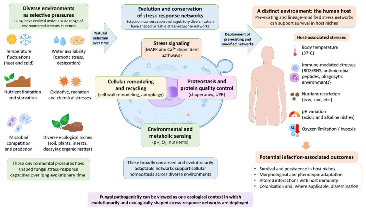
Figure 2.
Major fungal stress-response systems across environmental, host-associated, and therapeutic stresses. Fungi encounter overlapping stresses in environmental niches, during host colonization and infection, and following antifungal exposure. These inputs engage major cellular stress-response systems, including HOG MAPK and oxidative-stress signaling, the cell-wall integrity pathway, the calcineurin–Hsp90 network, the unfolded protein response (UPR) and proteostasis, and nutrient-sensing, translational-control, and metabolic-adaptation pathways. These systems regulate osmoadaptation, antioxidant defenses, cell-wall remodeling, ion homeostasis, protein quality control, autophagy, and metabolic adaptation. In pathogenic fungi, their activation can support survival in host niches, phenotypic adaptation, host interactions, and, where applicable, colonization and dissemination. Although core stress-response modules are broadly conserved, their sensors, regulatory connections, and phenotypic outputs can differ among fungal lineages.
Figure 2.
Major fungal stress-response systems across environmental, host-associated, and therapeutic stresses. Fungi encounter overlapping stresses in environmental niches, during host colonization and infection, and following antifungal exposure. These inputs engage major cellular stress-response systems, including HOG MAPK and oxidative-stress signaling, the cell-wall integrity pathway, the calcineurin–Hsp90 network, the unfolded protein response (UPR) and proteostasis, and nutrient-sensing, translational-control, and metabolic-adaptation pathways. These systems regulate osmoadaptation, antioxidant defenses, cell-wall remodeling, ion homeostasis, protein quality control, autophagy, and metabolic adaptation. In pathogenic fungi, their activation can support survival in host niches, phenotypic adaptation, host interactions, and, where applicable, colonization and dissemination. Although core stress-response modules are broadly conserved, their sensors, regulatory connections, and phenotypic outputs can differ among fungal lineages.
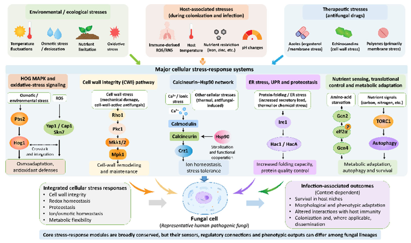
Figure 3.
Integration of fungal stress-response networks to maintain cellular homeostasis under combined stresses. Fungal cells often encounter multiple stresses simultaneously, requiring coordinated responses rather than activation of isolated pathways. Crosstalk among HOG MAPK, cell-wall integrity, calcineurin–Hsp90, oxidative-stress, unfolded protein response, autophagy, translational-control, and metabolic pathways enables shared regulators and downstream effectors to coordinate proteostasis, redox balance, ion and cell-wall homeostasis, nutrient use, and cellular remodeling. This networked organization supports adaptation to changing environmental and host-associated conditions, while the strength and direction of pathway interactions can vary among fungal species and physiological states.
Figure 3.
Integration of fungal stress-response networks to maintain cellular homeostasis under combined stresses. Fungal cells often encounter multiple stresses simultaneously, requiring coordinated responses rather than activation of isolated pathways. Crosstalk among HOG MAPK, cell-wall integrity, calcineurin–Hsp90, oxidative-stress, unfolded protein response, autophagy, translational-control, and metabolic pathways enables shared regulators and downstream effectors to coordinate proteostasis, redox balance, ion and cell-wall homeostasis, nutrient use, and cellular remodeling. This networked organization supports adaptation to changing environmental and host-associated conditions, while the strength and direction of pathway interactions can vary among fungal species and physiological states.
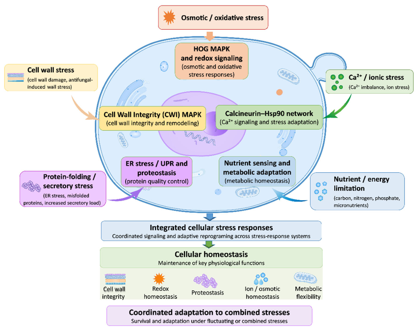

Table 1.
Overview of fungal stress-response systems described in Section 4. Core machinery is representative; pathway components, regulatory connections, and biological outputs can differ among fungal lineages.
Table 1.
Overview of fungal stress-response systems described in Section 4. Core machinery is representative; pathway components, regulatory connections, and biological outputs can differ among fungal lineages.
| Stress-response system | Core machinery | Principal functions | Comparative features and references |
|---|---|---|---|
| Heat shock and chaperones | Hsf1; Hsp70/Hsp90 and co-chaperones | Protein folding and stress-dependent signaling | Candida albicans, Candidozyma auris, Aspergillus fumigatus and Cryptococcus neoformans: distinct developmental and drug-response outputs [75,79,80,81]. |
| HOG signaling | Two-component or membrane inputs; Hog1-family MAPKs | Osmotic adjustment and glycerol regulation; additional stress outputs | A. fumigatus: SakA/MpkC; C. neoformans: unusual basal Hog1 phosphorylation and stress-dependent regulation [105,106,107]. |
| Cell wall integrity | Rho1–Pkc1–MAPK signaling | Wall remodeling and compensation for damage | C. albicans: Mkc1; Nakaseomyces glabratus: Slt2; A. fumigatus: MpkA and oxidative-stress connections [124,125,126]. |
| pH | Rim/Pac sensors and proteolytic activation of Rim101/PacC | pH-responsive transcription, surface and metabolic adaptation | Aspergillus nidulans: PacC; C. albicans: Rim101, including effects on iron acquisition [133,134,137]. |
| Hypoxia | Heme/Hap1–Rox1; Upc2 or SREBP-family regulators | Oxygen-dependent metabolism and sterol homeostasis | C. albicans uses Upc2; A. fumigatus uses SrbA. Shared physiological demands do not imply identical sensors [138,141,143]. |
| ER stress and UPR | Ire1/IreA; unconventional transcript splicing | ER folding capacity and secretory homeostasis | A. fumigatus: HacA-dependent and independent IreA functions; C. neoformans: divergent Hxl1 effector [150,151]. |
| STRIPAK | Striatin-associated PP2A complexes | Coordination of development, stress responses and selective autophagy | A. nidulans: StrA; C. neoformans: Far/Mob3 components with distinct effects on thermotolerance and virulence-associated traits [152,156]. |
| Melanin | DHN, DOPA or pyomelanin pathways | Pigmented cell-surface protection; effects depend on pathway and species | Aspergillus spp., Exophiala dermatitidis and Fonsecaea pedrosoi illustrate diverse pigment biology; radiation-associated growth is not proof of energy capture [157,162,169,170]. |
| Stress granules | RNA-binding proteins and stress-dependent condensates | Reorganization of mRNA processing during stress | A. nidulans: Pbp1-associated regulation; C. neoformans: calcineurin colocalization with RNA granules under thermal stress [175,176]. |
| Autophagy | ATG machinery; nutrient and TORC1 regulation | Bulk or selective recycling of cellular material | A. fumigatus, C. neoformans and C. albicans show different developmental, nutritional and biofilm phenotypes; virulence effects depend on model [185,186,187]. |
| ROS and thioredoxin | Gpx3–Yap1/Cap1; Skn7; thioredoxin system | Peroxide sensing, antioxidant defense and signal termination | C. albicans: Trx1 links Cap1, Hog1 and Rad53; C. neoformans and A. fumigatus differ in regulator contributions to virulence [192,194,196,197]. |
| Mitochondrial retrograde signaling | Rtg1–Rtg3; Rtg2/Mks1 and TOR inputs in S. cerevisiae | Nuclear transcriptional adaptation to respiratory dysfunction; metabolic and redox homeostasis | S. cerevisiae: regulated nuclear entry and CIT2 induction. C. albicans: constitutively nuclear Rtg1–Rtg3 and divergent targets [261,262,263]. |
Table 2.
Species-dependent antifungal responses and supporting mechanisms.
| Species | Selected antifungal responses and mechanisms | Sources |
|---|---|---|
| Candida albicans | Echinocandin resistance/tolerance involves Hsp90–calcineurin. Hog1 contributes to the response to AmB. Genetic and pharmacological evidence links proteostasis and MAPK signaling to drug response. | [216,217] |
| Candida tropicalis | Fluconazole resistance and acquired echinocandin resistance through target mutation or over expression, transporter over expression, or mitochondrial respiratory chain suppression. | [218] |
| Lodderomyces (Candida) parapsilosis complex | Fluconazole resistance occurs. Baseline echinocandin MICs are elevated due to amino acid sequence relative to several common Candida species. | [218,219,220] |
| Pichia kudriavzevii | Intrinsic fluconazole resistance; other azoles and echinocandins have different susceptibility profiles due to target mutation or overexpression, or transporter overexpression. | [218] |
| Meyerozyma guilliermondii | Reduced echinocandin susceptibility and azole resistance are reported; mechanisms and supporting evidence vary by drug and isolate. | [221] |
| Clavispora lusitaniae | Resistance to AmB, azoles, and echinocandins can emerge during therapy due to target mutation or overexpression, or transporter mutation or overexpression. | [222] |
| Candidozyma auris | Hsp90 affects azole tolerance; Cdr1 supports high-level azole resistance. Calcineurin and Crz1 influence responses to multiple drug classes. | [79,223] |
| Nakaseomyces glabratus complex | Fluconazole resistance varies among isolates; FKS-associated acquired echinocandin resistance involving target mutation or Hsp90–calcineurin. | [218,224] |
| Aspergillus fumigatus complex | SrbA connects sterol/hypoxia adaptation to azole responses. Caspofungin paradoxical growth involves Hsp90-dependent stress adaptation and recovery of Fks1 activity. | [80,143,225] |
| Aspergillus flavuscomplex | Most isolates had wild-type MICs to mold-active triazoles; non-wild-type isolates with ERG11 mutations or overexpression of ERG11 and transporter genes, but some strains with unknown mechanisms. Some isolates are intrinsically non-susceptible to AmB with increased peroxidase and superoxide-dismutase activities. | [226,227,228] |
| Aspergillus niger complex | Mold-active triazole profiles differ among isolates and from cryptic members of section Nigri. Report on paradoxical resistance to itraconazole in some strains. Decreased expression of peroxidative enzymes, increased expression of transporters, reduced ATP synthesis under itraconazole. | [227,229,230] |
| Aspergillus terreus | Frequently low AmB susceptibility with oxidative-stress defenses. mold-active triazoles have a different activity profile that cannot be inferred from ergosterol binding alone. | [227,231] |
| Aspergillus lentulus | Reduced azole susceptibility may reflect intrinsic Erg11 amino acid sequence. | [232] |
| Aspergillus calidoustus | Intrinsically poor susceptibility to several azoles, including posaconazole possibly due to unique amino acid sequence of Erg11. | [233] |
| Scedosporium apiospermum | Commonly high AmB MICs. PIG1 disruption alters melanization and stress phenotypes, but heterogeneous susceptibility results do not establish melanin as the principal resistance determinant. | [234,235] |
| Lomentospora prolificans | Broadly poor susceptibility to azoles, echinocandins, and AmB. Voriconazole exposure induces overexpression of Hsp70 and Serine-rich RNA polymerase I suppressor protein (Srp1). | [234,236] |
| Fusarium oxysporumand Neocosmospora solani | AmB and posaconazole exposure elicits species-dependent transcriptional responses involving sterols, redox pathways and transport. Expression associations alone do not establish causal resistance mechanisms. | [237,238] |
| Exophiala dermatitidis | Hsp90 reduces azole susceptibility. | [239] |
| Histoplasma capsulatum | Srb1 silencing increases itraconazole susceptibility. | [211] |
| Rhizopus arrhizus | Intrinsic Erg11 amino acid sequence rendering some azoles non susceptible in strain dependently. | [240] |
|
Mucor circinelloides Mucor lusitanicus |
AmB is active against many isolates. Azole responses involve Cyp51 biology and drug transporters; calcineurin also modulates antifungal responses. Its contribution, and that of Hsp90, to interspecies polyene susceptibility remains unresolved. | [241,242] |
Table 2 note. Entries summarize experimental mechanisms or susceptibility patterns, not clinical treatment recommendations. High MICs, tolerance, persistence, and paradoxical growth are distinct outcomes. Transcriptomic associations and proposed mechanisms are identified as such. AmB, amphotericin B; CWI, cell-wall integrity; MIC, minimum inhibitory concentration; ROS, reactive oxygen species; SREBP, sterol regulatory element-binding protein.
Disclaimer/Publisher’s Note: The statements, opinions and data contained in all publications are solely those of the individual author(s) and contributor(s) and not of MDPI and/or the editor(s). MDPI and/or the editor(s) disclaim responsibility for any injury to people or property resulting from any ideas, methods, instructions or products referred to in the content. |
© 2026 by the authors. Licensee MDPI, Basel, Switzerland. This article is an open access article distributed under the terms and conditions of the Creative Commons Attribution (CC BY) license (http://creativecommons.org/licenses/by/4.0/).
Copyright: This open access article is published under a Creative Commons CC BY 4.0 license, which permit the free download, distribution, and reuse, provided that the author and preprint are cited in any reuse.