

Submitted:
01 October 2026
Posted:
02 October 2026
You are already at the latest version
Abstract
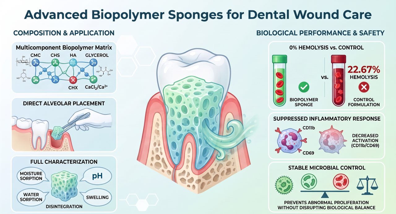
Biopolymer-based sponges incorporating chlorhexidine (CHX) and CaCl₂ were developed as potential topical biomaterials for post-dental extraction wound management. Carboxymethylcellulose (CMC) and Chitosan (CHS) sponges containing hyaluronic acid (HA), glycerol (G), chlorhexidine (CHX) and calcium chloride (CaCl₂, Ca2+) were characterized in terms of their physicochemical properties, hemocompatibility, cytotoxicity, neutrophil activation and microbial growth control. The CHX-loaded sponges exhibited no detectable hemolysis (0%), compared with 22.67% for the control formulation, and markedly reduced neutrophil activation, as indicated by CD11b and CD69 expression. Although no significant growth inhibition was observed, the sponges prevented abnormal microbial proliferation, maintaining a stable microbial population. Collectively, these findings highlight the potential of CHX-loaded biopolymer sponges as hemocompatible topical biomaterials for post-extraction wound management alveolar tissue repair.
Keywords:
carboxymethylcellulose
; chitosan
; sponges
; chlorhexidine
; calcium
; dental alveolar treatment
; Streptococcus mutans
1. Introduction
Recent advances in dentistry and biomaterials science have increasingly focused on patient-centered strategies for managing post-dental extraction wounds and supporting the repair of alveolar tissues. Tooth extraction creates an open wound that is exposed to the oral environment, where microbial colonization, local inflammation, and mechanical disturbances may interfere with normal healing. Alveolar osteitis, commonly known as dry socket, is one of the most painful complications associated with tooth extraction and may substantially impair patients’ quality of life. Although its pathogenesis is multifactorial, local fibrinolysis, bacterial activity, surgical trauma, and an inadequate inflammatory response have been implicated in its development. [1,2,3]. Local antimicrobial strategies have therefore been investigated as adjuncts to conventional post-extraction care. Chlorhexidine (CHX) is one of the most widely studied antiseptics in dentistry, and clinical evidence indicates that mouthrinses and intra-alveolar formulations containing CHX may reduce the incidence of alveolar osteitis in selected extraction settings. However, the clinical benefit depends on the concentration, formulation, and administration protocol, and the potential for adverse reactions should also be considered. [4,5]. Accordingly, the development of localized delivery systems capable of maintaining CHX at the wound site while minimizing repeated administration may provide a useful approach for the management of post-extraction alveolar wounds.
Chlorhexidine is a cationic biguanide with broad-spectrum activity against Gram-positive and Gram-negative bacteria [6]. Its antimicrobial mechanism involves electrostatic interaction with negatively charged bacterial membranes, followed by disruption of membrane permeability and leakage of intracellular components [6,7]. The biological effect is concentration-dependent: lower concentrations are generally associated with bacteriostatic activity, whereas higher concentrations can produce irreversible membrane damage and bacterial death [8]. These properties make CHX an attractive candidate for incorporation into topical biomaterials. Nevertheless, the biological performance of a CHX-containing material should not be assessed solely based on antimicrobial activity, because its interaction with blood components and immune cells may also influence its suitability for contact with injured oral tissues. Biopolymeric sponge are promising platforms for local drug delivery because their porous architecture can provide a high surface area, fluid absorption, conformal contact with irregular wound surfaces, and the potential for controlled release of active compounds. Carboxymethylcellulose (CMC), a water-soluble cellulose derivative, has been widely investigated in wound dressings and drug-delivery systems because of its hydrophilicity, chemical versatility, and favorable biocompatibility [9,10]. On the other hand, the chitosan (CHS), a natural polysaccharide derived from chitin, offers additional advantages, including biodegradability, mucoadhesion, film-forming capacity, and intrinsic antimicrobial activity [11,12,13]. Chitosan-based biomaterials have consequently been explored for wound dressings, tissue repair, and localized therapeutic delivery [14,15,16]. Hyaluronic acid (HA) is a naturally occurring, non-sulfated glycosaminoglycan and a major component of the extracellular matrix of connective tissues, where it plays a critical role in maintaining tissue hydration, structural integrity, and homeostasis. Beyond its structural and biophysical functions, HA actively regulates cell–matrix interactions, cellular migration and proliferation, inflammatory responses, and tissue remodeling and regeneration. These diverse biological activities are largely influenced by its molecular weight, tissue distribution, and interactions with specific cell-surface receptors and other extracellular matrix components [17]. Because of its biocompatibility, high water-retention capacity, and biological activity, HA has been incorporated into hydrogels, sponges, films, and other wound-dressing systems [18,19,20].The combination of CMC, CHS, and HA may generate a hydrated and mucoadhesive matrix with complementary physicochemical and biological properties. In addition, glycerol can act as a plasticizing agent, improving the flexibility and handling characteristics of polymeric membranes and sponges. Calcium chloride may further modify the structure and ionic environment of the polymeric network through interactions with anionic functional groups, while also serving as a source of Ca²⁺, an ion relevant to mineralized-tissue biology. The effects of CaCl₂, however, depend on its concentration, distribution, and interactions with the polymer components and should therefore be evaluated experimentally.
Beyond antimicrobial performance, the interaction of a biomaterial with blood and innate immune cells is an important component of its biological evaluation. Hemocompatibility assays can provide information about the material’s effects on erythrocytes and other blood components, whereas neutrophil-based assays can help identify potential cytotoxic, inflammatory, or immunomodulatory effects. Neutrophils are the most abundant circulating leukocytes and constitute an immediate first line of defense against tissue injury and microbial invasion. Following the detection of inflammatory signals, they are recruited to affected tissues, where they exert antimicrobial and immunomodulatory functions through phagocytosis, degranulation, reactive oxygen species generation, and the formation of neutrophil extracellular traps (NETs) [21,22]. Neutrophils have a particularly important role in maintaining oral mucosal homeostasis. Under physiological conditions, they continuously migrate through the gingival tissues and accumulate in the gingival crevice, where they contribute to microbial surveillance and containment within the highly diverse oral microbial environment. This surveillance helps control bacterial biofilms and protects the epithelial barrier from microbial invasion [23,24]. However, neutrophil activation must remain appropriately regulated because excessive degranulation, oxidative activity, or NET formation may contribute to tissue injury and persistent inflammation. Therefore, evaluating neutrophil responses to a CHX-loaded biomaterial may provide complementary information about its biological compatibility and help determine whether the material supports antimicrobial defense without inducing an undesirable inflammatory response. This consideration is particularly relevant for calcium-containing biomaterials, because calcium-mediated physicochemical changes at the material–tissue interface may influence neutrophil activation and inflammatory signaling.
In this study, we developed porous biopolymeric sponges based on CMC, CHS, HA, glycerol, and CaCl₂ containing CHX for potential use in localized dental alveolar applications. The sponges were characterized in terms of their physicochemical properties and evaluated for antibacterial activity against Streptococcus mutans. Their biological performance was further assessed through hemocompatibility and neutrophil-response assays. We hypothesized that the incorporation of CHX into the CMC/CHS/HA/G/Ca-based sponges would provide antibacterial activity while maintaining an acceptable interaction with blood components and neutrophils. Accordingly, the main was to develop porous biopolymeric sponges based on CMC, CHS, HA, Glycerol, and CaCl2 containing CHX for potential use in localized dental alveolar applications, characterize their physicochemical properties, and evaluate their antibacterial activity against Streptococcus mutans, as well as their biological performance through hemocompatibility and neutrophil-response assays. These findings may contribute to the development of multifunctional biopolymeric sponges for the localized management of post-extraction alveolar wounds.
2. Material and Methods
2.1. Formulation and Synthesis of Sponges
Chitosan (CHS, with a high molecular weight, deacetylation degree greater than 85%), Carboxymethylcellulose sodium (CMC, with a molecular weight of 90 kDa and a degree of substitution of 0.7), and CaCl2 were obtained from Sigma-Aldrich (Saint Louis, MO, USA). Glacial acetic acid and distilled water were purchased from Sigma Aldrich (USA). Glycerol spectrophotometric grade (99.5%+), was obtained from Acros, NJ, USA. Hyaluronic acid was purchased from RAAM De SAHUAYO Lab. S.A. DE C.V. Chlorhexidine gluconate commercial (Genhexis RC, 2%) was purchased from ZEYCO laboratory S.A. de C.V. Hyaluronic acid, with a high molecular weight, was purchased from Alma Tierra S.A. de C.V. Reagents for prothrombin time (PT) and activated partial thromboplastin time (APTT) were obtained from Spinreact (Estado de México, MEX). The hemolysis test was performed using cyanmethemoglobin reagent (Hycel Reactivos Químico, Jalisco, MEX) and standard hemoglobin was acquired from Spinreact.
Preparation of CMC/CHS/HA/G/CHX Sponges
The sponges were prepared using a modified casting and solvent evaporation method by Perez et al. 2025 [25]. To prepare 1g of CHS powder in 80 mL of deionized water was added to a flask and magnetically stirred (400 rpm) at 60 °C. An aqueous solution of acetic acid (2%, v/v) was then added to the mixture, and stirring continued for 10 min, then 1 g of CMC was added, resulting in a CMC/CHS solution. Simultaneously, 3 mL of hyaluronic acid (HA), 3 mL of glycerol (G), 6 mL of Chlorhexidine gluconate at 2% (CHX) and 6 mL of CaCl2 at 0.1M was added and stirring continued for 15 min until the CMC/CHS/HA/G/Ca/CHX solution transformed into a flocculent suspension. Four mL of the CHS suspension were poured into silicone mold (1.6 cm in diameter and 1 cm of height) on an analytical scale. The prepared molds were dried at 25 °C for 60 h. The final sample designation and amounts of CMC, CHS, HA, G, CHX and CaCl2 (SCHX) used for each sample were summarized in Table 1. The SControl (CMC, CHS, HA, G, Ca) was prepared with the same formulation.
2.2. In Vitro Characterization of the Sponges
2.2.1. Scanning Electron Microscopy Analysis
The surface images of the samples were observed using a scanning electron microscope (SEM, JEOL, JSM-6610 LV, Japan). The sponges were dried at room temperature for 7 days, after which the dried fragments were broken and coated with gold by sputtering to produce electric conductivity. SEM images of the sponge surface were captured under high vacuum condition operating at 20 kV.
2.2.2. Fourier Transform Infrared (FTIR-ATR) Spectra Analysis
The spectra of the CMC/CHS/HA/G/Ca samples and their CHX sample were obtained using Fourier transform infrared spectroscopy (FTIR-ATR) with (attenuated total reflectance/ATR) an accessory (Bruker, Vertex 70 model, Bulington, CAN), with 32 scans in the range between 400 and 4000 cm−1, and a resolution of 4 cm−1.
2.2.3. Mass of Sponges
The samples were weighed separately using the analytical balance (OHAUS Adventure Pro AV265C, New Jersey, EE.UU.: 260g (SD ± 0.1 mg) (l ± 0.3 mg), after 48 h of being stored at room temperature. The mean mass and standard deviation (SD) were calculated taking an average of the ten values per unit (n = 10).
2.2.4. Surface pH
A disk unit (n = 10) was placed in a flask containing 5 mL of deionized water. The formulation was allowed to swell for 5 min and was subsequently removed from the flask. The resulting pH of the liquid was recorded using a pH meter (ROCA-Model PHS-3CU) at 25 °C and was taken as an indication of the formulations surface pH [25].
2.2.5. Moisture Sorption
The sponge’s moisture sorption was studied by exposing them to 75% relative humidity (RH) using a desiccator with an oversaturated NaCl solution at room temperature. The units were pre-weighed (m1) and stored in a humid desiccator for 10 days (n = 10). Afterwards, they were re-weighed (m2). Moisture sorption was calculated in the same manner as swelling capacity and multiplied by 100 to obtain the sorption percentage [25]. The experiment was performed with ten samples.
2.2.6. Water Sorption Behavior
CMC/CHS/HA/G/Ca sponges and CHX loading sponges were cut into small disks (0.6 cm diameter, n = 10), desiccated overnight under vacuum, and weighed to determine their dry mass. The weighed sponges were placed in a flask containing 5 mL of phosphate-buffered saline (PBS, pH 7.4) at 37 °C. The swelling kinetics were evaluated by periodically measuring the weight increment of the samples with respect to the dry sponges, using an analytical scale with a precision of 0.001 g. After gently bottling the surface with a tissue to remove the excess PBS solution, the samples were weighed, and the respective values were recorded until day 2 [25].
The water sorption (W.S.) was calculated as follows:
where m1 expressed the weight of the dried sponges, while m2 expressed the weight of the swollen sponges.
2.2.7. Swelling Capacity
The swelling capacity of the sponges was evaluated using a modified test from the literature [26], where a membrane with a determined mass (m1) was placed in a glass flask and immersed in 5 mL of PBS (pH = 7.38) at 37 °C. The sponge was allowed to swell for 5 min. Its mass (m2) was recorded after gently wiping the product with a piece of tissue paper to remove the surface water. The swelling index represents the mass gained with respect to the mass of a dehydrated sponge and was calculated according to Equation (2). The experiment was carried out ten times.
2.2.8. Evaluation of Disintegration or Biodegradability
The disintegration test was performed using a modified disk method from the literature [25]. A sponge weighed was immersed into a flask containing 5 mL of simulated body fluids (SBFs) (buffer solution) at pH = 7.40 and at 37 °C to determine their biodegradability in human fluid. During the experiment, the disintegration time for separate units of membrane disks was observed. In cases where a coherent matrix remained after 168 h, the sample was recorded as not disintegrated. Ten parallels were tested for each formulation (n = 10).
2.3. Evaluation of Blood Coagulation: Extrinsic and Intrinsic Pathways
The study protocol was approved by the Research Ethics Committee of the School of Dentistry at the Universidad Veracruzana [25]. Prothrombin time (PT) was measured to assess the effect of the samples on the extrinsic coagulation pathway, whereas activated partial thromboplastin time (APTT) was determined to assess their effect on the intrinsic coagulation pathway. Both coagulation parameters were evaluated for the biopolymeric sponges, including the control formulation (SControl) and the chlorhexidine-loaded formulation (SCHX). Citrated blood was collected from a healthy adult human donor, and the blood was used in the experiments. Then, 60 mg of each biopolymeric sponge was incubated with 600 µL of whole blood in a 2 mL conical-bottom tube. The samples were incubated at 37 °C with oscillatory stirring at 100 rpm for 15 min (MaxQ 4450 Thermo Scientific, Massachusetts, USA.). Immediately after the incubation, the blood from each tube with the biopolymeric sponge sample was collected with a micropipette and then the blood was poured into a 1.5 mL conical-bottom tube; these new samples were centrifuged for 5 min at 3000 rpm to separate the plasma from the blood cells. The plasma was used to measure the PT and the APTT with the Biobas 10 coagulometer (Spinreact México), Estado de Mexico, MEX. Plasma from blood without contact with any sample was used as a physiological control. A normal control and a pathological control (Spinreact México) were used as the negative and positive control, respectively. The blood for these controls was also incubated at 37 °C, at oscillatory stirring (100 rpm, 15 min). The normal PT was set at 11.1–14.3 s according to the reagent instructions. The normal APTT was set at 24–36 s according to the reagent instructions. All experiments were performed by triplicate.
2.3.2. Hemolysis Test
Ethical approval for the study was granted by the Research Ethics Committee of the School of Dentistry at the Universidad Veracruzana [25]. A hemolysis test was performed according to the Standard Practice for Assessment of Hemolytic Properties of Materials (ASTM F756-00) to evaluate the hemolytic potential of the biopolymeric sponges, using blood from a healthy adult donor. The blood was collected with EDTA vacutainer tubes. Each experimental sponge (60 mg) was incubated with blood (600 µL) in the same way as described in the blood coagulation experiments. Also, blood was collected to separate the plasma from the blood cells in the same way as described for the blood coagulation experiments. The plasma (25 µL) obtained from the blood in contact with the experimental sponge was mixed with a cyanmethemoglobin reagent (225 µL) in a well of a 96-well microplate. Then, the hemoglobin in plasma was measured at 540 nm in a microplate reader (Multiskan FC Thermo Fisher Scientific, Massachusetts, USA.). The hemoglobin concentration was obtained from a calibration curve made with a solution of standard hemoglobin mixed with a cyanmethemoglobin reagent. Hemolysis was expressed as the percentage of hemoglobin released to the total content. Thus, we mixed a sample of blood with cyanmethemoglobin reagent to achieve the 100% of hemolysis. In addition, we used a NaCl solution as a positive control and a silicon sample as a negative control. A physiologic control was performed with plasma collected from blood from the donor. According to the ASTM standard, the material might be nonhemolytic (0–2% of hemolysis), or slightly hemolytic (2–5% of hemolysis), or hemolytic (>5% hemolysis). All experiments were performed in triplicate.
2.3. Neutrophil Response to Sponges
Neutrophil Isolation
Peripheral blood was collected from healthy adult donors into CPD-containing tubes (Citrate-Phosphate-Dextrose buffer) and processed immediately after collection. Neutrophils were isolated by density-gradient centrifugation using a Percoll-based protocol. Briefly, whole blood was diluted with sterile PBS and carefully layered onto a discontinuous Percoll density gradient. Following centrifugation at 200 g for 25 min at room temperature, the neutrophil-enriched fraction was collected and transferred to a new tube. Cells were washed twice with PBS and resuspended in RPMI-1640 medium supplemented with 10% heat-inactivated fetal bovine serum and antibiotics. Cell number and viability were determined by trypan blue exclusion, and preparations with viability >95% were used for subsequent experiments.
Neutrophil Treatment
Purified neutrophils were seeded at a density of 1x 10⁶ cells/mL in 24-well tissue-culture plates and allowed to equilibrate for 10 min at 37 °C in a humidified atmosphere containing 5% CO₂. Cells were subsequently exposed to the indicated biomaterials at the concentrations specified in each experiment. Untreated neutrophils incubated under identical conditions were used as controls. Cells were incubated with the biomaterials for 3 h at 37 °C and 5% CO₂. Following incubation, cells were gently collected and washed with cold PBS to remove unbound material before flow-cytometric analysis.
Following exposure to biomaterials, neutrophils were rinsed with PBS and fixed in 4% PFA for 20 minutes at 4°C. The cells were subsequently resuspended at a concentration of 1 × 10^6 cells per milliliter in ice-cold 5% BSA in PBS and incubated on ice for 20 minutes. After a 5-minute centrifugation at 400 g at 4°C, primary antibodies (CD69-PE and CD11b-biotin) at a concentration of 1 µg/ml were added and incubated for 20 minutes at 4°C in the dark. The cells were washed twice with PBA (PBS containing 1% BSA and 0.1% NaN3) via centrifugation at 400 g for 5 minutes. A streptavidin-FITC reagent was then added and incubated for 20 minutes at 4°C in the dark. Following this, the cells were washed three times with PBA, as previously described, and finally resuspended in 1 mL of ice-cold 1% PFA. The cell suspensions were subsequently analyzed using a BD Fortessa flow cytometer.
2.4. Assessing the Antibacterial Activity of Sponges
Bacterial Growth
S. mutans was initially cultured overnight in brain heart infusion (BHI) broth containing calf brain and bovine heart infusions, peptone, NaCl, glucose, and disodium phosphate. The culture was incubated for 24 h at 37 °C under a 5% CO₂ atmosphere using an ESCO CelCulture CO₂ incubator, without agitation. Following incubation, aliquots from the overnight culture were used to prepare the subcultures (Table 3), which were adjusted to an optical density at 600 nm (OD₆₀₀) of 0.05. This adjustment was performed to obtain a standardized initial inoculum, ensuring comparable bacterial loads and OD₆₀₀ values across all treatments evaluated. The bacterial concentration corresponding to an OD₆₀₀ of 0.05 was determined as colony-forming units per milliliter (CFU/mL) using the Massive Stamping Drop Plate method (MSDP) [27].
For the inhibition assays, SCHX and SControl were sterilized by exposure to UV light for 20 min in a Class II biosafety cabinet (LABCONCO Purifier® Biological Safety Cabinet). Following sterilization, individual sponge samples were aseptically introduced into the corresponding bacterial subcultures, while BHI broth without sponge was included as the untreated control. The samples were then incubated for 24 h under anaerobic conditions, as described above. After incubation, bacterial growth was quantified by determining the CFU/mL using the Massive Stamping Drop Plate (MSDP) method on BHI agar. As an additional control to assess potential bacterial growth associated with the sponge matrix itself, individual sponge samples were placed directly in BHI broth and incubated under the same conditions. All experiments were performed in triplicate, and CFU/mL measurements were conducted in quintuplicate for each experimental replicate.
2.5. Statistical Analysis
All values are presented as the mean ± SD (standard deviation). Statistical analyses were performed using Jamovi 2024, Sydney, AUS, to evaluate the data obtained. To compare the water absorbance capacity, swelling index, moisture sorption, surface pH and mass, an independent sample t-test was employed. The level of significance was set at p < 0.05.
3. Results and Discussion
3.1. Scanning Electron Microscopy Analysis
CMC and CHS is a water-soluble anionic biopolymer with excellent physicochemical properties, such as hydrophilicity, non-toxicity, and bio-adhesiveness. Figure 1 shows images at two levels. Section A and B shown the microphotographs of sponges at superficial and cross-sectional structure level to investigate the influence of CHX on the sponge morphology. Surface and internal structure analysis by SEM is a widely used technique to study the sub-microscopic and three-dimensional architecture of different drug delivery systems in biopolymeric matrices [28]. Figure 1A, SEM image at 100x show the top surface of a CMC/CHS/HA/G/Ca sponge (SControl), where the most compact and uniform surface morphology and few superficial agglomerations of CMC and CHS can be observed in the matrix resulting from a homogeneous synthesis by solvent casting method [25,29]. Similar results can be found in the literature with freeze-drying method [30]. The SEM image at 100× (Figure 1B) show the surface morphology of the CMC/CHS/HA/G/Ca sponges with CHX at 6% (SCHX), suggesting that the prepared sponges loaded with CHX had an irregular surface, with agglomerations in contrast to the unloaded sponges (SControl) (Figure 1A). CaCl2 particles were visible in the form of dots (Figure 1B) over the sponge but were absent from the rough surface of SControl (Figure 1A). The calcium chloride particles are leached out by immersing the biopolymeric matrix in the aqueous solution, resulting in highly porous scaffold [29]. The cross-section SEM image of SCHX (Figure 1D) present a large, open, channel-like structure with different size pores (~90,000 µm2) in contrast with bigger size pores (>300,000 µm2) of SControl (Figure 1C). Similar images were reported by Cai et al., with Gentamicin-loaded CMC/CHS composite sponge [30]. The pore network in the SCHX may be suitable for post-dental extraction due to its porous structure, which enhanced the swelling capacity of liquids into the sponge.
3.2. FTIR−ATR Analysis
To analyze the interaction between the CHX functional groups and the CMC/CHS/HA/G/Ca sponge, the FTIR spectra of the CMC/CHS/HA/G/Ca/CHX sponge and the CMC/CHS/HA/G/Ca sponge were characterized (Figure 2A and Figure 2B). The FTIR spectrum of sponges exhibited characteristic absorption peaks (towards higher frequencies) between 3100 and 3600 cm−1 of stretching vibration band of the -OH and =NH, -NH functional groups of CMC, CS, HA and G. A characteristic peak 1624.07 cm−1 from CHX molecule in the biopolymer matrix and a positive shift of intensification of the stretching vibration band of the C=N group. Additionally, band between 2800 and 3000 cm−1 are assigned to the typical C−H stretching vibration in alkyl −CH2 and −CH3 of biopolymer. Furthermore, the bands around 1530 cm−1 and 1640 cm−1 are common for C=O stretching vibration amide functional group, similarly to those reported by other authors [30,31,32]. When CHX was added to the CMC/CHS/HA/G/Ca sponge, another peak appears from 3296.43 cm−1 to 3297.49 cm−1 and 3204 cm−1 (Figure 2A), which indicates the N−H and the O−H bond. The similarity of biopolymer components can confirm that the sponges have a good compatibility and miscibility and that in the relatively higher pH sponges, the CMC and chitosan molecules combined mor closely [30]. In the spectra of the CHX-loaded CMC/CHS/HA/G/Ca sponge (Figure 2A), characteristic peaks indicated the presence of CHX in the CMC/CHS/HA/G/Ca sponges. A positive shift in spectra A is the peak 1530.67 cm-1 from CHX in the biopolymer sponge, a significant intensification of the stretching vibration band of the N=H. The possible metallic coordination between chlorhexidine and calcium ion through the nitrogen atoms of amino group (-C=NH) explain this peaks, as results reported by Kowalczuk et al. [33]. The peaks around 998 cm-1 and 1138 cm-1 in the spectra (Figure 2A and Figure 2B) refer to the saccharide structure and the ether C−O−C functional groups of biopolymers. The fingerprint of the C-Cl (chlorine bonded to aromatic carbon) stretches of chlorhexidine is manifested in the peak 822.47 cm−1.
3.3. Mass of the Sponges
The sponges are three-dimensional networks formed by cross-linking monomers or polymers with good mechanical hardness, biodegradability, and biocompatibility. The mass values of sponges, as shown in Table 2, demonstrated an increase in the mass of SCHX compared to SControl (0.83 ± 0.19 g and 0.75 ± 0.07 g, respectively). The difference in sponge mass values was not statistically significant. The CHX loading in the CMC/CHS/HA/G/Ca sponge no increase in mass values. The properties of the sponges demonstrate a high potential for use in the manufacture of dressings, with ability to be used to fill spaces, function as wound dressings, or serve as drug delivery systems. In practical situations, the adaptability of sponges can fill irregular alveolar spaces after dental extraction. The irregular surface of the sponge may also enhance tissue and promote cellular activity, and porous inner can be enhanced the swelling properties. The incorporation of CMC, HA and CHS in combination with G and Calcium further induced a softness character in the sponges. The cross-linking of these biomaterials results in the formation of an interpenetrating biopolymeric network, creating a rapid method for producing a drug delivery system with the necessary mass and 3D properties.
3.4. Surface pH Kinetic
The pH of a biopolymeric sponge over time exerts a direct and multifactorial impact on the exposed dental socket. CHX is a powerful antimicrobial agent, a cationic bisbiguanide (1,1’-hexamethylene bi [5-(p-chlorophenyl)biguanide], CHX2+ at range of pH between 5.5 to 7.0, while a high concentration of H+ ions competes with CHX+ on the sponge, influencing the performance of sponge. The antimicrobial efficiency of CHX in the biopolymeric matrix of the membranes is influenced by pH changes, in vitro, the anti-bacterial effects of CHX all related to altered cell membrane permeability, but low concentrations (0.02-0.06%) CHX causes displacement of Ca2+ and Mg+2 and loss of K+ from cell wall, resulting in a bacteriostatic effect [34,35]. The pH of the surface sponge was 5.43 and 5.47 in the buffer solution for SControl and SCHX, respectively. The pH of the sponge SCHX was superior at pH =5. The solution pH gradually decreased over time, reaching 4.8 after 2 h (Figure 3). This decrease may indicate the release of CHX from the biopolymeric matrix, consistent with previous reports of similar behavior for antibiotics released from polymeric systems [25,26,30,36]. However, drug release tests are necessary to fully evaluate CHX release from sponges. Additionally, at a pH above 2.8 the fibroblast remains viable, allowing them to utilize the local anion gap to proliferate and promote healing in a bacteria-free environment [25].
3.5. Moisture Sorption, Disintegration, Water Sorption Behavior and Swelling Capacity
The effect of CHX incorporation on sponge water sorption was evaluated, and the results are presented in Figure 4. The water sorption behavior was analyzed at 0.1, 0.17, 0.25, 0.33, 0.42, 0.5, 1, 2, 3, 24, and 48 h. Moisture sorption, water sorption behavior, and swelling properties of natural materials depend on the nature of the medium and on the swelling caused by solvent diffusion into the material structure from the extracellular medium. The introduction from charged chemical groups from chlorhexidine into the biopolymeric matrix can alter some sponge characteristics. The highest water sorption in both sponges was observed in PBS (pH = 7.4, 37 °C), with values 238.58 ± 30.42% at 24th hour, and 480.23 ± 56.21% at 48th hour, for SControl and SCHX, respectively (Table 2). CMC and CHS are commonly used as drug delivery systems due to their strong hygroscopicity. Similar results were reported by Cai et al. [30]. HA and G improve the hygroscopicity in biopolymeric matrices [26]. This increase was due to the incorporation of CHX into the biopolymeric matrix, likely through the interaction of protonated acid groups (biguanides) at pH of 5.47, which led to a greater water affinity of CMC and CHS to low pH [26,30]. The stability of CHX could be affected by sponge moisture sorption during storage, as it is a humidity sensitive drug. In addition to their capacity to swell and absorb wound exudate, these biopolymer membranes maintain wound moisture by delivering water molecules. Therefore, the impact of these charges on moisture sorption and water sorption behavior were evaluated (Figure 4).
Table 2 shows the moisture sorption weight gain recorded on the seventh day. The moisture sorption of these sponges follows the order of SControl < SCHX, with values of 40.77 ± 10.07% and 38.43 ± 1.93%, respectively. The difference is not statistically significant (p = 0.488). The process of hydration in polymer networks can absorb at least 10% of their dry weight in water and in some cases even thousands of times that amount. Due to their high water absorption capacity and biocompatibility, these sponges have been used in dressing and drug delivery systems [37] and in post-dental extraction wound. These sponges are cross-linked polymer networks that swell in a liquid medium. The embedded liquid acts as a selective filter, allowing the free diffusion of certain solute molecules, such as drugs, while the polymer network serves as a matrix to retain the liquid. The disintegration time of the sponges was evaluated in a buffer medium under physiological conditions. These conditions allowed determine their biodegradability in contact with human fluids. The disintegration time in both sponges was observed at 48th and 144th hour in PBS (pH = 7.40, 37 °C) for SControl and SCHX, respectively (Table 2).
The disintegration time of a biopolymeric sponge is a critical factor in its performance as a wound dressing, as it determines the balance between wound protection and tissue regeneration. If the dressing degrades too quickly or too slowly, it can disrupt the healing process. The results of disintegration observed show a higher time disintegration to sponges SCHX compared with the sponges SControl, 144h and 48h, respectively. The average time for visible gum tissue to close and heal after a tooth extraction is 7 to 14 days. The dressing must degrade in synchronization with the growth rate of the new tissue, gradually disappearing as the wound heals [38,39,40]. The swelling behavior of the biopolymeric sponges was evaluated in a buffer medium. The swelling index was determined to assess their fluid absorption capacity under conditions simulating contact with wound exudate at the alveolar tissue. Results indicated that sponges showed different swelling capacity to SControl and SCHX, 0.46 ± 0.05 and 1.16 ± 0.18, respectively (Table 2). The difference is not statistically significant (p < 0.001). This water sorption behavior in the time can be explained by the fact that CMC/CHS/HA/G/Ca is a good hydro dispersible biopolymer base that allows for the faster swelling of CHX, similar to the results reported with biopolymeric sponges loaded with Gentamicin [30] and other biopolymeric sponges with calcium ion [32]. These results indicate that 6mL of chlorhexidine gluconate at 2% and 0.07% of CaCl2 were the ideal amounts incorporated into a 1.00 g/80 mL CMC and 1.00 g/80mL of CHS powder polymer blended sponge.
3.6. Blood Coagulation
Hemostasis is the body's physiological response to injury and the first step in the wound-healing process. Topical hemostatic agent with polysaccharides-based materials, such as chitosan and cellulose, play an important role also employed in conjunction with conventional techniques for achieving hemostasis in a surgery setting [41]. The PT test and the APTT evaluated the effect of the sponges on the extrinsic and intrinsic pathways of blood coagulation, respectively. Table 2 shows the PT results (13.30±0.60 s and 17.10±1.40 s) and APTT (29.20±2.90 s and 26.00±3.90 s) for SControl and SCHX, respectively. The SControl induced a PT and APTT within the normal range. Similarly, the SCHX also induced APTT within the normal range, but the PT value is slightly above the normal time. These findings showed that the sponges did not interact with the blood coagulation pathways compared to the reported with other CHS and CMC sponges, which the interaction between the carboxyl groups of CMC and the amino groups in CHS, resulted with noticeable variation [42]. Although biomaterials composed of CMC and CHS have demonstrated in vitro and in vivo hemostatic properties [41,43], their hemostatic effect depends on both the membrane composition and the percentage of Ca2+ and other components. In this study, the samples were prepared with a percentage of CMC, CHS, HA and G that did not induce any effect on the PT and APPT. Additionally, CHX is an antiseptic with various biological effects; however, these do not include the induction or inhibition of blood coagulation.
3.7. Hemolytic Properties
The design of the biomaterials intended for wound care requires careful consideration of their interaction with biological systems. Given that the fabricated sponges are intended to come into direct contact with alveolar tissues, ensuring their blood compatibility is a critical requirement to prevent adverse reactions and promote healing. The CMC/CHS/HA/G/Ca-based sponges, widely recognized for their hemocompatibility, are generally well tolerated by the human body and do not induce an immune response or inflammation. The hemolytic properties of the membranes were evaluated by quantifying the extent of hemolysis induced by the CMC/CHS/HA/G/ca sponges (SControl = 22.67%) and the chlorhexidine-loading sponges (SCHX = 00.00%). According to the ASTM standard recommendation followed in the experiment, the SControl showed hemolytic properties (>5% of hemolysis), but the CHX-loaded sponge did not induce a hemolytic level. The results of the hemolysis assay offer valuable information regarding the hemocompatibility of the sponges, which es crucial to exhibit minimal hemolysis, or the destruction of red blood cells brought by shear stress or changes in osmotic pressure [42]. The CMC/CHS/HA/G/Ca-based sponge exhibited a hemolytic effect due to detergent action of the cationic charge in the biopolymeric matrix, which disrupts the red blood cells. Interestingly, the CHX-loaded sponges no induced hemolysis, suggesting that the addition of the antiseptic might reduce the cationic charges confirming their compatibility as reported literature [25,44]. Indeed, polymer-based films improved their blood compatibility properties when CHX was added to the formulation.
3.8. Effect of CMC/CHS/G/Ca/CHX Sponges on Neutrophil Activation
To assess the immune response of neutrophils to the biomaterials, human neutrophils isolated from peripheral blood were incubated for 3 hours in the presence of the CHX-loading CMC/CHS/HA/G/Ca-based sponge. Afterwards, the expression of activation markers CD11b and CD69 was evaluated via flow cytometry. As controls, untreated neutrophils and cells stimulated with LPS were included. Analysis of these cellular activation markers revealed that the control biomaterial induced a marked increase in CD11b expression, reaching levels higher than those observed in cells stimulated with LPS, which was used as a positive control for immune activation. These findings suggest that the composition of the SControl biomaterial is sufficient to induce a pronounced immune response. In contrast, biomaterial with alternative compositions, such as SCHX, showed reduced CD11b expression almost comparable to non-activated cells, indicating a lower degree of cellular activation than the control biomaterial (Figure 5a and Figure 5b).
A similar pattern was observed for CD69 expression. The SControl induced a greater increase in CD69 expression, whereas treatment with SCHX resulted in lower expression levels (Figure 5c and Figure 5d). Collectively, these findings suggest that biomaterial composition plays an important role in determining the magnitude of the immune activation response.
The notable increase in CD11b levels induced by the SControl, surpassing even the response observed following LPS stimulation, indicates a substantial modification in the neutrophil activation state prompted by the material. CD11b is promptly mobilized to the neutrophil surface upon stimulation and plays a crucial role in adhesion, migration, and effector functions. The response documented herein may be especially pertinent to the physicochemical attributes of the high-molecular-weight chitosan with a degree of deacetylation exceeding 85% employed in the formulation. The degree of deacetylation is a significant determinant of chitosan's charge and biological characteristics, and prior research has demonstrated that neutrophils exhibit varied responses to chitosans with different degrees of deacetylation [45]. Notably, highly deacetylated chitosan has been reported to elicit a neutrophil response that differs from traditional pro-inflammatory stimulation, without inducing substantial superoxide production or degranulation [45,46]. Consequently, the pronounced increase in CD11b observed with the SControl may reflect neutrophil priming or activation caused by the material interface, rather than a generalized inflammatory response. Although HA can also modulate neutrophil responses in a manner dependent on molecular weight and concentration, current evidence does not support attributing a specific effect on CD11b/CD69 to CMC or glycerol; these components are more suitably regarded as contributors to the overall physicochemical properties of the matrix [47].
A similar pattern was observed for CD69, with the SControl biomaterial eliciting the most pronounced response, whereas formulation containing SCHX demonstrated significantly reduced expression. CD69 serves as an early activation marker, while CD11b more accurately reflects alterations associated with neutrophil activation and effector functions; consequently, the concurrent decrease of both markers indicates that the inclusion of CHX modulates the overall neutrophil response rather than targeting a specific activation pathway. Importantly, however, the diminished expression of CD11b and CD69 should not be directly construed as an anti-inflammatory effect. Chlorhexidine is well recognized for its effects on neutrophil function and membrane integrity [48]. In vitro studies have shown that CHX concentrations exceeding 0.005% rapidly compromise neutrophil membranes, with the observed inhibition of neutrophil functions attributed to its lytic activity [48]. These findings are particularly pertinent given that the present biomaterial contains CHX; however, the nominal concentration incorporated into the material should not be equated with the actual concentration released and encountered by the cells [48].
Consequently, the attenuated expression of CD11b and CD69 observed with SCHX may indicate either specific modulation of neutrophil activation or a diminished cellular responsiveness secondary to CHX exposure. This distinction is imperative when evaluating the biological compatibility of the formulation. Prior research has demonstrated that CHX can influence neutrophil viability, chemotaxis, and oxidative metabolism at concentrations significantly lower than those employed in the present formulation [48,49]. Therefore, if neutrophil viability remains intact and the concentration of CHX released from the biomaterial stays within a non-cytotoxic range, then the concomitant reduction in CD11b and CD69 would serve as evidence of neutrophil activation modulation. Conversely, if CHX exposure undermines viability or sponge integrity, the decreased expression of these markers should be interpreted as functional impairment rather than beneficial immunomodulation. This distinction is particularly critical because the cytotoxicity of chlorhexidine is highly dependent on both concentration and duration of exposure [50].
Overall, these findings demonstrate that biomaterial composition is a major determinant of the neutrophil response. The strong induction of CD11b and CD69 by the control formulation indicates that the chitosan/HA-containing matrix generates a pronounced neutrophil-reactive environment, whereas incorporation of CHX substantially attenuates this phenotype. From a biomaterials perspective, reducing excessive neutrophil activation could be advantageous in limiting prolonged inflammation and tissue damage; however, the present results alone do not establish that CHX improves immunological compatibility. Rather, they demonstrate that addition of CHX fundamentally changes the interaction between the biomaterial and neutrophils, emphasizing the need to balance antimicrobial activity with preservation of innate immune-cell viability and function. Measurements of CHX release, viability, and additional functional parameters such as ROS generation or degranulation would therefore be important to determine whether the attenuated CD11b/CD69 response represents controlled immunomodulation or CHX-associated cellular dysfunction.
3.9. Antibacterial Activity of CMC/CHS/HA/G/Ca/CHX Sponges
The inhibition results for S. mutans using CMC/CHS/HA/G/Ca/CHX sponges indicated that the composition of these sponges does not exhibit inhibitory activity against the evaluated strain (Table 3). In both repetitions, a constant S. mutans growth was observed. The antibacterial activity of the biopolymeric sponges was evaluated against S. mutans by quantifying viable bacterial counts (Log CFU/mL) after exposure to the different sponge formulations. All experiments were initiated with a standardized bacterial suspension corresponding to an optical density (OD) of 0.05, equivalent to 6.27 ± 0.13 Log CFU/mL (mean of five replicates).
The antibacterial activity of the biopolymeric sponges was evaluated against S. mutans by quantifying viable bacterial counts (Log CFU/mL) after exposure to the different sponge formulations. All experiments were initiated with a standardized bacterial suspension corresponding to an optical density (OD) of 0.05, equivalent to 6.27 ± 0.13 Log CFU/mL (mean of five replicates).
Exposure of S. mutans to the unloaded control sponges (SControl) resulted in viable counts of 5.95 ± 0.20, 5.97 ± 0.19, and 5.12 ± 0.35 Log CFU/mL across the three independent replicates. Sponges containing chlorhexidine (SCHX) showed viable counts of 6.08 ± 0.16, 6.02 ± 0.12, and 5.81 ± 0.18 Log CFU/mL, respectively. Sponges containing chlorhexidine in combination with calcium exhibited viable counts of 5.86 ± 0.32, 5.93 ± 0.13, and 5.46 ± 0.24 Log CFU/mL. Untreated S. mutans showed viable counts of 6.28 ± 0.13, 6.06 ± 0.07, and 5.79 ± 0.33 Log CFU/mL (Table 3).

Table 3.
Inhibition of sponges against S. mutans. The average of 3 events is shown, 5 replicates in each event. Values are expressed as log₁₀ colony-forming units per milliliter (CFU/mL).
Table 3.
Inhibition of sponges against S. mutans. The average of 3 events is shown, 5 replicates in each event. Values are expressed as log₁₀ colony-forming units per milliliter (CFU/mL).
| Sponges | S. mutans |
| Control a | 6.04 ± 0.28 |
| SControl | 5.79 ± 0.31 |
| SCHX | 5.94 ± 0.46 |
a Refers to the number of bacteria without the presence of sponges.
The results demonstrated that all groups containing sponges exhibited CFU/mL values comparable to, or slightly lower than, those of the untreated control. Importantly, no evidence of bacterial overgrowth was observed in the presence of the biopolymeric matrix. These findings suggest that the sponges do not promote bacterial proliferation and, under the conditions evaluated, maintain microbial populations at levels similar to or lower than those observed in the absence of the material.
The present study evaluated the antibacterial performance of chlorhexidine-loaded biopolymeric sponges against S. mutans by quantifying bacterial counts (log CFU/mL). Although no reduction in CFU/mL was observed relative to the untreated control, all groups containing sponges exhibited CFU/mL values comparable to or slightly lower than those of the control, with no evidence of bacterial overgrowth. From a biomaterial’s perspective, these findings suggest that the CMC/CHS/HA/G/Ca-based matrix does not promote bacterial proliferation under the experimental conditions evaluated, indicating that its presence does not provide a favorable environment for bacterial growth.
The absence of bacterial overgrowth in the presence of the biopolymeric sponges is consistent with the composition of the matrix, which lacks readily metabolizable carbon sources for S. mutans. Carboxymethylcellulose, chitosan, and hyaluronic acid are high-molecular-weight polysaccharides that are not efficiently utilized by oral streptococci as primary carbon substrates under the conditions tested. This is in line with previous reports showing that certain biopolymeric matrices do not promote bacterial colonization unless supplemented with fermentable carbohydrates or other growth-promoting agents [51].
In contrast, studies evaluating chlorhexidine in solution or in conventional formulations (e.g., mouthwashes, gels) have consistently demonstrated significant reductions in S. mutans counts, often in the range of 1–3 log CFU/mL, depending on concentration, exposure time, and formulation [52,53,54]. The more modest effect observed in the present study may be attributed to the sustained-release nature of the sponge, the lower effective concentration of CHX at the bacteria–material interface, or the protective effect of the polymeric matrix. It is also possible that the experimental design, which involved direct contact between the bacterial suspension and the sponge, did not fully replicate the dynamic conditions of the oral cavity, where salivary flow and mechanical forces may influence antimicrobial efficacy.
Interestingly, some studies have reported that sublethal exposure to chlorhexidine can induce stress responses in S. mutans without causing substantial reductions in viable counts, suggesting that bacterial tolerance may develop under certain conditions [55]. This phenomenon could partially explain the limited reduction in Log CFU/mL observed in the present study, particularly if the CHX concentration released from the sponges was below the minimum bactericidal concentration for this strain.
From a clinical standpoint, the fact that the sponges do not support bacterial proliferation is a desirable property for a biomaterial intended for use in post-extraction alveolar wounds. Materials that serve as nutrient sources or that promote bacterial colonization may increase the risk of infection or delay healing. The present findings suggest that the CMC/CHS/HA/G/Ca-based sponges maintain microbial populations at levels similar to or below those observed in the absence of the material, without inducing overgrowth. This behavior is consistent with the concept of biocompatibility, which encompasses not only the absence of cytotoxicity but also the ability to avoid promoting undesirable microbial activity [51].
Future studies should evaluate the antibacterial activity of these sponges under more clinically relevant conditions, including biofilm formation assays, dynamic flow systems, and longer exposure times. Additionally, the incorporation of higher CHX concentrations or the combination with other antimicrobial agents (e.g., xylitol, essential oils) may enhance the bactericidal efficacy while maintaining the favorable biocompatibility profile observed in this study.
4. Conclusions
In this study, we successfully synthesized CMC/CHS/HA/G/Ca sponges loaded with chlorhexidine (CHX) and demonstrated their promising potential as a novel wound delivery system for topical applications, particularly for managing post-dental extraction wounds or supporting the repair of alveolar tissues. The incorporation of CHX into the biopolymeric matrix significantly enhanced the biopolymeric matrix of the sponges, exhibiting maintain microbial populations at levels similar to or below those observed in the absence of the material, without inducing overgrowth of S. mutans, a key pathogen involved in oral infections. The sponge also displayed excellent physicochemical characteristics, such as pH, moisture retention, and swelling behavior, all of which are crucial for their potential application in wound healing. Additionally, the low hemolytic levels and the improves immunological compatibility observed in our study indicate that the sponges are biocompatible and exhibit favorable interactions with blood cells and neutrophils, further supporting their suitability for clinical use. Our results demonstrate that the combination of CMC, CHS, HA, glycerol and calcium chloride provides a synergy that improves the mechanical strength and water retention capacity of the sponge, making them ideal for maintaining a moist wound environment, which is a critical factor in promoting wound healing. However, to fully assess their potential, further kinetic studies and in vivo evaluations are required to explore their effectiveness and long-term performance in clinical settings. These additional investigations will provide critical insights into the ability of these sponges to promote tissue regeneration, reduce infection rates, and improve patient compliance in the management of post-dental extraction.
Author Contributions
S.P.-G. and J.Á.R.-E.: investigation, methodology, project administration, supervision, writing—original draft, funding acquisition, resources. A.V.J.-D.: conceptualization, data curation, formal analysis, investigation, methodology, project administration, supervision, writing—original draft, writing—review and editing, software, visualization. L.A.P.-R.: methodology, data curation, formal analysis, investigation, writing—review and editing, writing—original draft. A.F.-L.: investigation, methodology, supervision, writing—review and editing. E.R.-C.: investigation, methodology, writing—review and editing. D.d.C.P.-G. and P.G.G.-G.: data curation, formal analysis, software, writing—review and editing. I.J.-D.: investigation, software, supervision, writing—review and editing B.I.C.-C.: supervision, writing—review and editing, funding acquisition, resources, validation, visualization. J.L.S.-F.: validation, writing—review and editing, resources, supervision. C.S.-V.: formal analysis, resources B.E.C.-S.: funding acquisition, resources, validation, visualization, writing—review and editing. G.N.R.d.C.-Q.: resources, supervision. A.G.M.-G.: validation, funding acquisition. All authors have read and agreed to the published version of the manuscript.
Funding
This research was funded by Faculty of Stomatology, Meritorious Autonomous University of Puebla, VIEP-BUAP 00264-PV/2018, 00507-PV/2025 and 00708-PV/2026.
Institutional Review Board Statement
Not applicable
Informed Consent Statement
Not applicable
Data Availability Statement
Data is contained within the article. The original contributions present in this study are included in the article. Further inquiries can be directed to the corresponding authors.
Acknowledgments
We thank the Bioengineering Department of the Tecnológico de Monterrey, Puebla campus, for the facilities provided to carry out part of the experiments presented, which were essential for obtaining relevant results that strengthen this publication. We also extended our gratitude to Bioengineering undergraduate student Pichón-Texis M. from Tecnológico de Monterrey for their valuable support during the experimental process. D.d.C.P.-G., L.A.P.-R., C.S.-V., A.F.-L., M.E.M., A.J.-D., O.L.-O. and B.I.C.-C. are members of the National Researchers System of SECIHTI and thank this Institution for the support provided. A.J.-D. and B.C.-S. are members of the Academic Group “Biomedicina y Nuevas Tecnologías Aplicadas a la Odontología”, BUAP-CA-383, PRODEP.
Conflicts of Interest
The authors declare no conflicts of interest.
References
- Daly, B.; Sharif, M.; Jones, K.; Worthington, H.; Beattie, A. Local Interventions for the Management of Alveolar Osteitis (Dry Socket). Cochrane Database Syst. Rev. 2022. [Google Scholar] [CrossRef] [PubMed]
- John Mamoun Dry Socket Etiology, Diagnosis, and Clinical Treatment Techniques. JKAOMS 2018, 44, 52–58. [CrossRef] [PubMed]
- Blum, I.R. Contemporary Views on Dry Socket (Alveolar Osteitis): A Clinical Appraisal of Standardization, Aetiopathogenesis and Management: A Critical Review. Int. J. Oral Maxillofac. Surg. 2002, 31, 309–317. [Google Scholar] [CrossRef] [PubMed]
- Teshome, A. The Efficacy of Chlorhexidine Gel in the Prevention of Alveolar Osteitis after Mandibular Third Molar Extraction: A Systematic Review and Meta-Analysis. BMC Oral Health 2017, 17, 82. [Google Scholar] [CrossRef] [PubMed]
- Caso, A.; Hung, L.-K.; Beirne, O.R. Prevention of Alveolar Osteitis with Chlorhexidine: A Meta-Analytic Review. Oral Surgery, Oral Medicine, Oral Pathology, Oral Radiology, and Endodontology 2005, 99, 155–159. [Google Scholar] [CrossRef] [PubMed]
- Poppolo Deus, F.; Ouanounou, A. Chlorhexidine in Dentistry: Pharmacology, Uses, and Adverse Effects. Int. Dent. J. 2022, 72, 269–277. [Google Scholar] [CrossRef] [PubMed]
- Brookes, Z.L.S.; Bescos, R.; Belfield, L.A.; Ali, K.; Roberts, A. Current Uses of Chlorhexidine for Management of Oral Disease: A Narrative Review. J. Dent. 2020, 103, 103497. [Google Scholar] [CrossRef] [PubMed]
- Mohammadi, Z.; Abbott, P.V. The Properties and Applications of Chlorhexidine in Endodontics. Int. Endod. J. 2009, 42, 288–302. [Google Scholar] [CrossRef] [PubMed]
- Pereira, R.F.; Bártolo, P.J. 3D Bioprinting of Photocrosslinkable Hydrogel Constructs. J. Appl. Polym. Sci. 2015, 132. [Google Scholar] [CrossRef]
- Zhong, N.; Cao, N.; Cheng, Z.; Fang, S.; Jiang, M.; Guan, J.; Wang, W.; Zang, X. Enhanced Carboxymethyl Cellulose-Based Hydrogels for Wound Dressings. ACS Appl. Bio Mater. 2025, 8, 5532–5546. [Google Scholar] [CrossRef] [PubMed]
- Jayakumar, R.; Prabaharan, M.; Sudheesh Kumar, P.T.; Nair, S.V.; Tamura, H. Biomaterials Based on Chitin and Chitosan in Wound Dressing Applications. Biotechnol. Adv. 2011, 29, 322–337. [Google Scholar] [CrossRef] [PubMed]
- Dash, M.; Chiellini, F.; Ottenbrite, R.M.; Chiellini, E. Chitosan—A Versatile Semi-Synthetic Polymer in Biomedical Applications. Prog. Polym. Sci. 2011, 36, 981–1014. [Google Scholar] [CrossRef]
- Shah, J.; Patel, D.; Rananavare, D.; Hudson, D.; Tran, M.; Schloss, R.; Langrana, N.; Berthiaume, F.; Kumar, S. Recent Advancements in Chitosan-Based Biomaterials for Wound Healing. J. Funct. Biomater. 2025, 16, 45. [Google Scholar] [CrossRef] [PubMed]
- Jin, S.G. Recent Applications of Chitosan-Based Biomaterials as Wound Dressings. Int. J. Mol. Sci. 2026, 27, 1637. [Google Scholar] [CrossRef] [PubMed]
- Bano, I.; Arshad, M.; Yasin, T.; Ghauri, M.A.; Younus, M. Chitosan: A Potential Biopolymer for Wound Management. Int. J. Biol. Macromol. 2017, 102, 380–383. [Google Scholar] [CrossRef] [PubMed]
- Feng, P.; Luo, Y.; Ke, C.; Qiu, H.; Wang, W.; Zhu, Y.; Hou, R.; Xu, L.; Wu, S. Chitosan-Based Functional Materials for Skin Wound Repair: Mechanisms and Applications. Front. Bioeng. Biotechnol. 2021, Volume 9-2021. [Google Scholar] [CrossRef] [PubMed]
- Abatangelo, G.; Vindigni, V.; Avruscio, G.; Pandis, L.; Brun, P. Hyaluronic Acid: Redefining Its Role. Cells 2020, 9, 1743. [Google Scholar] [CrossRef] [PubMed]
- Li, L.; Ma, C.; Wu, X.; Han, Y.; Wang, T.; Li, Z. Review of Recent Advances in Chitosan- and Hyaluronic-Acid-Based Wound Dressings: Fabrication, Application, and Types. Front. Pharmacol. 2026, 17–2026. [Google Scholar] [CrossRef] [PubMed]
- Graça, M.F.P.; Miguel, S.P.; Cabral, C.S.D.; Correia, I.J. Hyaluronic Acid—Based Wound Dressings: A Review. Carbohydr. Polym. 2020, 241, 116364. [Google Scholar] [CrossRef] [PubMed]
- Longinotti, C. The Use of Hyaluronic Acid Based Dressings to Treat Burns: A Review. Burn. Trauma 2014, 2, 2321–3868.142398. [Google Scholar] [CrossRef] [PubMed]
- Rosales, C. Neutrophil: A Cell with Many Roles in Inflammation or Several Cell Types? Front. Physiol. 2018, 9–2018. [Google Scholar] [CrossRef] [PubMed]
- Mayadas, T.N.; Cullere, X.; Lowell, C.A. The Multifaceted Functions of Neutrophils. Annu. Rev. Pathol. Mech. Dis. 2014, 9, 181–218. [Google Scholar] [CrossRef] [PubMed]
- Uriarte, S.M.; Edmisson, J.S.; Jimenez-Flores, E. Human Neutrophils and Oral Microbiota: A Constant Tug-of-War between a Harmonious and a Discordant Coexistence. Immunol. Rev. 2016, 273, 282–298. [Google Scholar] [CrossRef] [PubMed]
- Scott, D.A.; Krauss, J. Neutrophils in Periodontal Inflammation. In Periodontal Disease; Kinane, D.F., Mombelli, A., Eds.; S.Karger AG, 2011; Vol. 15, p. 0. ISBN 978-3-8055-9833-0. [Google Scholar]
- Pérez-Gutiérrez, S.; Ramírez-Enciso, J.Á.; Pazos-Rojas, L.A.; Flores-Ledesma, A.; Reyes-Cervantes, E.; Pazos-Guarneros, D.D.C.; Juárez-Díaz, I.; Gordillo-Guerra, P.G.; Cerda-Cristerna, B.I.; Suárez-Franco, J.L.; et al. Antibacterial Properties of Polymeric Membranes Containing Doxycycline for Potential Applications in Foot Ulcer Treatment. IJMS 2025, 26, 3274. [Google Scholar] [CrossRef] [PubMed]
- Hachity-Ortega, J.A.; Jerezano-Domínguez, A.V.; Pazos-Rojas, L.A.; Flores-Ledesma, A.; Pazos-Guarneros, D.D.C.; Parra-Solar, K.A.; Reyes-Cervantes, E.; Juárez-Díaz, I.; Medina, M.E.; González-Martínez, M.; et al. Effect of Glycerol on Properties of Chitosan/Chlorhexidine Membranes and Antibacterial Activity against Streptococcus mutans. Front. Microbiol. 2024, 15, 1430954. [Google Scholar] [CrossRef] [PubMed]
- Corral-Lugo, A.; Morales-García, Y.E.; Pazos-Rojas, L.A.; Ramírez-Valverde, A.; Martínez-Contreras, R.D.; Muñoz-Rojas, J. Cuantificación de bacterias cultivables mediante el método de “Goteo en Placa por Sellado (o estampado) Masivo.”. Rev. Colomb. De Biotecnol. 2012, XIV, 147–156. [Google Scholar]
- Kumar, P.; Sandeep, K.P.; Alavi, S.; Truong, V.D. Analytical Techniques for Structural Characterization of Biopolymer-Based Nanocomposites. Polym. Packag. Appl. [CrossRef]
- Abdelaziz, A.G.; Nageh, H.; Abdo, S.M.; Abdalla, M.S.; Amer, A.A.; Abdal-hay, A.; Barhoum, A. A Review of 3D Polymeric Scaffolds for Bone Tissue Engineering: Principles, Fabrication Techniques, Immunomodulatory Roles, and Challenges. Bioengineering 2023, 10, 204. [Google Scholar] [CrossRef] [PubMed]
- Cai, B.; Zhong, T.; Chen, P.; Fu, J.; Jin, Y.; Liu, Y.; Huang, R.; Tan, L. Preparation, Characterization and in vitro Release Study of Drug-Loaded Sodium Carboxy-Methylcellulose/Chitosan Composite Sponge. PLoS ONE 2018, 13, e0206275. [Google Scholar] [CrossRef] [PubMed]
- Guarnizo-Herrero, V.; Torrado-Salmerón, C.; Torres Pabón, N.S.; Torrado Durán, G.; Morales, J.; Torrado-Santiago, S. Study of Different Chitosan/Sodium Carboxymethyl Cellulose Proportions in the Development of Polyelectrolyte Complexes for the Sustained Release of Clarithromycin from Matrix Tablets. Polymers 2021, 13, 2813. [Google Scholar] [CrossRef] [PubMed]
- Liu, Z.; Zhang, S.; Gao, C.; Meng, X.; Wang, S.; Kong, F. Temperature/pH-Responsive Carboxymethyl Cellulose/Poly (N-Isopropyl Acrylamide) Interpenetrating Polymer Network Aerogels for Drug Delivery Systems. Polymers 2022, 14, 1578. [Google Scholar] [CrossRef] [PubMed]
- Kowalczuk, D.; Gładysz, A.; Pitucha, M.; Kamiński, D.M.; Barańska, A.; Drop, B. Spectroscopic Study of the Molecular Structure of the New Hybrid with a Potential Two-Way Antibacterial Effect. Molecules 2021, 26, 1442. [Google Scholar] [CrossRef] [PubMed]
- Brookes, Z.L.S.; Bescos, R.; Belfield, L.A.; Ali, K.; Roberts, A. Current Uses of Chlorhexidine for Management of Oral Disease: A Narrative Review. J. Dent. 2020, 103, 103497. [Google Scholar] [CrossRef] [PubMed]
- Pattananandecha, T.; Sirilun, S.; Apichai, S.; Ouirungroj, T.; Uirungroj, P.; Ogata, F.; Kawasaki, N.; Saenjum, C. Pharmaceutical Incompatibility of Lubricating Gel Formulation Reduces Antibacterial Activity of Chlorhexidine Gluconate: In Vitro Study in Northern Thailand. IJERPH 2022, 19, 12285. [Google Scholar] [CrossRef] [PubMed]
- Tang, R.; Wang, Z.; Muhammad, Y.; Shi, H.; Liu, K.; Ji, J.; Zhu, Y.; Tong, Z.; Zhang, H. Fabrication of Carboxymethyl Cellulose and Chitosan Modified Magnetic Alkaline Ca-Bentonite for the Adsorption of Hazardous Doxycycline. Colloids Surf. A Physicochem. Eng. Asp. 2021, 610, 125730. [Google Scholar] [CrossRef]
- Gulrez, S.K.H.; Al-Assaf, S.; Phillips, G.O. Hydrogels: Methods of Preparation, Characterization and Applications. In Progress in Molecular and Environmental Bioengineering-From Analysis and Modeling to Technology Applications; InTech, 2011; ISBN 978-953-307-268-5. [Google Scholar]
- Widyarman, A.S.; Wijayanti, D.M.H.; Sadono, M.H.; Hartanto, F.K.; Komariah; Poedjiastoeti, W. Effect of Probiotic Limosilactobacillus reuteri and Hyaluronic Acid Gel Combination Therapy on Neutrophil Response, Neovascularization, and Reepithelialization in the Wound-Healing Process Following Tooth Extraction: An in vivo Study. Eur. J. Dent. 2026, 20, 852–860. [Google Scholar] [CrossRef] [PubMed]
- Cirillo, E.; Ciribè, M.; Putrino, A.; Vanacore, S.; Litta, F.P.; Galeotti, A. The Management of a Post-Extraction Gingival Lesion in a Paediatric Patient: A Case Report. Children 2025, 12, 1331. [Google Scholar] [CrossRef] [PubMed]
- Alrayyes, Y.; Aloraini, S.; Alkhalaf, A.; Aljasser, R. Soft-Tissue Healing Assessment after Extraction and Socket Preservation Using Platelet-Rich Fibrin (PRF) in Smokers: A Single-Blinded, Randomized, Controlled Clinical Trial. Diagnostics 2022, 12, 2403. [Google Scholar] [CrossRef] [PubMed]
- Gheorghiță, D.; Moldovan, H.; Robu, A.; Bița, A.-I.; Grosu, E.; Antoniac, A.; Corneschi, I.; Antoniac, I.; Bodog, A.D.; Băcilă, C.I. Chitosan-Based Biomaterials for Hemostatic Applications: A Review of Recent Advances. IJMS 2023, 24, 10540. [Google Scholar] [CrossRef] [PubMed]
- Rozykulyyeva, L.; Widiyanti, P.; Queiroz, L.; Na, D.; Astuti, S.D. Carboxymethyl Cellulose and Chitosan-Based Tampon for Effective Epistaxis Management. Braz. J. Biol. 2025, 85, e290825. [Google Scholar] [CrossRef] [PubMed]
- Chen, R.; Du, F.; Yuan, Q. Multifunctional Sodium Hyaluronate/Chitosan Foam Used as an Absorbable Hemostatic Material. Bioengineering 2023, 10, 868. [Google Scholar] [CrossRef] [PubMed]
- Kruczkowska, W.; Kłosiński, K.K.; Grabowska, K.H.; Gałęziewska, J.; Gromek, P.; Kciuk, M.; Kałuzińska-Kołat, Ż.; Kołat, D.; Wach, R.A. Medical Applications and Cellular Mechanisms of Action of Carboxymethyl Chitosan Hydrogels. Molecules 2024, 29, 4360. [Google Scholar] [CrossRef] [PubMed]
- Simard, P.; Galarneau, H.; Marois, S.; Rusu, D.; Hoemann, C.D.; Poubelle, P.E.; El-Gabalawy, H.; Fernandes, M.J. Neutrophils Exhibit Distinct Phenotypes toward Chitosans with Different Degrees of Deacetylation: Implications for Cartilage Repair. Arthritis Res. Ther. 2009, 11, R74. [Google Scholar] [CrossRef] [PubMed]
- Foster, L.J.R.; Ho, S.; Hook, J.; Basuki, M.; Marçal, H. Chitosan as a Biomaterial: Influence of Degree of Deacetylation on Its Physiochemical, Material and Biological Properties. PLoS ONE 2015, 10, e0135153. [Google Scholar] [CrossRef] [PubMed]
- Safrankova, B.; Gajdova, S.; Kubala, L. The Potency of Hyaluronan of Different Molecular Weights in the Stimulation of Blood Phagocytes. Mediat. Inflamm. 2010, 2010, 380948. [Google Scholar] [CrossRef] [PubMed]
- Agarwal, S.; Piesco, N.P.; Peterson, D.E.; Charon, J.; Suzuki, J.B.; Godowski, K.C.; Southard, G.L. Effects of Sanguinarium, Chlorhexidine and Tetracycline on Neutrophil Viability and Functions in vitro. J. Periodontal Res. 1997, 32, 335–344. [Google Scholar] [CrossRef] [PubMed]
- Dundappa, J.; Kanteshwari, K. Comparative Evaluation of Effects of Chlorhexidine and Tetracycline on Neutrophil Viability and Functions in vitro. J. Indian Soc. Periodontol. 2012, 16, 342–349. [Google Scholar] [CrossRef] [PubMed]
- Hidalgo, E.; Dominguez, C. Mechanisms Underlying Chlorhexidine-Induced Cytotoxicity. Toxicol. Vitr. 2001, 15, 271–276. [Google Scholar] [CrossRef] [PubMed]
- Choi, W.; Hong, J. Modulating the Oral Microbiome with Dental Biomaterials: A Review of Challenges, Advances, and Future Perspectives. Biomaterials 2026, 325, 123610. [Google Scholar] [CrossRef] [PubMed]
- Kamalloo, H.; Moeininejad, M.; golshoui, M. kiani; Samadyar, F.; Shivaee, A.; Kalani, B.S. Comparative Analysis of Chlorhexidine Derivatives and Alternative Agents on Streptococcus mutans Viability, Biofilm Formation, and Gene Expression. MicrobiologyOpen 2026, 15, e70360. [Google Scholar] [CrossRef] [PubMed]
- Jothika, M.; Vanajassun, P.; Someshwar, B. Effectiveness of Probiotic, Chlorhexidine and Fluoride Mouthwash against Streptococcus mutans – Randomized, Single-Blind, in vivo Study. J. Int. Soc. Prev. Community Dent. 2015, 5, S44–S48. [Google Scholar] [CrossRef] [PubMed]
- Syed, M.; Chopra, R.; Shrivastava, V.; Sachdev, V. Comparative Evaluation of 0.2% Chlorhexidine Mouthwash, Xylitol Chewing Gum, and Combination of 0.2% Chlorhexidine Mouthwash and Xylitol Chewing Gum on Salivary Streptococcus mutans and Biofilm Levels in 8- to 12-Year-Old Children. Int. J. Clin. Pediatr. Dent. 2016, 9, 313–319. [Google Scholar] [CrossRef] [PubMed]
- Muehler, D.; Mao, X.; Czemmel, S.; Geißert, J.; Engesser, C.; Hiller, K.-A.; Widbiller, M.; Maisch, T.; Buchalla, W.; Al-Ahmad, A.; et al. Transcriptomic Stress Response in Streptococcus mutans Following Treatment with a Sublethal Concentration of Chlorhexidine Digluconate. Microorganisms 2022, 10, 561. [Google Scholar] [CrossRef] [PubMed]
Figure 1.
Surface morphology and cross-sectional of SControl and SCHX sponges; scanning electron microscope images of the surface of SControl (A) at 100× and SCHX (B) at 100×; scanning electron microscope images of the cross-section of SControl at 37× (C) and SCHX at 43× (D) magnifications.
Figure 1.
Surface morphology and cross-sectional of SControl and SCHX sponges; scanning electron microscope images of the surface of SControl (A) at 100× and SCHX (B) at 100×; scanning electron microscope images of the cross-section of SControl at 37× (C) and SCHX at 43× (D) magnifications.
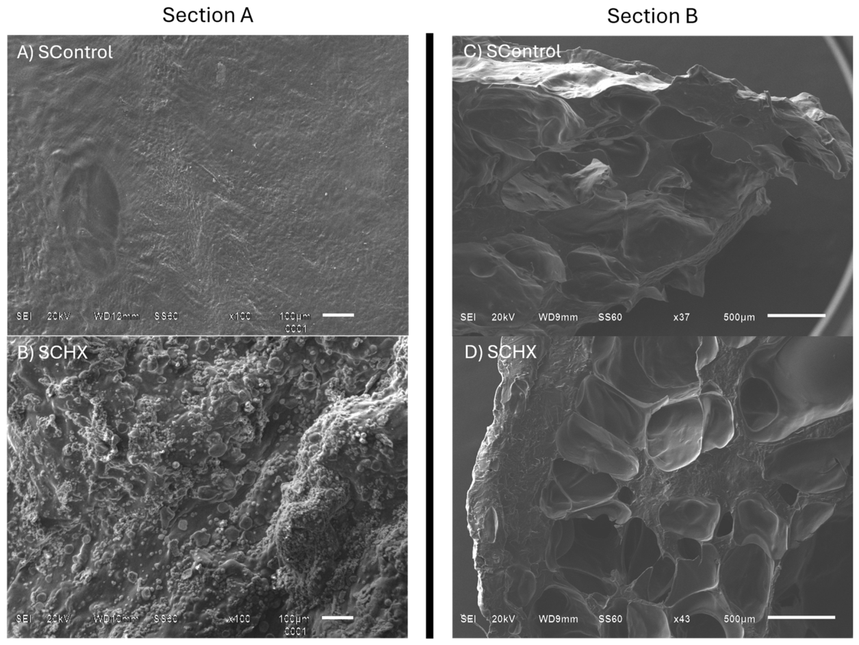
Figure 2.
FTIR-ATR spectra of (A) SCHX and (B) SControl.
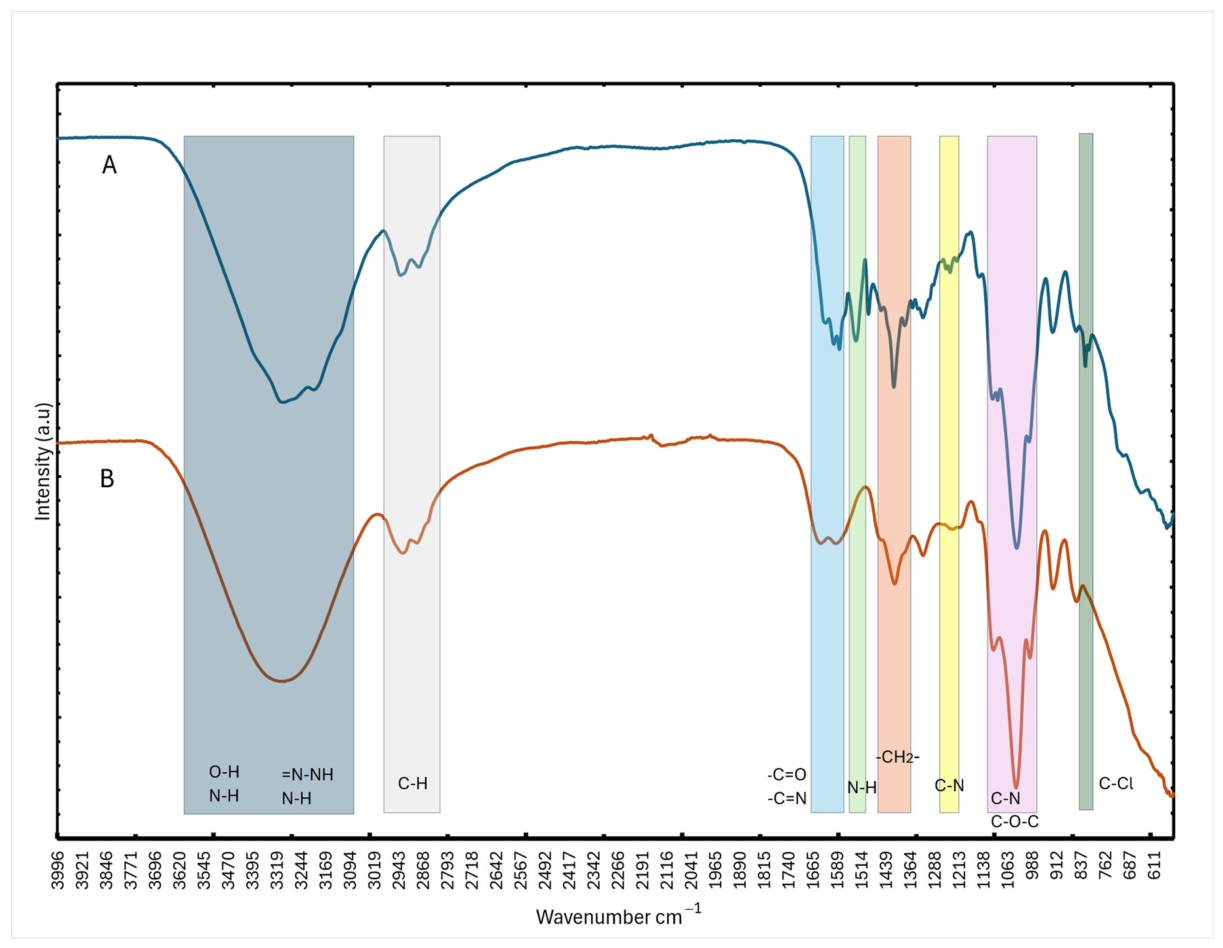
Figure 3.
Graphical presentation of surface pH kinetic of SControl and SCHX.
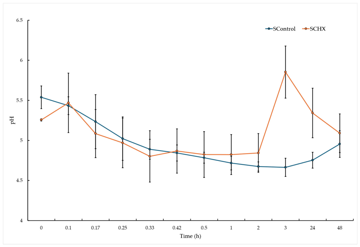
Figure 4.
Graphical presentation of water sorption behavior of SControl and SCHX.
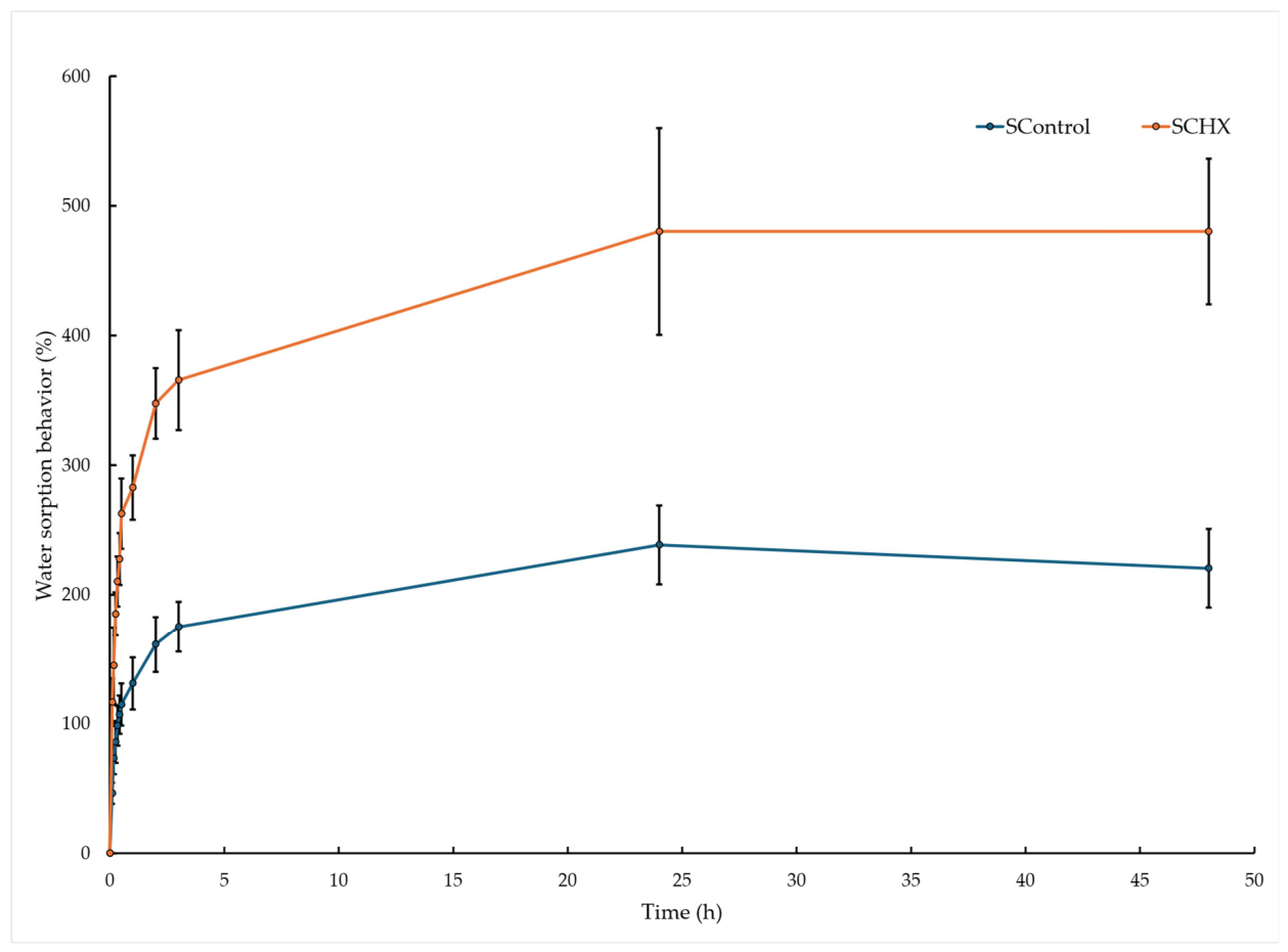
Figure 5.
SCHX reduces activation in human neutrophils. (A) CD11b expression in human neutrophils treated with LPS, SControl or SCHX biomaterials, or left unstimulated. The histogram shows CD11b expression in neutrophils (10,000 events). (B) CD11b expression in neutrophils treated with LPS, control or CHX biomaterials, or left unstimulated. A one-way ANOVA was used for these experiments (n = 4). (C) CD69 expression in human neutrophils treated with LPS, SControl or SCHX biomaterials, or left unstimulated. The histogram shows CD69 expression in neutrophils (10,000 events). (D) CD69 expression in neutrophils treated with LPS, SControl or SCHX biomaterials, or left unstimulated. A one-way ANOVA was used for these experiments (n = 4).
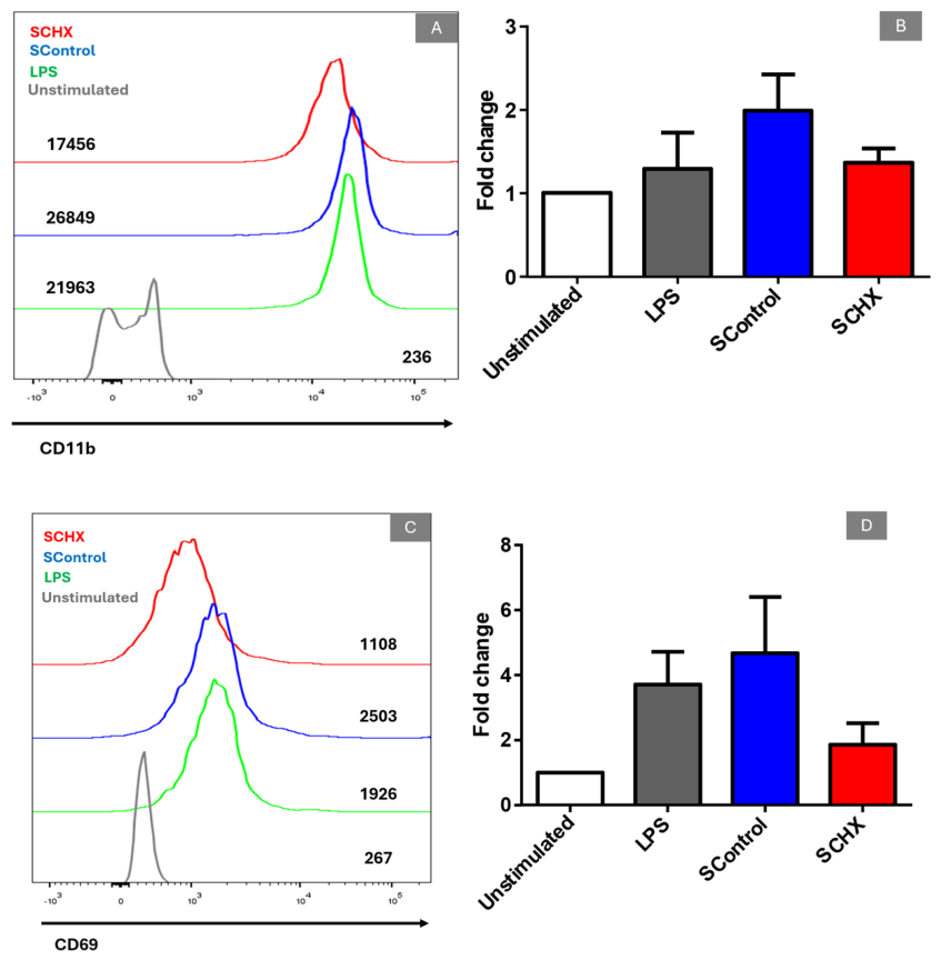
Table 1.
Sample designation and composition in terms of CMC, CHS, HA, G, Ca and CHX content.
| Sample |
SCHX (F/CMC/CHS/HA/G/Ca/CHX) (%) * |
SControl (F/CMC/CHS/HA/G/Ca) (%) * |
| CMC (p/p) | 1.25 | 1.25 |
| CHS (p/p) | 1.25 | 1.25 |
| HA (v/v) | 3.00 | 3.00 |
| G (v/v) | 3.00 | 3.00 |
| CHX (p/p) | 6.00 | 0.00 |
| CaCl2 (p/p) | 0.07 | 0.07 |
* Percentage of CMC, CHS, HA and G are based on the solution, and percentage of CHX and CaCl2 was based on biopolymers.
Table 2.
Overview of characteristics of CMC/CHS/HA/G/Ca (SControl) and CMC/CHS/HA/G/Ca/CHX (SCHX) sponges, including mass (mg ± SD) (n = 10), surface pH (mean ± SD) at 24 h (n = 10), moisture sorption after 10 days (n = 10), water sorption at 48 h (% ± SD) (n = 10), swelling index (mean ± SD) (n = 10), blood coagulation time (PT and TT) (values are given as s ± SD) (n = 3) and hemolysis (% ± SD) (n = 3).
|
SControl (CMC/CHS/HA/G/Ca) |
SCHX (CMC/CHS/HA/G/Ca/CHX) |
p Value | ||
| Mass (g ± SD) | 0.75 ± 0.07 | 0.83 ± 0.19 | a0.188 | |
| Surface pH (mean ± SD) | 5.43 ± 0.11 | 5.47 ± 0.37 | b0.774 | |
| Moisture sorption (% ± SD) | 40.77 ± 10.07 | 38.43 ± 1.93 | b0.488 | |
| Water sorption (% ± SD) | 238.58 ± 30.42 | 480.23 ± 56.21 | a<0.001 | |
| Swelling index (mean ± SD) | 0.46 ± 0.08 | 1.16 ± 0.18 | b<0.001 | |
| Disintegration time (h) | 48h | 144h | ||
| Blood coagulation time | PT (s ± SD) | 13.20 ± 0.60 | 17.10 ± 1.30 | |
| TT (s ± SD) | 29.20 ± 2.90 | 26.00 ± 3.90 | ||
| Hemolysis (%) | 22.67 | 0.00 | ||
aPrueba t de Student, bPrueba t de Welch, cW.S. at 24h, dW.S. at 48h.
Disclaimer/Publisher’s Note: The statements, opinions and data contained in all publications are solely those of the individual author(s) and contributor(s) and not of MDPI and/or the editor(s). MDPI and/or the editor(s) disclaim responsibility for any injury to people or property resulting from any ideas, methods, instructions or products referred to in the content. |
© 2026 by the authors. Licensee MDPI, Basel, Switzerland. This article is an open access article distributed under the terms and conditions of the Creative Commons Attribution (CC BY) license.
Copyright: This open access article is published under a Creative Commons CC BY 4.0 license, which permit the free download, distribution, and reuse, provided that the author and preprint are cited in any reuse.