

Submitted:
30 September 2026
Posted:
01 October 2026
You are already at the latest version
Abstract
Multi-walled carbon nanotubes (MWCNTs) can modulate plant growth and development, but the signaling mechanisms underlying their effects on organogenesis remain unclear. Here, we found that MWCNTs promote adventitious root (AR) formation and that RBOH-dependent hydrogen peroxide (H₂O₂) production plays a key role in this process in cucumber (Cucumis sativus L.) and Arabidopsis thaliana. MWCNTs treatment promoted AR formation in cucumber hypocotyls. Concomitantly, endogenous H₂O₂ levels increased rapidly, along with enhanced CsRBOHD expression and NADPH oxidase activity, suggesting activation of an RBOH-dependent H₂O₂ response. Consistent with this, scavenging H₂O₂ or inhibiting NADPH oxidase markedly reduced MWCNTs-induced rooting, whereas exogenous H₂O₂ partially restored AR formation under NADPH oxidase inhibition. Genetic evidence further supported this relationship, as Atrbohd and Atrbohf mutants showed reduced responses to MWCNTs but remained responsive to exogenous H₂O₂, suggesting that RBOH-mediated H₂O₂ generation is a critical step in MWCNTs-induced adventitious rooting. In parallel, MWCNTs and H₂O₂ treatments enhanced the expression of genes associated with adventitious root development. Together, these findings support a model in which MWCNTs promote adventitious rooting through RBOH-dependent H₂O₂ signaling, providing new insight into nanomaterial-mediated regulation of plant development.
Keywords:
MWCNTs
; adventitious root
; cucumber
; NADPH oxidase
; H₂O₂
1. Introduction
Over the past decade, engineered carbon-based nanomaterials—particularly multi-walled carbon nanotubes (MWCNTs)—have attracted growing interest in agriculture and plant biotechnology due to their unique tubular graphitic structures, high electrical conductivity, and remarkable cellular permeability [1,2,3]. Previous studies have demonstrated that carbon nanotubes can enter plant cells through endocytosis, aquaporins, or by creating new pores in the cell wall [4]. Low physiological concentrations of carbon nanomaterials have been demonstrated to stimulate seed germination, enhance photosynthetic performance, accelerate nutrient absorption, and promote root elongation across various crop species [5,6,7,8,9]. Similarly, both single- and multi-walled carbon nanotubes promote rice root growth by regulating root growth-related gene expression and inducing epigenetic changes in the root meristem [10]. MWCNTs have also been shown to enhance tobacco cell growth by upregulating genes related to cell division and water transport [11]. In addition to nanomaterials, endogenous signaling molecules such as gasotransmitters also regulate root system development [12,13], suggesting that diverse external and internal cues may converge on common downstream pathways. However, high-dose applications or improper chemical functionalization can induce cell wall damage, excessive reactive oxygen species (ROS) accumulation, and phytotoxicity [4,14,15].
Adventitious root (AR) formation is a crucial post-embryonic organogenesis process essential for vegetative propagation, clonal survival, and nursery establishment in horticultural crops [16,17]. AR initiation is orchestrated by a complex physiological signaling network governed by phytohormone crosstalk—primarily auxin—and reactive second messengers such as carbon monoxide (CO), methane (CH4), nitric oxide (NO), and hydrogen peroxide (H2O2) [12,18,19,20]. These small-molecule compounds enhance adventitious rooting by regulating antioxidant systems, water balance, and auxin transport [21]. The central role of auxin in the hormone cross-talk network controlling adventitious rooting has been extensively reviewed [22,23]. Specifically, transient physiological bursts of endogenous H2O2 function as indispensable secondary signals that trigger local cell division, cell wall loosening, and primordium activation during rhizogenesis [24,25]. In Arabidopsis hypocotyl cuttings, wound-induced ROS generated by RBOHD propagate through the tissue and regulate auxin biosynthesis and transport to initiate AR formation [26]. These H2O2-dependent processes do not operate in isolation; recent evidence indicates that H2O2 cooperates with other reactive messengers, particularly nitric oxide, to coordinate the sequential steps of adventitious root development [27].
In plant cells, plasma membrane-localized NADPH oxidases, designated as respiratory burst oxidase homologs (RBOHs), serve as the primary enzymatic engine generating apoplastic superoxide radicals, which rapidly dismutate into H2O2 [28,29,30]. RBOH-derived H2O2 signaling plays fundamental roles in stomatal regulation, cell expansion, root hair growth, and environmental stress adaptation [31,32]. In Arabidopsis thaliana, AtrbohD and AtrbohF represent the main catalytic subunits mediating stress- and hormone-induced ROS signaling cascades [28,33,34]. Downstream of ROS signals, cell cycle progression and auxin responsiveness drive the transition of differentiated hypocotyl cells into active root primordia [35,36].Crucially, recent studies in cucumber explants have demonstrated that gasotransmitters regulate adventitious rooting by modulating a conserved suite of organogenic marker genes, including the DNA replication licensing gene CsCDC6, the molecular chaperone CsDNAJ-1, auxin-responsive Aux/IAA genes (CsAux22B-like, CsAux22D-like), and calcium-dependent protein kinases (CsCDPK1, CsCDPK5) [12,19]. These genes have been demonstrated to be conserved downstream targets in various hormone- and gasotransmitter-induced rooting pathways [12,20]. Given the central role of RBOH-derived ROS in plant signaling, it is plausible that nanomaterial-induced ROS production might also converge on this conserved pathway. The crucial role of ROS and related metabolic signals in mediating plant responses to abiotic stimuli has been well documented, including salt-induced ABA accumulation [37] and heat-induced methylglyoxal signaling [38]. However, whether nanomaterial-induced ROS signaling follows a similar RBOH-dependent mechanism during adventitious rooting remains largely unexplored.
In the present study, we systematically investigated the physiological, cellular, and genetic mechanisms by which MWCNTs promote adventitious root formation in cucumber (Cucumis sativus) explants and Arabidopsis thaliana NADPH oxidase mutants (AtrbohD and AtrbohF). Our data reveal that MWCNTs physically penetrate hypocotyl tissues and accumulate in intercellular spaces and primary cell walls, where they activate plasma membrane-localized RBOH, leading to a rapid, biphasic H2O2 burst. This oxidative signal subsequently drives the upregulation of downstream cell cycle regulatory genes (CsCDC6, CsDNAJ-1) and auxin/calcium signaling genes (CsAux22, CsCDPKs), thus promoting root primordium initiation and elongation. Critically, genetic validation using AtrbohD and AtrbohF mutants confirms that the loss of RBOH function abolishes MWCNT-induced H2O2 accumulation and adventitious rooting, defects that are partially rescued by exogenous H2O2 supplementation. Collectively, these findings establish a conserved RBOH–H2O2 signaling cascade governing MWCNT-induced organogenesis, offering novel insights into nanomaterial–plant interactions.
2. Results
2.1. MWCNTs Promote Adventitious Root Formation in Cucumber in Association with H2O2 Signaling
The morphology and structural characteristics of the MWCNTs were first examined (Figure S1). TEM images showed entangled, hollow tubular structures characteristic of MWCNTs, with outer and inner diameters of approximately 10 and 4.5 nm, respectively (Figure S1A, B). XRD analysis revealed a prominent diffraction peak at approximately 2θ = 26°, corresponding to the (002) plane of graphitic carbon, together with a weaker reflection around 43° assigned to the (100)/(101) planes (Figure S1C). Raman spectra showed characteristic D and G bands at approximately 1350 and 1580 cm−1, respectively (Figure S1D), confirming the graphitic structure of the MWCNTs.
The effects of MWCNTs on adventitious root (AR) formation were then examined in NPA-pretreated cucumber hypocotyl explants. Treatment with MWCNTs at 0.05–50 mg·L−1 promoted AR formation to varying degrees compared with the control, with the strongest response observed at 5 mg·L−1 (Figure 1A–C). Both AR numbers and root length increased with MWCNTs concentration up to 5 mg·L−1, whereas the promotive effect declined at 50 mg·L−1. SNP, a known promoter of adventitious rooting, was included as a positive control and similarly enhanced AR formation (Figure 1A–C). These results indicate that MWCNTs promote adventitious rooting in cucumber explants within an appropriate concentration range. Given the established role of H2O2 in adventitious root development, we next examined whether H2O2 contributes to the promotive effect of MWCNTs. Both MWCNTs and exogenous H2O2 significantly increased AR number and root length, whereas these effects were markedly reduced by co-treatment with DMTU or AsA (Figure 1D,E). Together, these results suggest that H2O2 is involved in the promotion of adventitious root formation by MWCNTs in cucumber explants.
2.2. Uptake and Tissue Localization of MWCNTs in Cucumber Hypocotyls
To examine the uptake and distribution of MWCNTs in cucumber hypocotyl tissues, Rhodamine B-labeled MWCNTs (MWCNTs/Rho B) were visualized by confocal fluorescence microscopy, together with TEM observation of their ultrastructural localization (Figure 2). Cross-sections from MWCNTs/Rho B-treated hypocotyls showed clear red fluorescence throughout the basal tissues, including the epidermal region, cortical tissues, and vascular region, whereas only weak background fluorescence was observed in the control (Figure 2A). Consistently, relative fluorescence intensity was significantly higher in MWCNTs/Rho B-treated tissues than in the control (Figure 2B). Ultrastructural TEM observations further revealed electron-dense tubular structures associated with MWCNTs in the cell walls (CW) and intercellular spaces (IS) of hypocotyl cells, whereas no comparable structures were observed in the untreated control (Figure 2C). These observations indicate that MWCNTs can enter cucumber hypocotyl tissues and are mainly localized within apoplastic regions, including cell walls and intercellular spaces.
2.3. MWCNTs Induce Rapid and Sustained H2O2 Accumulation in Cucumber Hypocotyls
Since H2O2 scavenging reduced MWCNTs-induced adventitious rooting, we next examined the temporal changes in endogenous H2O2 levels following MWCNTs treatment (Figure 3). MWCNTs induced a rapid increase in H2O2 contents at the hypocotyl base, with a marked elevation detected during the early treatment period and the highest level observed at approximately 6 h (Figure 3A). Although H2O2 levels gradually declined thereafter, they remained significantly higher than those of the corresponding controls at later time points, including 48 h. These results indicate that MWCNTs treatment induces an early and sustained H2O2 response in cucumber hypocotyl tissues. The increase in H2O2 was further supported by H2DCF-DA fluorescence imaging. MWCNTs- and H2O2-treated hypocotyl bases showed markedly stronger green fluorescence than the control, whereas DMTU or AsA treatment substantially reduced the fluorescence signal (Figure 3B, C). Similarly, co-treatment with DMTU or AsA markedly decreased the fluorescence induced by MWCNTs or exogenous H2O2. Together, these results show that MWCNTs treatment is associated with enhanced H2O2 accumulation in the basal region of cucumber hypocotyls, supporting the involvement of H2O2 in MWCNT-induced adventitious root formation.
2.4. NADPH Oxidase Contributes to MWCNT-Induced H2O2 Production and Adventitious Root Formation
To determine the subcellular sites of H2O2 production following MWCNTs treatment, CeCl3 cytochemical staining coupled with TEM was first performed. Compared with the control, MWCNTs-treated hypocotyl cells showed abundant electron-dense cerium perhydroxide deposits, mainly associated with the plasma membrane and adjacent cell wall regions (Figure 4A). This spatial distribution suggested that a plasma membrane-associated ROS-producing system may contribute to MWCNTs-induced H2O2 accumulation. We therefore examined the involvement of respiratory burst oxidase homolog D (RBOHD), a plasma membrane-localized NADPH oxidase. MWCNTs treatment significantly increased CsRBOHD transcript abundance and NADPH oxidase activity, while DPI, an inhibitor of NADPH oxidase activity, markedly reduced enzyme activity without preventing the MWCNTs-induced increase in CsRBOHD expression (Figure 4B,C). Consistent with these responses, DPI markedly reduced the increases in adventitious root number and root length induced by MWCNTs (Figure 4D–F). The rooting phenotype of the MWCNTs + DPI group was similar to that of the DPI-alone group, indicating that inhibition of NADPH oxidase largely attenuated the promotive effect of MWCNTs on adventitious rooting. By contrast, exogenous H2O2 partially restored root formation in the presence of DPI, suggesting that H2O2 can act downstream of NADPH oxidase. Together, these results indicate that RBOHD-dependent NADPH oxidase contributes to MWCNT-induced H2O2 production and promotes adventitious root formation through an H2O2-dependent pathway.
2.5. RBOH Is Genetically Required for MWCNT-Induced Adventitious Rooting
Having established the involvement of RBOH-dependent NADPH oxidase in MWCNTs-induced H2O2 production, we next examined whether this signaling process was associated with changes in adventitious rooting-related gene expression. MWCNTs and exogenous H2O2 treatments significantly increased the transcript levels of CsCDC6, CsAux22B-like, CsAux22D-like, CsDNAJ-1, CsCDPK1, and CsCDPK5 (Figure S2). In contrast, DPI markedly attenuated the MWCNTs-induced upregulation of these genes. A similar reduction was observed when DPI was combined with H2O2. These expression patterns further support a link between RBOH-dependent H2O2 signaling and the transcriptional responses associated with adventitious root development.
To further test the role of RBOH genetically, adventitious root formation was examined in Col-0, Atrbohd and Atrbohf mutants (Figure 5). Under control conditions, both mutants showed reduced adventitious rooting compared with Col-0. MWCNTs treatment increased adventitious root formation in Col-0, whereas little or no response to MWCNTs was observed in either Atrbohd or Atrbohf, as reflected by adventitious root number and total root length (Figure 5A–C). By contrast, exogenous H2O2 markedly promoted root formation in both mutant backgrounds, partially restoring the reduced rooting phenotype. These results indicate that the response to MWCNTs depends on functional RBOHD/F. Consistent with the rooting phenotypes, MWCNTs treatment increased H2DCF-DA fluorescence and endogenous H2O2 levels in Col-0 but failed to induce comparable responses in the Atrbohd and Atrbohf mutants (Figure 5D–F). By contrast, exogenous H2O2 remained effective in both mutants, promoting adventitious rooting and restoring H2O2 signals. Together with the pharmacological and biochemical evidence obtained in cucumber, these genetic data support a signaling sequence in which RBOH-dependent H2O2 production contributes to MWCNTs-induced adventitious root formation.
3. Discussion
The integration of nanomaterials into crop propagation represents an innovative strategy for modern agriculture. However, a comprehensive understanding of the underlying physiological and molecular pathways is essential for their safe and effective application. In this study, we demonstrate that MWCNTs promote adventitious rooting in cucumber through an RBOH-mediated H2O2 signaling pathway. It is well established that root system architecture is highly plastic and is modulated by diverse endogenous and environmental signals, including gasotransmitters, hormones, and osmotic stress [12,13,39]. While previous studies have also established that ROS and metabolic signals are central to plant stress responses, such as salt-induced ABA accumulation [37] and heat-induced methylglyoxal-mediated protein modification [38], our findings reveal a distinct role for RBOH-derived H2O2 in promoting developmental organogenesis rather than stress adaptation. This highlights the versatile nature of ROS signaling in integrating external stimuli into diverse physiological outputs. Our physical characterizations confirmed that MWCNTs possess stable graphitic tubular structures (Figure S1). Confocal and TEM tracking provided direct evidence that MWCNTs penetrate outer hypocotyl tissues and lodge within cell walls and intercellular spaces (Figure 2). This cellular localization is consistent with previous observations that carbon nanotubes can traverse plant cell walls via endocytosis or create physical micro-channels [1,3].This physical presence may lead to local mechanical stress or directly interact with plasma membrane proteins, potentially serving as the initial trigger for the downstream signaling cascade.
H2O2 is a well-established secondary messenger in plant growth, stress adaptations, and hormone signaling networks [16]. In auxin-induced adventitious rooting, transient H2O2 accumulation is required to stimulate cell division in root primordia [19,20].Here, MWCNTs exposure induced a rapid H2O2 burst in cucumber hypocotyls (Figure 3). The complete reversal of MWCNTs-induced root promotion by H2O2 scavengers (DMTU and AsA) demonstrates that endogenous H2O2 is indispensable for MWCNTs-mediated root organogenesis. Using CeCl3 cytochemical localization and enzyme inhibitors, we definitively demonstrated that plasma membrane-bound NADPH oxidase (RBOH) serves as the primary enzymatic engine for MWCNTs-induced H2O2 generation (Figure 4). These results align closely with the central role of RBOH in hormone- and stress-induced ROS bursts previously documented in auxin-mediated rooting [19] and gasotransmitter signaling [20]. The fact that MWCNTs failed to rescue the rooting defect in Atrbohd/f mutants, combined with the recovery of this phenotype by exogenous H2O2, unequivocally confirms the evolutionary necessity of RBOH in mediating the signals from the nanoparticles. Furthermore, MWCNTs upregulated CsRBOH transcript levels and enhanced NADPH oxidase activity, whereas the NADPH oxidase inhibitor DPI abolished both H2O2 generation and root induction (Figure 4). To provide definitive genetic proof, we evaluated Arabidopsis NADPH oxidase mutants (Atrbohd and Atrbohf). Loss of RBOH function rendered these explants unresponsive to MWCNTs-induced rooting, a defect that could be rescued by exogenous H2O2 supplementation (Figure 5). These genetic results firmly establish the necessity and evolutionary conservation of the RBOH-H2O2 signaling axis in MWCNTs-induced organogenesis. Notably, this signaling cascade shares intriguing similarities with stress-induced ROS production, where mechanical stimuli or ion fluxes activate plasma membrane-localized RBOHs. Previous studies have shown that mechanosensitive channels (e.g., MCA1/MCA2) can initiate Ca2+ influx under cold stress [40], and Ca2+-dependent protein kinases (CDPKs) are known to activate RBOHs in various plant species. Our observation that MWCNTs treatment strongly upregulated CsCDPK1 and CsCDPK5 (Figure S2) further supports the hypothesis that MWCNTs may act as a physical trigger, potentially initiating a mechanosensitive pathway that converges on RBOH activation. This is consistent with our previous findings that CDPKs serve as key nodes in H2O2-mediated root development [12]. However, whether MWCNTs directly activate mechanosensitive channels or modulate membrane tension to trigger the Ca2+-CDPK-RBOH cascade remains to be experimentally validated in future studies.
Downstream of ROS generation, adventitious rooting requires precise cell cycle activation and auxin signaling [20]. Our qPCR analyses confirmed that MWCNTs significantly upregulate cell cycle regulatory genes (CsCDC6, CsDNAJ-1) and auxin/calcium-responsive genes (CsAux22B-like, CsAux22D-like, CsCDPK1, CsCDPK5) (Figure S2). The suppression of these inductive responses by DPI further confirms that MWCNTs regulate downstream molecular machinery via an upstream RBOH-H2O2 signaling cascade. While this nanomaterial-based approach holds great promise for crop propagation, its potential long-term environmental accumulation, stability, and the risk of trophic transfer in agricultural ecosystems must be carefully evaluated in future studies [1]. Additionally, future research should explore how MWCNTs interact with other environmental factors (e.g., temperature and light) to modulate plant developmental plasticity, as our recent work has demonstrated that light and temperature coordinately regulate phytomelatonin synthesis to maintain plant morphogenesis [41] and that 14-3-3 proteins suppress ethylene-mediated primary root growth inhibition [42].
4. Materials and Methods
4.1. Physicochemical Characterization of MWCNTs
Multi-walled carbon nanotubes (MWCNTs; outer diameter, 10 ± 1 nm; inner diameter, approximately 4.5 nm; length, 3–6 μm, according to the manufacturer’s specifications) were purchased from Sigma-Aldrich (USA). To prepare homogeneous suspensions for plant treatments, MWCNTs were dispersed in 0.01% (w/v) agar solution by bath ultrasonication (100 W, 40 kHz) for 2 h. The morphology of the MWCNTs was examined by transmission electron microscopy (TEM; Hitachi H-7650). The crystalline graphitic structure was characterized by X-ray diffraction (XRD; Rigaku D/max-2500), while graphitic structure and defect characteristics were further evaluated by Raman spectroscopy (Jobin Yvon HR800) using a 532 nm excitation laser.
4.2. Plant Materials, Growth Conditions, and Explant Preparation
Cucumber (Cucumis sativus L. cv. ‘Lufeng’) seeds were surface-sterilized with 0.1% (w/v) HgCl2 for 8 min, rinsed thoroughly with sterile distilled water, and germinated on moist filter paper in the dark at 25 ± 1 °C for 5 days. Etiolated hypocotyl explants (5 cm in length) were excised, and their primary root systems were removed. Adventitious rooting was induced in an auxin-depleted model, in which explants were pre-incubated with 10 μM N-1-naphthylphthalamic acid (NPA; a polar auxin transport inhibitor) for 12 h, following established protocols [12,20]. Arabidopsis thaliana wild-type (Col-0) and T-DNA insertion mutant lines Atrbohd (SALK_120299) and Atrbohf (SALK_034674) were obtained from the Arabidopsis Biological Resource Center (ABRC). Seeds were surface-sterilized, stratified at 4 °C for 3 days, and grown vertically on half-strength Murashige and Skoog (1/2 MS) agar medium under a 16/8 h light/dark photoperiod at 22 ± 1 °C. The genotyping and phenotypic characterization of Arabidopsis mutants were performed following the methods described in our recent work [41,42].
4.3. Experimental Treatments
Cucumber explants were divided into non-depleted and depleted groups. Non-depleted explants, which were not subjected to the depletion treatment described above, were maintained in distilled water and served as a physiological reference. Depleted explants were transferred to glass test tubes containing distilled water alone (control), various concentrations of MWCNTs (0.05, 0.5, 5, and 50 mg·L−1), or 10 μM sodium nitroprusside (SNP). For inhibitor and scavenger assays, depleted explants were co-treated with H2O2 scavengers [dimethylthiourea (DMTU, 5 mM) or ascorbic acid (AsA, 1 mM)] or the NADPH oxidase inhibitor diphenyleneiodonium (DPI, 2 μM), in combination with the indicated treatments. Exogenous H2O2 was applied at 1 mM for rescue experiments. After 4 days of treatment, adventitious roots longer than 1 mm were counted, and root length was measured for each explant.
4.4. Rhodamine B Labeling and TEM Cell-Penetration Imaging
To track physical distribution, MWCNTs were non-covalently labeled with Rhodamine B (Rho B). MWCNT suspensions were incubated with 0.1 mM Rho B for 12 h, followed by extensive washing and centrifugation to remove unbound dye. Transverse sections from the basal region of treated cucumber hypocotyls were examined using a Zeiss LSM 710 laser scanning confocal microscope. For ultrastructural observation, 2 mm segments from the hypocotyl bases were fixed in 2.5% (v/v) glutaraldehyde in 100 mM phosphate buffer (pH 7.2) for 12 h, post-fixed in 1% (w/v) OsO4, dehydrated through a graded ethanol series, embedded in Epon 812 resin, and sectioned for TEM examination.
4.5. Quantification and Cellular Visualization of H2O2
Endogenous H2O2 content in hypocotyl bases was quantified spectrophotometrically using the xylenol orange method, as described by Chen et al. [43]. In situ accumulation of H2O2 was visualized using 2,7-dichlorodihydrofluorescein diacetate (H2DCF-DA; 20 μM). Stained tissue bases were incubated for 30 min in the dark, washed with buffer, and imaged via confocal laser scanning microscopy (excitation: 488 nm, emission: 525 nm). Relative fluorescence intensity was quantified using ImageJ software.
The subcellular localization of H2O2 was evaluated using cerium chloride (CeCl3) cytochemistry. Tissues were incubated in 5 mM CeCl3 dissolved in 50 mM MOPS buffer (pH 7.2) for 1 h, followed by standard glutaraldehyde/OsO4 fixation and ultrathin sectioning to observe electron-dense cerium perhydroxide precipitates via TEM.
4.6. NADPH Oxidase Assays
Plasma membrane NADPH oxidase activity was determined according to the method described by Kabała et al. [44], with minor modifications. Plasma membrane fractions were isolated from the basal regions of cucumber hypocotyls, and NADPH oxidase activity was assayed by monitoring the superoxide-dependent reduction of XTT (2,3-bis(2-methoxy-4-nitro-5-sulfophenyl)-2H-tetrazolium-5-carboxanilide inner salt). The reaction mixture contained 0.5 mM XTT and 0.1 mM NADPH as the electron donor. XTT reduction was monitored spectrophotometrically at 470 nm in parallel reactions in the presence or absence of 50 U CuZn-superoxide dismutase (CuZn-SOD). NADPH oxidase activity was calculated from the SOD-sensitive rate of XTT reduction and normalized to the protein content of the plasma membrane fraction.
4.7. Quantitative Real-Time PCR (qPCR)
Total RNA was extracted from hypocotyl tissue bases (0–0.5 cm from the cutting edge) using the Plant RNA Kit (Omega Bio-tek). Reverse transcription was performed using the PrimeScript RT Reagent Kit (Takara). Quantitative real-time PCR (qPCR) was conducted on a StepOnePlus Real-Time PCR System (Applied Biosystems) using SYBR Premix Ex Taq (Takara). Relative transcript abundance was calculated using the 2−ΔΔCt method and normalized against internal reference genes CsCACS and CsTIP41, as validated in our previous work [12]. Gene-specific primers for CsCDC6, CsAux22B-like, CsAux22D-like, CsDNAJ-1, CsCDPK1, CsCDPK5, and CsRBOH are listed in Table S1.
4.8. Statistical Analysis
Data were collected from at least three independent biological replicates and expressed as mean ± standard deviation (SD) or standard error (SE), following the statistical framework established in our previous studies [41,42]. For single-factor comparisons (e.g., Figure 2B and Figure 3A), Student’s t-test was used. For multiple group comparisons, statistical significance was determined using one-way analysis of variance (ANOVA) followed by Duncan’s multiple range test. All analyses were performed with SPSS 22.0 software, and a value of P < 0.05 was considered statistically significant.
5. Conclusions
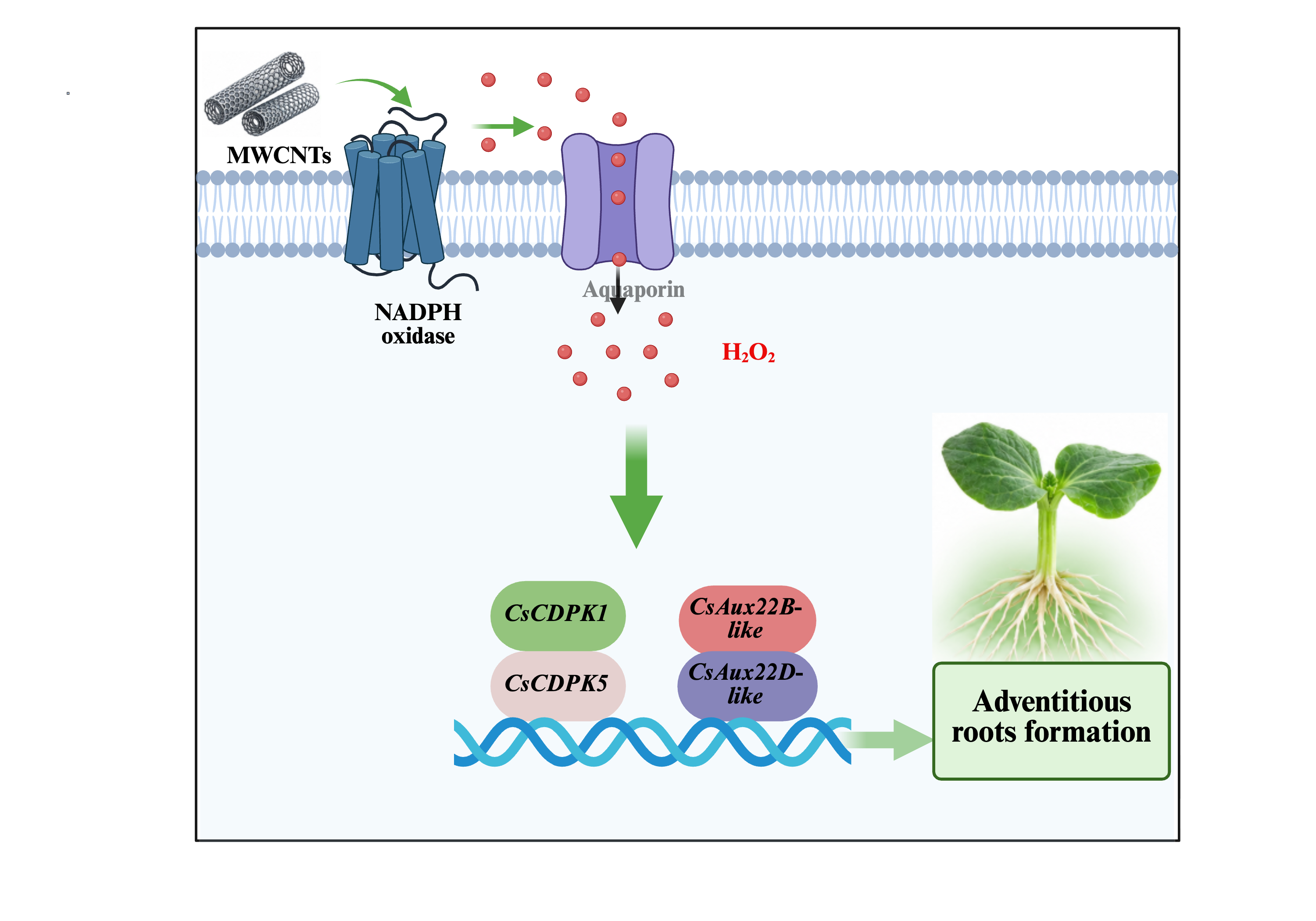
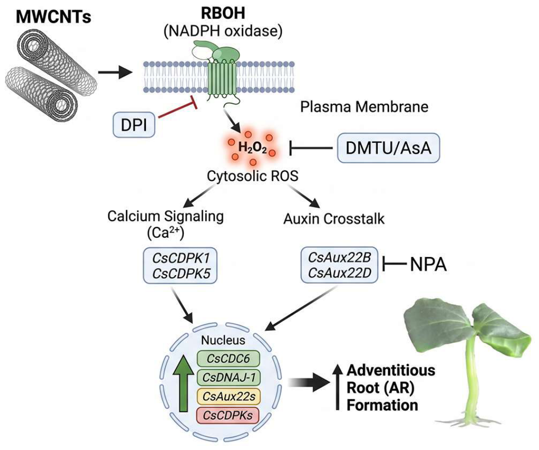
In summary, this study demonstrates that multi-walled carbon nanotubes (MWCNTs) promote adventitious root (AR) formation in cucumber and Arabidopsis thaliana. MWCNTs were detected in cucumber hypocotyl tissues, where they were mainly localized in the cell wall and intercellular spaces. MWCNTs treatment enhanced CsRBOHD expression and NADPH oxidase activity, leading to increased endogenous H2O2 accumulation. Pharmacological inhibition of NADPH oxidase or scavenging of H2O2 markedly reduced the promotive effect of MWCNTs on adventitious rooting, whereas exogenous H2O2 partially restored root formation when NADPH oxidase activity was inhibited. MWCNTs- and H2O2-induced rooting was also associated with altered expression of genes related to adventitious root development. Consistently, Atrbohd and Atrbohf mutants showed a reduced response to MWCNTs, while exogenous H2O2 retained its ability to promote adventitious rooting in these mutants. Together, these findings support a regulatory pathway in which MWCNTs promote adventitious root formation through RBOH-dependent H2O2 production (Figure 6). The results provide new insight into the interaction between carbon nanomaterials and endogenous ROS signaling during plant root development and may contribute to the development of nanomaterial-based approaches for vegetative propagation.
Supplementary Materials
The following supporting information can be downloaded at the website of this paper posted on Preprints.org. Figure S1. Structural characterization of MWCNTs; Figure S2. Expression profiles of adventitious rooting-related genes in cucumber hypocotyls; Table S1: Primer sequences used for RT-qPCR analysis.
Author Contributions
Conceptualization, M.X., S.W., Z.X. and F.D.; methodology, M.X. and S.W.; software, Y.Z.; validation, Y.R. and S.Z.; formal analysis, Y.Z. and Y.L.; investigation, M.X., S.W. and Q.T.; resources, Y.R. and S.Z.; data curation, Y.L.; writing—original draft preparation, M.X. and S.W.; writing—review and editing, Z.X. and F.D.; visualization, Y.R.; supervision, Z.X. and F.D.; project administration, M.X.; funding acquisition, Z.X. All authors have read and agreed to the published version of the manuscript.
Funding
This research was funded by the Hubei Key Laboratory of Biological Resources Protection and Utilization (Hubei Minzu University), grant numbers BS24045 and KYPT01.
Data Availability Statement
The data presented in this study are available on request from the corresponding author. The raw data supporting the conclusions of this article will be made available by the authors on reasonable request.
Acknowledgments
The authors thank the Hubei Key Laboratory of Biological Resources Protection and Utilization (Hubei Minzu University) for providing the experimental platform and technical support throughout this research. We also extend our sincere thanks to all laboratory colleagues for their continuous assistance in sample collection, experimental operation, and data sorting.
Conflicts of Interest
The authors declare no conflicts of interest. The funders had no role in the design of the study; in the collection, analyses, or interpretation of data; in the writing of the manuscript; or in the decision to publish the results.
References
- Lowry, G.V.; Avellan, A.; Gilbertson, L.M. Opportunities and Challenges for Nanotechnology in the Agri-Tech Revolution. Nat. Nanotechnol. 2019, 14, 517–522. [Google Scholar] [CrossRef]
- Cao, Z.Y.; Zhou, H.; Kong, L.S.; Li, L.N.; Wang, R.; Shen, W.B. A Novel Mechanism Underlying Multi-Walled Carbon Nanotube-Triggered Tomato Lateral Root Formation: The Involvement of Nitric Oxide. Nanoscale Res. Lett. 2020, 15, 49. [Google Scholar] [CrossRef]
- Zhao, G.; Zhao, Y.Y.; Lou, W.; Abdalmegeed, D.; Guan, R.Z.; Shen, W.B. Multi-Walled Carbon Nanotubes Can Promote Brassica Napus L. and Arabidopsis Thaliana L. Root Hair Development through Nitric Oxide and Ethylene Pathways. Int. J. Mol. Sci. 2020, 21, 9109. [Google Scholar] [CrossRef]
- Rico, C.M.; Majumdar, S.; Duarte-Gardea, M.; Peralta-Videa, J.R.; Gardea-Torresdey, J.L. Interaction of Nanoparticles with Edible Plants and Their Possible Implications in the Food Chain. J. Agric. Food Chem. 2011, 59, 3485–3498. [Google Scholar] [CrossRef]
- Ghorbanpour, M.; Hadian, J. Multi-Walled Carbon Nanotubes Stimulate Callus Induction, Secondary Metabolites Biosynthesis and Antioxidant Capacity in Medicinal Plant Satureja Khuzestanica Grown in Vitro. Carbon 2015, 94, 749–759. [Google Scholar] [CrossRef]
- Tripathi, D.K.; Shweta; Singh, S.; Singh, S.; Pandey, R.; Singh, V.P.; Sharma, N.C.; Prasad, S.M.; Dubey, N.K.; Chauhan, D.K. An Overview on Manufactured Nanoparticles in Plants: Uptake, Translocation, Accumulation and Phytotoxicity. Plant Physiol. Biochem. 2017, 110, 2–12. [Google Scholar] [CrossRef]
- Joshi, A.; Kaur, S.; Singh, P.; Dharamvir, K.; Nayyar, H.; Verma, G. Tracking Multi-Walled Carbon Nanotubes inside Oat (Avena Sativa L.) Plants and Assessing Their Effect on Growth, Yield, and Mammalian (Human) Cell Viability. Appl. Nanosci. 2018, 8, 1399–1414. [Google Scholar] [CrossRef]
- Chen, J.L.; Zeng, X.Y.; Yang, W.J.; Xie, H.J.; Ashraf, U.; Mo, Z.W.; Liu, J.H.; Li, G.K.; Li, W. Seed Priming with Multiwall Carbon Nanotubes (MWCNTs) Modulates Seed Germination and Early Growth of Maize under Cadmium (Cd) Toxicity. J. Soil Sci. Plant Nutr. 2021, 21, 1793–1805. [Google Scholar] [CrossRef]
- Ma, L.; Kong, L.L.; Gui, R.F.; Yang, X.J.; Zhang, J.W.; Gong, Q.; Qin, D.J.; Zhuang, M.S.; Ashraf, U.; Mo, Z.W. Application of Hydrogen-Rich Water Modulates Physio-Biochemical Functions and Early Growth of Fragrant Rice under Cd and Pb Stress. Environ. Sci. Pollut. Res. 2021, 28, 58558–58569. [Google Scholar] [CrossRef]
- Yan, S.H.; Zhang, H.; Huang, Y.; Tan, J.J.; Wang, P.; Wang, Y.P.; Hou, H.L.; Huang, J.; Li, L.J. Single-Wall and Multi-Wall Carbon Nanotubes Promote Rice Root Growth by Eliciting the Similar Molecular Pathways and Epigenetic Regulation. IET Nanobiotechnol 2016, 10, 222–229. [Google Scholar] [CrossRef]
- Khodakovskaya, M.V.; De Silva, K.; Biris, A.S.; Dervishi, E.; Villagarcia, H. Carbon Nanotubes Induce Growth Enhancement of Tobacco Cells. ACS Nano 2012, 6, 2128–2135. [Google Scholar] [CrossRef]
- Qi, F.; Xiang, Z.; Kou, N.; Cui, W.; Xu, D.; Wang, R.; Zhu, D.; Shen, W. Nitric Oxide Is Involved in Methane-Induced Adventitious Root Formation in Cucumber. Physiol. Plant. 2017, 159, 366–377. [Google Scholar] [CrossRef]
- Xiang, Z.X.; Li, W.; Lu, Y.T.; Yuan, T.T. Hydrogen Sulfide Alleviates Osmotic Stress-Induced Root Growth Inhibition by Promoting Auxin Homeostasis. Plant J. 2023, 114, 1369–1384. [Google Scholar] [CrossRef]
- Stampoulis, D.; Sinha, S.K.; White, J.C. Assay-Dependent Phytotoxicity of Nanoparticles to Plants. Environ. Sci. Technol. 2009, 43, 9473–9479. [Google Scholar] [CrossRef]
- Samadi, S.; Asgari Lajayer, B.; Moghiseh, E.; Rodríguez-Couto, S. Effect of Carbon Nanomaterials on Cell Toxicity, Biomass Production, Nutritional and Active Compound Accumulation in Plants. Environ. Technol. Innov. 2021, 21, 101323. [Google Scholar] [CrossRef]
- Bellini, C.; Pacurar, D.I.; Perrone, I. Adventitious Roots and Lateral Roots: Similarities and Differences. Annu. Rev. Plant Biol. 2014, 65, 639–666. [Google Scholar] [CrossRef]
- Steffens, B.; Rasmussen, A. The Physiology of Adventitious Roots. Plant Physiol. 2016, 170, 603–617. [Google Scholar] [CrossRef]
- Pagnussat, G.C.; Lanteri, M.L.; Lombardo, M.C.; Lamattina, L. Nitric Oxide Mediates the Indole Acetic Acid Induction Activation of a Mitogen-Activated Protein Kinase Cascade Involved in Adventitious Root Development. Plant Physiol. 2004, 135, 279–286. [Google Scholar] [CrossRef]
- Lanteri, M.L.; Pagnussat, G.C.; Lamattina, L. Calcium and Calcium-Dependent Protein Kinases Are Involved in Nitric Oxide- and Auxin-Induced Adventitious Root Formation in Cucumber. J. Exp. Bot. 2006, 57, 1341–1351. [Google Scholar] [CrossRef]
- Xuan, W.; Zhu, F.-Y.; Xu, S.; Huang, B.-K.; Ling, T.-F.; Qi, J.-Y.; Ye, M.-B.; Shen, W.-B. The Heme Oxygenase/Carbon Monoxide System Is Involved in the Auxin-Induced Cucumber Adventitious Rooting Process. Plant Physiol. 2008, 148, 881–893. [Google Scholar] [CrossRef]
- Deng, Y.Z.; Wang, C.L.; Wang, N.; Wei, L.J.; Li, W.F.; Yao, Y.D.; Liao, W.B. Roles of Small-Molecule Compounds in Plant Adventitious Root Development. Biomolecules 2019, 9, 420. [Google Scholar] [CrossRef]
- Pacurar, D.I.; Perrone, I.; Bellini, C. Auxin Is a Central Player in the Hormone Cross-Talks That Control Adventitious Rooting. Physiol. Plant. 2014, 151, 83–96. [Google Scholar] [CrossRef]
- Lakehal, A.; Bellini, C. Control of Adventitious Root Formation: Insights into Synergistic and Antagonistic Hormonal Interactions. Physiol. Plant. 2019, 165, 90–100. [Google Scholar] [CrossRef]
- Li, X.P.; Xu, Q.Q.; Liao, W.B.; Ma, Z.J.; Xu, X.T.; Wang, M.; Ren, P.J.; Niu, L.J.; Jin, X.; Zhu, Y.C. Hydrogen Peroxide Is Involved in Abscisic Acid-Induced Adventitious Rooting in Cucumber (Cucumis Sativus L.) under Drought Stress. J. Plant Biol. 2016, 59, 536–548. [Google Scholar] [CrossRef]
- Sharma, S.; Singh, H.P.; Batish, D.R.; Kohli, R.K. Nitric Oxide Induced Modulations in Adventitious Root Growth, Lignin Content and Lignin Synthesizing Enzymes in the Hypocotyls of Vigna Radiata. Plant Physiol. Biochem. 2019, 141, 225–230. [Google Scholar] [CrossRef]
- Huang, A.X. Reactive Oxygen Species Regulate Auxin Levels to Mediate Adventitious Root Induction in Arabidopsis Hypocotyl Cuttings. 2020, 62. [Google Scholar] [CrossRef]
- Roussos, P.A. Adventitious Root Formation in Plants: The Implication of Hydrogen Peroxide and Nitric Oxide. Antioxidants 2023, 12, 862. [Google Scholar] [CrossRef]
- Torres, M.A.; Dangl, J.L.; Jones, J.D.G. Arabidopsis Gp91phox Homologues AtrbohD and AtrbohF Are Required for Accumulation of Reactive Oxygen Intermediates in the Plant Defense Response. Proc. Natl. Acad. Sci. 2002, 99, 517–522. [Google Scholar] [CrossRef]
- Suzuki, N.; Miller, G.; Morales, J.; Shulaev, V.; Torres, M.A.; Mittler, R. Respiratory Burst Oxidases: The Engines of ROS Signaling. Curr. Opin. Plant Biol. 2011, 14, 691–699. [Google Scholar] [CrossRef]
- Guo, H.Y.; Bi, X.Y.; Wang, Z.P.; Jiang, D.; Cai, M.; An, M.N.; Xia, Z.H.; Wu, Y.H. Reactive Oxygen Species-Related Genes Participate in Resistance to Cucumber Green Mottle Mosaic Virus Infection Regulated by Boron in Nicotiana Benthamiana and Watermelon. Front. Plant Sci. 2022, 13, 1027404. [Google Scholar] [CrossRef]
- Foreman, J.; Demidchik, V.; Bothwell, J.H.F.; Mylona, P.; Miedema, H.; Torres, M.A.; Linstead, P.; Costa, S.; Brownlee, C.; Jones, J.D.G.; et al. Reactive Oxygen Species Produced by NADPH Oxidase Regulate Plant Cell Growth. Nature 2003, 422, 442–446. [Google Scholar] [CrossRef]
- Kwak, J.M.; Mori, I.C.; Pei, Z.M.; Leonhardt, N.; Torres, M.A.; Dangl, J.L.; Bloom, R.E.; Bodde, S.; Jones, J.D.G.; Schroeder, J.I. NADPH Oxidase AtrbohD and AtrbohF Genes Function in ROS-Dependent ABA Signaling in Arabidopsis. EMBO J. 2003, 22, 2623–2633. [Google Scholar] [CrossRef]
- Miller, G.; Schlauch, K.; Tam, R.; Cortes, D.; Torres, M.A.; Shulaev, V.; Dangl, J.L.; Mittler, R. The Plant NADPH Oxidase RBOHD Mediates Rapid Systemic Signaling in Response to Diverse Stimuli. Sci. Signal. 2009, 2. [Google Scholar] [CrossRef]
- Fonseca-García, C.; Nava, N.; Lara, M.; Quinto, C. An NADPH Oxidase Regulates Carbon Metabolism and the Cell Cycle during Root Nodule Symbiosis in Common Bean (Phaseolus Vulgaris). BMC Plant Biol. 2021, 21, 274. [Google Scholar] [CrossRef]
- Himanen, K.; Boucheron, E.; Vanneste, S.; De Almeida Engler, J.; Inzé, D.; Beeckman, T. Auxin-Mediated Cell Cycle Activation during Early Lateral Root Initiation. Plant Cell 2002, 14, 2339–2351. [Google Scholar] [CrossRef]
- Takahashi, F.; Sato-Nara, K.; Kobayashi, K.; Suzuki, M.; Suzuki, H. Sugar-Induced Adventitious Roots in Arabidopsis Seedlings. J. Plant Res. 2003, 116, 83–91. [Google Scholar] [CrossRef]
- Ding, F.; Zhang, B.L.; Li, F.; Li, Y.R.; Li, J.H.; Lu, Y.T. General Control Non-Repressible 20 Functions in the Salt Stress Response of Arabidopsis Seedling by Modulating ABA Accumulation. Environ. Exp. Bot. 2022, 198, 104856. [Google Scholar] [CrossRef]
- Ding, F.; Liu, Y.X.; Fan, S.H.; Hua, W.; Lu, Y.T.; Fu, Z.W. Heat-Induced Methylglyoxal Impairs Plant Thermotolerance by Repressing cpHSC70-1-Mediated Chloroplast Protein Import via Post-Translational Modification. Mol. Plant 2026, 19, 1837–1853. [Google Scholar] [CrossRef]
- Yuan, T.; Xiang, Z.; Li, W.; Gao, X.; Lu, Y. Osmotic Stress Represses Root Growth by Modulating the Transcriptional Regulation of PIN-FORMED3. New Phytol. 2021, 232, 1661–1673. [Google Scholar] [CrossRef]
- Mori, K.; Renhu, N.; Naito, M.; Nakamura, A.; Shiba, H.; Yamamoto, T.; Suzaki, T.; Iida, H.; Miura, K. Ca2+-Permeable Mechanosensitive Channels MCA1 and MCA2 Mediate Cold-Induced Cytosolic Ca2+ Increase and Cold Tolerance in Arabidopsis. Sci. Rep. 2018, 8, 550. [Google Scholar] [CrossRef]
- Xiang, Z.X.; Li, Y.R.; Zhang, N.X.; Zhang, Y.X.; Yuan, T.T. Light and Temperature Coordinately Regulate Phytomelatonin Synthesis to Maintain Plant Morphogenesis via the COP1-HY5 Module. J. Pineal Res. 2025, 77, e70059. [Google Scholar] [CrossRef]
- Xiang, Z.X.; Lv, Y.L.; Li, Y.R.; Zhou, Y.F.; Lu, Y.K.; Xu, M.; Ding, F. 14-3-3 λ Suppresses Ethylene-Mediated Root Growth Inhibition through EIN3/EIL1 in Aratbidopsis. Plant Physiol. 2026, 1–16. [Google Scholar] [CrossRef]
- Chen, Z.P.; Gu, Q.; Yu, X.L.; Huang, L.Q.; Xu, S.; Wang, R.; Shen, W.; Shen, W.B. Hydrogen Peroxide Acts Downstream of Melatonin to Induce Lateral Root Formation. Ann. Bot. 2018, 121, 1127–1136. [Google Scholar] [CrossRef]
- Kabała, K.; Reda, M.; Wdowikowska, A.; Janicka, M. Role of Plasma Membrane NADPH Oxidase in Response to Salt Stress in Cucumber Seedlings. Antioxidants 2022, 11, 1534. [Google Scholar] [CrossRef]
Figure 1.
Involvement of H2O2 in MWCNTs-induced adventitious root formation in cucumber explants. (A) Representative rooting phenotypes of cucumber explants treated with various concentrations of MWCNTs (0.05, 0.5, 5, and 50 mg·L-1) and SNP (10 μM). Scale bar = 5 mm. (B, C) Quantification of adventitious roots (AR) number (B) and average root length (C) per explant. (D, E) Effects of H2O2 scavengers DMTU (5 mM) and AsA (1 mM) on MWCNTs- and H2O2-induced AR number (D) and root length (E). Different lowercase letters above bars indicate statistically significant differences (P < 0.05, Duncan’s multiple range test). Data are expressed as mean ± SD (n = 15).
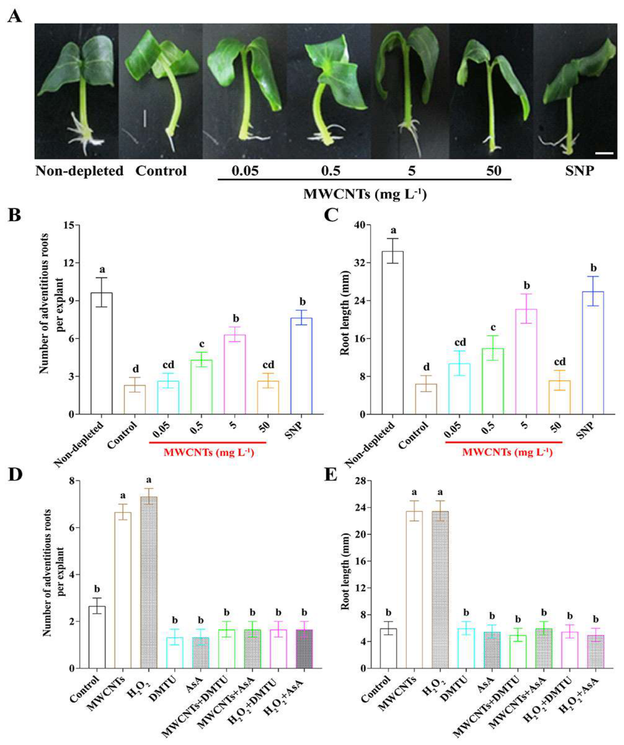
Figure 2.
Distribution and localization of MWCNTs in cucumber hypocotyl. (A) Bright-field and confocal fluorescence images of cross-sections from cucumber hypocotyl bases treated with Rhodamine B-labeled MWCNTs (MWCNTs/Rho B). Red fluorescence indicates the distribution of MWCNT/Rho B in hypocotyl tissues. Scale bar =0.5 mm. (B) Relative fluorescence intensity in control and MWCNTs/Rho B -treated hypocotyl tissues (P < 0.01, Student’s t-test). (C) TEM images displaying the physical accumulation of MWCNTs (black arrows) in cell walls (CW) and intercellular spaces (IS) of treated cells compared with control.
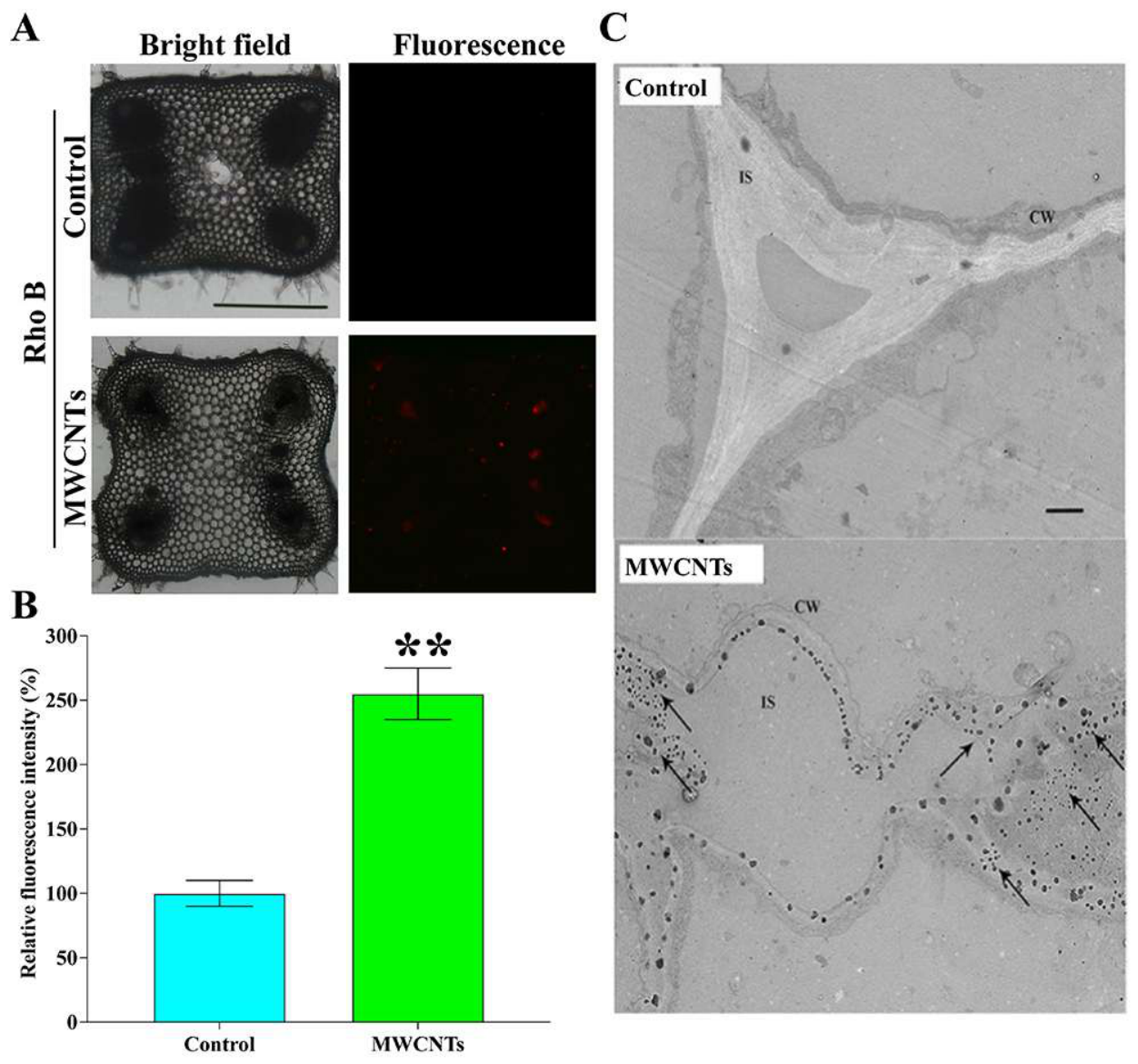
Figure 3.
MWCNTs induce rapid H2O2 accumulation in cucumber hypocotyl tissues. (A) Time-course changes in endogenous H2O2 contents in the basal region of cucumber hypocotyls following MWCNT treatment (ns, not significant; ** P < 0.01, *** P < 0.001, and **** P < 0.0001; Student’s t-test). (B) Representative confocal fluorescence images showing H2O2-associated H2DCF-DA signals in the basal region of cucumber hypocotyls under the indicated treatments. Scale bar = 200 μm. (C) Quantification of relative H2DCF-DA fluorescence intensity in hypocotyl tissues subjected to the indicated treatments. Different lowercase letters indicate significant differences among treatments (P < 0.05).
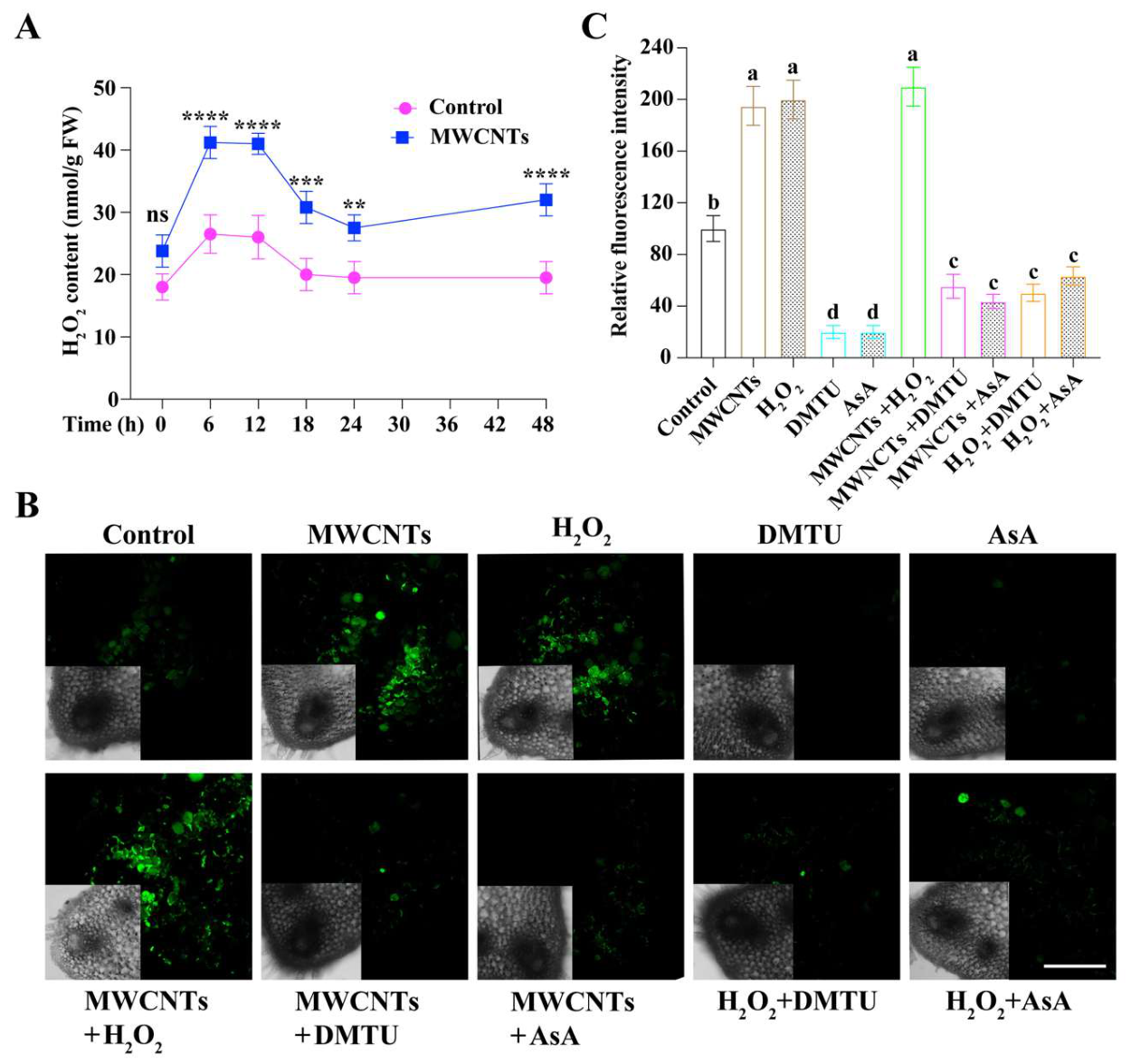
Figure 4.
NADPH oxidase contributes to MWCNT-induced H2O2 production and adventitious root formation in cucumber. (A) Cytochemical localization of H2O2 in cucumber hypocotyl cells by CeCl3 staining followed by TEM. Black arrows indicate electron-dense cerium perhydroxide deposits associated with the plasma membrane and cell wall. (B) Relative expression of CsRBOH in cucumber hypocotyl bases under the indicated treatments, as determined by RT-qPCR. (C) NADPH oxidase activity in cucumber hypocotyl tissues under the indicated treatments. (D–F) Effects of the NADPH oxidase inhibitor DPI on MWCNTs- and H2O2-induced adventitious root formation in cucumber explants. Representative phenotypes (D), adventitious root number (E), and root length (F) are shown. Different letters indicate significant differences among treatments (P < 0.05). .
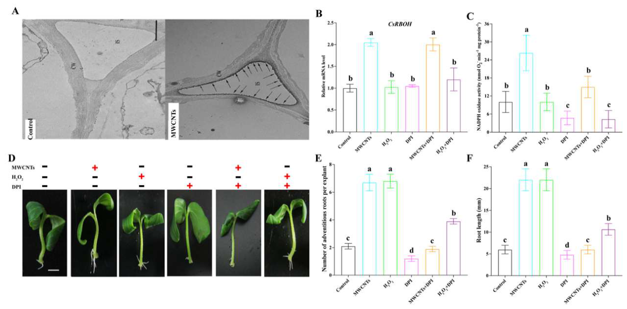
Figure 5.
RBOHD/F are required for MWCNTs-induced H2O2 accumulation and adventitious root formation in Arabidopsis. (A) Representative adventitious rooting phenotypes of Col-0, Atrbohd and Atrbohf mutants under control conditions or following treatment with MWCNTs or H2O2. Scale bar = 0.5 cm (B, C) Adventitious root number (B) and total root length (C) of Col-0, Atrbohd and Atrbohf mutants under the indicated treatments. (D) Representative H2DCF-DA fluorescence images showing H2O2 signals in roots of Col-0, Atrbohd and Atrbohf mutants under the indicated treatments. Scale bar = 200 μm. (E, F) Quantification of relative H2DCF-DA fluorescence intensity (E) and endogenous H2O2 contents (F). Statistical significance for the indicated comparisons is shown as ***P < 0.001; ns, no significant.
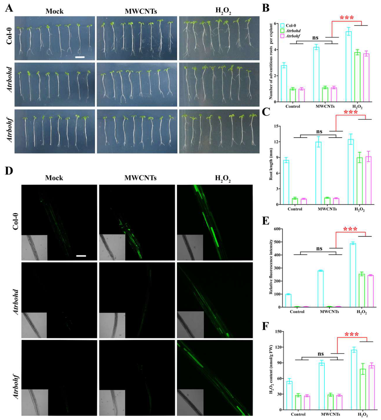
Figure 6.
Proposed working model for MWCNT-induced adventitious root formation in cucumber (Cucumis sativus L.). MWCNTs stimulate RBOH-dependent NADPH oxidase activity at the plasma membrane, leading to increased H2O2 accumulation. H2O2 subsequently modulates calcium- and auxin-related signaling, accompanied by the upregulation of genes associated with adventitious root development, thereby promoting root formation. DPI inhibits NADPH oxidase activity, whereas DMTU and AsA suppress H2O2 accumulation; NPA interferes with auxin transport.
Disclaimer/Publisher’s Note: The statements, opinions and data contained in all publications are solely those of the individual author(s) and contributor(s) and not of MDPI and/or the editor(s). MDPI and/or the editor(s) disclaim responsibility for any injury to people or property resulting from any ideas, methods, instructions or products referred to in the content. |
© 2026 by the authors. Licensee MDPI, Basel, Switzerland. This article is an open access article distributed under the terms and conditions of the Creative Commons Attribution (CC BY) license (http://creativecommons.org/licenses/by/4.0/).
Copyright: This open access article is published under a Creative Commons CC BY 4.0 license, which permit the free download, distribution, and reuse, provided that the author and preprint are cited in any reuse.