

Submitted:
30 September 2026
Posted:
30 September 2026
You are already at the latest version
Abstract
Plants have to deal with a wide variety of unfavourable and unavoidable environmental conditions, among which low temperature is one of the most harmful. The temperature conditions determine plant growth, development, and geographic distribution. Recently, climate change results in an increase in the frequency of unfavourable weather phenomena, including sudden frosts. Exposure to low temperature results in disturbance of cellular metabolism and structure, leading, among other negative effects, to the occurrence of oxidative stress. Photosynthetic apparatus is especially prone to damage caused by reactive oxygen species (ROS) formed because of its overexcitation occurring at low temperatures under light. Among the mechanisms of acclimatisation to cold stress, an important one is an increase in the share of polyunsaturated fatty acid chains (PUFA) in membrane lipids to maintain membrane fluidity. However, PUFA are very susceptible to lipid peroxidation. Therefore, antioxidant protection of lipids is crucial to provide tolerance to cold stress. Prenyllipids, such as carotenoids, isoprenoid chromanols, and isoprenoid quinols, are potent lipophilic antioxidants. Additionally, carotenoids play a crucial role in photoprotection. In this review we tackle the role of lipophilic antioxidants in low temperature-induced stress. Firstly, we describe ROS properties and action, sites of their formation, and the ways of their detoxication. Then, we summarise the current knowledge concerning chemical structures, occurrence, biosynthesis, and function of prenyllipid antioxidants. The third part is devoted to the effects of low temperature stress on plants and the mechanisms enabling acclimatisation and adaptation to cold and frost. The final part focusses on the literature data concerning the role of prenyllipid antioxidants in the response to cold stress.
Keywords:
carotenoids
; cold acclimatisation
; cold stress
; oxidative stress
; plastoquinone
; prenyllipid antioxidants
; tocopherol
1. Introduction
Being sessile organisms, plants have to deal with the wide variety of unfavourable and unavoidable environmental conditions, such as drought, high light, extreme temperatures, waterlogging, salinity, and many others. These stresses pose a threat to plant survival and reproduction, they also limit growth and yield. Among them, low temperature is one of the most harmful to higher plants [1,2].
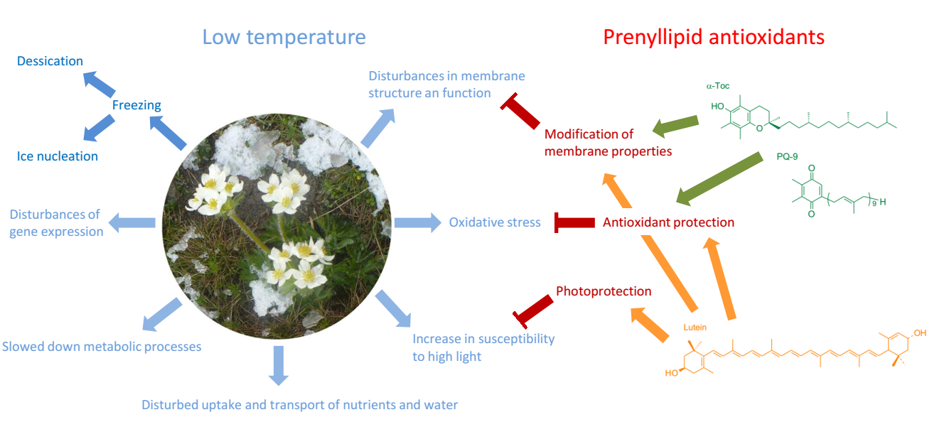
Exposure to environmental stresses usually leads to the excessive formation of reactive oxygen species (ROS), as a result of the disturbance of various metabolic processes [3]. The situation in which ROS generation in the cell exceeds the ability of their detoxication is called oxidative stress. Plants are especially prone to oxidative damage because (1) they perform oxygenic photosynthesis and therefore produce additional O2 in their green tissues (2) they contain a lot of chlorophyll (Chl) that can act as photosensitiser and transfer its excitation energy to O2 (3) they have a photosynthetic electron transfer chain from which electrons can leak to O2 [4]. If the photosynthetic apparatus absorbs more energy that can be used for beneficial reactions of photosynthesis, excess energy fuels the unwanted side reactions leading to enhanced ROS formation. In low temperatures, the chemical reactions of photosynthesis are slowed down, whereas the absorption of light by the photosynthetic apparatus is quite efficient, which results in a decrease of the threshold above which certain intensity of light becomes excessive [4,5]. Furthermore, among the mechanisms of acclimatisation to cold stress, an important one is the maintenance of membrane fluidity, necessary for the proper functioning of cellular metabolism, in particular photosynthetic and respiratory electron transfer chains, as well as proper membrane transport and membrane-related signalling. To achieve this objective, many organisms enhance the share of polyunsaturated fatty acid chains (PUFA) in their membrane lipids [6]. However, PUFA are very susceptible to oxidation [7]. Therefore, antioxidant protection of lipids is crucial to provide tolerance to cold stress. Prenyllipid antioxidants, such as carotenoids, and isoprenoid chromanols and quinols, are potent lipophilic antioxidants occurring in membranes and lipid storage sites [8]. Carotenoids are also crucial for safe dissipation of excess light energy absorbed by the photosynthetic apparatus [9]. Additionally, the compounds mentioned above participate, directly or indirectly, in signalling during stress acclimatisation [4,9].
In this review we tackle the role of lipophilic antioxidants in low temperature-induced stress. The first subchapter focusses on ROS, their properties and harmful action, the sites of their formation and the ways of their detoxication. The second chapter is devoted to prenyllipid antioxidants, their chemical structures, occurrence, biosynthesis and function. Particular emphasis was placed on the compounds present in chloroplasts, because these organelles are the major site of ROS production in plant green tissues. The third chapter is about the impact of low temperature on plants, while the fourth summarises the literature data concerning the role of prenyllipid antioxidants in the response to cold stress.
2. Reactive oxygen species, their sites of formation in plants, and their action on biomolecules
Molecular oxygen appeared on Earth as a product of oxygenic photosynthesis. This crucial metabolic process has evolved in ancient cyanobacteria and later was passed onto photosynthetic eukaryotes, along with the acquisition of plastids, organelles of direct or indirect cyanobacterial origin [10,11]. The presence of O2 in the atmosphere and waters enabled the subsequent evolution of energetically efficient aerobic respiration [12]. However, aerobic metabolism has its dark side, which is the formation of ROS as its by-products [13].
ROS consist of radical and non-radical forms. Molecular oxygen in its ground state is diradical that contains two unpaired electrons with parallel spins in two antibonding orbitals π*2p. Such a form is called triplet oxygen (3O2). This unusual electron configuration makes 3O2 less reactive than one could imply on the physicochemical characteristics of oxygen atoms [13]. Excitation of 3O2 results in spin reversal of one of the unpaired electrons, leading to the formation of singlet oxygen (1O2). Singlet oxygen can have electrons of opposite spins either in separate orbitals (very short-lived form, 1Σg+O2) or the same orbital (form 1ΔgO2 of lower energy and longer lifetime). Paired electrons make 1O2 much more reactive than 3O2 because the spin restriction is removed [14]. Singlet oxygen is thought to be the major ROS responsible for light-induced loss of photosystem II (PS II) activity and leaf damage [14]. In addition to excitation, 3O2 can also undergo reduction. The complete four-electron reduction leads to the formation of water, while all its intermediates belong to ROS [15]. Among them, superoxide radical (O2∙−), is often formed as a result of electron leakage from the electron transfer chains. In low pH, O2∙− undergoes protonation [3,15]. Dismutation of O2∙−/HO2∙ leads to the generation of H2O2, which is less reactive, but may react with free metal ions, resulting in the formation of hydroxyl radical (OH∙). Metal ions tend to bind to the surface of DNA and proteins. In such a case, the most dangerous type of ROS would be formed in the vicinity of the targets of its damaging action [16]. The most important types of ROS and their reactivity are shown in Figure 1. Other ROS worth mentioning are ozone (O3), alkoxy radicals (RO∙, where R is organic residue, for example fatty acid chain), peroxy radicals (ROO∙) and hydroperoxides (ROOH) [16].
Being prone to react with organic molecules, especially those containing unsaturated bonds, and with redox-active metal ions, ROS can disturb cell biochemistry and structure [3]. The most important biomolecules vulnerable to ROS and the results of their oxidative damage are shown in Figure 2. Taking into account the genetic material, mitochondrial and plastid DNA is more endangered than that present in the nucleus, due to the relatively high production of ROS in these organelles (see below) and the fact that, unlike nuclear DNA, it is not associated with histones [16]. Among lipids, PUFA residues are particularly susceptible to lipid peroxidation [17]. Such residues, when present in phospholipids, provide sufficient membrane fluidity; therefore, they are abundant in membranes, where electron transfer chains occur. Unfortunately, these electron transfer chains are important sources of ROS. Lipid peroxidation can occur through direct oxidation of lipids by 1O2 or through a radical-mediated process, which can be initiated by HO2∙, OH∙, NO2∙, perferryl radicals, carbon-centered radicals (R∙), RO∙, and ROO∙. Because radical-mediated peroxidation is a self-propagating chain reaction, low amounts of radical initiators may ultimately cause extensive damage [16]. However, the analysis of specific lipid peroxidation products showed that more than 80% of nonenzymatic peroxidation in the leaves of higher plants is caused by 1O2 [18]. In addition to the compounds mentioned in Figure 2, ROS are able to oxidise other compounds, such as carbohydrates and pigments [3,19].
In photosynthetic cells, ROS are usually formed as a result of electron leakage from electron transfer chains and the photosensitising action of Chls; they can also be products of some enzymatic reactions (Figure 3). The most important sites of ROS production are chloroplasts, mitochondria, and peroxisomes [20]. In green tissues under light, the highest amounts of ROS are formed in chloroplasts, due to the occurrence of Chl-binding proteins, the functioning of the photosynthetic electron transport chain, and the increased concentration of O2 because of its production at PS II [3]. Over-reduction of photosynthetic electron transfer chain leads to enhanced electron leakage and resulting formation of O2∙- . The electrons leak mainly from the Fe-S clusters of Photosystem I (PS I) and reduced ferredoxin, however O2∙- can be also formed at receptor side of PS II or within the pool of PQ [3,15,21]. Excessive reduction of photosynthetic chain elements also increases the risk of the formation of triplet excited states of Chl, which can transfer their excitation energy to 3O2, leading to the generation of 1O2 [22]. In heterotrophic eukaryotes, mitochondria are the dominant sites of cellular ROS production. In photosynthetic eukaryotes, these organelles are thought to produce ROS less abundantly than chloroplasts. For example, in C3 plants, H2O2 production in mitochondria is estimated to be 20 times lower than its production in chloroplasts. However, mitochondrial share in overall cellular ROS generation in plants is believed to be significant in darkness or in non-green tissues [3,23]. The major process responsible for mitochondrial ROS production is electron leakage from the respiratory electron transfer chain, mainly at complexes I and III; however, complex II and the over-reduced ubiquinone pool also play a role [23,24]. Some mitochondrial enzymes not involved in respiration are also known to produce ROS, but their contribution to mitochondrial ROS formation is minor [25]. In peroxisomes, H2O2 and O2∙- are generated by various catabolic enzymes. H2O2 is also a product of glycolate oxidase that participates in the photorespiratory pathway [26]. The ROS mentioned above can also be formed enzymatically at many other sites in the plant cell, which plays different roles, such as signalling, cell wall lignification, or response to pathogen [16].
To defend themselves against ROS, organisms have evolved various mechanisms based on low-molecular-weight antioxidants and enzymes (Figure 4). The former are able to quench 1O2 and scavenge various ROS; and can be subdivided into hydrophilic and hydrophobic ones. Lipophilic antioxidants belong to the prenyllipid class of compounds. They are crucial for the protection of cell membranes and lipid storage sites [4]. In addition to direct ROS scavenging, some low-molecular-weight antioxidants, such as ascorbate, glutathione, and some phenolic compounds are electron-providing co-substrates of ROS-detoxifying enzymes [27]. When enzymes are considered, crucial ones can directly detoxify certain types of ROS. Other enzymes participate in the recycling of low-molecular-weight antioxidants, oxidised as a result of ROS scavenging. There are also many enzymes responsible for the repair of oxidised biomolecules, especially proteins and nucleic acids. Glutathione-S-transferases, not shown in Figure 4, are a class of enzymes, members of which participate in various processes, including ROS detoxification, ascorbate recycling, and prevention of ROS formation due to detoxification of xenobiotics [3,16,27]. As plants are especially prone to oxidative stress, they have evolved a wide range of highly redundant antioxidant mechanisms [3]. Aerobic organisms have also evolved to use ROS for their benefit, in cellular signalling and to fight with pathogens [20]. Because various stress factors lead to the enhancement of ROS production in cells, these ROS may serve as signals, triggering plant responses [28]. Stress-induced ROS are known to participate in signalling pathways associated with mitogen-activated protein kinases (MAPK); they may also induce calcium signalling [3,29].
3. Plant prenyllipid antioxidants: structure, occurrence, biosynthesis, and function
3.1. Isoprenoid chromanols and quinones
Important plant prenyllipid antioxidants belonging to isoprenoid chromanols and quinones are shown in Figure 5, and those commonly occurring in chloroplasts are marked with a dark green colour. All of these compounds have amphipathic properties, because their molecules are composed of a polar head-group (a chromanol or quinone ring) and an apolar prenyl side-chain [31,32]. The main chromanol present in plant green tissues is α-tocopherol (α-Toc) [33]. The other forms of tocopherols (called β-, γ- and δ-vitamers) all contain phytol-derived saturated side-chain, but differ in the number and position of methyl substituents in the chromanol ring. Among them, γ-tocopherol (γ-Toc) is found in leaves in lesser amounts than α-Toc, however, it is often abundant in seed oils of many plant species [34]. The other vitamers can also be found in plant seeds, together with α-, β-, γ-, δ-tocotrienols (T3s), which contain geranylgeranyl-derived side-chain with three unsaturated bonds. The occurrence of certain Tocs and T3s in seeds and their relative abundance depend on the plant species [32]. In some species γ- and δ-Toc were also found in cuticular waxes [33]. Plastochromanol-8, which contains head-group same as γ-Toc and much longer, unsaturated side-chain is present in green tissues in minor amounts [35]. The other chromanols, such as tokomonoenols (T1s) and tokodienols, containing one and two unsaturated bonds in their side-chains, respectively, as well as other derivatives, including demethylated or phosphorylated forms, have recently been discovered. They occur in small amounts in some plant species [4].
Among isoprenoid benzoquinones, plastoquinone-9 (PQ-9), together with its reduced form, plastoquinol-9 (PQH2-9) that has hydroxyl instead of ketone groups in the ring, are present in chloroplasts in high amounts [31]. Its analogues containing shorter side-chains, which may also differ with the degree of saturation, as well as forms varying in the number of methyl groups in the quinone ring (i.e. 6-methyl-dihydrophytylplastoquinone, 3-demethylplastoquinone-9), have also been described in the literature [4,36]. Ubiquinone (UQ) and ubiquinol (UQH2) occur in the inner membrane of mitochondria [37]. The most common number of isoprenoid residues in their side-chains is 9 and 10, however, forms with shorter chains have also been found in minor amounts. The exact composition of UQs depends on the plant species [38,39]. Taking into account naphthoquinones, photosynthetic eukaryotes typically contain phylloquinone (PhQ), but in some algal species menaquinone-4, containing the side-chain derived from geranylgeranyl, instead of the phytol-derived one was found. The PhQ derivative that contains the -OH group in the side chain has also been described in the literature [40,41,42].
The oxidized derivatives of α-Toc, PC-8 and PQ-9, which are α-tocopheryloquinone (α-TQ), hydroxy-plastochromanol (PC-OH), and plastoquinone C (PQ C), respectively, were drawn in Figure 5 using a sap-green colour. PQ derivatives that contain more -OH groups in the side-chain have also been described in the literature [43]. All these compounds occur in plants in small amounts, but their level is known to increase under stress conditions [43,44,45].
Due to their crucial role in photosynthesis, PQ and PhQ are present in both cyanobacteria and photosynthetic eukaryotes. In the latter organisms, PQ occurs in chloroplasts: in thylakoid membranes, plastoglobules, which are lipid storage sites, as well as in the inner chloroplast envelope, where it is synthesised [4]. PhQ is a cofactor in PS I, but minor amounts of this compound were found in plastoglobules, chloroplast envelope, and cell membranes [31]. The occurrence of both PQ and PhQ is not limited to green tissues, as these prenyllipids are also present in small amounts in non-photosynthetic organs, such as flowers, fruits, roots and bulbs [31]. UQ is commonly present in eukaryotes and plays a key role in respiration. In mitochondria, it occurs in the inner membranes [31,37].
Isoprenoid chromanols are synthesised by most cyanobacteria. These compounds are also ubiquitous in photosynthetic eukaryotes, but their amounts vary depending on the taxonomic affiliation. In particular, higher plants are known to contain high levels of isoprenoid chromanols, which are also more structurally diversified [4,32]. In chloroplasts, isoprenoid chromanols occur in thylakoids, envelopes, and plastoglobules. Non-green plastids also contain chromanols. In particular, these prenyllipids can be stored in leukoplasts in seed cells. However, in addition to plastids, isoprenoid chromanols in seeds can also be found in oleosomes, which are lipid droplets surrounded by a protein-lipid monolayer and dispersed in the cytoplasm. These lipid bodies can store up to 40% of the seed chromanol pool [4,46,47]. The presence of chromanols in other cell compartments was reported in the literature, but the purity of the fractions used for the analyses has been questioned [33]. Concerning plant organs, the highest levels of isoprenoid chromanols occur in green tissues and seeds, in particular those accumulating lipids as the major storage material. These compounds can also be found in non-photosynthetic plant organs, but generally their content is lower compared to leaves [4]. Chromanol levels usually increase during acclimatisation to environmental stresses [48].
In the biosynthetic pathway of isoprenoid quinones and chromanols, the side-chain and head-group precursors are synthesised separately [4]. Subsequently, specific prenyltransferases catalyse their condensation, and the resulting compound is further modified. In this paper, the synthesis of Tocs, PQ and PC-8 is described in more detail, as these compounds are well known as efficient antioxidants present in chloroplasts.
The Toc biosynthesis pathway is shown in Figure 6. Homogentisate (HGA), which serves as a precursor of the head-group of isoprenoid chromanols, is synthesised from p-hydroxyphenylpyruvate, a compound derived from tyrosine [49]. The synthesis of HGA is catalysed by p-hydroxyphenylpyruvate dioxygenase (HPPD, encoded by the PDS1 gene) that is present in chloroplasts or cytosol, depending on the species [50]. The side-chain is synthesised via multistep condensation of five-carbon precursors, isopentenyl pyrophosphate (IPP), and dimethylallyl pyrophosphate (DMAPP). There are two pathways of IPP and DMAPP synthesis, that is, 1-deoxy-D-xylulose-5-phosphate (DXP) pathway, also called the methylerythritol phosphate pathway, and the mevalonate (MVA) pathway [51]. Cyanobacteria possess the DXP pathway, whereas higher plants have both, DXP in plastids and MVA in the cytosol [49]. Considering eukaryotic algae, most of them also contain both pathways, but there are eukaryotic algae that have lost one of the pathways [52,53]. It is thought that in plants the IPP and DMAPP used for the synthesis of plastid isoprenoids are mainly derived from the DXP pathway. However, it was shown that in the situation of overproduction of these precursors in the cytoplasm, they can be transported to chloroplasts [50]. Isoprenoid chains of different lengths are synthesised through condensation of IPP and DMAPP by various polyprenyl pyrophosphate synthases. Among them, geranylgeranyl pyrophosphate synthase (GGPPS) catalyses the subsequent head-to-tail condensation of four isoprenoid units. The resulting geranylgeranyl pyrophosphate (GGPP) is a common precursor for the biosynthesis of various isoprenoids produced in plastids [49,50]. In the Toc biosynthetic pathway, GGPP is then reduced to phytol pyrophosphate (PPP). Higher plants are also known to utilise phytol derived from degradation of Chls. In fact, this route of formation of Toc side-chain is thought to dominate in these organisms. Specific kinases, encoded by the VTE5 and VTE6 genes, catalyse subsequent phosphorylations of phytol to PPP [34,50]. Homogentisate phytyltransferase (HPT, encoded by the VTE2 gene) carries out the condensation of HGA with PPP. The product of this reaction can then undergo cyclisation by tocopherol cyclase (TC, encoded by the VTE1 gene), leading to the formation of δ-Toc, or methylation by 2-methyl-6-phytyl-1,4-benzoquinone methyltransferase (MPBQ MT, encoded by VTE3). Subsequent cyclisation of the methylated intermediate by TC results in γ-Toc formation. Both δ-Toc and γ-Toc are substrates for γ-tocopherol methyltransferase (γ-TMT, encoded by VTE4), and the products of these reactions are β-Toc and α-Toc, respectively [51]. HPT can also catalyse condensation of HGA with tetrahydrogeranylgeranyl pyrophosphate in the biosynthetic pathway of T1s [54]. In the synthesis of T3s, HGA is condensed with GGPP by homogentisate geranylgeranyl transferase and the subsequent reactions are catalysed by TC, MPBQ MT, and γ-TMT, like in the Toc and T1 biosynthetic pathways [51]. The last steps of synthesis of isoprenoid chromanols occur mostly in the chloroplast envelope, where HPT, MPBQ MT and γ-TMT localise. On the other hand, TC was found in plastoglobules [55].
The synthesis of PQ-9 and PC-8 is shown in Figure 7. In higher plants, HGA serves as a head-group precursor for PQ. On the other hand, cyanobacteria, which use HGA for the synthesis of Tocs similar to plants, for the synthesis of PQ use p-hydroxybenzoate, which is also a precursor of head-group in the biosynthesis of UQ in eukaryotes and those of proteobacteria that contain UQ [56,57]. The side-chain precursor, solanesyl pyrophosphate, is synthesised by the subsequent addition of five IPP to GGPP, carried out by solanesyl pyrophosphate synthase (SPS). In higher plants, homogentisate solanesyl transferase (HST) is an enzyme responsible for the condensation of SPS with HGA. The product of this reaction is then methylated by MPBQ MT, leading to the formation of PQH2-9. Enzymatic or non-enzymatic oxidation of PQH2-9 is a source of PQ-9. Some PQH2-9 may undergo cyclisation catalysed by TC, resulting in PC-8 formation [4]. The synthesis of PQ in cyanobacteria requires different prenyltransferase, which gene is homologue to the one participating in the synthesis of UQ, and different steps of further modifications of the head-group [56].
In the synthesis of PhQ head-group precursor is derived from chorismate, while PPP serves as the precursor of the side-chain. Most enzymes that participate in PhQ biosynthesis are located in plastids, but some steps take place in peroxisomes [58,59,60]. The synthesis of UQ occurs in mitochondria and its detailed description can be found in the review by Xu et al. [39].
Due to their amphipathic properties, isoprenoid chromanols and quinones localise in membranes and lipid storage sites, where they display their protective properties. Isoprenoid chromanols are well-known potent lipophilic antioxidants. Being the most common vitamer, α-Toc has been studied most extensively in this respect, both in vitro and in vivo. This compound was shown to be an efficient scavenger of radicals, including O2∙−/ HO2∙ and those formed during radical-mediated lipid peroxidation: R∙, RO∙, and ROO∙ [61,62]. α-Toc is also known to efficiently quench and scavenge 1O2 [63,64]. The antioxidant properties of other Toc vitamers, T3s and PC-8, have also been examined in various systems [63,64,65,66,67].
In radical scavenging reactions, isoprenoid chromanols donate a hydrogen atom from the hydroxyl group in their chromanol ring. Hydrogen-donating power depends on a ring structure, and decreases in order α > β > γ > δ [61]. In some assays this order corresponded well to the antioxidant efficiency of certain vitamers [66,67], in other cases the order of decreasing antioxidant potency was α > β ≈ γ > δ [65,66]. There were also cases in which α-Toc did not exceed other vitamers in its antioxidant action [66]. The chromanoxyl radical, formed as a result of radical scavenging by isoprenoid chromanols, is relatively stable because of delocalisation of electrons in the aromatic ring. It can be re-reduced to the corresponding Toc by ascorbate (Asc), reduced isoprenoid quinols such as PQH2-9, and phenolic compounds [64,68,69,70]. However, if the reductant is scarce or absent and when peroxidation occurs in dispersed systems, such as liposomes or low density lipoproteins (LDLs), tocopheroxyl radical lifetime can be prolonged, which favours hydrogen abstraction from other molecules and thus, chain-carrying pro-oxidant action [64,71,72,73]. Fortunately, chloroplasts are the site where Asc and PQH2-9 are usually abundant [4,74]. Considering the impact of the side-chain on the antioxidant action, α-T3 turned out to be more effective than α-Toc in counteracting radical-mediated peroxidation in liposomes [66,75]. Serbinova et al. postulated that this effect resulted from a higher recycling efficiency of α-T3 from chromanoxyl radicals, more uniform distribution of α-T3 in membrane bilayer, and a greater disordering of membrane lipids by α-T3, which makes interaction of chromanols with lipid radicals more efficient [75].
Taking into account the quenching of 1O2, the rate constants of reaction of α-Toc with 1O2 in organic solvents were higher than those of other vitamers and PC-8, while the rates of the T3s vitamers were comparable to their respective Tocs. However, for all compounds, the order of magnitude of the rate constants was the same [63]. Tocs 1O2 scavenging rates are lower than their 1O2 quenching rates for approximately two orders of magnitude and were shown to decrease in order α > γ > δ > β [76]. Scavenging of 1O2 by α-Toc results in the formation of 8a-hydroperoxy-α-tocopherone, which is unstable and may be further converted to α-TQ or re-reduced to α-Toc by Asc [55]. In chloroplasts α-TQ can be reduced to quinol form (α-TQH2) [77], which displays antioxidant properties similar to those of other isoprenoid quinols (see below) and can also be enzymatically recycled back to α-Toc [78]. The reactivity of PC-8 towards radicals and 1O2 is similar to that displayed by γ-Toc. However, due to its longer side-chain, PC-8 was suggested to be a particularly effective antioxidant in the hydrophobic core of the lipid bilayer [64].
While α-Toc has a fully substituted benzene ring in the chromanol head-group, γ-Toc has an unsubstituted 5-position and therefore is able to trap electrophiles, including reactive nitrogen species (RNS) [79,80].
In addition to their antioxidant properties, isoprenoid chromanols were also postulated to participate in signalling, mostly through interference with ROS and RNS-mediated pathways [8,81]. For example, the phytohormone jasmonic acid (JA) belongs to oxylipins. Because lipid hydroperoxide is an intermediate in its synthesis [82], it was postulated that by inhibition of lipid peroxidation α-Toc is able to limit JA synthesis [83]. Experiments in corn mutant sxd1 lacking α-Toc showed the importance of this prenyllipid for proper formation of plasmodesmata and phloem loading [84,85,86]. Isoprenoid chromanols have also an impact on membrane properties. α-Toc was shown to increase membrane rigidity and ordering, decrease ion leakage, and favour the formation of reversed hexagonal structures [33,87,88].
Isoprenoid quinones are the most recognised as electron carriers in electron transfer chains, such as photosynthetic, respiratory, and chlororespiratory ones. These compounds also function as enzyme cofactors and participate in cellular signalling [31,36,37]. Their reduced forms are well-known for their potent antioxidant properties [4]. Quinones may also act as antioxidants, but their activity is lower compared to that of quinols. Isoprenoid quinols can scavenge inorganic (HO2∙, perferryl radical) and lipid radicals, as well as quench and scavenge 1O2 [63,64,89,90,91]. PQH2 was also shown to scavenge H2O2 in organic solvents [92]. The scavenging of radicals by isoprenoid quinols results in the formation of semiquinone radicals, which undergo disproportionation to quinones and quinols [90]. Quinones are known to react with O2∙− which leads to the formation of semiquinone radical [93,94]. However, quinols and semiquinone radicals can also act as pro-oxidants [90].
The rate constants of 1O2 quenching by PQH2-9 and UQH2-10 in organic solvents were lower than those of α-Toc, but similar to values measured for other Toc vitamers. PQ-9 was also shown to quench 1O2, however, the rate constant of this reaction was a few times lower compared to that of PQH2-9 [63]. Taking into account the scavenging of 1O2 by PQ/PQH2, apart from the head group, unsaturated bonds in the side-chain can also be the target of oxidation, leading to the formation of hydroxy-derivatives. These products are considered markers of enhanced 1O2 formation in vivo, together with PC-OH [43,44]. An in vivo study on C. reinhardtii exposed to high-light stress showed that PQH2 is the main prenyllipid responsible for the detoxication of 1O2 in chloroplasts [91]. Similarly, transgenic A. thaliana containing increased amounts of PQH2 and PC-8 due to the over-expression of the sps1 gene was more resistant to high light than wild type, while such an effect was not observed in the line with increased levels of α- and γ-Toc [95]. The amount of PQ/PQH2 increases under stress conditions known to cause oxidative stress in plants [36]. The antioxidant function of UQ/UQH2 in plants has not been examined as widely as that of Toc and PQ/PQH2, but there are some literature data concerning this topic [37].
PQ is known to play a role in chloroplast-derived signalling. PQ pool is thought to act as a redox sensor responsible for the initiation of response to changing light conditions. In experiments carried out in plants and algae using inhibitors of photosynthetic electron transfer or selective excitation of PS I and II it was shown that the change in the PQH2/PQ ratio had an impact on the expression of important genes, both chloroplast and nuclear, such as those encoding PS II core peptide D1, PS I core peptides PsaA and PsaB, LHC II, ascorbate peroxidases, superoxide dismutase and nitrate reductase [96,97,98,99,100,101,102,103,104,105]. Similarly to Tocs, due to its antioxidant properties, PQ/PQH2 was postulated to participate in cellular signalling through interaction with ROS-mediated signalling pathways [36].
The redox state of the PQ pool is also known to regulate the activity of the kinase STN7 and its homologues, responsible for the induction of the state transition of the photosynthetic apparatus from state 1 to state 2. This process involves dissociation of LHC II antennae from PS II and their association with PS I, as well as aggregation of LHC II in thylakoid membranes. State transitions play a role in balancing the energy supply to photosystems and the regulation of cyclic electron transfer around PS I [106,107].
Furthermore, PQ is a cofactor of enzymes necessary for carotenoid biosynthesis in plants, algae, and cyanobacteria, i.e. phytoene desaturase and ζ-carotene desaturase [108].
3.2. Carotenoids
Carotenoids are a large class of natural pigments, ancient in their evolutionary origin, and relatively common among living organisms [109,110]. So far, more than 1100 natural compounds belonging to this group have been discovered [111]. Being accessory photosynthetic pigments, crucial for light harvesting and photoprotection, carotenoids are synthesised by all photosynthetic organisms, both prokaryotic and eukaryotic [9]. They are also produced by some non-photosynthetic bacteria, archaebacteria, protozoa, and fungi [112]. Animals need to obtain these pigments in food; however, there is a known aphid species that can produce carotenoids on its own [113].
Carotenoids are tetraterpenoids, which means that they are synthesised by condensation of eight isoprenoid units. However, there are known exceptions, that is, heliobacteria produce carotenoids made of six isoprenoid units as well [114]. The typical C40 backbone can also be further extended, producing C45 or C50 carotenoids, while shortening of the molecules leads to the formation of apocarotenoids [109]. These pigments are divided into two subgroups, that is, carotenes, which are hydrocarbons, and xanthophylls, which contain one or more oxygen atoms [9]. All carotenoids are long molecules, containing multiple double bonds, owning to which they efficiently absorb visible light, in the range 400-550 nm [112]. Taking into account the conformation of double bonds, all-trans carotenoids are more prevalent in nature; however, cis-isomers may be formed enzymatically or as a result of exposure to heat or light [111,115]. Some of these compounds are acyclic, whereas the rest contain ring(s), usually at their ends. They differ in degree of saturation, number of conjugated double bonds, and presence and distribution of various substituents [111,116]. Due to the diversity of substituents, there are known xanthophylls that are alcohols and their esters, aldehydes, ketones, acids and their esters, epoxides, ethers, and glycosides [9].
The detailed carotenoid composition of a given organism depends on the systematic affiliation; therefore, these compounds are valuable chemotaxonomic biomarkers [110,116,117]. Higher plants are known to synthesise a narrower array of these pigments, compared to photosynthetic bacteria and algae [116,118]. Carotenoids commonly occurring in plant green tissues are shown in Figure 8, with those present in higher amounts drawn in orange. Among them, lutein is the most abundant [119,120].
In addition to these compounds, the intermediates of their biosynthetic pathway, such as lycopene, δ- and γ-carotene, as well as some derivatives, such as epoxy-lutein, can also be found in small amounts and in non-green tissues [9,119,121]. The most abundant carotenoid of cyanobacteria is usually β-carotene, but they synthesize its hydroxy-derivatives zeaxanthin and nostoxanthin, as well as ketocarotenoids, such as canthaxanthin and echinenone, and carotenoid glycosides, for example myxoxanthophyll [122]. In eukaryotic algae, the diversity of carotenoids is remarkable [116]. The carotenoid composition of green algae is similar to that of higher plants; however, they also synthesise other compounds, such as astaxanthin, siphonaxanthin, or loroxanthin. The dominant carotenoid in unicellular red algae is zeaxanthin, whereas macrophytic red algae contain lutein as their main carotenoid. The major carotenoids of stramenopiles are diadinoxanthin and fucoxanthin. These compounds can be also found in haptophytes; however, diadinoxanthin is not as widespread as in the group mentioned above. Cryptophytes contain α-carotene and acetylene xanthophylls, mainly alloxanthin. In dinoflagellates, peridinin is a characteristic xanthophyll. Euglenids contain carotenoids typical of green algae, but also those present in stramenopiles, such as diadinoxanthin [117,123,124].
In cyanobacteria, carotenoids are present in thylakoids, but can also be found in the cell membrane and cell wall [109]. In photosynthetic eukaryotes, these pigments occur in chloroplasts, where they are bound to photosystems and antennae. However, some fraction diffuses freely in membranes, mainly thylakoid ones, and may be stored in plastoglobules. In non-green tissues, carotenoids are present in large amounts in chromoplasts and in minor amounts in leucoplasts [110]. In some algae, carotenoids also occur in the eyespot and oil droplets [25,125]. Considering organ distribution in higher plants, these pigments are present in all green tissues, but can also occur in certain flowers, fruits, roots, and bulbs [110,121]. Carotenoids can also be found in seeds and pollen [119].
Carotenoid biosynthesis starts with the condensation of IPP and DMAPP, resulting in the formation of GGPP. The synthesis of these precursors has been briefly described in Subchapter 3.1. Here, it is only worth mentioning that in higher plants carotenoids are synthesised in plastids; therefore, C5 precursors are derived from the DXP pathway [110]. Phytoene synthase catalyses the head-to-head 1-1 condensation of two GGPP molecules, resulting in the formation of 15-cis phytoene [112]. To obtain lycopene, which is a common precursor for the synthesis of many C40 carotenoids, 15-cis phytoene must undergo desaturation and isomerisation. In the so-called all-trans pathway, present in carotenoid-synthesising fungi, archaebacteria, and bacteria other than cyanobacteria, these reactions are catalysed by a single enzyme, called CrtI [108,126]. Cyanobacteria (with the known exception of Gloeobacter violaceus using the all-trans pathway) have another more complicated poly-cis pathway, and because plastids are of cyanobacterial origin, photosynthetic eukaryotes also use it [127]. This pathway involves four enzymes, two desaturases, and two isomerases (Figure 9). First, phytoene desaturase (PDS) in two steps introduces two double bonds in cis-conformation at 9 and 9’ positions, forming 9-15-9’-tri-cis-ζ-carotene and using PQ as electron acceptor. Next, 15-cis-ζ-carotene isomerase (Z-ISO) catalyses isomerisation at the 15 position. This reaction may also occur non-enzymatically, under high light. Resulting 9-9’-di-cis-ζ-carotene is utilized by ζ-carotene desaturase (ZDS) which in two steps introduces two double bonds in cis-conformation at the positions 7 and 7’, leading to the formation of 7,9,7’,9’-tetra-cis-lycopene. Similarly to PDS, ZDS uses PQ as a co-substrate. Finally, carotenoid isomerase (CRTISO) performs two steps of isomerisation, producing all-trans lycopene. Similarly to isomerisation at the earlier stage of lycopene synthesis, these reactions can also occur non-enzymatically in high light [108,112].
In plants, lycopene can be found in high amounts, for example in tomato and watermelon fruits, but more often this compound is further modified to obtain cyclic carotenoids [110]. Two cyclases, lycopene β-cyclase (LCYβ) and lycopene ε-cyclase (LCYε), are responsible for the formation of β- and ε-ionone rings at the end of the molecule, respectively. At this stage, the carotenogenic pathway of plants undergoes bifurcation (Figure 9). Both δ-carotene, containing ε-ionone ring, and γ-carotene, containing β-ionone ring, can be further cyclised by LCYβ, resulting in α- and β-carotene, respectively [112,128]. Second cyclisation of δ-carotene catalysed by LCYε, leading to the formation of ε-carotene is very rare in plants [127]. Other cyclisation reactions are also possible, but not common; for example, plants belonging to the pepper family are known to produce carotenoids with cyclopentane ring(s) [110]. Most cyanobacteria do not contain LCYε and therefore have only one branch of the pathway [122]. In eukaryotic algae, some clades have both branches, whereas the other have only LCYβ [128].
Further modifications of carotenes, carried out by various enzymes, mainly hydroxylases and epoxidase, result in the formation xanthophylls (Figure 10). Hydroxylases are specific for the ring, into which they introduce hydroxyl group, therefore they were called β- and ε- carotenoid hydroxylases (CHYβ and CHYε, respectively). Multiple genes of carotenoid hydroxylases were found in Arabidopsis and tomato [110]. Hydroxylation of both rings of α-carotene is necessary to produce lutein, whereas hydroxylation of both rings of β-carotene results in zeaxanthin (Zx) formation. The two-step epoxidation of Zx by zeaxanthin epoxidase (ZE) leads to the synthesis of violaxanthin (Vx). The reverse reaction, i.e. the two-step de-epoxidation of Vx to Zx, catalysed by violaxanthin de-epoxidase (VDE), is important for photoprotection and will be discussed later. Vx can also be converted to neoxanthin (Nx) by neoxanthin synthase (NSY) [121].
Being very efficient in light energy capture and absorbing light in a range not absorbed by Chls and bacteriochlorophylls (BChls), carotenoids are accessory photosynthetic pigments present in photosystems and the majority of antennae types [129]. Furthermore, these compounds are usually necessary for proper assembly and stabilisation of pigment-protein complexes of the photosynthetic apparatus [124,128,130].
In addition to their role in light-harvesting, carotenoids are crucial for photoprotection, because of their ability of fast and efficient deactivation of their excited states via thermal dissipation. Rapid quenching of triplet excited states of (B)Chls allows carotenoids to prevent the formation of 1O2 in the photosynthetic apparatus [9,129,131]. In addition, the dissipation of excess energy absorbed by antennae and photosystems diminishes the risk of O2∙− formation due to over-reduction of the photosynthetic electron transfer chain [15,132].
The crucial photoprotective mechanism of higher plants, called qE (Zx-dependent ΔpH-induced nonphotochemical quenching of chlorophyll fluorescence), requires a proton gradient across the thylakoid membrane and de-epoxidised xanthophyll Zx to function. The latter is formed in a violaxanthin cycle, i.e. interconversion of Vx, antheraxanthin (Ax) and Zx, depending on light conditions. Two opposite reactions of the cycle are catalysed by enzymes belonging to the lipocalin superfamily: Zx epoxidase (ZE) and Vx de-epoxidase (VDE) [133]. In non-stress conditions, the level of Zx in plant leaves is low, while the level of Vx is high. Under high light VDE, activated by low pH in the thylakoid lumen, removes epoxy-groups and introduces double bonds in both rings of Vx, producing Zx (Figure 10). In low light or darkness, ZE catalyses the reverse reaction of epoxidation of Zx to Vx [134]. The presence of Zx influences the interactions between PS II antennae and the PsbS protein crucial for qE function in higher plants [121]. In addition to qE, Zx is also known to participate in the photoprotective mechanism called qZ, which does not require a pH gradient across the thylakoid membrane for functioning. This mechanism was found to occur in plants and green algae [135,136].
The violaxanthin cycle is the most common cycle that involves photoprotection-related interconversion of xanthophylls. However, five other cycles have been discovered in certain groups of algae and some plant species, two of them being the incomplete violaxanthin cycle, while the rest are based on other xanthophylls. Among the latter, the most important is the diadinoxanthin cycle, in which diadinoxanthin is de-epoxidised to diatoxanthin under high light [137]. This cycle was found to occur in many clades of algae, including diatoms, brown algae, dinophytes, and haptophytes [138]. Interestingly, the contribution of the xanthophyll cycle to photoprotection in green algae depends on species and growth conditions and is rather low compared to land plants [139,140]. In these organisms, qE depends on the presence of LHCSR proteins, in which carotenoids, most probably lutein, are responsible for the dissipation of excitation energy [141]. LHCSR proteins were also found in mosses [142].
In cyanobacteria, the keto-carotenoid molecule is bound to Orange Carotenoid Protein (OCP), which participates in the regulation of energy dissipation under high light. The absorption of blue-green light causes the isomerisation of carotenoid and subsequent change in OCP conformation. The active, red form of OCP binds to the phycobilisome and facilitates the dissipation of excess energy captured. In low light or darkness, the red form is converted to an inactive orange form [143].
The mechanisms of thermal dissipation of excessive energy in other clades of algae require further study. Additionally, carotenoids can also act as sunscreens, reducing the penetration of light into the chloroplast or whole cell [144]. Being present in algal eyespots, carotenoids are also important for proper light perception and phototaxis [125].
Carotenoids not only prevent ROS formation, but are also potent lipophilic antioxidants, present both in pigment-protein complexes and freely diffusing in membranes [9]. These compounds are known to efficiently quench 1O2. The principal quenching mechanism is Dexter-type energy transfer, followed by dissipation of excitation energy as heat by skeletal vibration of the carotenoid molecule. Efficient energy release occurs in carotenoids having at least nine conjugated double bonds, and the higher the number of these bonds, the better is certain compound in quenching [145]. Therefore, lycopene is one of the most efficient antioxidants in this respect. The quenching of 1O2 by many natural carotenoids occurs with rate constants approaching diffusion control [76]. On the other hand, the formation of carotenoid aggregates was suggested to result in a decrease in quenching efficiency [146]. The rate constants for 1O2 quenching depend not only on the compound tested, but also on the model system used; however, in many experimental setups the values determined for carotenoids exceeded those of α-Toc [147]. The scavenging of 1O2 by carotenoids is also possible, but it is a minor side reaction compared to quenching, as it is estimated to be responsible for 0.05% of total 1O2 detoxifying activity by these compounds [76,145]. This process leads to the decomposition of carotenoids, and some products of their oxidation are known to be involved in stress signalling [148]. Carotenoids may also scavenge radicals, including O2∙−/ HO2∙ and lipid radicals R∙, RO∙, and ROO∙ [149,150,151]. Therefore, these compounds can inhibit lipid peroxidation. However, carotenoids act as chain-breaking antioxidants under low O2 concentrations, whereas at higher ones these pigments act as chain-carrying pro-oxidants. The pro-oxidant effect is due to the reaction of carotenoid radicals with O2, which leads to the formation of Car-OO• able to abstract hydrogen from the lipid molecule [149].
Carotenoids that freely diffuse in membranes have an impact on membrane structure and microviscosity. Therefore, they modulate the molecular dynamics of the lipid bilayers. The impact of certain compound on membrane properties depends on its chemical structure [112,128]. Carotenes, which are non-polar molecules, are thought to be entirely embedded in the hydrophobic core of the membrane. On the other hand, xanthophylls, containing polar group(s), usually at their ends, tend to localise so that these groups are anchored in the polar zone(s) of the lipid bilayer. Xanthophylls having all-trans conformation and polar groups at both ends are thought to be localised so that their terminal groups are anchored in the two opposite polar zones of the bilayer. Such a position enables the formation of hydrogen bonds in the polar zones and van der Waals interactions in the hydrophobic core. Both of these interactions modify membrane stability and rigidity, and have an impact on the efficiency of diffusion through the membrane [128]. Concerning their impact on membrane fluidity, carotenoids can be divided into (1) non-affecting xanthophylls, having polar group only at one end, (2) fluidizing carotenes, and (3) rigidifying xanthophylls, containing polar groups at both ends. Photosynthetic organisms take advantage of these properties of carotenoids, changing their composition to maintain the desired membrane fluidity under changing environmental conditions [128].
Cleavage and further modification of carotenoids leads to the formation of apocarotenoids, which participate in signalling or act as volatile aromatic compounds [112,127]. The most widely known example of such compounds is the phytohormone abscisic acid (ABA), which plays a crucial role in plant acclimatisation to drought and other abiotic stresses, and in the regulation of dormancy. ABA is also involved in the regulation of plant development and senescence [152]. Another example of carotenoid-derived plant hormones are strigolactones, which play a role in symbiotic interactions with arbuscular mycorrhizal fungi, regulate shoot and root growth, and leaf senescence [153]. Apocarotenoids other than ABA and strigolactones have also been postulated to participate in the regulation of plant growth, development, and stress response [110,148,154].
4. Cold stress in plants, its effects and protective mechanisms
Temperature conditions on Earth vary in a wide range, therefore living organisms differ with their tolerance to that environmental factor. On the basis of displayed thermal optimum for active metabolism they were classified into psychrophiles, mesophiles, thermophiles and hyperthermophiles. According to Goldstein [155], psychrophilic organisms have a growth maximum below 20 °C, mesophilic in the range 20 °C–45 °C, thermophilic in the range 45 °C–80 °C, and hyperthermophilic above 80 °C. However, the values given for certain subclass vary depending on the literature source (for comparison, see [156,157,158]). Photosynthetic eukaryotes are mainly psychrophiles and mesophiles, some thermophilic species can also be found [156,159]. Plant tolerance to temperature depends on the strategies evolved to adapt to climate conditions. Species that occur in tropical and subtropical areas lack the capacity to acclimate to low temperature and, therefore, are sensitive to cold stress. Plants growing in temperate climate can tolerate seasonal changes in temperature, while those living in subarctic and arctic zones can withstand severe frosts [2,160]. Temperature tolerance in dormant stages (seeds, spores) and organs is greater than that of metabolically active stages and tissues [158]. In actively metabolising plants, tolerance varies depending on organ, tissue, and age – generative organs, meristems, and seedlings are usually more sensitive to temperature stress [161,162]. When considering plant reproduction, especially pollen is vulnerable to chilling, which causes its sterility, inhibits pollen germination, and slows down the elongation of the pollen tube. Cold stress may also lead to abnormal ovule development [163,164]. The damage caused by low temperature depends on the length of cold period and the rate of cooling and rewarming. The sudden decrease in temperature is more damaging than the slow one [158,161]. The co-existence of other stress factors is also an important feature [156,158,165].
Low temperature stress can be subdivided into chilling stress and freezing stress, the latter occurring when the temperature drops below 0 °C causing the threat of the formation of ice crystals within the organism [2,160]. Chilling-sensitive plants, such as corn, are damaged at temperatures between 10 and 15 °C. They were subdivided into those having all organs sensitive to low temperature and those in the case of which only some organs, for example buds and flowers, are cold-sensitive [158]. Considering freezing stress, plants were subdivided into freezing-sensitive plants, which cannot withstand conditions in which water crystallisation occurs, and freezing-tolerant plants, able to cope with such conditions. In plants living in climate zones where frosts occur in winter, tolerance to freezing stress changes throughout the year, being developed in the autumn and lost in the spring [158,161].
Temperature has an impact on many crucial physicochemical parameters, including diffusion rates of metabolites and gases, solubility of compounds, rate of chemical reactions, viscosity of aqueous solutions, and strength of molecular interactions, the latter related to maintaining proper structure of biomolecules and membrane fluidity [158,166,167]. Proper membrane fluidity is a crucial factor for efficient functioning of electron transfer chains and other processes involving membrane-bound enzymes, membrane transport proteins, and membrane-bound receptors. In low temperature, the lipid bilayer can also undergo a transition from the liquid-crystalline phase to the rigid gel phase. The lipid phase transition, apart from its effects on lateral diffusion and structural changes in membrane-embedded proteins, causes membrane leakage, resulting in disturbance of ion homeostasis, ATP synthesis, and signalling [158,161,167,168]. Due to the reasons listed above, the decrease in temperature leads to the decrease in the general rate of metabolism and changes in cell structure [158,167].
As was mentioned in the Introduction, under low temperature photosynthetic apparatus is especially prone to damage, because under such conditions the efficiency of using absorbed light energy for desired photochemical reactions is lower, and therefore the threshold of light intensity above which light is excessive is decreased when compared to optimal temperature. The extra energy absorbed by photosynthetic apparatus fuels ROS formation, leading to the appearance of oxidative stress [4,162,169,170]. The increase in oxidative stress markers in cold-exposed plants is a phenomenon well-documented in the literature (Table 1). Another adverse effect of low temperature is the alteration of gene expression due to the formation of double-stranded RNA structures [171]. Cold stress disrupts water homeostasis. The malfunction of stomata regulation, loss of turgor, and inhibition of elongation growth are observed in plants exposed to low temperature. Under these conditions, water uptake is less efficient due to an increase in viscosity of aqueous solutions and a decrease in membrane fluidity [158,164,172,173]. The uptake of nutrients by roots, as well as xylem and phloem transport, are also slowed down at low temperatures [164,174,175]. Lower efficiency of phloem loading and transport adds to the decrease in photosynthesis rate, due to the negative feedback regulatory loop [176]. The macroscopic effects of cold stress observed in plants are growth inhibition, yield reduction, chloroses, necroses, wilting, curling, crinkling and browning of leaves [167,173,177].
At temperatures below 0 °C, water begins to crystallise in plant tissues. Because dissolved substances decrease the temperature of phase transition of the solution into solid state, the cytoplasm with its high concentrations of metabolites and ions needs lower temperature to freeze than the extracellular aqueous solutions [2,177]. Usually, ice formation starts in peripheral vascular bundles and intercellular spaces in aboveground organs. After the initiation of crystallisation in the bundles, the ice spreads rapidly in the xylem [158,193]. The formation of extracellular ice results in water efflux from the cells. Under such conditions, dessication and ion stress occur, with all their adverse effects on metabolism and cell structure [194,195,196]. As the water expands during freezing, mechanical strain is exerted on the plant tissues. Also, freezing and thawing of xylem sap leads to xylem cavitation and a drastic decrease in the efficiency of water transport [194,195]. In severe frosts, intracellular ice formation occurs, which is deadly to the cell, as growing crystals damage organelles [161]. Another adverse condition faced by plants during winter is physiological drought due to the freezing of water in the soil [197]. Additionally, snow cover causes light deprivation and limits oxygen availability in the soil. It can also mechanically damage plants [198,199].
The sensing of cold and the following signal transduction pathways have been the subject of intensive studies [164,173,200]. Their detailed description is beyond the scope of this review. Briefly, it is thought that the decrease in temperature is perceived at the membrane and cytoskeleton levels. The changes in pools of certain metabolites can also be an indicator of low temperature. Secondary messengers involved in cold stress signalling include Ca2+, ROS, and NO. Taking into account proteins, important roles are played by COLD-RESPONSIVE PROTEIN KINASE 1 (CRPK1), calcium/calmodulin-regulated kinases and MAPK cascade [200]. Phytohormones are other signalling compounds that play a role in response and acclimatisation to low temperature stress [173]. Photoreception is also related to cold sensing and acclimatisation, and the receptors involved are phytochromes and cryptochromes [173,200].
The mechanisms of acclimatisation and adaptation to low temperature are shown in Table 2. Relevant to this review are those related to membrane fluidity maintenance, antioxidant response, and photoprotection. The increase in the number of unsaturated double bonds in membrane fatty acids is an important mechanism of cold acclimatisation. However, PUFA are more prone to peroxidation, and the higher the share of these fatty acids in membrane lipid pool, the greater the need for their antioxidant protection [6,7]. In addition, as described in Chapter 3, prenyllipid antioxidants themselves have an impact on the physicochemical properties of the membrane, so they may be involved in fluidity adjustment. In the case of carotenoids, their role in photoprotection is also important. Finally, prenyllipids may play a role in cold stress signalling. Literature data concerning the role of these compounds in the response to cold stress are reviewed in the next chapter.
5. Prenyllipid antioxidants in cold stress response
The positive role of Toc in the response to low temperature was reported for the model cyanobacterium Synechocystis sp. strain PCC 6803 [206]. This microorganism is insensitive to low temperature (5 °C) in darkness, but loses viability when cold-treatment is accompanied by the exposure to light (100 μmol photons m-2s-1). Preconditioning at 15 °C greatly improved the tolerance of Synechocystis sp. PCC6803 to cold and light. The α-Toc content in acclimated cyanobacteria was about 25% higher compared to non-acclimated ones. Subsequent exposure to 5 °C under light resulted in the decrease in α-Toc level, however it was less pronounced in preconditioned Synechocystis, compared to non-preconditioned cyanobacteria. After 8 days of stress treatment the higher level of α-Toc was accompanied by the lower level of lipid hydroperoxides in cold-acclimated blue-green algae compared to non-acclimated ones. Toc-deficient mutants slr1736, slr1737, slr0089, and slr0090 almost completely lost the ability to acclimate to low temperature plus light, while no differences or only slight differences were observed compared to wild type when mutants were incubated at 5 °C in darkness. In mutants slr1737 and slr0089, examined in this respect, exposure to low temperature under light resulted in a pronounced increase in the content of lipid hydroperoxides. Complementation of the slr0089 mutation restored the ability to stress acclimatisation. The results obtained suggest that the role of α-Toc is to provide antioxidant protection under combined low temperature and light [206]. Analysis of prenyllipid content in eukaryotic algae showed that snow and permafrost green algae accumulated high amounts of α-Toc [207,208].
Tocs are important for antioxidant protection under low temperature and high light, also in higher plants [209]. When leaf discs of A. thaliana, wild type and mutant vte1, were exposed to 1000 μmol photons m-2s-1 and 10 °C for 30 h, progress in lipid peroxidation, accompanied by a decrease in Chl content, was observed in mutant, while in wild type Chl degradation was less pronounced and increased lipid peroxidation did not occur. However, pronounced pigment bleaching and fatty acid degradation were not observed in whole mutant plants exposed to long-term stress (7 days, 1000 μmol photons m-2s-1 and 8 °C), suggesting compensatory mechanisms for Toc deficiency. It was shown that at low temperature under high light, the mutant vte1 accumulated more Zx than the wild type, which was accompanied by the enhanced efficiency of NPQ induction. The double mutant vte1npq1, lacking both Toc and VDE, showed a significant decrease in the maximum quantum yield of PS II, pigment bleaching, and enhanced lipid peroxidation, compared to the control and single mutants vte1 and npq1, when grown under high light at low temperature. These results showed that α-Toc and Zx may complement each other under low temperature and high light stress [209]. Maize seedlings grown at 15/13°C day/night since 6th day after germination up to the three leaves stage contained 2.2-fold more α-Toc compared to seedlings grown at 25/22°C day/night. Cold-acclimated seedlings also had 25% more lutein and more than 2 times larger pool of violaxantin cycle pigments, which was highly de-epoxidised (Zx and Ax constituted half of the pool, whereas Vx was dominant in seedlings grown at normal conditions). They also displayed more efficient NPQ induction than non-stressed plants. Subsequent exposure to 1000 μmol photons m-2s-1 at 5 °C resulted in a less pronounced decrease in Chl, lutein, β-carotene, ascorbate, and glutathione in acclimated seedlings compared to non-acclimated ones [210]. The contents of α-Toc and total carotenoids increased approximately 2 times, and the de-epoxidised xanthophylls accumulated in the leaves of cold-hardened (5 weeks of growth at 4°C) winter rye (Secale cereale) when compared to non-hardened plants grown at 22 °C. Subsequent exposure to 500 μmol photons m-2s-1 at 4°C caused faster decrease in maximum quantum yield of PS II in non-acclimated plants [211]. The study on chilling-tolerant and chilling-sensitive genotypes of maize (Zea mays) showed that tolerance to low temperature might be correlated with an increase in α-tocopherol level, as well as with an increase in glutathione content and glutathione reductase activity [212]. The experiment on wheat cultivars showed that during cold acclimatisation (3 weeks at 5 °C, 130 μmol photons m-2s-1, photoperiod 8/16 h day/night), frost-tolerant cultivars accumulated more Tocs (all vitamers were present and α-Toc was dominant form) and β-carotene in leaves [213]. Transgenic tobacco (Nicotiana tabacum), accumulating high amounts of tocotrienols in leaves, exposed for 4 d to high light stress (700 μmol photons m-2s-1, 14/10 h photoperiod) at 10°C was able to maintain higher maximum quantum yield of PS II and higher content of Chls and carotenoids when compared to the wild type. The transgenic plants also showed less pronounced lipid peroxidation [214]. The Toc-deficient rice mutant sgd1-2 (the SGD1 gene is a homologue of VTE2) was hypersensitive to cold stress (3 days at 4 °C). The cold-treated mutant showed a more pronounced increase in proline level, compared to wild type plants, but this antioxidant was unable to compensate for Toc deficiency and provide survival after stress recovery [215]. On the contrary, exposure of Eucalyptus gunnii to 12 or 4°C caused a decrease both in the expression of EgSXD1 gene and α-Toc content [216].
An increase in the content of prenyllipid antioxidants under stress conditions is a common phenomenon, and cold stress is no exception in this regard [4]. Bergmüller et al. [217] observed a 2-fold increase in the content of Tocs in A. thaliana exposed to 4°C for 5 days. Interestingly, while α-Toc was a dominant vitamer in non-stress conditions, an increase in the share of γ-Toc in the leaf chromanol pool occurred under cold, heat (35°C), or high light (850 μmol photons m-2s-1) treatments. In both high light- and low temperature-exposed plants γ-Toc constituted about 25% of total Tocs. Slightly more of the other two vitamers were present in cold-treated Arabidopsis compared to high light-treated plants [217]. Experiments carried out for the sake of the Ph.D. thesis of the author of the present review showed that 14 days of exposure to high light or cold resulted in a significant increase in Toc content in the leaves of A. thaliana (Table 3). The sum of Tocs in high light- and low temperature-exposed plants was similar, however the share of γ-Toc in the chromanol pool was significantly higher in cold-treated plants, i.e., 28% versus 8% in high light-treated and 3% in control plants. Stress exposure also led to an increase in the content of α-TQ in leaves, as a result of enhanced oxidation of α-Toc. Unfortunately γ-TQ content was not assessed in this experiment [218]. However, both α-TQ and γ-TQ were shown to accumulate in runner bean (Phaseolus coccineus) seedlings after 12 h of exposure to low temperature (keeping the leaves on ice in darkness), followed by 3 h of high light exposure (750 μmol photons m-2s-1). Exposure to high light alone did not cause such an effect. It is worth to mention here that the young leaves of the runner bean contained unusually high level of γ-Toc [219]. An increased share of γ-Toc in total Tocs was observed in some species of plants that grow in the harsh climate of Patagonia [220]. It is an interesting question, if accumulation of γ-Toc is an effect of its slower or less efficient methylation, or the specific functions of this vitamer in cold-stressed plants.
Exposure of alfalfa (Medicago sativa) to 10 °C for 24 h in darkness led to an increase in α-Toc content, while during recovery at 25 °C the level of this antioxidant decreased [221]. Lepidium sativum seedlings grown for 5 days at 10 °C contained much more γ-Toc and PC-8, and slightly more α-Toc than plants grown at 22 °C [222]. The content of α-Toc increased during cold stress in varieties of wheat, Triticum aestivum and Triticum durum. The 6-day-old seedlings were transferred from normal conditions (24/16°C day/night) to low temperature conditions (5/-5°C day/night) and kept there for 6 days. Taking into account other prenyllipids, low temperature treatment caused an increase in the xanthophyll content and slight decrease in β-carotene content [223].
A high α-Toc content was found in several species of mosses that grow in Antarctica [224]. The 1.4-fold increase in α-Toc content during autumn and the persistence of an elevated level of this antioxidant during winter was observed in the evergreen shrub Ephedra monosperma that grows in Central Yakutia. These changes were accompanied by changes in carotenoid content, i.e., a decrease in β-carotene, increase in lutein, accumulation of rhodoxanthin, and de-epoxidation of Vx to Ax and Zx. Photoprotective xanthophylls were kept at elevated levels during winter [225,226]. The leaves and rhizomes of the seagrass, Cymodocea nodosa, contained more α-Toc in winter and spring than in summer [227]. In Mediterranean evergreen species an increase in α-tocopherol content accompanied by night retention of de-epoxidised xanthophylls was observed in response to an abrupt period of exceptional cold weather. The accumulation of Zx and Ax was correlated with a sustained decrease in maximum quantum yield of PS II [228]. The high mountain plant Soldanella alpina, collected at its natural sites, contained high levels of α-Toc and violaxanthin cycle pigments. The amounts of these compounds decreased when the plants were transferred to controlled conditions and grown at 22°C for 3 weeks. Interestingly, when S. alpina was grown for 3 weeks at 6 °C, α-Toc level decreased, but carotenoids remained at levels similar to those measured in plants growing in the mountains [229]. High levels of α-Toc were also detected in alpine grazing plants from Norway [230]. Desiccation-tolerant overwintering fern species contained more α-Toc, γ-Toc and violaxanthin cycle pigments than non-tolerant species. They also displayed an increased de-epoxidation ratio. All tested species accumulated Zx and Ax as a response to controlled freezing. These xanthophylls were shown to accumulate during natural freezing in the field at night, which was accompanied by a decrease in maximum quantum yield of PS II [231].
An interesting aspect of Toc functions under low temperature conditions is related to its role in signalling. As mentioned in Subchapter 3.1, Tocs are involved in the regulation of assimilate transport [85]. Disturbances in differentiation of plasmodesmata, their occlusion by callose, death of vascular parenchyma cells, and impaired sucrose export from leaves were observed in the sxd1 mutant of maize lacking tocopherol cyclase [84,85,86]. A photoassimilate export-defective phenotype and vascular-specific callose deposition were also observed in transgenic potato, in which the expression of StSXD1 was silenced using the RNAi method [232]. The Toc-deficient vte1 and vte2 mutants of A. thaliana did not display such a phenotype under optimal growth conditions; however, it appeared under low temperature [233]. Cold-treated mutants showed stunted growth, lower yield, callose deposition, and impaired carbohydrate export from leaves. Exposure to 7.5°C for 4 weeks resulted in about 5-fold increase in total Tocs in control plants. During this time, the content of Chls decreased transiently, while the carotenoid content slightly increased. A progressive decline in Chls and carotenoids was observed in mutants, which was accompanied by accumulation of anthocyanins. The fact that the observed changes were independent of light level and occurred in the absence of photoinhibition or lipid peroxidation let the authors to conclude that the mechanisms involved were independent of antioxidant functions of Toc [233]. Further research on the vte2 mutant showed that its lipid profile was different compared to the wild type, i.e., the cold-treated mutant contained a lower content of linolenic acid and a higher level of linoleic acid. The change in lipid composition turned out to be the result of the reduced conversion of dienoic to trienoic fatty acids in the endoplasmic reticulum (ER). Therefore, it was postulated that Toc plays a role in modulation of PUFA metabolism [234]. The cold-induced phenotype was suppressed in the double mutant vte2fad2 and to a lesser extent in the mutant vte2fad6, defective in both Tocs and fatty acid desaturases, playing a role in the conversion of monoenoic to dienoic fatty acids [234]. The suppression of the cold-induced phenotype of the vte2 mutant was also observed in plants that carry mutations in transporters responsible for the transport of lipids from the ER to the plastids [235].
The role of isoprenoid quinones in cold acclimatisation and resistance was examined to a lesser extent. Similarly to α-Toc, PQ and PQH2 levels increased in autumn in E. monosperma. The beginning of exposure to a freezing average daily temperature resulted in a depletion of the PQ pool by 18%. Even through, the content of PQtot in the winter months was approximately 1.5-fold higher than that measured at the end of July [226]. The content of PQ in chloroplasts isolated from rye (Secale cereale) grown at 5 °C was approximately 1.6-fold higher than that of the control grown at 20 °C [236]. Pea plants grown at 6 °C showed a higher PQ content normalised to Chl compared to plants grown at 18°C. However, such an effect was not observed in cucumber (Cucumis sativus), oats (Avena sativa) and Lolium tremulentum. In L. tremulentum, the PQ content was decreased in plants grown at 6 °C when compared to those grown at 18°C [237]. In Scots pine (Pinus sylvestris), the PQ content decreased in autumn and increased in late spring, which was correlated with changes in Chl content. These results suggest that the changes of the PQ pool in this species play a role in the adjustment of photosynthesis rather than in antioxidant protection [238]. The experiments concerning the UQ response to low temperature are scarce. A comparison of the cold response (3 days at 10°C followed by 3 days at 5°C) of chilling-sensitive mung bean (Vigna radiata) and chilling-tolerant pea (Pisum sativum) seedlings showed that the UQH2, UQ, and total ubiquinone (UQtot) content decreased in the mung bean, but not in the pea in response to the temperature treatment [239]. Therefore, the UQ content seems to be associated with the ability of metabolic adjustment.
In addition to experiments that measured both Tocs and carotenoids, there were many others that evaluated only the carotenoid content. Cold acclimatisation is usually accompanied by an increase in the carotenoid level. Exposure of the model green microalga Chlamydomonas reinhardtii, naturally living in moderate climate, and the snow alga Chlamydomonas nivalis to 4 °C under 100 µmols photons m-2s-1 for 72 h resulted in an increase in total carotenoid content compared to controls grown at 22 °C. This effect was more pronounced in C. nivalis [240]. C. reinhardtii grown at 10, 15, and 20 °C contained more carotenoids per cell than the algae cultivated at 25 °C [241]. In the red alga Betaphycus gelatinus 2 h of incubation at 15 °C was enough to cause a 2-fold increase in the total carotenoid content when compared to the control kept at 27 °C [242]. Stramenopile microalgae Nannochloropsis oceanica grown for 3 days at 15 °C showed a 2-fold increase in the content of total carotenoids, β-carotene and Vx, and approximately 1.5-fold increase in Zx level, compared to algae grown at 30 °C [243]. The cold acclimatisation of spinach plants (Spinacia oleracea L.) resulted in a 20% increase in total carotenoids normalised to Chl compared to the control. In this experiment, 5-week-old plants were kept at 18 °C or cold-acclimatised by gradual decrease of temperature from 18 to 1 °C for 10 days under the same light regime, 260-300 µmols photons m-2s-1 8/16 day/night [244]. A gradual increase in total carotenoids was observed in 2-week-old Calendula officinalis seedlings transferred to 4 °C for 5 days, while in control seedlings grown at 25 °C the level of these pigments remained unchanged [245]. Total carotenoid content in 8-week-old kale (Brassica oleracea var. acephala) exposed to 8 °C for 24 h was approximately 15% higher than that of the control kept at normal growth temperature, 21 °C [246]. Exposure of 4-week-old bell pepper (Capsicum annuum) plants to low temperature (15/10 °C day/night), UV-B (72 kJm-2, 6 h per day) or low temperature plus UV-B for 7 days led to an increase in carotenoid content. The increase was more pronounced when both stress factors were applied simultaneously [247]. The silencing of the CaLCYB gene encoding lycopene β-cyclase of C. annuum resulted in a decrease in α- and β-carotene, Vx, Zx, and lutein content, and a decrease in tolerance to low temperature (15/5 °C day/night versus control conditions 25/22 °C), assessed visually, and by measuring markers of oxidative stress and maximum quantum yield of PS II [248].
An increase in the amount of violaxanthin cycle pigments and the share of de-epoxidised xanthopylls in total carotenoid pool is a common element of cold acclimatisation. This response enables a more efficient NPQ and thus decreases the risk of ROS formation. Potato leaves pre-illuminated (1 h, 900 µmols photons m-2s-1 at 23 °C) to provide Vx de-epoxidation, and then exposed to 2000 µmols photons m-2s-1 at 3 °C, showed less pronounced lipid peroxidation than non-preilluminated leaves exposed to high light at low temperature [249]. Low temperature (5°C) and high light (1500 µmols photons m-2s-1) acted synergistically to induce an increase in violaxanthin cycle pigments in barley (Hordeum vulgare) seedlings exposed to stress conditions for 5 days [250]. Koroleva et al. [251] compared the activity of the xanthophyll cycle and NPQ in the chilling-sensitive domestic tomato, Lycopersicon esculentum cv. Abunda and two relatively chilling-tolerant lines of wild tomato (Lycopersicon peruvianum). Plants grown at 20-27 °C were transferred to low temperature and high light conditions (10 °C, 300 µmols photons m-2s-1, 12/12 day/night), and measurements were carried out after 2 weeks of stress exposure. For all genotypes tested, stress caused a substantial increase in the content of lutein, Nx, and violaxanthin cycle pigments normalised to Chl in leaves, which was associated by increased efficiency of NPQ induction, compared to the non-stressed control. In L. esculentum and L. peruvianum line LA 385 cold acclimatisation resulted in the increased activity of the xanthophyll cycle [251]. In another study on tomato cold tolerance, the cold response was compared in L. esculentum cv. Abunda, low-altitude wild species L. pimpinellifolium, and three high-altitude wild species L. peruvianum, L. hirsutum, and L. chilense. Five-leave-stage plants grown at 25/20 °C day/night, 225 µmols photons m-2s-1, 14/10 day/night were transferred to 10 °C, 75 µmols photons m-2s-1, 14/10 day/night for 2 weeks. In domestic and low-altitude species, cold exposure caused an increase in the content of violaxanthin cycle pigments normalised to Chl, whereas the de-epoxidation state increased in high-altitude species. Both responses resulted in higher Zx levels in the leaves of all cold-treated plants. The lutein level increased only in stress-exposed L. pimpinellifolium and L. hirsutum [252]. Ramalho et al. [253] examined the cold response in three coffea species, C. arabica, C. canephora and C. dewevrei, during a 20-day period of gradual temperature decrease, from 25/20 °C to 15/10 °C (day/night), during a subsequent chilling treatment (3 days with 15/4 °C, when 4 °C was kept at night and for the first 4 h of the light phase), and after 6 days of rewarming (25/20 °C day/night). C. arabica turned out to be more cold tolerant than two other species, which was correlated with a higher content of total carotenoids, lutein, Nx, and Zx observed during the whole experiment. This species showed the most pronounced increase in lutein and β-carotene content, during cold acclimatisation and during 15/4 °C treatment, respectively [253]. The comparison of two pepper cultivars, chilling-sensitive and relatively tolerant to chilling, showed that when five leaf stage plants, grown at 25/20 °C day/night, were transferred to 14 °C or 25 °C, both under 250 µmols photons m-2s-1, 12/12 day/night, for 5 days, the content of β-carotene and lutein was about 15-20% higher, while the content of total carotenoids was 20-30% higher in cold-acclimated plants than in non-acclimated ones. The cold-acclimated chilling-tolerant cultivar showed a 65% increase in violaxanthin cycle pigments compared to a 50% increase in the cold-acclimated chilling-sensitive cultivar; it also contained more Ax and Zx. Subsequent exposure of plants to 1200 µmols photons m-2s-1 at 4 °C caused more efficient induction of NPQ and more pronounced de-epoxidation of violaxanthin cycle pigments in cold-acclimated plants compared to non-acclimated plants, while the final values of the NPQ parameter and the ratio (Ax + Zx)/(Vx + Ax + Zx) were higher in the cultivar tolerant to chilling compared to chilling-sensitive one [254]. Cold acclimatisation (2 months at 9°C) of Oxyria digyna, naturally growing in arctic and alpine habitats, resulted in an increased capacity for Vx de-epoxidation during 20 min of incubation under 2000 µmols photons m-2s-1 at 4 °C, compared to non-acclimated plants grown at 18-25°C [255].
The role of carotenoids in cold response and cold tolerance was also studied in monocotyledon crop species. 3-day exposure of 3-week-old maize plants (Zea mays) to 15 °C resulted in a 1.6-fold increase in the content of violaxanthin cycle pigments and the de-epoxidation of their pool, compared to plants grown at 25/20 °C day/night [256]. P. Haldimann checked the cold response of three chilling-tolerant and three chilling-sensitive genotypes of maize. Five-day-old seedlings grown at 24/22 °C day/night were kept at this temperature or transferred to 14/12 °C, and then grown until the three leaf stage. Two light regimes were tested, 200 and 600 µmols photons m-2s-1. Cold-exposed plants showed an increase in carotenoid content normalised to Chl and the effect was more pronounced in the chilling-sensitive genotypes than in the tolerant ones, particularly under high light. Taking into account particular carotenoids, all genotypes showed an increased content of lutein, Nx, and violaxanthin cycle pigments, but a decreased content of β-carotene normalised to Chl, especially under high light. The increase in both violaxanthin cycle pigments and the share of Zx in their pool was more pronounced in stressed chilling-sensitive lines compared to chilling-tolerant genotypes [257]. When 2-week-old seedlings of two cold-tolerant japonica rice genotypes grown at 28 °C, 300 µmols photons m-2s-1, 16/8 day/night were exposed to 10 °C for 48 h, they showed an increase in the content of lutein and increased de-epoxidation state of violaxanthin cycle pigments [258]. In etiolated barley seedlings, grown at 20 or 5 °C and exposed to light (250 and 800 µmols photons m-2s-1 for control and 50 and 250 µmols photons m-2s-1 for cold-stressed plants), the most pronounced increase in the content of violaxanthin cycle pigments was observed for plants exposed to 250 µmols photons m-2s-1 at 5 °C. In addition, only in this series there was an increase in lutein and Nx. The seedlings etiolated at 5 °C under 250 µmols photons m-2s-1 displayed a higher value of the qN parameter representing the NPQ efficiency compared to the seedlings etiolated at 20 °C under the same light intensity [259].
An increase in the content of violaxanthin cycle pigments is often observed during frost-hardening in species adapted to a cold climate. Experiment in cold acclimatisation of the coniferous tree Picea glehnii, a cold-tolerant species of the boreal zone, showed that the exposure of 4-year-old plants to 15/10 °C day/night for 10 days and then to 5/0 °C day/night for another 10 days resulted in an increase in the content of violaxanthin cycle pigments, in particular Zx, and a slight, but not statistically significant increase in lutein and β-carotene, when compared to control plants grown at 25/20 °C day/night. Subsequent exposure of P. glehnii to 0 °C for 2 weeks caused an increase in violaxanthin cycle pigments, mainly Zx, in non-acclimated plants, while the content of these xanthophylls did not change in acclimated plants [260]. Three conifer species, Pinus ponderosa, Pseudotsuga menziesii and Picea pungens, contained higher levels of lutein and violaxanthin cycle pigments, and lower levels of α-carotene during winter. In addition, the de-epoxidation of Vx at midday was more pronounced in winter than in summer. Ax and Zx were retained at high levels overnight during winter. The predawn conversion state of the violaxanthin cycle was related to day to day variation in temperature, being more de-epoxidised on colder days [261]. A similar effect was observed in Scots pine, where the amount of lutein and violaxanthin cycle pigments increased 2-fold in winter, α-carotene level decreased, and the share of de-epoxidised xanthophylls remained high. The timing of seasonal changes indicated that de-epoxidation of xanthophylls correlates with the winter quenching of Chl fluorescence [262]. An independent study on this species by the Sofronova group showed a pronounced increase in total xanthophylls, lutein, and Zx, accompanied by pronounced decrease in β-carotene and slight decrease in Nx in autumn [263]. An increase in the content of lutein and violaxanthin cycle pigments, and the retention of de-epoxidized xanthophylls during winter was also reported for Taunton yew (Taxus x media). In this plant, an increase in the content of these pigments was more pronounced in sun leaves compared to shade leaves [264]. The amount of violaxanthin cycle pigments increased in autumn in both sun and shade leaves of Cryptomeria japonica. However, rhodoxanthin accumulation and retention of de-epoxidised xanthophylls at night were observed during winter only in sun leaves [265]. The evaluation of pigment composition in Qilian juniper (Sabina przewalskii) growing at its typical sites, wetter and drier, showed that an increased de-epoxidation state of xanthophylls occurred in winter, but not in summer, in plants growing at wet site, while high de-epoxidation of the violaxanthin cycle pool was sustained during whole year in plants from the dry site. On the other hand, increased level of rhodoxanthin in winter was reported for juniper growing at both sites [266]. An increase in the content of violaxanthin cycle pigments normalised to total Chl and increased de-epoxidation state of their pool in winter, as well as the retention of a highly de-epoxidised state of the pool during winter nights, was also observed in broad-leafed evergreen plants. In perennial shrub, Mahonia repens, this effect was the most pronounced in sun-exposed leaves and least pronounced in shade leaves [267]. An increase in the amount of violaxanthin cycle pigments, lutein and Nx in the autumn, accompanied by a decrease in β-carotene, as well as the retention of de-epoxidised xanthophylls during nights in winter were reported for two elevational ecotypes of Rhododendron catawbiense [268]. Analysis of pigment content and antioxidant content in winter-red (anthocyanic) and winter-green (acyanic) angiosperm evergreen species showed that anthocyanins likely function as a photoprotective strategy alternative to the one based on increased content of violaxanthin cycle pigments during winter [269]. The results of the experiment on two dominant arctic shrub species, Salix pulchra and Betula nana, support the hypothesis that xanthophyll cycle activity plays an important protective role in plants growing in harsh conditions. The high retention of de-epoxidised xanthophylls and saturation of the conversion of the violaxanthin cycle at low-light intensities were related to the high efficiency of NPQ measured in both species [270]. Analysis of the cold response of two ecotypes (Antarctic and Andes) of Colobanthus quitensis, which were cold acclimated at 4 °C for 3 weeks and then exposed to 1600 or 100 µmols photons m-2s-1 for 2 h, showed that in both ecotypes, in non-acclimated plants grown at 13-15 °C and cold-acclimated plants, high light exposure caused an increase in lutein content. The cold-acclimated Antarctic ecotype exposed to high light exhibited a lower de-epoxidation state of the violaxanthin cycle pigments compared to the cold-acclimated Andes ecotype, whereas such a difference was not observed for non-acclimated plants [271]. Analysis of violaxanthin cycle pigments and xanthophyll cycle activity in Antarctic plants, Polytrichum juniperinum (moss), and Deschampsia antarctica (grass) revealed that the total pool of xanthophyll cycle pigments was in the upper range of the values reported for most plant species, and that there was a strong conversion of Vx to Zx during the day. Overnight retention of Zx was not observed [272]. Antarctic moss species, Bryum pseudotriquetrum, Ceratodon purpureus, and Schistidium antarctici, showed sustained high levels of xanthophyll pigments, especially the plants growing at exposed sites [273]. An increase in carotenoids to Chl ratio in Ceratodon purpureus growing at light-exposed sites when compared to plants growing at shaded sites was also observed by other scientists. In particular, the Vx content was higher in moss growing in sun-exposed sites [274]. Another antarctic moss, Bryum argentum, contained relatively high amounts of violaxanthin cycle pigments. Sun-exposed moss contained more carotenoids than shade forms [275].
Recent studies on carotenoids and cold stress involve their biosynthesis and related signalling. Exposure of maize seedlings to 10 °C caused an increase in the expression of genes that encode phytoene synthase (ZnPSY2 and ZmPSY3), phytoene desaturase (ZmPDS), and violaxanthin deepoxidase (ZmVDE). On the other hand, the expression of ZmCRTISO, which encodes carotene isomerase, was lower in cold-treated seedlings compared to the control grown at 25 °C [276]. Li et al. [277] identified five genes that encode phytoene synthases in tobacco and examined their expression patterns. Among them, NtPSY1 was significantly induced by low temperature treatment. Overexpression of this gene enhanced β-carotene accumulation and improved cold tolerance in transgenic plants, assessed as survival rate after 2 weeks of growth at 10 °C [277]. The transgenic tobacco overexpressing tomato gene LeLUT1, encoding lycopene ε-synthase, had increased lutein level. When transgenic plants were exposed to low temperature (4 °C), they showed enhanced NPQ, lower levels of oxidative stress markers and membrane leakage, higher values of maximum quantum yield of PS II, net photosynthetic rate, and total Chl content, as well as less suppressed growth than cold-treated wild type [278]. Analysis of the expression of genes belonging to the CCD family in Forsythia suspensa showed that four of the five genes belonging to the 9-cis epoxycarotenoid dioxygenases (NCED) subfamily were significantly up-regulated under cold stress. Their products were postulated to participate in the synthesis of ABA under cold stress conditions [279].
6. Conclusions and further perspectives
The impact of low temperature on membrane properties, as well as the occurrence of oxidative stress in cold-exposed plants, especially under high light, are phenomena well documented in the scientific literature. The role of prenyllipid antioxidants, in particular carotenoids and Tocs, in cold acclimatisation has also been widely described. However, the vast majority of the literature focusses on the accumulation of these compounds as a response to chilling stress, the role of carotenoids in photoprotection, and the antioxidant action of prenyllipids. An interesting question would be to determine the exact impact of carotenoids and isoprenoid chromanols and quinones on the physicochemical properties of membranes whose composition is similar to that present in nature, in a wide range of temperatures. More experiments are needed on the roles of PQ, UQ, or phylloquinone in the response to cold stress, because these prenyllipids are examined to a lesser extent compared to Tocs and carotenoids. The very interesting research direction is the deciphering of the regulation of prenyllipid biosynthetic pathways in response to low temperature. The next step would be to obtain more lines of transgenic plants that overexpress genes crucial for the enhancement of the synthesis of carotenoids and Tocs to check if accumulation of these compounds would improve cold tolerance.
Funding
Subsidy for statutory activities from the Polish Ministry of Science and Higher Education No. 19000882 is acknowledged.
Acknowledgments
In the memory of my grandmother, Bożena Bajda, who taught me to value education, hard work, and assiduity.
Conflicts of Interest
The author declares no conflicts of interests.
References
- Hasanuzzaman, M.; Nahar, K.; Fujita, M. Role of tocopherol (vitamin E) in plants: Abiotic stress tolerance and beyond. In Emerging Technologies and Management of Crop Stress Tolerance; Ahmad, P., Rasool, S., Eds.; Academic Press, 2014; Vol. 2, pp. 267–289. ISBN 9780128010877. [Google Scholar]
- Ritonga, F.N.; Chen, S. Physiological and molecular mechanism involved in cold stress tolerance in plants. Plants 2020, 9, 560. [Google Scholar] [CrossRef] [PubMed]
- Gechev, T.S.; Van Breusegem, F.; Stone, J.M.; Denev, I.; Laloi, C. Reactive oxygen species as signals that modulate plant stress responses and programmed cell death. BioEssays 2006, 28, 1091–1101. [Google Scholar] [CrossRef] [PubMed]
- Nowicka, B.; Trela-Makowej, A.; Latowski, D.; Strzalka, K.; Szymańska, R. Antioxidant and signaling role of plastid-derived isoprenoid quinones and chromanols. Int. J. Mol. Sci. 2021, 22, 2950. [Google Scholar] [CrossRef] [PubMed]
- Ashrostaghi, T.; Aliniaeifard, S.; Shomali, A.; Azizinia, S.; Koohpalekani, J.A.; Moosavi-Nezhad, M.; Gruda, N.S. Light intensity: The role player in cucumber response to cold stress. Agronomy 2022, 12, 201. [Google Scholar] [CrossRef]
- Chen, L.-J.; Xiang, H.-Z.; Miao, Y.; Zhang, L.; Guo, Z.-F.; Zhao, X.-H.; Lin, J.-W.; Li, T.-L. An overview of cold resistance in plants. J. Agron. Crop Sci. 2014, 200, 237–245. [Google Scholar] [CrossRef]
- Catalá, A. Five decades with polyunsaturated fatty acids: Chemical synthesis, enzymatic formation, lipid peroxidation and its biological effects. J. Lipids 2013, 2013, 710290. [Google Scholar] [CrossRef] [PubMed]
- Kruk, J.; Szymańska, R.; Nowicka, B.; Dłużewska, J. Function of isoprenoid quinones and chromanols during oxidative stress in plants. N. Biotechnol. 2016, 33, 636–643. [Google Scholar] [CrossRef] [PubMed]
- Latowski, D.; Szymańska, R.; Strzałka, K. Carotenoids involved in antioxidant system of chloroplasts. In Oxidative damage to plants: Antioxidant networks and signaling; Ahmad, P., Ed.; Academic P, 2014; pp. 289–319. ISBN 1865843830. [Google Scholar]
- Keeling, P.J. Diversity and evolutionary history of plastids and their hosts. Am. J. Bot. 2004, 91, 1481–1493. [Google Scholar] [CrossRef] [PubMed]
- Olson, J.M.; Blankenship, R.E. Thinking about the evolution of photosynthesis. Photosynth. Res. 2004, 80, 373–386. [Google Scholar] [CrossRef] [PubMed]
- Grula, J.W. Evolution of photosynthesis and biospheric oxygenation contingent upon nitrogen fixation? Int. J. Astrobiol. 2005, 4, 251–257. [Google Scholar] [CrossRef]
- Halliwell, B. Reactive species and antioxidants. Redox biology is a fundamental theme of aerobic life. Plant Physiol. 2006, 141, 312–322. [Google Scholar] [CrossRef] [PubMed]
- Triantaphylidès, C.; Havaux, M. Singlet oxygen in plants: production, detoxification and signaling. Trends Plant Sci. 2009, 14, 219–228. [Google Scholar] [CrossRef] [PubMed]
- Edreva, A. Generation and scavenging of reactive oxygen species in chloroplasts: A submolecular approach. Agric. Ecosyst. Environ. 2005, 106, 119–133. [Google Scholar] [CrossRef]
- Nowicka, B. Heavy metal–induced stress in eukaryotic algae—mechanisms of heavy metal toxicity and tolerance with particular emphasis on oxidative stress in exposed cells and the role of antioxidant response. Environ. Sci. Pollut. Res. 2022, 29, 1–52. [Google Scholar] [CrossRef] [PubMed]
- Niki, E. Lipid peroxidation: Physiological levels and dual biological effects. Free Radic. Biol. Med. 2009, 47, 469–484. [Google Scholar] [CrossRef] [PubMed]
- Triantaphylidès, C.; Krischke, M.; Hoeberichts, F.A.; Ksas, B.; Gresser, G.; Havaux, M.; Van Breusegem, F.; Mueller, M.J. Singlet oxygen is the major reactive oxygen species involved in photooxidative damage to plants. Plant Physiol. 2008, 148, 960–968. [Google Scholar] [CrossRef] [PubMed]
- Pandhair, V.; Sekhon, B.S. Reactive oxygen species and antioxidants in plants: An overview. J. Plant Biochem. Biotechnol. 2006, 15, 71–78. [Google Scholar] [CrossRef]
- Van Breusegem, F.; Bailey-Serres, J.; Mittler, R. Unraveling the tapestry of networks involving reactive oxygen species in plants. Plant Physiol. 2008, 147, 978–984. [Google Scholar] [CrossRef] [PubMed]
- Pospíšil, P. Molecular mechanisms of production and scavenging of reactive oxygen species by photosystem II. Biochim. Biophys. Acta-Bioenerg. 2012, 1817, 218–231. [Google Scholar] [CrossRef] [PubMed]
- Krieger-Liszkay, A. Singlet oxygen production in photosynthesis. J. Exp. Bot. 2005, 56, 337–346. [Google Scholar] [CrossRef] [PubMed]
- Navrot, N.; Rouhier, N.; Gelhaye, E.; Jacquot, J.P. Reactive oxygen species generation and antioxidant systems in plant mitochondria. Proc. Physiol. Plant. 2007, Vol. 129, 185–195. [Google Scholar] [CrossRef]
- Zhao, R.Z.; Jiang, S.; Zhang, L.; Yu, Z. Bin Mitochondrial electron transport chain, ROS generation and uncoupling (Review). Int. J. Mol. Med. 2019, 44, 3–15. [Google Scholar] [CrossRef] [PubMed]
- Zhang, S.; He, Y.; Sen, B.; Wang, G. Reactive oxygen species and their applications toward enhanced lipid accumulation in oleaginous microorganisms. Bioresour. Technol. 2020, 307, 123234. [Google Scholar] [CrossRef] [PubMed]
- Del Río, L.A.; Sandalio, L.M.; Corpas, F.J.; Palma, J.M.; Barroso, J.B. Reactive oxygen species and reactive nitrogen species in peroxisomes. Production, scavenging, and role in cell signaling. Plant Physiol. 2006, 141, 330–335. [Google Scholar] [CrossRef] [PubMed]
- Rao, M.J.; Duan, M.; Zhou, C.; Jiao, J.; Cheng, P.; Yang, L.; Wei, W.; Shen, Q.; Ji, P.; Yang, Y.; et al. Antioxidant defense system in plants: Reactive oxygen species production, signaling, and scavenging during abiotic stress-induced oxidative damage. Horticulturae 2025, 11, 477. [Google Scholar] [CrossRef]
- Liu, R.; Wang, S.; Song, J. New progress in the production, oxidative damage, and scavenging mechanisms of reactive oxygen species in plants under abiotic stress. Front. Plant Sci. 2026, 17, 1774033. [Google Scholar] [CrossRef] [PubMed]
- Sood, M. Reactive oxygen species (ROS): plant perspectives on oxidative signalling and biotic stress response. Discov. Plants 2025, 2, 187. [Google Scholar] [CrossRef]
- Sevilla, F.; Camejo, D.; Ortiz-Espín, A.; Calderón, A.; Lázaro, J.J.; Jiménez, A. The thioredoxin/peroxiredoxin/sulfiredoxin system: Current overview on its redox function in plants and regulation by reactive oxygen and nitrogen species. J. Exp. Bot. 2015, 66, 2945–2955. [Google Scholar] [CrossRef] [PubMed]
- Nowicka, B.; Kruk, J. Occurrence, biosynthesis and function of isoprenoid quinones. Biochim. Biophys. Acta-Bioenerg. 2010, 1797, 1587–1605. [Google Scholar] [CrossRef] [PubMed]
- Szymańska, R.; Nowicka, B.; Trela, A.; Kruk, J. Vitamin E: Structure and forms. In Molecular Nutrition: Vitamins; Patel, V.B., Ed.; Elsevier, 2020; pp. 67–90. ISBN 9780128119075. [Google Scholar]
- Munné-Bosch, S.; Alegre, L. The function of tocopherols and tocotrienols in plants. CRC. Crit. Rev. Plant Sci. 2002, 21, 31–57. [Google Scholar] [CrossRef]
- Mène-Saffrané, L.; DellaPenna, D. Biosynthesis, regulation and functions of tocochromanols in plants. Plant Physiol. Biochem. 2010, 48, 301–309. [Google Scholar] [CrossRef] [PubMed]
- Kruk, J.; Szymańska, R.; Cela, J.; Munne-Bosch, S. Plastochromanol-8: Fifty years of research. Phytochemistry 2014, 108, 9–16. [Google Scholar] [CrossRef] [PubMed]
- Havaux, M. Plastoquinone in and beyond photosynthesis. Trends Plant Sci. 2020, 25, 1252–1265. [Google Scholar] [CrossRef] [PubMed]
- Liu, M.; Lu, S. Plastoquinone and ubiquinone in plants: Biosynthesis, physiological function and metabolic engineering. Front. Plant Sci. 2016, 7, 1898. [Google Scholar] [CrossRef] [PubMed]
- Ikeda, M.; Kagei, K. Ubiquinone content of eight plant species in cell culture. Phytochemistry 1979, 18, 1577–1578. [Google Scholar] [CrossRef]
- Xu, J.J.; Hu, M.; Yang, L.; Chen, X.Y. How plants synthesize coenzyme Q. Plant Commun. 2022, 3, 100341. [Google Scholar] [CrossRef] [PubMed]
- Ikeda, Y.; Komura, M.; Watanabe, M.; Minami, C.; Koike, H.; Itoh, S.; Kashino, Y.; Satoh, K. Photosystem I complexes associated with fucoxanthin-chlorophyll-binding proteins from a marine centric diatom, Chaetoceros gracilis. Biochim. Biophys. Acta-Bioenerg. 2008, 1777, 351–361. [Google Scholar] [CrossRef] [PubMed]
- Ziegler, K.; Maldener, I.; Lockau, W. 5′-Monohydroxyphylloquinone as a component of photosystem I. Z. Fur Naturforsch.-Sect. C 1989, 44, 468–472. [Google Scholar] [CrossRef]
- Yoshida, E.; Nakamura, A.; Watanabe, T. Reversed-phase HPLC determination of chlorophyll a′ and naphthoquinones in photosystem I of red algae: Existence of two menaquinone-4 molecules in photosystem I of Cyanidium caldarium. Anal. Sci. 2003, 19, 1001–1005. [Google Scholar] [CrossRef] [PubMed]
- Ferretti, U.; Ciura, J.; Ksas, B.; Rác, M.; Sedlářová, M.; Kruk, J.; Havaux, M.; Pospíšil, P. Chemical quenching of singlet oxygen by plastoquinols and their oxidation products in Arabidopsis. Plant J. 2018, 95, 848–861. [Google Scholar] [CrossRef] [PubMed]
- Szymańska, R.; Nowicka, B.; Kruk, J. Hydroxy-plastochromanol and plastoquinone-C as singlet oxygen products during photo-oxidative stress in Arabidopsis. Plant Cell Environ. 2014, 37, 1464–1473. [Google Scholar] [CrossRef] [PubMed]
- Kruk, J.; Szymańska, R.; Krupinska, K. Tocopherol quinone content of green algae and higher plants revised by a new high-sensitive fluorescence detection method using HPLC - Effects of high light stress and senescence. J. Plant Physiol. 2008, 165, 1238–1247. [Google Scholar] [CrossRef] [PubMed]
- Yang, J.; Plankensteiner, L.; de Groot, A.; Hennebelle, M.; Sagis, L.M.C.; Nikiforidis, C. V. The role of oleosins and phosphatidylcholines on the membrane mechanics of oleosomes. J. Colloid Interface Sci. 2025, 678, 1001–1011. [Google Scholar] [CrossRef] [PubMed]
- Nikiforidis, C. V. Structure and functions of oleosomes (oil bodies). Adv. Colloid Interface Sci. 2019, 274, 102039. [Google Scholar] [CrossRef] [PubMed]
- Munné-Bosch, S. The role of α-tocopherol in plant stress tolerance. J. Plant Physiol. 2005, 162, 743–748. [Google Scholar] [CrossRef]
- Bouvier, F.; Rahier, A.; Camara, B. Biogenesis, molecular regulation and function of plant isoprenoids. Prog. Lipid Res. 2005, 44, 357–429. [Google Scholar] [CrossRef] [PubMed]
- Pellaud, S.; Mène-Saffrané, L. Metabolic origins and transport of vitamin E biosynthetic precursors. Front. Plant Sci. 2017, 8, 1959. [Google Scholar] [CrossRef] [PubMed]
- Mène-Saffrané, L. Vitamin E biosynthesis and its regulation in plants. Antioxidants 2018, 7, 2. [Google Scholar] [CrossRef] [PubMed]
- Lange, B.M.; Rujan, T.; Martin, W.; Croteau, R. Isoprenoid biosynthesis: The evolution of two ancient and distinct pathways across genomes. Proc. Natl. Acad. Sci. U. S. A. 2000, 97, 13172–13177. [Google Scholar] [CrossRef] [PubMed]
- Lohr, M.; Schwender, J.; Polle, J.E.W. Isoprenoid biosynthesis in eukaryotic phototrophs: A spotlight on algae. Plant Sci. 2012, 185–186, 9–22. [Google Scholar] [CrossRef] [PubMed]
- Niu, Y.; Zhang, Q.; Wang, J.; Li, Y.; Wang, X.; Bao, Y. Vitamin E synthesis and response in plants. Front. Plant Sci. 2022, 13, 994058. [Google Scholar] [CrossRef] [PubMed]
- Szymańska, R.; Nowicka, B.; Kruk, J. Vitamin E - Occurrence, biosynthesis by plants and functions in human nutrition. Mini-Rev. Med. Chem. 2017, 17, 1039–1052. [Google Scholar] [CrossRef] [PubMed]
- Roger-Margueritat, M.; Beltran, M.; Schnoebelen, J.; Flandrin, L.; Rezali, W.; Michel, E.; Abby, S.S.; Pierrel, F. Heterologous plastoquinone production using a newly identified O2-dependent cyanobacterial hydroxylase. FEBS J. 2025, 293, 1708–1726. [Google Scholar] [CrossRef] [PubMed]
- Nowicka, B.; Kruk, J. Cyanobacteria use both p-hydroxybenozate and homogentisate as a precursor of plastoquinone head group. Acta Physiol. Plant. 2016, 38, 49. [Google Scholar] [CrossRef]
- Piller, L.E.; Besagni, C.; Ksas, B.; Rumeau, D.; Bréhélin, C.; Glauser, G.; Kessler, F.; Havaux, M. Chloroplast lipid droplet type II NAD(P)H quinone oxidoreductase is essential for prenylquinone metabolism and vitamin K 1 accumulation. Proc. Natl. Acad. Sci. U. S. A. 2011, 108, 14354–14359. [Google Scholar] [CrossRef] [PubMed]
- Fatihi, A.; Latimer, S.; Schmollinger, S.; Block, A.; Dussault, P.H.; Vermaas, W.F.J.; Merchant, S.S.; Basset, G.J. A dedicated type II NADPH dehydrogenase performs the penultimate step in the biosynthesis of vitamin K1 in synechocystis and arabidopsis. Plant Cell 2015, 27, 1730–1741. [Google Scholar] [CrossRef] [PubMed]
- Reumann, S. Biosynthesis of vitamin K1 (phylloquinone) by plant peroxisomes and its integration into signaling molecule synthesis pathways. In Peroxisomes and their Key Role in Cellular Signaling and Metabolism; Del Río, L.A., Ed.; Springer New York, 2013; Vol. 69, pp. 213–229. [Google Scholar]
- Kamal-Eldin, A.; Appelqvist, L.Å. The chemistry and antioxidant properties of tocopherols and tocotrienols. Lipids 1996, 31, 671–701. [Google Scholar] [CrossRef] [PubMed]
- Gotoh, N.; Niki, E. Rates of interactions of superoxide with vitamin E, vitamin C and related compounds as measured by chemiluminescence. Biochim. Biophys. Acta-Gen. Subj. 1992, 1115, 201–207. [Google Scholar] [CrossRef] [PubMed]
- Gruszka, J.; Pawlak, A.; Kruk, J. Tocochromanols, plastoquinol, and other biological prenyllipids as singlet oxygen quenchers-determination of singlet oxygen quenching rate constants and oxidation products. Free Radic. Biol. Med. 2008, 45, 920–928. [Google Scholar] [CrossRef] [PubMed]
- Nowicka, B.; Gruszka, J.; Kruk, J. Function of plastochromanol and other biological prenyllipids in the inhibition of lipid peroxidation - A comparative study in model systems. Biochim. Biophys. Acta-Biomembr. 2013, 1828, 233–240. [Google Scholar] [CrossRef] [PubMed]
- Mitarai, A.; Ouchi, A.; Mukai, K.; Tokunaga, A.; Mukai, K.; Abe, K. Kinetic studies of the free radical-scavenging actions of tocopherol metabolites (α-, γ-, and δ-carboxyethyl-6-hydroxychroman) and trolox in ethanol and micellar solutions. J. Agric. Food Chem. 2008, 56, 84–91. [Google Scholar] [CrossRef] [PubMed]
- Müller, L.; Theile, K.; Böhm, V. In vitro antioxidant activity of tocopherols and tocotrienols and comparison of vitamin E concentration and lipophilic antioxidant capacity in human plasma. Mol. Nutr. Food Res. 2010, 54, 731–742. [Google Scholar] [CrossRef] [PubMed]
- Yoshida, Y.; Niki, E.; Noguchi, N. Comparative study on the action of tocopherols and tocotrienols as antioxidant: Chemical and physical effects. Chem. Phys. Lipids 2003, 123, 63–75. [Google Scholar] [CrossRef] [PubMed]
- Mukai, K.; Itoh, S.; Morimoto, H. Stopped-flow kinetic study of vitamin E regeneration reaction with biological hydroquinones (reduced forms of ubiquinone, vitamin K, and tocopherolquinone) in solution. J. Biol. Chem. 1992, 267, 22277–22281. [Google Scholar] [CrossRef]
- Mukai, K.; Nishimura, M.; Kikuchi, S. Stopped-flow investigation of the reaction of vitamin C with tocopheroxyl radical in aqueous triton X-100 micellar solutions: The structure-activity relationship of the regeneration reaction of tocopherol by vitamin C. J. Biol. Chem. 1991, 266, 274–278. [Google Scholar] [CrossRef]
- Pazos, M.; Andersen, M.L.; Medina, I.; Skibsted, L.H. Efficiency of natural phenolic compounds regenerating α-tocopherol from α-tocopheroxyl radical. J. Agric. Food Chem. 2007, 55, 3661–3666. [Google Scholar] [CrossRef] [PubMed]
- Bowry, V.W.; Stocker, R. Tocopherol-mediated peroxidation. The prooxidant effect of vitamin E on the radical-initiated oxidation of human low-density lipoprotein. J. Am. Chem. Soc. 1993, 115, 6029–6044. [Google Scholar] [CrossRef]
- Upston, J.M.; Terentis, A.C.; Stocker, R. Tocopherol-mediated peroxidation of lipoproteins: implications for vitamin E as a potential antiatherogenic supplement. FASEB J. 1999, 13, 977–994. [Google Scholar] [CrossRef] [PubMed]
- Bowry, V.W.; Ingold, K.U.; Stocker, R. Vitamin E in human low-density lipoprotein. When and how this antioxidant becomes a pro-oxidant. Biochem. J. 1992, 288, 341–344. [Google Scholar] [CrossRef] [PubMed]
- Gest, N.; Gautier, H.; Stevens, R. Ascorbate as seen through plant evolution: The rise of a successful molecule? J. Exp. Bot. 2013, 64, 33–53. [Google Scholar] [CrossRef] [PubMed]
- Serbinova, E.; Kagan, V.; Han, D.; Packer, L. Free radical recycling and intramembrane mobility in the antioxidant properties of alpha-tocopherol and alpha-tocotrienol. Free Radic. Biol. Med. 1991, 10, 263–275. [Google Scholar] [CrossRef] [PubMed]
- Sies, H.; Stahl, W. Vitamins E and C, β-carotene, and other carotenoids as antioxidants. Am. J. Clin. Nutr. 1995, 62, 1315S–1321S. [Google Scholar] [CrossRef] [PubMed]
- Kruk, J.; Holländer-Czytko, H.; Oettmeier, W.; Trebst, A. Tocopherol as singlet oxygen scavenger in photosystem II. J. Plant Physiol. 2005, 162, 749–757. [Google Scholar] [CrossRef] [PubMed]
- Kobayashi, N.; DellaPenna, D. Tocopherol metabolism, oxidation and recycling under high light stress in Arabidopsis. Plant J. 2008, 55, 607–618. [Google Scholar] [CrossRef] [PubMed]
- Desel, C.; Krupinska, K. The impact of tocochromanols on early seedling development and NO release. J. Plant Physiol. 2005, 162, 771–776. [Google Scholar] [CrossRef] [PubMed]
- Jiang, Q. Natural forms of vitamin E: Metabolism, antioxidant, and anti-inflammatory activities and their role in disease prevention and therapy. Free Radic. Biol. Med. 2014, 72, 76–90. [Google Scholar] [CrossRef] [PubMed]
- Mesa, T.; Munné-Bosch, S. α-Tocopherol in chloroplasts: Nothing more than an antioxidant? Curr. Opin. Plant Biol. 2023, 74, 102400. [Google Scholar] [CrossRef] [PubMed]
- Yan, Y.; Borrego, E.; Kolomiets, M. V. Jasmonate biosynthesis, perception and function in plant development and stress responses. In Lipid Metabolism; Baez, R.V., Ed.; IntechOpen, 2013; p. 16. [Google Scholar]
- Munné-Bosch, S.; Weiler, E.W.; Alegre, L.; Müller, M.; Düchting, P.; Falk, J. α-Tocopherol may influence cellular signaling by modulating jasmonic acid levels in plants. Planta 2007, 225, 681–691. [Google Scholar] [CrossRef] [PubMed]
- Botha, C.E.J.; Cross, R.H.M.; Van Bel, A.J.E.; Peter, C.I. Phloem loading in the sucrose-export-defective (SXD-1) mutant maize is limited by callose deposition at plasmodesmata in bundle sheath-vascular parenchyma interface. Protoplasma 2000, 214, 65–72. [Google Scholar] [CrossRef]
- Falk, J.; Munné-Bosch, S. Tocochromanol functions in plants: Antioxidation and beyond. J. Exp. Bot. 2010, 61, 1549–1566. [Google Scholar] [CrossRef] [PubMed]
- Provencher, L.M.; Miao, L.; Sinha, N.; Lucas, W.J. Sucrose export defective1 encodes a novel protein implicated in chloroplast-to-nucleus signaling. Plant Cell 2001, 13, 1127–1141. [Google Scholar] [CrossRef] [PubMed]
- Atkinson, J.; Epand, R.F.; Epand, R.M. Tocopherols and tocotrienols in membranes: A critical review. Free Radic. Biol. Med. 2008, 44, 739–764. [Google Scholar] [CrossRef] [PubMed]
- Hincha, D.K. Effects of α-tocopherol (vitamin E) on the stability and lipid dynamics of model membranes mimicking the lipid composition of plant chloroplast membranes. FEBS Lett. 2008, 582, 3687–3692. [Google Scholar] [CrossRef] [PubMed]
- Bentinger, M.; Brismar, K.; Dallner, G. The antioxidant role of coenzyme Q. Mitochondrion 2007, 7, S41–S50. [Google Scholar] [CrossRef] [PubMed]
- James, A.M.; Smith, R.A.J.J.; Murphy, M.P. Antioxidant and prooxidant properties of mitochondrial Coenzyme Q. Arch. Biochem. Biophys. 2004, 423, 47–56. [Google Scholar] [CrossRef] [PubMed]
- Nowicka, B.; Kruk, J. Plastoquinol is more active than α-tocopherol in singlet oxygen scavenging during high light stress of Chlamydomonas reinhardtii. Biochim. Biophys. Acta-Bioenerg. 2012, 1817, 389–394. [Google Scholar] [CrossRef] [PubMed]
- Khorobrykh, S.; Tyystjärvi, E. Plastoquinol generates and scavenges reactive oxygen species in organic solvent: Potential relevance for thylakoids. Biochim. Biophys. Acta-Bioenerg. 2018, 1859, 1119–1131. [Google Scholar] [CrossRef] [PubMed]
- Kruk, J.; Jemioła-Rzemińska, M.; Burda, K.; Schmid, G.H.; Strzałka, K. Scavenging of superoxide generated in photosystem I by plastoquinol and other prenyllipids in thylakoid membranes. Biochemistry 2003, 42, 8501–8505. [Google Scholar] [CrossRef] [PubMed]
- Maroz, A.; Anderson, R.F.; Smith, R.A.J.; Murphy, M.P. Reactivity of ubiquinone and ubiquinol with superoxide and the hydroperoxyl radical: implications for in vivo antioxidant activity. Free Radic. Biol. Med. 2009, 46, 105–109. [Google Scholar] [CrossRef] [PubMed]
- Ksas, B.; Becuwe, N.; Chevalier, A.; Havaux, M. Plant tolerance to excess light energy and photooxidative damage relies on plastoquinone biosynthesis. Sci. Rep. 2015, 5, 10919. [Google Scholar] [CrossRef] [PubMed]
- Chang, C.C.C.; Ball, L.; Fryer, M.J.; Baker, N.R.; Karpinski, S.; Mullineaux, P.M. Induction of ASCORBATE PEROXIDASE 2 expression in wounded Arabidopsis leaves does not involve known wound-signalling pathways but is associated with changes in photosynthesis. Plant J. 2004, 38, 499–511. [Google Scholar] [CrossRef] [PubMed]
- Chen, Y.B.; Durnford, D.G.; Koblizek, M.; Falkowski, P.G. Plastid regulation of Lhcb1 transcription in the chlorophyte alga Dunaliella tertiolecta. Plant Physiol. 2004, 136, 3737–3750. [Google Scholar] [CrossRef] [PubMed]
- Fey, V.; Wagner, R.; Bräutigam, K.; Pfannschmidt, T. Photosynthetic redox control of nuclear gene expression. J. Exp. Bot. 2005, 56, 1491–1498. [Google Scholar] [CrossRef] [PubMed]
- Lepetit, B.; Sturm, S.; Rogato, A.; Gruber, A.; Sachse, M.; Falciatore, A.; Kroth, P.G.; Lavaud, J. High light acclimation in the secondary plastids containing diatom Phaeodactylum tricornutum is triggered by the redox state of the plastoquinone pool. Plant Physiol. 2013, 161, 853–865. [Google Scholar] [CrossRef] [PubMed]
- Pfannschmidt, T.; Nilsson, A.; Allen, J.F. Photosynthetic control of chloroplast gene expression. Nature 1999, 397, 625–628. [Google Scholar] [CrossRef]
- Yang, D.H.; Andersson, B.; Aro, E.M.; Ohad, I. The redox state of the plastoquinone pool controls the level of the light-harvesting chlorophyll a/b binding protein complex II (LHC II) during photoacclimation. Photosynth. Res. 2001, 68, 163–174. [Google Scholar] [CrossRef] [PubMed]
- Giordano, M.; Chen, Y.B.; Koblizek, M.; Falkowski, P.G. Regulation of nitrate reductase in Chlamydomonas reinhardtii by the redox state of the plastoquinone pool. Eur. J. Phycol. 2005, 40, 345–352. [Google Scholar] [CrossRef]
- Karpinski, S.; Reynolds, H.; Karpinska, B.; Wingsle, G.; Creissen, G.; Mullineaux, P. Systemic signaling and acclimation in response to excess excitation energy in Arabidopsis. Science (80-. ) . 1999, 284, 654–657. [Google Scholar] [CrossRef] [PubMed]
- Sherameti, I.; Sopory, S.K.; Trebicka, A.; Pfannschmidt, T.; Oelmüller, R. Photosynthetic electron transport determines nitrate reductase gene expression and activity in higher plants. J. Biol. Chem. 2002, 277, 46594–46600. [Google Scholar] [CrossRef] [PubMed]
- Steinbrenner, J.; Linden, H. Light induction of carotenoid biosynthesis genes in the green alga Haematococcus pluvialis: Regulation by photosynthetic redox control. Plant Mol. Biol. 2003, 52, 343–356. [Google Scholar] [CrossRef] [PubMed]
- Allen, J.F. State transitions - A question of balance. Science (80-. ) . 2003, 299, 1530–1532. [Google Scholar] [CrossRef] [PubMed]
- Ünlü, C.; Drop, B.; Croce, R.; Van Amerongen, H. State transitions in Chlamydomonas reinhardtii strongly modulate the functional size of photosystem II but not of photosystem i. Proc. Natl. Acad. Sci. U. S. A. 2014, 111, 3460–3465. [Google Scholar] [CrossRef] [PubMed]
- Sandmann, G. Evolution of carotene desaturation: The complication of a simple pathway. Arch. Biochem. Biophys. 2009, 483, 169–174. [Google Scholar] [CrossRef] [PubMed]
- Hirschberg, J.; Chamovitz, D. Carotenoids in cyanobacteria. In The Molecular Biology of Cyanobacteria; Bryant, D.A., Ed.; Springer Netherlands: Dordrecht, 1994; pp. 559–579. [Google Scholar]
- Nisar, N.; Li, L.; Lu, S.; Khin, N.C.; Pogson, B.J. Carotenoid metabolism in plants. Mol. Plant 2015, 8, 68–82. [Google Scholar] [CrossRef] [PubMed]
- Swapnil, P.; Meena, M.; Singh, S.K.; Dhuldhaj, U.P.; Harish; Marwal, A. Vital roles of carotenoids in plants and humans to deteriorate stress with its structure, biosynthesis, metabolic engineering and functional aspects. Curr. Plant Biol. 2021, 26, 100203. [Google Scholar] [CrossRef]
- Moise, A.R.; Al-Babili, S.; Wurtzel, E.T. Mechanistic aspects of carotenoid biosynthesis. Chem. Rev. 2014, 114, 164–193. [Google Scholar] [CrossRef] [PubMed]
- Takemura, M.; Maoka, T.; Koyanagi, T.; Kawase, N.; Nishida, R.; Tsuchida, T.; Hironaka, M.; Ueda, T.; Misawa, N. Elucidation of the whole carotenoid biosynthetic pathway of aphids at the gene level and arthropodal food chain involving aphids and the red dragonfly. BMC Zool. 2021, 6, 19. [Google Scholar] [CrossRef] [PubMed]
- Nowicka, B.; Kruk, J. Powered by light: Phototrophy and photosynthesis in prokaryotes and its evolution. Microbiol. Res. 2016, 186–187, 99–118. [Google Scholar] [CrossRef] [PubMed]
- Telegina, T.A.; Vechtomova, Y.L.; Aybush, A. V.; Buglak, A.A.; Kritsky, M.S. Isomerization of carotenoids in photosynthesis and metabolic adaptation. Biophys. Rev. 2023, 15, 887–906. [Google Scholar] [CrossRef] [PubMed]
- Takaichi, S. Carotenoids in algae: Distributions, biosyntheses and functions. Mar. Drugs 2011, 9, 1101–1118. [Google Scholar] [CrossRef] [PubMed]
- Tamaki, S.; Mochida, K.; Suzuki, K. Diverse biosynthetic pathways and protective functions against environmental stress of antioxidants in microalgae. Plants 2021, 10, 1250. [Google Scholar] [CrossRef] [PubMed]
- Takaichi, S. Distribution and biosynthesis of carotenoids. In The purple phototrophic bacteria; Hunter, C.N., Daldal, F., Thurnauer, M.C., Beatty, J.T., Eds.; Springer Netherlands: Dordrecht, 2009; pp. 97–117. [Google Scholar]
- Davies, B.H. Carotenoids in higher plants. In Lipids and Lipid Polymers in Higher Plants; Tevini, M., Lichtenthaler, H.K., Eds.; Springer Berlin Heidelberg: Berlin, 1977; pp. 199–217. [Google Scholar]
- Dall’Osto, L.; Lico, C.; Alric, J.; Giuliano, G.; Havaux, M.; Bassi, R. Lutein is needed for efficient chlorophyll triplet quenching in the major LHCII antenna complex of higher plants and effective photoprotection in vivo under strong light. BMC Plant Biol. 2006, 6, 32. [Google Scholar] [CrossRef] [PubMed]
- Sun, T.; Rao, S.; Zhou, X.; Li, L. Plant carotenoids: recent advances and future perspectives. Mol. Hortic. 2022, 2, 3. [Google Scholar] [CrossRef] [PubMed]
- Takaichi, S.; Mochimaru, M. Carotenoids and carotenogenesis in cyanobacteria: Unique ketocarotenoids and carotenoid glycosides. Cell. Mol. Life Sci. 2007, 64, 2607–2619. [Google Scholar] [CrossRef] [PubMed]
- Mc Gee, D.; Gillespie, E. The bioactivity and chemotaxonomy of microalgal carotenoids. In Biodiversity and chemotaxonomy; Ramawat, K.G., Ed.; Springer Switzerland: Cham, 2019; pp. 215–237. [Google Scholar]
- Takaichi, S. Distribution, biosynthesis, and function of carotenoids in oxygenic phototrophic algae. Mar. Drugs 2025, 23, 62. [Google Scholar] [CrossRef] [PubMed]
- Tamaki, S.; Shinomura, T.; Mochida, K. Illuminating the diversity of carotenoids in microalgal eyespots and phototaxis. Plant Signal. Behav. 2023, 18, 2257348. [Google Scholar] [CrossRef] [PubMed]
- De Ridder, E.; Vandamme, P.; Willems, A. Carotenoid biosynthesis in bacteria: the crt gene products and their functional roles in the carotenogenic pathways. Crit. Rev. Microbiol. 2025, 52, 64–83. [Google Scholar] [CrossRef] [PubMed]
- Liang, M.H.; Zhu, J.; Jiang, J.G. Carotenoids biosynthesis and cleavage related genes from bacteria to plants. Crit. Rev. Food Sci. Nutr. 2018, 58, 2314–2333. [Google Scholar] [CrossRef] [PubMed]
- Domonkos, I.; Kis, M.; Gombos, Z.; Ughy, B. Carotenoids, versatile components of oxygenic photosynthesis. Prog. Lipid Res. 2013, 52, 539–561. [Google Scholar] [CrossRef] [PubMed]
- Blankenship, R.E. Photosynthetic pigments: structure and spectroscopy. In Molecular mechanisms of photosynthesis; Blackwell Science: Oxford, 2002; pp. 42–60. [Google Scholar]
- Chi, S.C.; Mothersole, D.J.; Dilbeck, P.; Niedzwiedzki, D.M.; Zhang, H.; Qian, P.; Vasilev, C.; Grayson, K.J.; Jackson, P.J.; Martin, E.C.; et al. Assembly of functional photosystem complexes in Rhodobacter sphaeroides incorporating carotenoids from the spirilloxanthin pathway. Biochim. Biophys. Acta-Bioenerg. 2015, 1847, 189–201. [Google Scholar] [CrossRef] [PubMed]
- Frank, H.A.; Cogdell, R.J. Carotenoids in photosynthesis. Photochem. Photobiol. 1996, 63, 257–264. [Google Scholar] [CrossRef] [PubMed]
- Chaux, F.; Peltier, G.; Johnson, X. A security network in PSI photoprotection: Regulation of photosynthetic control, NPQ and O2 photoreduction by cyclic electron flow. Front. Plant Sci. 2015, 6, 875. [Google Scholar] [CrossRef] [PubMed]
- Nowicka, B.; Strzałka, W.; Strzałka, K. New transgenic line of Arabidopsis thaliana with partly disabled zeaxanthin epoxidase activity displays changed carotenoid composition, xanthophyll cycle activity and non-photochemical quenching kinetics. J. Plant Physiol. 2009, 166, 1045–1056. [Google Scholar] [CrossRef] [PubMed]
- Jahns, P.; Latowski, D.; Strzalka, K. Mechanism and regulation of the violaxanthin cycle: The role of antenna proteins and membrane lipids. Biochim. Biophys. Acta-Bioenerg. 2009, 1787, 3–14. [Google Scholar] [CrossRef] [PubMed]
- Jahns, P.; Holzwarth, A.R. The role of the xanthophyll cycle and of lutein in photoprotection of photosystem II. Biochim. Biophys. Acta-Bioenerg. 2012, 1817, 182–193. [Google Scholar] [CrossRef] [PubMed]
- Troiano, J.M.; Perozeni, F.; Moya, R.; Zuliani, L.; Baek, K.; Jin, E.; Cazzaniga, S.; Ballottari, M.; Schlau-Cohen, G.S. Identification of distinct pH- and zeaxanthin-dependent quenching in LHCSR3 from Chlamydomonas reinhardtii. Elife 2021, 10, e60383. [Google Scholar] [CrossRef] [PubMed]
- Kuczynska, P.; Jemiola-Rzeminska, M.; Nowicka, B.; Jakubowska, A.; Strzalka, W.; Burda, K.; Strzalka, K. The xanthophyll cycle in diatom Phaeodactylum tricornutum in response to light stress. Plant Physiol. Biochem. 2020, 152, 125–137. [Google Scholar] [CrossRef] [PubMed]
- Latowski, D.; Kuczyńska, P.; Strzałka, K. Xanthophyll cycle - a mechanism protecting plants against oxidative stress. Redox Rep. 2011, 16, 78–90. [Google Scholar] [CrossRef] [PubMed]
- Masojídek, J.; Kopecký, J.; Koblížek, M.; Torzillo, G. The xanthophyll cycle in green algae (Chlorophyta): Its role in the photosynthetic apparatus. Plant Biol. 2004, 6, 342–349. [Google Scholar] [CrossRef] [PubMed]
- Quaas, T.; Berteotti, S.; Ballottari, M.; Flieger, K.; Bassi, R.; Wilhelm, C.; Goss, R. Non-photochemical quenching and xanthophyll cycle activities in six green algal species suggest mechanistic differences in the process of excess energy dissipation. J. Plant Physiol. 2015, 172, 92–103. [Google Scholar] [CrossRef] [PubMed]
- Zheng, M.; Pang, X.; Chen, M.; Tian, L. Ultrafast energy quenching mechanism of LHCSR3-dependent photoprotection in Chlamydomonas. Nat. Commun. 2024, 15, 4437. [Google Scholar] [CrossRef] [PubMed]
- Gerotto, C.; Alboresi, A.; Giacometti, G.M.; Bassi, R.; Morosinotto, T. Role of PSBS and LHCSR in Physcomitrella patens acclimation to high light and low temperature. Plant Cell Environ. 2011, 34, 922–932. [Google Scholar] [CrossRef] [PubMed]
- Kirilovsky, D.; Kerfeld, C.A. The Orange Carotenoid Protein: A blue-green light photoactive protein. Photochem. Photobiol. Sci. 2013, 12, 1135–1143. [Google Scholar] [CrossRef] [PubMed]
- Varela, J.C.; Pereira, H.; Vila, M.; León, R. Production of carotenoids by microalgae: Achievements and challenges. Photosynth. Res. 2015, 125, 423–436. [Google Scholar] [CrossRef] [PubMed]
- Wada, N.; Sakamoto, T.; Matsugo, S. Multiple roles of photosynthetic and sunscreen pigments in cyanobacteria focusing on the oxidative stress. Metabolites 2013, 3, 463–483. [Google Scholar] [CrossRef] [PubMed]
- Cantrell, A.; McGarvey, D.J.; Truscott, T.G.; Rancan, F.; Böhm, F. Singlet oxygen quenching by dietary carotenoids in a model membrane environment. Arch. Biochem. Biophys. 2003, 412, 47–54. [Google Scholar] [CrossRef] [PubMed]
- Fukuzawa, K.; Inokami, Y.; Tokumura, A.; Terao, J.; Suzuki, A. Rate constants for quenching singlet oxygen and activities for inhibiting lipid peroxidation of carotenoids and α-tocopherol in liposomes. Lipids 1998, 33, 751–756. [Google Scholar] [CrossRef] [PubMed]
- Havaux, M. Carotenoid oxidation products as stress signals in plants. Plant J. 2014, 79, 597–606. [Google Scholar] [CrossRef] [PubMed]
- El-Agamey, A.; Lowe, G.M.; McGarvey, D.J.; Mortensen, A.; Phillip, D.M.; Truscott, T.G.; Young, A.J. Carotenoid radical chemistry and antioxidant/pro-oxidant properties. Arch. Biochem. Biophys. 2004, 430, 37–48. [Google Scholar] [CrossRef] [PubMed]
- Mortensen, A.; Skibsted, L.H. Reactivity of β-carotene towards peroxyl radicals studied by laser flash and steady-state photolysis. FEBS Lett. 1998, 426, 392–396. [Google Scholar] [CrossRef] [PubMed]
- Mortensen, A.; Skibsted, L.H.; Sampson, J.; Rice-Evans, C.; Everett, S.A. Comparative mechanisms and rates of free radical scavenging by carotenoid antioxidants. FEBS Lett. 1997, 418, 91–97. [Google Scholar] [CrossRef] [PubMed]
- Chen, K.; Li, G.J.; Bressan, R.A.; Song, C.P.; Zhu, J.K.; Zhao, Y. Abscisic acid dynamics, signaling, and functions in plants. J. Integr. Plant Biol. 2020, 62, 25–54. [Google Scholar] [CrossRef] [PubMed]
- Zwanenburg, B.; Pospíšil, T.; Ćavar Zeljković, S. Strigolactones: new plant hormones in action. Planta 2016, 243, 1311–1326. [Google Scholar] [CrossRef] [PubMed]
- Beltrán, J.; Wurtzel, E.T. Carotenoids: resources, knowledge, and emerging tools to advance apocarotenoid research. Plant Sci. 2025, 350, 112298. [Google Scholar] [CrossRef] [PubMed]
- Goldstein, R.A. Amino-acid interactions in psychrophiles, mesophiles, thermophiles, and hyperthermophiles: Insights from the quasi-chemical approximation. Protein Sci. 2007, 16, 1887–1895. [Google Scholar] [CrossRef] [PubMed]
- Nievola, C.C.; Carvalho, C.P.; Carvalho, V.; Rodrigues, E. Rapid responses of plants to temperature changes. Temperature 2017, 4, 371–405. [Google Scholar] [CrossRef] [PubMed]
- Trent, J.D. Extremophiles in astrobiology: per Ardua ad Astra. Gravit. Space Biol. Bull. 2000, 13, 5–11. [Google Scholar] [PubMed]
- Źróbek-Sokolnik, A. Temperature stress and responses of plants. In Environmental Adaptations and Stress Tolerance of Plants in the Era of Climate Change; Ahmad, P., Prasad, M.N. V., Eds.; Springer New York: New York, 2012; pp. 113–134. ISBN 9781461408154. [Google Scholar]
- Ciniglia, C.; Yoon, H.S.; Pollio, A.; Pinto, G.; Bhattacharya, D. Hidden biodiversity of the extremophilic Cyanidiales red algae. Mol. Ecol. 2004, 13, 1827–1838. [Google Scholar] [CrossRef] [PubMed]
- Janská, A.; Maršík, P.; Zelenková, S.; Ovesná, J. Cold stress and acclimation - what is important for metabolic adjustment? Plant Biol. 2010, 12, 395–405. [Google Scholar] [CrossRef] [PubMed]
- Beck, E.H.; Heim, R.; Hansen, J. Plant resistance to cold stress: Mechanisms and environmental signals triggering frost hardening and dehardening. J. Biosci. 2004, 29, 449–459. [Google Scholar] [CrossRef] [PubMed]
- Lambers, H.; Oliveira, R.S. Plant energy budgets: Effects of radiation and temperature. In Plant Physiological Ecology; Springer International Publishing, 2019; pp. 279–290. [Google Scholar]
- Kai, H.; Iba, K. Temperature stress in plants. In Encyclopedia of Life Sciences; Wiley: Chichester, 2014; pp. 1–7. [Google Scholar]
- Li, H.; Cui, L.; Liu, S.; Zhang, T.; Wang, Y.; Wang, X.; Jiang, H. Plant responses to cold stress: molecular and physiological adaptive mechanisms with breeding advances. Front. Plant Sci. 2026, 17, 1810324. [Google Scholar] [CrossRef] [PubMed]
- Jiang, Z.; van Zanten, M.; Sasidharan, R. Mechanisms of plant acclimation to multiple abiotic stresses. Commun. Biol. 2025, 8, 655. [Google Scholar] [CrossRef] [PubMed]
- Abalasei, M.E.; Toma, D.; Dorus, M.; Teodosiu, C. The impact of climate change on water quality: A critical analysis. Water 2025, 17, 3108. [Google Scholar] [CrossRef]
- Thakur, P.; Nayyar, H. Facing the cold stress by plants in the changing environment: Sensing, signaling, and defending mechanisms. In Plant Acclimation to Environmental Stress; Tuteja, N., Gill, S.S., Eds.; Springer New York: New Yor, 2013; pp. 29–69. ISBN 9781461450016. [Google Scholar]
- Heidarvand, L.; Maali Amiri, R. What happens in plant molecular responses to cold stress? Acta Physiol. Plant. 2010, 32, 419–431. [Google Scholar] [CrossRef]
- Allen, D.J.; Ort, D.R. Impacts of chilling temperatures on photosynthesis in warm-climate plants. Trends Plant Sci. 2001, 6, 36–42. [Google Scholar] [CrossRef] [PubMed]
- Devi, V.; Kaur, A.; Sethi, M.; Avinash, G. Perspective chapter: Effect of low-temperature stress on plant performance and adaptation to temperature change. In Plant Abiotic Stress Responses and Tolerance Mechanisms; Hussain, S., Awan, T.H., Waraich, E.A., Awan, M., Eds.; Intech Open, 2023; pp. 1–27. [Google Scholar]
- Sasaki, K.; Imai, R. Pleiotropic roles of cold shock domain proteins in plants. Front. Plant Sci. 2012, 2, 116. [Google Scholar] [CrossRef]
- Lambers, H.; Oliveira, R.S. Growth and allocation. In Plant Physiological Ecology; Springer International Publishing, 2019; pp. 385–449. [Google Scholar]
- Yu, M.; Zhaxi, L.; Deqing, Z.; Wei, X.; Tang, Y. Advances in plant response to low-temperature stress. Plant Growth Regul. 2025, 105, 167–185. [Google Scholar] [CrossRef]
- Cavender-Bares, J. Impacts of freezing on long distance transport in woody plants. In Vascular Transport in Plants; Holbrook, N.M., Zwieniecki, M.A., Eds.; Academic Press, 2005; pp. 401–424. ISBN 9780120884575. [Google Scholar]
- Wen, L.; Wang, X.; Xu, Q.; Tong, J.; Zhao, W.; Zhang, Y.; Lv, J.; Cai, L.; Xia, C. Impacting factors of changes in dynamic viscosity and interfacial tension of wood xylem sap. Forests 2023, 14, 1344. [Google Scholar] [CrossRef]
- Lambers, H.; Oliveira, R.S. Photosynthesis, respiration, and long-distance transport: Photosynthesis. In Plant Physiological Ecology; Springer International Publishing, 2019; pp. 11–114. [Google Scholar]
- Yadav, S.K. Cold stress tolerance mechanisms in plants. A review. Agron. Sustain. Dev. 2009, 30, 515–527. [Google Scholar] [CrossRef]
- Jouve, L.; Engelmann, F.; Noirot, M.; Charrier, A. Evaluation of biochemical markers (sugar, proline, malonedialdehyde and ethylene) for cold sensitivity in microcuttingsof two coffee species. Plant Sci. 1993, 91, 109–116. [Google Scholar] [CrossRef]
- Nayyar, H.; Chander, S. Protective effects of polyamines against oxidative stress induced by water and cold stress in chickpea. J. Agron. Crop Sci. 2004, 190, 355–365. [Google Scholar] [CrossRef]
- Khaledian, Y.; Maali-Amiri, R.; Talei, A. Phenylpropanoid and antioxidant changes in chickpea plants during cold stress. Russ. J. Plant Physiol. 2015, 62, 772–778. [Google Scholar] [CrossRef]
- Posmyk, M.M.; Bailly, C.; Szafrańska, K.; Janas, K.M.; Corbineau, F. Antioxidant enzymes and isoflavonoids in chilled soybean (Glycine max (L.) Merr.) seedlings. J. Plant Physiol. 2005, 162, 403–412. [Google Scholar] [CrossRef] [PubMed]
- Öktem, H.A.; Eyidodan, F.; Demirba, D.; Bayraç, A.T.; Öz, M.T.; Özgür, E.; Selçuk, F.; Yücel, M. Antioxidant responses of lentil to cold and drought stress. J. Plant Biochem. Biotechnol. 2008, 17, 15–21. [Google Scholar] [CrossRef]
- Apostolova, P.; Yordanova, R.; Popova, L. Response of antioxidative defence system to low temperature stress in two wheat cultivars. Gen. Appl. Plant Physiol. 2008, 34, 281–294. [Google Scholar]
- Kader, D.Z.A.; Saleh, A.A.H.; Elmeleigy, S.A.; Dosoky, N.S. Chilling-induced oxidative stress and polyamines regulatory role in two wheat varieties. J. Taibah Univ. Sci. 2011, 5, 14–24. [Google Scholar] [CrossRef]
- Liu, W.; Yu, K.; He, T.; Li, F.; Zhang, D.; Liu, J. The low temperature induced physiological responses of Avena nuda L., a cold-tolerant plant species. Sci. World J. 2013, 2013, 658793. [Google Scholar] [CrossRef]
- Guo, Y.; Liu, S.; Yang, Z.; Tian, S.; Sui, N. Responses of unsaturated fatty acid in membrane lipid and antioxidant enzymes to chilling stress in sweet sorghum (Sorghum bicolor (L.) Moench) seedling. J. Agric. Sci. 2016, 8, 71–83. [Google Scholar] [CrossRef]
- Han, Q.H.; Huang, B.; Ding, C.B.; Zhang, Z.W.; Chen, Y.E.; Hu, C.; Zhou, L.J.; Huang, Y.; Liao, J.Q.; Yuan, S.; et al. Effects of melatonin on anti-oxidative systems and photosystem II in cold-stressed rice seedlings. Front. Plant Sci. 2017, 8, 785. [Google Scholar] [CrossRef] [PubMed]
- Diao, Q.; Song, Y.; Shi, D.; Qi, H. Interaction of polyamines, abscisic acid, nitric oxide, and hydrogen peroxide under chilling stress in tomato (Lycopersicon esculentum mill.) seedlings. Front. Plant Sci. 2017, 8, 203. [Google Scholar] [CrossRef] [PubMed]
- Ghanbari, F.; Sayyari, M. Controlled drought stress affects the chilling-hardening capacity of tomato seedlings as indicated by changes in phenol metabolisms, antioxidant enzymes activity, osmolytes concentration and abscisic acid accumulation. Sci. Hortic. . 2018, 229, 167–174. [Google Scholar] [CrossRef]
- Liu, T.; Ye, X.; Li, M.; Li, J.; Qi, H.; Hu, X. H2O2 and NO are involved in trehalose-regulated oxidative stress tolerance in cold-stressed tomato plants. Environ. Exp. Bot. 2020, 171, 103961. [Google Scholar] [CrossRef]
- Xue, M.; Guo, T.; Ren, M.; Wang, Z.; Tang, K.; Zhang, W.; Wang, M. Constitutive expression of chloroplast glycerol-3-phosphate acyltransferase from Ammopiptanthus mongolicus enhances unsaturation of chloroplast lipids and tolerance to chilling, freezing and oxidative stress in transgenic Arabidopsis. Plant Physiol. Biochem. 2019, 143, 375–387. [Google Scholar] [CrossRef] [PubMed]
- Xu, C.; Wang, Y.; Yang, H.; Tang, Y.; Liu, B.; Hu, X.; Hu, Z. Cold acclimation alleviates photosynthetic inhibition and oxidative damage induced by cold stress in citrus seedlings. Plant Signal. Behav. 2023, 18, e2285169. [Google Scholar] [CrossRef] [PubMed]
- Hacker, J.; Neuner, G. Ice propagation in plants visualized at the tissue level by infrared differential thermal analysis (IDTA). Tree Physiol. 2007, 27, 1661–1670. [Google Scholar] [CrossRef] [PubMed]
- Charrier, G.; Nolf, M.; Leitinger, G.; Charra-Vaskou, K.; Losso, A.; Tappeiner, U.; Améglio, T.; Mayr, S. Monitoring of freezing dynamics in trees: A simple phase shift causes complexity. Plant Physiol. 2017, 173, 2196–2207. [Google Scholar] [CrossRef] [PubMed]
- Lambers, H.; Oliveira, R.S. Plant water relations. In Plant Physiological Ecology; Springer International Publishing, 2019; pp. 187–263. [Google Scholar]
- Mathivanan, S. Abiotic stress-induced molecular and physiological changes and adaptive mechanisms in plants. In Abiotic Stress in Plants; Fahad, S., Saud, S., Chen, Y., Wu, C., Wang, D., Eds.; Intech Open, 2021; pp. 1–21. [Google Scholar]
- Charrier, G.; Ngao, J.; Saudreau, M.; Améglio, T. Effects of environmental factors and management practices on microclimate, winter physiology, and frost resistance in trees. Front. Plant Sci. 2015, 6, 259. [Google Scholar] [CrossRef] [PubMed]
- Fan, Q.; Yang, H.; Li, P.; Duan, Y.; Guo, D.; Zhang, Q. Ecological response of forest vegetation communities to snow damage: A meta-analysis. Forests 2024, 15, 1989. [Google Scholar] [CrossRef]
- Vuosku, J.; Martz, F.; Hallikainen, V.; Rautio, P. Changing winter climate and snow conditions induce various transcriptional stress responses in Scots pine seedlings. Front. Plant Sci. 2022, 13, 1050903. [Google Scholar] [CrossRef] [PubMed]
- Ding, Y.; Shi, Y.; Yang, S. Advances and challenges in uncovering cold tolerance regulatory mechanisms in plants. New Phytol. 2019, 222, 1690–1704. [Google Scholar] [CrossRef] [PubMed]
- Bhandari, K.; Nayyar, H. Low temperature stress in plants: An overview of roles of cryoprotectants in defense. In Physiological Mechanisms and Adaptation Strategies in Plants Under Changing Environment; Ahmad, P., Wani, M.R., Eds.; Springer New York: New York, 2014; Vol. 1, pp. 193–265. ISBN 9781461485919. [Google Scholar]
- Lubbe, F.C.; Klimešová, J.; Henry, H.A.L. Winter belowground: Changing winters and the perennating organs of herbaceous plants. Funct. Ecol. 2021, 35, 1627–1639. [Google Scholar] [CrossRef]
- Kuprian, E.; Tuong, T.D.; Pfaller, K.; Wagner, J.; Livingston, D.P.; Neuner, G. Persistent supercooling of reproductive shoots is enabled by structural ice barriers being active despite an intact xylem connection. PLoS ONE 2016, 11, e0163160. [Google Scholar] [CrossRef] [PubMed]
- Ployet, R.; Soler, M.; Carocha, V.; Ladouce, N.; Alves, A.; Rodrigues, J.C.; Harvengt, L.; Marque, C.; Teulières, C.; Grima-Pettenati, J.; et al. Long cold exposure induces transcriptional and biochemical remodelling of xylem secondary cell wall in Eucalyptus. Tree Physiol. 2018, 38, 409–422. [Google Scholar] [CrossRef] [PubMed]
- Yuanyuan, M.; Yali, Z.; Jiang, L.; Hongbo, S. Roles of plant soluble sugars and their responses to plant cold stress. Afr. J. Biotechnol. 2009, 8, 2004–2010. [Google Scholar] [CrossRef]
- Yang, Y.; Yin, C.; Li, W.; Xu, D. α-Tocopherol is essential for acquired chill-light tolerance in the cyanobacterium Synechocystis sp. strain PCC 6803. J. Bacteriol. 2008, 190, 1554–1560. [Google Scholar] [CrossRef] [PubMed]
- Estevez, M.S.; Malanga, G.; Puntarulo, S. UV-B effects on Antarctic Chlorella sp cells. J. Photochem. Photobiol. B Biol. 2001, 62, 19–25. [Google Scholar] [CrossRef] [PubMed]
- Leya, T.; Rahn, A.; Lütz, C.; Remias, D. Response of arctic snow and permafrost algae to high light and nitrogen stress by changes in pigment composition and applied aspects for biotechnology. FEMS Microbiol. Ecol. 2009, 67, 432–443. [Google Scholar] [CrossRef] [PubMed]
- Havaux, M.; Eymery, F.; Porfirova, S.; Rey, P.; Dörmann, P. Vitamin E protects against photoinhibition and photooxidative stress in Arabidopsis thaliana. Plant Cell 2005, 17, 3451–3469. [Google Scholar] [CrossRef] [PubMed]
- Leipner, J.; Fracheboud, Y.; Stamp, P. Acclimation by suboptimal growth temperature diminishes photooxidative damage in maize leaves. Plant Cell Environ. 1997, 20, 366–372. [Google Scholar] [CrossRef]
- Streb, P.; Shang, W.; Feierabend, J. Resistance of cold-hardened winter rye leaves (Secale cereale L.) to photo-oxidative stress. Plant Cell Environ. 1999, 22, 1211–1223. [Google Scholar] [CrossRef]
- Leipner, J.; Fracheboud, Y.; Stamp, P. Effect of growing season on the photosynthetic apparatus and leaf antioxidative defenses in two maize genotypes of different chilling tolerance. Environ. Exp. Bot. 1999, 42, 129–139. [Google Scholar] [CrossRef]
- Janeczko, A.; Dziurka, M.; Pociecha, E. Increased leaf tocopherol and β-carotene content is associated with the tolerance of winter wheat cultivars to frost. J. Agron. Crop Sci. 2018, 204, 594–602. [Google Scholar] [CrossRef]
- Matringe, M.; Ksas, B.; Rey, P.; Havaux, M. Tocotrienols, the unsaturated forms of vitamin E, can function as antioxidants and lipid protectors in tobacco leaves. Plant Physiol. 2008, 147, 764–778. [Google Scholar] [CrossRef] [PubMed]
- Wang, D.; Wang, Y.; Long, W.; Niu, M.; Zhao, Z.; Teng, X.; Zhu, X.; Zhu, J.; Hao, Y.; Wang, Y.; et al. SGD1, a key enzyme in tocopherol biosynthesis, is essential for plant development and cold tolerance in rice. Plant Sci. 2017, 260, 90–100. [Google Scholar] [CrossRef] [PubMed]
- El Kayal, W.; Keller, G.; Debayles, C.; Kumar, R.; Weier, D.; Teulieres, C.; Marque, C. Regulation of tocopherol biosynthesis through transcriptional control of tocopherol cyclase during cold hardening in Eucalyptus gunnii. Physiol. Plant. 2006, 126, 212–223. [Google Scholar] [CrossRef]
- Bergmüller, E.; Porfirova, S.; Dörmann, P. Characterization of an Arabidopsis mutant deficient in γ-tocopherol methyltransferase. Plant Mol. Biol. 2003, 52, 1181–1190. [Google Scholar] [CrossRef] [PubMed]
- Nowicka, B. Badania funkcji antyoksydacyjnych oraz szlaków biosyntezy chinonów prenylowych u roślin [Studies on antioxidant functions and biosynthetic pathways of isoprenoid quinones in plants]. 2012. [Google Scholar] [PubMed]
- Szymańska, R.; Kruk, J. γ-Tocopherol dominates in young leaves of runner bean (Phaseolus coccineus) under a variety of growing conditions: The possible functions of γ-tocopherol. Phytochemistry 2008, 69, 2142–2148. [Google Scholar] [CrossRef] [PubMed]
- Strzałka, K.; Szymańska, R.; Ewa, Ś.; Skorupińska-Tudek, K.; Suwalsky, M. Tocochromanols, plastoquinone and polyprenols in selected plant species from Chilean Patagonia. Acta Biol. Cracoviensia Ser. Bot. 2009, 51, 39–44. [Google Scholar]
- Bafeel, S.O.; Ibrahim, M.M. Antioxidants and accumulation of α-tocopherol induce chilling tolerance in Medicago sativa. Int. J. Agric. Biol. 2008, 10, 593–598. [Google Scholar]
- Sattari Vayghan, H.; Tavalaei, S.; Grillon, A.; Meyer, L.; Ballabani, G.; Glauser, G.; Longoni, P. Growth temperature influence on lipids and photosynthesis in Lepidium sativum. Front. Plant Sci. 2020, 11, 745. [Google Scholar] [CrossRef] [PubMed]
- Keleş, Y.; Öncel, I. Response of antioxidative defence system to temperature and water stress combinations in wheat seedlings. Plant Sci. 2002, 163, 783–790. [Google Scholar] [CrossRef]
- Strzałka, K.; Szymańska, R.; Suwalsky, M. Prenyllipids and pigments content in selected antarctic lichens and mosses. J. Chil. Chem. Soc. 2011, 56, 808–811. [Google Scholar] [CrossRef]
- Sofronova, V.E.; Chepalov, V.A.; Dymova, O. V.; Golovko, T.K. The role of pigment system of an evergreen dwarf shrub Ephedra monosperma in adaptation to the climate of Central Yakutia. Russ. J. Plant Physiol. 2014, 61, 246–254. [Google Scholar] [CrossRef]
- Sofronova, V.E.; Nokhsorov, V. V.; Protopopov, F.F.; Nowicka, B.; Jemiola-Rzeminska, M.; Strzalka, K. Exploring lipophilic antioxidants accumulation in field-grown low temperature-stressed Ephedra monosperma. Biol. Plant. 2023, 67, 285–293. [Google Scholar] [CrossRef]
- Munné-Bosch, S.; Puig, S.; Fenollosa, E.; Casadesús, A.; Fernández, E. Vitamin E protects from lipid peroxidation during winter stress in the seagrass Cymodocea nodosa. Planta 2022, 255, 41. [Google Scholar] [CrossRef] [PubMed]
- García-Plazaola, J.I.; Olano, J.M.; Hernández, A.; Becerril, J.M. Photoprotection in evergreen Mediterranean plants during sudden periods of intense cold weather. Trees-Struct. Funct. 2003, 17, 285–291. [Google Scholar] [CrossRef]
- Streb, P.; Aubert, S.; Gout, E.; Bligny, R. Reversibility of cold- and light-stress tolerance and accompanying changes of metabolite and antioxidant levels in the two high mountain plant species Soldanella alpina and Ranunculus glacialis. J. Exp. Bot. 2003, 54, 405–418. [Google Scholar] [CrossRef] [PubMed]
- Sickel, H.; Bilger, W.; Ohlson, M. High levels of α-tocopherol in Norwegian alpine grazing plants. J. Agric. Food Chem. 2012, 60, 7573–7580. [Google Scholar] [CrossRef] [PubMed]
- Fernández-Marín, B.; Arzac, M.I.; López-Pozo, M.; Laza, J.M.; Roach, T.; Stegner, M.; Neuner, G.; García-Plazaola, J.I. Frozen in the dark: Interplay of night-time activity of xanthophyll cycle, xylem attributes, and desiccation tolerance in fern resistance to winter. J. Exp. Bot. 2021, 72, 3168–3184. [Google Scholar] [CrossRef] [PubMed]
- Hofius, D.; Hajirezaei, M.R.; Geiger, M.; Tschiersch, H.; Melzer, M.; Sonnewald, U. RNAi-mediated tocopherol deficiency impairs photoassimilate export in transgenic potato plants. Plant Physiol. 2004, 135, 1256–1268. [Google Scholar] [CrossRef] [PubMed]
- Maeda, H.; Song, W.; Sage, T.L.; DellaPenna, D. Tocopherols play a crucial role in low-temperature adaptation and phloem loading in Arabidopsis. Plant Cell 2006, 18, 2710–2732. [Google Scholar] [CrossRef] [PubMed]
- Maeda, H.; Sage, T.L.; Isaac, G.; Welti, R.; DellaPennaa, D. Tocopherols modulate extraplastidic polyunsaturated fatty acid metabolism in Arabidopsis at low temperature. Plant Cell 2008, 20, 452–470. [Google Scholar] [CrossRef] [PubMed]
- Song, W.; Maeda, H.; Dellapenna, D. Mutations of the ER to plastid lipid transporters TGD1, 2, 3 and 4 and the ER oleate desaturase FAD2 suppress the low temperature-induced phenotype of Arabidopsis tocopherol-deficient mutant vte2. Plant J. 2010, 62, 1004–1018. [Google Scholar] [CrossRef] [PubMed]
- Griffith, M.; Elfman, B.; Camm, E.L. Accumulation of plastoquinone A during low temperature growth of winter rye. Plant Physiol. 1984, 74, 727–729. [Google Scholar] [CrossRef] [PubMed]
- Chapman, D.J.; De-Felice, J.; Barber, J. Lipid, protein and plastoquinone-A content of chloroplast thylakoids: Effect of plant growth temperature. In Advances in Photosynthesis Research; Sybesma, C., Ed.; Springer Netherlands, 1984; pp. 275–278. [Google Scholar]
- Martin, B.; Matensson, O.; Oquist, G. Seasonal effects on photosynthetic electron transport and fluorescence properties in isolated chloroplasts of Pinus silvestris. Physiol. Plant. 1978, 44, 102–109. [Google Scholar] [CrossRef]
- Munro, K.D.; Hodges, D.M.; DeLong, J.M.; Forney, C.F.; Kristie, D.N. Low temperature effects on ubiquinone content, respiration rates and lipid peroxidation levels of etiolated seedlings of two differentially chilling-sensitive species. Physiol. Plant. 2004, 121, 488–497. [Google Scholar] [CrossRef]
- Zheng, Y.; Xue, C.; Chen, H.; He, C.; Wang, Q. Low-temperature adaptation of the snow alga Chlamydomonas nivalis is associated with the photosynthetic system regulatory process. Front. Microbiol. 2020, 11, 1233. [Google Scholar] [CrossRef] [PubMed]
- Potijun, S.; Yaisamlee, C.; Sirikhachornkit, A. Pigment production under cold stress in the green microalga Chlamydomonas reinhardtii. Agriculture 2021, 11, 564. [Google Scholar] [CrossRef]
- Deng, Y.; Xu, S.; Liao, K.; He, L. Physiological and molecular response mechanisms of Betaphycus gelatinus to low- and high-temperature stress. Int. J. Mol. Sci. 2026, 27, 593. [Google Scholar] [CrossRef] [PubMed]
- Chua, E.T.; Dal’Molin, C.; Thomas-Hall, S.; Netzel, M.E.; Netzel, G.; Schenk, P.M. Cold and dark treatments induce omega-3 fatty acid and carotenoid production in Nannochloropsis oceanica. Algal Res. 2020, 51, 102059. [Google Scholar] [CrossRef]
- Schöner, S.; Krause, G.H. Protective systems against active oxygen species in spinach: response to cold acclimation in excess light. Planta 1990, 180, 383–389. [Google Scholar] [CrossRef] [PubMed]
- Jan, N.; Majeed, U.; Andrabi, K.I.; John, R. Cold stress modulates osmolytes and antioxidant system in Calendula officinalis. Acta Physiol. Plant. 2018, 40, 73. [Google Scholar] [CrossRef]
- Ljubej, V.; Karalija, E.; Salopek-Sondi, B.; Šamec, D. Effects of short-term exposure to low temperatures on proline, pigments, and phytochemicals level in kale (Brassica oleracea var. acephala). Horticulturae 2021, 7, 341. [Google Scholar] [CrossRef]
- León-Chan, R.G.; López-Meyer, M.; Osuna-Enciso, T.; Sañudo-Barajas, J.A.; Heredia, J.B.; León-Félix, J. Low temperature and ultraviolet-B radiation affect chlorophyll content and induce the accumulation of UV-B-absorbing and antioxidant compounds in bell pepper (Capsicum annuum) plants. Environ. Exp. Bot. 2017, 139, 143–151. [Google Scholar] [CrossRef]
- Li, J.; Li, J.; Luo, J.; Li, J.; Sohail, H.; Zhu, K.; Yang, P. Brassinosteroid-mediated carotenoid regulation enhances chilling tolerance in pepper. BMC Genom. 2025, 26, 601. [Google Scholar] [CrossRef] [PubMed]
- Sarry, J.E.; Montillet, J.L.; Sauvaire, Y.; Havaux, M. The protective function of the xanthophyll cycle in photosynthesis. FEBS Lett. 1994, 353, 147–150. [Google Scholar] [CrossRef] [PubMed]
- Montané, M.H.; Petzold, B.; Kloppstech, K. Formation of early-light-inducible-protein complexes and status of xanthophyll levels under high light and cold stress in barley (Hordeum vulgare L.). Planta 1999, 208, 519–527. [Google Scholar] [CrossRef]
- Koroleva, O.Y.; Krause, G.H.; Brüggemann, W. Effects of long-term chilling under excessive light on xanthophyll cycle activity and non-photochemical fluorescence quenching in Lycopersicon genotypes. J. Plant Physiol. 2000, 156, 341–349. [Google Scholar] [CrossRef]
- Venema, J.H.; Posthumus, F.; De Vries, M.; Van Hasselt, P.R. Differential response of domestic and wild Lycopersicon species to chilling under low light: Growth, carbohydrate content, photosynthesis and the xanthophyll cycle. Physiol. Plant. 1999, 105, 81–88. [Google Scholar] [CrossRef]
- Ramalho, J.C.; Quartin, V.L.; Leitão, E.; Campos, P.S.; Carelli, M.L.C.; Fahl, J.I.; Nunes, M.A. Cold acclimation ability and photosynthesis among species of the tropical Coffea genus. Plant Biol. 2003, 5, 631–641. [Google Scholar] [CrossRef]
- Liu, P.; Meng, Q.; Zou, Q.; Zhao, S.; Liu, Q. Effects of cold-hardening on chilling-induced photoinhibition of photosynthesis and on xanthophyll cycle pigments in sweet pepper. Photosynthetica 2001, 39, 467–472. [Google Scholar] [CrossRef]
- Koroleva, O.Y.; Brüggemann, W.; Krause, G.H. Photoinhibition, xanthophyll cycle and in vivo chlorophyll fluorescence quenching of chilling-tolerant Oxyria digyna and chilling-sensitive Zea mays. Physiol. Plant. 1994, 92, 577–584. [Google Scholar] [CrossRef]
- Savitch, L. V.; Ivanov, A.G.; Gudynaite-Savitch, L.; Huner, N.P.A.; Simmonds, J. Effects of low temperature stress on excitation energy partitioning and photoprotection in Zea mays. Funct. Plant Biol. 2009, 36, 37–49. [Google Scholar] [CrossRef] [PubMed]
- Haldimann, P. Low growth temperature-induced changes to pigment composition and photosynthesis in Zea mays genotypes differing in chilling sensitivity. Plant Cell Environ. 1998, 21, 200–208. [Google Scholar] [CrossRef]
- Bonnecarrère, V.; Borsani, O.; Díaz, P.; Capdevielle, F.; Blanco, P.; Monza, J. Response to photoxidative stress induced by cold in japonica rice is genotype dependent. Plant Sci. 2011, 180, 726–732. [Google Scholar] [CrossRef] [PubMed]
- Król, M.; Ivanov, A.G.; Jansson, S.; Kloppstech, K.; Huner, N.P.A. Greening under high light or cold temperature affects the level of xanthophyll-cycle pigments, early light-inducible proteins, and light-harvesting polypeptides in wild-type barley and the chlorina f2 mutant. Plant Physiol. 1999, 120, 193–203. [Google Scholar] [CrossRef] [PubMed]
- Bae, J.J.; Choo, Y.S.; Ono, K.; Sumida, A.; Hara, T. Photoprotective mechanisms in cold-acclimated and nonacclimated needles of Picea glehnii. Photosynthetica 2010, 48, 110–116. [Google Scholar] [CrossRef]
- Adams, W.W.; Demmig-Adams, B. Carotenoid composition and down regulation of photosystem II in three conifer species during the winter. Physiol. Plant. 1994, 92, 451–458. [Google Scholar] [CrossRef]
- Ottander, C.; Campbell, D.; Öquist, G. Seasonal changes in photosystem II organisation and pigment composition in Pinus sylvestris. Planta 1995, 197, 176–183. [Google Scholar] [CrossRef]
- Sofronova, V.E.; Dymova, O. V.; Golovko, T.K.; Chepalov, V.A.; Petrov, K.A. Adaptive changes in pigment complex of Pinus sylvestris needles upon cold acclimation. Russ. J. Plant Physiol. 2016, 63, 433–442. [Google Scholar] [CrossRef]
- Verhoeven, A.S.; Swanberg, A.; Thao, M.; Whiteman, J. Seasonal changes in leaf antioxidant systems and xanthophyll cycle characteristics in Taxus x media growing in sun and shade environments. Physiol. Plant. 2005, 123, 428–434. [Google Scholar] [CrossRef]
- Han, Q.; Katahata, S.; Kakubari, Y.; Mukai, Y. Seasonal changes in the xanthophyll cycle and antioxidants in sun-exposed and shaded parts of the crown of Cryptomeria japonica in relation to rhodoxanthin accumulation during cold acclimation. Tree Physiol. 2004, 24, 609–616. [Google Scholar] [CrossRef]
- Wen, L.; Chen, T. Responses of pigmentation and δ13C in Qilian juniper to cold and drought stresses under natural conditions in the Qilian Mountains, China. Acta Physiol. Plant. 2014, 36, 2211–2218. [Google Scholar] [CrossRef]
- Logan, B.A.; Grace, S.C.; Adams, W.W., III; Demmig-Adams, B. Seasonal differences in xanthophyll cycle characteristics and antioxidants in Mahonia repens growing in different light environments. Oecologia 1998, 116, 9–17. [Google Scholar] [CrossRef] [PubMed]
- Liu, B.; Xia, Y.; Krebs, S.L.; Medeiros, J.; Arora, R. Seasonal responses to cold and light stresses by two elevational ecotypes of Rhododendron catawbiense: A comparative study of overwintering strategies. Environ. Exp. Bot. 2019, 163, 86–96. [Google Scholar] [CrossRef]
- Hughes, N.M.; Burkey, K.O.; Cavender-Bares, J.; Smith, W.K. Xanthophyll cycle pigment and antioxidant profiles of winter-red (anthocyanic) and winter-green (acyanic) angiosperm evergreen species. J. Exp. Bot. 2012, 63, 1895–1905. [Google Scholar] [CrossRef] [PubMed]
- Magney, T.S.; Logan, B.A.; Reblin, J.S.; Boelman, N.T.; Eitel, J.U.H.; Greaves, H.E.; Griffin, K.L.; Prager, C.M.; Vierling, L.A. Xanthophyll cycle activity in two prominent arctic shrub species. Arct. Antarct. Alp. Res. 2017, 49, 277–289. [Google Scholar] [CrossRef]
- Bravo, L.A.; Saavedra-Mella, F.A.; Vera, F.; Guerra, A.; Cavieres, L.A.; Ivanov, A.G.; Huner, N.P.A.; Corcuera, L.J. Effect of cold acclimation on the photosynthetic performance of two ecotypes of Colobanthus quitensis (Kunth) Bartl. J. Exp. Bot. 2007, 58, 3581–3590. [Google Scholar] [CrossRef] [PubMed]
- García-Plazaola, J.I.; López-Pozo, M.; Fernández-Marín, B. Xanthophyll cycles in the juniper haircap moss (Polytrichum juniperinum) and Antarctic hair grass (Deschampsia antarctica) on Livingston Island (South Shetland Islands, Maritime Antarctica). Polar Biol. 2022, 45, 1247–1256. [Google Scholar] [CrossRef]
- Lovelock, C.E.; Robinson, S.A. Surface reflectance properties of antarctic moss and their relationship to plant species, pigment composition and photosynthetic function. Plant Cell Environ. 2002, 25, 1239–1250. [Google Scholar] [CrossRef]
- Post, A. Photoprotective pigment as an adaptive strategy in the antarctic moss Ceratodon purpureus. Polar Biol. 1990, 10, 241–245. [Google Scholar] [CrossRef]
- Schroeter, B.; Green, T.G.A.; Kulle, D.; Pannewitz, S.; Schlensog, M.; Sancho, L.G. The moss Bryum argenteum var. muticum Brid. is well adapted to cope with high light in continental Antarctica. Antarct. Sci. 2012, 24, 281–291. [Google Scholar] [CrossRef]
- Xiang, N.; Zhang, B.; Hu, J.; Li, K.; Guo, X. Modulation of carotenoid biosynthesis in maize (Zea mays L.) seedlings by exogenous abscisic acid and salicylic acid under low temperature. Plant Cell Rep. 2024, 43, 1. [Google Scholar] [CrossRef] [PubMed]
- Li, X.; Luo, W.; Tuo, S.; Chen, L.; Huang, J.; Feng, Y.; Lv, X.; Sun, J.; Xu, G.; Cai, J.; et al. Diversification of tobacco phytoene-synthase-encoding genes coordinates carotenoid flux, strigolactone biosynthesis, and cold responses. Front. Plant Sci. 2026, 17, 1750572. [Google Scholar] [CrossRef]
- Zhou, B.; Deng, Y.-S.; Kong, F.-Y.; Li, B.; Meng, Q.-W. Overexpression of a tomato carotenoid ɛ-hydroxylase gene alleviates sensitivity to chilling stress in transgenic tobacco. Plant Physiol. Biochem. 2013, 70, 235–245. [Google Scholar] [CrossRef] [PubMed]
- Zhao, X.-L.; Yang, Y.-L.; Xia, H.-X.; Li, Y. Genome-wide analysis of the carotenoid cleavage dioxygenases gene family in Forsythia suspensa: Expression profile and cold and drought stress responses. Front. Plant Sci. 2022, 13, 998911. [Google Scholar] [CrossRef] [PubMed]
Figure 1.
Formation and reactivity of the most important reactive oxygen species. Based on information from [16].
Figure 1.
Formation and reactivity of the most important reactive oxygen species. Based on information from [16].
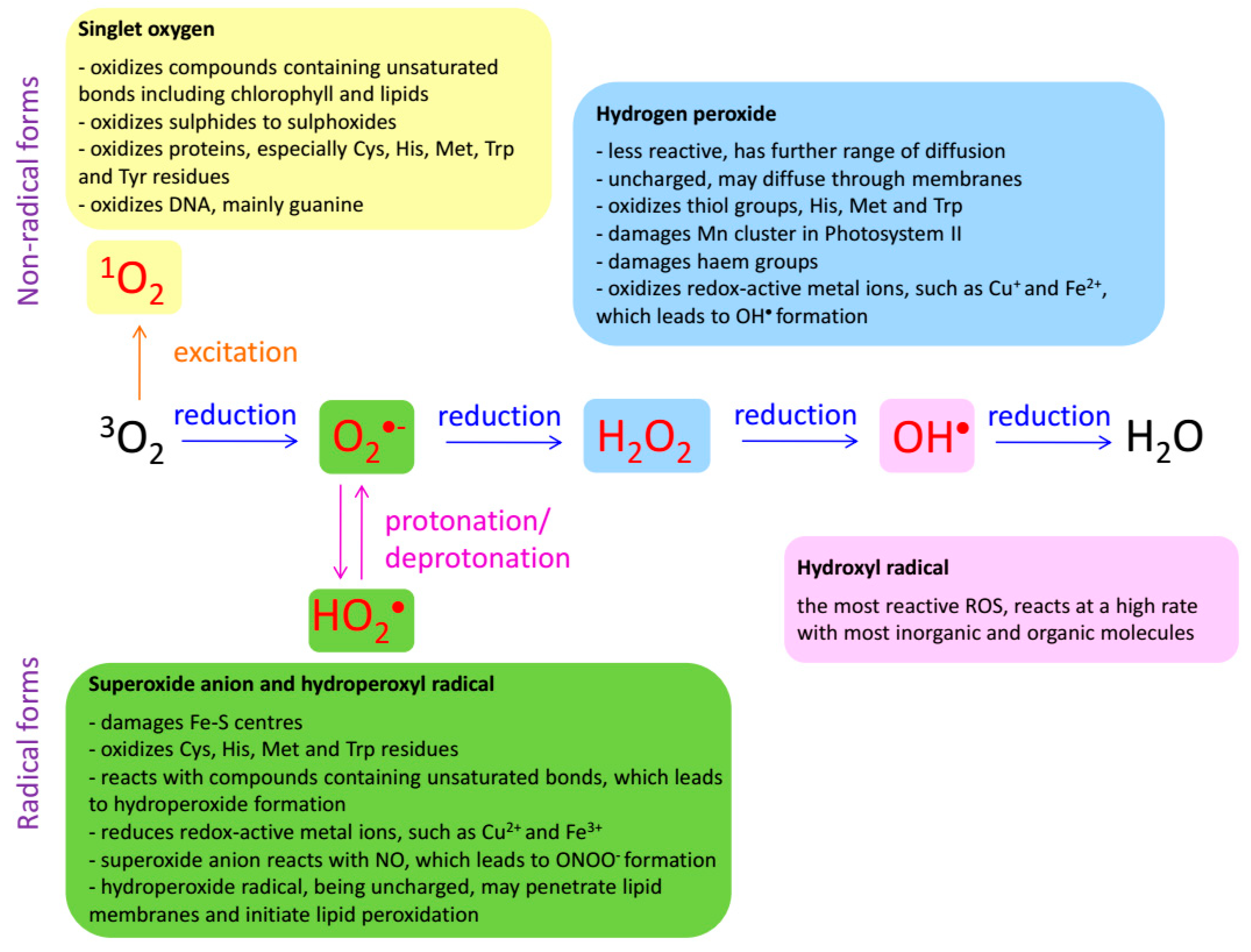
Figure 2.
Damaging action of ROS on the main types of biomolecules and its effects. Based on information from [16].
Figure 2.
Damaging action of ROS on the main types of biomolecules and its effects. Based on information from [16].
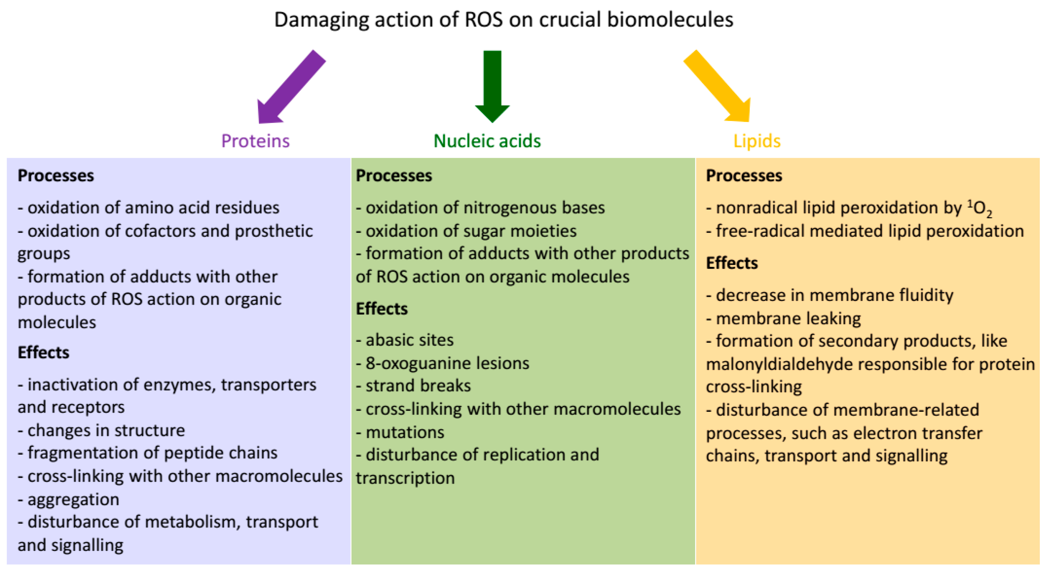
Figure 3.
The major sites of ROS production in photosynthetic cells. Based on information from [16].
Figure 3.
The major sites of ROS production in photosynthetic cells. Based on information from [16].
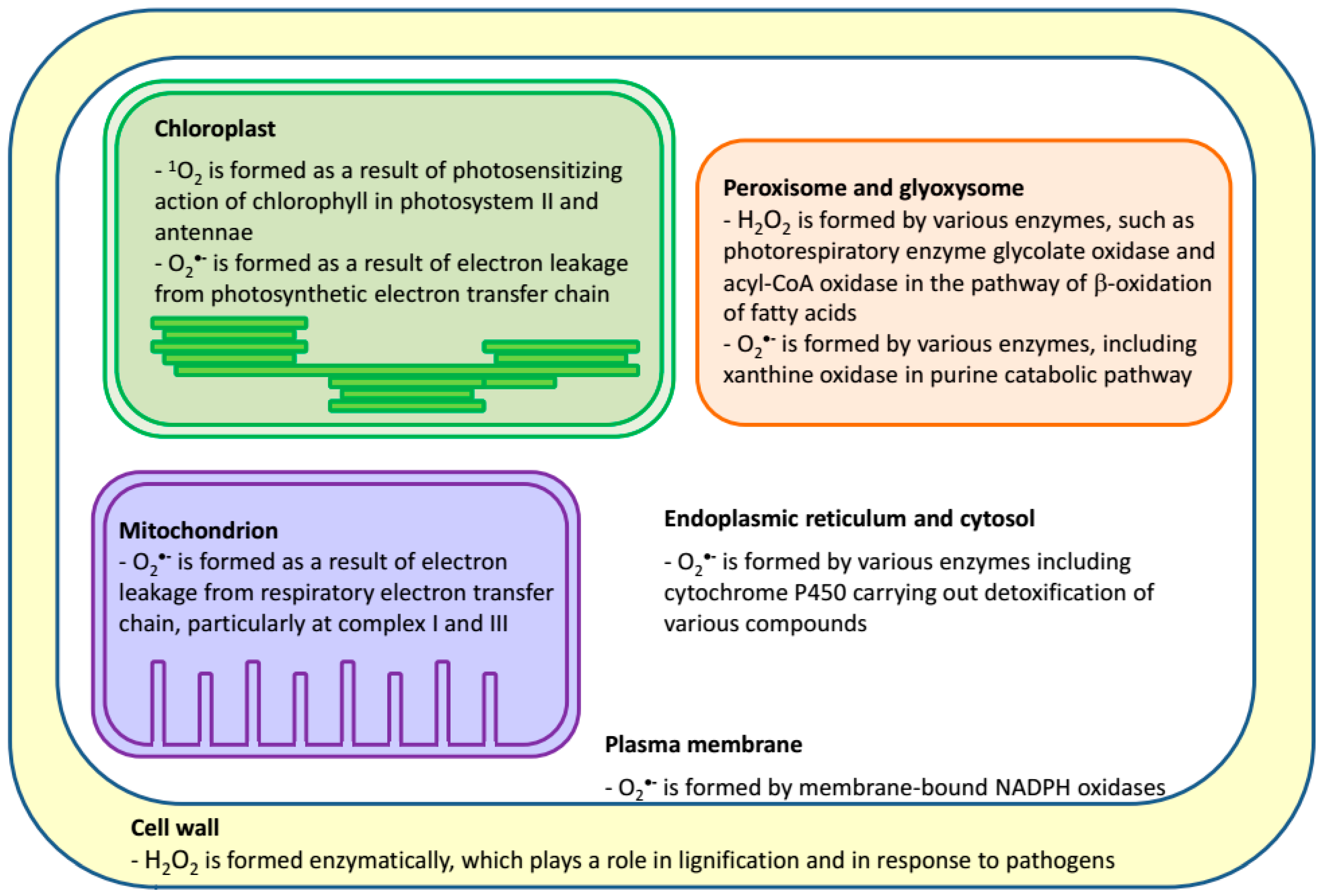
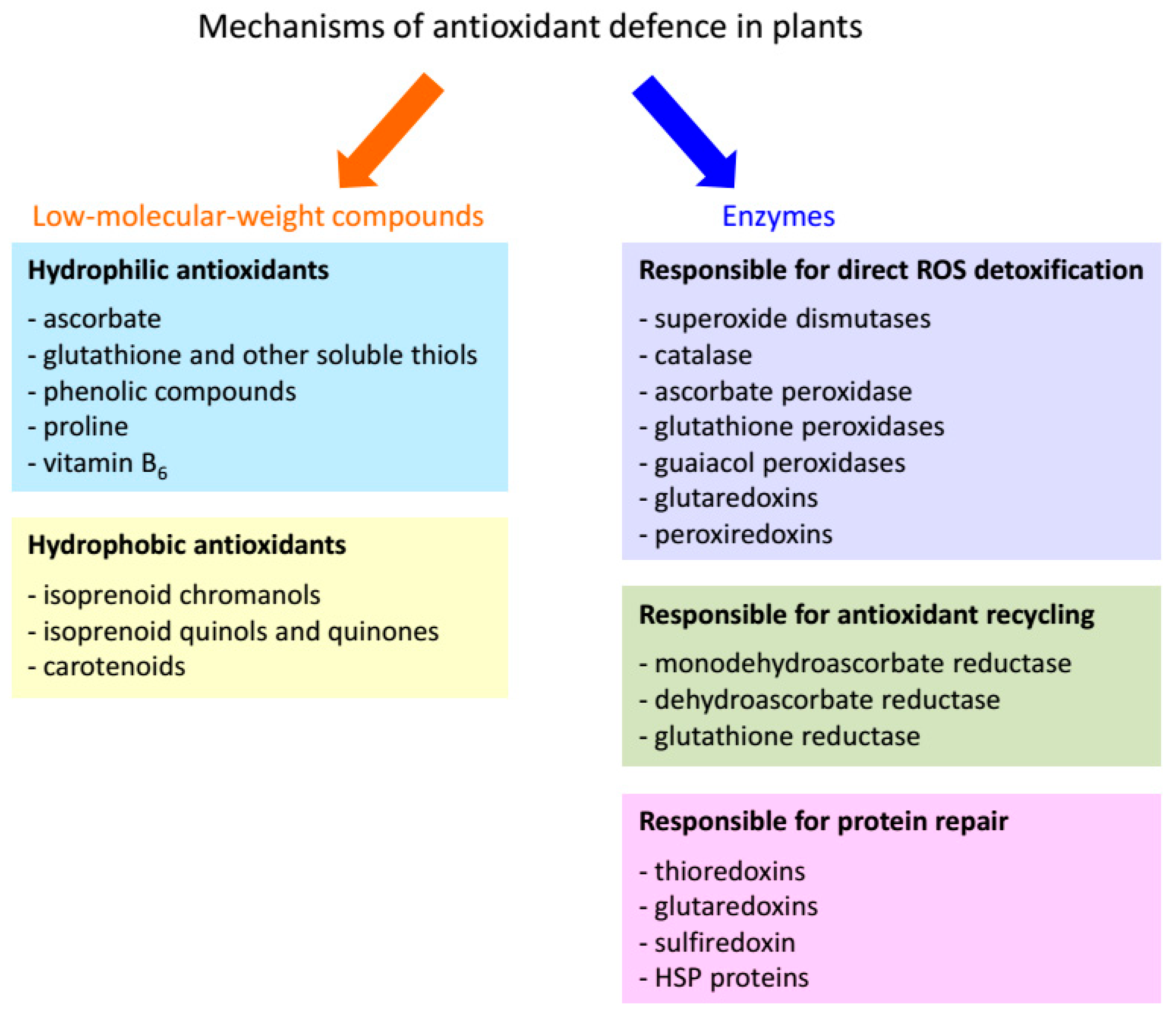
Figure 5.
Plant isoprenoid chromanols and quinones. The compounds commonly occurring in chloroplasts are marked with a dark-green colour, and the examples of their oxidation products are marked with a sap-green colour. α-T3, α-tocotrienol; α-Toc, α-tocopherol; β-Toc, β-tocopherol; γ-Toc, γ-tocopherol; δ-Toc, δ-tocopherol; α-TQ, α-tocopheryloquinone; PC-8, plastochromanol-8; PC-OH, hydroxy-plastochromanol; PhQ, phylloquinone; PQ-9, plastoquinone-9; PQ C, plastoquinone C; UQ-9, ubiquinone-9.
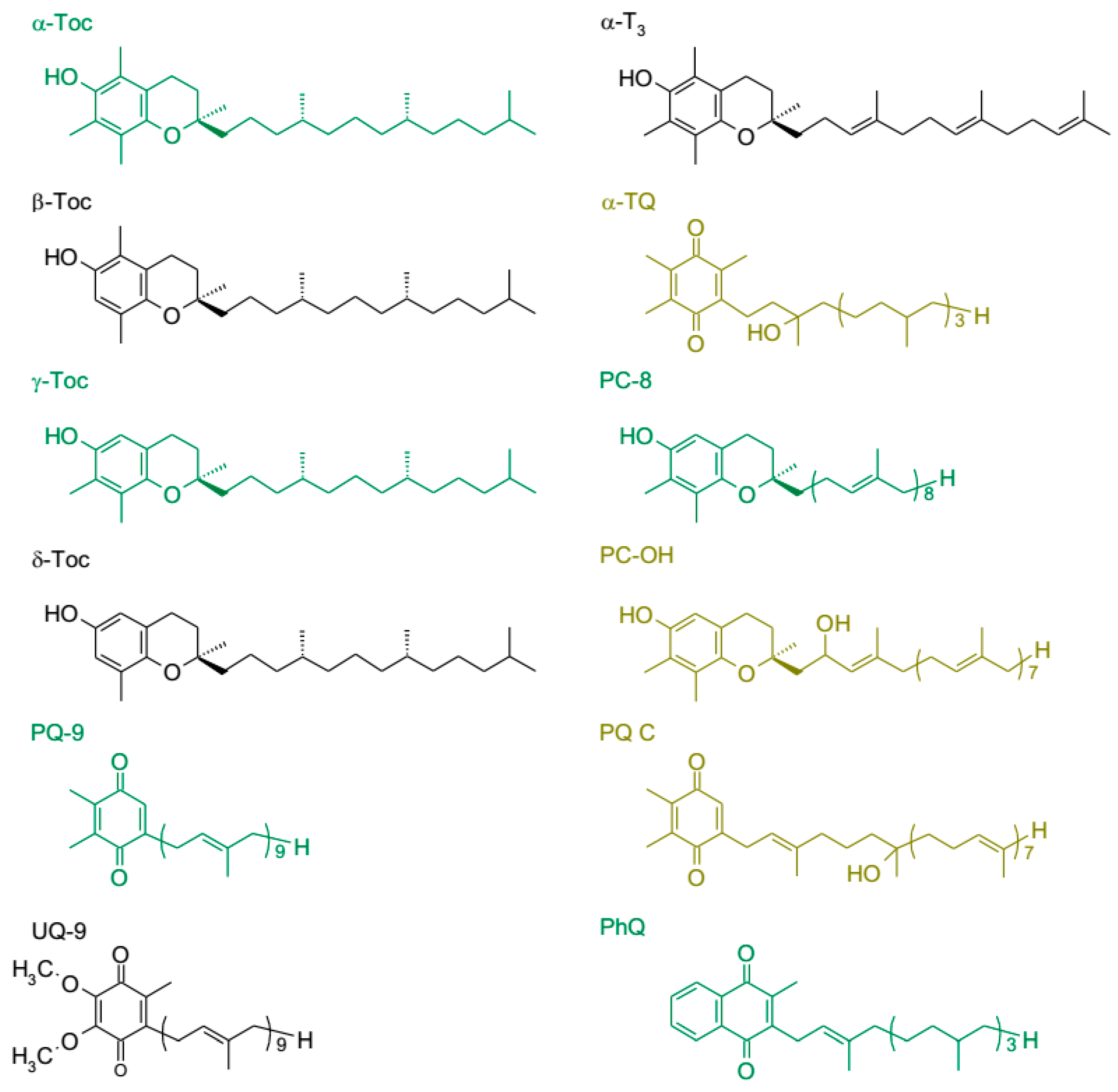
Figure 6.
Synthesis of tocopherols in higher plants. DMAPP, dimethylallyl pyrophosphate; DMPBQ, 2,3-dimethyl-6-phytyl-1,4-benzoquinone; GGDR, geranylgeranyl pyrophosphate reductase; GGPP, geranylgeranyl pyrophosphate; GGPPS, geranylgeranyl pyrophosphate synthase; HGA, homogentisic acid; HPP, p-hydroxyphenylpyruvate; HPPD, p-hydroxyphenylpyruvate dioxygenase; HPT, homogentisate phytyltransferase; IPP, isopentenyl pyrophosphate; MPBQ, 2-methyl-6-phytyl-1,4-benzoquinone; MPBQ MT, 2-methyl-6-phytyl-1,4-benzoquinone methyltransferase; PK, phytol kinase; PPK, phytyl-P kinase; PPP, phytyl pyrophosphate; TAT, tyrosine aminotransferase; TC, tocopherol cyclase; γ-TMT, γ-tocopherol methyltransferase; α-Toc, α-tocopherol; β-Toc, β-tocopherol; γ-Toc, γ-tocopherol; δ-Toc, δ-tocopherol; Tyr, tyrosine.
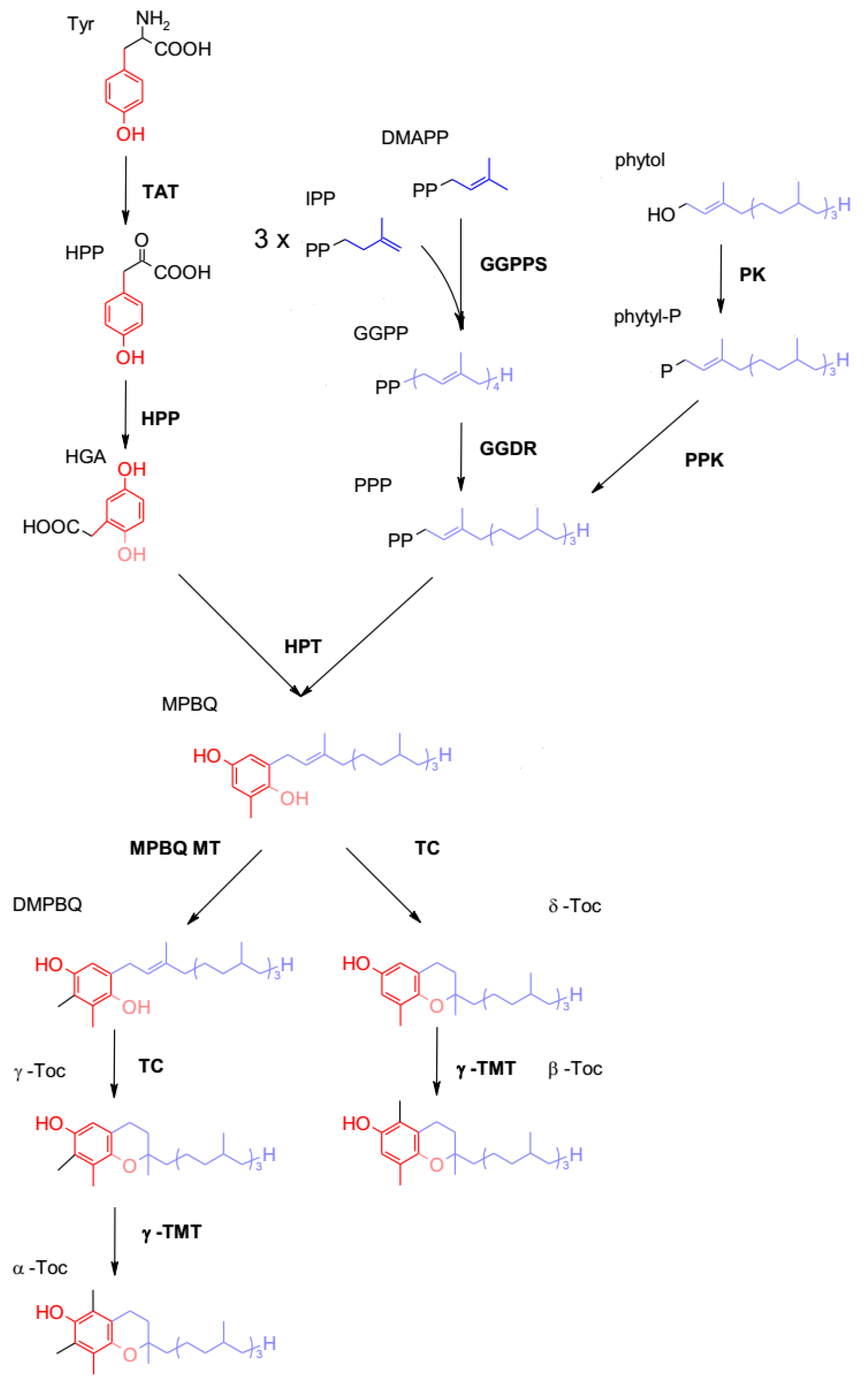
Figure 7.
Synthesis of plastoquinone and plastochromanol-8 in higher plants. GGPP, geranylgeranyl pyrophosphate; HGA, homogentisic acid; HST, homogentisate solanesyl transferase; IPP, isopentenyl pyrophosphate; MPBQ MT, 2-methyl-6-phytyl-1,4-benzoquinone methyltransferase; MSBQ, 2-methyl-6-solanesyl-1,4-benzoquinone; PC-8, plastochromanol-8; PQ-9, plastoquinone-9; PQH2-9, plastoquinol-9; SPP, solanesyl pyrophosphate; SPS, solanesyl pyrophosphate synthase; TC, tocopherol cyclase.
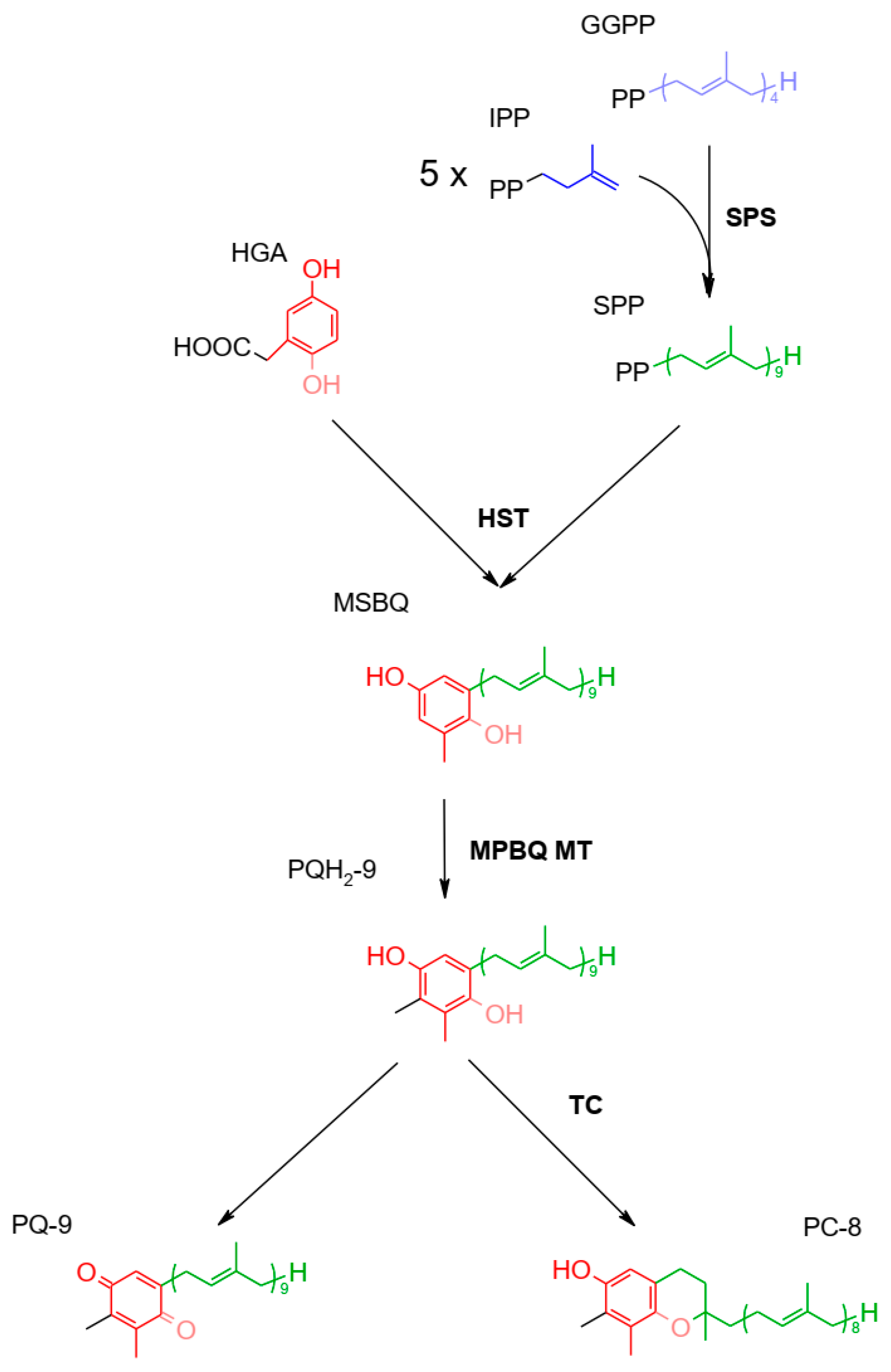
Figure 8.
Major carotenoids present in chloroplasts of higher plants. Compounds that occur in higher amounts are marked with an orange colour. α-Car, α-carotene; β-Car, β-carotene.
Figure 8.
Major carotenoids present in chloroplasts of higher plants. Compounds that occur in higher amounts are marked with an orange colour. α-Car, α-carotene; β-Car, β-carotene.
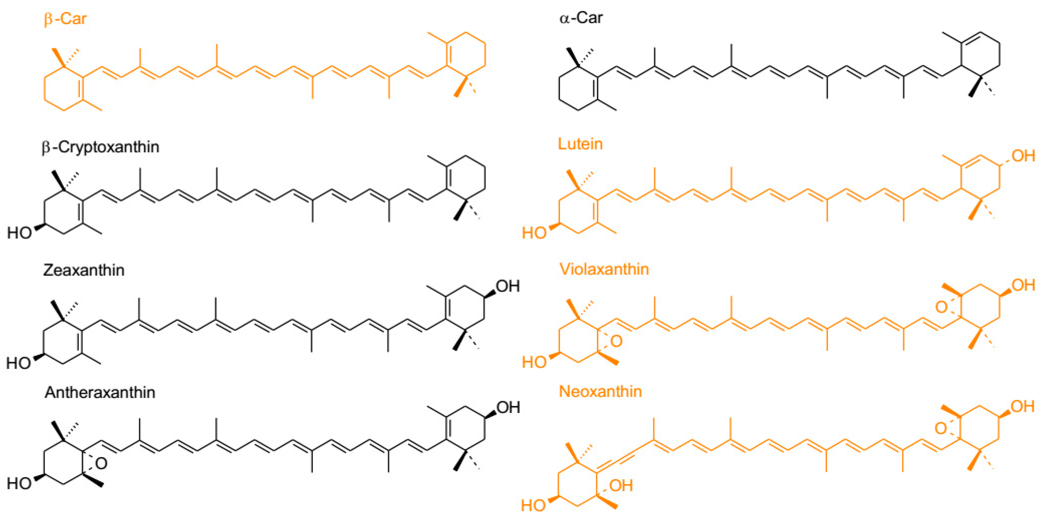
Figure 9.
Synthesis of carotenes in higher plants. α-Car, α-Carotene; β-Car, β-Carotene; γ-Car, γ-Carotene; δ-Car, δ-Carotene; CRTISO, carotenoid isomerase; GGPP, geranylgeranyl pyrophosphate; LCYβ, lycopene β-cyclase; LCYε, lycopene ε-cyclase; PDS, phytoene desaturase; PSY, phytoene synthase; ZDS, ζ-carotene desaturase; ZISO, ζ-carotene isomerase.
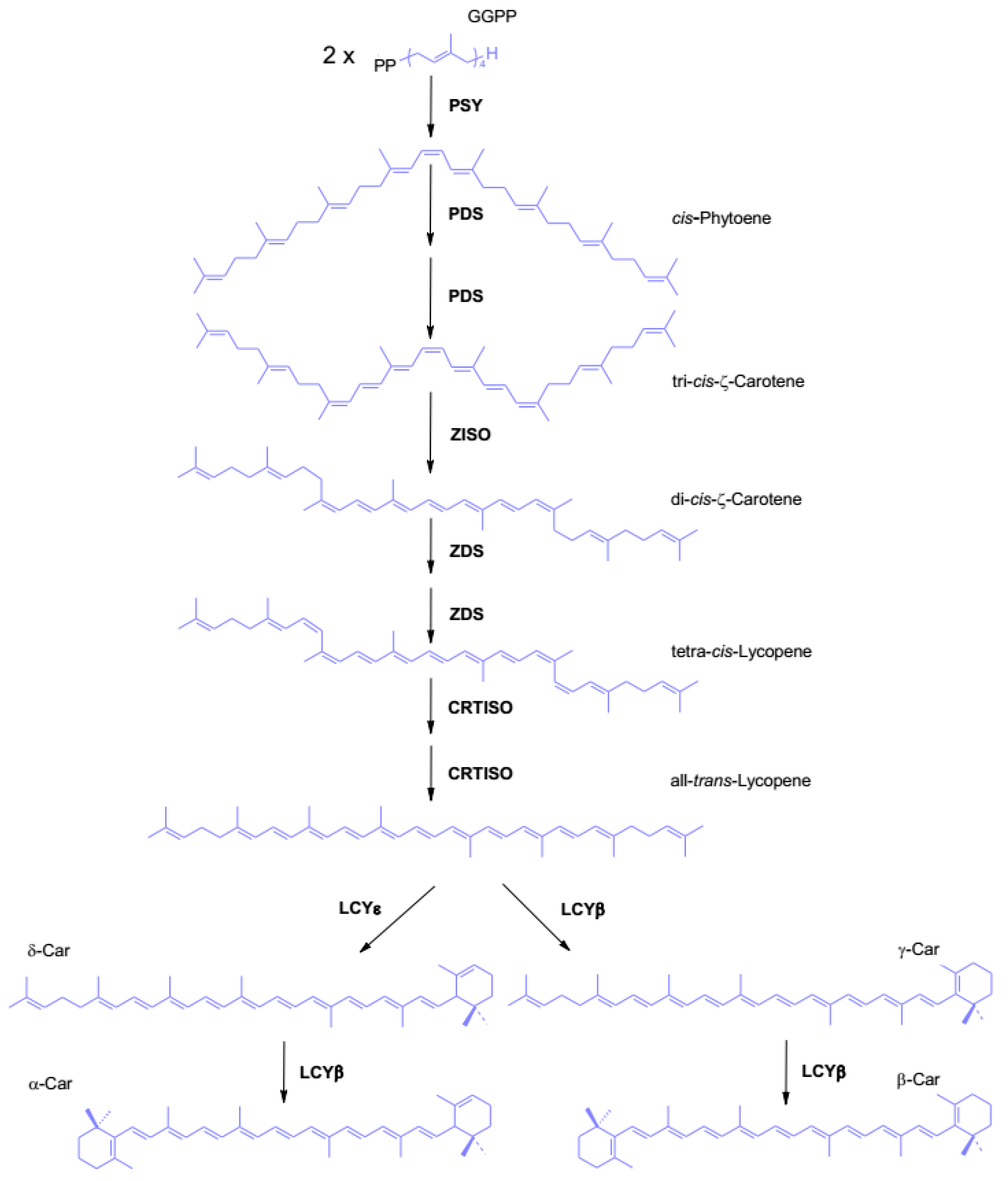
Figure 10.
Synthesis of xanthophylls in higher plants. For better clarity of the picture, the variant of lutein biosynthesis, in which the hydroxylation of α-ring precedes the hydroxylation of β-ring, and α-cryptoxanthin is an intermediate instead of zeinoxanthin, was not shown. Enzymes and reactions of the xanthophyll cycle are marked in orange. α-Car, α-Carotene; β-Car, β-Carotene; CHYβ, β-carotenoid hydroxylase; CHYε, ε-carotenoid hydroxylase; NSY, neoxanthin synthase; VDE, violaxanthin de-epoxidase; ZE, zeaxanthin epoxidase.
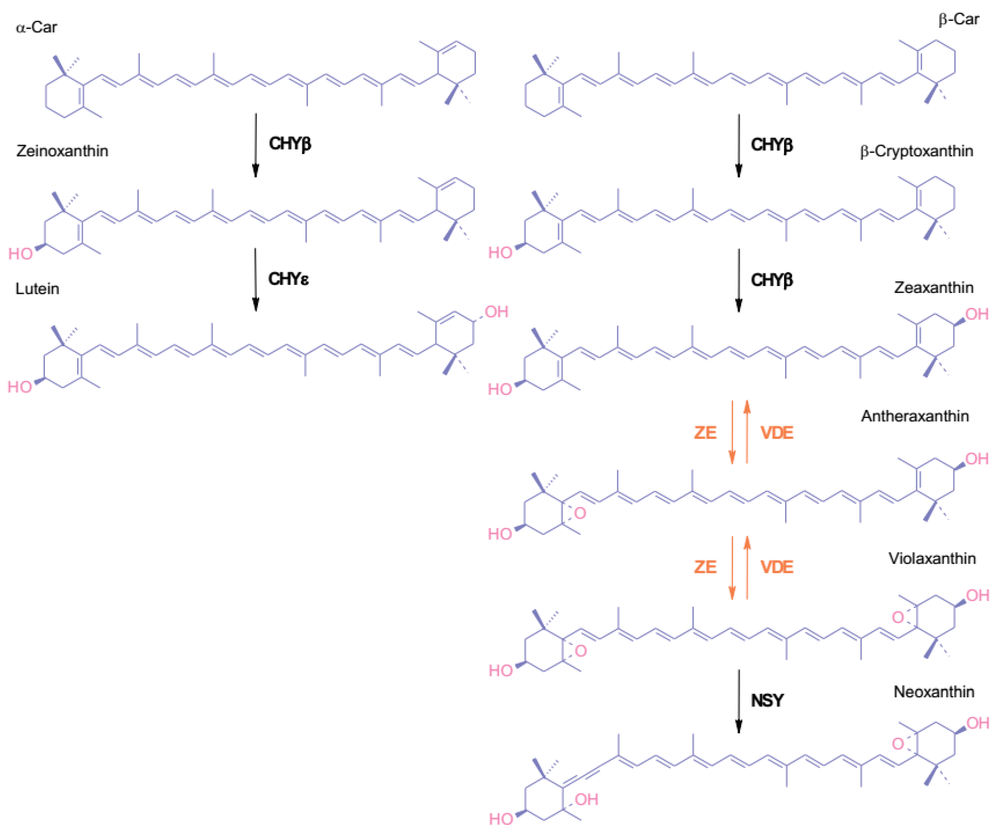

Table 1.
Impact of low temperature stress on oxidative stress markers in exposed plants. DAB, 3,3′-diaminobenzidine; MDA, malonyldialdehyde, product of lipid peroxidation; PS II, photosystem II.
Table 1.
Impact of low temperature stress on oxidative stress markers in exposed plants. DAB, 3,3′-diaminobenzidine; MDA, malonyldialdehyde, product of lipid peroxidation; PS II, photosystem II.
| Species | Experimental setup | Observation | Reference |
| coffee: Coffea arabica, Coffea canephora, differing with cold tolerance | microcuttings conserved in vitro at 12, 17, 22, 27 °C; measurements carried out at t 0, 1, 2, 4, 8, 12, 16, 20 weeks | increase in MDA content inversely correlated with the survival rate and cold sensitivity of tested species | [178] |
| chickpea (Cicer arietinum) |
15-day-old seedlings grown 24/21 °C day/night, transferred to 10 °C; measurements carried out at t 1, 2, 3, 4, 5, 6, 7 days | 5-fold increase in H2O2 in time until day 6, 4-fold increase in MDA in time until day 5 | [179] |
| chickpea (Cicer arietinum) |
3-week-old seedlings grown at 23 °C, kept for the next 10 days at 23 or 10 °C, then exposed to 4 °C; measurements carried out at t 2, 4 days of incubation at 4 °C | increase in H2O2 in cold-stressed non-acclimated seedlings, up to 40% of pre-stressed value | [180] |
| soybean (Glycine max) | 3-day-old seedlings grown at 25 °C, transferred to 1 °C; measurements carried out at t 0, 2, 4, 6, 8 days | in roots MDA content increased over time, reaching 3-fold of the initial value on the 8th day, in hypocotyls the increase was not statistically significant | [181] |
| lentil (Lens culinaris) | 10-day-old seedlings grown at 23 °C were subjected to 4 °C or 23 °C for 5 days; measurements carried out at the end of treatment | 3.7-fold increase in H2O2 in shoots compared to control, increase in MDA content in shoots and roots compared to control, 2.25- and 2.15-fold, respectively | [182] |
| wheat (Triticum aestivum), two varieties: hardy winter wheat Sadovo-1 and non-hardy spring wheat San Pastore |
12-day-old seedlings grown 20/16 °C day/night, transferred to 2 °C, after 7 days of cold acclimatisation exposed to freezing stress: 24 h in darkness with gradually decreasing temperature 1 °C per h, to final temperature -6 °C, then recovered at 20/16 °C for 24 h; measurements carried out at the end of cold hardening, freezing stress, and recovery period | increase in H2O2 after cold-hardening phase compared to non-stressed control, 1.4-fold for winter wheat and 2-fold for spring wheat, increase in MDA content after cold hardening phase, compared to control, 2.75-fold for winter wheat and 2.3-fold for spring wheat, 1.7-fold increase in MDA content during recovery phase for spring variety | [183] |
| wheat (Triticum aestivum), two varieties Sids 1 and Gimmieza 7 |
15-day-old seedlings grown at 21/15 °C day/night, exposed to 5 °C for 6 and 9 hours or not stress-treated, then again kept at 21/15 °C; measurements carried out at 25 days post germination | increase in H2O2 compared to control, about 1.6-fold for Sids 1 and 2.7-fold for Gimmieza 7, 2-fold increase in MDA content compared to control |
[184] |
| naked oats (Avena nuda) | seedlings grown at 20 °C until they reached the four-leaf stage, transferred to 1 °C or kept at 20 °C; measurements carried out at t 0, 1, 3, 5, 7 days | 2.75-fold increase in MDA in cold-stressed plants, no increase in control series | [185] |
| sweet sorghum (Sorghum bicolor), two inbred lines M-81E and Roma, differing with cold tolerance | seedlings grown at 28/20 °C day/night until they reached the four-leaf stage, then transferred to 10 °C; measurements carried out at t 0, 12, 24, 36 and 48 h | increase in MDA content over time inversely correlated with the maximum quantum yield of PS II and the sensitivity of tested line | [186] |
| rice (Oryza sativa) | 14-day-old seedlings grown 28/22 °C day/night, transferred to 12 °C or kept at 28/22 °C; measurements carried out after 6 days | 3-fold increase in MDA content compared to control | [187] |
| tomato (Solanum lycopersicum) | seedlings grown at 25/15 °C day/night until they reached the five-leaf stage + 7 days, then subjected to 4 °C for 24 h, recovered at 25/15 °C; measurements carried out at t 0, 12, 24 h of cold incubation and after 10 h of recovery | about 65% increase in H2O2 after 12 h of cold treatment, then slow decrease | [188] |
| tomato (Solanum lycopersicum) | seedlings grown at 25/18 °C day/night until they reached the four-leaf stage + 7 days, then subjected to 3 °C for 6 h/day for 6 days or not stress-treated, recovered for 72 h; measurements carried out at the end | 2.25-fold increase in H2O2 compared to control | [189] |
| tomato (Solanum lycopersicum) | seedlings grown at 25/18 °C day/night until they reached the five-leaf stage, transferred to 15 °C/8 °C (day/night) or non-stressed; measurements carried out after 48 h | 30% increase in MDA content, correlated with decrease in maximum quantum yield of PS II | [190] |
| thale cress (Arabidopsis thaliana) | 2-week-old plants grown at 22 °C, transferred to 4 °C; measurements carried out after 24 h | increase in H2O2 visualised using DAB staining compared to t 0, 2-fold increase in MDA content compared to non-stressed control | [191] |
| Nanfeng tangerine (Citrus reticulate) | 3-year-old seedlings, kept at 28/18 °C day/night for 7 days, acclimated at 10/7 °C day/night for 3 days or kept under non-stressful conditions, exposed to 7/4 °C day/night for 3 days; measurements carried out at the end of experiment | statistically non-significant increase in H2O2 compared to control, slightly lower in cold-acclimated seedlings compared to non-acclimated ones, increase in MDA content compared to control, about 1.9-fold for non-acclimated seedlings and 1.2-fold for cold-acclimated ones | [192] |
Table 2.
Mechanisms of acclimatisation and adaptations to low temperature. Based on the information from [2,158,160,162,164,168,173,174,176,195,201,202,203,204,205].
| Stress type | Character of changes and adaptations | Mechanism |
| cold stress | biochemical |
- changes in the content of membrane lipids
- increased capacity for PS II repair - decrease in the amount of antennae and chlorophyll content - enhanced photoprotection - switching off feedback down-regulation by accumulated sugars
|
| morphological |
|
|
| freezing stress | biochemical |
- act as osmoprotectants - protect structures of macromolecules and membranes - act as antioxidants - enable protoplasm vitrification in extremely frost-resistant species
- enhanced pectin synthesis to fill the extracellular spaces |
| tissue and organ level |
|
|
| developmental |
- surviving winter as dormant, underground storage organs, such as bulbs, tubers, and rhizomes - shedding freeze-sensitive organs, mainly leaves, before winter |
Table 3.
The content of α-tocopheryloquinone (α-TQ), α-tocopherol (α-Toc), δ-tocopherol (δ-Toc), and γ-tocopherol (γ-Toc), expressed as mols/100 mols of total chlorophyll, in leaves of A. thaliana exposed to stress conditions for 14 days.
Table 3.
The content of α-tocopheryloquinone (α-TQ), α-tocopherol (α-Toc), δ-tocopherol (δ-Toc), and γ-tocopherol (γ-Toc), expressed as mols/100 mols of total chlorophyll, in leaves of A. thaliana exposed to stress conditions for 14 days.
| α-TQ | δ-Toc | γ-Toc | α-Toc | |
| Control, 80 μmols photons m-2s-1, 21°C | 0,004 ± 0,004 | 0,011 ± 0,004 | 0,034 ± 0,006 | 1,064 ± 0,168 |
| 200 μmols photons m-2s-1, 21°C | 0,191 ± 0,020 | 0,347 ± 0,062 | 4,448 ± 0,666 | 52,012 ± 6,870 |
| 80 μmols photons m-2s-1, 4°C | 0,148 ± 0,070 | 0,506 ± 0,031 | 14,454 ± 0,882 | 36,176 ± 2,885 |
Disclaimer/Publisher’s Note: The statements, opinions and data contained in all publications are solely those of the individual author(s) and contributor(s) and not of MDPI and/or the editor(s). MDPI and/or the editor(s) disclaim responsibility for any injury to people or property resulting from any ideas, methods, instructions or products referred to in the content. |
© 2026 by the authors. Licensee MDPI, Basel, Switzerland. This article is an open access article distributed under the terms and conditions of the Creative Commons Attribution (CC BY) license (http://creativecommons.org/licenses/by/4.0/).
Copyright: This open access article is published under a Creative Commons CC BY 4.0 license, which permit the free download, distribution, and reuse, provided that the author and preprint are cited in any reuse.