

Submitted:
28 September 2026
Posted:
30 September 2026
You are already at the latest version
Abstract
Acute kidney injury (AKI) and chronic kidney disease (CKD), such as ischemia reperfusion injury, renal sepsis, diabetic nephropathy and renal fibrosis, remain major causes of morbidity and mortality around the world. Despite current therapeutic advances, progression to renal dysfunction and end-stage renal diseases continue to be a major clinical concern. Honokiol, a low molecular weight natural product derived from Magnolia officinalis, has demonstrated numerous renoprotective effects on kidney disorders. The objective of this review was to discuss the latest findings on the protective effects of honokiol on a variety of models of kidney injury. These models include diabetic nephropathy, CKD by adenine administration, unilateral ureteral obstruction (UUO)-induced renal fibrosis, renal ischemia reperfusion, and sepsis-induced acute kidney injury as well as cyclosporin A, cadmium, and cisplatin renal toxicity. Numerous investigations on honokiol's protective mechanisms focus on critical pathological processes such as mitochondrial dysfunction, reactive oxygen species generation, inflammatory signaling, and tissue remodeling. These studies suggest that honokiol demonstrates renoprotective effects by modulating multiple pathways, including SIRT3-mediated mitochondrial homeostasis, NLRP3 inflammasome activation, oxidative stress responses, Wnt/β-catenin signaling, and miR-218-5p/heme oxygenase-1 (HO-1) signaling. These mechanisms contribute to the preservation of mitochondrial homeostasis, reduction of renal inflammation, prevention of extracellular matrix accumulation, and improvement of kidney function in various experimental models. Our review ends with suggestions for future studies on the potential of honokiol as a promising therapeutic agent for the prevention and treatment of renal disorders.
Keywords:
honokiol
; kidney acute kidney injury
; chronic kidney disease
; sirt3
; oxidative stress
; mitochondrial dysfunction
1. Introduction
The kidney, as a homeostasis organ [1,2], is one of the hardest working organs in our body and plays a critical role in maintaining good health. Each kidney works hard to filter all the blood in the body every thirty minutes. They also remove excess fluid, toxins, drug metabolites, and waste to prevent toxic accumulation that cause damage to the kidneys, which can result in acute kidney injury (AKI) and chronic kidney diseases (CKD) [3,4]. In particular, CKD affects millions of individuals and more than 1 in 10 American adults has CKD, and as many as 9 in 10 don’t know they have it [5]. Despite that CKD can be treated and many efforts have been proven by utilizing renin-angiotensin system inhibitors [6], sodium glucose cotransporter-2 inhibitors [7,8,9], and mineralocorticoid receptor antagonist [10], many patients still progress to renal failure or end-stage kidney disease. This concern highlights the need for more therapeutic approaches that can target the underlying mechanism of disease progression.
The pathogenesis of kidney disease is complex and involves multiple interconnected pathways. Current studies have identified mitochondrial dysfunction, oxidative stress, inflammation, and fibrosis as major contributors to renal injury. While basal level of reactive oxygen species (ROS) plays a beneficial redox signaling role in cellular function [11,12], excessive production of ROS usually disrupts cellular homeostasis and damage proteins [13,14], lipids [15,16], and DNA [17,18], which can all lead to impaired renal function. Mitochondrial dysfunction further exacerbates mitochondrial ROS production [19,20], which activates nucleotide-binding oligomerization domain-like receptor protein 3 (NLRP3) inflammasome [21]. Furthermore, persistent inflammation leads to excessive extracellular matrix (ECM) deposition, tubular injury and activation of pro-fibrotic signaling pathways such as TGF-β/Smad [22,23,24], ultimately contributing to the progression of CKD or AKI transition to CKD [25,26,27]. It should be noted that in addition to mitochondria, xanthine oxidase [28,29] and NADPH oxidases (NOX), in particular, NOX4 [30,31,32,33], has also been implicated in oxidative stress and cellular dysfunction in a variety of kidney disorders.
Honokiol (HKL) (Figure 1), a naturally occurring biphenolic molecule derived from Magnolia officinalis [34,35], has gained attention for its therapeutic potential in a variety of disease models. HKL was first isolated in 1972 [35,36]. Due to its hydrophobicity property, it can cross the blood brain barrier [37]. Therefore, its neuroprotective effects have been studied in a variety of neurological disorders [38,39] including Alzheimer’s disease [40,41] and Parkinson’s disease [42,43]. Nonetheless, HKL has also been demonstrated to exhibit other bioactive functions such as antitumor, anti-inflammation, antioxidation, antibacterial, and antivirus. HKL is metabolized in the liver whereby it undergoes biotransformation [37]. Oral administration in rats reveals that it could rapidly distribute in the brain, the liver, and the kidney [37]. Indeed, recent investigations on the kidneys reveal that HKL has considerable renoprotective benefits by modulating numerous signaling pathways implicated in disease progression.
Recent research has highlighted the importance of SIRT3, a mitochondrial deacetylase, in mediating the protective effects of HKL [44]. In microenvironments, SIRT3 plays a critical role in controlling fibrotic gene networks and signaling molecules [45,46]. In the case of kidney, as a high-energy-demanding organ, SIRT3 helps to regulate mitochondrial protein acetylation levels [47,48]. This effect influences mitochondrial energy production and ultimately kidney function. Unfortunately, this process is disrupted in renal fibrosis, whereby the role of HKL becomes more critical. In experimental models of diabetic nephropathy, HKL attenuated renal injury by activating SIRT3, reducing mitochondrial ROS production, and suppressing NLRP3 inflammasome-mediated pyroptosis [10,21,49]. Similarly, studies of renal fibrosis have shown that HKL suppresses extracellular matrix deposition and epithelial-mesenchymal transition through SIRT3-dependent inhibition of Wnt/ β-catenin signaling [50]. Furthermore, in ischemia-reperfusion injury, HKL enhances endogenous antioxidant defenses by promoting glutathione (GSH) biosynthesis and activating cryoprotective signaling pathways [51]. In the case of renal sepsis, HKL is known to ameliorate acute kidney injury by modulating miR-218-5p/heme oxygenase-1 (HO-1) signaling [52].
2. Renoprotective Effects of Honokiol on Renal Injuries
2.1. Honokiol and Diabetic Nephropathy
Diabetes mellitus has always been associated with several complications and presented as a critical global public health challenge. Among the most severe microvascular complications, diabetic nephropathy (DN) or diabetic kidney disease (DKD) has shown a parallel increase in incidence and persisted as the leading cause of chronic kidney disease (CKD) and end-stage renal disease worldwide. It has been estimated that approximately 40% of diabetic patients can develop DKD [53,54]. The progression of DKD is characterized by persistent hyperglycemia-induced oxidative stress, mitochondrial impairment, chronic inflammation and cell death, ultimately resulting in irreversible renal damage. However, many efforts including inhibitors of renin-angiotensin system (RAS) [6,55], sodium-glucose cotransporter-2 (SGLT2) inhibitors [7,8,56], and novel nonsteroidal mineralocorticoid receptor antagonists (MRAs) [57,58], have partially attenuated the progression of renal pathology. Unfortunately, these strategies of DN treatments fail to completely halt the progression, which demands the need for further research into underlying mechanisms and novel intervention.
In terms of the role of HKL in diabetic nephropathy, it has been found that HKL mitigates renal dysfunction and pathological renal injury in mouse model of diabetic nephropathy. As compared with the control group, db/db diabetic mice presented with high levels of serum creatinine (SCr), blood urea nitrogen (BUN), and 24-hour urinary protein (UP) [10]. These markedly elevated parameters indicate significant renal dysfunction in db/db mice. In contrast, the HKL-treated group (5mg/kg HKL for 8 weeks) ameliorated these changes in renal function parameters (Figure 2). Also, HKL has been proven to inhibit NLRP3/GSDMD-mediated pyrotosis in the kidneys of diabetic mice. Immunohistochemical results revealed that the renal tissue of db/db mice has significantly high expression of pyroptosis-marker proteins. With a specific focus on NLRP3 inflammasome triggered pyrotosis as a key contributor, caspase-1 activation mediates cleavage of Gasdermin D (GSDMD) [59,60], generating its membrane-pore-forming N-terminal domain (GSDMD-N). This disruption of membrane allows the release of inflammatory mediators IL-18 and IL-1β, resulting in an endless cycle [61]. Thus, HKL-treatment has suppressed the upregulation of these pyrosis-related proteins.
Mechanistically, HKL restores mitochondrial homeostasis through activation of SIRT3, resulting in decreased mitochondrial ROS (mtROS) generation and improved cellular antioxidant capacity. In the HKL-treated group, western blot and immunofluorescence further confirmed that activation of SIRT3 also suppresses NLRP3 inflammasome activation, thereby reducing all the key inflammatory mediators in renal tissues [10,62]. In addition to inhibiting inflammation, HKL has been shown to preserve mitochondrial function by maintaining mitochondrial membrane potential and preventing oxidative damage induced by chronic hyperglycemia. The reduction of mtROS not only attenuates NLRP3 inflammasome activation but also interrupts the vicious loop of oxidative stress and mitochondrial injury that drives diseases progression. These findings suggest that HKL exhibits renal protection through the modulation of the SIRT3-mtROS- NLRP3 axis [10,62]. In high fat diet/streptozotocin induced diabetic nephropathy, HKL was found to mitigate hyperglycemia, redox imbalance, and inflammation, which led to improvement in renal function [63].
2.2. Honokiol and renal Fibrosis
Renal fibrosis is the common pathological process occurring in nearly all CKD [64,65], and is characterized by injury, inflammation, ECM deposition, and renal parenchyma destruction [66,67]. As mentioned in the previous section, SIRT3’s function in controlling fibrogenesis gene network and signaling molecules was the growing focus in the kidney microenvironment, but the fibrogenesis mechanisms are more complex. For this reason, the activation of Wnt/β-Catenin has been associated with the alterations at the level of transcripts and proteins and has been identified as the key mitochondrial drivers of fibrogenesis [68,69].
First, HKL suppresses renal fibrosis through attenuation of the Wnt/β-Catenin signaling axis in unilateral ureteral obstruction (UUO) mice [50]. Using immunohistochemical staining of renal section, a reduced β-Catenin expression in UUO-HKL treated mic was observed. To access the regulation of the Wnt/β-Catenin signaling axis, Western blot analysis was conducted by inspecting balance of phosphorylation and dephosphorylation of key protein β-Catenin [50]. With this method, HKL treated UUO-mice showed a decrease in this key protein expression and an increase in phospho- β-Catenin, as compared to the UUO mice treated with vehicle. Therefore, treatment with HKL has proven to downregulate β-Catenin accumulation and thereby limiting downstream profibrotic gene. This inhibition was accompanied by decreased expression of fibrosis-related genes like collagen I, Snail1, Twist, MMP-7, Fibronectin, and α-SMA, reducing excessive EMT and ameliorating the accumulation of ECM [70]. These findings indicate that HKL effectively demonstrates anti-fibrotic effect on renal structure and function.
Another principal mechanism underlying the anti-fibrotic effects of HKL is the activation of mitochondrial SIRT3. It was found that UUO-induced mitochondrial fission could be reversed by HKL via sirt3 activation [71]. Loss of SIRT3 usually results in mitochondrial dysfunction, increased oxidative stress, and activation of profibrotic signaling pathways. In the study of fibrotic kidney and TGF-β stimulated HK-2 cells, HKL has been shown to restore SIRT3 expression and limit mitochondrial ROS accumulation. This finding proves preservation of mitochondrial function is a critical component of renoprotective activity. On the other hand, silencing SIRT3 diminished the full effect of HKL. Studies have explored further to scrutinize specific functions of SIRT3 by conceptualizing a model of conditional SIRT3 knockout (cKO) mice [50]. Experiments revealed a high expression of β-Catenin and decreased appearance of SIRT3 in UUO-cKO group as compared with the WT group. A qPCR analysis showed significantly inhibited mRNA levels of SIRT3 and upregulated mRNA levels of other fibrotic proteins like Collagen III, α-SMA, and β-Catenin in the UUO-cKO group. This above model solidifies the role of SIRT3 in control of renal fibrosis more specifically and directly, proving that SIRT3 is a critical upstream mediator which downregulates the Wnt/β-Catenin signaling axis.
2.3. Honokiol and Renal Ischemia-Reperfusion
Renal ischemia-reperfusion injury (IR) is a major cause of acute kidney injury (AKI) and is characterized by an ischemic period of lacking oxygen and nutrients by blood blockage. Although sudden restoration occurs after an ischemic period, reperfusion paradoxically exacerbates tissue injury due to sudden outburst of ROS generation, leading to endothelial and organ dysfunction. These events culminate in tubular epithelial injury, impaired renal function and may contribute to the progression from AKI to CKD. The reperfusion of ischemic tissue also creates an imbalance between ROS generation and tissue’s ability to detoxify ROS. Renal tissues subjected to IR presented a marked accumulation of xanthine oxidase (XO) substrates (hypoxanthine, xanthine and succinate), one of the major sources of ROS production [72]. In addition, the XO inhibitors such as febuxostat and allopurinol are known to suppress renal IR injury via reduced oxidative stress. Another cause of oxidative stress under IR is by reduced or impaired antioxidant activities. Among many antioxidant systems protecting renal tissue from oxidative stress, glutathione (GSH) is a pivotal antioxidant that has been investigated in many studies for its antioxidative ability in protecting cells by neutralizing hydrogen peroxide [73,74]. GSH synthesis is primarily regulated by the rate-limiting enzyme glutamate-cysteine ligase (Gcl). The catalytic subunit of Gcl (Gclc), whose expression is induced by oxidative stress and regulated by nuclear factor-erythroid 2 related factor 2 (Nrf2) signaling [51,75]. Consequently, renal IR is associated with significant depletion of GSH, which contributes to oxidative stress and tissue damage, making the restoration of GSH biosynthesis a promising therapeutic strategy.
Honokiol is known to significantly increase GSH levels by upregulating the catalytic and modulatory subunits of Gcl, Gclc and Gclm (glutamate-cysteine ligase modifier subunit), respectively. Experimental data has proven that the relative mRNA levels of Gclc, Gclm and glutathione synthetase increased significantly in cells treated with 1 or 2 µM of honokiol for 3,6, or 12 hours [51]. HKL also increased the protein levels of GSH subunits but the level of glutathione synthetase was not significantly changed after treatment. In addition, these GSH biosynthetic enzymes are activated transcriptionally by Nrf2, thus the HKL-induced upregulation is through Nrf2-mediated transcriptional activation (Figure 3). Activation of this pathway enables renal cells to effectively neutralize ROS production during reperfusion, thus reducing oxidative damage and preserving cellular integrity. Since mitochondria are both the primary source and major target of ROS during reperfusion, HKL’s role in limiting ROS accumulation through Nrf2- mediated GSH production also interrupts this self-perpetuating cycle of mitochondrial dysfunction and oxidative stress. This preservation of mitochondria continues ATP production, maintains cellular viability, and improves renal recovery following ischemic injury.
The Nrf2 transcriptional activity has shown to be enhanced by signaling kinases, specifically phosphokinase 2-kinases (PI3K)/AKT and protein kinases C (PKC) [76,77]. Pharmacological inhibition of PI3K/Akt or PKC signaling has experimentally attenuated HKL-induced Nrf2 activation [51]. This evidence demonstrates that both pathways are critical mediators of HKL’s antioxidant effects. By stimulating these pathways, HKL has proven to enhance GSH production and reduce ROS-mediated oxidative damage during renal ischemia-reperfusion injury. This finding implies that HKL not only exerts renoprotection by directly scavenging free radicals but also by activating endogenous signaling pathways that maintain redox homeostasis. It should be noted that while Nrf2 phosphorylation is not always needed for Nrf2 activation and translocation [78,79,80], phosphorylation itself does enhance the Nrf2 signaling process [81,82,83].
2.4. Honokiol and Renal Sepsis
Another protective influence of Honokiol is on sepsis-associated acute kidney injury (SA-AKI). Sepsis is characterized by a dysregulated host response to infection and remains one of the leading causes of AKI in critically ill patients. SA-AKI is one of the most severe complications of sepsis, which is associated with high morbidity and mortality. While the pathogenesis of SA-AKI is complex, oxidative stress and inflammatory signaling are thought to be implicated. Zhang et al. [52] found that HKL can ameliorate AKI in septic mice and lipopolysaccharide (LPS)-induced glomerular mesangial cell (GMC) dysfunction by targeting miR-218-5p/heme oxygenase-1 (HO-1) signaling.
HO-1 is an inducible enzyme [84,85] and has a protective effect against AKI in various animal models such as sepsis-, ischemia-reperfusion- and kidney transplantation-induced AKI [86,87,88]. Evidence from multiple rodent models supports the role of HO-1 as a cytoprotective enzyme that is adaptively upregulated in response to tissue injury [89]. Knockout of this enzyme will result in enhancement of inflammatory response and renal structural injury. In addition, microRNAs (miRNAs) are also the target of this study. miRNA such as miR-107, miR-124, miR-204 have been known as mediators of sepsis-induced AKI for their ability to specifically bind to 3’-untranslated regions (3’-UTRs) to inhibit translation of target genes [52]. For this reason, the authors aimed to target the role of miR-218-5p and its target gene HO-1 in SA-AKI to further increase our understanding of the renoprotective effect of HKL.
LPS-induced AKI is widely used as experimental model for investigating the pathophysiology of sepsis-associated renal injury [90,91,92]. Using this model, Zhang et el. Demonstrated that HKL significantly alleviated LPS-induced renal injury by improving renal histopathological changes and reducing serum creatinine and blood urea nitrogen levels. Treatment with HKL also suppressed the production of pro-inflammatory cytokines such as TNF- α, IL-6, and IL-1 β, thus ameliorating oxidative stress and glomerular mesangial cell apoptosis in renal tissues [52]. Also, mechanistically HKL downregulated mi-R-218-5p expression, which helps to restore HO-expression and exert antioxidative defense. This regulation ultimately reduces oxidative injury and inflammation in septic kidneys.
2.5. Honokiol and Adenine Induced Chronic Kidney Disease
The protective effects of HKL on adenine-induced CKD have also been investigated using Sprague-Dawley rats. Liu et al [93] treated rats with 0.75% (w/w) adenine-containing diet for 3 weeks to induce CKD while HKL (5mg/kg/day) was given concurrently via gavage for 4 weeks. The authors found that HKL decreased blood urea nitrogen and serum creatinine. Moreover, HKL also mitigated adenine-induced renal tubular atrophy and tubulointerstitial fibrosis (Figure 4). Importantly, HKL was found to improve fatty acid oxidation in the kidney of adenine-treated rats, which led to the authors’ conclusion that HKL protects against adenine induced CKD likely via the regulation of lipid metabolism. Using this same model of CKD, Wei et al [94] also found that excessive renal mitophagy induced by adenine was decreased by HKL which also suppressed the expression of Bcl-2 interacting protein 3 (BNIP3) and BNIP3-like pathways.
2.6. Honokiol and Cadmium-Induced Kidney Injury
Cadmium is a heavy metal pollutant that is highly toxic to the kidneys [95]. It usually accumulates in the kidney and can be hard to eliminate. Therefore, cadmium-induced kidney injury has been widely used to evaluate the renoprotective effects of a variety of natural compounds and plant extracts [95]. Using quail as an animal model, Zhang et al [96] investigated the protective mechanisms of HKL. It was found that cadmium increased the expression of autophagy-associated and apoptosis-related genes, while decreased the expression of genes related to mitochondrial unfolded protein response and lysosomal dysfunction. These detrimental effects could be mitigated by HKL that also counteracted oxidative damage and cell death by repairing autophagy dysfunction. Therefore, dietary supplement of HKL is beneficial to the kidneys.
2.7. Honokiol and Cisplatin-Induced Kidney Injury
Cisplatin is a cancer drug, and its efficacy is highly limited due to its renal toxicity [97]. Similar to cadmium, cisplatin-induced kidney injury has also been widely used as animal models to study the mechanisms of kidney injury pathology and to evaluate the effectiveness of a variety of natural compounds and antioxidants [97,98,99,100]. Using this model, Mao et al [101] studied the renoprotective mechanisms of HKL. The authors particularly focused on mitochondrial dynamics that are involved in mitochondrial remodeling and apoptosis. They found that cisplatin induced renal mitochondrial fragmentation with elevated levels of ROS production and increased release of apoptosis-inducing factors. These impairments could be attenuated by HKL, which recovered sirt3 expression and activated AMPK and maintained Drp1 phosphorylation. This maintenance blocked Drp1 translocation in mitochondria, prevented mitochondrial fission or fragmentation and subsequent cell death [101]. Additionally, nanoparticulated HKL was also found to mitigate cisplatin-induced kidney injury by maintaining mitochondrial antioxidant capacity [102]. Therefore, HKL may protect the kidneys against cisplatin toxicity in cancer patients.
2.8. Honokiol and Cyclosporine A-Induced Kidney Injury
Cyclosporine A (CsA) is an immunosuppressant and calcineurin inhibitor [103,104]. Its role is to mitigate immune rejection of transplanted organs such as the liver and the kidney. CsA’s therapeutical effect can be compromised by its renal toxicity. Therefore, studies targeting CsA’s renal toxicity have widely been conducted by numerous investigators [105,106,107]. In a recent published study, Kim et al [108] explored the mechanisms of mitigating CsA’s renal toxicity. The authors focused on mitochondrial sirt3 and mitochondrial homeostasis and the beneficial effects of HKL on these parameters. They found that CsA treatment inhabited sirt3 expression in tubular epithelial cells and this inhibition could aggravate CsA induced cell death. Furthermore, mitochondrial dysfunction was worsened by CsA. All these detrimental effects of CsA could be partially reversed by sirt3 overexpression that was shown to restore mitochondrial homeostasis. In sirt3 knockout mouse model, CsA’s nephrotoxicity was further enhanced, indicating the key role of sirt3 in renoprotection against CsA’s renal toxicity. In wildtype mice, HKL treated animals showed sirt3 activation that was linked to renoprotection. This study further demonstrates that HKL activation of sirt3 plays an important role in renal protection against CsA toxicity, suggesting that HKL and CsA can be administered together to mitigate kidney injury while enhancing CsA’s therapeutic potential.
3. Conclusions and Discussion
Kidney diseases, including AKI, DKD, drugs and toxins toxicity, renal fibrosis, ischemia-reperfusion, and sepsis-associated injury continue to impose a substantial burden to human health and disease. Current evidence suggests that oxidative stress, mitochondrial dysfunction, inflammation, pyroptosis and apoptosis are the main contributors that drive the initiation and progression of renal injury [109,110,111,112,113]. These complications posed on kidneys need therapeutic agents that are capable of simultaneously targeting multiple pathogenic pathways rather than treatments directed at a single molecular target.
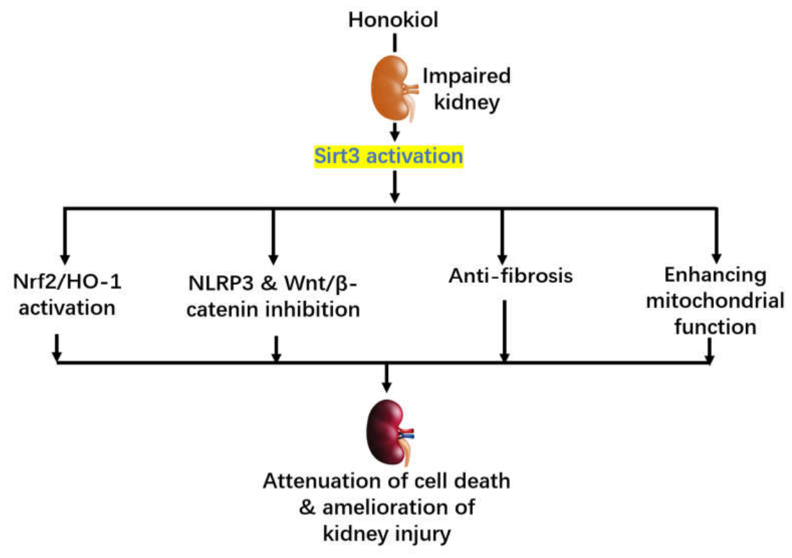
In this review, focusing on studies derived from various animal models (Figure 5), we have discussed the beneficial effects of honokiol on variety of animal models of kidney disease (Figure 6). Numerous studies demonstrate that HKL, by binding and activating sirt3 [44,114,115], exerts broad renoprotective properties across many experimental models of kidney disease. HKL treatment improves survival in septic rats and attenuates oxidative stress, inflammatory cytokine production, and nitric oxide (NO) levels in sepsis-associated AKI [116,117]. Although the underlying mechanism varies along with different disease context, HKL still preserves renal function by modulating key molecular pathways. In diabetic nephropathy, HKL reduces mitochondrial ROS production via SIRT3 activation and inhibits NLRP2 inflammasome-mediated pyroptosis to reduce the key inflammatory cytokines that can worsen renal injury. During renal fibrosis, HKL attenuates extracellular matrix accumulation [118] and epithelial-mesenchymal transition through SIRT3-dependent inhibition of Wnt/β-Catenin signaling. However, this approach has some limitations, including the need for further studies to investigate the organ-specific activity of SIRT3 and the long-term effects of HKL. Nonetheless, it has been established that HKL enhances endogenous antioxidant defense by activating PI3K/Akt-PKC-Nrf2 signaling pathway, thereby promoting GSH biosynthesis and reducing oxidative damage in ischemia-reperfusion [119]. In addition, in SA-AKI, HKL alleviates renal injury via regulation of the miR-218-5p/HO-1 signaling axis, leading to reduction of oxidative stress, inflammation, and apoptosis. Altogether, these studies highlight HKL as a promising natural therapeutic compound that targets several pathways responsible for kidney disease progression.
Despite these promising findings, several challenges remain before HKL can be translated into clinical practice. Current discussion is predominantly derived from animal models, with limited information regarding HKL’s efficacy and safety in humans. Thus, further investigation is required for optimal dosing, pharmacokinetic properties, and bioavailability of HKL. Since kidney diseases are highly heterogenous, future studies should also determine whether the molecular mechanisms identified in experimental models are conserved across different patient populations and stages of diseases. Continued preclinical and clinical investigations are needed to fully elucidate HKL’s therapeutic potential and facilitate the translation of HKL into novel treatment for acute and chronic kidney disorders.
Finally, we would also like to point out that while HKL shows extensive renoprotective effects in a variety of animal models of kidney injury, whether HKL is a caloric restriction mimetic that can also slow down kidney aging is yet to be comprehensively assessed. Nonetheless, given the findings that HKL can activate both sirtuins and AMPK [108,120,121], the two signaling pathways that are activated by caloric restriction to extend lifespan of whole animal or individual organs [122,123,124,125], it is plausible that HKL may well exhibit a caloric restriction effect on kidney aging. This plausibility needs to be tested in future studies, which may be facilitated using D-galactose accelerated kidney aging models [126,127].
Author Contributions
Original draft preparation, P.P.P and J.T.N.; review and editing, L-J.Y. All authors have read and agreed to the published version of the manuscript.
Funding
This research received no external funding.
Institutional Review Board Statement
Not applicable.
Informed Consent Statement
Not applicable.
Data Availability Statement
No new data were created or analyzed in this study.
Conflicts of Interest
The authors declare no conflicts of interest.
References
- Ruby, M.; Gifford, C.C.; Pandey, R.; Raj, V.S.; Sabbisetti, V.S.; Ajay, A.K. Autophagy as a Therapeutic Target for Chronic Kidney Disease and the Roles of TGF-beta1 in Autophagy and Kidney Fibrosis. Cells 2023, 12. [CrossRef]
- Montes-de-Oca-Saucedo, C.R.; Villa-Cedillo, S.A.; Rios-Vazquez, C.S.; Alvarez-Tostado-Ojeda, E.A.; Murguia-Perez, M.; Agustin-Godinez, E.; Ruiz-Salazar, R.; Soto-Dominguez, A.; Saucedo-Cardenas, O. Caloric restriction attenuates age-related liver and kidney alterations in mice: Histological evidence. Histol Histopathol 2026, 25065. [CrossRef]
- Alhassani, R.Y.; Bagadood, R.M.; Balubaid, R.N.; Barno, H.I.; Alahmadi, M.O.; Ayoub, N.A. Drug Therapies Affecting Renal Function: An Overview. Cureus 2021, 13, e19924. [CrossRef]
- Gutierrez, J.P.; Diep, T.N.; Niu, S.; Yan, L.J. Renoprotection by 5-Methoxytryptophan in Kidney Disease. Biomolecules 2026, 16. [CrossRef]
- Bragg-Gresham, J.; Xu, F.; Han, Y.; Licon, A.L.; Kiryakos, J.; Heung, M.; Morgenstern, H.; Weitzel, W.; Veinot, T.; Gillespie, B.; et al. Epidemiology of Kidney Disease in the United States: Highlights from the Centers for Disease Control and Prevention Kidney Disease Surveillance System. Clin J Am Soc Nephrol 2026, 21, 1456-1468. [CrossRef]
- Sparks, M.A.; Crowley, S.D.; Gurley, S.B.; Mirotsou, M.; Coffman, T.M. Classical Renin-Angiotensin system in kidney physiology. Compr Physiol 2014, 4, 1201-1228. [CrossRef]
- Viggiano, D.; Joshi, R.; Borriello, G.; Cacciola, G.; Gonnella, A.; Gigliotti, A.; Nigro, M.; Gigliotti, G. SGLT2 Inhibitors: The First Endothelial-Protector for Diabetic Nephropathy. J Clin Med 2025, 14. [CrossRef]
- Tuttle, K.R.; Brosius, F.C., 3rd; Cavender, M.A.; Fioretto, P.; Fowler, K.J.; Heerspink, H.J.L.; Manley, T.; McGuire, D.K.; Molitch, M.E.; Mottl, A.K.; et al. SGLT2 Inhibition for CKD and Cardiovascular Disease in Type 2 Diabetes: Report of a Scientific Workshop Sponsored by the National Kidney Foundation. Diabetes 2021, 70, 1-16. [CrossRef]
- Scheen, A.J. SGLT2 Inhibitors: Benefit/Risk Balance. Curr Diab Rep 2016, 16, 92. [CrossRef]
- Yu, K.; Liu, M.; Zhang, T.; Guo, C.; Du, L.; Li, M.; Wang, Q.; Han, N.; Chi, Y.; Li, Y. Honokiol attenuates diabetic nephropathy by targeting SIRT3 to suppress mitochondrial ROS-induced pyroptosis. Diabetol Metab Syndr 2025, 18, 7. [CrossRef]
- Cai, Z.; Yan, L.J. Protein oxidative modifications: Beneficial roles in disease and health. Journal of Biochemical and Pharmacological Research 2013, 1, 15-26.
- Yan, L.J. Positive oxidative stress in aging and aging-related disease tolerance. Redox Biology 2014, 2, 165-169. [CrossRef]
- Fedorova, M.; Bollineni, R.C.; Hoffmann, R. Protein carbonylation as a major hallmark of oxidative damage: update of analytical strategies. Mass Spectrom Rev 2014, 33, 79-97. [CrossRef]
- Yan, L.J. Analysis of oxidative modification of proteins. Curr Protoc Protein Sci 2009, Chapter 14, Unit14 14.
- Sayre, L.M.; Lin, D.; Yuan, Q.; Zhu, X.; Tang, X. Protein adducts generated from products of lipid oxidation: focus on HNE and one. Drug Metab Rev 2006, 38, 651-675.
- Yan, L.J.; Lodge, J.K.; Traber, M.G.; Matsugo, S.; Packer, L. Comparison between copper-mediated and hypochlorite-mediated modifications of human low density lipoproteins evaluated by protein carbonyl formation. J Lipid Res 1997, 38, 992-1001.
- Beckman, K.B.; Ames, B.N. Oxidative decay of DNA. J Biol Chem 1997, 272, 19633-19636.
- Shigenaga, M.K.; Park, J.W.; Cundy, K.C.; Gimeno, C.J.; Ames, B.N. In vivo oxidative DNA damage: measurement of 8-hydroxy-2’-deoxyguanosine in DNA and urine by high-performance liquid chromatography with electrochemical detection. Methods Enzymol 1990, 186, 521-530.
- Liu, H.; Xiang, X.; Shi, C.; Guo, J.; Ran, T.; Lin, J.; Dong, F.; Yang, J.; Miao, H. Oxidative stress and inflammation in renal fibrosis: Novel molecular mechanisms and therapeutic targets. Chem Biol Interact 2025, 421, 111784. [CrossRef]
- Kudin, A.P.; Bimpong-Buta, N.Y.; Vielhaber, S.; Elger, C.E.; Kunz, W.S. Characterization of superoxide-producing sites in isolated brain mitochondria. J Biol Chem 2004, 279, 4127-4135.
- Wu, K.; Wang, Q.; Zhang, Z.; Luo, W.; Peng, J.; Ma, X.; Wang, L.; Xie, C.; Guo, W. Honokiol ameliorates pyroptosis in intestinal ischemia-reperfusion injury by regulating the SIRT3-mediated NLRP3 inflammasome. Int J Mol Med 2025, 55. [CrossRef]
- Jin, S.; Fu, C.; Zhang, H.; Ji, Y.; Jiao, Q.; Liu, P. Acute Kidney Injury-to-Chronic Kidney Disease Transition-Associated Macrophage Subtypes: Biological Functions and Intercellular Crosstalk. Int J Mol Sci 2026, 27. [CrossRef]
- Liu, Y.; Liu, Z.; Liu, L.; Su, Y.; Chen, J.; Qing, B.; Tang, C.; Zhang, Z.; Ji, R.; Cao, X.; et al. Methionine restriction alleviates kidney fibrosis through epigenetic repression of the TGF-beta-Smad3-Hoxc8/P-TEFb axis. Nat Commun 2025, 17, 1301. [CrossRef]
- Jiao, H.; Zhang, M.; Chen, L.; Zhang, Z. Traditional Chinese Medicine targeting the TGF-beta/Smad signaling pathway as a potential therapeutic strategy for renal fibrosis. Front Pharmacol 2025, 16, 1513329. [CrossRef]
- Huang, X.; Wang, M.; Yu, B.; Ren, P.; Bian, X.; Wang, P.; Yao, X.; Lin, W. Transition from acute kidney injury to chronic kidney disease: molecular mechanisms and therapeutic interventions. Mol Biomed 2026, 7. [CrossRef]
- Gan, J.X.; Li, M.F.; Deng, H.H.; Liu, Q.T.; Chen, Z.X.; Liang, C.L.; Wang, L.X.; Liu, X.S.; Gu, Y.Y. Ablation of the renal tubular gluconeogenic enzyme PCK1 drives AKI-to-CKD transition by negatively regulating the TGF-beta/Smad3 signaling pathway. Cell Mol Life Sci 2026. [CrossRef]
- Kulkarni, H.V.; Mulay, S.R.; Gaikwad, A.B. The AKI-CKD Continuum: Mechanisms and Therapeutic Interventions. Nephrol Dial Transplant 2026. [CrossRef]
- Chiang, C.H.; Chen, C.; Fang, S.Y.; Lin, S.C.; Chen, J.W.; Chang, T.T. Xanthine oxidase/NADPH oxidase inhibition by hydralazine attenuates acute kidney injury and prevents the transition of acute kidney injury to chronic kidney disease. Life Sci 2023, 327, 121863. [CrossRef]
- Song, H.; Ren, W.; Liu, T.; Zou, S.; Yin, D.; Yu, J. Effects of Eucommia ulmoides leaf extract on xanthine oxidase and chronic kidney disease induced by adenine in rats. Nat Prod Res 2025, 39, 5340-5345. [CrossRef]
- Yang, Q.; Wu, F.R.; Wang, J.N.; Gao, L.; Jiang, L.; Li, H.D.; Ma, Q.; Liu, X.Q.; Wei, B.; Zhou, L.; et al. Nox4 in renal diseases: An update. Free Radic Biol Med 2018, 124, 466-472. [CrossRef]
- Schiffer, T.A.; Carvalho, L.; Guimaraes, D.; Boeder, A.; Wikstrom, P.; Carlstrom, M. Specific NOX4 Inhibition Preserves Mitochondrial Function and Dampens Kidney Dysfunction Following Ischemia-Reperfusion-Induced Kidney Injury. Antioxidants (Basel) 2024, 13. [CrossRef]
- Li, X.; Ma, T.K.; Wang, P.; Shi, H.; Hai, S.; Qin, Y.; Zou, Y.; Zhu, W.T.; Li, H.M.; Li, Y.N.; et al. HOXD10 attenuates renal fibrosis by inhibiting NOX4-induced ferroptosis. Cell Death Dis 2024, 15, 398. [CrossRef]
- Li, J.; Wang, L.; Wang, B.; Zhang, Z.; Jiang, L.; Qin, Z.; Zhao, Y.; Su, B. NOX4 is a potential therapeutic target in septic acute kidney injury by inhibiting mitochondrial dysfunction and inflammation. Theranostics 2023, 13, 2863-2878. [CrossRef]
- Li, X.; Yuan, Z.; Wang, Y.; Wang, W.; Shi, J. Recent advances of honokiol:pharmacological activities, manmade derivatives and structure-activity relationship. Eur J Med Chem 2024, 272, 116471. [CrossRef]
- Jiang, W. Pharmacological advances of honokiol: Mechanisms, targets and therapeutic potential (Review). Biomed Rep 2026, 25, 103. [CrossRef]
- Fujita, M.; Itokawa, H.; Sashida, Y. Honokiol, a New Phenolic Compound isolated from the Bark of Magnolia obovata THUNB. CHEMICAL & PHARMACEUTICAL BULLETIN 1972, 20, 212-213. [CrossRef]
- Nooralishahi, B.; Dehghani, M.; Rezaee Tazangi, F. EXPRESS: Honokiol and analgesia: a mechanistic review on the current capacities and challenges. Mol Pain 2025, 21, 17448069251377447. [CrossRef]
- Dai, X.; Xie, L.; Liu, K.; Liang, Y.; Cao, Y.; Lu, J.; Wang, X.; Zhang, X.; Li, X. The Neuropharmacological Effects of Magnolol and Honokiol: A Review of Signal Pathways and Molecular Mechanisms. Curr Mol Pharmacol 2023, 16, 161-177. [CrossRef]
- Faysal, M.; Khan, J.; Zehravi, M.; Nath, N.; Singh, L.P.; Kakkar, S.; Perusomula, R.; Khan, P.A.; Nainu, F.; Asiri, M.; et al. Neuropharmacological potential of honokiol and its derivatives from Chinese herb Magnolia species: understandings from therapeutic viewpoint. Chin Med 2023, 18, 154. [CrossRef]
- Wang, D.; Dong, X.; Wang, C. Honokiol Ameliorates Amyloidosis and Neuroinflammation and Improves Cognitive Impairment in Alzheimer’s Disease Transgenic Mice. J Pharmacol Exp Ther 2018, 366, 470-478. [CrossRef]
- Li, H.; Sun, J.; Wu, Y.; Yang, Y.; Zhang, W.; Tian, Y. Honokiol relieves hippocampal neuronal damage in Alzheimer’s disease by activating the SIRT3-mediated mitochondrial autophagy. CNS Neurosci Ther 2024, 30, e14878. [CrossRef]
- Cheng, G.; Hardy, M.; Feix, J.B.; Kalyanaraman, B. Inhibition of levodopa metabolism to dopamine by honokiol short-chain fatty acid derivatives may enhance therapeutic efficacy in Parkinson’s disease. Sci Rep 2025, 15, 20004. [CrossRef]
- Gan, Q.; Xian, Y.; Zhou, T.; Fu, X.; Gou, S.; Zhou, S. Honokiol targets cyclophilin D and blocks mitochondrial DNA release to inhibit microglial NLRP3 inflammasome activation and alleviate neurodegeneration in Parkinson’s disease models. Chem Biol Interact 2026, 438, 112305. [CrossRef]
- Xiang, F.; Zhang, Z.; Li, Y.; Xia, B.; Liao, Y.; Yang, C.; He, Z.; Lin, L.; Xie, J. Honokiol Targeting SIRT3: From Molecular Mechanisms to Therapeutic Opportunities. FASEB J 2025, 39, e70798. [CrossRef]
- Hirschey, M.D.; Shimazu, T.; Jing, E.; Grueter, C.A.; Collins, A.M.; Aouizerat, B.; Stancakova, A.; Goetzman, E.; Lam, M.M.; Schwer, B.; et al. SIRT3 deficiency and mitochondrial protein hyperacetylation accelerate the development of the metabolic syndrome. Mol Cell 2011, 44, 177-190. [CrossRef]
- Wang, Y.; Zhang, X.; Wang, P.; Shen, Y.; Yuan, K.; Li, M.; Liang, W.; Que, H. Sirt3 overexpression alleviates hyperglycemia-induced vascular inflammation through regulating redox balance, cell survival, and AMPK-mediated mitochondrial homeostasis. J Recept Signal Transduct Res 2019, 39, 341-349. [CrossRef]
- Azzouz, R.S.; Yan, L.J. The Role of Sirt3 in Kidney Health and Disease. Pharmaceuticals (Basel) 2025, 18. [CrossRef]
- Morigi, M.; Perico, L.; Benigni, A. Sirtuins in Renal Health and Disease. J Am Soc Nephrol 2018, 29, 1799-1809. [CrossRef]
- Tian, X.; Zeng, Y.; Tu, Q.; Jiao, Y.; Yao, S.; Chen, Y.; Sun, L.; Xia, Q.; Luo, Y.; Yuan, L.; et al. Butyrate alleviates renal fibrosis in CKD by regulating NLRP3-mediated pyroptosis via the STING/NF-kappaB/p65 pathway. Int Immunopharmacol 2023, 124, 111010. [CrossRef]
- Guo, Z.; Wang, J.; Li, X.; Ren, X.; Yin, Y.; Tian, Y.; Qin, Z.; Yuan, K.; Zeng, X. Honokiol attenuates renal fibrosis via SIRT3-Mediated regulation of Wnt/beta-Catenin signaling. Funct Integr Genomics 2026, 26, 42. [CrossRef]
- Park, E.J.; Dusabimana, T.; Je, J.; Jeong, K.; Yun, S.P.; Kim, H.J.; Kim, H.; Park, S.W. Honokiol Protects the Kidney from Renal Ischemia and Reperfusion Injury by Upregulating the Glutathione Biosynthetic Enzymes. Biomedicines 2020, 8. [CrossRef]
- Zhang, T.; Xiang, L. Honokiol alleviates sepsis-induced acute kidney injury in mice by targeting the miR-218-5p/heme oxygenase-1 signaling pathway. Cell Mol Biol Lett 2019, 24, 15. [CrossRef]
- Zheng, L.; Chen, W.; Yao, K.; Lin, Y.; Liao, C.; Zhou, T. Mechanism of mesenchymal stem cells in treating diabetic kidney disease. Stem Cell Res Ther 2025, 16, 629. [CrossRef]
- Li, Q.; Shang, J.; Inagi, R. Control of Mitochondrial Quality: A Promising Target for Diabetic Kidney Disease Treatment. Kidney Int Rep 2025, 10, 994-1010. [CrossRef]
- Rastogi, A.; Sarkar, M.; Kawai, K.; Munoz, G.; Simon, L.; Hwang, M.; Khalid, M.; Rhee, C. The Impact of Renin-Angiotensin Aldosterone System Inhibitors in IgA Nephropathy: A Meta-Analysis of Randomized Controlled Trials. Clin J Am Soc Nephrol 2026. [CrossRef]
- DeFronzo, R.A.; Reeves, W.B.; Awad, A.S. Pathophysiology of diabetic kidney disease: impact of SGLT2 inhibitors. Nat Rev Nephrol 2021, 17, 319-334. [CrossRef]
- Yao, L.; Liang, X.; Wang, P. Therapeutic perspective: evolving evidence of nonsteroidal mineralocorticoid receptor antagonists in diabetic kidney disease. Am J Physiol Endocrinol Metab 2023, 324, E531-E541. [CrossRef]
- Lerma, E.; White, W.B.; Bakris, G. Effectiveness of nonsteroidal mineralocorticoid receptor antagonists in patients with diabetic kidney disease. Postgrad Med 2023, 135, 224-233. [CrossRef]
- Yang, S.; Li, X.; Zhang, C.; Ma, S.; Zhou, Y. Recent advances in PANoptosis research in kidney disease: mechanistic networks, pathological roles, and potential intervention strategies. Front Immunol 2026, 17, 1902148. [CrossRef]
- Li, W.; Hao, F.; Lijia, X. Azilsartan Ameliorates Diabetic Kidney Disease Through Modulation of Inflammation, Pyroptosis, and Mitochondrial Dysfunction. J Diabetes Res 2026, 2026, e2555220. [CrossRef]
- Wang, L.; Hauenstein, A.V. The NLRP3 inflammasome: Mechanism of action, role in disease and therapies. Mol Aspects Med 2020, 76, 100889. [CrossRef]
- Wei, X.H.; Chen, J.; Wu, X.F.; Zhang, Q.; Xia, G.Y.; Chu, X.Y.; Xia, H.; Lin, S.; Shang, H.C. Salvianolic acid B alleviated myocardial ischemia-reperfusion injury via modulating SIRT3-mediated crosstalk between mitochondrial ROS and NLRP3. Phytomedicine 2025, 136, 156260. [CrossRef]
- Rather, I.A.; Khan, N.; Kushwah, A.S.; Surampalli, G.; Kumar, M. Nephroprotective effects of honokiol in a high-fat diet-streptozotocin rat model of diabetic nephropathy. Life Sci 2023, 320, 121543. [CrossRef]
- Hsu, Y.H.; Chiu, I.J.; Lin, Y.F.; Chen, Y.J.; Lee, Y.H.; Chiu, H.W. Lactoferrin Contributes a Renoprotective Effect in Acute Kidney Injury and Early Renal Fibrosis. Pharmaceutics 2020, 12. [CrossRef]
- Chen, Y.Y.; Chen, X.G.; Zhang, S. Druggability of lipid metabolism modulation against renal fibrosis. Acta Pharmacol Sin 2021. [CrossRef]
- Nogueira, A.; Pires, M.J.; Oliveira, P.A. Pathophysiological Mechanisms of Renal Fibrosis: A Review of Animal Models and Therapeutic Strategies. In Vivo 2017, 31, 1-22. [CrossRef]
- Wu, R.; Li, J.; Tu, G.; Su, Y.; Zhang, X.; Luo, Z.; Rong, R.; Zhang, Y. Comprehensive Molecular and Cellular Characterization of Acute Kidney Injury Progression to Renal Fibrosis. Front Immunol 2021, 12, 699192. [CrossRef]
- Chen, G.; Zhou, Y.; Lin, M.; Li, A.; Lu, S.; Li, J.; Xiao, L.; Hong, X. Wnt/beta-catenin/ATF3 signaling promotes the progression from acute kidney injury to chronic kidney disease by regulating mitophagy in mice. Biochem Pharmacol 2025, 242, 117444. [CrossRef]
- Wang, K.E.; Yu, Q.Q.; Wang, W.F.; Wang, Y.N.; Yang, J.; Miao, H.; Liu, L.M.; Zhao, Y.Y. Natural product therapy in diabetic kidney disease: emerging multiomics-mediated signalling pathway and molecular target. Chin Med 2026, 21. [CrossRef]
- Li, S.S.; Sun, Q.; Hua, M.R.; Suo, P.; Chen, J.R.; Yu, X.Y.; Zhao, Y.Y. Targeting the Wnt/beta-Catenin Signaling Pathway as a Potential Therapeutic Strategy in Renal Tubulointerstitial Fibrosis. Front Pharmacol 2021, 12, 719880. [CrossRef]
- Quan, Y.; Park, W.; Jin, J.; Kim, W.; Park, S.K.; Kang, K.P. Sirtuin 3 Activation by Honokiol Decreases Unilateral Ureteral Obstruction-Induced Renal Inflammation and Fibrosis via Regulation of Mitochondrial Dynamics and the Renal NF-kappaBTGF-beta1/Smad Signaling Pathway. Int J Mol Sci 2020, 21. [CrossRef]
- Chouchani, E.T.; Pell, V.R.; Gaude, E.; Aksentijevic, D.; Sundier, S.Y.; Robb, E.L.; Logan, A.; Nadtochiy, S.M.; Ord, E.N.J.; Smith, A.C.; et al. Ischaemic accumulation of succinate controls reperfusion injury through mitochondrial ROS. Nature 2014, 515, 431-435. [CrossRef]
- Masella, R.; Di Benedetto, R.; Vari, R.; Filesi, C.; Giovannini, C. Novel mechanisms of natural antioxidant compounds in biological systems: involvement of glutathione and glutathione-related enzymes. J Nutr Biochem 2005, 16, 577-586. [CrossRef]
- Fiser, B.; Jojart, B.; Csizmadia, I.G.; Viskolcz, B. Glutathione--hydroxyl radical interaction: a theoretical study on radical recognition process. PLoS One 2013, 8, e73652. [CrossRef]
- Ramani, K.; Tomasi, M.L.; Yang, H.; Ko, K.; Lu, S.C. Mechanism and significance of changes in glutamate-cysteine ligase expression during hepatic fibrogenesis. J Biol Chem 2012, 287, 36341-36355. [CrossRef]
- Dang, Y.F.; Qiu, T.X.; Song, D.W.; Liu, L. PMA-triggered PKCepsilon activity enhances Nrf2-mediated antiviral response on fish rhabdovirus infection. Fish Shellfish Immunol 2019, 87, 871-878. [CrossRef]
- Huang, H.C.; Nguyen, T.; Pickett, C.B. Regulation of the antioxidant response element by protein kinase C-mediated phosphorylation of NF-E2-related factor 2. Proc Natl Acad Sci U S A 2000, 97, 12475-12480. [CrossRef]
- Zheng, H.; Whitman, S.A.; Wu, W.; Wondrak, G.T.; Wong, P.K.; Fang, D.; Zhang, D.D. Therapeutic potential of Nrf2 activators in streptozotocin-induced diabetic nephropathy. Diabetes 2011, 60, 3055-3066.
- Wu, J.; Li, R.; Li, W.; Ren, M.; Thangthaeng, N.; Sumien, N.; Liu, R.; Yang, S.; Simpkins, J.W.; Forster, M.J.; et al. Administration of 5-methoxyindole-2-carboxylic acid that potentially targets mitochondrial dihydrolipoamide dehydrogenase confers cerebral preconditioning against ischemic stroke injury. Free Radic Biol Med 2017, 113, 244-254. [CrossRef]
- Wu, J.; Jin, Z.; Yang, X.; Yan, L.J. Post-ischemic administration of 5-methoxyindole-2-carboxylic acid at the onset of reperfusion affords neuroprotection against stroke injury by preserving mitochondrial function and attenuating oxidative stress. Biochem Biophys Res Commun 2018, 497, 444-450. [CrossRef]
- Li, Z.; Mo, R.L.; Gong, J.F.; Han, L.; Wang, W.F.; Huang, D.K.; Xu, J.G.; Sun, Y.J.; Chen, S.; Han, G.C.; et al. Dihydrotanshinone I inhibits gallbladder cancer growth by targeting the Keap1-Nrf2 signaling pathway and Nrf2 phosphorylation. Phytomedicine 2024, 129, 155661. [CrossRef]
- Pan, Z.; Li, M.; Chen, B.; Lu, J.; Wang, F.; Meng, C.; Wang, X.; Shao, M.; He, Q.; Wang, H. Naringenin ameliorates iron overload-associated osteoporosis via Tfeb/p62/Nrf2-mediated antioxidation. Chin J Nat Med 2026, 24, 912-928. [CrossRef]
- Cabrera, S.N.S.; Huber, S.; Braunbock-Muller, B.; Heiss, E.H. Metformin-restricted motility of an NRF2-activated lung cancer cell line involves NAD+ depletion, rather than AMPK- or BACH1 signaling. Sci Rep 2026. [CrossRef]
- Matsuoka, Y.; Okazaki, M.; Kitamura, Y. Induction of inducible heme oxygenase (HO-1) in the central nervous system: is HO-1 helpful or harmful? Neurotox Res 1999, 1, 113-117. [CrossRef]
- Yoo, S.J.; Nakra, N.K.; Ronnett, G.V.; Moon, C. Protective Effects of Inducible HO-1 on Oxygen Toxicity in Rat Brain Endothelial Microvessel Cells. Endocrinol Metab (Seoul) 2014, 29, 356-362. [CrossRef]
- Holzen, J.P.; August, C.; Bahde, R.; Minin, E.; Lang, D.; Heidenreich, S.; Dietl, K.H.; Spiegel, H.U. Influence of heme oxygenase-1 on microcirculation after kidney transplantation. J Surg Res 2008, 148, 126-135. [CrossRef]
- Shimizu, H.; Takahashi, T.; Suzuki, T.; Yamasaki, A.; Fujiwara, T.; Odaka, Y.; Hirakawa, M.; Fujita, H.; Akagi, R. Protective effect of heme oxygenase induction in ischemic acute renal failure. Crit Care Med 2000, 28, 809-817. [CrossRef]
- Nath, K.A. Heme oxygenase-1 and acute kidney injury. Curr Opin Nephrol Hypertens 2014, 23, 17-24. [CrossRef]
- Wang, G.; Hamid, T.; Keith, R.J.; Zhou, G.; Partridge, C.R.; Xiang, X.; Kingery, J.R.; Lewis, R.K.; Li, Q.; Rokosh, D.G.; et al. Cardioprotective and antiapoptotic effects of heme oxygenase-1 in the failing heart. Circulation 2010, 121, 1912-1925. [CrossRef]
- Sun, X.; Wang, H.; Liu, Y.; Yang, Y.; Wang, Y.; Liu, Y.; Ai, S.; Shan, Z.; Luo, P. 5-Methoxytryptophan Alleviates Lipopolysaccharide-Induced Acute Kidney Injury by Regulating Nrf2-Mediated Mitophagy. J Inflamm Res 2024, 17, 9857-9873. [CrossRef]
- Liu, L.; Song, Y.; Zhao, M.; Yi, Z.; Zeng, Q. Protective effects of edaravone, a free radical scavenger, on lipopolysaccharide-induced acute kidney injury in a rat model of sepsis. Int Urol Nephrol 2015, 47, 1745-1752. [CrossRef]
- Dou, X.; Yan, D.; Ma, Z.; Gao, N.; Shan, A. Sodium butyrate alleviates LPS-induced kidney injury via inhibiting TLR2/4 to regulate rBD2 expression. J Food Biochem 2022, 46, e14126. [CrossRef]
- Liu, X.; Gao, L.; Huang, X.; Deng, R.; Wei, X.; Lu, J.; Li, S. Lipidomics reveals the potential mechanism of honokiol against adenine-induced chronic kidney disease. Front Pharmacol 2022, 13, 1019629. [CrossRef]
- Wei, X.; Wang, Y.; Lao, Y.; Weng, J.; Deng, R.; Li, S.; Lu, J.; Yang, S.; Liu, X. Effects of honokiol protects against chronic kidney disease via BNIP3/NIX and FUNDC1-mediated mitophagy and AMPK pathways. Mol Biol Rep 2023, 50, 6557-6568. [CrossRef]
- Yan, L.J.; Allen, D.C. Cadmium-Induced Kidney Injury: Oxidative Damage as a Unifying Mechanism. Biomolecules 2021, 11. [CrossRef]
- Zhang, K.; Dong, W.; Li, J.; Gong, Z.; Liu, W.; He, S.; Zou, H.; Song, R.; Liu, G.; Liu, Z. Honokiol Antagonizes Cadmium-Induced Nephrotoxicity in Quail by Alleviating Autophagy Dysfunction, Apoptosis and Mitochondrial UPR Inhibition with Its Antioxidant Properties. Life (Basel) 2022, 12. [CrossRef]
- Iskander, A.; Yan, L.J. Cisplatin-Induced Kidney Toxicity: Potential Roles of Major NAD(+)-Dependent Enzymes and Plant-Derived Natural Products. Biomolecules 2022, 12. [CrossRef]
- Wang, T.J.; Liu, H.T.; Lai, Y.H.; Jan, T.R.; Nomura, N.; Chang, H.W.; Chou, C.C.; Lee, Y.J.; Tsai, P.J. Honokiol, a Polyphenol Natural Compound, Attenuates Cisplatin-Induced Acute Cytotoxicity in Renal Epithelial Cells Through Cellular Oxidative Stress and Cytoskeleton Modulations. Front Pharmacol 2018, 9, 357. [CrossRef]
- Tang, C.; Livingston, M.J.; Safirstein, R.; Dong, Z. Cisplatin nephrotoxicity: new insights and therapeutic implications. Nat Rev Nephrol 2023, 19, 53-72. [CrossRef]
- Xu, Z.; Zhang, M.; Wang, W.; Zhou, S.; Yu, M.; Qiu, X.; Jiang, S.; Wang, X.; Tang, C.; Li, S.; et al. Dihydromyricetin attenuates cisplatin-induced acute kidney injury by reducing oxidative stress, inflammation and ferroptosis. Toxicol Appl Pharmacol 2023, 473, 116595. [CrossRef]
- Mao, R.W.; He, S.P.; Lan, J.G.; Zhu, W.Z. Honokiol ameliorates cisplatin-induced acute kidney injury via inhibition of mitochondrial fission. Br J Pharmacol 2022, 179, 3886-3904. [CrossRef]
- Liu, H.T.; Wang, T.E.; Hsu, Y.T.; Chou, C.C.; Huang, K.H.; Hsu, C.C.; Liang, H.J.; Chang, H.W.; Lee, T.H.; Tsai, P.S. Nanoparticulated Honokiol Mitigates Cisplatin-Induced Chronic Kidney Injury by Maintaining Mitochondria Antioxidant Capacity and Reducing Caspase 3-Associated Cellular Apoptosis. Antioxidants (Basel) 2019, 8. [CrossRef]
- Gooch, J.L.; King, C.; Francis, C.E.; Garcia, P.S.; Bai, Y. Cyclosporine A alters expression of renal microRNAs: New insights into calcineurin inhibitor nephrotoxicity. PLoS One 2017, 12, e0175242. [CrossRef]
- Yilmaz, D.E.; Kirschner, K.; Demirci, H.; Himmerkus, N.; Bachmann, S.; Mutig, K. Immunosuppressive calcineurin inhibitor cyclosporine A induces proapoptotic endoplasmic reticulum stress in renal tubular cells. J Biol Chem 2022, 298, 101589. [CrossRef]
- Yen, N.T.H.; Tien, N.T.N.; Anh, N.T.V.; Le, Q.V.; Eunsu, C.; Kim, H.S.; Moon, K.S.; Nguyen, H.T.; Kim, D.H.; Long, N.P. Cyclosporine A-induced systemic metabolic perturbations in rats: A comprehensive metabolome analysis. Toxicol Lett 2024, 395, 50-59. [CrossRef]
- Naderi, R.; Seyhani, A.; Shirpoor, A.; Jafari, A.; Eyvani, K. Effects of curcumin on cyclosporine A-induced oxidative stress, autophagy, and apoptosis in rat heart. Mol Biol Rep 2025, 52, 310. [CrossRef]
- Wang, J.; Li, Z.; Qing, B.; Xia, Z.; Yuan, Y.; Zhang, L.; Liu, Z. Cyclosporine A promotes renal fibrosis through activating endoplasmic reticulum stress by targeting miR-212-5p/ATF6 axis. J Transl Med 2026, 24. [CrossRef]
- Kim, J.E.; Jo, M.J.; Bae, S.Y.; Ahn, S.Y.; Ko, G.J.; Kwon, Y.J. Mitochondrial SIRT3 as a protective factor against cyclosporine A-induced nephrotoxicity. Sci Rep 2024, 14, 10143. [CrossRef]
- Fiorentino, M.; Grandaliano, G.; Gesualdo, L.; Castellano, G. Acute Kidney Injury to Chronic Kidney Disease Transition. Contrib Nephrol 2018, 193, 45-54. [CrossRef]
- Ratliff, B.B.; Abdulmahdi, W.; Pawar, R.; Wolin, M.S. Oxidant Mechanisms in Renal Injury and Disease. Antioxid Redox Signal 2016, 25, 119-146. [CrossRef]
- Zhang, Y.; Yu, C.; Li, X. Kidney Aging and Chronic Kidney Disease. Int J Mol Sci 2024, 25. [CrossRef]
- Zuo, Z.; Jing, K.; Wu, H.; Wang, S.; Ye, L.; Li, Z.; Yang, C.; Pan, Q.; Liu, W.J.; Liu, H.F. Mechanisms and Functions of Mitophagy and Potential Roles in Renal Disease. Front Physiol 2020, 11, 935. [CrossRef]
- Zoja, C.; Xinaris, C.; Macconi, D. Diabetic Nephropathy: Novel Molecular Mechanisms and Therapeutic Targets. Front Pharmacol 2020, 11, 586892. [CrossRef]
- Li, N.; Zhang, J.; Yan, X.; Zhang, C.; Liu, H.; Shan, X.; Li, J.; Yang, Y.; Huang, C.; Zhang, P.; et al. SIRT3-KLF15 signaling ameliorates kidney injury induced by hypertension. Oncotarget 2017, 8, 39592-39604. [CrossRef]
- Locatelli, M.; Zoja, C.; Zanchi, C.; Corna, D.; Villa, S.; Bolognini, S.; Novelli, R.; Perico, L.; Remuzzi, G.; Benigni, A.; et al. Manipulating Sirtuin 3 pathway ameliorates renal damage in experimental diabetes. Sci Rep 2020, 10, 8418. [CrossRef]
- Xia, S.; Lin, H.; Liu, H.; Lu, Z.; Wang, H.; Fan, S.; Li, N. Honokiol Attenuates Sepsis-Associated Acute Kidney Injury via the Inhibition of Oxidative Stress and Inflammation. Inflammation 2019, 42, 826-834. [CrossRef]
- Li, N.; Xie, H.; Li, L.; Wang, J.; Fang, M.; Yang, N.; Lin, H. Effects of honokiol on sepsis-induced acute kidney injury in an experimental model of sepsis in rats. Inflammation 2014, 37, 1191-1199. [CrossRef]
- Chiang, C.K.; Sheu, M.L.; Lin, Y.W.; Wu, C.T.; Yang, C.C.; Chen, M.W.; Hung, K.Y.; Wu, K.D.; Liu, S.H. Honokiol ameliorates renal fibrosis by inhibiting extracellular matrix and pro-inflammatory factors in vivo and in vitro. Br J Pharmacol 2011, 163, 586-597. [CrossRef]
- Yu, Y.; Li, M.; Su, N.; Zhang, Z.; Zhao, H.; Yu, H.; Xu, Y. Honokiol protects against renal ischemia/reperfusion injury via the suppression of oxidative stress, iNOS, inflammation and STAT3 in rats. Mol Med Rep 2016, 13, 1353-1360. [CrossRef]
- Ma, L.; Wang, R.; Wang, H.; Zhang, Y.; Zhao, Z. Long-term caloric restriction activates the myocardial SIRT1/AMPK/PGC-1alpha pathway in C57BL/6J male mice. Food Nutr Res 2020, 64. [CrossRef]
- Chen, Y.; Yang, Z.; Bai, J.; Wang, X.; Yuan, Q.; Mi, Y.; Zhang, C. Bioactive Lignan Honokiol Alleviates Ovarian Oxidative Stress in Aging Laying Chickens by Regulating SIRT3/AMPK Pathway. Antioxidants (Basel) 2024, 13. [CrossRef]
- de Souza Nunes Faria, M.S.; Pimentel, V.E.; Helaehil, J.V.; Bertolo, M.C.; Santos, N.T.H.; da Silva-Neto, P.V.; Thomazini, B.F.; de Oliveira, C.A.; do Amaral, M.E.C. Caloric restriction overcomes pre-diabetes and hypertension induced by a high fat diet and renal artery stenosis. Mol Biol Rep 2022, 49, 5883-5895. [CrossRef]
- Zhang, S.; Sun, S.; Wei, X.; Zhang, M.; Chen, Y.; Mao, X.; Chen, G.; Liu, C. Short-term moderate caloric restriction in a high-fat diet alleviates obesity via AMPK/SIRT1 signaling in white adipocytes and liver. Food Nutr Res 2022, 66. [CrossRef]
- Guo, Z.; Wang, M.; Ying, X.; Yuan, J.; Wang, C.; Zhang, W.; Tian, S.; Yan, X. Caloric restriction increases the resistance of aged heart to myocardial ischemia/reperfusion injury via modulating AMPK-SIRT(1)-PGC(1a) energy metabolism pathway. Sci Rep 2023, 13, 2045. [CrossRef]
- Nassar, K.; El-Mekawey, D.; Elmasry, A.E.; Refaey, M.S.; El-Sayed Ghoneim, M.; Elshaier, Y. The significance of caloric restriction mimetics as anti-aging drugs. Biochem Biophys Res Commun 2024, 692, 149354. [CrossRef]
- Niu, S.; Azzouz, R.S.; Yan, L.-J. Rodent Models of D-Galactose Induction of Accelerated Aging: A Platform for Exploring Kidney Aging Mechanisms and Anti-Kidney Aging Strategies. Cells 2026, 15, 766.
- Kobroob, A.; Maneechote, C.; Sivasinprasasn, S.; Chattipakorn, N.; Wongmekiat, O. Inhibition of Mitochondrial Fission by Mdivi-1 Alleviates Doxorubicin-Induced Nephrotoxicity in a Rat Model of D-Galactose-Induced Accelerated Renal Aging. Biomolecules 2026, 16. [CrossRef]
Figure 1.
Chemical structure of honokiol (HKL).
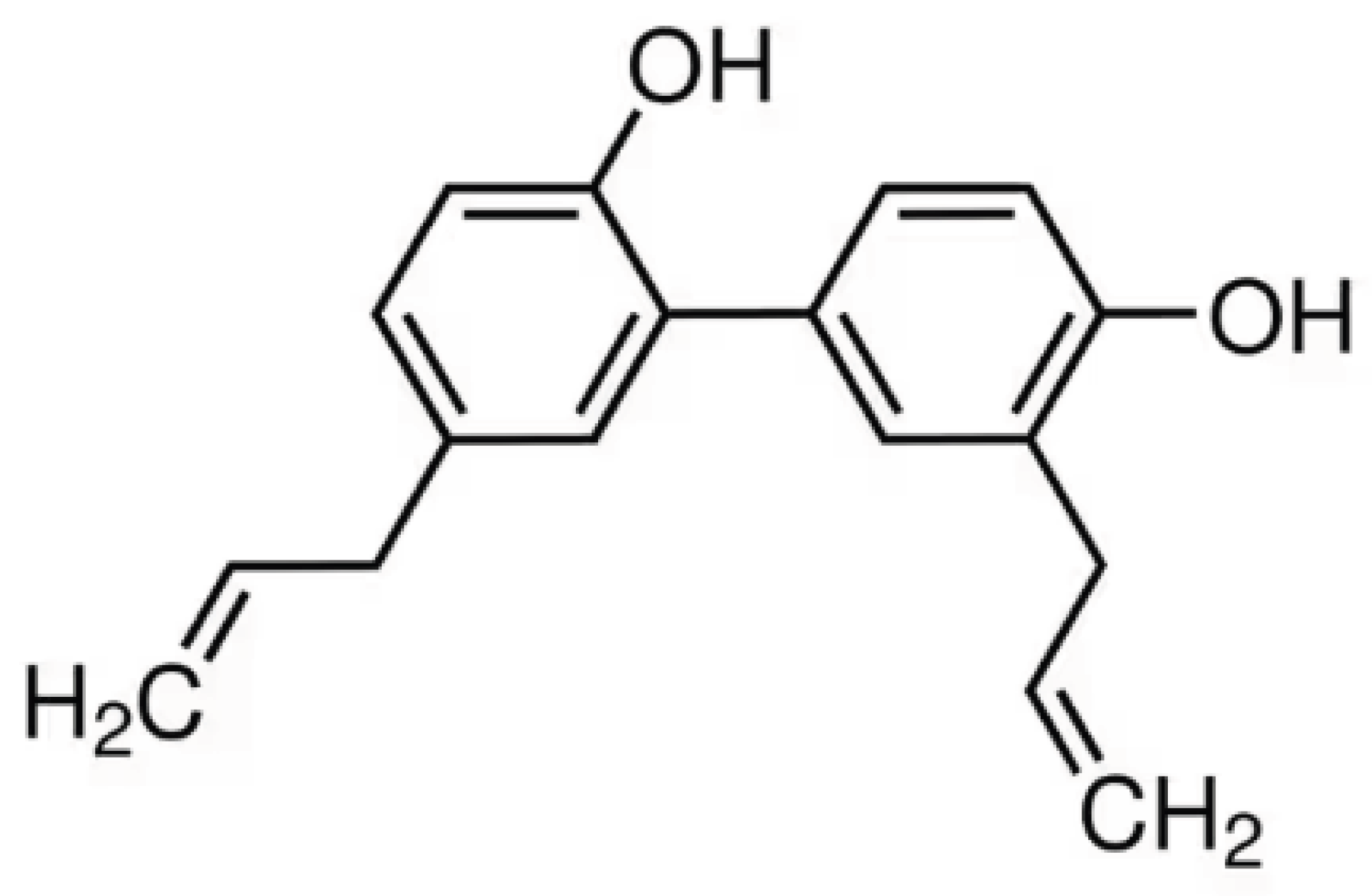
Figure 2.
Honokiol mitigates diabetic kidney disease in db/db mice via improving kidney function and histopathology. Shown are renal sections stained with hematoxylin and erosin (HE), periodic acid-Schiff (PAS), and Masson’s trichrome, respectively. This figure was reproduced from reference [10].
Figure 2.
Honokiol mitigates diabetic kidney disease in db/db mice via improving kidney function and histopathology. Shown are renal sections stained with hematoxylin and erosin (HE), periodic acid-Schiff (PAS), and Masson’s trichrome, respectively. This figure was reproduced from reference [10].
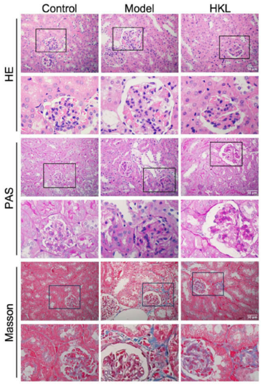
Figure 3.
Proposed mechanism of HKL-mediated protection against renal ischemia-reperfusion injury. GCLC: glutamate-cysteine ligase catalytic subunit; GCLM: glutamate-cysteine ligase modifier subunit; GSS: glutathione synthetase; ARE: antioxidant response element.
Figure 3.
Proposed mechanism of HKL-mediated protection against renal ischemia-reperfusion injury. GCLC: glutamate-cysteine ligase catalytic subunit; GCLM: glutamate-cysteine ligase modifier subunit; GSS: glutathione synthetase; ARE: antioxidant response element.
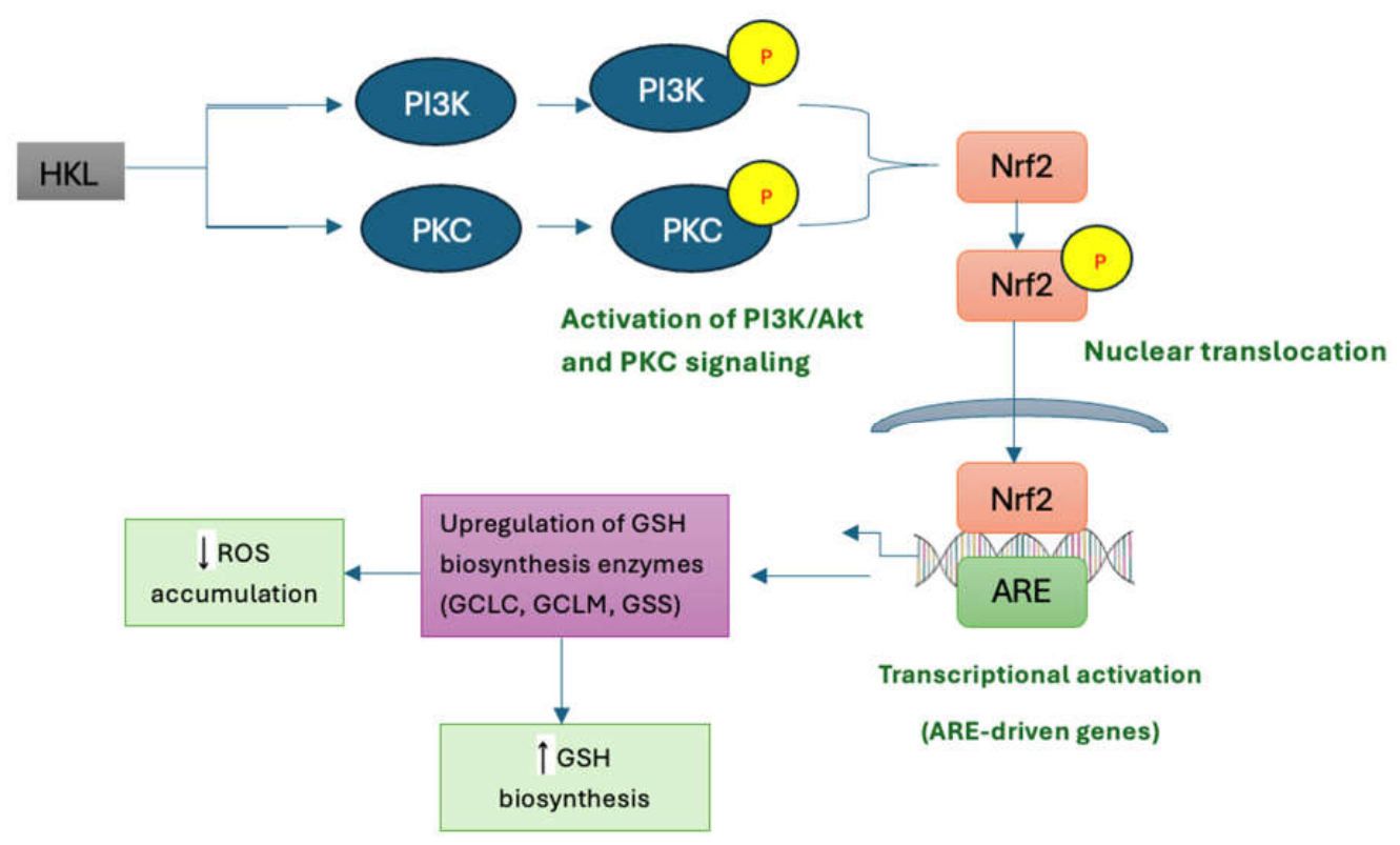
Figure 4.
Renoprotection of kidney tissues by honokiol in adenine induced CKD. Show is periodic acid-Schiff staining and Masson’s trichrome staining, respectively. The figure was reproduced from reference [93].
Figure 4.
Renoprotection of kidney tissues by honokiol in adenine induced CKD. Show is periodic acid-Schiff staining and Masson’s trichrome staining, respectively. The figure was reproduced from reference [93].
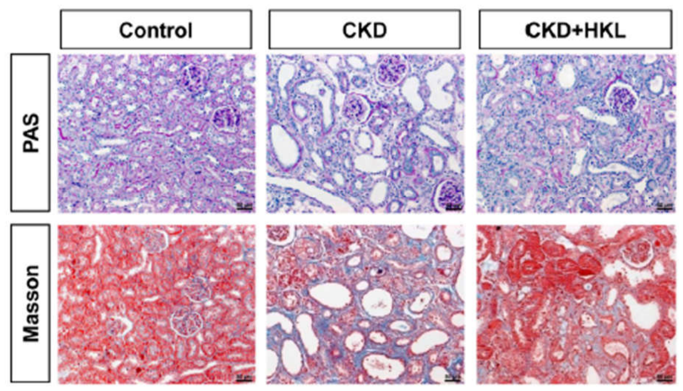
Figure 5.
Animal models of kidney disease discussed in this review article.
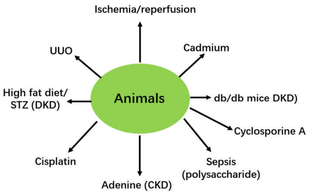
Figure 6.
Mechanisms by which honokiol protects against renal injury discussed in this review article. Honokiol can physically bind and activate sirt3, which then induces various protective signaling pathways that counteract kidney injury induced by a variety of stressors as shown in Figure 5.
Figure 6.
Mechanisms by which honokiol protects against renal injury discussed in this review article. Honokiol can physically bind and activate sirt3, which then induces various protective signaling pathways that counteract kidney injury induced by a variety of stressors as shown in Figure 5.
Disclaimer/Publisher’s Note: The statements, opinions and data contained in all publications are solely those of the individual author(s) and contributor(s) and not of MDPI and/or the editor(s). MDPI and/or the editor(s) disclaim responsibility for any injury to people or property resulting from any ideas, methods, instructions or products referred to in the content. |
© 2026 by the authors. Licensee MDPI, Basel, Switzerland. This article is an open access article distributed under the terms and conditions of the Creative Commons Attribution (CC BY) license (http://creativecommons.org/licenses/by/4.0/).
Copyright: This open access article is published under a Creative Commons CC BY 4.0 license, which permit the free download, distribution, and reuse, provided that the author and preprint are cited in any reuse.