

Submitted:
25 September 2026
Posted:
29 September 2026
You are already at the latest version
Abstract
The degradation of autophagic bodies in the plant vacuole is required to complete autophagic flux, yet the hydrolases involved in membrane breakdown and cargo processing remain incompletely defined. We investigated candidate vacuolar lipases and vacuolar processing enzymes (VPEs) in embryonic axes of white lupin (Lupinus albus L.) and Andean lupin (Lupinus mutabilis Sweet) exposed to carbon starvation. Carbon starvation increased total p-nitrophenyl-palmitate-hydrolyzing and Ac-ESEN-MCA-hydrolyzing activities, whereas asparagine and pharmacological perturbation of autophagic or vacuolar processes attenuated these responses. AlphaFold 3 models, enhanced conformational sampling, and molecular docking indicated that selected candidate lipases possess α/β-hydrolase-like architectures and can accommodate several membrane-associated lipids; however, these predictions do not establish catalytic activity or physiological substrate preference. Vacuole-targeted XP_019420123.1 failed to restore Ape1 maturation or AtAtg8a-GFP processing in Saccharomyces cerevisiae atg15Δ cells, indicating that the construct did not functionally substitute for Atg15 under the conditions tested. By contrast, partial inhibition of VPE-like activity with Ac-YVAD-cmk increased the number of intravacuolar autophagic-body-like structures while phagophores and autophagosomes remained detectable. Transcriptomic and RT-qPCR analyses further revealed gene-specific responses of VPE/legumain transcripts to carbon status and pharmacological treatments. Collectively, the results support an association between VPE-like proteolytic activity and efficient late intravacuolar degradation during carbon starvation. The analyzed lipases remain candidates for vacuolar lipid metabolism, but direct biochemical and genetic evidence is required before assigning them a role in autophagic-body membrane hydrolysis.
Keywords:
asparagine
; autophagic body
; carbon starvation
; legumain
; lipase
; Lupinus albus
; Lupinus mutabilis
; seed germination
; vacuolar processing enzyme
; vacuole
1. Introduction
Macroautophagy is an evolutionarily conserved pathway that delivers cytoplasmic material to lytic compartments for degradation and recycling. In plants, a cup-shaped phagophore expands around cytoplasmic cargo and closes to form a double-membrane autophagosome [1]. Fusion of the autophagosome outer membrane with the tonoplast releases the inner vesicle, termed the autophagic body, into the vacuolar lumen. Efficient breakdown of the autophagic-body membrane and its cargo is required for metabolite salvage and completion of autophagic flux [2,3,4]. The molecular machinery controlling selective cargo recognition, phagophore formation, autophagosome maturation, and vacuolar delivery is substantially better characterized than the terminal hydrolytic steps [5,6]. In Saccharomyces cerevisiae, autophagic-body degradation requires the vacuolar phospholipase Atg15 [7,8] and vacuolar proteases, including proteinase A (Pep4) and proteinase B (Prb1) [9]. Atg15 is delivered to the vacuole and disrupts intravacuolar membranes after proteolytic activation, thereby allowing other hydrolases to access the enclosed cargo [7,8,9,10,11,12,13]. Nevertheless, no clear Atg15 homolog has been identified outside fungi. Therefore, organisms in other lineages are expected to employ structurally unrelated enzymes that perform analogous membrane-degrading functions.
Recent work has provided direct evidence that the atypical phospholipases, lecithin-cholesterol acyltransferases, LCAT3 and LCAT4 cooperate in autophagic-body membrane hydrolysis in Arabidopsis thaliana. The two enzymes reach autophagic bodies through distinct trafficking routes, and their combined loss significantly delays, but does not completely abolish, autophagic-body degradation, indicating that additional hydrolases may contribute to the terminal phase of plant autophagy [14]. This discovery strengthens the rationale for investigating other vacuolar phospholipases or lipases and emphasizes that sequence similarity to yeast Atg15 is not sufficient to infer equivalent cellular function. Our previous transcriptome-based survey identified 38 lipase-encoding transcripts with predicted vacuolar localization in embryonic axes of white lupin (Lupinus albus L.) and Andean lupin (Lupinus mutabilis Sweet). Three encoded proteins contained regions similar to the catalytic region of yeast Atg15 (up to 80% sequence identity and 90% sequence similarity) and were therefore selected as candidates for further study. Most putative vacuolar lipase transcripts decreased under carbon starvation, particularly following asparagine supplementation, whereas the three candidates showed different expression patterns [15]. These observations suggested a possible association of vacuolar lipases with carbon-starvation responses but did not demonstrate involvement in autophagic body degradation.
Plant vacuolar processing enzymes (VPEs, also called legumains) are cysteine proteases of the asparaginyl endopeptidase family. Like yeast Pep4, they undergo pH-dependent autocatalytic maturation and process numerous vacuolar proteins and hydrolases (van den [16,17]. Plant VPEs have established roles in seed protein maturation, development, stress responses, and several forms of programmed cell death [18,19,20]. A contribution to autophagic degradation has been proposed, but direct experimental evidence remains limited [20,21,22]. Because VPE activity depends on an acidic vacuolar environment and may influence the maturation of other hydrolases, VPEs are plausible components of the enzymatic network that degrades autophagic material in plant cells.
Embryonic axes isolated from germinating lupin seeds provide a tractable system for studying carbon-starvation-induced autophagy. Sucrose deprivation in darkness stimulates autophagy, whereas exogenous asparagine attenuates several starvation responses and is accompanied by accumulation of autophagic bodies in enlarged vacuoles. The two lupin species differ in storage composition: white lupin seeds are protein-rich and relatively oil-poor, whereas Andean lupin seeds contain substantial amounts of both protein and lipids. This comparative system has therefore been useful for examining links among carbon status, reserve mobilization, redox regulation, and autophagy [23,24,25].
The present study is a continuation of our research on plant autophagy and had four objectives: (i) to determine how total lipolytic and VPE activities respond to carbon starvation, asparagine, and pharmacological perturbation of autophagic or vacuolar processes; (ii) to extend the structural analysis of candidate vacuolar lipases and test whether XP_019420123.1 can substitute for Atg15 in yeast; (iii) to assess whether partial inhibition of VPE activity alters vacuolar ultrastructure without preventing formation of earlier autophagic structures; and (iv) to identify and profile VPE-encoding transcripts in both lupin species. We interpret the pharmacological, biochemical, and ultrastructural findings as evidence of association and explicitly distinguish them from direct demonstration of enzyme-specific function.
2. Results
2.1. Pharmacological Perturbation Produced the Expected Ultrastructural Phenotypes
Carbon starvation was induced using the previously established lupin embryonic-axis culture system [23,24,25]. Ultrastructural analysis of root-meristematic-zone cells confirmed the previously reported increase in cell vacuolization of sucrose-starved (-S) embryonic axes relative to sucrose-fed (+S; 60 mM) axes. In -S cells, the vacuolar lumen was largely electron-lucent and contained little persistent material (Figure 1). Because autophagy-related lupin mutants are not available, pharmacological inhibitors were used to perturb different stages of the pathway. 3-Methyladenine (3MA) inhibits class III phosphatidylinositol 3-kinase and thereby interferes with autophagosome formation [26]. Concanamycin A (ConA) and bafilomycin A1 (BafA1) inhibit vacuolar H+-ATPase-dependent acidification and can impair late degradative stages of autophagy [27,28,29]. The effects of 5 mM 3MA, 10 µM ConA, and 10 µM BafA1 on root and hypocotyl length and on fresh and dry mass were assessed first (Supplementary Figure S1). ConA promoted retention of autophagic bodies within vacuoles, whereas 3MA markedly reduced the number of detectable autophagosomes (Figure 1). BafA1 did not reproducibly induce accumulation of morphologically identifiable autophagic bodies in either species and was therefore excluded from subsequent biochemical and transcriptional analyses. Electron-dense remnants resembling storage-protein deposits were also observed after ConA treatment (Figure 1). As reported previously, asparagine (+Asn) supplementation of -S axes produced a ConA-like accumulation of autophagic bodies in both species [23,24,25] (Figure 1).
2.2. Total Lipolytic Activity Responded to Carbon Status and Pharmacological Treatments
Total hydrolysis of p-nitrophenyl palmitate was higher in carbon-starved than in sucrose-fed axes of both lupin species, and asparagine attenuated this increase, especially in -S axes. ConA and 3MA also reduced the activity relative to the corresponding untreated conditions in both lupin species (Figure 2). Because the assay used a general artificial substrate at pH 9.0, these data indicate changes in aggregate lipolytic capacity and cannot be assigned to a specific vacuolar lipase or interpreted as a direct measurement of autophagic-body membrane hydrolysis.
2.3. Candidate Lipase Transcripts Were More Responsive to Vacuolar Perturbation than to Carbon Starvation Alone
RT-qPCR revealed gene-specific responses of the selected lipase transcripts to carbon availability, asparagine, ConA, and 3MA. ConA caused the largest increases in levels of all investigated transcripts, sometimes exceeding 200-fold relative to the +S calibrator, whereas responses to 3MA were more variable (Figure 3). In contrast to the earlier RNA-seq screen [15], most starvation-associated increases in candidate lipase transcripts were not statistically significant by RT-qPCR. Thus, independent validation supported responsiveness to perturbation of vacuolar/autophagic processes more consistently than a simple induction by carbon starvation.
2.4. Structural Organization and Molecular Docking Studies of Lipases
2.4.1. Structural Organization
We previously identified candidate lupin lipases with putative vacuolar localization and amino acid sequence similarities to yeast Atg15. The candidates comprised XP_019420123.1 (WL1) and XP_019425758.1 (WL2) from white lupin, and XP_019420123.1 (AL1), XP_019425758.1 (AL2), and XP_019430451.1 (AL3) from Andean lupin [15]. In this study, the structures of these five lipases were predicted using AlphaFold 3 [30]. The obtained models were characterized by generally high predicted local distance difference test (pLDDT) scores across the structured core regions. Lower prediction confidence was predominantly observed at the N- and C-terminal regions and in regions corresponding to the putative lipid lid (Supplementary Figure S2). The reduced confidence in the lid region is consistent with its expected conformational flexibility and the ability to adopt different conformations associated with the closed and open states of lipases. Pairwise sequence analysis revealed a high degree of sequence conservation between white lupin and Andean lupin proteins XP_019420123.1 and XP_019425758.1, with sequence identities ranging from 84.1% to 93.8% (Supplementary Table S2). Consistent with their high sequence similarity, structural comparison of the AF3 models showed that these four proteins also share highly similar overall folds with a 1.2-1.3 Å RMSD value calculated over Cα atoms (Figure 4a). Their structures resemble the architecture of previously characterized lipases represented by the 3TGL [31].
In contrast, protein XP_019430451.1 (AL3) from Andean lupin showed substantially lower sequence identity to the other analyzed proteins (17.6-20.9%, Supplementary Table S2) and exhibited a distinct structural organization. Structural comparison indicated that AL3 resembles the experimentally determined structure of dog gastric lipase (PDB:1K8Q) [32]. Based on these sequence and structural comparisons, XP_019420123.1 was selected as a representative of the highly conserved lupin lipase (WL1/WL2/AL1/AL2) group, whereas XP_019430451.1 (AL3) protein from Andean lupin was chosen as the sequence- and structurally divergent representative for subsequent detailed structural and molecular docking analyses. Detailed structural analysis of the two selected proteins revealed characteristic features of lipases belonging to the α/β-hydrolase family. Both proteins contain the conserved catalytic Ser residue, corresponding to Ser 174 in XP_019420123.1 and Ser 211 in XP_019430451.1 (Figure 4ab). In both structures, the catalytic serine occupies the nucleophilic position within the active site. Structural comparison with the corresponding experimentally determined lipase structures also enabled identification of the putative lipid-lid regions, comprising residues 110-120 in XP_019420123.1 and 260-280 in XP_019430451.1 (Figure 4cd).
2.4.2. Molecular Docking Studies
The lipid lid is an important structural element that controls substrate accessibility to the catalytic site. In the closed conformation, the lid covers the active-site region and restricts access to the catalytic machinery, whereas displacement toward the open conformation exposes the catalytic pocket and hydrophobic substrate-binding surface, enabling binding of lipid substrates. Consequently, molecular docking requires an open-lid conformation for a meaningful assessment of substrate binding. The experimentally determined structures related to XP_019420123.1, including 3TGL and 4TGL [33], provide structural examples of this characteristic open/closed organization. Similarly, the structural architecture of XP_019430451.1 resembles that observed in 1K8Q and 1HGL [34], which provide reference structures for the lipid-lid region and the accessible catalytic pocket. Because the initial AF3 models of both XP_019420123.1 and XP_019430451.1 adopted predominantly closed-lid conformations, metadynamics simulations were employed to sample the conformational dynamics of the lid and obtain structures with increased accessibility of the catalytic site. The resulting metadynamics simulation data were subjected to clustering, followed by visual and structural comparison of representative conformations with experimentally determined open-state reference structures. The open form of 4TGL was used as the structural reference for XP_019420123.1, whereas 1K8Q was used as the open-state reference for XP_019430451.1. Among the representative conformations obtained from the metadynamics simulations, the structures showing the lowest RMSD values relative to the corresponding open-state reference structures were selected for subsequent molecular docking (Figure 5).
Molecular docking was performed for 22 lipid substrates including triacylglycerols, phospholipids and galactolipids, whose names and abbreviations are provided in Supplementary Table S3. The substrates were designed with oleoyl or linoleoyl moieties because oleic acid (C18:1) and linoleic acid (C18:2) dominate the fatty acid spectra of white and Andean lupin embryonic axes [35]. Poses with docking scores more favorable than −6.0 kcal mol-1 were retained and visually inspected, with particular attention to the orientation of the potential substrate ester bond relative to the catalytic serine, S174 or S211. For XP_019420123.1, the corresponding top-ranked substrates were phosphatidylinositol (PI; 18:2/18:2; −7.16 kcal mol-1), monogalactosyldiacylglycerol (MGDG; 18:2/18:2; −7.15 kcal mol-1), and digalactosyldiacylglycerol (DGDG; 18:2/18:2; −7.01 kcal mol-1). For XP_019430451.1, the top-ranked substrates were DGDG (18:2/18:2; −7.47 kcal mol-1), MGDG (18:2/18:2; −6.59 kcal mol-1), and PI (18:2/18:2; −6.32 kcal mol-1) (Supplementary Table S4). The subsequent positions in the docking score ranking were mainly occupied by phospholipids such as phosphatidylethanolamine, phosphatidylserine, and phosphatidylcholine. Surprisingly, typical lipase substrates such as tri-, di-, and monoacylglycerols achieved even lower docking scores (Supplementary Table S4). The lower ranking of acylglycerols relative to several polar membrane lipids raises the hypothesis that the analyzed proteins may preferentially interact with membrane-associated lipids rather than storage triacylglycerols. This possibility requires direct biochemical validation. Overall, the docking results suggest that both analyzed lipases can accommodate different lipid substrates within their active sites, indicating a potentially broad substrate-binding capacity. However, because these analyses did not account for the membrane environment or catalytic turnover, they should be considered preliminary hypotheses about substrate binding.
2.5. XP_019420123.1 Did Not Complement the Yeast Atg15Δ Phenotype
To test the proposed functional analogy with yeast Atg15, XP_019420123.1 was expressed in atg15Δ cells and targeted to the yeast vacuole using the carboxypeptidase Y (CPY) signal sequence. The lipase XP_019420123.1 was selected based on its highest predicted vacuolar localization and highest amino acid sequence similarity to yeast Atg15 [15]. To test whether the lupin lipase restores the atg15D phenotype, we analyzed Aminopeptidase 1 (Ape1) maturation by western blot. Ape1 is delivered to the vacuole as a precursor protein (prApe1) via the selective cytoplasm-to-vacuole targeting (Cvt) pathway and subsequently processed and matured to mApe1 by vacuolar hydrolases. In the absence of Atg15, Ape1 is not efficiently released into the vacuolar lumen and therefore remains detectable predominantly as the immature precursor form [11]; Figure 6a, compare atg15Δ to WT cells). Expression of CPY-XP_019420123.1 did not restore the atg15δ phenotype, as shown by the absence of mApe1 (Figure 6a), whereas CPY-LCAT3 did [14]. This was confirmed by assessing autophagy flux in the same heterologous system using the GFP-ATG8 degradation assay in atg15Δ cells expressing GFP-AtATG8a. Free GFP was not detected in the yeast strains expressing lupin lipase XP_019420123.1 with a CPY signal sequence; the GFP signal remained associated with AtAtg8a, indicating impaired degradation of autophagic bodies under nitrogen-starvation conditions (Figure 6b). These findings show that XP_019420123.1 did not functionally replace Atg15 under the conditions tested. However, a definitive mechanistic interpretation requires direct confirmation of the protein’s expression, stability, processing, and vacuolar localization.
2.6. VPE-like Activity Increased During Carbon Starvation and Was Sensitive to Autophagic and Vacuolar Perturbation
Hydrolysis of Ac-ESEN-MCA increased significantly in -S relative to +S axes of both species, and asparagine attenuated the starvation-associated increase. ConA and 3MA also reduced activity in starved axes (Figure 7). The strongest reduction was observed after 3MA treatment. These results show that total VPE-like activity responds to carbon status and pharmacological perturbation of autophagic and vacuolar processes. They do not, by themselves, establish that VPE activity is strictly autophagy-dependent.
2.7. Partial VPE Inhibition Was Associated with Retention of Material in the Vacuole
Ac-YVAD-cmk reduced total VPE-like activity in a concentration-dependent manner. Concentrations causing approximately 50% inhibition were 100 µM for white lupin axes and 25 µM for Andean lupin axes (Figure 8a). These concentrations were used for ultrastructural analysis. Following treatment, vacuoles contained electron-dense aggregates and structures resembling autophagic bodies (Figure 8b,c), whereas phagophores and autophagosomes remained detectable. The observations are consistent with impaired late intravacuolar degradation rather than complete blockade of early autophagosome formation. Nevertheless, because this is a pharmacological analysis, the data do not identify a specific VPE isoform or exclude off-target effects.
2.8. Lupin VPE/Legumain-like Genes Displayed Isoform-Specific Expression Patterns
Five VPE/legumain-encoding transcripts were identified in the embryonic-axis transcriptomes of both species: XM_019561080.1, XM_019573985.1, XM_019582316.1, XM_019603061.1, and XM_019591087.1 (Figure 9a). RNA-seq indicated starvation-associated increases for XM_019561080.1 in white lupin and for XM_019561080.1 and XM_019582316.1 in Andean lupin (Figure 9a). RT-qPCR confirmed a significant -S versus +S increase only for XM_019561080.1 in Andean lupin (Figure 9b). XM_019573985.1 could not be quantified reliably in Andean lupin axes because available primer sites did not provide sufficient paralogue specificity. ConA and 3MA increased the abundance of most investigated VPE/legumain transcripts in both -S and +S axes of both species (Figure 9b). Increases under ConA may reflect transcriptional compensation for impaired vacuolar acidification or degradation, but this interpretation remains provisional. Overall, the activity and transcript data were not tightly coupled, consistent with post-transcriptional regulation, pH-dependent maturation, and functional differentiation among VPE isoforms.
Comparison of the proteins encoded by these transcripts with Arabidopsis VPEs revealed that four of them showed the highest pairwise similarity to Arabidopsis γVPE (XP_019416625.1 73.1%, XP_019429530.1 70.7%, XP_019437861.1 72.2%, and XP_019458606.1 69.2%) at the same time showing high similarity to αVPE (XP_019416625.1 70.1%, XP_019429530.1 66.9%, XP_019437861.1 68.8%, and XP_019458606.1 66.7%), whereas XP_019446632.1 was most similar to βVPE (68.4%) (Supplementary Table S7).
3. Discussion
3.1. Candidate Vacuolar Lipases: Evidence, Negative Complementation, and Remaining Questions
Disruption of intravacuolar membranes is required before soluble hydrolases can access autophagic cargo. In yeast, this step depends primarily on Atg15 [11,13], whereas Arabidopsis LCAT3 and LCAT4 act cooperatively in autophagic-body membrane hydrolysis [14]. The absence of recognizable Atg15 homologs outside fungi and the structural diversity of known membrane-active enzymes indicate that limited sequence similarity alone is insufficient to identify a plant functional analog.
In our lupin system, total p-nitrophenyl-palmitate-hydrolyzing activity increased during carbon starvation and decreased after asparagine, ConA, or 3MA treatment (Figure 2). These reproducible responses in both species establish a relationship between carbon status, pharmacological autophagy perturbation, and aggregated lipolytic capacity. However, using an artificial and non-specific substrate precludes assigning this response to a particular vacuolar enzyme, a phospholipase, or either candidate protein. The activity data should therefore be viewed as physiological context rather than enzyme-specific evidence. Our previous transcriptome screening of lupin embryonic axes identified transcripts of 38 genes coding lipases with putative vacuolar localization, and three lipases shared amino acid sequence similarities with yeast Atg15 [15]. In white lupin embryonic axes, sugar starvation downregulated transcription of most lipase-encoding genes with predicted vacuolar localization, except for the three lipases sharing similarities to yeast Atg15 [15]. In this study, ConA and, to a lesser extent, 3MA strongly elevated some lipase transcript levels (Figure 3). These results suggest that specific lipases serve distinct functions during metabolic adaptation to carbon deficiency and in response to changes in vacuolar and autophagic processes. The strong induction of several candidate transcripts by ConA is compatible with a compensatory response to impaired vacuolar function. ConA changes vacuolar acidification and broadly affects trafficking, hydrolase maturation, and metabolite homeostasis; therefore, transcriptional induction after ConA treatment cannot be interpreted as evidence of direct participation in autophagic-body degradation. The lack of robust starvation induction by RT-qPCR further supports a conservative interpretation. Future work should prioritize protein localization, loss-of-function analysis in lupin or a tractable heterologous plant system, and substrate-resolved enzymology.
The predicted α/β-hydrolase architecture, conserved catalytic serine, and ability to accommodate lipid ligands are consistent with a putative lipase function. However, these computational observations do not establish lipid hydrolysis or physiological substrate preference. Molecular docking suggested that the lupin lipases XP_019420123.1 and XP_019425758.1 can accommodate structurally diverse lipid substrates within their active-site regions. However, docking experiments alone do not provide sufficient evidence to define substrate specificity, particularly given the lipase lid’s potential role in controlling substrate access to the catalytic site. For white lupin XP_019420123.1, an open conformation could be modeled with relatively high confidence based on its structural similarity to experimentally determined open lipase structures, including 1EIN [36] and 4TGL [33] (RMSD 3.63 Å). In contrast, obtaining a reliable open conformation for the Andean lupin lipase XP_019430451.1 (Figure 4b) was considerably more challenging. The lid region covering the catalytic Ser211 showed relatively low confidence in the AF3 model, and no closely related experimental structure in an appropriate open state was available. Moreover, this region showed substantially lower structural similarity to the open lipase structure 1K8Q (RMSD 11.43 Å). Therefore, differences in docking scores between the two lipases, XP_019420123.1 and XP_019430451.1, may partly reflect the particular open-state conformations used for docking rather than intrinsic differences in substrate preference. This limitation is particularly relevant for large lipid substrates, whose access to the catalytic site may strongly depend on lipase lid conformation and the resulting shape of the substrate-access channel. The lipase lid controls access to the catalytic site and can influence interfacial activation, activity, and substrate selectivity [37]. Moreover, conventional docking provides only a static representation and does not account for transitions among closed, partially open, and fully open lid conformations. Lid dynamics may therefore represent an important factor affecting substrate accessibility and potentially substrate specificity. Further studies incorporating multiple open and partially open conformations, for example using molecular dynamics or enhanced-sampling simulations, could provide a more reliable description of substrate recognition and help distinguish intrinsic substrate preferences from effects related to lid conformation.
The heterologous assay can provide a more stringent functional test. Vacuole-targeted XP_019420123.1 did not restore Ape1 maturation or AtAtg8a-GFP processing in yeast atg15Δ cells (Figure 6). Accordingly, these data do not support classifying XP_019420123.1 as a functional Atg15 substitute. However, the negative result does not exclude a potential role of XP_019420123.1 in plant vacuolar lipid metabolism. One possible limiting factor may be the heterologous AtAtg8a protein used in the yeast system. As reported for other autophagy-related proteins, the lupin lipase may interact with Atg8a through an Atg8-interacting motif (AIM), and such an interaction may depend on species-specific molecular determinants. Alternatively, XP_019420123.1’s functionality may depend on a specific membrane environment or interaction with an additional protein or activating factor absent in the heterologous yeast system. This requirement would be consistent with the proposed mode of action of Atg15, whose lipase activity depends on prior proteolysis by proteinase A [11]. Nevertheless, these possibilities remain hypotheses, and confirming fusion-protein abundance and localization is essential before resolving the causes of failed complementation.
3.2. VPE-like Activity in the Context of Storage-Protein Mobilization and Late Intravacuolar Degradation
Lupin embryonic axes provide a physiologically complex system in which carbon-starvation-induced autophagy occurs concurrently with the mobilization of seed storage reserves. Lupin seeds accumulate large quantities of storage proteins, predominantly conglutins, in protein-storage vacuoles. During germination, these reserves are progressively degraded to supply amino acids and nitrogen-containing compounds required for embryonic-axis growth and seedling establishment. Studies of Lupinus angustifolius documented the gradual mobilization of storage proteins from protein bodies during germination, whereas quantitative proteomic and metabolic analyses of Lupinus albus revealed extensive redistribution of nitrogen from storage proteins to functional proteins during seedling establishment [38,39]. These processes form an important physiological background for interpreting changes in VPE-like activity in the present experimental system.
Storage-protein mobilization requires the coordinated activity, trafficking, and maturation of vacuolar proteases. Proteases already present in protein bodies may contribute to the initiation of storage-protein degradation during early germination, whereas newly synthesized endopeptidases and exopeptidases sustain subsequent large-scale reserve mobilization [40]. Studies of germinating mung bean seeds further demonstrated that vacuolar sorting receptors and multivesicular bodies participate in the delivery of cysteine proteases to protein-storage vacuoles, illustrating that reserve-protein degradation depends on regulated remodeling of the endomembrane and vacuolar proteolytic systems [41]. VPEs are particularly relevant in this context because they were initially characterized as enzymes responsible for the maturation of seed storage proteins and other vacuolar proproteins. Genetic analyses in Arabidopsis thaliana demonstrated that VPEs, particularly the seed-type βVPE, are required for the correct processing of storage-protein precursors, including 12S globulins and 2S albumins [42]. More broadly, VPEs may influence vacuolar proteolysis by processing other vacuolar proteins and hydrolases and by contributing to the organization of proteolytic cascades [18,19,20].
Against this physiological background, the increase in Ac-ESEN-MCA-hydrolyzing activity observed during carbon starvation probably reflects remodeling of the vacuolar proteolytic system rather than an exclusively autophagy-specific response. VPE-like activity increased in carbon-starved axes of both lupin species and was attenuated by asparagine, which suppresses several starvation- and autophagy-related responses in this experimental system [23,24,25]. ConA and 3MA also reduced the measured activity (Figure 7). The effect of ConA is mechanistically consistent with disruption of vacuolar acidification, because VPE precursor maturation and enzymatic activity depend on an acidic environment. The effect of 3MA may reflect interference with autophagic trafficking, including the proposed delivery of VPEs to the vacuole through an autophagy-related route, but additional effects on cellular metabolism and membrane trafficking cannot be excluded [43]. Consequently, the pharmacological results do not demonstrate that VPE activity is strictly autophagy-dependent. Rather, they show that VPE-like activity responds to carbon status and to perturbation of autophagic and vacuolar processes in a tissue undergoing simultaneous reserve mobilization and intracellular recycling.
More direct evidence for the involvement of VPE-like proteolytic activity in late intravacuolar degradation was obtained using Ac-YVAD-cmk. Partial inhibition of Ac-ESEN-MCA-hydrolyzing activity increased the number of intravacuolar autophagic bodies or autophagic-body-like structures, whereas phagophores and autophagosomes remained detectable (Figure 8). This phenotype is consistent with delayed degradation after delivery of autophagic material to the vacuole rather than complete inhibition of autophagosome formation. A general requirement for effective vacuolar proteolysis during autophagy was established in the classical yeast study by [9], in which deficiency of vacuolar proteases, particularly proteinase B, caused accumulation of membrane-bound autophagic bodies containing cytoplasmic material. The disappearance of these structures after restoration of proteolytic activity demonstrated that they represented intermediates of the normal autophagic pathway. This yeast phenotype provides a useful functional comparison with the accumulation observed in Ac-YVAD-cmk-treated lupin cells, but it does not imply molecular equivalence between plant VPEs and yeast proteinase B. The enzymes belong to different protease families, and the similarity concerns the broader requirement for an effective vacuolar proteolytic network.
The present results can therefore be interpreted within a model in which VPEs contribute to late intravacuolar degradation through several, potentially overlapping functions. VPEs may cleave selected cargo proteins directly, promote the maturation or activation of other vacuolar hydrolases, or participate in broader remodeling of the proteolytic machinery associated with storage-protein mobilization. The partial correspondence between VPE-like activity, inhibitor-induced ultrastructural changes, and VPE/legumain transcript abundance supports such a role but does not identify the responsible isoform or its direct substrates. Moreover, Ac-YVAD-cmk may partially inhibit cysteine proteases other than VPEs, particularly at the concentrations required for intact lupin tissues. The accumulation of autophagic body or autophagic-body-like structures should therefore be attributed conservatively to reduced VPE-like proteolytic capacity rather than exclusively to inhibition of a specific VPE isoform.
The broadly similar direction of the responses in white and Andean lupin strengthens this interpretation. Carbon starvation increased VPE-like activity in both species, whereas asparagine, ConA, and 3MA generally attenuated the response. Partial inhibition with Ac-YVAD-cmk also promoted intravacuolar accumulation in both lupins. Nevertheless, the inhibitor concentration required to reduce VPE-like activity by approximately 50% differed between the species, and individual VPE/legumain transcripts showed species-specific expression patterns. These differences may reflect variation in inhibitor uptake or metabolism, basal abundance and composition of vacuolar proteases, reserve mobilization, or regulation of vacuolar acidification. Although white and Andean lupin differ in seed protein and lipid contents, the current experiments do not establish a direct causal relationship between reserve composition and VPE regulation. The interspecific differences should therefore be regarded as biologically informative observations requiring targeted comparative investigation.
Overall, the biochemical, pharmacological, transcriptional, and ultrastructural evidence supports an association between VPE-like proteolytic activity and efficient late intravacuolar degradation during carbon starvation. At the same time, the physiology of germinating lupin seeds indicates that the measured activity is likely to integrate several vacuolar processes, including storage-protein mobilization, maturation of vacuolar hydrolases, and degradation of material delivered by autophagy. Genetic perturbation of individual VPE/legumain genes, isoform-specific activity probes, targeted proteomics, or independent pharmacological approaches will be required to distinguish these functions and establish the direct contribution of particular VPE isoforms to autophagic-body and cargo degradation.
3.3. Isoform-Specific Regulation and Post-Transcriptional Control
Studies investigating the functional roles of VPEs have shown that a subset of VPEs may exhibit distinct functional specificities and organ-specific expression patterns [19,44]. We identified five VPE-encoding transcripts expressed in the embryonic axes of both lupin species, and only two, which are the most similar to Arabidopsis γVPE, showed higher transcript abundance in carbon-starved axes in RNA-seq analysis. Interestingly, ConA treatment induced transcript levels of both XM_019561080.1 and XM_019582316.1, which may indicate a compensatory response to impaired autophagic flux or vacuolar degradation. In contrast to decreasing tendencies in VPE activity under impaired autophagy and intravacuolar processes, we also observed transcript abundance of other genes encoding VPE in lupin embryonic axes treated with ConA and 3MA despite carbon availability (+S, Figure 9b). These observations suggest that autophagy inhibition has also a pronounced effect on transcription of genes encoding proteins associated with vacuolar metabolism, as carbon starvation itself does. The observed differences in expression patterns of individual VPE genes, together with the apparent compensatory response to autophagy inhibition, suggest that not all VPE contribute equally to intravacuolar degradation during carbon starvation, and future research should emphasize this issue. Although five distinct VPE/legumain transcripts were identified (Figure 9a), pairwise sequence similarity alone does not permit definitive assignment of the encoded proteins to the α-, β-, γ-, or δVPE clades. A phylogenetic framework is therefore needed before finalizing isoform nomenclature. Even with improved classification, the activity assay measures the combined contribution of several VPE-like enzymes. Isoform-specific antibodies, activity-based probes, targeted proteomics, or gene disruption will be needed to determine which member or members are responsible for the starvation- or autophagy-associated activity.
3.4. Working Model and Study Limitations
Together, the results support a working model in which carbon starvation increases autophagic delivery and the hydrolytic capacity of the vacuole. Efficient terminal degradation probably requires the coordinated activity of membrane-active lipases or phospholipases, proteases, and other acid hydrolases. Within this network, VPE-like proteolytic activity may facilitate late intravacuolar degradation through direct cargo processing and/or maturation of additional vacuolar hydrolases. The broadly similar responses observed in white and Andean lupin indicate that this association is not restricted to a single lupin species, although the interspecific differences in inhibitor sensitivity and transcript regulation require further investigation.
The analyzed lupin lipases remain candidates for vacuolar lipid metabolism, but no direct membrane-hydrolysis activity was demonstrated. Computational modeling and docking generated hypotheses concerning lipid binding, whereas XP_019420123.1 did not restore Atg15-dependent processing in yeast under the conditions tested. The principal limitations of the study include the pharmacological basis of the VPE evidence, the use of total enzyme activities measured in crude extracts, the possible inhibition of cysteine proteases other than VPEs by Ac-YVAD-cmk, the absence of direct verification of CPY-XP_019420123.1 abundance and vacuolar delivery, the limited spatial sampling inherent to TEM quantification, and the predictive nature of the computational analyses. These limitations define the boundary between the observed associations and the proposed mechanisms and identify the need for isoform-specific, biochemical, localization, and genetic studies.
4. Materials and Methods
4.1. Plant Material and Experimental Treatments
Seeds of white lupin (Lupinus albus L.) and Andean lupin (Lupinus mutabilis Sweet) were germinated, and embryonic axes were isolated and cultured in vitro as described previously [25]. Axes were maintained for 96 h in darkness at 25 °C in liquid mineral [45] medium containing 60 mM sucrose (+S) or lacking sucrose (-S), with or without 35 mM asparagine (+Asn). For pharmacological treatments, axes were transferred after 72 h to fresh medium containing 5 mM 3-methyladenine (3MA), 10 µM concanamycin A (ConA), 10 µM bafilomycin A1 (BafA1), or Ac-YVAD-cmk and cultured for a further 24 h. Ac-YVAD-cmk was applied at 100 µM to white lupin and 25 µM to Andean lupin, concentrations that reduced measured VPE activity by approximately 50%. All compounds were prepared as concentrated stocks in dimethyl sulfoxide (DMSO). Solvent controls contained the corresponding final DMSO concentration, which did not exceed 0.1% (v/v) in all treatments with autophagy inhibitors and 0.5% (v/v) in treatments with Ac-YVAD-cmk. The final DMSO concentration was the same in all autophagy inhibitor and solvent-control treatments. Because solvent controls did not differ significantly from untreated controls, only untreated controls are shown.
4.2. Transmission Electron Microscopy
Plant material for TEM observation was prepared as described by [24]. Root tips, not exceeding 4 mm, were collected from embryonic axes after 96 h of culture. Samples were fixed in a 1:1 (v/v) mixture of 4% glutaraldehyde and 4% formaldehyde prepared in 0.05 M cacodylate buffer, pH 6.8, post-fixed in 1% osmium tetroxide, stained en bloc with 2% uranyl acetate, and embedded in LR White resin. Ultrathin sections (70 nm) were prepared using an Ultracut S ultramicrotome (Reichert, Depew, NY, USA), counterstained with 5% uranyl acetate and 0.5% lead citrate, and examined using JEOL TEM-1200EX and JEM-1400Flash transmission electron microscopes (JEOL, Tokyo, Japan). Autophagic bodies were identified as single-membrane-bound intravacuolar structures containing cytoplasmic material. Structures for which membrane continuity or cargo identity could not be resolved were conservatively described as autophagic-body-like structures or electron-dense aggregates. Representative images were selected from independent biological samples.
The average number of autophagic bodies per cell cross-section was quantified for root meristematic zone cells of sugar-starved axes (-S) and -S axes treated for 24 h with 100 µM (white lupin) and 25 µM (Andean lupin) Ac-YVAD-cmk inhibitor. Three independent grids were analyzed for each experimental condition, and 10 randomly selected cells with a visible vacuole in the section were examined per grid.
4.3. Enzymatic Assays
4.3.1. Lipolytic Activity
Total lipolytic activity was measured using p-nitrophenyl palmitate as an artificial substrate, following [46]. Embryonic axes were homogenized at 1:2 (w/v) in 0.05 M phosphate buffer (pH 7.5) containing 0.01% β-mercaptoethanol, and centrifuged for 20 min at 22,000 × g. The 3 mL reaction mixture contained 2.75 mL of 0.05 M glycine-NaOH buffer (pH 9.0), 0.067% Triton X-100, 100 mL substrate solution, and 50 µL supernatant. Control reactions contained water instead of substrate solutions. Absorbance at 420 nm was recorded with a UV–vis 160A (Shimadzu) spectrophotometer after 5 and 15 min at 35 °C, and activity was calculated from the change in p-nitrophenol concentration using a calibration curve for solutions ranging from 0.005 to 0.08 mM. Activity was normalized to protein content measured by the [47] method, using bovine serum albumin (BSA) as a standard.
4.3.2. VPE Activity
Activity was measured as described by [48]. Embryonic axes were homogenized on ice at 1:3 (w/v) in 50 mM sodium acetate (pH 5.5) with 50 mM NaCl, 1 mM EDTA, 100 mM DTT, and 1 mM PMSF, and homogenates were centrifuged at 12,000 × g for 15 min at 4 °C. The reaction mixture contained 15 µL of supernatant and 100 µM Ac-ESEN-MCA (Peptide Institute, Cat. No.: MCA-3227-v) in a final volume of 100 µL. Fluorescence was measured at 37 °C using excitation and emission wavelengths of 380 and 460 nm, respectively, at 5-min intervals for 60 min. Rates were calculated from the linear phase after subtraction of substrate-only and extract-only blanks. Released 7-amino-4-methylcoumarin (AMC) was quantified from a calibration curve, and activity was expressed as pmol AMC min-1 mg-1 protein. Protein content was quantified by the [47] method using BSA as the standard.
4.4. Structural Modeling and Molecular Docking
4.4.1. Structure Prediction and Preparation
Structure prediction was performed using the standalone version of AlphaFold 3 (AF3) [30]. The resulting structures were subsequently post-processed by removing N- and C-terminal regions with very low model confidence scores (pLDDT < 50) and prepared using the Protein Preparation Wizard [49]. Protonation states were assigned at pH 7.0 ± 2.0 using the default settings. The structures were then minimized using the Prime module [50] with the OPLS4 force field [51] and an RMS gradient convergence criterion of 0.3 kcal mol− 1 Å− 1. The prepared and minimized structures were used for further analyses.
4.4.2. Ligand Preparation
Selected potential lipase substrates were prepared using the LigPrep module implemented in Schrödinger Suite, assigning protonation states at pH 7.0 ± 2.0 using Epik [52]. The analyzed substrates included acylglycerols (triacylglycerol, TAG; diacylglycerol, DAG; and monoacylglycerol, MAG), phospholipids (phosphatidylcholine, PC; phosphatidylethanolamine, PE; phosphatidylinositol, PI; phosphatidylglycerol, PG; phosphatidic acid, PA; and phosphatidylserine, PS), and glycolipids (monogalactosyldiacylglycerol, MGDG; and digalactosyldiacylglycerol, DGDG). Each substrate was analyzed in variants containing either oleoyl (C18:1) or linoleoyl (C18:2) acyl chains (Supplementary Table S3).
4.4.3. Binding Site and Grid Generation for Docking Studies
The receptor grids were generated using the Glide [53] module implemented in Schrodinger, with the docking region centered on the catalytic serine residue: Ser174 in the White lupin lipase XP_019420123.1 and Ser211 in the Andean lupin XP_019430451.1 lipase.
4.4.4. Molecular Docking
Molecular docking of the selected lipid substrates into the lipase structures was performed using Glide [53]. During docking, the protein receptor was treated as rigid, whereas the substrates were allowed conformational flexibility. The OPLS2005 [54] force field was applied throughout the docking calculations. Docking was performed using the Glide Standard Precision (SP) protocol, and at least ten alternative binding poses were generated for each substrate. The resulting poses were initially ranked according to the docking score. The final poses were selected based on both favorable docking scores and catalytically relevant binding geometries, with particular emphasis on the positioning of the substrate ester group relative to the catalytic Ser residue.
4.4.5. Metadynamics Simulations
Metadynamics simulations were performed using the Desmond package [55] to explore the conformational transition of the lipid lid from the closed to the open state. Each system was solvated in an orthorhombic box using the three-point water model (TIP3P), neutralized, and supplemented with NaCl to a final concentration of 0.15 M. The OPLS4 force field was used throughout the simulations. Prior to metadynamics, the solvated systems were subjected to the standard Desmond relaxation protocol. Three independent 25 ns metadynamics simulations were performed for each system. To generate independent replicas, different random seeds were used to assign the initial atomic velocities, thereby providing distinct initial conditions for each simulation. Production simulations were carried out in the NPT ensemble at 300 K and 1.01325 bar using the Martyna–Tobias–Klein barostat. A distance-based collective variable (CV) describing the position of the lipid-lid region relative to the protein core was used to enhance sampling of the transition between the closed and open lid conformations. Gaussian potentials with a height of 0.03 kcal mol− 1 and a width of 0.05 Å were deposited every 0.09 ps. A multiple-time-step integration scheme of 2/2/6 fs was employed, with a 9 Å cutoff for short-range nonbonded interactions. Trajectory frames were recorded every 25 ps. The resulting trajectories were analyzed to identify conformations corresponding to the open state of the lipid-lid. Structures sampled across the three independent metadynamics simulations were compared with the open-lid conformation of lipase proteins that were experimentally determined and deposited in the PDB database (4TGL [33] and 1K8Q [32]). The conformation showing the closest structural similarity to the reference open state was selected for subsequent molecular docking calculations.
4.5. Heterologous Expression of XP_019420123.1 in Yeast
4.5.1. Yeast Transformation and Culture
Yeast strains used in this study are listed in Supplementary Table S5. To generate the PYES3-CPY-XP_019420123.1 plasmid, the coding sequence of the corresponding gene XM_019564578.1 and a DNA fragment encoding the first 50 amino acids of the CPY protein (CPY1-50; from the PRC1 gene) were fused using the OE-PCR method as described by [14]. The primers used are listed in Supplementary Table S1. The obtained construct, flanked by attB1 and attB2 sequences, was cloned into pDONR221 by BP reaction and then cloned into the pYES3 plasmid using an LR reaction. At each step, the constructs were sequenced to confirm successful cloning. To express CPY-XP_019420123.1, SEY6210 and YAB613 yeast cells were transformed with the plasmids, and transformants were selected on synthetic minimal medium lacking tryptophan (SMD; 0.67% yeast nitrogen base, 2% glucose, 0.2% dropout -Trp). As a negative control, yeast cells were transformed with the empty pPYES3 plasmid. To induce transgene expression, yeast cells were grown overnight in medium lacking tryptophan (gal-SMD; 0.67% yeast nitrogen base, 2% galactose, 0.2% dropout -Trp). Cultures were diluted back to 0.2 OD, and yeast cells were grown until mid-log phase (OD 0.6-1).
4.5.2. Immunoblotting
Western blots were carried out as described by [14]. In short, 1 OD of each culture was harvested, and total protein was extracted as described by [56]. Equal amounts of protein were separated on 12% SDS-PAGE gels and transferred onto nitrocellulose membranes using a Trans-Blot Turbo transfer system (Bio-Rad, Grand Junction, CO, USA). Equal protein loading and even transfer were checked by stain-free imaging, and then membranes were blocked in 5% skim milk in TBS solutions for 1 h at RT with agitation. Immunoblotting with a primary antibody against Ape1 dissolved in a 1:10 000 ratio (v/v) in 2.5% skim milk in TBS was performed overnight at 4 °C with agitation and followed by three 5-min membrane washes with TBS-T. Incubation with peroxidase-conjugated goat anti-rabbit secondary antibody (Bio-Rad, Hercules, CA, USA, Cat. No.: 1706515) dissolved in 2.5% skim milk in TBS (1:10 000 v/v) was performed for 2 h at RT with agitation and followed by three 5-min membrane washes with TBS-T. To measure Atg8a turnover, yeast cells were transferred to nitrogen-depleted medium (SGal-N; 0.17% yeast nitrogen base without amino acids, 2% galactose) and starved for 6 h. 1 OD of each culture was harvested, and the procedure was performed as described by [56]. Equal amounts of protein were separated on 12% SDS-PAGE gels and transferred to nitrocellulose membranes. The membranes were incubated overnight at 4 °C with agitation in primary anti-GFP antibody (Roche, Cat. No.: 11814460001) at a 1:3000 (v/v) ratio in 5% skim milk in TBS, followed by three 5-min membrane washes with TBS-T. As a secondary antibody, peroxidase-coupled goat anti-mouse antibody (Bio-Rad, Hercules, CA, USA, Cat. No.: 1706516) was used at a 1:10 000 (v/v) ratio in 2.5% skim milk in TBS. After 2 h incubation at RT with agitation, the membrane was washed as described above. Bands were detected by a ChemiDoc MP Imaging System (Bio-Rad, Hercules, CA, USA) using Clarity Western ECL Blotting Substrate (Bio-Rad, Hercules, CA, USA).
4.6. Transcriptomics
4.6.1. Next-Generation Sequencing (NGS)
NGS-based quantitative transcriptomic analysis was performed following the procedure described by [24]. In brief, total RNA was isolated from powdered, liquid-nitrogen-frozen embryonic axes (50 mg) using the RNeasy Plant Mini Kit (Qiagen, Hilden, Germany). NGS cDNA libraries were prepared from purified mRNA using the TruSeq Stranded mRNA Sample Preparation kit v2 (RS-122-2101, Illumina, San Diego, CA, USA) according to the manufacturer’s protocol. Libraries were sequenced using a 150 bp paired-end protocol. Raw transcriptomic data have been deposited in the SRA database (accession numbers: PRJNA953600 for white lupin https://www.ncbi.nlm.nih.gov/sra/PRJNA953600 (accessed on 11 April 2023) and PRJNA953433 for Andean lupin https://www.ncbi.nlm.nih.gov/sra/PRJNA953433 (accessed on 9 April 2023). After demultiplexing, raw sequencing data were cleaned by eliminating adapter reads, N-base reads, and low-quality reads using the CLC Genomics Workbench trim sequences module (Qiagen version 20). The expression level of each gene in each library was calculated by quantifying the number of Illumina reads that mapped to the LupAngTanjil_v1.0 reference sequence annotated with cDNAs (57,263 contigs) using the CLC Genomics Workbench RNA-Seq Analysis module. The raw gene expression counts were normalized using the RPKM (reads per kilobase of the exon model per million mapped reads) method described by [57]. Heatmaps for selected records were generated using the online software Heatmapper (https://server.heatmapper2.ca, accessed on 14 April 2026), based on normalized means from three independent sequencing runs (Supplementary Table S6).
4.6.2. RT-qPCR
RT-qPCR analysis was performed following the procedure described by [24]. Total RNA was isolated from 200 mg of liquid-nitrogen-ground embryonic axes using the RNeasy Plant Mini Kit (Qiagen, Cat. No.: 74904, Hilden, Germany). RNA concentration was measured using a NanoDrop 2000 spectrophotometer (Thermo Scientific, Waltham, MA, USA). The quality of RNA was assessed, and only samples with a 260/280 ratio between 1.9 and 2.1 were used. RNA integrity was assessed by agarose gel electrophoresis, and purity was confirmed by PCR using actin-specific primers (Supplementary Table S1). Genomic DNA contamination was tested by PCR and, when detected, RNA was treated with the TURBO DNA-free Kit (Invitrogen, Thermo Fisher Scientific, AM1907) and reassessed. cDNA was synthesized using the High-Capacity cDNA Reverse Transcription Kit (Applied Biosystems, Thermo Fisher Scientific, 4368814). Quantitative PCR was performed with iTaq Universal SYBR Green Supermix (Bio-Rad) using a CFX Duet Real-Time PCR System (Bio-Rad) and gene-specific primers listed in Supplementary Table S1. Primer specificity was assessed from melting curves and amplification products, and amplification efficiencies were determined from serial cDNA dilutions. Relative transcript abundance was calculated using the 2-ΔΔCT method [58], with the +S treatment as the calibrator. Actin was selected after verification of stable expression under experimental conditions based on NGS-derived expression data across the analyzed trophic variants and preliminary PCR assessment. For statistical analysis, the gene expression data were Log2-transformed to meet distribution and variance assumptions.
4.7. Sequence Comparison
Lupin VPE amino acid sequences were aligned with functionally characterized VPEs from Arabidopsis using the software CLUSTAL W https://www.genome.jp/tools-bin/clustalw, (accessed on 4 December 2024).
4.8. Statistical Analysis
Data are presented as means ± SD from at least three independent biological experiments, each derived from a separate in vitro culture, with at least three technical replicates per experiment. Exact biological replicate numbers are provided in the figure legends. RNA-seq was performed using three independent biological replicates. When assumptions of normality and variance homogeneity were met, data were analyzed by one-way ANOVA followed by Tukey’s HSD multiple-range test. The number of autophagic bodies per cell cross-section was compared using the Mann-Whitney U test. Analyses were performed separately for white and Andean lupin using Statistica version 13 (TIBCO Software Inc., Palo Alto, CA, USA), and p ≤ 0.05 was considered statistically significant.
5. Conclusions
Carbon starvation increased total p-nitrophenyl-palmitate-hydrolyzing and VPE-like activities in embryonic axes of both lupin species, whereas asparagine and pharmacological perturbation of autophagic or vacuolar processes attenuated these responses. The broadly similar direction of these responses in white and Andean lupin indicates that the association between vacuolar hydrolase activity and carbon-starvation-induced autophagy is not restricted to a single lupin species, despite differences in seed reserve composition. Partial inhibition with Ac-YVAD-cmk increased the number of autophagic bodies or autophagic-body-like structures without eliminating phagophores or autophagosomes. Together with the gene-specific transcript responses, these findings support an association between VPE-like proteolytic activity and efficient late intravacuolar degradation during carbon starvation. The pharmacological approach does not identify the responsible isoform, establish a direct VPE substrate, or exclude contributions from other cysteine proteases.
The candidate lipases displayed α/β-hydrolase-like structural features and computationally plausible binding poses for several membrane-associated lipids. These predictions are suitable for prioritizing substrates for experimental testing but do not establish catalytic activity or physiological specificity. CPY-XP_019420123.1 did not complement the yeast atg15Δ phenotype under the conditions tested, and the absence of direct verification of construct abundance and vacuolar delivery limits mechanistic interpretation. Direct enzymology with defined membranes, localization studies, and genetic perturbation are therefore required before assigning these proteins a role in autophagic-body membrane hydrolysis. Overall, the results favor a model in which terminal autophagic degradation in lupin depends on a coordinated network of vacuolar hydrolases rather than on a single enzyme.
Supplementary Materials
The following supporting information can be downloaded at the website of this paper posted on Preprints.org.
Author Contributions
K.W. co-conceived and designed the research, performed the majority of the experiments, co-analyzed the results, co-prepared the figures and supplementary materials, and was the principal contributor to manuscript preparation and writing. M.J. performed bioinformatic structure analysis and participated in manuscript preparation. K.N. performed NGS analyses and assisted with RT-qPCR. M.P.-B. supported RT-qPCR experiments and VPE activity assays. A.B. assisted with designing yeast complementation assays, including cloning and immunoblotting of Ape1 and GFP; I.T.M. and M.A. assisted with yeast culture maintenance. G.N. assisted with TEM imaging. E.P.-L. supported OE-PCR experiments. S.S. obtained and supervised funding; contributed to NGS analyses and participated in manuscript preparation. A.L. supervised bioinformatic structure analysis and participated in manuscript preparation. S.B. conceived the original research concept, co-designed the study, secured and supervised funding, supervised the research, co-prepared all figures and supplementary materials, and critically revised the manuscript. All authors have read and approved the final version of the manuscript.
Funding
This research was funded by the Polish National Science Centre (grant number 2021/41/N/NZ3/01929).
Institutional Review Board Statement
Not applicable.
Informed Consent Statement
Not applicable.
Data Availability Statement
Transcriptomics data (NGS) have been deposited in the SRA database. The BioProject accession number for white lupin: PRJNA953600, https://www.ncbi.nlm.nih.gov/sra/PRJNA953600, deposited on 11 April 2023, and Andean lupin: PRJNA953433, https://www.ncbi.nlm.nih.gov/sra/PRJNA953433, deposited on 9 April 2023.
Acknowledgments
We thank Stanisław Stawiński and Radosław Kazuś, the heads of the Plant Breeding Station Smolice division in Przebędowo, Murowana Goślina, Poland, for providing seeds for the study. The Ape1 antibody was kindly provided by Professor Daniel Klionsky (University of Michigan, USA) to Amélie Bernard (Laboratory of Membrane Biogenesis, CNRS, France).
Conflicts of Interest
The authors declare no conflicts of interest.
References
- Gomez, R.E.; Chambaud, C.; Lupette, J.; Pascal, S.; Brocard, L.; Noack, L.C.; Jaillais, Y.; Joubès, J.; Bernard, A. Phosphatidylinositol-4-phosphate controls autophagosome formation in Arabidopsis thaliana. Nat. Commun. 2022, 13, 4385. [Google Scholar] [CrossRef]
- Marshall, R.S.; Vierstra, R.D. Autophagy: The master of bulk and selective recycling. Annu. Rev. Plant Biol. 2018, 69, 173–208. [Google Scholar] [CrossRef]
- Zhuang, X.; Li, B.; Jiang, L. Autophagosome biogenesis and organelle homeostasis in plant cells. Plant Cell 2024, 36, 3009–3024. [Google Scholar] [CrossRef]
- Lv, Q.; Soltani, A.; Lei, M.; Ma, G. Autophagy in plants: Molecular mechanisms and roles in abiotic stress responses. Front. Plant Sci. 2026, 17, 1861141. [Google Scholar] [CrossRef]
- Parzych, K.R.; Klionsky, D.J. Vacuolar hydrolysis and efflux: Current knowledge and unanswered questions. Autophagy 2019, 15, 212–227. [Google Scholar] [CrossRef]
- Yagyu, M.; Yoshimoto, K. New insights into plant autophagy: Molecular mechanisms and roles in development and stress responses. J. Exp. Bot. 2024, 75, 1234–1251. [Google Scholar] [CrossRef]
- Epple, U.D.; Suriapranata, I.; Eskelinen, E.-L.; Thumm, M. Aut5/Cvt17p, a putative lipase essential for disintegration of autophagic bodies inside the vacuole. J. Bacteriol. 2001, 183, 5942–5955. [Google Scholar] [CrossRef]
- Teter, S.A.; Eggerton, K.P.; Scott, S.V.; Kim, J.; Fischer, A.M.; Klionsky, D.J. Degradation of lipid vesicles in the yeast vacuole requires function of Cvt17, a putative lipase. J. Biol. Chem. 2001, 276, 2083–2087. [Google Scholar] [CrossRef]
- Takeshige, K.; Baba, M.; Tsuboi, S.; Noda, T.; Ohsumi, Y. Autophagy in yeast demonstrated with proteinase-deficient mutants and conditions for its induction. J. Cell Biol. 1992, 119, 301–311. [Google Scholar] [CrossRef]
- Hirata, E.; Shirai, K.; Kawaoka, T.; Sato, K.; Kodama, F.; Suzuki, K. Atg15 in Saccharomyces cerevisiae consists of two functionally distinct domains. Mol. Biol. Cell 2021, 32, 645–663. [Google Scholar] [CrossRef]
- Kagohashi, Y.; Sasaki, M.; May, A.I.; Kawamata, T.; Ohsumi, Y. The mechanism of Atg15-mediated membrane disruption in autophagy. J. Cell Biol. 2023, 222, e202306120. [Google Scholar] [CrossRef]
- Marquardt, L.; Montino, M.; Mühe, Y.; Schlotterhose, P.; Thumm, M. Topology and function of the Saccharomyces cerevisiae autophagy protein Atg15. Cells 2023, 12, 2056. [Google Scholar] [CrossRef]
- Watanabe, Y.; Iwasaki, Y.; Sasaki, K.; Motono, C.; Imai, K.; Suzuki, K. Atg15 is a vacuolar phospholipase that disintegrates organelle membranes. Cell Rep. 2023, 42, 113567. [Google Scholar] [CrossRef]
- Castets, J.; Buridan, M.; Toboso Moreno, I.; Wattelet-Boyer, V.; Sánchez de Medina Hernández, V.; Gomez, R.E.; Dittrich-Domergue, F.; Lupette, J.; Chambaud, C.; Pascal, S.; et al. A dual phospholipase system instructs membrane hydrolysis during the final stages of plant autophagy. Nat. Commun. 2026, 17, 6444. [Google Scholar] [CrossRef]
- Wleklik, K.; Stefaniak, S.; Nuc, K.; Pietrowska-Borek, M.; Borek, S. Identification and potential participation of lipases in autophagic body degradation in embryonic axes of lupin (Lupinus spp.) germinating seeds. Int. J. Mol. Sci. 2024, 25, 90. [Google Scholar] [CrossRef]
- van den Hazel, H.B.; Kielland-Brandt, M.C.; Winther, J.R. Autoactivation of proteinase A initiates activation of yeast vacuolar zymogens. Eur. J. Biochem. 1992, 207, 277–283. [Google Scholar] [CrossRef]
- Hiraiwa, N.; Nishimura, M.; Hara-Nishimura, I. Vacuolar processing enzyme is self-catalytically activated by sequential removal of the C-terminal and N-terminal propeptides. FEBS Lett. 1999, 447, 213–216. [Google Scholar] [CrossRef]
- Vorster, B.J.; Cullis, C.A.; Kunert, K.J. Plant vacuolar processing enzymes. Front. Plant Sci. 2019, 10, 479. [Google Scholar] [CrossRef]
- Yamada, K.; Basak, A.K.; Goto-Yamada, S.; Tarnawska-Glatt, K.; Hara-Nishimura, I. Vacuolar processing enzymes in the plant life cycle. New Phytol. 2020, 226, 21–31. [Google Scholar] [CrossRef]
- Wleklik, K.; Borek, S. Vacuolar processing enzymes in plant programmed cell death and autophagy. Int. J. Mol. Sci. 2023, 24, 1198. [Google Scholar] [CrossRef]
- Thompson, A.R.; Vierstra, R.D. Autophagic recycling: Lessons from yeast help define the process in plants. Curr. Opin. Plant Biol. 2005, 8, 165–173. [Google Scholar] [CrossRef]
- Yang, Y.; Xiang, Y.; Niu, Y. An overview of the molecular mechanisms and functions of autophagic pathways in plants. Plant Signal. Behav. 2021, 16, 1977527. [Google Scholar] [CrossRef]
- Borek, S.; Paluch-Lubawa, E.; Pukacka, S.; Pietrowska-Borek, M.; Ratajczak, L. Asparagine slows down the breakdown of storage lipid and degradation of autophagic bodies in sugar-starved embryo axes of germinating lupin seeds. J. Plant Physiol. 2017, 209, 51–67. [Google Scholar] [CrossRef]
- Borek, S.; Stefaniak, S.; Nuc, K.; Wojtyla, Ł.; Ratajczak, E.; Sitkiewicz, E.; Wleklik, K.; Pietrowska-Borek, M. Sugar starvation disrupts lipid breakdown by inducing autophagy in embryonic axes of lupin (Lupinus spp.) germinating seeds. Int. J. Mol. Sci. 2023, 24, 11773. [Google Scholar] [CrossRef]
- Stefaniak, S.; Wleklik, K.; Nuc, K.; Wojtyla, Ł.; Samardakiewicz, S.; Pietrowska-Borek, M.; Sitkiewicz, E.; Malinowska, A.; Świderska, B.; Borek, S. Asparagine-guided regulation of redox status and autophagy in sugar-starved lupin (Lupinus spp.) embryonic axes: A transcriptomic and proteomic approach. Int. J. Mol. Sci. 2026, 27, 6851. [Google Scholar] [CrossRef]
- Dauphinee, A.N.; Denbigh, G.L.; Rollini, A.; Fraser, M.; Lacroix, C.R.; Gunawardena, A.H.L.A.N. The function of autophagy in lace plant programmed cell death. Front. Plant Sci. 2019, 10, 1198. [Google Scholar] [CrossRef]
- Yano, K.; Yanagisawa, T.; Mukae, K.; Niwa, Y.; Inoue, Y.; Moriyasu, Y. Dissection of autophagy in tobacco BY-2 cells under sucrose starvation conditions using the vacuolar H+-ATPase inhibitor concanamycin A and the autophagy-related protein Atg8. Plant Signal. Behav. 2015, 10, e1082699. [Google Scholar] [CrossRef]
- Wang, R.; Wang, J.; Hassan, A.; Lee, C.-H.; Xie, X.-S.; Li, X. Molecular basis of V-ATPase inhibition by bafilomycin A1. Nat. Commun. 2021, 12, 1782. [Google Scholar] [CrossRef]
- Qi, H.; Wang, Y.; Bao, Y.; Bassham, D.C.; Chen, L.; Chen, Q.-F.; Hou, S.; Hwang, I.; Huang, L.; Lai, Z.; et al. Studying plant autophagy: Challenges and recommended methodologies. Adv. Biotechnol. 2023, 1, 2. [Google Scholar] [CrossRef]
- Abramson, J.; Adler, J.; Dunger, J.; et al. Accurate structure prediction of biomolecular interactions with AlphaFold 3. Nature 2024, 630, 493–500. [Google Scholar] [CrossRef]
- Brzozowski, A.M.; Derewenda, Z.S.; Dodson, E.J.; Dodson, G.G.; Turkenburg, J.P. Structure and molecular model refinement of Rhizomucor miehei triacylglyceride lipase: A case study of the use of simulated annealing in partial model refinement. Acta Crystallogr. B 1992, 48, 307–319. [Google Scholar] [CrossRef]
- Roussel, A.; Miled, N.; Berti-Dupuis, L.; Rivière, M.; Spinelli, S.; Berna, P.; Gruber, V.; Verger, R.; Cambillau, C. Crystal structure of the open form of dog gastric lipase in complex with a phosphonate inhibitor. J. Biol. Chem. 2002, 277, 2266–2274. [Google Scholar] [CrossRef]
- Derewenda, U.; Brzozowski, A.M.; Lawson, D.M.; Derewenda, Z.S. Catalysis at the interface: The anatomy of a conformational change in a triglyceride lipase. Biochemistry 1992, 31, 1532–1541. [Google Scholar] [CrossRef]
- Roussel, A.; Canaan, S.; Egloff, M.-P.; Rivière, M.; Dupuis, L.; Verger, R.; Cambillau, C. Crystal structure of human gastric lipase and model of lysosomal acid lipase, two lipolytic enzymes of medical interest. J. Biol. Chem. 1999, 274, 16995–17002. [Google Scholar] [CrossRef]
- Borek, S.; Pukacka, S.; Michalski, K. Regulation by sucrose of storage compound breakdown in germinating seeds of yellow, white, and Andean lupin. II. Mobilization of storage lipid. Acta Physiol. Plant. 2012b, 34, 1199–1206. [Google Scholar] [CrossRef]
- Brzozowski, A.M.; Savage, H.; Verma, C.S.; Turkenburg, J.P.; Lawson, D.M.; Svendsen, A.; Patkar, S. Structural origins of the interfacial activation in Thermomyces (Humicola) lanuginosa lipase. Biochemistry 2000, 39, 15071–15082. [Google Scholar] [CrossRef]
- Khan, F.I.; Lan, D.; Durrani, R.; Huan, W.; Zhao, Z.; Wang, Y. The lid domain in lipases: Structural and functional determinant of enzymatic properties. Front. Bioeng. Biotechnol. 2017, 5, 16. [Google Scholar] [CrossRef]
- Escudero-Feliu, J.; Lima-Cabello, E.; Rodríguez de Haro, E.; Morales-Santana, S.; Jimenez-Lopez, J.C. Functional association between storage protein mobilization and redox signaling in narrow-leafed lupin (Lupinus angustifolius L.) seed germination and seedling development. Genes 2023, 14, 1889. [Google Scholar] [CrossRef]
- Angermann, C.; Heinemann, B.; Hansen, J.; Töpfer, N.; Braun, H.-P.; Hildebrandt, T.M. Proteome reorganization and amino acid metabolism during germination and seedling establishment in Lupinus albus. J. Exp. Bot. 2024, 75, 4891–4903. [Google Scholar] [CrossRef]
- Müntz, K.; Belozersky, M.A.; Dunaevsky, Y.E.; Schlereth, A.; Tiedemann, J. Stored proteinases and the initiation of storage protein mobilization in seeds during germination and seedling growth. J. Exp. Bot. 2001, 52, 1741–1752. [Google Scholar] [CrossRef]
- Wang, J.; Li, Y.; Lo, S.W.; Hillmer, S.; Sun, S.S.M.; Robinson, D.G.; Jiang, L. Protein mobilization in germinating mung bean seeds involves vacuolar sorting receptors and multivesicular bodies. Plant Physiol. 2007, 143, 1628–1639. [Google Scholar] [CrossRef]
- Shimada, T.; Yamada, K.; Kataoka, M.; Nakaune, S.; Koumoto, Y.; Kuroyanagi, M.; Tabata, S.; Kato, T.; Shinozaki, K.; Seki, M.; et al. Vacuolar processing enzymes are essential for proper processing of seed storage proteins in Arabidopsis thaliana. J. Biol. Chem. 2003, 278, 32292–32299. [Google Scholar] [CrossRef]
- Teper-Bamnolker, P.; Danieli, R.; Peled-Zehavi, H.; Belausov, E.; Abu-Abied, M.; Avin-Wittenberg, T.; Sadot, E.; Eshel, D. Vacuolar processing enzyme translocates to the vacuole through the autophagy pathway to induce programmed cell death. Autophagy 2021, 17, 3109–3123. [Google Scholar] [CrossRef]
- Hatsugai, N.; Yamada, K.; Goto-Yamada, S.; Hara-Nishimura, I. Vacuolar processing enzyme in plant programmed cell death. Front. Plant Sci. 2015, 6, 234. [Google Scholar] [CrossRef]
- Heller, R. Recherches sur la nutrition minérale des tissus végétaux cultivés in vitro. Ann. Sci. Nat. Bot. Biol. Vég. 1954, 14, 1–223. [Google Scholar]
- Borek, S.; Ratajczak, W.; Ratajczak, L. Ultrastructural and enzymatic research on the role of sucrose in mobilization of storage lipids in germinating yellow lupine seeds. Plant Sci. 2006, 170, 441–452. [Google Scholar] [CrossRef]
- Bradford, M.M. A rapid and sensitive method for the quantitation of microgram quantities of protein utilizing the principle of protein-dye binding. Anal. Biochem. 1976, 72, 248–254. [Google Scholar] [CrossRef]
- Labudda, M.; Różańska, E.; Prabucka, B.; Muszyńska, E.; Marecka, D.; Kozak, M.; Dababat, A.A.; Sobczak, M. Activity profiling of barley vacuolar processing enzymes provides new insights into the plant-cyst nematode interaction. Mol. Plant Pathol. 2020, 21, 38–52. [Google Scholar] [CrossRef]
- Sastry, G.M.; Adzhigirey, M.; Day, T.; Annabhimoju, R.; Sherman, W. Protein and ligand preparation: Parameters, protocols, and influence on virtual screening enrichments. J. Comput.-Aided Mol. Des. 2013, 27, 221–234. [Google Scholar] [CrossRef]
- Jacobson, M.P.; Friesner, R.A.; Xiang, Z.; Honig, B. On the role of the crystal environment in determining protein side-chain conformations. J. Mol. Biol. 2002, 320, 597–608. [Google Scholar] [CrossRef]
- Lu, C.; Wu, C.; Ghoreishi, D.; Chen, W.; Wang, L.; Damm, W.; Ross, G.A.; Dahlgren, M.K.; Russell, E.; Von Bargen, C.D.; et al. OPLS4: Improving force field accuracy on challenging regimes of chemical space. J. Chem. Theory Comput. 2021, 17, 4291–4300. [Google Scholar] [CrossRef]
- Greenwood, J.R.; Calkins, D.; Sullivan, A.P.; Shelley, J.C. Towards the comprehensive, rapid, and accurate prediction of the favorable tautomeric states of drug-like molecules in aqueous solution. J. Comput.-Aided Mol. Des. 2010, 24, 591–604. [Google Scholar] [CrossRef]
- Friesner, R.A.; Murphy, R.B.; Repasky, M.P.; Frye, L.L.; Greenwood, J.R.; Halgren, T.A.; Sanschagrin, P.C.; Mainz, D.T. Extra precision Glide: Docking and scoring incorporating a model of hydrophobic enclosure for protein-ligand complexes. J. Med. Chem. 2006, 49, 6177–6196. [Google Scholar] [CrossRef]
- Banks, J.L.; Beard, H.S.; Cao, Y.; Cho, A.E.; Damm, W.; Farid, R.; Felts, A.K.; Halgren, T.A.; Mainz, D.T.; Maple, J.R.; et al. Integrated Modeling Program, Applied Chemical Theory (IMPACT). J. Comput. Chem. 2005, 26, 1752–1780. [Google Scholar] [CrossRef]
- Bowers, K.J.; Chow, E.; Xu, H.; Dror, R.O.; Eastwood, M.P.; Gregersen, B.A.; Klepeis, J.L.; Kolossváry, I.; Moraes, M.A.; Sacerdoti, F.D.; et al. Scalable algorithms for molecular dynamics simulations on commodity clusters. In Proceedings of the ACM/IEEE Conference on Supercomputing (SC06), Tampa, FL, USA, 11-17 November 2006. [Google Scholar] [CrossRef]
- Bernard, A.; Jin, M.; González-Rodríguez, P.; Füllgrabe, J.; Delorme-Axford, E.; Backues, S.K.; Joseph, B.; Klionsky, D.J. Rph1/KDM4 mediates nutrient-limitation signaling that leads to the transcriptional induction of autophagy. Curr. Biol. 2015, 25, 546–555. [Google Scholar] [CrossRef]
- Mortazavi, A.; Williams, B.A.; McCue, K.; Schaeffer, L.; Wold, B. Mapping and quantifying mammalian transcriptomes by RNA-Seq. Nat. Methods 2008, 5, 621–628. [Google Scholar] [CrossRef]
- Schmittgen, T.D.; Livak, K.J. Analyzing real-time PCR data by the comparative C-T method. Nat. Protoc. 2008, 3, 1101–1108. [Google Scholar] [CrossRef]
Figure 1.
Ultrastructure of root meristematic-zone cells of white and Andean lupin embryonic axes cultured in vitro for 96 h on a mineral liquid medium with (+S) and without (-S) 60 mM sucrose, and with 35 mM asparagine (+Asn) (green frames). After 72 h, the -S medium was supplemented with 10 µM concanamycin A (ConA), 5 mM 3-methyladenine (3MA), or 10 μM bafilomycin A1 (BafA1) (red frames). ConA and BafA1 inhibit vacuolar H+-ATPase-dependent acidification; 3MA perturbs phosphatidylinositol 3-kinase-dependent autophagosome formation. AB, autophagic body; CW, cell wall; M, mitochondrion; N, nucleus; Nu, nucleolus; OB, oil body; Ph, phagophore; S, starch; SP, storage protein; V, vacuole. Scale bars represent 2 μm.
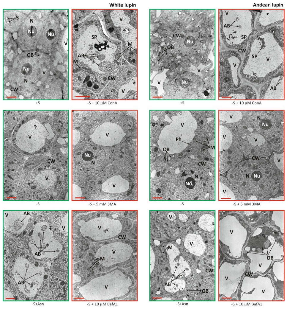
Figure 2.
Total p-nitrophenyl-palmitate-hydrolyzing activity in white and Andean lupin embryonic axes cultured in vitro for 96 h on a mineral liquid medium with (+S) and without (-S) 60 mM sucrose, and with 35 mM asparagine (+Asn). After 72 h, the +S and -S media were supplemented with 10 µM concanamycin A (ConA) or 5 mM 3-methyladenine (3MA). Data are means ± SD from three independent biological experiments. Different letters indicate significant differences at p ≤ 0.05 (ANOVA, Tukey’s HSD multiple-range test).
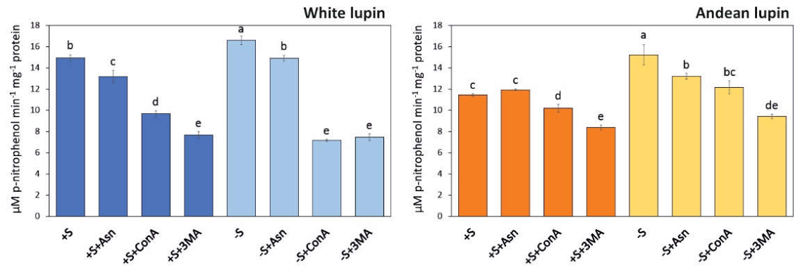
Figure 3.
Relative abundance of selected putative vacuolar-lipase transcripts in white and Andean lupin embryonic axes cultured in vitro for 96 h on a mineral liquid medium with (+S) and without (-S) 60 mM sucrose, and with 35 mM asparagine (+Asn). After 72 h, the +S and -S media were supplemented with 10 µM concanamycin A (ConA) or 5 mM 3-methyladenine (3MA). Fold changes were calculated relative to +S. Data are means ± SD from three independent biological experiments. Different letters indicate significant differences at p ≤ 0.05 (ANOVA, Tukey’s HSD multiple-range test). Primers used in this experiment are listed in Supplementary Table S1.
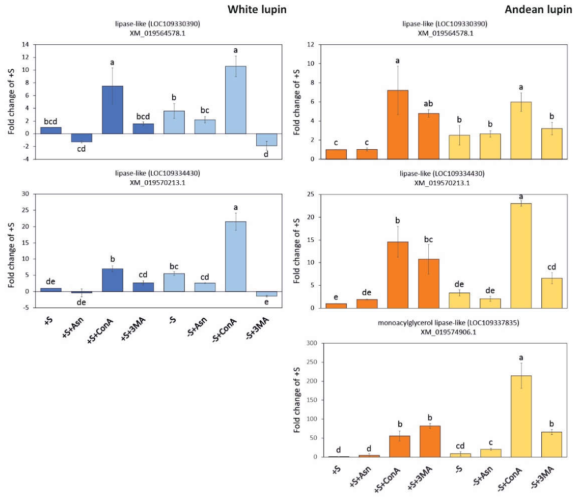
Figure 4.
Structural comparison of the analyzed lipases and reference lipase structures. (a) Structural superposition of the predicted models of XP_019420123.1 (yellow) and XP_019425758.1 (violet) from white lupin, and XP_019420123.1 (pink) and XP_019425758.1 (turquoise) from Andean lupin, with the catalytic serine (S174) indicated. (b) Predicted structure of XP_019430451.1 (dark green) from Andean lupin, with the catalytic serine (S211) highlighted in red. (c) Structural superposition of the closed (pdb id: 3TGL, magenta) and open (pdb id: 4TGL, green) conformations of the reference lipase, illustrating the conformational rearrangement of the lipid lid. (d) Structural superposition of the open (pdb id: 1K8Q, orange) and closed (pdb id: 1HGL, light green) conformations of a reference lipase, highlighting the alternative positions of the lipid lid.
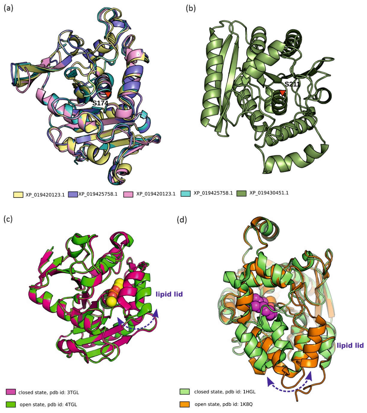
Figure 5.
Molecular docking studies. Superposition of lipase structures illustrating lipid-lid dynamics for XP_019420123.1 (a) and XP_019430451.1 (b). Best-ranked molecular docking poses obtained for XP_019420123.1 (c) and XP_019430451.1 (d). PI, phosphatidylinositol; MGDG, monogalactosyldiacylglycerol; DGDG, digalactosyldiacylglycerol; 18:2, linoleoyl moiety.
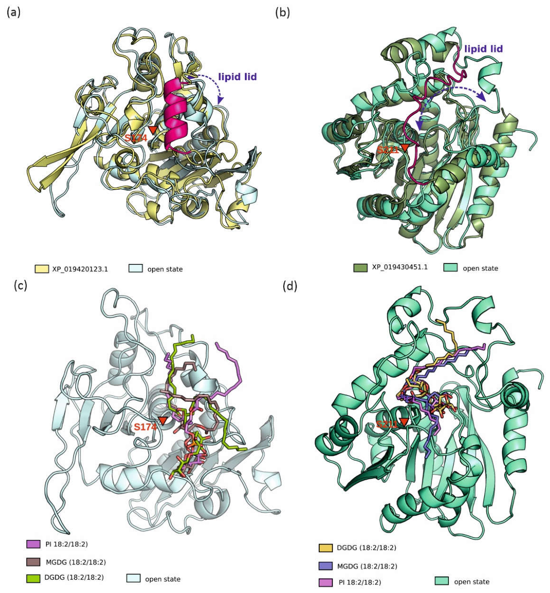
Figure 6.
Immunoblot analyses of (a) Ape1 in WT or atg15Δ AtAtg8a-GFP cells transformed with an empty plasmid (e.p.) or CPY-XP_019420123.1 (2 replicates). The experiment was repeated with similar results, showing prApe1-to-mApe1 turnover only in WT. These results indicate that CPY-XP_019420123.1 did not restore Atg15-dependent processing under the conditions tested. In the yeast strain atg15Δ AtAtg8a-GFP expressing CPY-XP_019420123.1, (b) AtAtg8a-GFP processing was not restored in the atg15Δ, as evidenced by the absence of free GFP and successful processing by CPY-LCAT3. Yeast cells were grown in nutrient-rich selective media (+N) or were exposed to nitrogen starvation (-N) conditions. Stain-free images were used as a loading control. Yeast strains used in the experiment are listed in Supplementary Table S5.
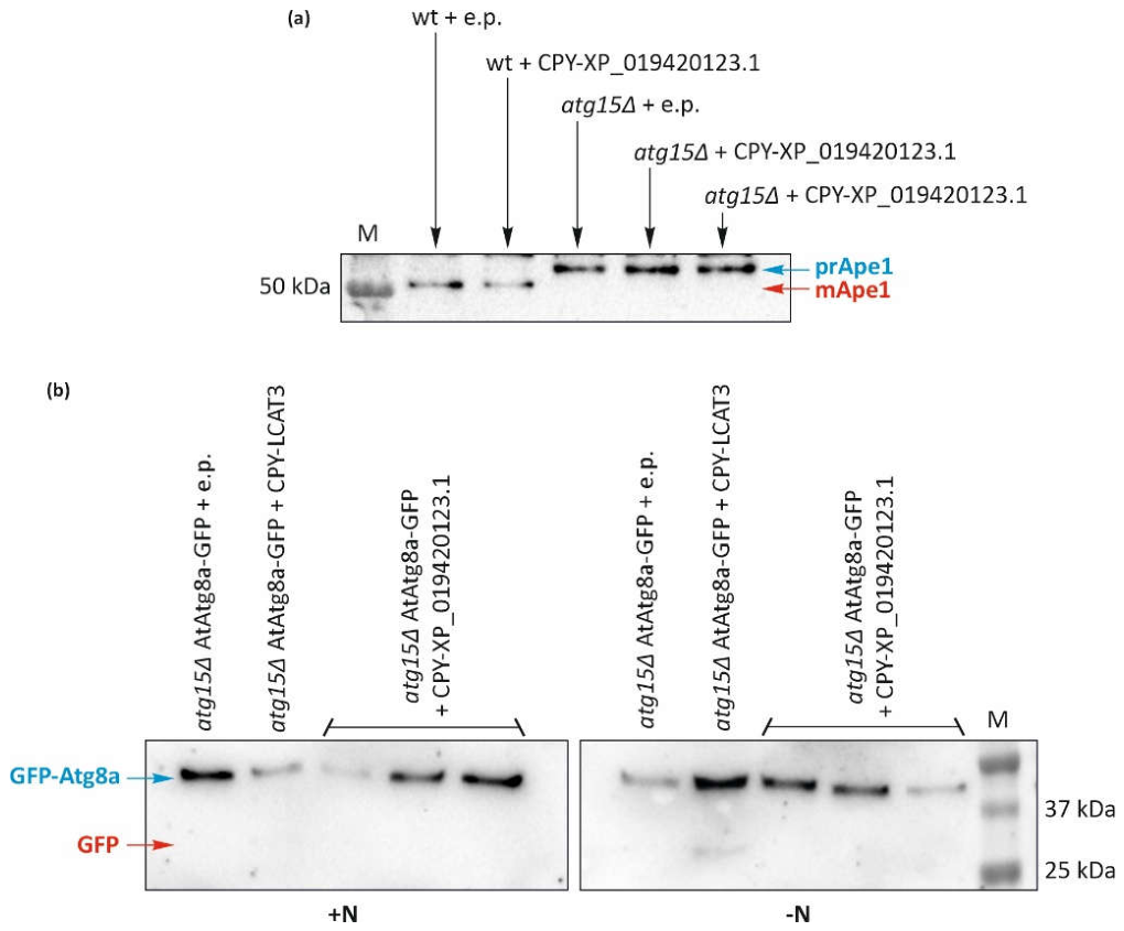
Figure 7.
Total Ac-ESEN-MCA-hydrolyzing activity in white and Andean lupin embryonic axes cultured in vitro for 96 h on a mineral liquid medium with (+S) and without (-S) 60 mM sucrose, and with 35 mM asparagine (+Asn). After 72 h, the +S and -S media were supplemented with 10 µM concanamycin A (ConA) or 5 mM 3-methyladenine (3MA). Data are means ± SD from four to six independent biological experiments. Different letters indicate significant differences at p ≤ 0.05 (ANOVA, Tukey’s HSD multiple-range test).
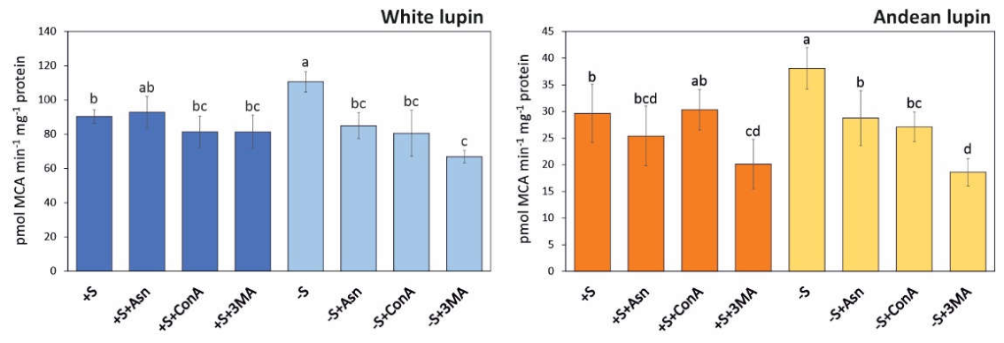
Figure 8.
Effect of Ac-YVAD-cmk on VPE-like activity (a) after 24 h exposure to 0-150 µM inhibitor in carbon-starved (-S) white and Andean lupin axes cultured in vitro for 96 h. Ultrastructure (b) and autophagic body number (c) of root meristematic zone cells after 24-h treatment with 100 µM and 25 µM Ac-YVAD-cmk of white and Andean embryonic axes, respectively. Data are means ± SD from three to four independent biological experiments. Different letters indicate significant differences at p ≤ 0.05 (ANOVA, Tukey’s HSD multiple-range test). AB, autophagic body; Au, autophagosome; CW, cell wall; M, mitochondrion; N, nucleus; Nu, nucleolus; OB, oil body; V, vacuole.
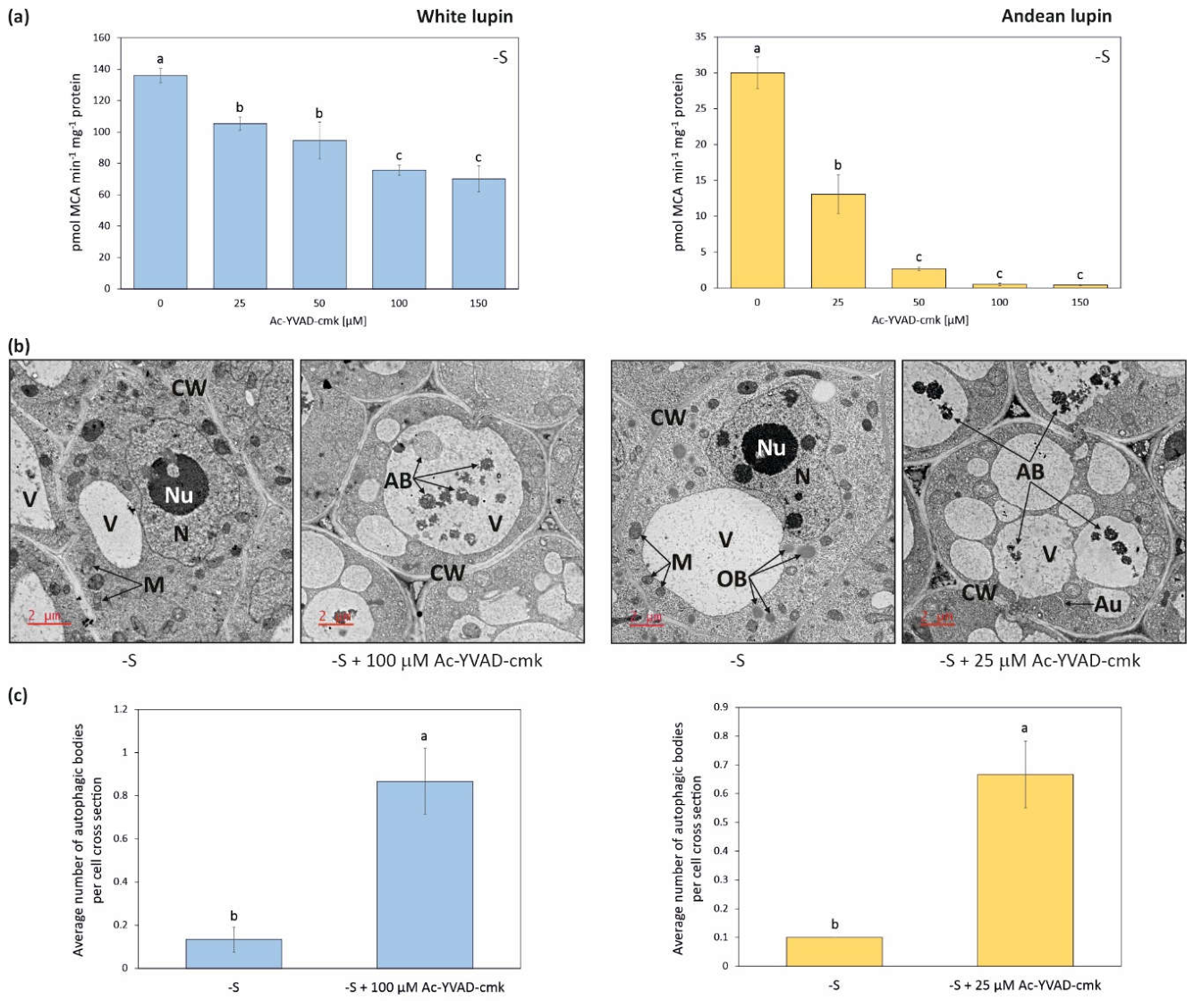
Figure 9.
Heatmaps showing the relationships in the levels of transcripts of the genes encoding vacuolar processing enzyme (VPE)/legumain (a) and relative changes in selected VPE/legumain gene transcript levels determined by RT-qPCR in embryonic axes of white and Andean lupin cultured in vitro for 96 h on a mineral liquid medium with (+S) and without (-S) 60 mM sucrose, and with 35 mM asparagine (+Asn). After 72 h, the +S and -S media were supplemented with 10 µM concanamycin A (ConA) or 5 mM 3-methyladenine (3MA). Fold changes were calculated relative to +S. Data are means ± SD from three independent biological experiments. Different letters indicate significant differences at p ≤ 0.05 (ANOVA, Tukey’s HSD multiple-range test). The transcriptomics data used to prepare the heatmaps are presented in Supplementary Table S6. Primer sequences are listed in Supplementary Table S1.
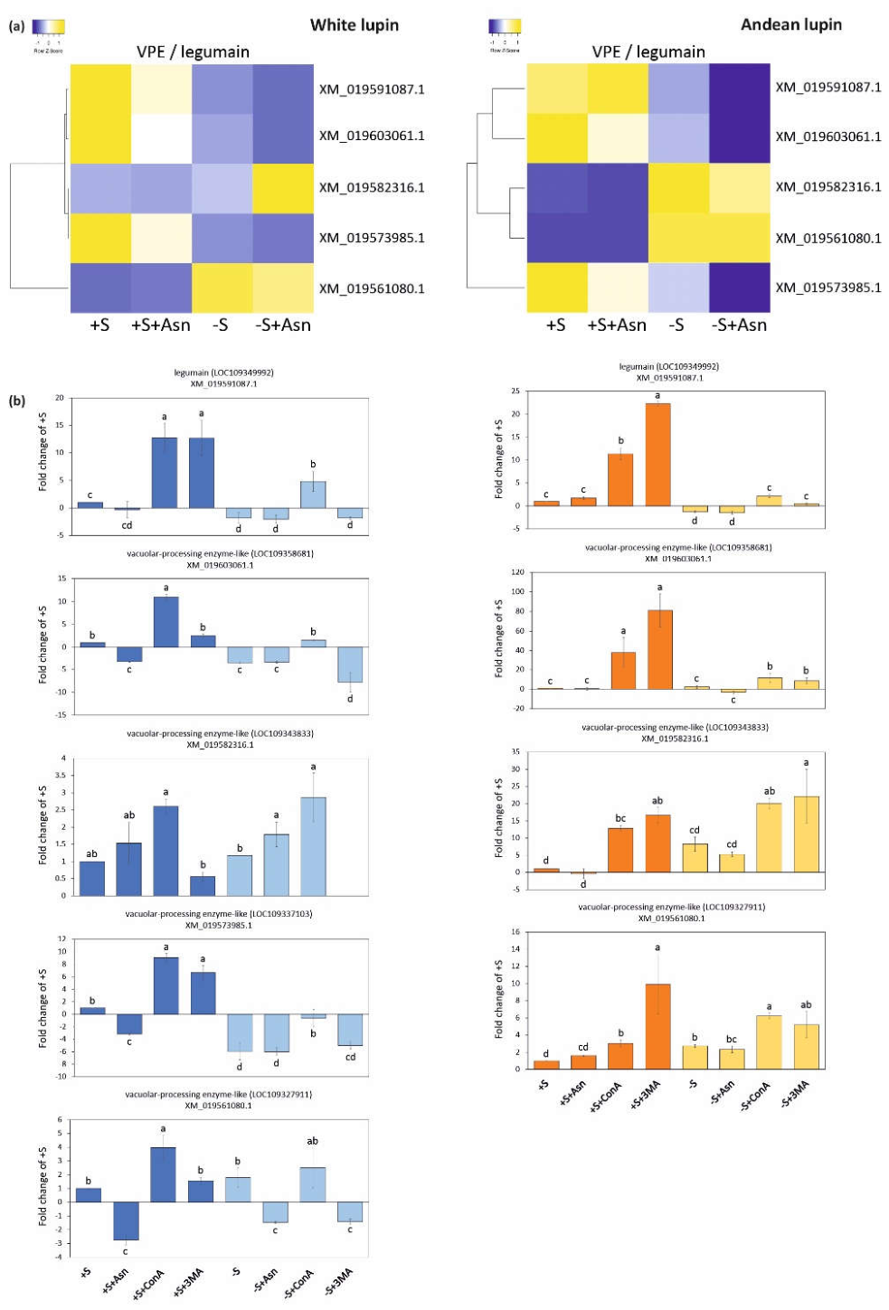
Disclaimer/Publisher’s Note: The statements, opinions and data contained in all publications are solely those of the individual author(s) and contributor(s) and not of MDPI and/or the editor(s). MDPI and/or the editor(s) disclaim responsibility for any injury to people or property resulting from any ideas, methods, instructions or products referred to in the content. |
© 2026 by the authors. Licensee MDPI, Basel, Switzerland. This article is an open access article distributed under the terms and conditions of the Creative Commons Attribution (CC BY) license.
Copyright: This open access article is published under a Creative Commons CC BY 4.0 license, which permit the free download, distribution, and reuse, provided that the author and preprint are cited in any reuse.