

Submitted:
25 September 2026
Posted:
28 September 2026
You are already at the latest version
Abstract
Protecting crops from bacterial diseases is a major challenge for sustainable agriculture. Bacterial spot caused by Xanthomonas arboricola pv. pruni (Xap) threatens stone fruit and almond production, while restrictions on agrochemicals in the European Union have increased the need for safe alternatives. Bacteriophages (phages) are promising biotools for controlling bacterial plant pathogens. However, there is limited research on Xap-specific phages in Europe, particularly under Mediterranean conditions. The aim of this study was to isolate and characterise phages from environmental sources with lytic activity against Spanish Xap strains from different Prunus species and geographical locations. Six Xap phages were isolated from different environmental water samples and characterised using genomic, phylogenetic, morphological and biological analyses. These studies enabled three of the phages to be assigned to different species and revealed a putative novel genus of previously undescribed phages with myovirus morphology. In vitro assays confirmed their specificity and ability to infect and lyse multiple Spanish Xap strains, significantly reducing bacterial populations by phage-based biocontrol. Five of them were selected and combined into different four-phage cocktails. The selected phages demonstrated high stability in vitro and in planta. Following optimisation of foliar spray application, two selected cocktails significantly reduced bacterial spot in Prunus plants. To our knowledge, these are the first European Xap phage cocktails to be effective in Prunus. They therefore represent promising, safe and sustainable biocontrol agents for managing bacterial spot in stone fruit and almond crops under Mediterranean climatic conditions. They could also be integrated into broader disease management strategies for woody crops and could help to reduce dependence on agrochemicals.
Keywords:
phytopathogenic bacterium
; plant disease
; biological control
; phage
; woody crop
; One Health
; SDGs
1. Introduction
Protecting crops from bacterial diseases is an ongoing challenge. A major threat to the production of stone fruit and almond trees (Prunus spp.), which are highly valued for their nutritional and health benefits (Lara et al., 2020), is bacterial spot, caused by the bacterium Xanthomonas arboricola pv. pruni (Xap). This pathogen is able to infect a wide range of Prunus species, including but not limited to almond, apricot, nectarine, peach and plum (Garita-Cambronero et al., 2018; Sakata and Ishiga, 2025). Generally, in Prunus spp., Xap causes necrotic spots and cankers on leaves, fruit, twigs and branches or trunks. The disease caused by Xap is commonly referred to as bacterial spot of stone fruits, although other names are also used depending on the host species and the predominant symptoms (EPPO, 2026). This phytopathogen has been shown to have a detrimental effect on the quality and marketability of the fruits, resulting in significant economic losses for growers around the world (Stefani, 2010; Garita-Cambronero et al., 2018; Sakata and Ishiga, 2025; Cuesta-Morrondo et al., 2026). At present, Xap is considered a protected-zone quarantine pest (PZQP) for specified areas and a regulated non-quarantine pest (RNQP) for specified plant material in the European Union (EU) (EPPO, 2026). It is also included in the EPPO A2 List and subjected to phytosanitary regulation in several countries worldwide (European Commission, 2019; EPPO, 2026). The frequency and severity of the disease caused by Xap increase in temperate and humid climates, favouring the pathogen proliferation and infection (Morales et al., 2018). However, the perennial nature of Prunus crops makes eradication especially difficult once the pathogen has become established, since it can survive overwintering on infected plant material (dormant buds, leaf scars and cankers) and plant debris (mummified almond fruits and fallen leaves), which serve to spread the pathogen (Zaccardelli et al., 1998; Garita-Cambronero et al., 2018; Haack et al., 2020; EPPO, 2026; Panth et al., 2025; Sakata and Ishiga, 2025).
In Europe, it has been determined that a theoretical outbreak of the disease in a plum orchard in the Emilia-Romagna region (Northern Italy) with a 30% reduction in fruit yield could result in an economic loss of over 10,000 €per hectare (Stefani, 2010). In Spain, the bacterial spot of stone fruit and almond trees was first observed on Japanese plum in Extremadura region (South-West Spain) in 2002. Subsequent years witnessed the detection of this phytopathogen in various Autonomous Communities in Eastern and Northern Spain, affecting a range of cultivars including Japanese plum, nectarine, peach and almond trees. Notably, outbreaks of Xap on almond trees in Spain were the first to be documented within the EU (Roselló et al., 2012; EPPO, 2026). Yield losses in commercial almond orchards in Northern Spain ranged from 23% to 47% of production in 2013 and 2014 (Palacio-Bielsa et al., 2014). Recent studies confirm that Xap continues to be a major constraint for peach and other Prunus crops, with particular concern in regions where warm and humid periods favour disease development (Sakata and Ishiga, 2025).
Bacterial spot of stone fruits is particularly difficult to manage in commercial orchards because it combines an epiphytic phase, latent infections, survival in infected woody tissues and dissemination through rain splash, wind-driven rain, contaminated plant material and orchard operations (Palacio-Bielsa et al., 2011; Garita-Cambronero et al., 2018). Current management relies on an integrated strategy that includes the use of healthy planting material, sanitation, pruning practices, reduction of inoculum sources, choice of less susceptible cultivars and repeated applications of copper-based compounds when permitted (Garita-Cambronero et al., 2018). Nevertheless, these measures often provide only partial control. The problem is a complex matter, primarily due to a paucity of effective agrochemicals, with cupric compounds being the mainstay of treatments (Stefani, 2010). However, the emergence of Xap strains resistant or tolerant to copper (Giovanardi et al., 2017; Cox et al., 2022; Sakata and Ishiga, 2025) together with copper accumulation in soils and its phytotoxicity to some of the Xap-affected fruit trees, has prompted interest in alternative treatment options. The antibiotics streptomycin and oxytetracycline (Dhanvantari et al., 1978) have been shown to be effective in controlling this pathogen but, their use is not permitted in the EU, and oxytetracycline- and streptomycin-resistance genes have been detected in Xap strains (Herbert et al., 2022). Thus, once this phytopathogen establishes itself in an orchard, it is very difficult to control (Garita-Cambronero et al., 2018). This is further exacerbated by growing societal concerns regarding bacterial resistance to antibiotics and the deleterious effects of agrochemicals in the environment and global health (Grenni et al., 2018). Consequently, there has been an increasing focus on the development of more specific, health-safe and sustainable strategies.
Bacteriophages (phages) have emerged as a hopeful option for the control of bacterial pathogens in agriculture (Buttimer et al., 2017; Álvarez et al., 2019; Grace et al., 2021; Holtappels et al., 2021; Biosca et al., 2024, 2026; Choudhary et al., 2025). These natural bacterial predators have the unique ability to selectively infect and destroy bacteria, with minimal expected disturbance of beneficial plant microbiota if they are properly selected and characterised. Thus, it relates to a highly specific and environmentally friendly biological control method, in line with the green EU policies, the One Health strategy and the achievement of the Sustainable Development Goals (SDGs) (Álvarez and Biosca, 2025). Lytic phages are being evaluated as promising biotools to be included in Integrated Pest Management (IPM) programmes (Jones et al., 2012; Holtappels et al., 2021; Choudhary et al., 2025). For diseases caused by Xanthomonas spp., the development of phage-based biocontrol has also progressed from empirical assays to the genome-informed selection of strictly lytic phages and the design of phage cocktails. Recent reviews emphasize that phage efficacy depends on host range, lytic activity, efficacy in the plant, environmental persistence, resistance management, formulation, timing and delivery method (Grace et al., 2021; Nakayinga et al., 2021; Stefani et al., 2021; Greer et al., 2024; Choudhary et al., 2025). Furthermore, recent studies with other economically significant Xanthomonas species support the use of locally isolated phages and cocktails adapted to the climatic conditions of the target crop area. In Mediterranean conditions, a European X. euvesicatoria pv. euvesicatoria phage cocktail was shown to significantly reduce bacterial growth in vitro and control bacterial spot symptoms in pepper plants (Biosca et al., 2026).
Despite this potential, studies on Xap-specific phages remain scarce, particularly in Europe. The first studies were published in the 1970s, when Civerolo and Kiel demonstrated that X. pruni phages inhibited bacterial spot on peach foliage (Civerolo and Kiel, 1969). Civerolo then characterized the relationships between the phages and their bacterial hosts (Civerolo, 1972). In the 1990s, Xap phages were isolated and characterized from peach orchards in Northern Italy (Zaccardelli et al., 1992; Saccardi et al., 1993), enabling the selection of a candidate with a broad host range for Xap biocontrol. Nevertheless, its efficacy under field conditions was inconsistent, due to reduced survival and the emergence of bacterial resistance to a single phage. Thereafter, only two recent North American studies have renewed interest in Xap-infecting phages (D’Amico-Willman et al., 2025, 2026). The first study reported that genetically similar Xap strains and their phages, which were isolated from peach orchards in North Carolina over several decades, displayed phenotypic and genotypic variation (D’Amico-Willman et al., 2025). A second genomic study by these authors characterized several Xap phages, which had remained stable and adapted over decades in North Carolina peach orchards (D’Amico-Willman et al., 2026). However, these phages were not tested for biocontrol in planta.
The paucity of recent research on Xap-specific phages in Europe highlights the need for studies in this area, considering the potential of phages as biocontrol agents in the management of bacterial diseases in Prunus spp. crops. Thus, the aim of this study was to isolate and characterise a collection of phages with lytic activity against Spanish strains of Xap isolated over a 23-year period from different Prunus species and geographical locations. Xap phages were isolated from environmental water samples based on the hypothesis that they could provide an effective and sustainable means of biological control under Mediterranean climatic conditions. Six phages were initially characterized through genomic and phylogenetic analyses. This enabled their taxonomic assignment to different genera, as well as the identification of previously uncharacterised phages displaying myovirus-like morphology. Subsequently, in vitro assays confirmed their ability to infect and lyse multiple Spanish Xap strains from different hosts and locations, as well as their specificity. Phage-based in vitro control assays also confirmed their ability to significantly reduce bacterial populations. These findings supported the selection of five individual phages and their combination in two phage cocktails. The selected phages demonstrated high stability in vitro and in planta. Following the optimization of their application via a foliar spray, both phage cocktails provided significant protection against bacterial spot in Prunus plants. These novel phage combinations are the first European cocktails able of effectively controlling Xap in planta. They could therefore be used to reinforce current strategies for the control of bacterial spot in stone fruit and almond crops, as part of integrated disease-management programmes for woody plants.
2. Material and Methods
1.1. Bacterial Strains, Culture Conditions and Preservation
The Spanish Xap reference strain CITA 33 (Centro de Investigación y Tecnología Agroalimentaria de Aragón, Zaragoza, Spain), isolated from almond (P. dulcis), was used as the host strain for bacteriophage isolation and propagation and in subsequent phage characterisation and biocontrol assays. Additional Spanish and reference Xap strains isolated from different Prunus hosts, geographical origins and years of isolation within a period of up to 23 years were included for host range determination (Table 1). To assess phage specificity, a panel comprising strains of other Xanthomonas species (Table 1) together with representative bacterial species from different genera was included (Supplementary Table 1). All strains were routinely cultured in Luria-Bertani (LB) broth [tryptone 10 g/L, yeast extract 5 g/L, NaCl 10 g/L] or Nutrient Broth (NB) [peptone 5 g/L, beef extract 3 g/L, NaCl 5 g/L], or on Nutrient Agar (NA; NB supplemented with 15 g/L agar), depending on the experimental requirements. Liquid cultures were incubated at 28 °C with shaking at 150 rpm for 24 h, whereas plates were incubated at 28 °C for 24 or 48 h, depending on the bacterial species.
The handling of bacterial strains was carried out in accordance with their biosafety levels (1 and 2) in consideration of their designated biological risk classification and the quantity of inoculum employed in specific experiments. All bacterial strains were preserved in NB supplemented with either 20% or 25% (v/v) glycerol at -20 °C or -80 °C, respectively.
1.2. X. Arboricola pv. Pruni Identity and Pathogenicity Confirmation
The identity of Xap strains was routinely confirmed by conventional PCR following the protocol described by Pagani et al. (2004), according to the EPPO Standard PM 7/064 (2) (2021). The specific primers Y17CoF (5′- GAC GTG GTG ATCAGC GAG TCA TTC-3′) and Y17CoR (5′- GAC GTG GTG ATGATG ATC TGC-3′) were used. Xap strain CITA 33 and X. vesicatoria strain CFBP 1941 were included as the positive amplification control (PAC) and negative amplification control (NAC), respectively, and water was additionally used as (NAC)
The peach-almond hybrid rootstock GF677 (Prunus amygdalus × P. persica), which was obtained by micropropagation from Agromillora Group (Spain), was used to confirm the pathogenicity of the Spanish reference strain CITA 33 prior to detached-leaf and in planta biocontrol assays, due to its susceptibility to bacterial spot disease. Assays were carried out using the standard Xap pathogenicity testing method according to EPPO PM 7/064 (2) (EPPO, 2021) and involved the use of the leaf syringe infiltration technique. Bacterial suspensions were adjusted to 10⁷ or 10⁸ CFU/mL and 6 plants were evaluated for each concentration. Young fully expanded leaves were infiltrated using a needleless plastic syringe, applying gentle and consistent pressure to the abaxial surface until the mesophyll exhibited a water-soaked appearance.
1.3. Isolation, Propagation and Preservation of X. Arboricola pv. Pruni Bacteriophages
Bacteriophages were isolated from environmental water samples taken from irrigation channels surrounding Valencia (Eastern Spain), using the Xap strain CITA 33 as the bacterial host. Filtered samples (0.22 μm pore size) were enriched with an equal volume of 2× LB medium and incubated overnight at 28 °C with shaking at 150 rpm, following the procedure described by Biosca et al. (2024; 2026). The enrichment cultures were then centrifuged at 10,000 rpm for 10 min, and the resulting supernatants were filtered through sterile 0.22 μm filters to remove bacterial cells. Filtrates were screened for the presence of bacteriophages using spot assays on bacterial lawns. Plates were incubated at 28 °C for 18–24 h and examined for plaque formation. Individual plaques were picked using sterile pipette tips and eluted in SM buffer (50 mM Tris-HCl, pH 7.5; 100 mM NaCl; 10 mM MgSO₄; 0.01% gelatin). Purification was achieved through at least four successive rounds of plaque isolation and serial tenfold dilution using the double-layer agar method until pure phage isolates were obtained.
Purified phages were propagated by infecting exponentially growing cultures of the bacterial host and incubating at 28 °C for at least 18 h with shaking at 150 rpm. Phage titres were determined using the double-layer agar method following serial tenfold dilution. Amplified phage stocks were stored at 4 °C for short-term preservation and at −80 °C in 30% glycerol for long-term storage.
1.4. X. Arboricola pv. Pruni Bacteriophage DNA Extraction, Whole-Genome Sequencing, Bioinformatic and Phylogenetic Analysis
The isolation of genomic DNA from a selection of phages was conducted in accordance with the methodology outlined by Biosca et al. (2021, 2024). In summary, the process involved the removal of bacterial nucleic acids from filtered (0.22 μm pore diameter filters) phage lysates by DNase and RNase treatment for one hour at 37 °C, followed by thermal inactivation with EDTA for 10 minutes at the same temperature. Then, the isolation of phage DNA was undertaken using the NucleoSpin® plasmid isolation kit (Macherey-Nagel, Düren, Germany), following the low-copy plasmid isolation protocol.
Total DNA quantification was conducted using a Qubit 4TM Fluorometer (ND-2000, ThermoFisher, Wilmington, DE, USA) with the dsDNA HS Assay KitTM and dsDNA integrity was assessed by electrophoresis on a 1% agarose gel. The construction of DNA libraries was then undertaken using the Nextera XT Library Preparation Kit (Illumina, San Diego, California, USA), in accordance with the manufacturer’s specifications. Subsequently, the Illumina MiSeq platform was utilized for sequencing, employing 2 × 250 bp paired-end sequencing (Illumina, San Diego, California, USA). Thereafter, the raw data read outputs were subjected to quality control, trimming, and de novo assembly with the CLC Genomics Workbench 10.1.1 software (QIAGEN, Hilden, Germany). De novo contigs were identified using BLASTn and BLASTx (Altschul et al., 1997) implemented in ViPTree (Nishimura et al., 2017). Annotation of phage contigs was performed using Pharokka (V1.7.5) (Bouras et al., 2023). The annotated genomes were further analysed using PhageScope to screen for putative virulence factors and antimicrobial resistance genes as part of the genomic characterisation (Wang et al., 2024). Intergenomic nucleotide similarities were calculated using VIRIDIC v1.1 (Moraru et al., 2020). The analysis included EW15 and EW17 together with representative bacteriophages selected from the ViPTree analysis according to genome-wide similarity score (SG). Genome organization and comparative genomic maps were visualized using LoVis4u (Egorov and Atkinson 2025). Pairwise comparisons were performed using the default settings, and results were reported as similarity. Species and genus thresholds were set to 95% and 70%, respectively.
1.5. Virion Visualisation of Novel Phages by Transmission Electron Microscopy
Selected bacteriophages were morphologically characterised by transmission electron microscopy (TEM). In summary, 5 μL aliquot of high-titre phage stocks (≥10⁹ PFU/mL) were placed on carbon-coated grids and negatively stained with 1% uranyl acetate for 5 minutes. Excess stain was removed, and the grids were allowed to air dry. Grids were examined using an FEI Tecnai G2 Spirit transmission electron microscope (Thermo Fisher Scientific, Waltham, USA). Capsid diameter and tail length were measured from TEM micrographs of at least 10 virions per bacteriophage.
1.6. Host Range and Specificity Determination
The host range and specificity of the selected bacteriophages were evaluated by spot assays. Briefly, bacterial lawns were prepared by mixing 0.2 mL of a bacterial suspension adjusted to OD₆₀₀ = 0.5 with molten top agar (0.6% w/v), and the mixture was poured onto NBA plates and allowed to solidify for approximately 30 min. Thereafter, 10 μL of filtered phage lysates (≥10⁸ PFU/mL) were spotted onto the bacterial lawns and allowed to dry at room temperature. The Xap strain CITA 33 was included as the positive control. Plates were incubated at 28 °C for 18–24 h and examined for the presence of lytic activity. Complete clearing under the spotted area was recorded as lysis, partial clearing without complete lysis was classified as weak lysis, and the absence of visible clearing was considered a negative result.
Host range was determined using a collection of Spanish and reference Xap strains isolated from different Prunus hosts, geographical origins and years of isolation (Table 1). To further assess phage specificity, the selected bacteriophages were also tested against a panel of non-target bacterial species, including representatives of other Xanthomonas species and bacterial species from different genera (Supplementary Table 1). All assays were repeated in independent experiments.
1.7. In Vitro Curves of Phage-Mediated Bacterial Biocontrol
The lytic activity of bacteriophages against the Xap reference strain CITA 33 was evaluated in vitro by monitoring bacterial growth in co-culture, following the methodology described by Biosca et al. (2026) with minor modifications. A 24-h bacterial culture grown in LB medium at 28 °C was adjusted to an OD₆₀₀ of 0.1, corresponding to approximately 10⁸ CFU/mL and subsequently ten-fold diluted to a final concentration of 10⁷ CFU/ mL. Phage suspensions were individually adjusted to 10⁸ PFU/mL and evaluated individually against both bacterial concentrations, corresponding to MOIs of 100 and 10, respectively. Following the evaluation of individual phages, the fifteen possible four-phage cocktail combinations generated from the six selected bacteriophages were screened against Xap strain CITA 33 at a MOI of 10, using the bacterial concentration of 10⁷ CFU/mL.
Phage–bacteria interactions were monitored in 96-well microplates by recording OD₆₀₀ every 120 min for 48 h at 28 °C with continuous shaking using a Tecan Infinite M Nano microplate reader (Männedorf, Switzerland). Wells containing LB medium alone and bacterial suspensions without bacteriophages were included as negative and positive growth controls, respectively. At least three technical replicates per condition were included in two independent experiments.
1.8. Method to Test X. Arboricola pv. Pruni Pathogenicity in Prunus for Performing Bacterium-Phage Biocontrol Assays
The method was set up in this work. The Prunus hybrid GF677 was used as the experimental host to establish the Xap inoculation conditions for pathogenicity and for subsequent in planta biocontrol assays. Two inoculation methods were evaluated for pathogenicity using bacterial suspensions of strain CITA 33 adjusted to 10⁶, 10⁷ or 10⁸ CFU/mL. Six leaves detached from the plant were used in the first method, while the second one involved the use of six plants per bacterial concentration.
Detached-leaf assays were initially performed to assess the suitability of ex vivo inoculations as a rapid preliminary approach for evaluating symptom development caused by Xap. The assay was performed following the methodology described by Licciardello et al. (2022), with modifications based on Álvarez et al. (2025). Fully expanded Prunus leaves were surface-disinfected by immersion in 0.5% sodium hypochlorite for 30 s and 70% ethanol for 30 s, rinsed three times with sterile distilled water, and placed abaxial side up on Petri dishes containing 1% water agar. Four inoculation sites were established on each leaf, two on each side of the central vein. Each site was wounded five times with a sterile needle before applying a 10 μL drop of bacterial suspension at the corresponding concentration. Plates were sealed with flexible film (Parafilm®M) and incubated at 28 °C. Symptom development was monitored periodically for up to three weeks.
In vivo inoculation assays were subsequently conducted using foliar spraying with potted plants that were grown in a commercial substrate under greenhouse conditions and transplanted into 6.5-cm diameter pots. Plants that were approximately 15 cm tall and were bearing about14 fully expanded leaves were selected for all in planta assays. For foliar spray inoculations, bacterial suspensions of Xap were prepared following the protocol by Tamir-Ariel et al. (2007), but applied using the protocol outlined by Biosca et al. (2026). Plants were sprayed with the pathogen until runoff using a handheld sprayer, ensuring complete coverage of leaf surfaces. After inoculation, plants were enclosed in transparent plastic bags to maintain high relative humidity. Bags were replaced every 10 days to prevent excessive condensation while preserving humid conditions throughout the experimental period. Inoculated plants were maintained in a Panasonic MLR-352-PE controlled-environment growth chamber at 26 °C under a 16-h light/8-h dark photoperiod for four weeks. Symptom development was monitored periodically for up to four weeks.
1.9. Bacteriophage Stability in Vitro and in Planta
1.9.1. In Vitro Phage Stability Under Different Temperature and pH Conditions
The stability of selected bacteriophages was evaluated individually under different temperature and pH values over a three-week period. Phage suspensions were prepared in SM buffer and adjusted to a final concentration of 108 PFU/mL. To assess thermal stability, phage suspensions in SM buffer (pH 7.2) were incubated at 28 °C, or 37 °C.
For pH stability assays, phage suspensions were prepared in SM buffer adjusted to pH 5.5, 7.2, or 8.0 and incubated at 28 °C. Samples were collected at 0, 1, 2, and 3 weeks for phage quantification.
Serial tenfold dilutions were prepared in SM buffer, and phage titres were determined using the double-layer agar method. Briefly, aliquots of the appropriate dilutions were mixed with exponentially growing host cells (OD₆₀₀nm = 0.6) and molten top agar before being poured on NA plates, which were incubated at 28 °C for 24 h prior to plaque enumeration. All assays were performed using two independent biological replicates, and plaque counts were determined from three technical replicates per sample. Phage survival was expressed as log of PFU/mL.
In Planta Phage Stability
Phage stability was evaluated on Prunus leaves using the foliar spray method described above for Xap inoculation. Each bacteriophage was sprayed individually onto three plants per treatment. Phage suspensions were adjusted to 10⁸ PFU/mL immediately prior to their application. To evaluate in planta phage survival in the absence of the bacterial host, plants were sprayed individually with each phage.
Leaf samples were collected immediately after the sprayed suspension had dried on the leaf surface (defined as 0 days post-inoculation (dpi), approximately 1h after application), and subsequently at 1, 3, 7, and 20 dpi. At each sampling time, leaves from the three plants within the same treatment were pooled to obtain 1 g of fresh tissue, placed in sterile mesh extraction bags, and homogenized in 9 mL of SM buffer. The resulting homogenates were centrifuged at 5,000 rpm for 5 min to remove plant debris, and the supernatants were subsequently filtered through 0.22 μm syringe filters. Serial tenfold dilutions of the filtrates were prepared in SM buffer, and phage titres were determined using the double-layer agar method as described above. Negative control plants sprayed with SM buffer alone were included and processed following the same sampling procedure. The experiment was performed twice with independently prepared phage suspensions, yielding two biological replicates for each phage. Each sample was titrated in duplicate as technical replicates. Phage stability was expressed as PFU/g fresh weight (FW).
1.10. In Planta Evaluation of Two Bacteriophage Cocktails Against X. Arboricola pv. Pruni
The efficacy of two bacteriophage cocktails in reducing symptom development caused by Xap strain CITA 33 was evaluated in Prunus GF677 plants. The experimental design included six treatments: (i) Phosphate Buffered Saline (PBS)-treated plants as negative controls, [PBS: 137 mM NaCl, 10 mM Na2HPO4, 2.7 mM KCl, 1.9 mM KH2PO4, pH 7.4]; (ii) plants sprayed with Cocktail 1 alone; (iii) plants sprayed with Cocktail 2 alone; (iv) plants inoculated with the bacterial pathogen alone; and (v–vi) plants co-inoculated with the pathogen and either Cocktail 1 or Cocktail 2.
Cocktail 1 consisted of four selected phages (EW1, EW3, EW5 and EW17), whereas in Cocktail 2, phage EW5 was replaced by phage EW16, resulting in a combination comprising phages EW1, EW3, EW16 and EW17. Phage stocks were individually adjusted to 1-2 x 10⁸ PFU/mL and mixed in equal proportions immediately before use to prepare each cocktail. The experiment consisted of three independent biological replicates, each including six plants per treatment (n = 18 plants per treatment; total n = 108 plants).
Bacterial suspensions were prepared from 48-h cultures grown on NBA plates at 28 ºC and adjusted to 5 x 10⁷ CFU/mL. For co-inoculation treatments, bacterial and phage suspensions were mixed immediately prior to application. All treatments were applied using the foliar spray inoculation protocol detailed above. Disease development was assessed by counting bacterial spot lesions on all leaves of each plant, similarly to Biosca et al. (2026) but at 30 dpi. To account for differences in plant size, lesion counts were normalized by the total leaf area of each plant and expressed as lesion density (lesion/cm2). Leaf area was determined from digital images using ImageJ software (version 1.54p, National Institutes of Health, Bethesda, MD, USA).
To confirm the presence of Xap in plants exhibiting disease symptoms, bacteriological analyses were performed on a subset of symptomatic plants. Leaf tissues showing characteristic lesions were processed, and yellow, presumptive Xap colonies were isolated on NA plates. A selection of purified isolates was subsequently subjected to PCR-based identification using the Xap-specific primers Y17CoF and Y17CoR, according to the diagnostic protocol established by Pagani et al. (2004).
1.11. Statistical Analysis
Data were analysed using one- or two-way analysis of variance (ANOVA), as appropriate. When significant effects were detected, means were compared using Tukey’s honestly significant difference (HSD) test. Normality and homogeneity of variance were assessed prior to analysis. Differences were considered statistically significant at P< 0.05. Statistical analyses were performed using R version 4.4.2 (R Foundation for Statistical Computing, Vienna, Austria). Curve plots were prepared using GraphPad Prism version 10.2.0 (GraphPad Software, Boston, MA, USA).
2. Results and Discussion
2.1. Confirmation of X. Arboricola pv. Pruni Identity and Pathogenicity
Identification of Xap strains was confirmed by PCR using the Xap-specific primers Y17CoF and Y17CoR according to Pagani et al. (2004) and the EPPO Standard PM 7/064 (2) (2021). Positive results were obtained for all Xap strains included in the present study (Table 1).
Pathogenicity assays were carried out using the Spanish reference strain CITA 33 and the leaf syringe infiltration technique, which is the standard method for testing the pathogenicity of Xap according to EPPO PM 7/064 (2). Strain CITA 33 induced symptom development under controlled conditions after 14 dpi, confirming its pathogenicity. The symptoms remained confined to the infiltrated areas, indicating a highly localized response resulting from direct mesophyll inoculation. No substantial differences in the affected area were observed among the two bacterial concentrations tested (107and 10⁸ CFU/mL).
2.2. X. Arboricola pv. Pruni Bacteriophages were Isolated from Different Environmental Waters
Six bacteriophages were selected following the enrichment of several environmental water samples taken from agricultural irrigation channels, with the reference Xap Spanish strain CITA 33 acting as the bacterial host. This supports that surface water can act as a reservoir for phages that infect Xap. This broadens the range of ecological sources of Xap phages, which have primarily been found in symptomatic peach tissues or materials associated with Prunus orchards (D’Amico-Willman et al., 2024, 2026). The phages were initially named according to the water sample from which they were isolated (EW1, EW3, EW5, EW15, EW16, and EW17). The prefix ‘EW’ denotes environmental water, and the subsequent number indicates the number of the water sample from the irrigation channel that was sampled. No phages were obtained by direct isolation, indicating the low abundance of phages able to infect Xap in environmental water. All six phages produced clear, defined plaques on NA plates, indicative of lytic activity. The sizes of these plaques ranged from approximately 1.5 to 7 mm, indicating productive infection and suggesting phenotypic variability (Figure 1).
2.3. Selected X. Arboricola pv. Pruni Bacteriophages Belong to Different Genera
De novo assembly of the selected bacteriophages resulted in contigs ranging from 43,449 to 44,626 bp in length. BLASTN analysis against the NCBI nucleotide collection database revealed that the assembled genomes of phages EW1, EW3 and EW16 showed high nucleotide similarity to previously described Xanthomonas phages belonging to the class Caudoviricetes. Specifically, EW1 phage showed 97.8% nucleotide identity (91% query coverage (QC)) to Xanthomonas phage Sopo (43,861 bp; GenBank accession LR743529.1), which is currently not classified and was designated as vXap-EW1 (Clavijo-Coppens et al., 2021). EW3 phage showed 98.6% identity (88% QC) to Xanthomonas phage JUN5 (42,912 bp; OK913679.1) and EW16 exhibited 97.0% identity (95% QC) to Xanthomonas phage Tenjo (43,850 bp; LR743531.1), both belonging to the genus Tabiovirus (Clavijo-Coppens et al., 2021) and then named as vXapT-EW3 and vXapT-EW16. It has recently been proposed that the phages Sopo, JUN5 and Tenjo should be grouped together in a new genus (Clavijo-Coppens et al., 2021) not yet accepted by the International Committee on Taxonomy of Viruses (ICTV). In contrast, phage EW5 showed 97.0% nucleotide identity and 97% QC to Xanthomonas phage vB_Xar_IVIA-DoCa4 (43,007 bp; ON932078), classified within the genus Pradovirus, and then named as vXapP-EW5. BLASTN analysis revealed no significant nucleotide similarities to sequences available in the NCBI database for phages EW15 and EW17. However, pairwise comparison of their assembled genomes revealed 100% nucleotide identity, indicating that both phages are genomically indistinguishable and likely represent the same bacteriophage species. All assembled phage genomes were deposited in the NCBI GenBank database under accession numbers PZ832393, PZ832394, PZ832395, PZ832396, PZ832397, and PZ832398.
ViPTree proteomic analysis placed EW15 and EW17 within the phylum Uroviricota and the class Caudoviricetes, forming a distinct branch from the reference phages included in the proteomic tree. The highest genome-wide similarity score (SG) was observed with Stenotrophomonas phage vB_SM_ytsc_ply2008005c (NC_072504; SG = 0.2205). The next closest matches corresponded to Xanthomonas phages Langgrundblatt1 (NC_073093; SG = 0.1864), Langgrundblatt2 (NC_073094; SG = 0.1840), Pfeifenkraut (NC_073095; SG = 0.1815), and Suba (NC_073092; SG = 0.1725) (Figure 2A). Other related phages showed SG values below 0.16. In the proteomic tree constructed against 3,171 viral genomes, EW15 did not cluster within any of the neighboring established genera, but instead formed an independent branch positioned between several distinct groups of Caudoviricetes. This placement, together with the low SG values obtained for its closest reference phages and the absence of significant nucleotide similarity, suggested that EW15 and EW17 represent a highly divergent lineage. In addition, to evaluate the genomic relatedness of EW15 and EW17, pairwise intergenomic nucleotide identities were calculated using VIRIDIC. As expected from the ViPTree analysis, both phages shared 100% nucleotide identity with each other, confirming that they belong to the same viral species. In contrast, nucleotide identity with the closest reference phages remained extremely low, reaching a maximum of only 16.6% again with Stenotrophomonas phage vB_SM_ytsc_ply2008005c and below 9% for all Xanthomonas reference phages (Figure 2B), further supporting that EW15 and EW17 represent a highly divergent lineage distinct from currently recognized caudoviral genera.
Comparative genome visualization based on Pharokka annotations showed that phages vXap-EW1, vXapT-EW3, vXapT-EW16 and vXapP-EW5 displayed a highly conserved genomic architecture relative to their closest GenBank sequences, maintaining the overall organization of the structural, DNA packaging, lysis, and DNA replication modules despite minor differences in gene content. In contrast, phages EW15 and EW17 exhibited identical genomic architectures to each other but a markedly different genome organization compared with the other phages analysed and showed no significant nucleotide similarity to genomes currently available in the GenBank database (Figure 3). EW15 and EW17 combine a complete lysis module, a conserved structural gene cluster and an extensive DNA replication/recombination region despite lacking closely related genomes in GenBank, suggesting that they represent a previously undescribed genomic lineage of Xanthomonas phages.
Additionally, screening of the predicted proteomes against the Virulence Factor Database (VFDB) and the Comprehensive Antibiotic Resistance Database (CARD) databases did not identify any known virulence factors or antimicrobial resistance genes in any of the six bacteriophage genomes.
In summary, phages vXap-EW1, vXapT-EW3, vXapT-EW16 and vXapP-EW5 were readily assigned to previously described viral groups based on their high genomic similarity to known bacteriophages. Regarding EW15 and EW17 phages, based on the combined evidence obtained from BLASTN, ViPTree, VIRIDIC and comparative functional annotations, both phages likely represent a previously undescribed viral genus. Should future taxonomic classification confirm this hypothesis, the name Xanthovalenvirus is proposed in this work, combining Xanthomonas, the bacterial host genus, with Valencia, the province where these bacteriophages were isolated and characterised for the first time; therefore, here designated as vXapX-EW15 and vXapX-EW17.
2.4. Transmission Electron Microscopy of Novel X. Arboricola pv. Pruni Bacteriophages
Transmission electron microscopy confirmed that both newly isolated bacteriophages possessed isometric icosahedral capsids and contractile tails, displaying a myovirus-like morphology (Figure 4). Mean capsid diameters were 54.8 ± 2.4 nm for vXapX-EW15 and 52.8 ± 1.4 nm for vXapX-EW17, whereas mean tail lengths were 148.1 ± 2.8 nm and 145.5 ± 5.5 nm, respectively (mean ± SD; n ≥ 10 virions per phage). Overall, the observed virion morphology was consistent with their placement within the class Caudoviricetes.
2.5. Selected Phages Infect Multiple X. Arboricola pv. Pruni Strains and Display High Specificity
Host range analysis revealed that all six bacteriophages lysed 25 out of the 26 Xap strains that were evaluated, including isolates that were recovered from different Prunus species, cultivars and geographical regions of Spain between 2002 and 2025 (Figure 5, Table 1). With respect to the remaining Xap strain, there was only weak lysis by four of the bacteriophages (Figure 5, Table 1). Weak lytic activity was observed against one strain each of X. arboricola pv. juglandis and X. arboricola pv. corylina, whereas no lytic activity was detected against the strains of X. axonopodis, X. euvesicatoria, and X. vesicatoria included in this study.
To further assess host specificity, the six bacteriophages were also tested against a panel of non-Xanthomonas bacterial species (Supplementary Table S1). No lytic activity was detected against any of the strains evaluated. The consistent susceptibility observed across the 26 Xap strains tested contrasts with the heterogeneous susceptibility reported by D’Amico-Willman et al. (2025), where some Xap strains were susceptible to all six tested phages, while others were resistant to all of them, and one strain showed susceptibility to only one phage. This highlights the importance of evaluating candidate biocontrol phages against geographically and temporally representative Xap collections. The limited activity observed outside Xap further supports the preferential specificity of these phages for the target pathovar, although only one strain of Xaj and Xac were examined.
2.6. Two Phage Cocktails Were Selected Against X. Arboricola pv. Pruni in Vitro
2.6.1. Individual Bacteriophage Killing-Activity
Bacteria-killing ability of the six selected bacteriophages against Xap was evaluated over time by monitoring the reduction in bacterial population using single phages. The activity of the selected bacteriophages was first evaluated individually against the Xap reference strain CITA 33 at MOIs of 100 and 10 (Figure 6A,B, respectively) at 28 ºC. Under both conditions, all phages markedly reduced bacterial population compared with the untreated control and exhibited highly similar killing profiles throughout the experimental period of 48 h. Although the new phage vXapX-EW17 consistently produced the highest bacterial growth suppression, no significant differences were observed with respect to the other phages.
Quantification of bacterial growth by the area under the growth curve confirmed a significant effect of phage treatment at both MOIs (one-way ANOVA, P< 0.001). A MOI of 10 was sufficient to markedly suppress bacterial growth, with no clear additional benefit observed at a MOI of 100 under the conditions tested. Post hoc comparisons (Tukey’s HSD) showed that each individual phage significantly reduced bacterial growth relative to the untreated control (P< 0.001), whereas no significant differences were detected among the six phages. These results demonstrate that all six bacteriophages displayed comparable lytic activity against strain CITA 33 and therefore constituted a suitable pool for the development of multi-phage formulations.
2.6.2. Two-Step Cocktail Screening Identifies Four-Phage Combinations with High Growth-suppressive Activity
A two-step screening strategy was employed to identify the most effective phage combinations with strong and sustained growth-suppressive activity against Xap. The strategy involved a systematic comparison of fifteen four-phage cocktails, generated from six selected bacteriophages against the Xap reference strain in an exploratory screening experiment comprising two biological replicates per phage combination (Figure 7A). Bacterial killing, quantified as the area under the growth curve, differed significantly among the fifteen combinations (one-way ANOVA, P< 0.001), indicating that the phage composition of the cocktail strongly influences the killing of the Xap reference strain. The five cocktails with the lowest area under the curve values were selected for validation in a second independent experiment (Figure 7B).
The five candidate cocktails again exhibited strong suppression of bacterial growth, with no significant differences in the area under the curve among them (P> 0.05), indicating comparable antibacterial activity (Figure 7B). The recurrent presence of vXap-EW1, XapT-EW3 and XapX-EW17 among the best-performing combinations suggested that these phages could contribute importantly to the observed growth suppression. Therefore, two cocktails sharing this common three-phage backbone but differing only in the inclusion of either vXapP-EW5 or vXapT-EW16 were selected for subsequent characterisation. These formulations were designated Cocktail 1 (vXap-EW1, XapT-EW3, vXapP-EW5 and XapX-EW17) and Cocktail 2 (vXap-EW1, XapT-EW3, XapT-EW16 and XapX-EW17).
The use of phage cocktails is generally considered advantageous, as combining phages with different host-recognition or infection mechanisms may broaden antibacterial activity and reduce the likelihood of treatment failure due to resistance to a single phage. This strategy has been supported in phytopathogenic Xanthomonas, where phage combinations delay the emergence of resistant populations (Choudhary et al., 2025). Similarly, Biosca et al. (2026) reported that a three-phage cocktail targeting X. euvesicatoria pv. euvesicatoria produced strong and sustained growth suppression in vitro and outperformed the individual phages under the conditions tested. In the present study, several four-phage combinations likewise produced pronounced and prolonged suppression of Xap growth, with OD₆₀₀nm values at 48 h approximately 63% lower than those of the untreated control. Thus, supporting their selection for subsequent biocontrol evaluation.
2.7. Method for Pathogenicity Testing and Biocontrol of X. Arboricola pv. Pruni by Spray-Inoculation on Prunus Plants
While the leaf syringe infiltration method is suitable for assessing Xap pathogenicity in planta, its mechanical nature, limited whole-plant representativeness, and limited apparent differences among inoculum concentrations render it unsuitable for subsequent phage-based biocontrol assays where phages are administered by spray-inoculation, mimicking the natural entry routes. Therefore, two different inoculation approaches were evaluated to establish a reproducible method for assessing the pathogenicity of Xap in Prunus plants. This was done prior to evaluating the stability of the phages and their efficacy as biocontrol agents in planta.
Ex vivo assays using detached leaves were first used as a rapid approach to assess symptom development caused by Xap in Prunus tissue (Figure 8A). Symptoms appeared as water-soaked areas around the wounded inoculation sites and later progressed to local tissue deterioration only after 10 dpi, with similar concentration-dependent pattern (from 106 to 108 CFU/mL). However, this method involves artificial wound-associated tissue damage, making this approach unsuitable as a robust model for subsequent phage biocontrol assays.
In contrast, foliar spray inoculation of whole plants reproduced characteristic bacterial spot symptoms on Prunus leaves (Figure 8B) and was compatible with the intended phage application strategy. Symptom onset was observed from approximately 20 dpi, and by 30 dpi visible and well-developed lesions were present in plants inoculated with 10⁷ or 10⁸ CFU/mL. Among the bacterial concentrations tested, 10⁶ CFU/mL produced only weak symptom development, whereas 10⁷ and 10⁸ CFU/mL resulted in comparable bacterial spot symptoms, with similar mean numbers of spots per plant at 30 dpi (44.1 ± 19.4 and 50.6 ± 26.4, respectively; mean ± SE). Therefore, 10⁷ CFU/mL was selected as the lowest concentration able to induce reproducible symptoms without increasing the bacterial inoculum. No symptoms were observed in PBS-treated control plants. Based on symptom reproducibility, compatibility with foliar phage delivery, and suitability for quantitative disease assessment, foliar spray inoculation of Prunus plants with Xap was selected for subsequent phage persistence and biocontrol assays.
2.8. X. Arboricola pv. Pruni Bacteriophages Exhibit High Stability in Vitro and in Planta
The stability of the five selected bacteriophages was assessed under both in vitro (temperature and pH) and in planta conditions to determine their ability to maintain infectivity under environments relevant to practical application in biocontrol.
Under in vitro conditions, vXap-EW1, vXapT-EW3, vXapP-EW5, vXapT-EW16, and vXapX-EW17 exhibited high stability, with titres generally remaining within the same range throughout the three-week incubation period, across all temperature or pH values tested (P> 0.05) (Figure 9A). In contrast, vXapP-EW5 exhibited a distinct stability profile. Under acidic conditions (pH 5.5), titres showed a slight but significant reduction after one week of incubation (P < 0.001), after which they remained stable for the rest of the experiment. No changes were observed at pH 7.2 or 8.0. Similarly, phage titres remained stable at 28 °C, whereas incubation at 37 °C resulted in a significant reduction after one week, followed by stable titres for the rest of the experiment. Comparable long-term stability for Xap phages under different pH and temperature conditions remain scarce. However, comparable results for three lytic bacteriophages infecting X. euvesicatoria pv. euvesicatoria were recently published (Biosca et al., 2026), with titres remaining close to 10⁸ PFU/mL for at least one month at temperatures between 4 °C and 37 °C and pH values ranging from 5.5 to 8.0.
In planta conditions, infective phages were recovered from Prunus leaves at all sampling times up to 20 dpi, although recoverable phage populations gradually declined after application (Figure 9B). Stability on leaf surfaces differed significantly among the five bacteriophages (P < 0.001). From 3 days after application onwards, two contrasting persistence patterns became apparent: vXap-EW1, vXapT-EW16 and vXapX-EW17 maintained higher recoverable titres than vXapT-EW3 and vXapP-EW5 throughout the rest of the experiment, with vXapT-EW16 and vXapX-EW17 retaining approximately 4 log units (PFU/g FW) at 20 days after application. These differences indicate that persistence in the phyllosphere is phage-dependent. The distinct persistence profiles remained evident until the end of the experiment, with vXapT-EW16 and vXapX-EW17 retaining approximately 4 log units (PFU/g FW) at 20 days after application, supporting their suitability for foliar application. By contrast, vXapP-EW5 exhibited the lowest persistence (≈2 log PFU/g FW), consistent with its greater sensitivity to acidic pH and elevated temperature observed under in vitro conditions. However, the absence of a progressive decline in this phage after the first week suggests an early loss of infective particles rather than continuous phage inactivation. Notably, vXapT-EW3 remained stable under all tested in vitro conditions but showed poorer persistence on leaves.
Overall, the prolonged recovery period for environmental water Xap phages is particularly noteworthy given the limited published information available for Xap-specific phages. Zaccardelli et al. (1992) evaluated the survival of the Xap F8 phage, which was isolated from a diseased peach leaf, on detached leaves that were kept in a climatic chamber, as well as on leaves from peach tree branches in an orchard. They recovered Xap F8 phage from field-grown leaves for at least five days after application in the absence of the bacterial host. However, phage survival was approximately two logs lower than on leaves maintained under controlled chamber conditions, where the phage F8 population decreased by two logs over six days. Consequently, some of the phages isolated from environmental water in the present study exhibited stronger environmental stability than previously reported. However, the stability of phages in a buffer solution at different temperatures and pH values is not sufficient to predict their survival in the phyllosphere. The same applies to survival on detached leaves, in climate chamber plants or in the field.
Sensitivity to UV radiation, dehydration, adsorption/entrapment in leaf structures, plant exudates, wash-off, and efficiency of infective particle recovery from surfaces are additional factors. In experiments involving the application of phages to tomato leaves, solar UV radiation was found to be the main environmental factor limiting phage survival (Iriarte et al., 2007). Nevertheless, phage persistence may benefit from protective formulations or optimized application schedules (Jones et al., 2012; Choudhary et al., 2025). Previous studies have shown that specialized formulations can increase phage persistence on plant surfaces and improve control of bacterial spot caused by xanthomonads (Balogh et al., 2003; Jones et al. 2012; Choudhary et al., 2025). Moreover, applying phages late in the day, when UV exposure is reduced, and repeating the application could improve the duration of effective phage titres (Jones et al., 2012; Choudhary et al., 2025).
2.9. Two Phage Cocktails Significantly Reduced Bacterial Spot Symptoms Caused by X. Arboricola pv. Pruni in Prunus Plants
The efficacy of the two selected phage cocktails for the biocontrol of Xap on Prunus plants [Cocktail 1 (vXap-EW1, XapT-EW3, vXapP-EW5 and XapX-EW17) and Cocktail 2 (vXap-EW1, XapT-EW3, XapT-EW16 and XapX-EW17)] was assessed using the optimised foliar spray inoculation protocol set up in this work. The selection of phage cocktails was based on bacterial killing curves, and was furtherly confirmed after evaluation of phage stability data under conditions relevant to biocontrol.
The two phage cocktails co-inoculated with the pathogen on Prunus plants significantly reduced bacterial spot symptoms compared with plants inoculated with Xap alone (Figure 10A). The mean lesion density was 0.456 lesions/cm² in plants inoculated with Xap, decreasing to 0.266 lesions/cm² with the co-inoculation of Cocktail 1 (vXap-EW1, vXapT-EW3, vXapT-EW16 and vXapX-EW17), corresponding to a 41.7% reduction. Cocktail 2 (vXap-EW1, vXapT-EW3, vXapP-EW5 and vXapX-EW17) reduced lesion density to 0.163 lesions/cm², which is a 64.2% reduction. Post hoc comparisons confirmed significant reductions relative to Xap alone for both Cocktail 1 (P = 0.0003) and Cocktail 2 (P< 0.0001). Although Cocktail 2 yielded a lower mean lesion density than Cocktail 1, this difference was not statistically significant (P = 0.0677). No disease symptoms were observed in plants treated with PBS or either phage cocktail alone, indicating no visible phytotoxic effects associated with the phage preparations.
Figure 10B-D shows representative photographs of biocontrol assays of Xap in Prunus plants treated with Cocktail 2. At 30 dpi, leaves from plants inoculated with Xap alone exhibited more bacterial spot lesions than leaves from plants co-inoculated with Xap and Cocktail 2 (Figure 10B). Similarly, representative whole-plant images showed more pronounced symptoms and greater defoliation in plants inoculated with Xap alone (Figure 10C) than in plants treated with Xap and Cocktail 2 (Figure 10D).
The present results are consistent with some reported data supporting the use of phages to control bacterial spot caused by Xap. Preventive application of Xap phages to peach leaves reduced the disease by approximately 42% (Civerolo and Kiel, 1969; Frampton et al., 2012), and the Xap phage F8 prevented symptom development in 92.12% of nectarine fruits when applied prior to bacterial inoculation (Zaccardelli et al., 1992). Subsequent field trials with the same phage reported a 55% reduction in diseased fruits in a peach orchard under low disease pressure (Saccardi et al., 1993). However, direct quantitative comparison with the present study should be approached with caution, as these earlier studies differed in terms of plant material, application timing and treatment regime, and the use of single phages rather than cocktail treatments.
The efficacy observed in this study is also consistent with more recent studies using phage combinations against phytopathogenic Xanthomonas. A three-phage cocktail against X. euvesicatoria pv. euvesicatoria reduced bacterial spot lesions on pepper plants by between 44.2% and 77.6%, depending on bacterial strain and application method (Biosca et al., 2026). Similarly, Retamales et al. (2022) demonstrated that repeated applications of a three-phage cocktail against X. arboricola pv. juglandis reduced the incidence and severity of walnut blight under field conditions. Together, these findings support the potential of multi-phage formulations for the biocontrol of diseases caused by phytopathogenic Xanthomonas and highlight the importance of subsequent field evaluation. Both Xap cocktails therefore represent promising candidates for further assessment, with Cocktail 2 yielding the lowest mean lesion density under the conditions tested.
Taken together, six water-borne phages targeting Xap were isolated from different surface environmental water samples of Valencia surroundings, in Eastern Spain. Genomic and taxonomic analyses assigned three of the phages to two established species, while two corresponded to previously undescribed phages exhibiting myovirus-like morphology. These phages were also able to infect multiple Spanish Xap strains isolated from different hosts and geographical locations over a 23-year period and showed high specificity for Xap. Bacterial killing curves identified five promising phages and five four-phage combinations that strongly suppressed bacterial growth in vitro. These data, along with the evaluation of the stability of the phages under different temperature and pH conditions, and on Prunus leaves, enabled the selection of two four-phage cocktails. After optimising a foliar spray pathogenicity assay in planta, both cocktails significantly reduced the density of bacterial spot lesions in Prunus plants, with one yielding the lowest mean disease severity. Given their effectiveness in protecting plants against Xap, these cocktails support the further development of safe and sustainable biotools for controlling bacterial spot in stone fruit and almond crops under Mediterranean conditions. This is consistent with the One Health strategy and the Sustainable Development Goals of the United Nations.
Author Contributions
IS-L: Data curation, Formal analysis, Investigation, Methodology, Visualization, Writing—original draft, Writing—review & editing, Validation. FM: Data curation, Formal analysis, Investigation, Methodology, Validation, Visualization, Writing—original draft, Writing—review & editing. EGB: Conceptualization, Formal analysis, Funding acquisition, Investigation, Methodology, Project administration, Resources, Supervision, Validation, Visualization, Writing—original draft, Writing—review & editing. BÁ: Investigation, Methodology, Writing—review & editing.
Funding
This research was supported by Grant PID2021-123600OR-C44 funded by MICIU/AEI/10.13039/501100011033 and “ERDF A way of making Europe”.
Data Availability Statement
The genome sequences of the Xanthomonas arboricola pv. pruni bacteriophages characterised in this study have been deposited in the NCBI GenBank database under Accession Numbers PZ832393, PZ832394, PZ832395, PZ832396, PZ832397, and PZ832398.
Acknowledgments
We would like to express our gratitude to A. Palacio-Bielsa from Centro de Investigación y Tecnología Agroalimentaria de Aragón (CITA), Zaragoza (Spain) and the Plant Health Services of the Spanish autonomous communities for their valuable contribution of the strains used in this study. Special thanks to M. Betrán (Aragón Government), M. M. López and E. Marco (Instituto Valenciano de Investigaciones Agrarias, Valencia, Spain) and C. Arribas and R. Santiago (Extremadura Regional Government). We would also like to thank the technicians at the Central Service for Experimental Research (SCSIE) at the University of Valencia (UV), particularly M. Durán, and at the Centro de Investigación Príncipe Felipe (CIPF), for their expert support with transmission electron microscopy. Finally, Félix Morán would like to express his gratitude for the opportunity to undertake a postdoctoral fellowship in the BACPLANT Group (reference GIUV2015-219) at the UV, which enabled him to contribute to this study.
Conflicts of Interest
The authors declare that the research was conducted in the absence of any commercial or financial relationships that could be construed as a potential conflict of interest.
References
- Altschul, S. F., Madden, T. L., Schäffer, A. A., Zhang, J., Zhang, Z., Miller, W., and Lipman, D. J. (1997). Gapped BLAST and PSI-BLAST: a new generation of protein database search programs. Nucleic Acids Res. 25, 3389–3402. [CrossRef]
- Álvarez, B., and Biosca, E.G. (2025). Harnessing the activity of lytic bacteriophages to foster the Sustainable Development Goals and the “One Health” strategy. Viruses 17(4):549. [CrossRef]
- Álvarez, B., López, M. M., and Biosca, E. G. (2019). Biocontrol of the major plant pathogen Ralstonia solanacearum in irrigation water and host plants by novel waterborne lytic bacteriophages. Front. Microbiol. 10:2813. [CrossRef]
- Álvarez, B., Salas, I., Castellón, T., Palacio-Bielsa, A., and Biosca, E. G. (2025). Potential sustainable antagonistic biocontrol strategy against Xanthomonas vesicatoria. Biol. Life Sci. Forum 46:5. [CrossRef]
- Balogh, B., Jones, J. B., Momol, M. T., Olson, S. M., Obradovic, A., King, P., and Jackson, L. E. (2003). Improved efficacy of newly formulated bacteriophages for management of bacterial spot on tomato. Plant Dis. 87, 949–954. [CrossRef]
- Biosca E.G., Català-Senent J.F., Figàs-Segura À., Bertolini E., López M.M., Álvarez B. (2021). Genomic analysis of the first European bacteriophages with depolymerase activity and biocontrol efficacy against the phytopathogen Ralstonia solanacearum. Viruses 13:2539. [CrossRef]
- Biosca, E. G., Delgado Santander, R., Morán, F., Figàs-Segura, À., Vázquez, R., Català-Senent, J. F., and Álvarez, B. (2024). First European Erwinia amylovora lytic bacteriophage cocktails effective in the host: characterization and prospects for fire blight biocontrol. Biology 13:176. [CrossRef]
- Biosca, E. G., Salas-Lastres, I., Català-Senent, J. F., Morán, F., Palacio-Bielsa, A., and Álvarez, B. (2026). Development of the first European Xanthomonas euvesicatoria pv. euvesicatoria lytic bacteriophage cocktail effective in controlling bacterial spot disease in pepper plants. Front. Microbiol. 17:1821339. [CrossRef]
- Bouras, G., Nepal, R., Houtak, G., Psaltis, A. J., Wormald, P. J., and Vreugde, S. (2023). Pharokka: a fast scalable bacteriophage annotation tool. Bioinformatics 39(1):btac776. [CrossRef]
- Buttimer, C., McAuliffe, O., Ross, R. P., Hill, C., O’Mahony, J., and Coffey, A. (2017). Bacteriophages and bacterial plant diseases. Front. Microbiol. 8:34. [CrossRef]
- Civerolo, E. L., and Kiel, H. L. (1969). Inhibition of bacterial spot of peach foliage by Xanthomonas pruni bacteriophage. Phytopathology 59, 1966–1967.
- Civerolo, E. L. (1972). Interaction between bacteria and bacteriophages on plant surfaces and in plant tissues. In Plant Pathogenic Bacteria 1971, 25–37. The Netherlands.
- Choudhary, M., Bankole, I. A., McDuffee, S. T., Parajuli, A., Poudel, M., Balogh, B., Paret, M. L., and Jones, J. B. (2025). Bacteriophages as agents for plant disease control: where are we after a century? Viruses 17:1033. [CrossRef]
- Clavijo-Coppens, F., Ginet, N., Cesbron, S., Briand, M., Jacques, M. A., and Ansaldi, M. (2021). Novel virulent bacteriophages infecting Mediterranean isolates of the plant pest Xylella fastidiosa and Xanthomonas albilineans. Viruses 13:725. [CrossRef]
- Cox, B. M., Wang, H., and Schnabel, G. (2022). Copper tolerance in Xanthomonas arboricola pv. pruni in South Carolina peach orchards. Plant Dis. 106, 1549–1555. [CrossRef]
- Cuesta-Morrondo, S., Palacio-Bielsa, A., Popović, T., Obradović, A., Garita-Cambronero, J., and Cubero, J. (2026). Revisiting the identification of Xanthomonas arboricola strains isolated from cherry in Montenegro through genome analysis and pathogenicity testing. Phytopathology 116, 340–346. [CrossRef]
- D’Amico-Willman, K. M., Joglekar, P., Ritchie, D. F., Smith, A. M., Heiberger, H., Huerta, A. I., et al. (2025). Genetically similar Xanthomonas arboricola pv. pruni strains and associated phages display phenotypic and genotypic variation across 35 years. Phytobiomes J. 9, 75–87. [CrossRef]
- D’Amico-Willman, K. M., Joglekar, P., Turner, D., Flaherty, M., Ritchie, D. F., and Huerta, A. I. (2026). Four decades of genomic stability and adaptive divergence in Xanthomonas phages: defining Duraznoxanthovirus arenicola and its evolutionary framework. Front. Microbiol. 17:1779411. [CrossRef]
- Dhanvantari, B.N., Dirks, V.A. and Brown, R.J. (1978) Effectiveness of antibiotics for control of bacterial spot of peach in Southwestern Ontario. Can. J. Plant Sci. 58,953–959.
- Egorov AA, Atkinson GC (2025). LoVis4u: a locus visualization tool for comparative genomics and coverage profiles. NAR Genom. Bioinform.7(1), lqaf009. [CrossRef]
- European and Mediterranean Plant Protection Organization (EPPO) (2021). PM 7/064 (2) Xanthomonas arboricola pv. pruni. EPPO Bull. 51, 336–349. [CrossRef]
- European and Mediterranean Plant Protection Organization (EPPO) (2026). Xanthomonas arboricola pv. pruni. EPPO datasheets on pests recommended for regulation. https://gd.eppo.int (accessed 2026-08-06).
- European Commission. (2019). Commission Implementing Regulation (EU) 2019/2072 of 28 November 2019 establishing uniform conditions for the implementation of Regulation (EU) 2016/2031 as regards protective measures against pests of plants. Official Journal of the European Union (OJEU) L319, 1–279.
- Frampton, R. A., Pitman, A. R., and Fineran, P. C. (2012). Advances in bacteriophage-mediated control of plant pathogens. Int. J. Microbiol. 2012:326452. [CrossRef]
- Garita-Cambronero, J., Palacio-Bielsa, A., and Cubero, J. (2018). Xanthomonas arboricola pv. pruni, causal agent of bacterial spot of stone fruits and almond: its genomic and phenotypic characteristics in the X. arboricola species context. Mol. Plant Pathol. 19, 2053–2065. [CrossRef]
- Giovanardi, D., Dallai, D., and Stefani, E. (2017). Population features of Xanthomonas arboricola pv. pruni from Prunus spp. orchards in Northern Italy. Eur. J. Plant Pathol. 147, 761–771. [CrossRef]
- Grace, E. R., Rabiey, M., Friman, V. P., and Jackson, R. W. (2021). Seeing the forest for the trees: use of phages to treat bacterial tree diseases. Plant Pathol. 70, 1987–2001. [CrossRef]
- Greer, S. F., Rabiey, M., Studholme, D. J., and Grant, M. (2024). The potential of bacteriocins and bacteriophages to control bacterial disease of crops with a focus on Xanthomonas spp. J. R. Soc. N. Z. 55, 302–326. [CrossRef]
- Grenni, P., Ancona, V., and Barra Caracciolo, A. (2018). Ecological effects of antibiotics on natural ecosystems: a review. Microchem. J. 136, 25–39. [CrossRef]
- Haack, S. E., Wade, L., Förster, H., and Adaskaveg, J. E. (2020). Epidemiology and management of bacterial spot of almond caused by Xanthomonas arboricola pv. pruni, a new disease in California. Plant Dis. 104, 1953–1963. [CrossRef]
- Herbert, A., Hancock, C. N., Cox, B., Schnabel, G., Moreno, D., Carvalho, R., et al. (2022). Oxytetracycline and streptomycin resistance genes in Xanthomonas arboricola pv. pruni, the causal agent of bacterial spot in peach. Front. Microbiol. 13:821808. [CrossRef]
- Holtappels, D., Fortuna, K., Lavigne, R., and Wagemans, J. (2021). The future of phage biocontrol in integrated plant protection for sustainable crop production. Curr. Opin. Biotechnol. 68, 60–71. [CrossRef]
- Iriarte, F. B., Balogh, B., Momol, M. T., Smith, L. M., Wilson, M., and Jones, J. B. (2007). Factors affecting survival of bacteriophage on tomato leaf surfaces. Appl. Environ. Microbiol. 73, 1704–1711. [CrossRef]
- Jones, J. B., Vallad, G. E., Iriarte, F. B., Obradović, A., Wernsing, M. H., Jackson, L. E., et al. (2012). Considerations for using bacteriophages for plant disease control. Bacteriophage 2, 208–214. [CrossRef]
- Lara, M. V., Bonghi, C., Famiani, F., Vizzotto, G., Walker, R. P., and Drincovich, M. F. (2020). Stone fruit as biofactories of phytochemicals with potential roles in human nutrition and health. Front. Plant Sci. 11:562252. [CrossRef]
- Licciardello, G., Caruso, P., Bella, P., Boyer, C., Smith, M. W., Pruvost, O., et al. (2022). Pathotyping citrus ornamental relatives with Xanthomonas citri pv. citri and X. citri pv. aurantifolii refines our understanding of their susceptibility to these pathogens. Microorganisms 10:986. [CrossRef]
- Morales, G., Moragrega, C., Montesinos, E., and Llorente, I. (2018). Effects of leaf wetness duration and temperature on infection of Prunus by Xanthomonas arboricola pv. pruni. PLoS One 13:e0193813. [CrossRef]
- Moraru, C., Varsani, A., and Kropinski, A. M. (2020). VIRIDIC—a novel tool to calculate the intergenomic similarities of prokaryote-infecting viruses. Viruses 12:1268. [CrossRef]
- Nakayinga, R., Makumi, A., Tumuhaise, V., and Tinzaara, W. (2021). Xanthomonas bacteriophages: a review of their biology and biocontrol applications in agriculture. BMC Microbiol. 21:291. [CrossRef]
- Nishimura, Y., Yoshida, T., Kuronishi, M., Uehara, H., Ogata, H., and Goto, S. (2017). ViPTree: the viral proteomic tree server. Bioinformatics 33, 2379–2380. [CrossRef]
- Pagani, M. C. (2004). An ABC transporter protein and molecular diagnoses of Xanthomonas arboricola pv. pruni causing bacterial spot of stone fruits. Dissertation. North Carolina State University, Raleigh, NC.
- Palacio-Bielsa, A., Cambra, M. A., Cubero, J., Rosselló, M., and López, M. (2014). Mancha bacteriana de los frutales de hueso y del almendro (Xanthomonas arboricola pv. pruni), una grave enfermedad emergente en España. Phytoma Spain 259,36-42.
- Palacio-Bielsa, A., Cubero, J., Cambra, M. A., Collados, R., Berruete, I. M., and López, M. M. (2011). Development of an efficient real-time quantitative PCR protocol for detection of Xanthomonas arboricola pv. pruni in Prunus species. Appl. Environ. Microbiol. 77, 89–97. [CrossRef]
- Panth, M., Noh, E., Sherratt, A., Schnabel, G., Anco, D. J., Carvalho, R., et al. (2025). Overwintering capability of Xanthomonas arboricola pv. pruni strains with different bactericidal sensitivities on peach trees. Phytopathology 115, 1100–1108. [CrossRef]
- Retamales, J., Núñez, P., Alvarado, R., Campan, E.D.M., Otto, T., Segovia, C., Vasquez, I., Santander, J. (2022). Characterization of Xanthomonas arboricola pv. juglandis bacteriophages against bacterial walnut blight and field evaluation. Viruses. 14(7):1380. [CrossRef]
- Roselló, M., Santiago, R., Palacio-Bielsa, A., García-Figueres, F., Montón, C., Cambra, M. A., et al. (2012). Current status of bacterial spot of stone fruits and almond caused by Xanthomonas arboricola pv. pruni in Spain. J. Plant Pathol. 94(Suppl. 1), S98. [CrossRef]
- Saccardi, A., Gambin, E., Zaccardelli, M., Barone, G., and Mazzucchi, U. (1993). Xanthomonas campestris pv. pruni control trials with phage treatments on peaches in the orchard. Phytopathol. Mediterr. 32, 206–210.
- Sakata, N., and Ishiga, Y. (2025). Advancing sustainable management of bacterial spot of peaches: insights into Xanthomonas arboricola pv. pruni pathogenicity and control strategies. Bacteria 4:27. [CrossRef]
- Stefani, E. (2010). Economic significance and control of bacterial spot/canker of stone fruits caused by Xanthomonas arboricola pv. pruni. J. Plant Pathol. 92(Suppl. 1), S99–S103.
- Stefani, E., Obradović, A., Gašić, K., Altin, I., Nagy, I. K., and Kovács, T. (2021). Bacteriophage-mediated control of phytopathogenic xanthomonads: a promising green solution for the future. Microorganisms 9:1056. [CrossRef]
- Tamir-Ariel, D., Navon, N., and Burdman, S. (2007). Identification of genes in Xanthomonas campestris pv. vesicatoria induced during its interaction with tomato. J. Bacteriol. 189, 6359–6371. [CrossRef]
- Wang RH, Yang S, Liu Z, Zhang Y, Wang X, Xu Z, Wang J and Li SC (2024). PhageScope: a well-annotated bacteriophage database with automatic analyses and visualizations. Nucleic Acids Res. 52(D1), D756–D761. [CrossRef]
- Zaccardelli, M., Saccardi, A., Gambin, E., and Mazzucchi, U. (1992). Xanthomonas campestris pv. pruni bacteriophages on peach trees and their potential use for biological control. Phytopathol. Mediterr. 31, 133–140.
- Zaccardelli, M., Malaguti, S., and Bazzi, C. (1998). Biological and epidemiological aspects of Xanthomonas arboricola pv. pruni on peach in Italy. J. Plant Pathol. 80, 125–132.
Figure 1.
Plaque morphology of the six Xanthomonas arboricola pv. pruni bacteriophages isolated from environmental water samples. Representative plaques produced by phages EW1 (A), EW3 (B), EW5 (C), EW15 (D), EW16 (E), and EW17 (F) on nutrient agar (NA) plates using Xap strain CITA 33 as the host after 48 h of incubation at 28 ºC. All phages formed clear plaques, characteristic of lytic infection, with plaque diameters ranging from approximately 1.5 to 7 mm. Scale bar = 2 mm.
Figure 1.
Plaque morphology of the six Xanthomonas arboricola pv. pruni bacteriophages isolated from environmental water samples. Representative plaques produced by phages EW1 (A), EW3 (B), EW5 (C), EW15 (D), EW16 (E), and EW17 (F) on nutrient agar (NA) plates using Xap strain CITA 33 as the host after 48 h of incubation at 28 ºC. All phages formed clear plaques, characteristic of lytic infection, with plaque diameters ranging from approximately 1.5 to 7 mm. Scale bar = 2 mm.
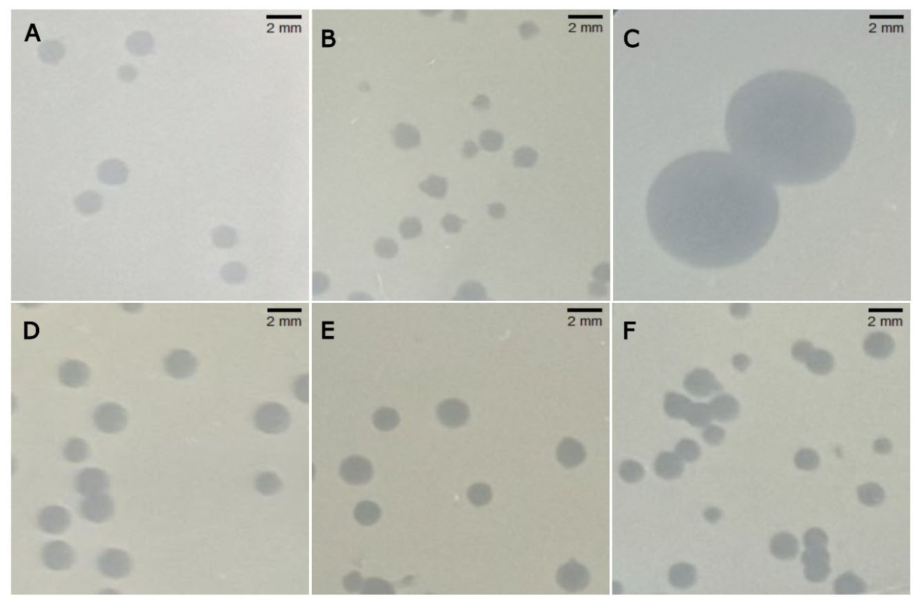
Figure 2.
Proteomic and intergenomic comparison of Xanthomonas arboricola pv. pruni bacteriophages EW15 and EW17 with representative reference bacteriophages. (A) Extract of the ViPTree proteomic tree generated by BLASTX analysis against 3,171 reference viral genomes, showing the phylogenetic position of EW15 and EW17 relative to neighboring bacteriophage genera. (B) Pairwise intergenomic similarity matrix generated with VIRIDIC for EW15, EW17 and the representative bacteriophages selected from the ViPTree analysis. Values represent intergenomic nucleotide similarities (%). Colored shading indicates the different viral genera represented in the tree and similarity matrix, and red stars in A identify the highest genome-wide similarity score with EW15 and EW17.
Figure 2.
Proteomic and intergenomic comparison of Xanthomonas arboricola pv. pruni bacteriophages EW15 and EW17 with representative reference bacteriophages. (A) Extract of the ViPTree proteomic tree generated by BLASTX analysis against 3,171 reference viral genomes, showing the phylogenetic position of EW15 and EW17 relative to neighboring bacteriophage genera. (B) Pairwise intergenomic similarity matrix generated with VIRIDIC for EW15, EW17 and the representative bacteriophages selected from the ViPTree analysis. Values represent intergenomic nucleotide similarities (%). Colored shading indicates the different viral genera represented in the tree and similarity matrix, and red stars in A identify the highest genome-wide similarity score with EW15 and EW17.
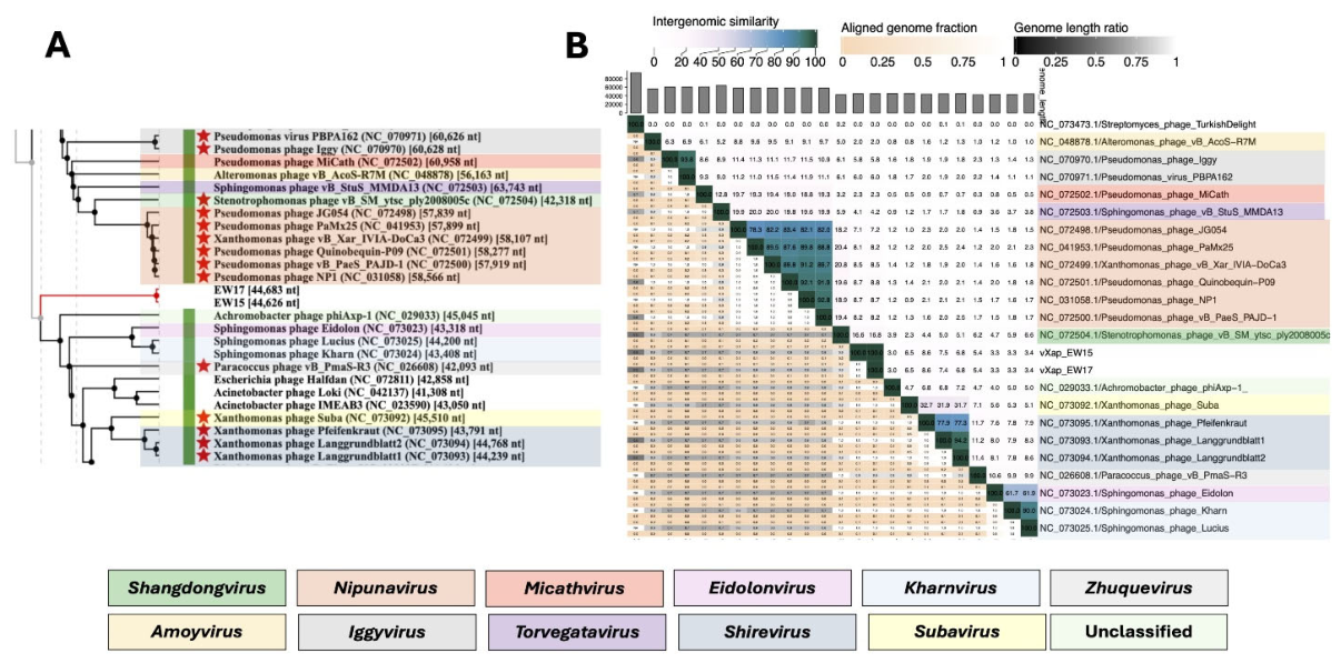
Figure 3.
Comparative genome organisation of Xanthomonas arboricola pv. pruni (Xap) bacteriophages vXap-EW1, vXapT-EW3, vXapP-EW5, vXapX-EW15, vXapT-EW16, vXapX-EW17, and their closest homologous phages identified in GenBank. Conserved genomic regions are linked by shaded blocks and predicted proteins are coloured according to their functional categories.
Figure 3.
Comparative genome organisation of Xanthomonas arboricola pv. pruni (Xap) bacteriophages vXap-EW1, vXapT-EW3, vXapP-EW5, vXapX-EW15, vXapT-EW16, vXapX-EW17, and their closest homologous phages identified in GenBank. Conserved genomic regions are linked by shaded blocks and predicted proteins are coloured according to their functional categories.
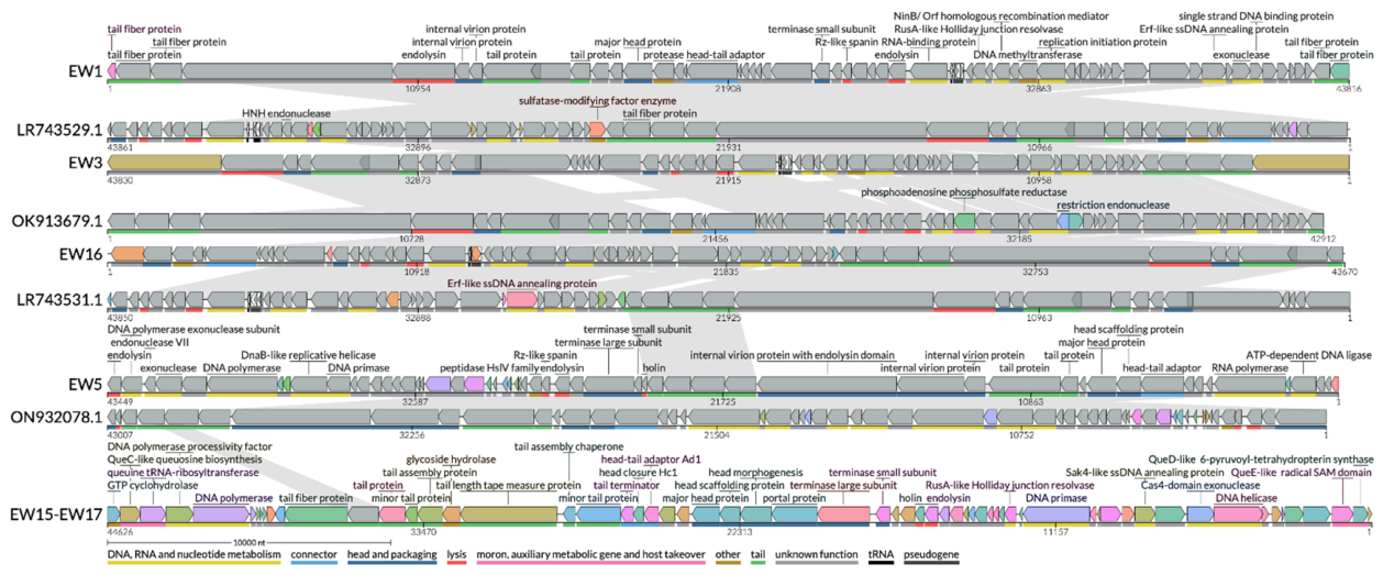
Figure 4.
Virion representative micrographs of the new Xanthomonas arboricola pv. pruni bacteriophages after negative staining. (A) vXapX-EW15 and (B) vXapX-EW17. Both phages exhibited isometric icosahedral capsids and contractile tails, displaying a myovirus-like morphology. Scale bars100 nm.
Figure 4.
Virion representative micrographs of the new Xanthomonas arboricola pv. pruni bacteriophages after negative staining. (A) vXapX-EW15 and (B) vXapX-EW17. Both phages exhibited isometric icosahedral capsids and contractile tails, displaying a myovirus-like morphology. Scale bars100 nm.
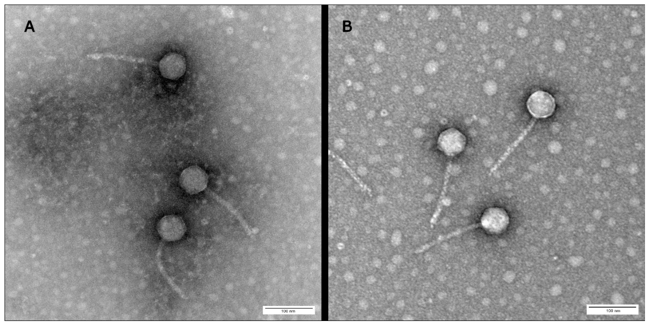
Figure 5.
Host range of six Xanthomonas arboricola pv. pruni (Xap) bacteriophages across Xanthomonas strains. Lytic activity was evaluated against a collection of 26 Xap strains, together with one strain each of X. arboricola pv. juglandis (Xaj) and X. arboricola pv. corylina (Xac), and representative strains of other Xanthomonas species.
Figure 5.
Host range of six Xanthomonas arboricola pv. pruni (Xap) bacteriophages across Xanthomonas strains. Lytic activity was evaluated against a collection of 26 Xap strains, together with one strain each of X. arboricola pv. juglandis (Xaj) and X. arboricola pv. corylina (Xac), and representative strains of other Xanthomonas species.
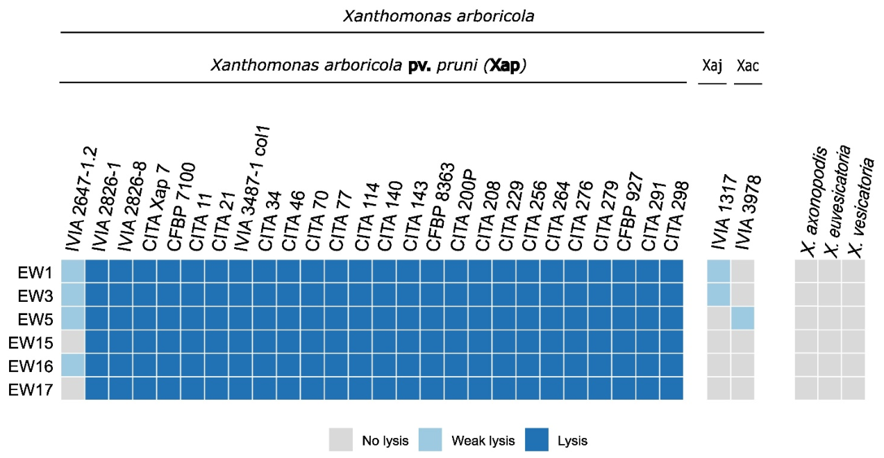
Figure 6.
Xanthomonas arboricola pv. pruni (Xap) killing curves by single specific bacteriophages. Growth curves of strain CITA 33 following treatment with individual bacteriophages at (A) MOI 100 (10⁸ PFU/mL:10⁶ CFU/mL) and (B) MOI 10 (10⁸ PFU/mL:10⁷ CFU/mL). Bacteriophages vXap-EW1, vXapT-EW3, vXapP-EW5, vXapX-EW15, vXapT-EW16, vXapX-EW17 are abbreviated as EW1, EW3, EW5, EW15, EW16 and EW17. Bacterial growth was monitored as optical density at 600 nm (OD600nm) over 48 h at 28 ºC. Symbols and lines represent the mean ± standard error (SE). Asterisks indicate significant differences between phage-treated cultures and the untreated bacterial control (P< 0.001).
Figure 6.
Xanthomonas arboricola pv. pruni (Xap) killing curves by single specific bacteriophages. Growth curves of strain CITA 33 following treatment with individual bacteriophages at (A) MOI 100 (10⁸ PFU/mL:10⁶ CFU/mL) and (B) MOI 10 (10⁸ PFU/mL:10⁷ CFU/mL). Bacteriophages vXap-EW1, vXapT-EW3, vXapP-EW5, vXapX-EW15, vXapT-EW16, vXapX-EW17 are abbreviated as EW1, EW3, EW5, EW15, EW16 and EW17. Bacterial growth was monitored as optical density at 600 nm (OD600nm) over 48 h at 28 ºC. Symbols and lines represent the mean ± standard error (SE). Asterisks indicate significant differences between phage-treated cultures and the untreated bacterial control (P< 0.001).
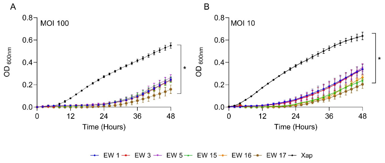
Figure 7.
Xanthomonas arboricola pv. pruni (Xap) killing curves by different combinations of four-phage cocktails. (A) Growth curves obtained for the fifteen possible four-phage cocktail combinations generated from the six selected Xap EW bacteriophages (abbreviated as 1, 3, 5, 15, 16 and 17). Bacterial growth was monitored as optical density at 600 nm (OD₆₀₀nm) over 48 h at 28 ºC. The shaded formulations correspond to the five cocktails selected for further validation based on their low area under the growth curve values. (B) Growth curves obtained for validation of the five selected four-phage cocktails. The two cocktails highlighted in bold, designated Cocktail 1 (vXap-EW1, XapT-EW3, vXapP-EW5 andXapX-EW17) and Cocktail 2 (vXap-EW1, XapT-EW3, XapT-EW16 andXapX-EW17) were selected for further assays with Prunus plants. Symbols and lines represent the mean ± standard error (SE). Asterisks indicate significant differences relative to the untreated bacterial control (P< 0.001).
Figure 7.
Xanthomonas arboricola pv. pruni (Xap) killing curves by different combinations of four-phage cocktails. (A) Growth curves obtained for the fifteen possible four-phage cocktail combinations generated from the six selected Xap EW bacteriophages (abbreviated as 1, 3, 5, 15, 16 and 17). Bacterial growth was monitored as optical density at 600 nm (OD₆₀₀nm) over 48 h at 28 ºC. The shaded formulations correspond to the five cocktails selected for further validation based on their low area under the growth curve values. (B) Growth curves obtained for validation of the five selected four-phage cocktails. The two cocktails highlighted in bold, designated Cocktail 1 (vXap-EW1, XapT-EW3, vXapP-EW5 andXapX-EW17) and Cocktail 2 (vXap-EW1, XapT-EW3, XapT-EW16 andXapX-EW17) were selected for further assays with Prunus plants. Symbols and lines represent the mean ± standard error (SE). Asterisks indicate significant differences relative to the untreated bacterial control (P< 0.001).
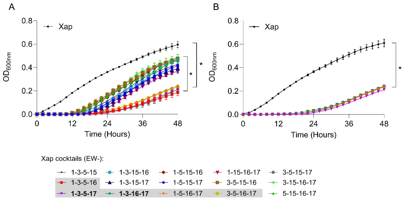
Figure 8.
Optimisation of a spray-inoculation pathogenicity method for Xanthomonas arboricola pv. pruni in Prunus plants. (A) Detached-leaf assay showing localized water-soaked lesions 10 days post-inoculation (dpi) following Xap inoculation at four sites on a wounded leaf. (B) Representative bacterial spot symptoms on a Prunus plant at 30 dpi following spray inoculation with Xap. The inset shows a close-up of representative leaf lesions.
Figure 8.
Optimisation of a spray-inoculation pathogenicity method for Xanthomonas arboricola pv. pruni in Prunus plants. (A) Detached-leaf assay showing localized water-soaked lesions 10 days post-inoculation (dpi) following Xap inoculation at four sites on a wounded leaf. (B) Representative bacterial spot symptoms on a Prunus plant at 30 dpi following spray inoculation with Xap. The inset shows a close-up of representative leaf lesions.
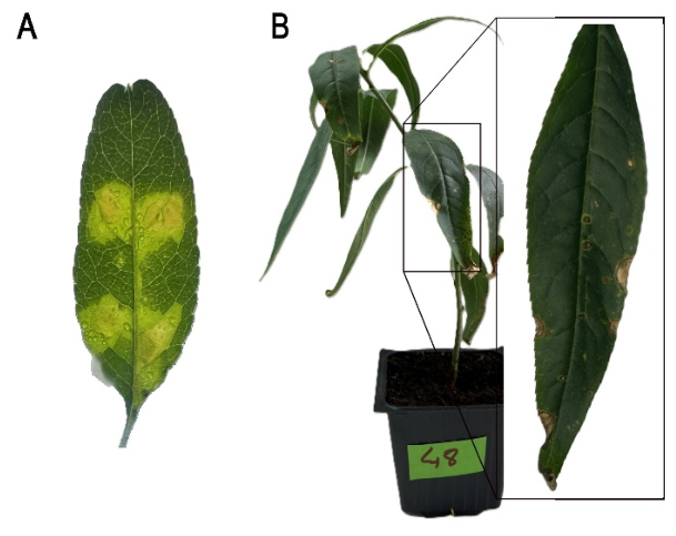
Figure 9.
Stability of Xanthomonas arboricola pv. pruni bacteriophages under in vitro and in planta conditions. (A) In vitro stability of bacteriophages vXap-EW1, vXapT-EW3, vXapP-EW5, vXapT-EW16, and vXapX-EW17 (abbreviated as EW1, EW3, EW5, EW16 and EW17) following incubation under different pH (5.5, 7.2, and 8.0) and temperature (28 ºC and 37 °C) conditions over three weeks. Phage infectivity was expressed as log (PFU/mL). (B) In planta persistence of the same bacteriophages on Prunus leaves following spray application. Infective phage particles recovered from leaf tissue were expressed as log [PFU/g fresh weight (FW)]. Time 0, 1, 3, 7, and 20 indicate days after application, with time 0 corresponding to 1 h post inoculation. Points represent mean values and error bars indicate the standard error (SE).
Figure 9.
Stability of Xanthomonas arboricola pv. pruni bacteriophages under in vitro and in planta conditions. (A) In vitro stability of bacteriophages vXap-EW1, vXapT-EW3, vXapP-EW5, vXapT-EW16, and vXapX-EW17 (abbreviated as EW1, EW3, EW5, EW16 and EW17) following incubation under different pH (5.5, 7.2, and 8.0) and temperature (28 ºC and 37 °C) conditions over three weeks. Phage infectivity was expressed as log (PFU/mL). (B) In planta persistence of the same bacteriophages on Prunus leaves following spray application. Infective phage particles recovered from leaf tissue were expressed as log [PFU/g fresh weight (FW)]. Time 0, 1, 3, 7, and 20 indicate days after application, with time 0 corresponding to 1 h post inoculation. Points represent mean values and error bars indicate the standard error (SE).
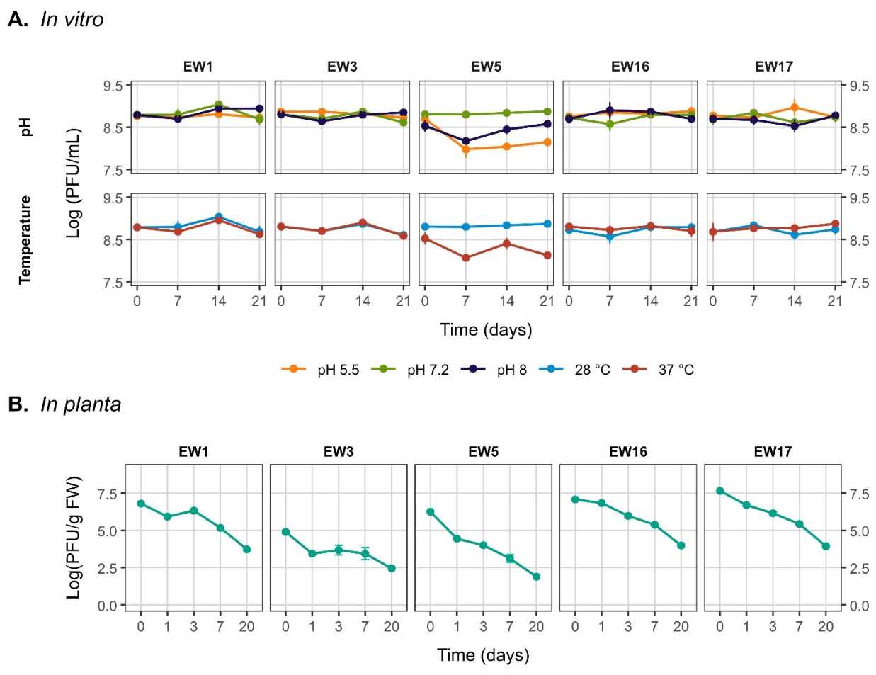
Figure 10.
Efficacy of two bacteriophage cocktails against Xanthomonas arboricola pv. pruni in Prunus plants. (A) Quantification of disease severity as lesion density (lesions/cm²) at 30 dpi. Each point represents one plant (n = 18 plants per treatment, from three independent experiments, and each one with 6 plants). Boxes represent the interquartile range (IQR), the horizontal line indicates the median, and whiskers extend to 1.5 × IQR. Different letters indicate significant differences among treatments according to Tukey’s post hoc test (P< 0.05). Biocontrol assays comprised the following treatments: Xap alone as positive control, Xap co-inoculated either with phage Cocktail 1 or Cocktail 2, and two types of negative controls (plants treated with PBS as well as phage cocktails alone), both of which remained symptomless. (B) Representative leaves collected from plants inoculated with Xap alone (left) or co-inoculated with Xap and the phage Cocktail 2 (right). (C) Representative Prunus plants inoculated with Xap alone (positive control) at 30 dpi, where some defoliation can be observed, as well as leaf lesions. (D) Representative Prunus plants co-inoculated with Xap and the phage Cocktail 2 (vXap-EW1, XapT-EW3, XapP-EW5, XapX-EW17) at 30 dpi.
Figure 10.
Efficacy of two bacteriophage cocktails against Xanthomonas arboricola pv. pruni in Prunus plants. (A) Quantification of disease severity as lesion density (lesions/cm²) at 30 dpi. Each point represents one plant (n = 18 plants per treatment, from three independent experiments, and each one with 6 plants). Boxes represent the interquartile range (IQR), the horizontal line indicates the median, and whiskers extend to 1.5 × IQR. Different letters indicate significant differences among treatments according to Tukey’s post hoc test (P< 0.05). Biocontrol assays comprised the following treatments: Xap alone as positive control, Xap co-inoculated either with phage Cocktail 1 or Cocktail 2, and two types of negative controls (plants treated with PBS as well as phage cocktails alone), both of which remained symptomless. (B) Representative leaves collected from plants inoculated with Xap alone (left) or co-inoculated with Xap and the phage Cocktail 2 (right). (C) Representative Prunus plants inoculated with Xap alone (positive control) at 30 dpi, where some defoliation can be observed, as well as leaf lesions. (D) Representative Prunus plants co-inoculated with Xap and the phage Cocktail 2 (vXap-EW1, XapT-EW3, XapP-EW5, XapX-EW17) at 30 dpi.
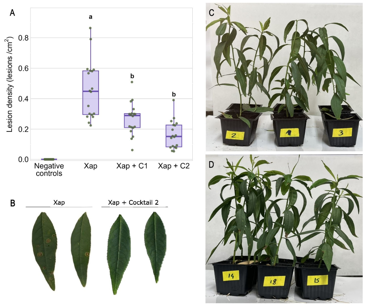

Table 1.
Strains of Xanthomonas arboricola pv. pruni and other plant pathogenic Xanthomonas spp. used in this study.
Table 1.
Strains of Xanthomonas arboricola pv. pruni and other plant pathogenic Xanthomonas spp. used in this study.
| Bacterial species and strain | Host plant |
Geographical origin |
Isolation year |
| X. arboricola pv. pruni (Xap) | |||
| IVIAª 2647-1.2 | P. salicina | Badajoz | 2002 |
| IVIA 2826-1 | P. domestica cv. Anne-Gold | Valencia | 2003 |
| IVIA 2826-8 | P. persica var. Nectarina cv. Zephiz | Valencia | 2003 |
| CITAbXap 7 | P. persica | Valencia | 2004 |
| IVIA 3162-2 col3 = CFBPc 7100 | P. dulcis cv. Rumbeta | Alicante | 2006 |
| CITA 11 | P. persica cv. Richard Lady | Huesca | 2008 |
| CITA 21 | P. dulcis | Huesca | 2008 |
| IVIA 3487-1 col1 | P. armeniaca | Huesca | 2008 |
| CITA 34 | P. dulcis cv. Guara | Teruel | 2009 |
| CITA 46 | P. persica cv. Summer Lady | Navarra | 2009 |
| CITA 70 | P. dulcis x P. persica | Zaragoza | 2010 |
| CITA 77 | P. dulcis cv. Guara | Navarra | 2010 |
| CITA 114 | P. dulcis | Zaragoza | 2011 |
| CITA 140 | P. armeniaca | Teruel | 2012 |
| CITA 143 | P. dulcis | Lérida | 2012 |
| CITA 177 = CFBP 8363 | P. dulcis cv. Guara | Teruel | 2014 |
| CITA 200P | P. pérsica cv. Big Nectared | Huesca | 2015 |
| CITA 208 | P. persica cv. Nectarina Honey Royale | Huesca | 2016 |
| CITA 229 | P. pérsica var. platycarpa | Huesca | 2016 |
| CITA 256 | P. dulcis cv. Vayro | Huesca | 2016 |
| CITA 264 | P. persica | Huesca | 2017 |
| CITA 276 | P. salicina cv. Angeleno | Huesca | 2018 |
| CITA 279 | P. persica cv. Honey Royale | Huesca | 2019 |
| CITA 33 = CFBP 927 | P. dulcis cv. Guara | Teruel | 2009 |
| CITA 291 | P. dulcis cv. Vayro | Zaragoza | 2025 |
| CITA 298 | P. pérsica var. Nectarina cv. Honey Late | Zaragoza | 2025 |
| X. arboricola pv. juglandis (Xaj) | |||
| IVIA 1317 | Juglans regia | Badajoz | 1993 |
| X. arboricola pv. corylina (Xac) | |||
| IVIA 3978 | Corylus avellana cv. Tonda Gentile | Tarragona | 2011 |
| X. axonopodis | |||
| CECTd 914 | Phaseolus vulgaris | Hungary | 2005 |
| X. euvesicatoria pv. euvesicatoria (Xee) | |||
| LSVe 302 | C. annuum cv. Jaranda | Cáceres | 2024 |
| X. euvesicatoria pv. perforans (Xep) | |||
| CFBP 7993 | Solanum lycopersicum | Mauritius Island | 2010 |
| X. vesicatoria (Xv) | |||
| CFBP 1941 | S. lycopersicum | Murcia | 1978 |
ªIVIA: Instituto Valenciano de Investigaciones Agrarias, Valencia, Spain; bCITA: Centro de Investigación y Tecnología Agroalimentaria de Aragón, Zaragoza, Spain; cCIRM-CFBP: International Centre of Microbial Resource—French Collection for Plant-associated Bacteria, Institute National de Recherche pourl ’Agriculture, l’Alimentation et l’Environnement, France; dCECT: Colección Española de Cultivos Tipo, Valencia, Spain; eLSV: Laboratorio de Diagnóstico de Sanidad Vegetal, Junta de Extremadura, Badajoz, Spain.
Disclaimer/Publisher’s Note: The statements, opinions and data contained in all publications are solely those of the individual author(s) and contributor(s) and not of MDPI and/or the editor(s). MDPI and/or the editor(s) disclaim responsibility for any injury to people or property resulting from any ideas, methods, instructions or products referred to in the content. |
© 2026 by the authors. Licensee MDPI, Basel, Switzerland. This article is an open access article distributed under the terms and conditions of the Creative Commons Attribution (CC BY) license (http://creativecommons.org/licenses/by/4.0/).
Copyright: This open access article is published under a Creative Commons CC BY 4.0 license, which permit the free download, distribution, and reuse, provided that the author and preprint are cited in any reuse.