

Submitted:
18 September 2026
Posted:
20 September 2026
You are already at the latest version
Abstract
Photosynthetic energy conversion requires continuous coordination between light-driven electron transport and downstream metabolic demand. Chlorophyll fluorescence, P700 spectroscopy and electrochromic-shift measurements have transformed our understanding of photosynthetic regulation by resolving PSII operating efficiency, excitation pressure, PSI redox poise and proton-motive-force dynamics. However, these measurements primarily describe the photochemical state of the photosynthetic apparatus and do not directly determine how much ATP and NADPH are produced, consumed or redistributed through cellular metabolism. Metabolomics provides an essential connection between photosynthetic regulation and downstream energy metabolism, but metabolite abundance alone cannot distinguish altered production from altered consumption or quantify pathway turnover. Here, I argue that the next major step in photosynthesis research will require integration of photosynthetic biophysics with metabolomics, isotope tracing and metabolic flux analysis. Such an approach could connect electron and proton transport with ATP/NADPH production, carbon allocation, reductant export and alternative electron pathways to construct quantitative chloroplast energy budgets. I discuss how this framework can help distinguish the metabolic functions of cyclic electron flow, oxygen-dependent electron sinks and chloroplast-mitochondrial exchange and propose an experimental progression from measurements of photochemical state to quantitative energy-flux modelling. Moving from descriptive photosynthetic parameters toward integrated bioenergetic accounting should improve our ability to understand acclimation, predict metabolic compensation and rationally engineer photosynthesis under changing environmental conditions.
Keywords:
photosynthesis
; metabolomics
; fluxomics
1. Introduction
Reduction of one molecule of CO₂ through the Calvin–Benson–Bassham (CBB) cycle requires approximately three ATP and two NADPH, corresponding to an ATP/NADPH demand ratio of about 1.5 (Kramer and Evans, 2010; Walker et al., 2020). Linear electron flow (LEF) is estimated to generate an ATP/NADPH ratio of approximately 1.28-1.5, depending on the assumed proton-to-ATP stoichiometry (Kramer and Evans, 2010). However, cellular energetic demand is highly dynamic because photorespiration, nutrient assimilation, protein repair, ion transport, carbon-concentrating mechanisms and stress metabolism continuously alter both ATP and NADPH requirements (Kramer and Evans, 2010; Walker et al., 2020).
Photosynthetic organisms compensate through a network of alternative electron pathways. Cyclic electron flow (CEF) around PSI increases proton-motive force without net NADPH production (Yamori and Shikanai, 2016; Yamori et al., 2016), flavodiiron-dependent electron transport can divert electrons toward O2 (Chaux et al., 2017), plastid terminal oxidase (PTOX) can contribute to plastoquinol oxidation (Krieger-Liszkay and Feilke, 2016), and Mehler-type reactions provide additional oxygen-dependent electron sinks (Badger et al., 2000; Makino, Miyake and Yokota, 2002). Reducing equivalents can also be exchanged between chloroplasts and mitochondria through metabolite shuttles (Raghavendra and Padmasree, 2003; Fakhimi and Grossman, 2024). These pathways therefore operate as an interconnected network in which perturbation of one component can be compensated by others (Levin, 2024; Peltier et al., 2024).
Although genetic disruption of individual pathways has enabled their characterisation, compensation makes their quantitative contribution under physiological or stress conditions difficult to determine (Walker et al., 2020; Peltier et al., 2024). Chlorophyll fluorescence, P700 absorbance and electrochromic-shift measurements effectively describe PSII, PSI and proton-motive-force states (Klughammer and Schreiber, 1994; Baker, 2008; Mathiot and Alric, 2021), but do not independently quantify ATP production, NADPH consumption, carbon allocation or reductant export. Conversely, metabolomics reveals energetic and carbon-metabolic states, but metabolite abundance does not directly report turnover (Ratcliffe and Shachar-Hill, 2006; Sweetlove and Ratcliffe, 2011; Kruger and Ratcliffe, 2021).
The central challenge is therefore quantitative: how much electron flux enters each pathway, how much ATP is generated, where reducing power is consumed or exported, and how these processes influence carbon allocation and growth. Recent studies have quantified contributions of CEF, pseudocyclic electron flow (PCEF) and chloroplast-to-mitochondrial electron flow (CMEF) under carbon-concentrating conditions and demonstrated substantial functional compensation among them (Burlacot et al., 2022; Peltier et al., 2024).
Addressing this challenge requires integrating photochemical measurements with metabolomics and flux-based approaches. Stable-isotope tracing and metabolic-flux analysis can resolve pathway turnover and carbon partitioning (Ratcliffe and Shachar-Hill, 2006; Ma et al., 2014; Kruger and Ratcliffe, 2021), while membrane-inlet mass spectrometry can quantify simultaneous O₂ evolution and consumption (Burlacot et al., 2022; Peltier et al., 2024). Here, I examine how combining these approaches could enable quantitative chloroplast energy budgets and reveal the physiological functions of alternative electron pathways.
2. What Do Conventional Photosynthetic Measurements Actually Tell Us?
Conventional approaches include chlorophyll fluorescence, P700 absorption spectroscopy, electrochromic-shift (ECS) measurements and gas exchange. Pulse-amplitude-modulated (PAM) fluorescence is widely used to quantify PSII efficiency, excitation pressure and non-photochemical quenching (NPQ), thereby describing fate of the absorbed energy (Genty, Briantais and Baker, 1989; Baker, 2008; Murchie and Lawson, 2013). Time-resolved methods, such as, TCSPC and streak-camera spectroscopy can additionally resolve excitation-transfer and trapping kinetics of PSI and PSII (Chukhutsina, Holzwarth and Croce, 2019). Interestingly, similar fluorescence phenotypes can arise from very different metabolic states. Reduced PSII yield, for example, may reflect photoprotection, carbon-sink limitation, altered ATP/NADPH demand or changes in downstream electron allocation (Baker, 2008; Murchie and Lawson, 2013; Bag et al., 2020). Fluorescence therefore reveals the state of the photosynthetic apparatus but cannot directly determine ATP production, NADPH consumption or the identity of downstream electron sinks. P700 measurements complement fluorescence by reporting PSI redox state and distinguishing donor-side from acceptor-side limitation (Klughammer and Schreiber, 1994; Schreiber and Klughammer, 2016). Acceptor-side limitation can arise when electrons reach PSI faster than NADP+ and other stromal acceptors can be regenerated. Nevertheless, P700 oxidation does not identify where those electrons ultimately go. Increased P700 oxidation could result from enhanced carbon metabolism, alternative electron sinks, altered cyclic electron flow (CEF) or export of reducing equivalents. P700 therefore reports PSI redox poise rather than the metabolic fate of photosynthetic reducing power.
Electrochromic-shift (ECS) measurements provide information about proton-motive force (pmf), proton flux and thylakoid proton conductivity (Sacksteder and Kramer, 2000; Avenson et al., 2005; Mathiot and Alric, 2021). Because ATP synthesis is driven by proton flow through the chloroplast ATP synthase, ECS brings photosynthetic analysis closer to the energetic output of the light reactions than electron transport measurements alone. This becomes particularly important when alternative electron pathways modify proton translocation relative to NADPH formation. CEF, for example, can enhance pmf formation without net NADPH production and therefore adjust the ATP/NADPH output (Avenson et al., 2005; Kramer and Evans, 2010). However, increased pmf or proton flux cannot be interpreted as a quantitative measurement of metabolic ATP availability. ATP production is ultimately coupled to ATP synthase activity and downstream ATP consumption, while the resulting ATP pool is simultaneously influenced by numerous competing metabolic processes. ECS therefore provides an essential component of chloroplast energetic accounting, but not the complete energy budget.
CO₂ exchange provides a measure of net carbon assimilation and its biochemical or diffusional limitations (Long and Bernacchi, 2003). O₂ exchange, in contrast, contains contributions from photosynthetic water oxidation (Shevela et al., 2016) together with oxygen-consuming processes including mitochondrial respiration (Smith, Strand and Walker, 2024), photorespiration (Bauwe, Hagemann and Fernie, 2010) and alternative electron-transfer pathways (Burlacot et al., 2022). Membrane-inlet mass spectrometry (MIMS), particularly when combined with isotopic O2 or CO2 exchange, can resolve simultaneous gas production and consumption and quantify oxygen-dependent electron sinks that are difficult to distinguish (Beckmann et al., 2009; Burlacot, Burlacot, et al., 2020). Together, fluorescence, P700, ECS and gas exchange provide complementary information on photochemistry, redox state, proton energetics and gas flux. Their major limitation is that none independently determines how ATP, NADPH and fixed carbon are subsequently consumed or redistributed through metabolism.
3. From Photochemistry to Metabolic Flux
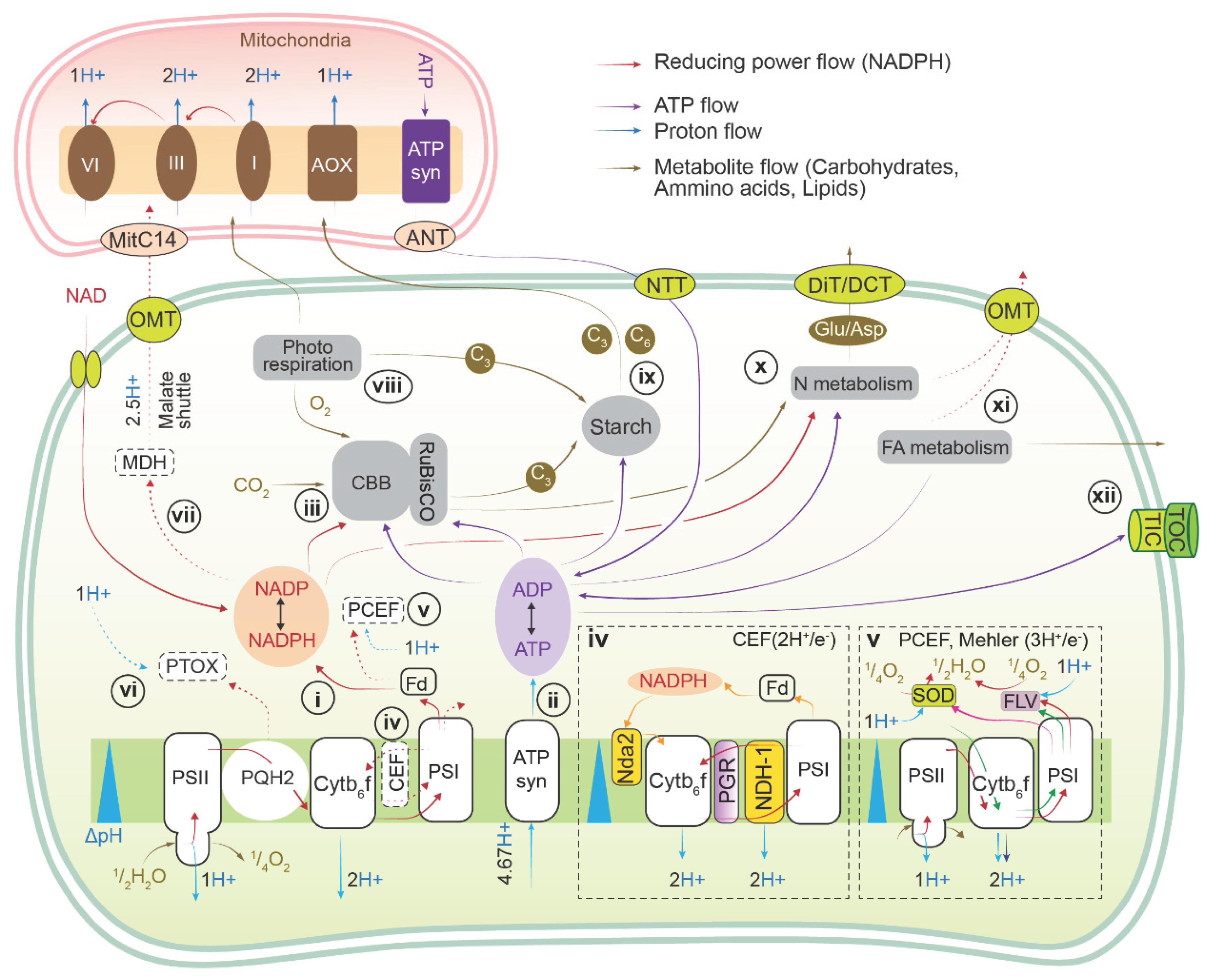
A complete understanding of photosynthetic acclimation requires connecting regulation of the light reactions with chloroplast metabolic homeostasis (Figure 1). Metabolite profiling provides an important bridge by revealing whether changes in electron transport are accompanied by altered pools of Calvin-Benson-Bassham (CBB) cycle intermediates, organic acids, sugars and other metabolites (Szecowka et al., 2013; Westhoff and Weber, 2024). Targeted metabolite measurements and genetically encoded biosensors can further resolve energetic and redox components such as ATP, ADP and pyridine nucleotides (De Col et al., 2017; Voon et al., 2018). Such measurements therefore reveal metabolic consequences of altered photochemistry that cannot be inferred from fluorescence, P700 or ECS alone (Bailleul et al., 2015).
ATP provides a particularly clear example. Photophosphorylation generates ATP, whereas carbon assimilation, ion transport and numerous other metabolic processes simultaneously consume it. Measurements of stromal ATP or ATP/ADP status therefore provide information that cannot be obtained directly from measurements of electron or proton transport (De Col et al., 2017; Voon et al., 2018). Recently, Degen et al. (2026) showed using a chloroplast-targeted MgATP2- biosensor that Arabidopsis mutants impaired in cyclic electron flow (CEF) maintained stromal MgATP2- levels comparable with wild type despite substantially reduced proton-motive force. This disequilibrium challenges the assumption that CEF-dependent increases in proton-motive force necessarily translate proportionally into increased steady-state stromal ATP and highlights the importance of distinguishing energetic state from energetic flux (Degen, Schwarzländer and Johnson, 2026).
A similar problem applies to NADP+/NADPH. NADPH supplies reducing power to the CBB cycle and other stromal reactions, while NADP⁺ regeneration maintains electron withdrawal from PSI. Chloroplast NADP(H) status is consequently closely linked to redox homeostasis (Hashida et al., 2018). However, increased NADPH cannot distinguish enhanced production from reduced consumption or impaired reductant redistribution, particularly under stress when carbon assimilation is restricted and alternative electron pathways are activated.
More generally, metabolite abundance should not be equated with metabolic flux. ATP, NADPH, CBB-cycle intermediates, malate and carbohydrate pools may remain relatively stable despite substantial changes in turnover, whereas metabolite accumulation may result from reduced downstream consumption rather than increased production. Time-resolved ¹³C labelling has demonstrated that substantial flux can occur through pools whose steady-state abundance changes only modestly (Szecowka et al., 2013; Ma et al., 2014). Consequently, metabolomics provides essential information about metabolic state but cannot by itself determine whether changes in photosynthesis alter ATP/NADPH production or consumption, redirect reducing power towards alternative sinks, or modify carbon export.
Subcellular compartmentation adds a further layer of complexity. Whole-cell metabolite measurements may not accurately reflect chloroplast energy or redox state because photosynthetic metabolism is distributed across chloroplasts, mitochondria, peroxisomes and the cytosol (Kueger et al., 2012; Dietz, 2017). The malate/oxaloacetate system can redistribute reducing equivalents between compartments, triose-phosphate export connects chloroplast carbon fixation with cytosolic metabolism, and photorespiration couples chloroplast, peroxisomal and mitochondrial metabolism through rapid exchange of intermediates (Selinski and Scheibe, 2019) (Figure 1). Thus, understanding photosynthetic energy balance ultimately requires information not only on metabolite abundance, but also on the rate and location of metabolic turnover.
The key distinction is therefore between metabolic state and metabolic flux. Metabolomics can reveal whether photosynthetic perturbations are accompanied by changes in ATP, NADPH, carbon and redox pools, but determining where photosynthetically derived energy and carbon ultimately go requires flux-based measurements. Stable-isotope tracing, metabolic-flux analysis and complementary approaches can provide this missing quantitative layer by resolving rates of production, consumption, export and recycling.
4. Flux-Based Approaches: Resolving Rate and Fate
The next step is to move from describing photosynthetic and metabolic states towards determining the rates at which energy and carbon move through the system. No single technique can provide a complete chloroplast energy budget. Instead, complementary approaches are required to determine where newly fixed carbon is allocated, how rapidly metabolic pathways operate, and how changes in flux influence chloroplast ATP and redox state.
4.1. Stable-Isotope Tracing: Following Newly Fixed Carbon
Stable-isotope labelling provides a direct means of distinguishing metabolite abundance from metabolic turnover. Pulse labelling with 13CO2 in plants or 13C -bicarbonate in photosynthetic microorganisms can follow newly fixed carbon through the CBB cycle, photorespiration, starch and sucrose synthesis, organic-acid metabolism and amino-acid biosynthesis (Rühl et al., 2012; Szecowka et al., 2013; Ma et al., 2014; Niedenführ, Wiechert and Nöh, 2015). When combined with time-resolved LC-MS metabolomics, the appearance and redistribution of labelled isotopolouges can reveal pathway dynamics that remain invisible in steady-state metabolite pools (Rühl et al., 2012; Szecowka et al., 2013; Emwas et al., 2022).
This distinction is particularly important in photosynthetic systems, where central metabolites may turn over rapidly while maintaining relatively stable concentrations. Stable-isotope tracing can therefore establish whether an apparently unchanged metabolite pool is metabolically static or instead sustained by rapid and balanced production and consumption. It also provides information on carbon partitioning between competing metabolic destinations, allowing photochemical changes to be connected with downstream carbon allocation.
4.2. From Isotope Enrichment to Metabolic Flux
Isotope tracing becomes most powerful when incorporated into metabolic-flux analysis. Rather than simply determining whether a metabolite becomes labelled, mathematical modelling of the temporal appearance and redistribution of labelled isotopologues can be used to estimate reaction and pathway fluxes (Ratcliffe and Shachar-Hill, 2006; Szecowka et al., 2013).
For example, isotopically non-stationary 13C metabolic-flux analysis revealed substantial reorganisation of central carbon metabolism during high-light acclimation in Arabidopsis that could not be inferred from metabolite concentrations alone (Ma et al., 2014). Such approaches begin to provide the quantitative information required for photosynthetic energy accounting: how rapidly fixed carbon enters competing pathways, whether metabolic demand changes following perturbation of electron transport, and whether apparently similar photosynthetic phenotypes are associated with fundamentally different downstream fluxes.
An important future objective will therefore be to integrate estimates of electron and proton transport with carbon-flux measurements obtained under the same physiological conditions. Such integration could provide a quantitative connection between changes in photosynthetic energy production and the metabolic processes ultimately consuming that energy.
4.3. Linking Flux to Energetic and Redox State
Flux measurements alone do not reveal whether changing pathway activity alters the energetic state of the chloroplast. Genetically encoded sensors provide a complementary layer by enabling ATP and redox metabolites to be followed in specific cellular compartments. Fluorescent sensors have been used to monitor MgATP2- dynamics in plant cells and have revealed substantial differences in ATP status between cellular compartments (De Col et al., 2017; Voon et al., 2018). Similarly, iNAP and SoNar have enabled monitoring of NADPH abundance and NADH/NAD⁺ status during photosynthesis and photorespiration, while NERNST permits monitoring of NADP(H) redox poise within living cells, including chloroplasts (Lim et al., 2020; Molinari et al., 2023).
These measurements are particularly valuable because changes in electron or carbon flux need not produce proportional changes in steady-state ATP or NADPH pools. Conversely, apparently stable energetic pools may conceal substantial changes in their rates of production and consumption. Sensors therefore provide the energetic and redox context needed to interpret flux measurements, especially when combined with fluorescence, P700 and ECS measurements on comparable material (Degen, Schwarzländer and Johnson, 2026). Their greatest value may consequently arise from integration with isotope tracing and metabolic-flux analysis. Photochemical measurements can define the operating state of the light reactions, sensors can reveal the corresponding energetic and redox state, and isotope-based approaches can determine how rapidly carbon is redistributed through metabolism. Together, these measurements begin to connect photosynthetic state, rate and fate.
Oxygen-resolved measurements can provide an additional, pathway-specific layer. Membrane-inlet mass spectrometry (MIMS), particularly with isotopic O2, can distinguish simultaneous O2 evolution and consumption and has been valuable for quantifying oxygen-dependent electron sinks such as FLV-mediated photoreduction (Chaux et al., 2017; Burlacot, Richaud, et al., 2020; Bag et al., 2023). However, because O₂ consumption can arise from several processes, MIMS is most informative when combined with genetic or physiological perturbations and with fluorescence, P700 or ECS measurements (Burlacot, Richaud, et al., 2020; Fitzpatrick, Aro and Tiwari, 2022; Bag et al., 2023). It therefore complements, rather than replaces, the broader metabolic and flux-based framework described above.
5. Test Cases: Pathway Activity Does Not Equal Metabolic Function
Alternative electron pathways illustrate why pathway activation cannot be interpreted independently of metabolic context. The same pathway may contribute to ATP supply, redox balancing or photoprotection depending on cellular demand.
CEF around PSI provides an important example. Because electrons are recycled into the plastoquinone/cytochrome b6f pathway rather than used for net NADPH production, CEF can increase pmf formation relative to NADPH output (Shikanai, 2014; Alric and Johnson, 2017). However, CEF also contributes to photosynthetic control, PSI protection and redox regulation (Dang et al., 2014; Johnson et al., 2014; Kono et al., 2017; Buchert et al., 2020; Bag et al., 2025; Degen, Schwarzländer and Johnson, 2026). Recent measurements showing near-wild-type stromal MgATP²⁻ levels in CEF-impaired Arabidopsis despite reduced proton-motive force further demonstrate that CEF activity cannot be equated directly with ATP availability (Buchert et al., 2020; Degen, Schwarzländer and Johnson, 2026). Determining its metabolic function therefore requires linking CEF and proton flux with ATP status, carbon assimilation and metabolic turnover.
Oxygen-dependent electron sinks provide a similar example. Flavodiiron proteins can divert electrons from PSI towards O2, relieving acceptor-side pressure while maintaining upstream electron and proton transfer (Chaux et al., 2017; Ilík et al., 2017; Bag et al., 2023). Because this pathway draws electrons away from PSI acceptors while allowing continued upstream electron and proton transfer, it can simultaneously influence redox poise, photoprotection and the energetic output of photosynthesis (Allen, 2002; Alric, 2015). The relative importance of these functions is likely context dependent. Quantifying FLV activity therefore requires not only demonstrating O2 or NO (nitric oxide) reduction (Burlacot, Richaud, et al., 2020; Burlacot et al., 2022; Bag et al., 2023) but integrating that flux with LEF, pmf formation, ATP demand and carbon metabolism. However, measuring O2 reduction alone does not reveal whether this flux primarily supports photoprotection, energetic balance or compensation for restricted carbon metabolism. Other O2-consuming pathways further complicate this interpretation. Together, these examples illustrate the central problem: identifying where electrons flow does not necessarily explain what physiological function that flux serves. Resolving function requires integration of pathway activity with energetic, redox and metabolic flux measurements.
6. A Framework for Quantitative Photosynthetic Bioenergetics
A realistic future objective is to construct integrated chloroplast energy map in which photochemical measurements, metabolite pools and flux measurements are analysed as complementary layers rather than independent datasets. Such an approach could proceed through a sequential experimental framework.
First, define the photochemical state. Chlorophyll fluorescence would quantify PSII operating efficiency, excitation pressure and energy dissipation; P700 measurements would resolve PSI redox poise; and ECS would provide information on proton-motive-force formation and proton flux.
Second, quantify major carbon and oxygen fluxes. Gas exchange would establish net CO2 assimilation, while oxygen-resolved measurements such as MIMS could quantify O2 evolution and oxygen-dependent alternative electron pathways.
Third, determine energetic and metabolic state. Measurements of ATP/ADP, NADPH/NADP+ and central carbon metabolites would establish whether changes in photochemistry are accompanied by corresponding changes in energy, redox and carbon pools.
Fourth,determine turnover rather than abundance alone.13CO2 or 13C-bicarbonate pulse labelling combined with LC-MS and metabolic-flux analysis could reveal whether observed changes result from altered production, consumption, export or recycling.
Finally, integrate these layers into a common energy model. Such models should seek to account for electron flow through LEF and alternative pathways, proton-motive-force generation, ATP synthesis and consumption, NADPH production and utilisation, carbon fixation and allocation, and exchange of reducing equivalents with other cellular compartments. The objective is therefore not to replace fluorescence, P700 or ECS with omics. Rather, each measurement addresses a different level of the photosynthetic energy system. Photochemical measurements define what the photosynthetic machinery is doing; metabolomics describes the metabolic state associated with that activity; and fluxomics begins to reveal where the resulting energy and carbon actually go.
7. Outlook: From Descriptive Photosynthesis to Predictive Bioenergetics
Photosynthetic biophysics has provided exceptionally powerful tools for resolving the behaviour of PSII, PSI and the thylakoid proton circuit. Metabolomics has simultaneously revealed extensive reorganisation of carbon, energy and redox metabolism during environmental acclimation. Yet these areas are still frequently interpreted separately. The next major advance should therefore be to connect them quantitatively. Fluorescence, P700 and ECS can define the photochemical state of photosynthesis, while metabolomics can establish whether downstream metabolism maintains ATP, NADPH, carbon and redox homeostasis. Flux-based approaches are required to determine how rapidly electrons, ATP, reductant and carbon move through competing pathways.
Measurements must increasingly be performed under comparable physiological conditions and, ideally, on the same biological material. Temporal resolution will be essential because photosynthetic regulation can change within seconds whereas metabolite pools and long-term acclimation operate over very different timescales. Cellular compartmentation must also be addressed, particularly when chloroplast and mitochondrial metabolism exchange carbon and reducing equivalents. Finally, energetic models will require careful accounting of what is measured directly and what is inferred from stoichiometric assumptions.
The goal should be to progress beyond identifying whether a photosynthetic pathway is activated. Instead, future studies should ask: how much electron flux enters the pathway, how does it alter proton and ATP production, where is the resulting reductant consumed or exported, and what physiological function does this redistribution ultimately support?
Integrating photosynthetic biophysics, metabolomics and fluxomics offers a route from descriptive stress physiology toward predictive photosynthetic bioenergetics. Such quantitative energy budgets could reveal why apparently similar photochemical phenotypes produce very different metabolic outcomes, explain compensatory responses to photosynthetic engineering, and ultimately provide a more rational basis for improving photosynthetic resilience and productivity.
Acknowledgments
This work was supported by the Alexander von Humboldt Foundation through a postdoctoral fellowship (grant no. SWE-1246473-HFST-P) awarded for the project “CRYSTAL” to PB.
Conflicts of Interest
The author declares no conflict of interest.
References
- Allen, J.F. Photosynthesis of ATP-electrons, proton pumps, rotors, and poise. Cell 2002, 110(3), 273–276. [Google Scholar] [CrossRef] [PubMed]
- Alric, J. The plastoquinone pool, poised for cyclic electron flow? Front. Plant Sci. 2015, 6, 540–540. [Google Scholar] [CrossRef] [PubMed]
- Alric, J.; Johnson, X. Alternative electron transport pathways in photosynthesis: a confluence of regulation. 37 Physiol. Metab. 2017 2017, 37, 78–86. [Google Scholar] [CrossRef] [PubMed]
- Avenson, T.J.; et al. Regulating the proton budget of higher plant photosynthesis. Proc. Natl. Acad. Sci. USA 2005, 102(27), 9709–9713. [Google Scholar] [CrossRef] [PubMed]
- Badger, M.R.; et al. Electron flow to oxygen in higher plants and algae: rates and control of direct photoreduction (Mehler reaction) and rubisco oxygenase. Philos. Trans. R. Soc. London. Ser. B Biol. Sci. 2000, 355(1402), 1433–1446. [Google Scholar] [CrossRef] [PubMed]
- Bag, P.; et al. Direct energy transfer from photosystem II to photosystem I confers winter sustainability in Scots Pine. Nat. Commun. 2020, 11(1), 6388–6388. [Google Scholar] [CrossRef] [PubMed]
- Bag, P.; et al. Flavodiiron-mediated O2 photoreduction at photosystem I acceptor-side provides photoprotection to conifer thylakoids in early spring. Nat. Commun. 2023, 14(1), 3210–3210. [Google Scholar] [CrossRef] [PubMed]
- Bag, P.; et al. Photosynthetic advantages of conifers in the boreal forest. Trends Plant Sci. 2025, 30(4), 409–423. [Google Scholar] [CrossRef] [PubMed]
- Bailleul, B.; et al. Energetic coupling between plastids and mitochondria drives CO2 assimilation in diatoms. Nature 2015, 524(7565), 366–369. [Google Scholar] [CrossRef] [PubMed]
- Baker, N.R. Chlorophyll Fluorescence: A Probe of Photosynthesis In Vivo. Annu. Rev. Plant Biol. 2008, 59, 89–113. [Google Scholar] [CrossRef] [PubMed]
- Bauwe, H.; Hagemann, M.; Fernie, A.R. Photorespiration: players, partners and origin. Trends Plant Sci. 2010, 15(6), 330–336. [Google Scholar] [CrossRef] [PubMed]
- Beckmann, K.; et al. On-line mass spectrometry: membrane inlet sampling. Photosynth. Res. 2009, 102(2), 511–522. [Google Scholar] [CrossRef] [PubMed]
- Buchert, F.; et al. PGR5 is required for efficient Q cycle in the cytochrome b6f complex during cyclic electron flow. Biochem. J. 2020, 477(9), 1631–1650. [Google Scholar] [CrossRef] [PubMed]
- Burlacot, A.; Richaud, P.; et al. Algal photosynthesis converts nitric oxide into nitrous oxide. Proc. Natl. Acad. Sci. 2020, 117(5), 2704–2709. [Google Scholar] [CrossRef] [PubMed]
- Burlacot, A.; Burlacot, F.; et al. Membrane Inlet Mass Spectrometry: A Powerful Tool for Algal Research. Front. Plant Sci. 2020, 11. [Google Scholar] [CrossRef] [PubMed]
- Burlacot, A.; et al. Alternative photosynthesis pathways drive the algal CO2-concentrating mechanism. Nature 2022, 605(7909), 366–371. [Google Scholar] [CrossRef] [PubMed]
- Chaux, F.; et al. Flavodiiron proteins promote fast and transient O2 photoreduction in Chlamydomonas. Plant Physiol. 2017, 174(3), 1825–1836. [Google Scholar] [CrossRef] [PubMed]
- Chukhutsina, V.U.; Holzwarth, A.R.; Croce, R. Time-resolved fluorescence measurements on leaves: principles and recent developments. Photosynth. Res. 2019, 140(3), 355–369. [Google Scholar] [CrossRef] [PubMed]
- Dang, K.-V.; et al. Combined Increases in Mitochondrial Cooperation and Oxygen Photoreduction Compensate for Deficiency in Cyclic Electron Flow in Chlamydomonas reinhardtii. Plant Cell 2014, 26(7), 3036–3050. [Google Scholar] [CrossRef] [PubMed]
- De Col, V.; et al. ATP sensing in living plant cells reveals tissue gradients and stress dynamics of energy physiology. eLife 2017, 6, e26770. [Google Scholar] [CrossRef] [PubMed]
- Degen, G.E.; Schwarzländer, M.; Johnson, M.P. Disequilibrium between chloroplast proton motive force and ATP levels in Arabidopsis. Nat. Plants 2026, 1–9. [Google Scholar] [CrossRef] [PubMed]
- Dietz, K.-J. Subcellular metabolomics: the choice of method depends on the aim of the study. J. Exp. Bot. 2017, 68(21–22), 5695–5698. [Google Scholar] [CrossRef] [PubMed]
- Emwas, A.-H.; et al. Fluxomics - New Metabolomics Approaches to Monitor Metabolic Pathways. Front. Pharmacol. 2022, 13. Available online: https://www.frontiersin.org/journals/pharmacology/articles/10.3389/fphar.2022.805782. [CrossRef] [PubMed]
- Fakhimi, N.; Grossman, A.R. Photosynthetic Electron Flows and Networks of Metabolite Trafficking to Sustain Metabolism in Photosynthetic Systems. Plants 2024, 13(21), 3015. [Google Scholar] [CrossRef] [PubMed]
- Fitzpatrick, D.; Aro, E.-M.; Tiwari, A. True oxygen reduction capacity during photosynthetic electron transfer in thylakoids and intact leaves. Plant Physiology 2022. [Google Scholar] [CrossRef] [PubMed]
- Genty, B.; Briantais, J.-M.; Baker, N.R. The relationship between the quantum yield of photosynthetic electron transport and quenching of chlorophyll fluorescence. Biochim. Et. Biophys. Acta (BBA) -General. Subj. 1989, 990(1), 87–92. [Google Scholar] [CrossRef]
- Hashida, S.-N.; et al. Ferredoxin/thioredoxin system plays an important role in the chloroplastic NADP status of Arabidopsis. Plant J. Cell Mol. Biol. 2018, 95(6), 947–960. [Google Scholar] [CrossRef] [PubMed]
- Ilík, P.; et al. Alternative electron transport mediated by flavodiiron proteins is operational in organisms from cyanobacteria up to gymnosperms. New Phytol. 2017, 214(3), 967–972. [Google Scholar] [CrossRef] [PubMed]
- Johnson, X.; et al. Proton gradient regulation 5-mediated cyclic electron flow under ATP- or redox-limited conditions: a study of ΔATpase pgr5 and ΔrbcL pgr5 mutants in the green alga Chlamydomonas reinhardtii. Plant Physiol. 2014, 165(1), 438–452. [Google Scholar] [CrossRef] [PubMed]
- Klughammer, C.; Schreiber, U. An improved method, using saturating light pulses, for the determination of photosystem I quantum yield via P700+-absorbance changes at 830 nm. Planta 1994, 192, 261–268. [Google Scholar] [CrossRef]
- Kono, M.; et al. Photoprotection of PSI by Far-Red Light Against the Fluctuating Light-Induced Photoinhibition in Arabidopsis thaliana and Field-Grown Plants. Plant Cell Physiol. 2017, 58(1), 35–45. [Google Scholar] [CrossRef] [PubMed]
- Kramer, D.; Evans, J. The Importance of Energy Balance in Improving Photosynthetic Productivity. Plant Physiol. 2010, 155, 70–8. [Google Scholar] [CrossRef] [PubMed]
- Krieger-Liszkay, A.; Feilke, K. The dual role of the plastid terminal oxidase PTOX: between a protective and a pro-oxidant function. Front. Plant Sci. 2016, 6, 1147–1147. [Google Scholar] [CrossRef] [PubMed]
- Kruger, N.J.; Ratcliffe, R.G. Whither metabolic flux analysis in plants? J. Exp. Bot. 2021, 72(22), 7653–7657. [Google Scholar] [CrossRef] [PubMed]
- Kueger, S.; et al. High-resolution plant metabolomics: from mass spectral features to metabolites and from whole-cell analysis to subcellular metabolite distributions. Plant J. 2012, 70(1), 39–50. [Google Scholar] [CrossRef] [PubMed]
- Levin, G. Off the beaten pathway: Powering carbon capture with alternative photosynthetic electron transfer pathways. Plant Cell 2024, 36(10), 3899–3900. [Google Scholar] [CrossRef] [PubMed]
- Lim, S.-L.; et al. In planta study of photosynthesis and photorespiration using NADPH and NADH/NAD+ fluorescent protein sensors. Nat. Commun. 2020, 11(1), 3238. [Google Scholar] [CrossRef] [PubMed]
- Long, S.P.; Bernacchi, C.J. Gas exchange measurements, what can they tell us about the underlying limitations to photosynthesis? Procedures and sources of error. J. Exp. Bot. 2003, 54(392), 2393–2401. [Google Scholar] [CrossRef] [PubMed]
- Ma, F.; et al. Isotopically nonstationary 13C flux analysis of changes in Arabidopsis thaliana leaf metabolism due to high light acclimation. Proc. Natl. Acad. Sci. USA 2014, 111(47), 16967–16972. [Google Scholar] [CrossRef] [PubMed]
- Makino, A.; Miyake, C.; Yokota, A. Physiological functions of the water-water cycle (Mehler reaction) and the cyclic electron flow around PSI in rice leaves. Plant Cell Physiol. 2002, 43(9), 1017–1026. [Google Scholar] [CrossRef] [PubMed]
- Mathiot, C.; Alric, J. Standard units for ElectroChromic Shift measurements in plant biology. J. Exp. Bot. 2021, 72(18), 6467–6473. [Google Scholar] [CrossRef] [PubMed]
- Molinari, P.E.; et al. NERNST: a genetically-encoded ratiometric non-destructive sensing tool to estimate NADP(H) redox status in bacterial, plant and animal systems. Nat. Commun. 2023, 14(1), 3277. [Google Scholar] [CrossRef] [PubMed]
- Murchie, E.H.; Lawson, T. Chlorophyll fluorescence analysis: a guide to good practice and understanding some new applications. J. Exp. Bot. 2013, 64(13), 3983–3998. [Google Scholar] [CrossRef] [PubMed]
- Niedenführ, S.; Wiechert, W.; Nöh, K. How to measure metabolic fluxes: a taxonomic guide for 13C fluxomics. Syst. Biol. • Nanobiotechnology 2015, 34, 82–90. [Google Scholar] [CrossRef] [PubMed]
- Peltier, G.; et al. Alternative electron pathways of photosynthesis power green algal CO2 capture. Plant Cell 2024, 36(10), 4132–4142. [Google Scholar] [CrossRef] [PubMed]
- Raghavendra, A.S.; Padmasree, K. Beneficial interactions of mitochondrial metabolism with photosynthetic carbon assimilation. Trends Plant Sci. 2003, 8(11), 546–553. [Google Scholar] [CrossRef] [PubMed]
- Ratcliffe, R. g.; Shachar-Hill, Y. Measuring multiple fluxes through plant metabolic networks. Plant J. 2006, 45(4), 490–511. [Google Scholar] [CrossRef] [PubMed]
- Rühl, M.; et al. 13C-flux Analysis Reveals NADPH-balancing Transhydrogenation Cycles in Stationary Phase of Nitrogen-starving Bacillus subtilis*. J. Biol. Chem. 2012, 287(33), 27959–27970. [Google Scholar] [CrossRef] [PubMed]
- Sacksteder, C.A.; Kramer, D.M. Dark-interval relaxation kinetics (DIRK) of absorbance changes as a quantitative probe of steady-state electron transfer. Photosynth. Res. 2000, 66(1–2), 145–158. [Google Scholar] [CrossRef] [PubMed]
- Schreiber, U.; Klughammer, C. Analysis of Photosystem I Donor and Acceptor Sides with a New Type of Online-Deconvoluting Kinetic LED-Array Spectrophotometer. Plant Cell Physiol. 2016, 57(7), 1454–1467. [Google Scholar] [CrossRef] [PubMed]
- Shevela, D.; et al. Biogenesis of water splitting by photosystem II during de-etiolation of barley (Hordeum vulgare L.). Plant Cell Environ. 2016, 39(7), 1524–1536. [Google Scholar] [CrossRef] [PubMed]
- Shikanai, T. Central role of cyclic electron transport around photosystem I in the regulation of photosynthesis. Curr. Opin. Biotechnol. 2014, 26, 25–30. [Google Scholar] [CrossRef] [PubMed]
- Smith, K.; Strand, D.D.; Walker, B.J. Evaluating the contribution of plant metabolic pathways in the light to the ATP:NADPH demand using a meta-analysis of isotopically non-stationary metabolic flux analyses. Photosynth. Res. 2024, 161(3), 177–189. [Google Scholar] [CrossRef] [PubMed]
- Sweetlove, L.J.; Ratcliffe, R.G. Flux-Balance Modeling of Plant Metabolism. Front. Plant Sci. 2011, 2. [Google Scholar] [CrossRef] [PubMed]
- Szecowka, M.; et al. Metabolic Fluxes in an Illuminated Arabidopsis Rosette[W][OA]. Plant Cell 2013, 25(2), 694–714. [Google Scholar] [CrossRef] [PubMed]
- Voon, C.P.; et al. ATP compartmentation in plastids and cytosol of Arabidopsis thaliana revealed by fluorescent protein sensing. Proc. Natl. Acad. Sci. USA 2018, 115(45), E10778–E10787. [Google Scholar] [CrossRef] [PubMed]
- Walker, B.J.; et al. Flexibility in the Energy Balancing Network of Photosynthesis Enables Safe Operation under Changing Environmental Conditions. Plants 2020, 9(3). [Google Scholar] [CrossRef] [PubMed]
- Westhoff, P.; Weber, A.P.M. The role of metabolomics in informing strategies for improving photosynthesis. J. Exp. Bot. 2024, 75(6), 1696–1713. [Google Scholar] [CrossRef] [PubMed]
- Yamori, W.; et al. “A physiological role of cyclic electron transport around photosystem I in sustaining photosynthesis under fluctuating light in rice,”. nature.comW Yamori, A Makino, T ShikanaiScientific reports, 2016•nature.com 2016. [Google Scholar] [CrossRef] [PubMed]
- Yamori, W.; Shikanai, T. Physiological functions of cyclic electron transport around photosystem I in sustaining photosynthesis and plant growth. Annu. Rev. Plant Biol. 2016, 67, 81–106. [Google Scholar] [CrossRef] [PubMed]
Figure 1.
Integrated chloroplast-mitochondrial network controlling ATP/NADPH and metabolic energy balance. Schematic representation of the major pathways generating, consuming and redistributing ATP, reducing equivalents, protons and metabolites within the chloroplast and between chloroplasts and mitochondria. Red arrows indicate reducing-power flow, purple arrows ATP/adenylate flow, blue arrows proton translocation and brown arrows metabolite flow; dotted arrows indicate indirect or multistep connections. In the thylakoid membrane, (i) linear electron flow (LEF) transfers electrons from water through PSII, PQ/PQH₂, Cytb6f, PSI and ferredoxin (Fd) to NADP⁺, generating NADPH and a proton motive force. (ii) ATP synthase uses this proton gradient to generate ATP. ATP and NADPH support (iii) the Calvin-Benson-Bassham (CBB) cycle and CO₂ fixation. The ATP/NADPH output can be adjusted by (iv) cyclic electron flow (CEF) through Nda2-, PGR- and NDH-associated routes, which increase proton translocation without net NADPH formation, and by (v) pseudocyclic electron flow (PCEF)/Mehler-type pathways, including FLV- and SOD-associated reactions, which provide alternative electron sinks. (vi) PTOX provides an additional route for plastoquinol oxidation. Redox and metabolic exchange further connect photosynthesis with cellular metabolism. (vii) The malate shuttle, involving MDH and envelope transporters such as OMT, transfers reducing equivalents indirectly across the chloroplast envelope. Major energy-consuming metabolic pathways include (viii) photorespiration, (ix) starch metabolism, (x) nitrogen and amino-acid metabolism, and (xi) fatty-acid metabolism. DiT/DCT-type transporters exchange dicarboxylates and amino-acid-related metabolites, while NTT mediates adenylate exchange across the plastid envelope. Finally, (xii) TOC/TIC-mediated protein import represents an ATP-dependent chloroplast process. Together, these interconnected pathways show that chloroplast ATP/NADPH balance is determined not only by LEF and CEF, but by coordinated electron transport, metabolic demand, redox shuttling and intraorganellar energy exchange.
Disclaimer/Publisher’s Note: The statements, opinions and data contained in all publications are solely those of the individual author(s) and contributor(s) and not of MDPI and/or the editor(s). MDPI and/or the editor(s) disclaim responsibility for any injury to people or property resulting from any ideas, methods, instructions or products referred to in the content. |
© 2026 by the authors. Licensee MDPI, Basel, Switzerland. This article is an open access article distributed under the terms and conditions of the Creative Commons Attribution (CC BY) license (http://creativecommons.org/licenses/by/4.0/).
Copyright: This open access article is published under a Creative Commons CC BY 4.0 license, which permit the free download, distribution, and reuse, provided that the author and preprint are cited in any reuse.