

Submitted:
16 September 2026
Posted:
16 September 2026
You are already at the latest version
Abstract
The bacterial cytoplasm is increasingly recognized as a dynamic, out-of-equilibrium environment in which biochemical reactions occur. Here, we discuss recent advances in our understanding of the generation of microscopic forces in the cytoplasm, particle dynamics, and spatial compartmentalization. Current data argue against the dominant roles of nucleoid displacement or actin-like structures and support a model in which stochastic, energy-dependent interactions among metabolites and macromolecules generate active forces that fluidize the cytoplasm. These forces promote protein mobility, conformational transitions, and biochemical interactions, thereby enhancing intracellular coordination. Bacterial cytoplasmic dynamics are best understood as an emergent property of collective, non-equilibrium processes, providing a unifying framework for intracellular organization and function and redefining the physical basis of bacterial cell biology. We explain how the excitation of nano- and macromolecules in the cytoplasm generates an out-of-equilibrium force that changes the cytoplasm from a glass-like to a semi-liquid phase. We critically evaluate proposed drivers of cytoplasmic fluidity, including nucleoid-associated processes and non-equilibrium active forces.
Keywords:
bacteria
; cytoplasm
; particle dynamics
; spatial compartmentalization
1. Introduction
The cytoplasm of bacteria lacks organelles, yet functions as a multi-part machine that responds to external stimuli. Densely packed macromolecular structures (e.g., granules, the nucleoid, proteins, enzymatic megacomplexes, and ribosomes), atoms, ions, and microcompartments create a highly constrained, microscopic meshwork that behaves like a glass-forming liquid in cells metabolically inactive or dormant [1,2,3,4]. In Escherichia coli, approximately 70% of the cytoplasmic space (1 μm3) is occupied by the 4.6 × 10⁶ bps nucleoid [5]. The average mesh size of an E. coli nucleoid is 50nm [5], which implies that macromolecular assemblies exceeding 50nm are excluded from the nucleoid. However, since the density of the nucleoid varies, smaller macromolecules may penetrate at random positions. The more compact the nucleoid, the smaller its mesh size (due to “shrinking”), and the more difficult it would be for proteins and other macromolecules to navigate a highly compressed DNA mesh [6]. The tight spacing slows down the diffusion of macromolecules, which may delay critical cellular processes such as transcription, replication, and DNA repair. Ribosomal subunits with Stokes radii of 15 to 20 nm and enzymes with a radius of 5 nm diffuse freely into the nucleoid [7,8,9,10]. This implies that 30S and 50S subunits diffuse freely, whilst fully assembled 70S ribosomes and polyribosomes are excluded from the nucleoid-occupied region [11]. The cytoplasm is a poor solvent, which supports genome compaction [5]. It is thus not surprising that for the largest part of the cell’s life cycle, the nucleoid is localized at the poles of the cell and the periphery of the cytoplasm [5].
The behavior of macromolecular structures in the cytoplasm is explained by using the ‘spacers-and-stickers’ model, in which multivalent interactions between binding motifs (‘stickers’) are separated by flexible regions (‘spacers’) [12]. In the ‘associative polymer with percolation and phase separation’ model [13], the spatial orientation of the nucleoid is explained according to the formation of polymer networks (percolation) and the separation of phases. The movement of macromolecules such as ribonucleoproteins (BRs), RNA polymerases, and heat-unstable (HU) nucleoid-associated protein (NAP) (HUNP)–DNA condensates in the bacterial cytoplasm is best described by liquid-liquid phase separation (LLPS) [2,14,15,16].
In a dynamic (metabolically active) state, the cytoplasm becomes less glassy and more fluidic [2]. Large and small particles in the cytoplasm interact directly or group in vacuole-like structures called “cages” [1]. Larger particles that migrate in cages include protein condensates, aggregates, and agglomerates; ribulose-1,5-bisphosphate carboxylase/oxygenase (RuBisCO); DNA and DNA-binding proteins from starved cells; polyphosphate biopolymers; ribonucleoprotein bodies; and glycogen [17]. Toward the end of the cell’s life, HUNP relaxes the highly compacted nucleoid, particles and cages rearrange, and open areas (“gaps”) form in the cytoplasm [1]. At this stage, the nucleoid divides, the mesosome forms, and cell division starts [2,5].
Protein-bound cages contribute to cytoplasmic viscosity [18]. In a study using HeLa, VMKO HeLa, and MDCK cells, Ebata et al. [19] concluded that the cytoplasm of metabolically active cells exhibited a single-exponent power-law rheology, expressed as G = G1(−iω)0.5 (G = complex shear modulus, combining elastic storage and viscous loss moduli; G1 = material-specific scale factor or amplitude coefficient; −i = imaginary unit rotation representing phase lag; ω = angular frequency of the applied oscillation; 0.5 = the power-law exponent, representing critical jamming behavior). This is also observed in sheared, concentrated, starlike micelles [20] and dense, motile bacterial suspensions [21].
Several studies have suggested that ribosomes [22,23], pH [24], and ATP [1] controls cytoplasmic rheology. However, more recently, Losa et al. [25] demonstrated that cytoplasmic rheology is also controlled by protein agglomerates. When highly expressed, protein agglomerates obstruct the diffusion of nanoparticles. Membrane-bound organelles are rare and are only found in planctomyces (anammoxosomes), cyanobacteria (gas vescicles), and magnetotactic bacteria (magnetosomes) [26,27,28]. The rheology of the cytoplasm may also be altered by proteinaceous membrane-less microcompartments, also known as metabolosomes. Examples include carboxysomes encapsulating CO2, and Pdu, Etu, and Eut organelles containing enzymes that metabolize 1,2-propanediol, ethanol, and ethanolamine, respectively [29]. Crystals of magnetite (Fe3O4) and greigite (Fe3S4) found in magnetotactic bacteria (Magnetospirillum, Magnetovibrio and Magnetococcus) are enclosed in nano-magnetosomes [28].
This review discusses how particle dynamics and spatial compartmentalization influence the formation of microscopic forces in the cytoplasm. We identify the drivers of cytoplasmic fluidity responsible for the generation of an out-of-equilibrium force that changes the cytoplasm from a glass-like to a semi-liquid phase. The review does not cover interactions between proteins and the cell membrane. The behavior of proteins near cell structures is influenced by electrostatic and/or hydrophobic interactions, as well as repulsive volume-exclusion interactions, and warrants a separate discussion. We acknowledge that interfacial forces such as hydrophobic, electrostatic, and hydrogen interactions may act as physical catalysts by lowering the energy barrier required for molecules to self-assemble. This may cause conformational shifts in proteins and may expose intrinsically disordered regions (IDRs) that act as drivers of condensate formation. These interfacial forces are mostly associated with membranes and nucleic acid scaffolds, which are not discussed in this review.
2. Physicochemical Properties of the Cytoplasm
Densely packed macromolecules (referred to as crowding) lead to the formation of large open areas/gaps (Figure 1) [1,2,3,4] and give the cytoplasm a glass-like appearance [1,2]. Crowding increases the effective concentration of molecules and may enhance biochemical reactions as molecules associate (Figure 1). At the same time, the dissociation constant (KD) decreases (Figure 1) [30,31]. Because KD is inversely proportional to the association rate constant (ka in equation 1), a higher ka (faster turning “on” of reactions) or a lower dissociation rate kd (slower turning “off” of reactions), or both, decreases molecular dissociation and increases binding affinity (Figure 1). A decrease in KD does not inherently (automatically) increase the reaction rate constant (kreact in equation 2). In a very stable enzyme-substrate (ES) complex (extremely low KD), the initial free energy in the reaction is significantly lower, and the reacting molecules are in a deep thermodynamic pit, often referred to as a “substrate sticky trap” (visualized as a thermodynamic valley). Extremely stable enzyme-substrate complexes raise the overall activation energy barrier, making it harder to reach the transition state. In other words, in a deep thermodynamic pit (overly stable state), molecules collide less frequently and the energy difference between the ground state and transition state increases. Since fewer molecules have sufficient kinetic energy to reach the transition state, kreact is reduced (the overall reaction rate decreases) (Figure 1). For reactions driven by a high ka, e.g., covalent inhibitors or proximity-induced reactions, a low KD does not directly increase the chemical reactivity rate constant (kreact), but it increases the overall rate of product formation by driving the system toward the reactive complex.
With an increase in ka, the sum of the inverse values of the rate constant under diffusion control (kD) and kreact decreases (equation 3). This means the combined resistance (1/kD + 1/kreact) decreases.
In a reduced volume (crowded space) with high viscosity, fewer separate undissociated reactions form, and the activation energy barrier is much lower. Thus, molecular motion is slowed in a higher-viscosity medium, and fast reactions that depend on random interactions are hindered [32,33,34]. The reaction between ribonuclease (RNase) and RNA is an example of ES complexes regulated by a low KD. (approximately 10-15 M, as opposed to standard biological ES interactions at 10-9 to 10-6 M). At this low KD, the ribonuclease inhibitor (RI) protein wraps around RNase and serves as a biological “kill-switch”, protecting RNA from destruction.
Macromolecular crowding affects gene expression by altering the diffusion constants of transcription factors (TFs), as illustrated in Figure 2 [35]. Nucleoid-associated proteins (NAPs) bind to the genome and alter its three-dimensional structure, forming a compact nucleoid. Transcription factors (TFs) bind either specifically or non-specifically (randomly) to the nucleoid. The strong association with DNA causes the TFs to rotate more slowly along the grooves of the DNA molecule, a process referred to as facilitated diffusion/sliding (Figure 2). The physical distortion of DNA by NAPs disrupts the continuous helical path required for the TF to glide smoothly from one base pair to the next, severely shortening its one-dimensional sliding length (Figure 2) and causing premature dissociation. The rotational motion is associated with friction, proportional to the volume of the protein, and slows down the movement of TFs [36]. One-dimensional diffusion coefficients for TFs are substantially (two orders of magnitude) smaller than three-dimensional diffusion constants, suggesting that sliding down the nucleoid occurs much more slowly than diffusion through the cytoplasm. Norred et al. [37] have shown that macromolecular crowding influences the rate at which genes are expressed and described how mRNA and proteins are produced in bursts, a phenomenon associated with cytoplasmic noise (increased protein levels). This occurs at modest changes in mRNA levels, attributed to the non-homogeneous spatial distribution of mRNA in a crowded cytoplasm [37]. Thus, mRNA spatial noise leads to large temporal fluctuations in protein levels. Our understanding of the interplay between macromolecular crowding, gene expression, and spatial distribution (diffusion) of mRNA is limited and requires in-depth research. A better understanding of these processes will facilitate research in cell-based and synthetic biology.
The cytoplasm of metabolically active cells is in a homeostatic state, i.e., the physical and chemical conditions, such as macromolecular crowding, pH, ionic strength, and turgor pressure, are carefully controlled to maintain balance (reviewed by Poolman [38]). Protons, which participate in biochemical reactions as reactants and/or regulators of enzyme activity, serve as a source of electrochemical energy (proton motive force, PMF) and may influence LLPS (Figure 1). The PMF is influenced by the membrane potential (ΔΨ), which is typically negative in the cytoplasm, and the difference in cellular and external pH (ΔpH) (equation 4).
PMF = ΔΨ-ZΔpH
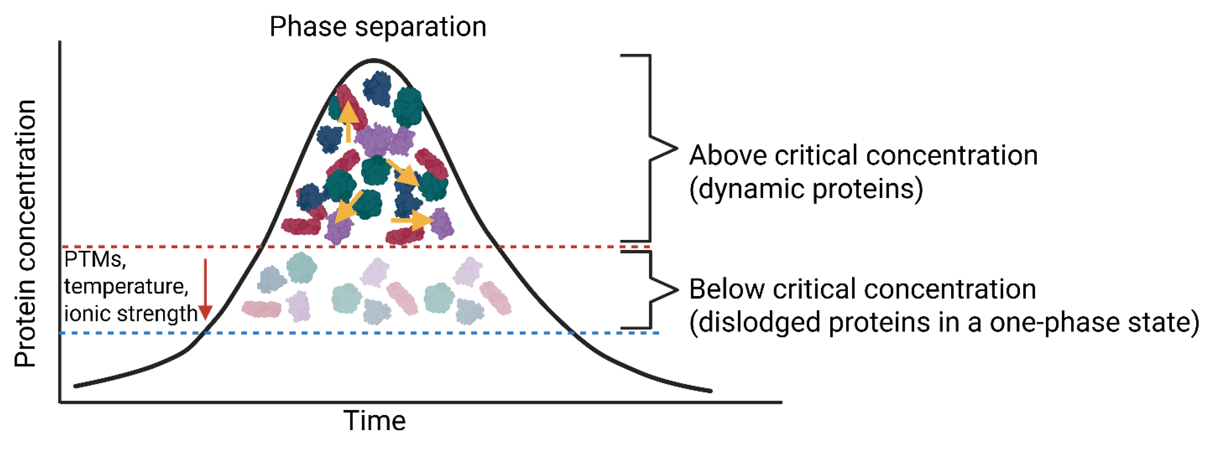
The symbol “Z” denotes the relation between the gas constant (R), absolute temperature (T), and the Faraday constant (F), formulated as 2.3RT/F [38]. Thus, to maintain a constant PMF, cells need to maintain electron charge (ion levels), temperature, and regulate thermodynamic and kinetic forces. PMF is generated by electron transport, light-driven proton translocation, ATP-driven proton pumps, or coupling of electrogenic transport to a metabolic reaction [39]. Each of these mechanisms increases the internal pH, which is kept in balance by cation/H+ antiporters, anion/H+ antiporters, and metabolite decarboxylation [38]. Protons are pumped out by respiration or other mechanisms and pumped back into the cell by PMF-consuming processes such as ATP synthesis or nutrient uptake [38]. Ionic strength influences the binding of proteins to nucleic acids, protein aggregation, the activity of enzymes, the structure of intrinsically disordered proteins (IDPs), ion channels, transporters, and phase separations [40,41,42]. Changes in the physical interactions between macromolecules in a crowded cytoplasm, e.g., due to increased ionic strength or lower pH, may affect the translational diffusion of proteins [43] and compromise proteome stability [44]. Proteins or protein domains that unfold as a result of, e.g., heat stress may represent IDPs and increase the viscosity of that specific area in the cytoplasm, causing the proteins to become entangled [45]. Below a critical concentration, proteins group and form a single-phase state (Figure 3). Changes in temperature, ionic strength, and post-translational modifications (PTMs) lower the critical threshold for phase separation. This allows proteins to disperse and associate at much lower concentrations (Figure 3). Proteins in separate liquid-liquid phases will attract each other, nucleic acids and other macromolecules in a nonspecific manner, and form an associative LLPS. Nonspecific interactions between molecules in the two phases may lead to the formation of segregated LLPS [46]. In this case, protein associations are enhanced via volume exclusion, resulting in one phase enriched in protein and the other phase depleted of protein, relative to the total composition. Macromolecules may also be dispersed via substrate channeling, mediated by metabolons (transient, noncovalently assembled enzyme clusters) that enable direct metabolite transfer between active sites. Examples are intramolecular tunnels (e.g., tryptophan synthase), electrostatic channels (e.g., between malate dehydrogenase and citrate synthase), chemical swing arms (e.g., pyruvate dehydrogenase), cluster channeling (e.g., purinosomes), and glycolytic bodies (reviewed by Fukuhara et al. [47]).
The behavior of proteins in a crowded cytoplasm was originally thought to be dictated by diffusion according to the StokesEinstein (SE) relation (τ∝η/T), where τ is the diffusion time, η is the viscosity of the medium, and T is the experimental temperature [48,49]. Recent findings on cytoplasm dynamics proved the SE relation incorrect. The translational diffusion coefficient (D) of proteins (study performed on Escherichia coli) is determined by the mass of a protein (polypeptide), multiplied by the oligomeric state, and not abundance [8,50]. Furthermore, D = αMβ, where M is the complex mass and α and β are fitting parameters. In the ES equation, β would be -0.33 for globular proteins that do not interact with each other. Instead, β was determined to be -0.6 for the diffusion of proteins in the cytoplasm of E. coli. [8,51]. This implies that the dependence on molecular mass is much stronger than originally anticipated in the SE equation (due to macromolecular crowding). This also indicates that the cytoplasm is a dilatant, non-Newtonian fluid that increases in viscosity during stress. A modified version of the SE equation was proposed (equation 5):
where ηMW represents the perceived viscosity as a function of the molecular weight. The perceived macromolecular viscosity varies from 9.9 cP to 18.1 cP (centipoise) for proteins of 26 kDa to 318.9 kDa [52]. Deviation from the SE equation was confirmed by using fluorescence recovery after photobleaching (FRAP) and nuclear magnetic resonance (NMR) [53]. If diffusion is related to viscosity, as shown in the fractional Debye-Stokes-Einstein (FDE) equation τ∝ (η/T)α (τ = time for a molecule to structurally rearrange or rotate, η = the fluid’s internal resistance to flow, T = absolute temperature, and α = the fractional exponent/scaling parameter of typically 1.0), an α value of less than 1.0 represents inconsistent diffusion [54]. Proteins in complex fluids near the glass transition have α-values of 0.7 to 1.0, depending on the degree of decoupling [55]. Junker et al. [56] have also shown a consistent negative deviation from the SE relation as the molecular weight increases. The diffusion of proteins in the mass range of 26-319 kDa correlates with the average 50-nm mesh size of the E. coli genome [5]. This means proteins with a Stokes radius up to 5 nm may not be affected by the genome’s position in the cytoplasm. Proteins at the poles of E. coli diffused more slowly than those in the rest of the cytoplasm [8,57]. Computer simulations demonstrated that the decrease in protein diffusion exceeds the constraining effects of the cell membrane. The accumulation of proteins most likely begins with genetic mutations, environmental stress (e.g., heat or oxidation), or aging. Misfolded proteins are cleared by chaperones that refold proteins and the proteasome. In bacteria, damaged proteins accumulate at cell poles [57]. As expected, the rate at which cytoplasmic proteins diffuse varies among species. An increase in macromolecular crowders may alter protein conformations from a relaxed, unfolded state to compact, quasi-spherical forms [58]. Proteins with no stable three-dimensional form under physiological conditions (referred to as intrinsically disordered proteins, IDPs) may become compacted in a crowded cytoplasm. Examples are carboxyamidated RNase T1 in the presence of dextran [59] and flavodoxin, apoflavodoxin, and Borrelia burgdorferi VisE in the presence of ficoll-70 k (ficoll PM 70), a neutral, highly branched, hydrophilic synthetic polymer of approximately 70 kDa [60].
Macromolecular crowding helps maintain the three-dimensional structure of DNA and proteins, regulates the kinetic and thermodynamic behavior of enzymes [61,62], facilitates the binding of molecules to ligands, the formation of insoluble aggregates, and the compaction/folding of proteins [4,63,64]. The nucleoid is kept intact by the ParA/MinD ATPase system, as shown in Figure 1 (reviewed by Dicks [2]). ParA orchestrates the segregation of genomic DNA, and MinD positions the divisome (the protein complex responsible for bacterial cell division) at the center of the cell [65]. Protein ParB binds to parS, a centromere-like site on the genome, and forms a large partition complex. ParB stimulates the ATPase of ParA and the ParA-ADP complexes. The ParA-ADP complex dissociates from the genome and is replaced by the next available ParA-ATP complex. This continuous cycle of ParA-ADP and ParA-ATP “shifts” the genome toward areas of higher ParA-ATP density located at opposite poles of the cell [66]. Genome shifting forms a “wave” [67]. Plasmids (and large proteins) are anchored by the helical actin homolog, protein MreB, positioned beneath the inner cell membrane. MreB also maintains cell shape and directs peptidoglycan synthesis [68]. The tubulin protein FtsZ, positioned at the center of the cell, tethers genomic DNA to the Z-ring, where cell division occurs [69]. Other cell particles are displaced in an equally well-controlled manner.
3. Drivers of Cytoplasm Dynamics
Metabolic activities fluidize the cytoplasm and allow large “pockets” of particles to group in overcrowded regions (nanopools), indicated in Figure 1. Regions in the cell with nanopools are metabolically more active [70], which implies that the rearrangement (turnover) of nanopools would promote the displacement of “pockets” over longer distances in the cytoplasm. Meng et al. [70] argued that the positioning of the “pockets”, ranging from subnanometers to micrometers in size, and the difference in diffusion rates of these “pockets” in the cytoplasm, may hold the answer to movement (cytoplasmic fluidity). In a dynamic state (metabolically active cytoplasm), particles are energized by the random “moving” of ATP, amino acids, and other metabolites [71,72,73,74]. Parry et al. [1], however, argued that DNA-related processes (e.g., transcription), the displacement of the nucleoid, and the turnover of cytoplasmic content due to metabolic processes (by definition, also particle “pockets”) are not the driving force behind cytoplasmic fluidity. This coincides with the view formulated by Monti et al. [75]. Both scientific groups, Parry et al. [1] and Monti et al. [75], argued that collisions between particles, or particles in “pockets,” occur randomly and thus out of equilibrium. For the fluctuation-dissipation (FD) theorem to apply, particles in the cytoplasm must be in equilibrium [75]. In the absence of FD, the internal fluctuations (e.g., Brownian motion), are not identical to the dissipation of energy in a weak force or agitation (with reference to conditions in the cytoplasm). Put differently, the fluctuations of particles must correspond to fluctuations in their energy to generate a fluidic cytoplasmic force that would force interactions between particles. For further reading on the FD theorem, the reader is referred to Monti et al. [75].
In modeling a bacterial cell, Meng et al. [70] have shown that cytoplasmic dynamics arise from a combination of metabolic processes. The authors determined the magnitude of the active force (F) in the cytoplasm to be 0.57 pN (pico Newton). This, they argue, is generated by the combined collisions between small molecules, amino acids, proteins, and ions that are out of equilibrium. Based on the size of a typical protein (10 nm, as defined by the authors), the active force generated by such a protein is 0.4 pN. This was calculated using the equation F = kBT/a, where F is the force expressed in N, kB is the Boltzmann constant [≈ 1.38 × 10⁻²³ J/K (Joules per Kelvin)], T is the temperature in K, and a is the size (in this case 10 nm). The Boltzmann constant relates the average microscopic thermal energy of particles to the macroscopic thermodynamic temperature of a system [76]. According to Meng et al. [70], a force of 0.4 pN is sufficient to overcome thermal fluctuations, propel proteins in a specific direction when signaling in the cytoplasm changes, and support three-dimensional conformational changes in proteins.
4. Size Matters in Diffusion
The rate at which macromolecules diffuse across the cytoplasm determines the speed or frequency of collisions. This is true when reactions are diffusion-limited, i.e., when the association rate constant (ka) depends only on the translational diffusion coefficient (D). However, the ka of a protein diffusing in the cytoplasm with D = 10 μm2/s and reacting with another molecule is approximately 108 M-1/s (M = molar). Since most proteins have catalytic regions and are not reactive across their entire surface, a more realistic diffusion-limited ka would be 105 to 106 M-1/s [74]. The Barnase-Barstar protein pair of Bacillus amyloliquefaciens has a ka of 108 to 1010 M-1/s, which is beyond the diffusion limit. The interaction between these proteins is mediated by electrostatic attraction [77,78,79,80], enabling ribonuclease binding to the inhibitor protein at orders of magnitude above the non-electrostatic diffusion limit. Other diffusion-limited protein reactions with high ka include the ternary complex of aminoacyl-tRNA, EF-TU, and GTP that interacts with the ribosome, known as tRNA selection or decoding [81,82]; proteins in low copy numbers and with longer distances to cover; and proteins binding to membranes or other large structures (e.g., the Min oscillation system) [83].
Protein-protein interactions, such as those between cationic Barnase and anionic Barstar, are rare in bacteria. Only 35 proteins in E. coli have a net surface charge >+10. Of these, 18 are ribosomal, 14 are DNA/RNA-associated, and 3 have unknown functions [52]. It thus seems that the bacterial proteome is largely anionic, thereby supporting rapid diffusion through the cytoplasm. It also implies that endosymbionts with highly cationic proteomes have evolved specialized mechanisms to avoid slow diffusion and impair ribosomal function. As crowding increases, the D of macromolecules decreases by orders of magnitude, and many reaction-limited interactions between macromolecules become diffusion-limited. Small molecules, such as sugars trapped in supramolecular protein aggregates, are not severely affected by osmotic upshifts and diffuse throughout the cytoplasm, even at NaCl concentrations of 1 M or higher [49,84]. The cytoplasm thus acts as a molecular sieve under high and low osmotic stress, but with different mesh sizes. This allows the rapid diffusion of ions, small metabolites, and signaling molecules across a crowded cytoplasm.
5. Conclusions
It is unlikely that cytoplasmic flow is caused by displacement of the nucleoid or by metabolic processes, but more likely by particles in “pockets” (“cages”) that move randomly in a non-equilibrium state, localized in nanopools at the two poles of the cell. The propulsion (displacement or movement) of proteins causes cytoplasmic streaming (flow) and increases biochemical reactions, thereby increasing metabolic activity. Macromolecular crowding is a key contributor to intracellular complexity and plays a major role in regulating protein-protein, protein-nucleic acid, and protein-lipid interactions. The spatial orientation of biomolecular condensates (macrocompartments) serves the role normally fulfilled by organelles and intracellular membranes. These condensates/compartments are regulated by supramolecular structures such as DNA and RNA, as well as by PTMs and the cell membrane. The exact manner in which PTMs regulate phase separation must be confirmed. The size of the nucleoid-free space in the cytoplasm and the concentration of proteins are likely determining the size of a condensate. Further research is required on the ultrastructure of condensates and the arrangement of their components, especially for heterotypic condensates. We do not know to what extent the stoichiometry of complexes in a semi-solid (dense, highly viscous, glass-like) cytoplasm changes during turnover to a semi-liquid phase. Examining crowding effects on the structure, function, and regulation of biomolecular condensates is challenging. Not all biomolecular condensates are manipulated by macromolecular crowding. In some cases, condensation is triggered by ionic strength, pH, ATP, and membrane surfaces. Many questions remain unanswered or not fully understood, e.g., the effect of ribosomes on protein mobility, the effect of protein levels on reaction rates and diffusion coefficients, the influence of metabolic conditions on LLPS formation, differences in the mobility of old and new proteins at cell poles, conditions or reactions affecting the compaction of the nucleoid, factors determining the size of biomolecular condensates, and the role of nucleotides in the assembly of condensates.
Funding
This research received no external funding.
Institutional Review Board Statement
Not applicable.
Informed Consent Statement
Not applicable.
Data Availability Statement
Not applicable.
Conflicts of Interest
The author has no conflicts of interest.
References
- Parry, B.R.; Surovtsev, I.V.; Cabeen, M.T.; O’Hern, C.S.; Dufresne, E.R.; Jacobs-Wagner, C. The bacterial cytoplasm has glass-like properties and is fluidized by metabolic activity. Cell 2014, 156, 183–194. [Google Scholar] [CrossRef]
- Dicks, L.M.T.; Pohl, C.; Botha, A. Bacterial sentience is determined by the stochastic, chaotic, and deterministic behavior of cytoplasmic particles. Curr. Issues Mol. Biol. 2026, 48, 777. [Google Scholar] [CrossRef]
- Zimmerman, S.B.; Trach, S.O. Estimation of macromolecule concentrations and excluded volume effects for the cytoplasm of Escherichia coli. J. Mol. Biol. 1991, 222(3), 599−620. [Google Scholar] [CrossRef]
- Rivas, G.; Minton, A.P. Influence of nonspecific interactions on protein associations: Implications for biochemistry in vivo. Annu. Rev. Biochem. 2022, 91(1), 321−351. [Google Scholar] [CrossRef]
- Xiang, Y.; Surovtsev, I.V.; Chang, Y.; Govers, S.K.; Parry, B.R.; Liu, J.; Jacobs-Wagner, C. Interconnecting solvent quality, transcription, and chromosome folding in Escherichia coli. Cell 2021, 184(14), 3626−3642.e14. [Google Scholar] [CrossRef]
- Kaljevic, ́. J.; Saaki, T.N.V.; Govers, S.K.; Remy, O.; Van Raaphorst, R.; Lamot, T.; Laloux, G. Chromosome choreography during the non-binary cell cycle of a predatory bacterium. Curr. Biol. 2021, 31(17), 3707−3720.e5. [Google Scholar] [CrossRef]
- Bakshi, S.; Siryaporn, A.; Goulian, M.; Weisshaar, J.C. Superresolution imaging of ribosomes and RNA polymerase in live Escherichia coli cells. Mol. Microbiol. 2012, 85(1), 21−38. [Google Scholar] [CrossRef]
- Ś migiel, W.M.; Mantovanelli, L.; Linnik, D.S.; Punter, M.; Silberberg, J.; Xiang, L.; Xu, K.; Poolman, B. Protein diffusion in Escherichia coli cytoplasm scales with the mass of the complexes and is location dependent. Sci. Adv. 2022, 8(32), No. eabo5387. [Google Scholar] [CrossRef]
- Lewis, P.J.; Thaker, S.D.; Errington, J. Compartmentalization of transcription and translation in Bacillus subtilis. EMBO J. 2000, 19(4), 710−718. [Google Scholar] [CrossRef]
- Van Gijtenbeek, L.A.; Robinson, A.; Van Oijen, A.M.; Poolman, B.; Kok, J. On the spatial organization of mRNA, plasmids, and ribosomes in a bacterial host overexpressing membrane proteins. PLoS Genet. 2016, 12(12), No. e1006523. [Google Scholar] [CrossRef]
- Sanamrad, A.; Persson, F.; Lundius, E.G.; Fange, D.; Gynnå, A.H.; Elf, J. Single-particle tracking reveals that free ribosomal subunits are not excluded from the Escherichia coli nucleoid. Proc. Natl. Acad. Sci. USA 2014, 111, 11413–11418. [Google Scholar] [CrossRef]
- Ginell, G.M.; Holehouse, A.S. An introduction to the stickers-and- spacers framework as applied to biomolecular condensates. In Phase-Separated Biomolecular Condensates; Zhou, H.-X., Spille, J.-H., Banerjee, P. R., Eds.; Springer US, 2026; pp. 95–116. [Google Scholar]
- Mittag, T.; Pappu, R.V. A conceptual framework for understanding phase separation and addressing open questions and challenges. Mol. Cell 2022, 82, 2201–2214. [Google Scholar] [CrossRef]
- Lasker, K.; Boeynaems, S.; Lam, V.; Scholl, D.; Stainton, E.; Briner, A.; Jacquemyn, M.; Daelemans, D.; Deniz, A.; Villa, E.; Holehouse, A.S.; Gitler, A.D.; Shapiro, L. The material properties of a bacterial-derived biomolecular condensate tune biological function in natural and synthetic systems. Nat. Commun. 2022, 13, 5643. [Google Scholar] [CrossRef]
- Perego, E.; Zappone, S.; Castagnetti, F.; Mariani, D.; Vitiello, E.; Rupert, J.; Zacco, E.; Tartaglia, G.G.; Bozzoni, I.; Slenders, E.; Vicidomini, G. Single-photon microscopy to study biomolecular condensates. Nat. Commun. 2023, 14, 8224. [Google Scholar] [CrossRef]
- Hoang, Y.; Azaldegui, C.A.; Dow, R.E.; Ghalmi, M.; Biteen, J.S.; Vecchiarelli, A.G. An experimental framework to assess biomolecular condensates in bacteria. Nat. Commun. 2024, 15, 3222. [Google Scholar] [CrossRef]
- Linnik, D.; Poolman, B. Spatiotemporal organization of the bacterial cytoplasm. Curr. Opin. Microbiol. 2025, 87, 102635. [Google Scholar] [CrossRef]
- Edwardson, T.G.W.; Levasseur, M.D.; Tetter, S.; Steinauer, A.; Hori, M.; Hilvert, D. Protein cages: from fundamentals to advanced applications. Chem. Rev. 2022, 122, 9145–9197. [Google Scholar] [CrossRef]
- Ebata, H.; Nishizawa, K.; van Esterik, F.A.S.; Maeda, T.; Tao, Y.; Inokuchi, S.; Ise, H.; Mizuno, D. Single power-law rheology of crowded cytoplasm in living cells. Cell Rep. Phys. Sci. 2026, 7, 103059. [Google Scholar] [CrossRef]
- Jacob, A.R.; Poulos, A.S.; Semenov, A.N.; Vermant, J.; Petekidis, G. Flow dynamics of concentrated starlike micelles: Asuperposition rheometry investigation into relaxation mechanisms. J. Rheol. 2019, 63, 641–653. [Google Scholar] [CrossRef]
- Sugino, Y.; Ebata, H.; Sowa, Y.; Ikeda, A.; Mizuno, D. Non-equilibrium fluidization of dense active suspension. arXiv 2024. [Google Scholar] [CrossRef]
- Xie, Y.; Shu, T.; Liu, T.; Spindler, M.C.; Mahamid, J.; Hocky, G.M.; Gresham, D.; Holt, L.J. Polysome collapse and RNA condensation fluidize the cytoplasm. Mol. Cell 2024, 84, 2698–2716.e9. [Google Scholar] [CrossRef]
- Delarue, M.; Brittingham, G.P.; Pfeffer, S.; Surovtsev, I.V.; Pinglay, S.; Kennedy, K.J.; Schaffer, M.; Gutierrez, J.I.; Sang, D.; Poterewicz, G.; Chung, J.J.; Plitzko, J.M.; Groves, J.T.; Jacobs-Wagner, C.; Engel, B.D.; Holt, L.J. mTORC1 controls phase separation and the biophysical properties of the cytoplasm by tuning crowding. Cell 2018, 174, 338–349.e20. [Google Scholar] [CrossRef]
- Munder, M.C.; Midtvedt, D.; Franzmann, T.; Nüske, E.; Otto, O.; Herbig, M.; Ulbricht, E.; Müller, P.; Taubenberger, A.; Maharana, S.; Malinovska, L.; Richter, D.; Guck, J.; Zaburdaev, V.; Alberti, S. A pH-driven transition of the cytoplasm from a fluid- to a solid-like state promotes entry into dormancy. eLife 2016, 5, e09347. [Google Scholar] [CrossRef]
- Losa, J.; Simon, F.; Linnik, D.; Kaya, S.G.; Stuart, M.C.A.; Stetsenko, A.; de Boer, R.; Ho, F.Y.; Incarnato, D.; Stevens, J.; van Eck, J.; Fraaije, M.W.; Weiss, L.E.; van Teeffelen, S.; Abeln, S.; Guskov, A.; Marrink, S.-J.; Poolman, B.; Heinemann, M. Condition-dependent amorphous protein agglomerates control cytoplasmic rheology. Mol. Cell 2026, 86(9), 1780–1796.e11. [Google Scholar] [CrossRef]
- Niftrik, L.A.; Fuerst, J.A.; Damsté, J.S.S.; Kuenen, J.G.; Jetten, M.S.M.; Strous, M. The anammoxosome: an intracytoplasmic compartment in anammox bacteria. FEMS Microbiol. Lett. 2004, 233, 7–13. [Google Scholar] [CrossRef]
- Walsby, A. E. Gas vesicles. Microbiol. Rev. 1994, 58, 94–144. [Google Scholar] [CrossRef]
- Hossain, S.; Bahreini, B.; Pasteur, E.; Sadoh, A.; Bala, R.M.; Ravindra, N. Magnetotactic bacteria and magnetosomes – an overview. MSEIJ 2024, 8, 83–100. [Google Scholar] [CrossRef]
- Held, M.; Quin, M.B.; Schmidt-Dannert, C. Eut bacterial microcompartments: insights into their function, structure, and bioengineering applications. Micro. Physiol. 2013, 23, 308–320. [Google Scholar] [CrossRef]
- Huang, J.-H.; Ferrell, J.E., Jr. How does cytoplasmic crowding affect reaction rates? Mol. Cell. 2026, 86(1), 9–23. [Google Scholar] [CrossRef]
- Zhou, H.-X. Protein folding and binding in confined spaces and in crowded solutions. J. MolRecognit. 2004, 17, 368–375. [Google Scholar] [CrossRef]
- Minton, A.P. Holobiochemistry: An integrated approach to the understanding of biochemical mechanism that emerges from the study of proteins and protein associations in volume-occupied solutions. In Structural and Organizational Aspects of Metabolic Regulation; Srere, P., Jones, M.E., Mathews, C., Eds.; Alan R. Liss, 1989; pp. 291–306. [Google Scholar]
- Zhou, H.X.; Rivas, G.; Minton, A.P. Macromolecular crowding and confinement: Biochemical, biophysical, and potential physiological consequences. Annu. Rev. Biophys. 2008, 37, 375−397. [Google Scholar] [CrossRef]
- Zimmerman, S.B.; Minton, A.P. Macromolecular Crowding: Biochemical, Biophysical, and Physiological Consequences. Annu. Rev. Biophys. Biomol. Struct. 1993, 22(1), 27−65. [Google Scholar] [CrossRef]
- Tabaka, M.; Kalwarczyk, T.; Hołyst, R. Quantitative influence of macromolecular crowding on gene regulation kinetics. Nucl. Acids Res. 2014, 42(2), 727–738. [Google Scholar] [CrossRef]
- Blainey, P.C.; Luo, G.; Kou, S.C.; Mangel, W.F.; Verdine, G.L.; Bagchi, B.; Xie, X.S. Nonspecifically bound proteins spin while diffusing along DNA. Nat. Struct. Mol. Biol. 2009, 16, 1224–1229. [Google Scholar] [CrossRef]
- Norred, S.E.; Caveney, P.M.; Chauhan, G.; Collier, L.K.; Collier, C.P.; Abel, S.M.; Simpson, M.L. Macromolecular Crowding induces spatial correlations that control gene expression bursting patterns. ACS Synth. Biol. 2018, 7(5), 1251–1258. [Google Scholar] [CrossRef]
- Poolman, B. Physicochemical homeostasis in bacteria. FEMS Microbiol. Revs. 2023, 47, 1–7. [Google Scholar] [CrossRef]
- Sikkema, H.R.; Gaastra, B.F.; Pols, T.; Poolman, B. Cell fuelling and metabolic energy conservation in synthetic cells. ChemBioChem 2019, 20(20), 2581−2592. [Google Scholar] [CrossRef]
- König, I.; Zarrine-Afsar, A.; Aznauryan, M.; Soranno, A.; Wunderlich, B.; Dingfelder, F.; Stüber, J. C.; Plückthun, A.; Nettels, D.; Schuler, B. Single-molecule spectroscopy of protein conformational dynamics in live eukaryotic cells. Nat. Methods 2015, 12(8), 773−779. [Google Scholar] [CrossRef]
- Syeda, R.; Qiu, Z.; Dubin, A.E.; Murthy, S.E.; Florendo, M.N.; Mason, D.E.; Mathur, J.; Cahalan, S.M.; Peters, E.C.; Montal, M.; Patapoutian, A. LRRC8 Proteins form volume-regulated anion channels that sense ionic strength. Cell 2016, 164(3), 499−511. [Google Scholar] [CrossRef]
- Elbaum-Garfinkle, S.; Kim, Y.; Szczepaniak, K.; Chen, C.C.-H.; Eckmann, C.R.; Myong, S.; Brangwynne, C.P. The disordered P granule protein LAF-1 drives phase separation into droplets with tunable viscosity and dynamics. Proc. Natl. Acad. Sci. USA. 2015, 112(23), 7189−7194. [Google Scholar] [CrossRef]
- Xie, Y.; Gresham, D.; Holt, L.J. Increased mesoscale diffusivity in response to acute glucose starvation. MicroPubl. Biol. 2023. [Google Scholar] [CrossRef]
- Leuenberger, P.; Ganscha, S.; Kahraman, A.; Cappelletti, V.; Boersema, P.J.; Von Mering, C.; Claassen, M.; Picotti, P. Cell-wide analysis of protein thermal unfolding reveals determinants of thermostability. Science 2017, 355, 6327. [Google Scholar] [CrossRef]
- Di Bari, D.; Timr, S.; Guiral, M.; Giudici-Orticoni, M.-T.; Seydel, T.; Beck, C.; Petrillo, C.; Derreumaux, P.; Melchionna, S.; Sterpone, F.; Peters, J.; Paciaroni, A. Diffusive dynamics of bacterial proteome as a proxy of cell death. ACS Cent. Sci. 2023, 9(1), 93− 102. [Google Scholar] [CrossRef]
- Abyzov, A.; Blackledge, M.; Zweckstetter, M. Conformational dynamics of intrinsically disordered proteins regulate biomolecular condensate chemistry. Chem. Rev. 2022, 122, 6719–6748. [Google Scholar] [CrossRef]
- Fukuhara, E.; Dunkley, E.; Bevilacqua, A.; Laurino, P. Dynamic cytoplasm: A physical regulator of enzymatic function. Biochem. 2025, 64, 2699–2711. [Google Scholar] [CrossRef]
- Das, N.; Sen, P. Dynamic heterogeneity and viscosity decoupling: origin and analytical prediction. Phys. Chem. Chem. Phys. 2021, 23, 15749–15757. [Google Scholar] [CrossRef]
- Khan, T.; Tarif, E.; Awano, Y.; Lozada, L.S.; Das, N.; Tominaga, K.; Sen, P. Multiple evidence for molecular level heterogeneity in a non-ionic biocatalytic deep eutectic solvent. J. Mol. Liq. 2023, 389, 122882. [Google Scholar] [CrossRef]
- Fulton, A.B. How crowded is the cytoplasm? Cell 1982, 30, 345–347. [Google Scholar] [CrossRef]
- Xiang, L.; Chen, K.; Yan, R.; Li, W.; Xu, K. Single-molecule displacement mapping unveils sign-asymmetric protein charge effects on intraorganellar diffusion. Nano Lett. 2023, 23(5), 1711–1716. [Google Scholar] [CrossRef]
- Monterroso, B.; Margolin, W.; Boersma, A.J.; Rivas, G.; Poolman, B.; Zorrilla, S. Macromolecular Crowding, phase separation, and homeostasis in the orchestration of bacterial cellular functions. Chem. Revs. 2024, 124(4), 1899–1949. [Google Scholar] [CrossRef]
- Feder, T.J.; Brust-Mascher, I.; Slattery, J.P.; Baird, B.; Webb, W.W. Constrained diffusion or immobile fraction on cell surfaces: a new interpretation. Biophys. J. 1996, 70, 2767–2773. [Google Scholar] [CrossRef]
- Höfling, F.; Franosch, T. Anomalous transport in the crowded world of biological cells. Rep. Prog. Phys. 2013, 76, 046602. [Google Scholar] [CrossRef]
- Wachsmuth, M.; Waldeck, W.; Langowski, J. Anomalous diffusion of fluorescent probes inside living cells investigated by spatially-resolved fluorescence correlation spectroscopy. J. Mol. Biol. 2000, 298, 677–689. [Google Scholar] [CrossRef]
- Junker, N.O.; Vaghefikia, F.; Albarghash, A.; Höfig, H.; Kempe, D.; Walter, J.; Otten, J.; Pohl, M.; Katranidis, A.; Wiegand, S.; Fitter, J. Impact of molecular crowding on translational mobility and conformational properties of biological macromolecules. J. Phys. Chem. B 2019, 123, 4477–4486. [Google Scholar] [CrossRef]
- Mantovanelli, L.; Linnik, D.S.; Punter, M.; Kojakhmetov, H.J.; Ś migiel, W.M.; Poolman, B. Simulation-based reconstructed diffusion unveils the effect of aging on protein diffusion in Escherichia coli. PLoS Comput. Biol. 2023, 19(9), e1011093. [Google Scholar] [CrossRef]
- Minton, A.P. Excluded volume as a determinant of macromolecular structure and reactivity. Biopolymers 1981, 20, 2093–2120. [Google Scholar] [CrossRef]
- Qu, Y.; Bolen, D.W. Efficacy of macromolecular crowding in forcing proteins to fold. Biophys. Chem. 2002, 101–102, 155–165. [Google Scholar] [CrossRef]
- Perham, M.; Stagg, L.; Wittung-Stafshede, P. Macromolecular crowding increases structural content of folded proteins. FEBS Lett. 2007, 581, 5065–5069. [Google Scholar] [CrossRef]
- Ellis, R.J. Macromolecular crowding: obvious but underappreciated. Trends Biochem. Sci. 2001, 26, 597–604. [Google Scholar] [CrossRef]
- Dhar, A.; Samiotakis, A.; Ebbinghaus, S.; Nienhaus, L.; Homouz, D.; Gruebele, M.; Cheung, M.S. Structure, function, and folding of phosphoglycerate kinase are strongly perturbed by macromolecular crowding. Proc. Natl. Acad. Sci. USA. 2010, 107(41), 17586–17591. [Google Scholar] [CrossRef]
- Rivas, G.; Minton, A.P. Macromolecular crowding in vitro, in vivo, and in between. Trends Biochem. Sci. 2016, 41(11), 970−981. [Google Scholar] [CrossRef]
- Van Den Berg, J.; Boersma, A.J.; Poolman, B. Microorganisms maintain crowding homeostasis. Nat. Rev. Microbiol. 2017, 15(5), 309−318. [Google Scholar] [CrossRef]
- Pulianmackal, L.T.; Vecchiarelli, A.G. Positioning of cellular components by the ParA/MinD family of ATPases. Curr. Opin. Microbiol. 2024, 79, 102485. [Google Scholar] [CrossRef]
- Ietswaart, R.; Szardenings, F.; Gerdes, K.; Howard, M. Competing ParA structures space bacterial plasmids equally over the nucleoid. PLoS Comput. Biol. 2014, 10(12), e1004009. [Google Scholar] [CrossRef]
- Köhler, R.; Kaganovitch, E.; Murray, S.M. High-throughput imaging and quantitative analysis uncovers the nature of plasmid positioning by ParABS. Elife 2022, 11, e78743. [Google Scholar] [CrossRef]
- Wang, Y.; Jiang, Y.; Song, Z.; Zhu, C.; Tang, Y.; Peng, J.; Liu, P. MreB: unraveling the molecular mechanisms of bacterial shape, division, and environmental adaptation. Cell Commun. Signal. 2025, 23(1), 377. [Google Scholar] [CrossRef]
- Barrows, J.M.; Goley, E.D. FtsZ dynamics in bacterial division: What, how, and why? Curr. Opin. Cell Biol. 2021, 68, 163–172. [Google Scholar] [CrossRef]
- Meng, L.; Jin, Y.; Guan, Y.; Xu, J.; Lin, J. Diffusion enhancement in bacterial cytoplasm through an active random force. Phys. Rev. Res. 2023, 5, L032018. [Google Scholar] [CrossRef]
- Wilhelm, C. Out-of-equilibrium microrheology inside living cells. Phys. Rev. Lett. 2008, 101, 028101. [Google Scholar] [CrossRef]
- Gnesotto, F.S.; Mura, F.; Gladrow, J.; Broedersz, C.P. Broken detailed balance and non-equilibrium dynamics in living systems: a review. Rep. Prog. Phys. 2018, 81, 066601. [Google Scholar] [CrossRef]
- Solon, A.P.; Stenhammar, J.; Wittkowski, R.; Kardar, M.; Kafri, Y.; Cates, M.E.; Tailleur, J. Pressure and phase equilibria in interacting active Brownian spheres. Phys. Rev. Lett. 2015, 114, 198301. [Google Scholar] [CrossRef]
- Solon, A.P.; Fily, Y.; Baskaran, A.; Cates, M.E.; Kardar, M.; Tailleur, J. Pressure is not a state function for generic active fluids. Nat. Phys. 2015, 11, 673. [Google Scholar] [CrossRef]
- Monti, J.M.; Perl, Y.S.; Tagliazucchi, E.; Kringelbach, M.L.; Decoet, G. Fluctuation-dissipation theorem and the discovery of distinctive off-equilibrium signatures of brain states. Phys. Rev. Res. 2025, 7, 013301. [Google Scholar] [CrossRef]
- Jørgensen, S.E. Information theory and energy. In Encyclopedia of Energy; Cleveland, C. J., Ed.; Elsevier, 2004; pp. 439–449. [Google Scholar]
- Schavemaker, P.E.; Boersma, A.J.; Poolman, B. How important is protein diffusion in prokaryotes? Front. Mol. Biosci. 2018, 5, 93. [Google Scholar] [CrossRef]
- Alsallaq, R.; Zhou, H.-X. Electrostatic rate enhancement and transient complex of protein−protein association. Proteins Struct. Funct. Bioinforma. 2008, 71(1), 320−335. [Google Scholar] [CrossRef]
- Schreiber, G.; Fersht, A.R. Interaction of barnase with its polypeptide inhibitor barstar studied by protein engineering. Biochem. 1993, 32(19), 5145−5150. [Google Scholar] [CrossRef]
- Wallis, R.; Moore, G.R.; James, R.; Kleanthous, C. Protein-protein interactions in colicin E9 DNase-immunity protein complexes. 1. Diffusion-controlled association and femtomolar binding for the cognate complex. Biochem. 1995, 34(42), 13743−13750. [Google Scholar] [CrossRef]
- Klumpp, S.; Scott, M.; Pedersen, S.; Hwa, T. Molecular crowding limits translation and cell growth. Proc. Natl. Acad. Sci. U.S.A. 2013, 110(42), 16754−16759. [Google Scholar] [CrossRef]
- Zhang, G.; Fedyunin, I.; Miekley, O.; Valleriani, A.; Moura, A.; Ignatova, Z. Global and local depletion of ternary complex limits translational elongation. Nucl. Acids Res. 2010, 38(14), 4778−4787. [Google Scholar] [CrossRef]
- Loose, M.; Kruse, K.; Schwille, P. Protein self-organization: Lessons from the Min system. Annu. Rev. Biophys. 2011, 40(1), 315−336. [Google Scholar] [CrossRef]
- Mika, J.T.; Van Den Bogaart, G.; Veenhoff, L.; Krasnikov, V.; Poolman, B. Molecular sieving properties of the cytoplasm of Escherichia coli and consequences of osmotic stress: Molecule diffusion and barriers in the cytoplasm. Mol. Microbiol. 2010, 77(1), 200−207. [Google Scholar]
Figure 1.
Gram-negative bacterial cell with spatially organized intracellular (cytoplasmic) structures. The nucleoid, plasmids, subatomic particles (electrons, neutrons, positrons, protons), ions, atoms, inorganic and organic molecules, proteins, ribosomes, enzymatic megacomplexes, granules, and microcompartments (“pockets” or cages”) give the cytoplasm a glass-like appearance. Metabolic processes fluidize the cytoplasm and separate particles, changing the glass-like appearance from semi-liquid to semi-solid. Particles in “pockets” (grey spheres) rearrange (indicated by black dashed arrows) to form open areas (“gaps”) in the cytoplasm. The movement of particles generates dynamic heterogeneity within the cytoplasm. The nucleoid is kept intact by the ParA/MinD ATPase complex. The exchange between ADP and ATP in ParA “shifts” the genome towards opposite poles of the cell, forming a “wave”-like movement. MreB, a helical actin homolog in the cytoplasm, positioned underneath the cell membrane, associates with a complex of proteins in the inner membrane and the periplasm. The tubulin protein FtsZ, positioned at the center of the cell, tethers genomic DNA to the Z-ring, where cell division takes place. RNA polymerases and other molecules form dense regions, a process known as liquid–liquid phase separation (LLPS). mRNAs are shown as free or as bound to ribosomes or proteins. Biomacromolecules (indicated in yellow) contribute to cytoplasmic crowding and can aggregate to form intracellular bodies. Open spaces (“gaps”) in the cytoplasm serve as metabolite pools (red dashed circle) and allow molecules to move in and out of the pool (red arrows). The image on the right summarizes the changes in forces during crowding. Red arrows pointing up = increase in activity, arrows pointing down = decrease in activity. KD = dissociation constant, ka = association rate constant (a higher ka turns reaction “on”), kd = dissociation rate (a low kd turns reaction “off”), kreact = reaction rate constant, PMF = proton motive force. Modified from a figure published by Dicks et al. [2]. Constructed using BioRender (https://biorender.com/, assessed on 9 September 2026).
Figure 1.
Gram-negative bacterial cell with spatially organized intracellular (cytoplasmic) structures. The nucleoid, plasmids, subatomic particles (electrons, neutrons, positrons, protons), ions, atoms, inorganic and organic molecules, proteins, ribosomes, enzymatic megacomplexes, granules, and microcompartments (“pockets” or cages”) give the cytoplasm a glass-like appearance. Metabolic processes fluidize the cytoplasm and separate particles, changing the glass-like appearance from semi-liquid to semi-solid. Particles in “pockets” (grey spheres) rearrange (indicated by black dashed arrows) to form open areas (“gaps”) in the cytoplasm. The movement of particles generates dynamic heterogeneity within the cytoplasm. The nucleoid is kept intact by the ParA/MinD ATPase complex. The exchange between ADP and ATP in ParA “shifts” the genome towards opposite poles of the cell, forming a “wave”-like movement. MreB, a helical actin homolog in the cytoplasm, positioned underneath the cell membrane, associates with a complex of proteins in the inner membrane and the periplasm. The tubulin protein FtsZ, positioned at the center of the cell, tethers genomic DNA to the Z-ring, where cell division takes place. RNA polymerases and other molecules form dense regions, a process known as liquid–liquid phase separation (LLPS). mRNAs are shown as free or as bound to ribosomes or proteins. Biomacromolecules (indicated in yellow) contribute to cytoplasmic crowding and can aggregate to form intracellular bodies. Open spaces (“gaps”) in the cytoplasm serve as metabolite pools (red dashed circle) and allow molecules to move in and out of the pool (red arrows). The image on the right summarizes the changes in forces during crowding. Red arrows pointing up = increase in activity, arrows pointing down = decrease in activity. KD = dissociation constant, ka = association rate constant (a higher ka turns reaction “on”), kd = dissociation rate (a low kd turns reaction “off”), kreact = reaction rate constant, PMF = proton motive force. Modified from a figure published by Dicks et al. [2]. Constructed using BioRender (https://biorender.com/, assessed on 9 September 2026).
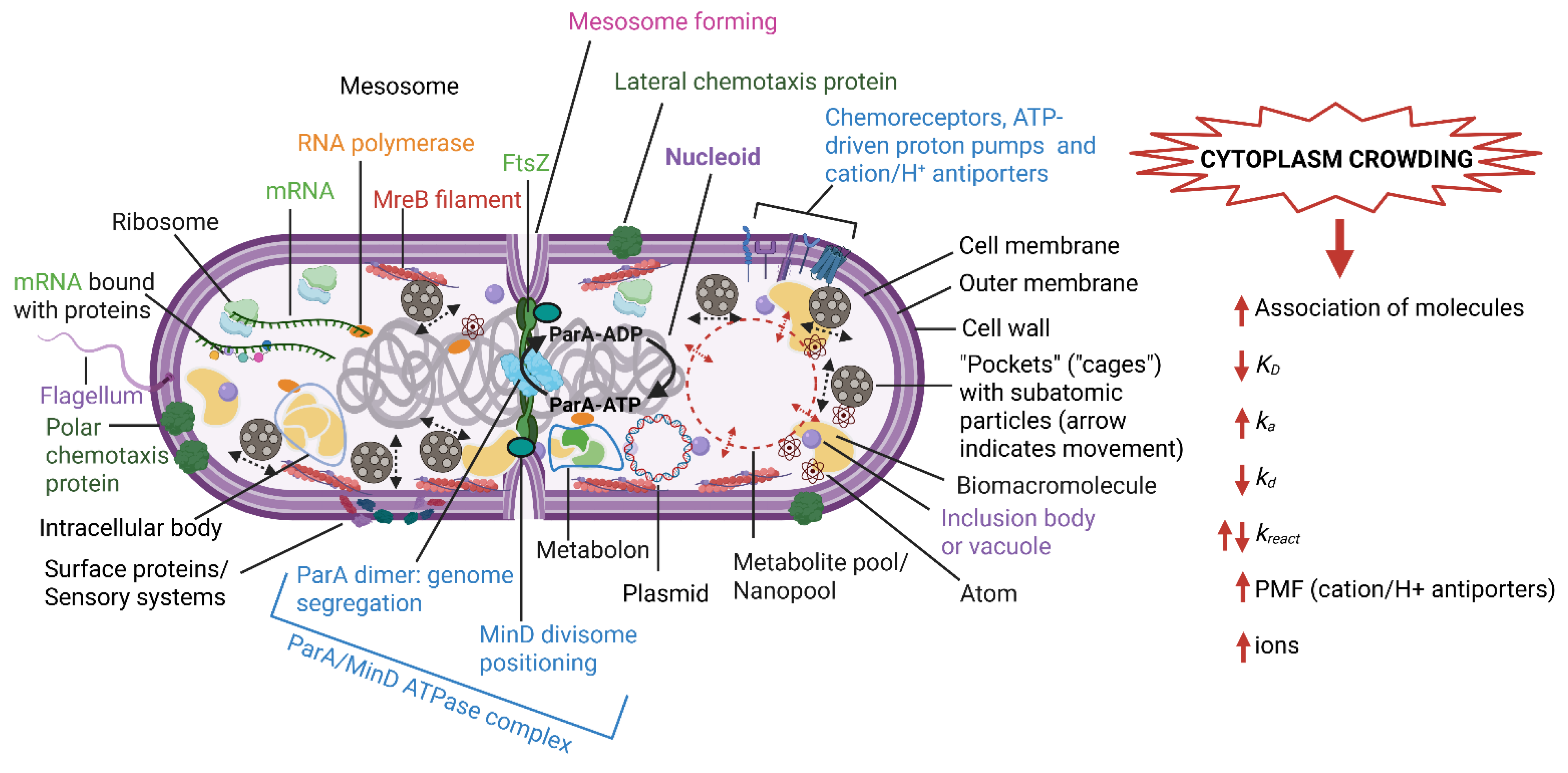
Figure 2.
The influence of macromolecular crowding on gene regulation kinetics. Linking nucleoid-associated proteins (NAPs) link DNA strands, whilst bending NAPs bend the DNA and force the nucleoid into a compact conformation. This increases transcription factor (TF) binding to the nucleoid. The TFs rotate along the grooves of the DNA molecule, described as facilitated diffusion/sliding. Sliding is hampered by bending NAPs (right image) but facilitated in the absence of NAP binding (bottom image). The overall target search (TF binding to DNA) is accelerated by crowding (a smaller reaction volume). Constructed using BioRender (https://biorender.com/, assessed on 11 September 2026).
Figure 2.
The influence of macromolecular crowding on gene regulation kinetics. Linking nucleoid-associated proteins (NAPs) link DNA strands, whilst bending NAPs bend the DNA and force the nucleoid into a compact conformation. This increases transcription factor (TF) binding to the nucleoid. The TFs rotate along the grooves of the DNA molecule, described as facilitated diffusion/sliding. Sliding is hampered by bending NAPs (right image) but facilitated in the absence of NAP binding (bottom image). The overall target search (TF binding to DNA) is accelerated by crowding (a smaller reaction volume). Constructed using BioRender (https://biorender.com/, assessed on 11 September 2026).
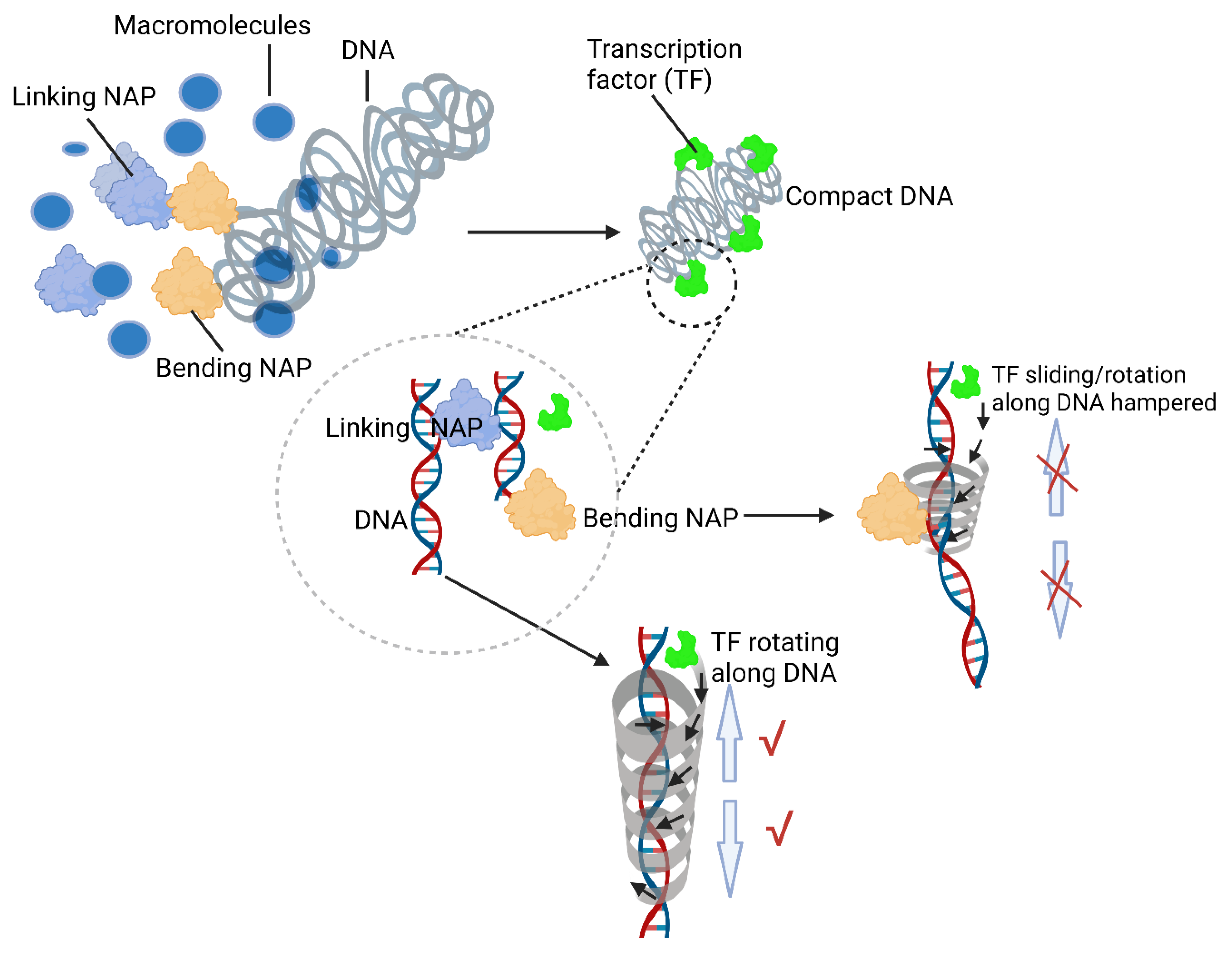
Figure 3.
Phase separation of proteins. Proteins above a critical concentration are dynamic and form microcompartments in a shorter time than proteins that have been dislodged due to post-translational modifications (PTMs), or changes in temperature and ionic strength and group below the critical concentration point. Constructed using BioRender (https://biorender.com/, assessed on 9 September 2026).
Figure 3.
Phase separation of proteins. Proteins above a critical concentration are dynamic and form microcompartments in a shorter time than proteins that have been dislodged due to post-translational modifications (PTMs), or changes in temperature and ionic strength and group below the critical concentration point. Constructed using BioRender (https://biorender.com/, assessed on 9 September 2026).
Disclaimer/Publisher’s Note: The statements, opinions and data contained in all publications are solely those of the individual author(s) and contributor(s) and not of MDPI and/or the editor(s). MDPI and/or the editor(s) disclaim responsibility for any injury to people or property resulting from any ideas, methods, instructions or products referred to in the content. |
© 2026 by the authors. Licensee MDPI, Basel, Switzerland. This article is an open access article distributed under the terms and conditions of the Creative Commons Attribution (CC BY) license (http://creativecommons.org/licenses/by/4.0/).
Copyright: This open access article is published under a Creative Commons CC BY 4.0 license, which permit the free download, distribution, and reuse, provided that the author and preprint are cited in any reuse.