

Submitted:
14 September 2026
Posted:
15 September 2026
You are already at the latest version
Abstract
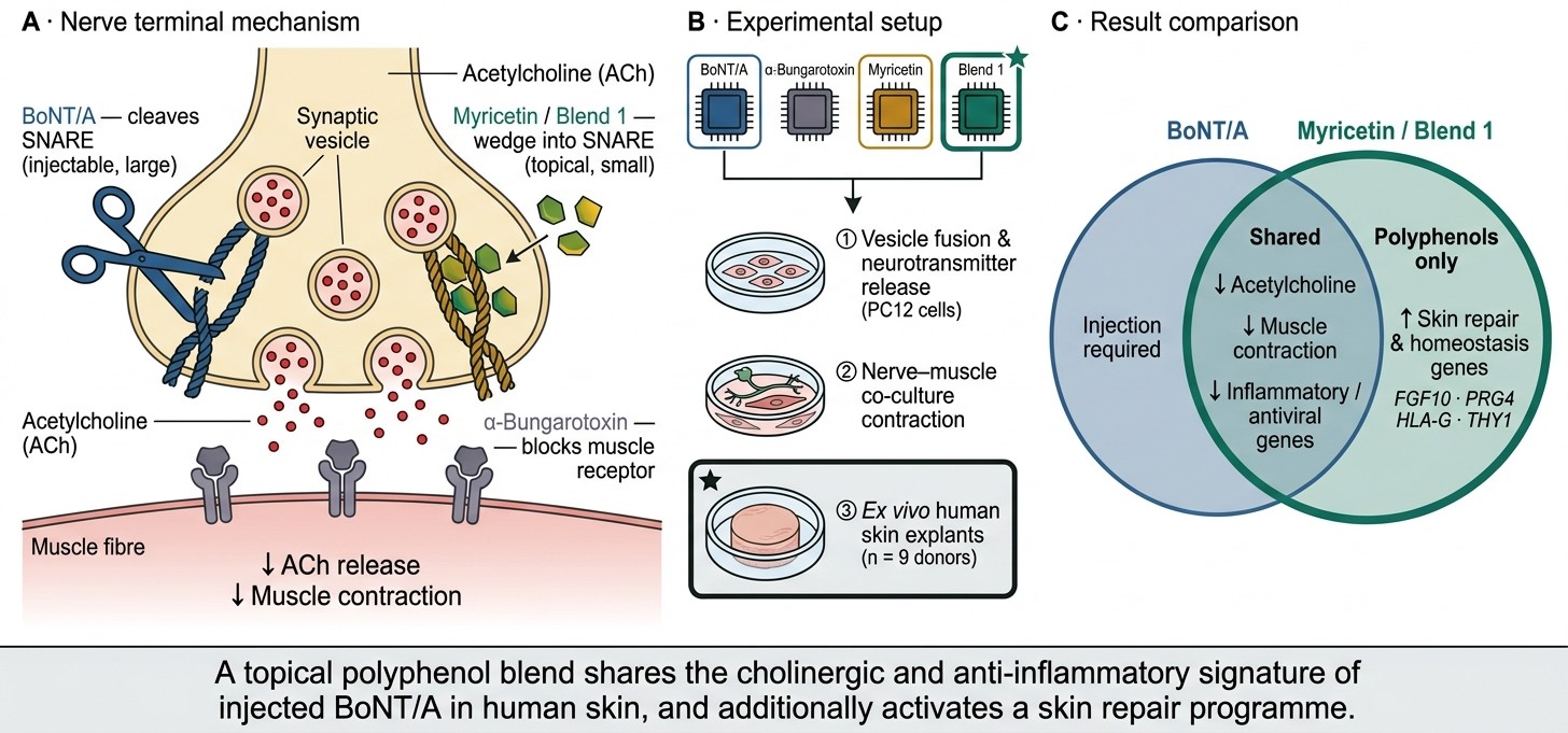
Injected botulinum neurotoxins (BoNT) reduce dynamic wrinkles by cleaving SNARE proteins at the neuromuscular junction; however, their large size mostly limits their use to injectable procedures. Certain polyphenols, including myricetin, have been shown in neuronal systems to reduce acetylcholine (ACh)-induced muscle contraction via inhibition of SNARE complex zippering, suggesting potential for topical use as modulators of skin cholinergic activity. This study aimed to identify a natural extract blend similarly capable of attenuating ACh release followed by characterization of the activity of this extract on human skin explants, alongside reference molecules BoNT/A, α-bungarotoxin, and myricetin. The natural extract blend (“Blend 1”) inhibited vesicle fusion and attenuated norepinephrine release in a cell culture system, inhibited muscle cell contraction within nerve/muscle co-culture, and attenuated ACh release consistent with myricetin and reference neurotoxins within human skin explants. Bulk transcriptomics of treated ex vivo skin explants revealed that BoNT/A, myricetin and Blend 1 all shared a similar downregulated inflammatory and anti-viral gene profile separate from α-bungarotoxin, but that myricetin and Blend 1 shared a large degree of overlap for upregulated genes associated with skin homeostasis and repair that was not observed for either toxin. These data support Blend 1 as a candidate for topical cosmetic use through modulation of BoNT/A-like cholinergic pathways, alongside activation of additional pathways associated with skin homeostasis and repair.
Keywords:
botulinum toxin type A
; myricetin
; polyphenol
; SNARE complex
; acetylcholine
; ex vivo human skin explant
; bulk transcriptomics
; neurocutaneous
; topical cosmetic ingredient
1. Introduction
The primary layers of the skin, the stratum corneum, epidermis and dermis, are subject to the natural process of chronological aging, and extrinsic exposome factors such as UV and pollution which promote skin aging. While changes to these skin layers lead to commonly observed signs of aging such as uneven skin tone and wrinkling, the deeper parts of the skin such as the hypodermis and the underlying skeletal muscle also have critical contributions to skin appearance. Broader changes in skin and facial appearance with age such as skin laxity and deeper wrinkles are more a consequence of changes in hypodermis fat quality and muscle basal tone than changes in the more superficial skin layers, and as such are a common target for anti-aging procedures.
One of the most common and effective anti-aging cosmetic procedures is the injection of botulinum neurotoxins type (BoNT), widely known by trade names such as Botox. Botulinum neurotoxins enter the motor nerve terminals and the neuromuscular junction via receptor-mediated endocytosis, where endosome acidification results in the cleaving of the light chain and transfer into the neuronal cytosol [1]. The primary target for BoNT is the neuronal soluble N-ethylmaleimide-sensitive factor (SNARE) complex, which consists of vesicle-associated membrane protein 2 (VAMP2) on the vesicle membrane, and syntaxin-1 and SNAP-25 on the neuronal plasma membrane. To initiate the process of fusion these proteins interact to form the parallel four-helix protein bundle SNARE complex [2]. While there are various hypotheses to explain subsequent events, the greatest evidence is for a zippering process whereby the complex forms at the N-terminus and progressively zippers towards the membranes, allowing the membranes of the vesicle and neuron to come sufficiently close to initiate hemifusion, allowing for the release of vesicle contents across the synaptic cleft [3]. Different BoNT serotypes can target different parts of the SNARE complex; BoNT serotypes A, C and E all cleave SNAP-25, with BoNT/C also able to cleave syntaxin-1; BoNT serotypes B, D, F and G act on the vesicle-associated VAMP2/synaptobrevin [4]. Of these BoNT serotypes, it is BoNT/A that is commonly used within medical aesthetics clinics. The SNARE inhibition-induced attenuation of ACh by BoNT results in paralysis of the affected muscles, leading to a reduction in wrinkle magnitude when used clinically.
While the clinical efficacy of BoNT/A has led to its widespread adoption within the medical aesthetics industry, the cost and invasive requirement for injections by well-trained medical practitioners reduce patient accessibility. There has therefore been considerable research into potential alternatives focused on peptides and molecules from natural sources that have lower molecular weights and more favorable skin penetration profiles. One molecular family of particular interest is polyphenols, a structurally diverse family of naturally occurring phenols with a history of safe use in cosmetics [5]. Yang and colleagues screened a panel of 39 polyphenolic compounds for their ability to prevent SNARE complex formation and vesicle membrane fusion, identifying several structures capable of preventing SNARE-mediated fusion and thus potentially possessing similar biological activity to BoNT/A family proteins [6]. In contrast to the irreversible cleavage of SNAP proteins (specifically SNAP-25) by BoNT/A, these polyphenols act by preventing SNARE zippering, albeit via slightly different mechanisms. Delphinidin and cyanidin inhibit the N-terminal nucleation of the SNARE complex independently of Ca2+ [7], in contrast to myricetin which binds to the middle region of the SNARE complex, arresting the hemifusion process [6]. Myricetin, a flavonoid found within a diverse range of dietary sources, is of particular interest due to its low molecular weight and high efficacy within these neuronal cell culture systems. Animal studies have demonstrated that injection of myricetin, as well as delphinidin and cyanidin, inhibit muscular contraction [8]. While the magnitude of this response was smaller compared to a reference BoNT/A, the onset of action was faster, a possible consequence of the lack of requirement for cellular processing as seen for BoNT/A, and there was no toxicity observed even at very high concentrations. These data suggest the potential for natural products to mimic the effect of BoNT/A via similar mechanisms, but with a superior safety profile and in a reversible manner.
Within the present study we aimed to explore topical alternatives to BoNT/A by examining the ability of a myricetin and a natural extract blend containing myricetin and structurally similar polyphenols to inhibit neuromuscular contraction in line with a SNARE-mediated mechanism. We then extended these approaches to whole human skin, examining the impact of neurotoxin and natural extract treatment on ACh levels within ex vivo human skin explants, followed by characterization of the biological response of these compounds via bulk transcriptomics analysis.
2. Materials and Methods
2.1. Extract Materials and Extraction
The plant materials used in this study included the fruits and leaves of guava (Psidium guajava), the bulbs of garlic (Allium sativum), the fruits of luffa (Luffa cylindrica), the leaves of black tea (Camellia sinensis), and the flowers of white water lily (Nymphaea alba). Each material was washed thoroughly with purified water, dried, and pulverized into particles ranging from 0.1 to 3 mm in size using a laboratory mill.
For each dried sample, purified water was added at a ratio of 1:100 (w/v), and the mixture was extracted with continuous stirring at room temperature for 3 days. The extract was filtered through Whatman No. 2 filter paper, and the remaining residue was subjected to the identical extraction and filtration procedure two additional times. The combined filtrates were concentrated under reduced pressure using a rotary evaporator (Eyela, Tokyo, Japan) and lyophilized using a freeze dryer to yield dry powders of each respective plant extract.
2.2. Preparation of the Complex Extract Blends
To formulate the complex extract, the five individual plant extracts were precisely weighed and combined in specific proprietary proportions based on their respective extraction yields. The mixture was thoroughly homogenized to ensure uniformity, yielding the final complex extract designated as Blend 1. The prepared Blend 1 was stored at −20 °C until further use in the cellular assays.
2.3. Membrane Fusion Quantification
The inhibitory effects of test compounds on SNARE-mediated membrane fusion were evaluated using an in vitro FRET-based lipid-mixing assay. To prepare the assay components, recombinant SNARE proteins (SNAP-25, Syntaxin 1A, and VAMP-2) were expressed in Escherichia coli Rosetta (DE3), purified via glutathione agarose affinity chromatography, and validated by SDS-PAGE. These proteins were then reconstituted into two distinct populations of liposomes. Unlabeled t-vesicles (POPC/DOPS, 65:35 mol%; 50 mM lipid) were reconstituted with a pre-assembled binary t-SNARE complex (SNAP-25 and Syntaxin 1a, 1:1 molar ratio) at a 1:100 protein-to-lipid ratio. Fluorescently labeled v-vesicles (POPC/DOPS/NBD-PE/Rhodamine-PE, 62:35:1.5:1.5 mol%; 10 mM lipid) were reconstituted with VAMP-2 at a 1:50 ratio. Following extensive dialysis to remove unincorporated proteins, the v-vesicles and t-vesicles were mixed at a 1:9 volume ratio in the presence or absence of the test samples. The fusion reaction was continuously monitored in independent triplicates at 37 °C for 90–120 min using a SpectraMax M2 multimode microplate reader (Molecular Devices, San Jose, CA, USA). The NBD-PE donor was excited at 465 nm (emission 530 nm), and the Rhodamine-PE acceptor emission was simultaneously recorded at 625 nm.
2.4. PC12 Cell Neurotransmitter (Norepinephrine) Secretion Quantification
PC12 cells were seeded at a density of 1 × 10⁵ cells/well in type IV collagen-coated 24-well plates. To induce neuronal differentiation, cells were cultured in low-serum DMEM (1% horse serum) supplemented with 50 ng/mL nerve growth factor (NGF) for 5-7 days until neurite outgrowth was confirmed. For the secretion assay, differentiated cells were washed and pre-incubated in Locke’s buffer (154 mM NaCl, 5.6 mM KCl, 3.6 mM NaHCO3, 5.6 mM glucose, 0.1% BSA, pH 7.4) for 15 min at 37° C. Cells were then treated with test samples for 30 min, followed by stimulation with NGF (50 ng/mL) in Locke’s solution for an additional 15 min. Conditioned media were collected, centrifuged at 300 × g for 5 min to remove cellular debris, and analyzed using a Norepinephrine ELISA kit (Abcam, Cambridge, UK, Cat No. ab287789) according to the manufacturer’s instructions. Absorbance was measured at 450 nm using a microplate reader, and the percentage of inhibition was calculated relative to the NGF-stimulated vehicle control.
2.5. Nerve and Muscle Cell Depolarization Co-Culture Evaluation
Human skeletal muscle cells (Lonza, Basel, Switzerland) were seeded in flasks as previously published [9] and maintained cultivated with SkGM-2 skeletal Muscle Cell Growth Medium BulletKit as per manufacturer’s instructions. When cells reached about 80% confluency, muscle cells were transferred to 96 wells plates in the proliferation medium. After 7 days of incubation, medium was changed to differentiation medium. In these conditions muscle cells form fibers. The correct formation of fibers was validated morphologically before proceeding to the co-culture step. Motor neurons derived from hiPS cells from commercial origin were thawed and transferred in the 96 wells plates containing muscle cells in a medium containing 2/3rds of motor neurons commercial medium (iCell Motor Neuron Kit, Fujifilm Cellular Dynamics; Madison WI, USA) and 1/3rds of muscle differentiation medium. The Co-culture was maintained 10 days and culture medium was changed every two or three days. In these conditions motor neurons maturate and form junction with muscle fibers. Spontaneous contractions are observed within 5 days. Co-culture was observed, and localization of contractile muscle fibers was registered. Movies of co-culture were recorded during 1 minute before treatment with an In Cell 2200 automated microscope (General Electric, Boston, MA, USA). Cell cultures were then treated with reference compound α-bungarotoxin at 2 µM or indicated compounds and contractions recorded for 2 minutes. The cultures were then incubated again at 37 °C and 5% CO2 for 24 hours before a further 2-minute recording. The contraction frequency was then calculated from the 2-minute recording. Six replicates were analyzed per condition. Data is representative of two independent experiments.
2.6. Skin Explant Source and Ethical Approval
Human skin was obtained from female donors immediately after abdominal or breast surgery, following written informed consent. Ethical approval for this study was granted by the Institutional Review Board (IRB) of Seoul National University Bundang Hospital (Seongnam, Republic of Korea; approval number B-2201-732-302), and all procedures were conducted in accordance with the Declaration of Helsinki.
2.7. Skin Explant Culture and Treatment
Upon collection, the explants were cultured in Dulbecco’s Modified Eagle Medium (DMEM) supplemented with 10% fetal bovine serum (FBS), 1% antibiotics, 10 µg/mL insulin, 10 ng/mL hydrocortisone, and 2 mM L-glutamine. The explants were maintained at 37 °C in a humidified atmosphere of 5% CO2.
For pharmacological treatments, the explants were treated with botulinum toxin type A (Hugel, Chuncheon, Republic of Korea) at 1 and 10 U/mL, α-bungarotoxin (Sigma-Aldrich, St. Louis, MO, USA) at 1 and 3 µM, Blend 1 at 5 and 15 µg/mL, or myricetin at 0.45 and 4.5 µg/mL. The treatments were administered by replacing the culture medium with fresh medium containing the respective agents once daily for three consecutive days. Explants treated with an equivalent volume of DMSO served as vehicle controls.
All experiments were performed using independent skin samples from nine different donors (biological replicates, n = 9), processed in three independent experimental runs (three donors per run). For each donor, three explants were allocated per treatment condition (technical replicates, n = 3).
2.8. Acetylcholine Quantification
Following the treatments, the explants were washed with ice-cold PBS and homogenized in the assay buffer provided in the Choline/Acetylcholine Assay Kit (Abcam, Cambridge, UK, Cat No. ab65345). The tissue homogenates were centrifuged at 12,000 rpm for 5 min at 4 °C to pellet the cellular debris. The resulting supernatants were collected and subjected to acetylcholine quantification strictly following the manufacturer’s protocol. Fluorescence intensity was measured at Ex/Em = 535/587 nm using a SpectraMax 190 microplate reader. The acetylcholine concentrations were extrapolated using a standard curve generated concurrently. To account for variations in tissue size, the measured acetylcholine levels were normalized to the total protein concentration of each sample, which was determined using a BCA protein assay kit. All biochemical measurements were performed in technical triplicates.
2.9. Bulk Transcriptomics of Human Skin Explants
Human skin explants were treated with the respective compounds or a vehicle control for three days. The experiment comprised five groups (one vehicle and four treatment groups). The entire experiment was independently repeated three times, each with skin samples from three different donors, resulting in nine biological replicates (n = 9) per group and a total of 45 samples. Following the treatments, the explants were rinsed with ice-cold PBS, immediately immersed in RNAlater stabilization solution (AM7020), and shipped to Macrogen (Seoul, Republic of Korea). Total RNA extraction, library construction, and sequencing were performed by Macrogen using their standard protocols. RNA quality and quantity were evaluated using an Agilent 2100 Bioanalyzer, and only samples exhibiting an RNA integrity number (RIN) > 7.0 were utilized for library preparation.
Sequencing libraries were prepared using the TruSeq Stranded mRNA Library Prep Kit (Illumina, San Diego, CA, USA), and sequencing was subsequently performed on an Illumina NovaSeq X platform to generate 150 bp paired-end reads. For transcriptomic data analysis, raw sequencing reads were quality-checked, aligned to the human reference genome (GRCh38), and quantified using Macrogen’s standard bioinformatics pipeline. For data pre-processing, low-expressed genes were filtered out to improve statistical power. Normalization and differential expression analysis were subsequently performed using DESeq2, which internally applies median-of-ratios normalization.
Differentially expressed genes (DEGs) between the treated and vehicle control groups were identified using the negative binomial Wald test (nbinomWaldTest) via the DESeq2 R package, with sequencing batch included as a covariate in the design formula (~batch + treatment) to account for potential batch effects. Genes satisfying the criteria of Log2 fold change 0.585 and adjusted p-value < 0.05 were considered statistically significant. Gene Ontology (GO) enrichment analysis and network analysis of the identified DEGs was done using the String Database v12.0 (https://string-db.org/). Volcano plots were created using BioSketchArt Volcano Plot Maker (https://biosketch.art/volcano-plot). The upset plot was created using ChiPlot tools (https://www.chiplot.online/upset_plot.html), and the heatmap was created using Heatmapper (https://heatmapper.ca/).
2.10. Statistical Analysis
Data from the membrane fusion, norepinephrine secretion, muscle contraction and acetylcholine quantification assays are presented as the mean ± standard deviation unless stated otherwise in the corresponding figure legend, and were compared using one-way ANOVA with Holm–Sidak correction for multiple comparisons. The number of replicates for each experiment is given in the corresponding figure legend. Differential expression analysis of the RNA sequencing data was performed as described in Section 2.9. A p-value < 0.05 was considered statistically significant. Statistical analyses were performed using GraphPad Prism version 11 (GraphPad Software, Boston, MA, USA).
3. Results
3.1. Natural Extract Blend Evaluation for Inhibition of Vesicle Fusion and Norepinephrine Release
Previous reports have indicated that myricetin inhibits vesicle fusion and norepinephrine (NE) release within cell systems, however pure polyphenol structures are often not permissible for cosmetic use. To identify extracts that possess similar SNARE-inhibition profiles to myricetin, we evaluated a series of preparations from natural sources that contain myricetin and associated polyphenolic structures and validated their biological activity for membrane fusion within PC12 cells. Since PC12 cells serve as a highly established in vitro model for studying SNARE-dependent exocytosis, we utilized NE secretion as a functional surrogate marker to evaluate general vesicle fusion inhibition. While different preparations were able to inhibit membrane fusion in our system, one blend, which we termed Blend 1, inhibited membrane fusion to the same extent as myricetin in a dose-dependent manner (Figure 1a) and induced the greatest level of NE release inhibition (Figure 1b). Our data demonstrates that our extract preparation Blend 1 can inhibit membrane fusion and NE release to a comparable degree as myricetin and BoNT/A.
3.2. Neurotoxins and Natural Extracts Inhibit Muscle Cell Depolarization
Previous reports have demonstrated myricetin and similar polyphenol structures to inhibit presynaptic membrane fusion therefore reduce muscle contraction, so we examined the ability of both myricetin and natural extract to inhibit muscle contraction frequency within a co-culture system of iPSC-derived neurons and skeletal muscle cells. In this system the neuronal cells self-arrange to form synaptic connections with the muscle cells, resulting in a basal level of muscle contraction. The cultures were treated with either positive control α-bungarotoxin, a competitive antagonist of the nicotinic acetylcholine receptor (nAChR), our reference polyphenol myricetin, or Blend 1. None of the compounds at any concentration tested showed any evidence of toxicity (data not shown). When the cell cultures were treated for 2 hours only Blend 1 at the highest concentration tested showed a reduction in muscle contraction (Figure 2a). After 24 hours post-treatment however, all treatments examined exhibited a significant decrease in muscle contraction frequency, and in some cases there was a near complete abolition of muscle contraction, suggesting almost total inhibition of neuromuscular junction signaling (Figure 2b). These data are consistent with previous reports of myricetin inhibiting vesicle fusion, and demonstrate the same physiological properties for Blend 1.
3.3. BoNT/A and Natural Extracts Reduce ACh Levels within Human Skin Explants
We next examined if this mechanism extended into skin by targeting ACh, the primary physiological transmitter for muscle contraction. To this end, we established an ACh release assay system whereby human skin explants were cultured in media and treated with positive controls BoNT/A or α-bungarotoxin, and myricetin and Blend 1 for either a single treatment, or three consecutive treatments, followed by ACh quantification within homogenized whole skin explants. Following a single day treatment we saw a decrease in ACh levels within cultures treated with either myricetin and Blend 1, but not BoNT/A or α-bungarotoxin (Figure 3a), however when treatment was extended to three consecutive days BoNT/A treatment resulted in a decrease in ACh, while both myricetin and Blend 1 continued to reduce ACh release (Figure 3b). As anticipated α-bungarotoxin did not decrease ACh release, as this toxin acts post-synaptically on nicotinic ACh receptors rather than inhibiting ACh release in the manner of BoNT/A. It increased ACh however, which may be explained by a transient increase in ACh due to reduced α7-nAChR binding or dysregulated ACh release negative feedback. These data demonstrate that the mechanisms we see behind the reduction of muscle contraction observed within our study similarly extends to skin.
3.4. Ex Vivo Skin Explants Treated with BoNT/A and Blend 1 Share Overlapping Transcriptomic Profiles
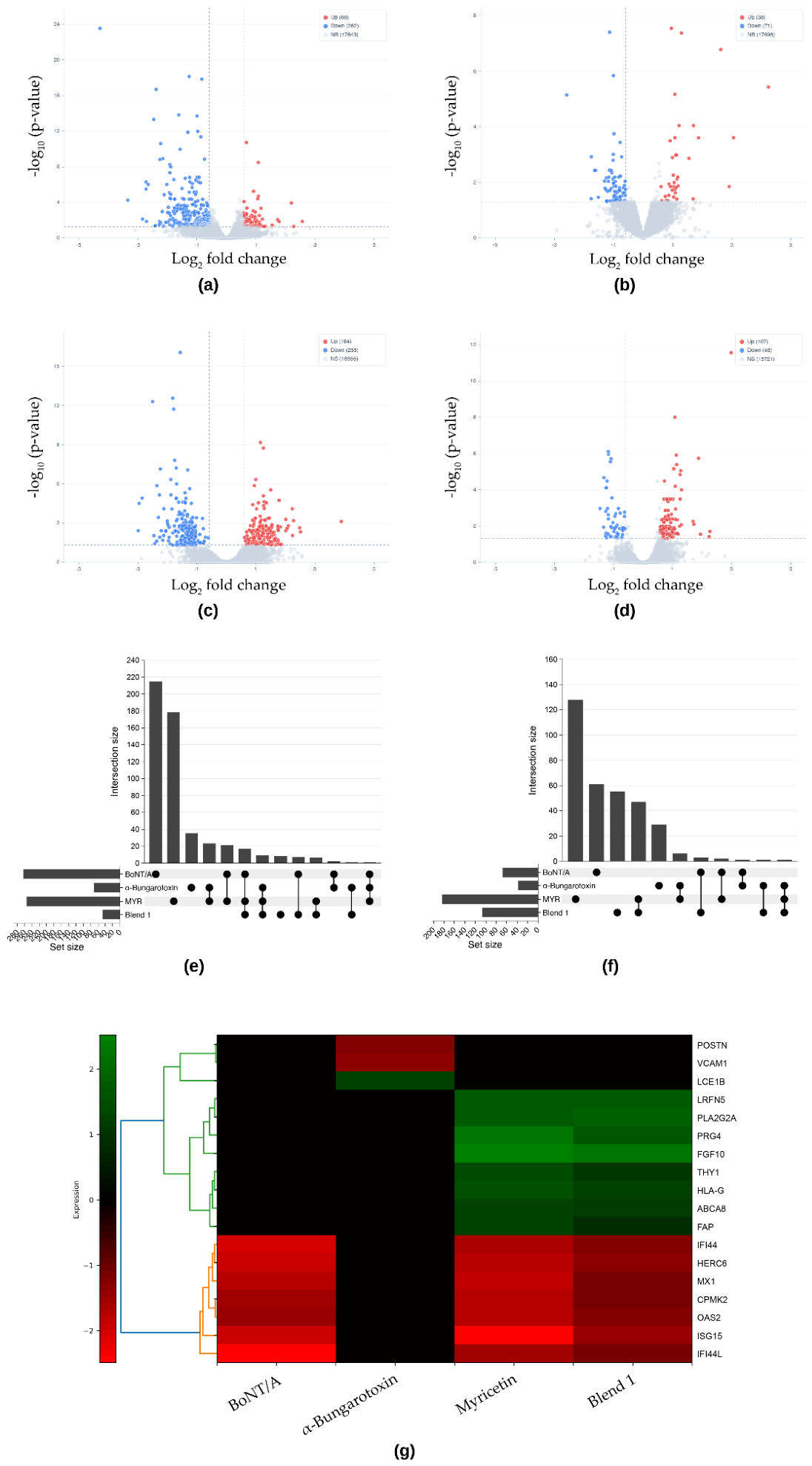
Transcriptomic analysis of treated skin explants showed BoNT/A treatment induced 328 differentially expressed mRNAs (66 up, 262 down) compared to untreated explants (absolute log2 fold change >0.58, adjusted p-value <0.05), α-bungarotoxin 109 genes (38 up, 71 down), myricetin 439 genes (184 up, 255 down) and Blend 1 155 genes (107 up, 48 down) (Figure 4A–D). Gene Ontology enrichment analysis for BoNT/A-downregulated pathways revealed impact on pathways associated with viral processes and defense against virus, consistent with dampening of virus-associated innate immune responses and a reduction of a basal inflammation/stress response state (Figure S1A). This may be particularly prominent within a skin explant system, where injury response and tissue stress may be enhanced. Such pathways have a strong neuronal component [10] that would be a natural target for BoNT/A-induced attenuation. Our α-bungarotoxin-treated explants however showed a very different transcriptomic signature, with pathways associated with vascular development and angiogenesis being downregulated (Figure S1B). Despite both agents acting on the neuromuscular junction, there was very limited overlap in downregulated genes, with only 2 of the 262 genes downregulated by BoNT/A also impacted by α-bungarotoxin treatment (Figure 4E) and only a single upregulated gene shared between conditions (Figure 4F). There were limited functional networks upregulated by BoNT/A treatment, precluding GO analysis and highlighting that BoNT/A treatment within skin explants at the timepoint measure was primarily suppressive. α-Bungarotoxin treatment upregulated processes associated with keratinization, epithelial cell development and differentiation (Figure S2A), demonstrating a moderate impact of epidermal cornification and skin barrier adaptation.
Treatment of skin explants with myricetin shared downregulated genes with both BoNT/A (21 of 255 genes) and α-bungarotoxin (23 of 255 genes)-treatments (Figure 4E). Myricetin-treated samples demonstrated significant similarities to α-bungarotoxin as demonstrated by GO analysis, with shared impact on stromal and vascular pathways (Figure S1C). At the DEG level however myricetin shares a strong inflammatory/interferon-suppression core like BoNT/A-treated explants, including IFI44L, ISG15, OAS2, CMPK2, MX1, IFI6 and related innate immune genes. (Figure S3 and Figure S5) consistent with a shared presynaptic inhibitory signature. Upregulated genes pathways were significantly different however, with myricetin pathways showing similarities to α-bungarotoxin in GO analysis, with overlapping terms for keratinization, differentiation and epidermis development (Figure S2 A&B). However, myricetin treatment induced a broader induced program that included both epithelial-associated genes and stromal/remodeling-related transcripts such as FGF10, CXCL12, LYVE1, and THY1 (Figure S5B), compared to the more barrier/cornification signature of α-bungarotoxin (Figure S4B).
Blend 1 treatment shares considerable overlap with both BoNT/A and myricetin, with 17 of the 48 downregulated Blend 1 genes also being downregulated with both BoNT/A and myricetin (Figure 4E). GO analysis similarly reveals a similar pathway modulation signature to BoNT/A, focused on defense response to virus and interferon-associated pathways (Figure S1D). The upregulated transcriptomic response was much more like myricetin than either BoNT/A or α -bungarotoxin, with 47 of the 107 genes upregulated by Blend 1 also impacted by myricetin, compared to only 3 and 1 genes when compared to BoNT/A & α-bungarotoxin respectively (Figure 4F). Of particular interest is Blend 1’s impact on stromal/vascular remodeling genes such as FGF10, CXCL12, LYVE1, and THY1 (Figure S6A), which are associated with skin homeostasis [11].
To compare transcriptional responses across treatment groups, we generated a heatmap of 18 representative DEGs selected to capture three distinct biological programs (Table S1, Figure 4G). A cluster of interferon-stimulated genes (ISGs), including ISG15, MX1, IFI44L and OAS2, were all downregulated in BoNT/A, myricetin and Blend 1 treated explants, but was not detected among α-bungarotoxin DEGs, defining a shared innate immune suppression signature. In contrast, α-bungarotoxin showed a distinct response characterized by downregulation of VCAM1 and POSTN and upregulation of the terminal differentiation marker LCE1B, consistent with a postsynaptic response profile [12,13]. Myricetin and Blend 1 additionally shared upregulation of stromal and immune-associated genes, including FGF10, PRG4 and HLA-G, that were absent from both neurotoxin groups, defining a polyphenol-associated skin homeostasis and repair signature. Together, these patterns support the interpretation that BoNT/A, myricetin and Blend 1 possess a shared inflammatory suppression program, whereas myricetin and Blend 1 additionally induce transcriptional response in skin consistent with skin regeneration.
4. Discussion
Within the present study we examined the ability of a natural polyphenol complex, polyphenol myricetin and reference toxins to inhibit neural activation within an in vitro model of neuromuscular contraction and extended these findings to examine ACh release induced within ex vivo skin. We also extended these data to transcriptomic signatures of these treatments within ex vivo skin explants. Our data supports the potential for a natural extract preparation to improve skin quality not only via neuromuscular junction pathways, but likely through pathways likely independent of the neuromuscular junction.
The cholinergic system in skin is comprised of both neuronal and non-neuronal components. The sympathetic cholinergic nerve fibers of the skin neuronal system primarily act on the eccrine sweat glands but also influence the cutaneous vasculature and sensory C-fibers [14]. The non-neuronal cholinergic system is more extensive in skin and operates independent of neuronal inputs. Keratinocytes synthesize and release ACh, and express both nicotinic and muscarinic ACh receptors which act in a mutually antagonistic manner to regulate cutaneous ACh function [15]. Fibroblasts express α7-nAChR which regulates ECM production and growth factor responsiveness [16]. Inhibition of nicotinic ACh receptors by a small peptide increased keratinocyte viability, further demonstrating the multifaceted nature of ACh within skin cells [17]. Cutaneous immune cells such as macrophages, T cells and dendritic cells also express nAChRs. The ubiquity of cholinergic pathways in the skin makes it an interesting target for modulating skin quality and inflammatory responses by agents such as BoNT/A beyond utilization solely as injectables for anti-wrinkle activity.
Research into topical uses of BoNT/A have revealed promising results. A preparation of BoNT/A in a topical gel formulation reduced the prominence of lateral canthal area lines compared to the vehicle control in a randomized clinical study of 17-19 adult patients [18], while topical application of BoNT/A improved after ablative CO2 fractional laser of lateral periorbital wrinkles compared to laser treatment alone within a small cohort of female patients [19]. The authors of this latter study note that in their previous pilot study topical BoNT/A alone had no effect, suggesting that enhanced delivery systems that improve skin penetration are likely to be extremely important given the very large size of BoNT/A proteins. This has led to recent studies examining the effect of topical BoNT/A in conjunction with microneedling to allow access of the BoNT/A protein into the epidermis and dermis. While these studies remain relatively small scale, there is promising evidence for the use of topical BoNT/A in conjunction with microneedling or intradermal droplet injection for reduction in crow’s feet wrinkles appearance [20] and reducing atrophic post-acne scar appearance [21]. Nevertheless, the extremely large size of BoNT/A proteins has led to the development of smaller peptides to inhibit the SNARE complex. The most wide-spread example of this approach is that of hexapeptide Ac-EEMQRR-NH2, commonly known by the brand name Argireline or acetyl hexapeptide-8. Argireline mimics the aa12-17 sequence of SNAP-25 and therefore blocks SNARE complex formation and resultant neurotransmitter release [22]. Dipeptide diaminobutyroyl benzylamide, more popularly known by its commercial trademark of Syn-ake®, was developed to possess the same biological activity as Waglerin-1, a protein originally isolated from viper venom which acts as a nicotinic ACh receptor antagonist [23]. Topical administration of Dipeptide diaminobutyroyl benzylamide in formula with additional peptides and antioxidants improved skin wrinkling [24]. A similar approach has been taken using a neuromuscular Nav1.4 Na+ channel blocking μ-Conotoxin [25]. Despite these impressive in vitro data and clinical results within small-scale trials, such peptides still have relatively high molecular weights. Within the present study we examined natural structures and extracts with the aim of identifying solutions with potentially more favorable skin penetration profiles and are of natural sources.
Our PC12 cell line system indicated that Blend 1 of our extract gave the most promising results for SNARE inhibition as determined by attenuation of membrane fusion and NE release. We then utilized a more physiological system by co-culturing iPSC-derived motor neurons together with muscle cells to measure muscle contraction frequency as a direct functional assay for the consequences of ACh blockade. Our data demonstrates both myricetin and Blend 1 extract inhibit muscle contraction in line with the results observed for α-bungarotoxin, consistent with the previously reported data for myricetin [6,8,26,27]. While these data suggest that myricetin contributes to the neuronal effects of Blend 1, such natural extract preparations are typically a complex mixture of structures and are likely to contain many different polyphenols. Indeed, myricetin is not the only flavonoid possessing the ability to inhibit SNARE zippering, as both delphinidin and cyanidin inhibit muscular contraction [8]. It is therefore possible that there are additional structures within our extract that contribute to the inhibition of muscle contract we observed within our system.
While our data supports existing studies of the biological activity of polyphenols on neuromuscular junction activity inhibition, there have been limited studies to date extending this approach specifically to skin. To examine the impact of our molecules of interest we extended our study of human skin, measuring ACh release within cultured human skin explants. The inhibition of ACh release within this system by both BoNT/A, myricetin and Blend 1 is consistent with our data obtained within the neuronal system, suggesting that SNARE-mediated inhibition of vesicle fusion and the resultant decrease in neurotransmitter release is responsible for the inhibition of muscle cell depolarization within our system. These data are inconsistent with the findings of Schlereth and co-workers (2005), who found that BoNT injected into the skin of volunteers did not result in a decrease of ACh levels as determined by HPLC analysis of dermal perfusion effluent that was run through the skin [28]. They concluded that a significant proportion of the ACh they measured was from non-neuronal sources, and thus not impacted by BoNT inhibition of exocytotic ACh release. Our system is considerably different, however. We performed ACh quantification on whole skin homogenates which captures whole skin ACh pools including vesicular and intracellular ACh, rather than only the extracellularly fraction measured within the microdialysis. These considerable differences in experimental systems may explain our discordant results. Interestingly α-bungarotoxin increased ACh within our skin explant system. We do not have data to explain this observation, although given the role of nAChR of ACh release regulation [29] interruption of feedback mechanisms regulating ACh release by blockade of AChRs by α-bungarotoxin would be consistent with our observations.
Having established the ability of our natural extract blend to modulate ACh release, we wanted to examine the physiological consequences of these treatments within human skin via bulk transcriptomics. Our analysis revealed striking differences across treatment groups. At the timepoint measured, BoNT/A was predominantly suppressive, with substantially more downregulated than upregulated DEGs. GO analysis identified marked suppression of pathways related to antiviral defense and innate immune activation, particularly interferon-stimulated genes (ISGs). Although initially unexpected, this pattern may reflect the stressed nature of the ex vivo skin explant system, in which surgical excision and culture are likely to elevate basal inflammatory signaling. Under these conditions compounds with anti-inflammatory or presynaptic inhibitory activity may reduce this background response. This interpretation is consistent with previous reports describing anti-inflammatory effects of BoNT/A through both neuronal and non-neuronal mechanisms [30,31]. Injection of BoNT/A resulted in a decrease in TNCB-induced inflammation within an animal model of atopic dermatitis [32], while BoNT/A treatment of rosacea patients resulted in clinically significant improvements in erythema scores [33]. This signature is shared with both myricetin and Blend 1, however, consistent with the shared pre-synaptic activity between both compounds and BoNT/A. SNARE family proteins VAMP-3, SNAP-29 and syntaxin-4 have all been implicated in cytokine release from primary human keratinocytes, with VAMP-3 identified as a target of particular interest for reducing inflammation within atopic dermatitis lesions [34]. These data all link the SNARE complex and therefore compounds that act on the complex to be associated with immune suppression. That α-bungarotoxin did not exhibit such an effect despite potent blockade of α7-nAChR suggests that the ISG arm of the cutaneous cholinergic system is mediated through an α7-nAChR-independent mechanism.
Where myricetin and Blend 1 diverge from BoNT/A is the upregulated DEG profile. BoNT/A treatment is overwhelmingly suppressive with the large majority of its DEGs being downregulated, and it shares no upregulated gene signature with the polyphenol treatments. We observed 47 genes upregulated exclusively by myricetin and Blend 1 which are consistent with a skin homeostasis and repair transcriptional program that these polyphenols activate independent of their neuronal effects. This signature encompasses multiple aspects of skin tissue homeostasis and repair such as FGF10 and FAP (both implicated in fibroblast and keratinocyte signaling and tissue repair [35,36]), PRG4 (anti-fibrotic ECM organization associated with dermal remodeling [37]), HLA-G (immune tolerance induction [38]), and THY1 (fibroblast activation, consistent with a pro-regenerative fibroblast state [39]). Myricetin protects against UV-B induced damage and wrinkling in a mouse model of photoaging [40], and protects human keratinocytes from UV-B-induced photodamage [41], consistent with our observations. These data collectively indicate that myricetin and Blend 1 not only recapitulate the innate immune-suppressive signature of BoNT/A through their shared presynaptic cholinergic activity but also possess a skin homeostatic and repair program profile that BoNT/A does not. These dual pathways suggest these molecules may offer a functional advantage over BoNT/A for topical skin use.
5. Conclusions
Within the present study, we demonstrate that BoNT/A reduces ACh release in human skin explants, consistent with our data for neuronal neurotransmitter release and neuromuscular junction activity. We also identified a transcriptomic signature of human skin explants treated with BoNT/A which shares a partial overlap with the profiles observed following myricetin and Blend 1 treatment, consistent with a shared modulation of neurocutaneous signaling. We also demonstrate Blend 1 impacts additional transcriptomic pathways associated with skin homeostasis and repair-related process beyond those shared with BoNT/A, collectively suggesting this extract holds promise as a topical treatment to improve cosmetic skin quality.
Supplementary Materials
The following supporting information can be downloaded at the website of this paper posted on Preprints.org: Figure S1: GO analysis for downregulated pathways; Figure S2: GO analysis for upregulated pathways; Figure S3: Network analysis of DEGs in BoNT/A-treated explants; Figure S4: Network analysis of DEGs in α-bungarotoxin-treated explants; Figure S5: Network analysis of DEGs in myricetin-treated explants; Figure S6: Network analysis of DEGs in Blend 1-treated explants; Table S1: Gene list for heatmap clustering analysis; Table S2: Differentially expressed genes in BoNT/A-treated explants; Table S3: Differentially expressed genes in α-bungarotoxin-treated explants; Table S4: Differentially expressed genes in myricetin-treated explants; Table S5: Differentially expressed genes in Blend 1-treated explants.
Author Contributions
Conceptualization, H.K., R.J.B., R.Y.K. and S.K.; methodology, H.K., J.W.M., R.J.B., and R.Y.K.; formal analysis, H.K., F.J., and R.J.B.; investigation, H.K., F.J., S.B., J.W.M. and Y.N.; resources, D.G.L., Y.M.H., J.W.M., and C.Y.H.; writing—original draft preparation, R.J.B.; writing—review and editing, R.J.B., H.K., F.J., and R.Y.K.; supervision, R.J.B. and R.Y.K.; project administration, R.Y.K., S.K. and C.P. All authors have read and agreed to the published version of the manuscript.
Funding
This research was funded by L’Oréal Research & Innovation and Cosmax BTI; the funders had a role in the design of the study; in the collection, analysis and interpretation of the data; in the writing of the manuscript; and in the decision to publish the results, through the participation of their employees as authors, as detailed in the Author Contributions section.
Institutional Review Board Statement
The study was conducted in accordance with the Declaration of Helsinki and approved by the Institutional Review Board of Seoul National University Bundang Hospital, Seongnam, Republic of Korea (protocol code B-2201-732-302, approved on 23 December 2021).
Informed Consent Statement
Written informed consent was obtained from all donors of the human skin samples used in this study.
Data Availability Statement
The differentially expressed gene lists supporting the conclusions of this article are provided in the Supplementary Materials (Table S2–S5). The underlying raw RNA sequencing data are not publicly available because they were generated from human skin explants under donor consent that does not permit the public release of individual-level sequence data, and because they relate to a proprietary cosmetic ingredient under commercial development. Requests to access the processed datasets should be directed to the corresponding author.
Acknowledgments
The authors thank Teng Yao of L’Oréal Research and Innovation China for her valuable advice on bulk transcriptomics. During the preparation of this manuscript, the authors used Google Gemini 2.5 Flash Image (“Nano Banana”) for the purpose of generating draft artwork for the graphical abstract. The authors have reviewed and edited the output and take full responsibility for the content of this publication. No generative AI tools were used to generate, analyze or interpret the experimental data, or to write the text of this manuscript.
Conflicts of Interest
H.K., Y.M.H., Y.N., D.G.L. and C.P. are employees of Cosmax BTI. S.K. is an employee of Cosmax. J.W.M. is an employee of GFC. R.J.B., F.J., S.B. and R.Y.K. were all employees of L’Oréal Research & Innovation when the study was initiated. C.Y.H. is a faculty member of Seoul National University Bundang Hospital and declares no competing financial interests. Blend 1 is a proprietary material under commercial development. This research was funded by L’Oréal Research & Innovation; the funder had a role in the design of the study; in the collection, analysis and interpretation of the data; in the writing of the manuscript; and in the decision to publish the results, through the participation of its employees as authors, as detailed in the Author Contributions section.
References
- Nigam, P.K.; Nigam, A. Botulinum Toxin. Indian J. Dermatol. 2010, 55, 8–14. [Google Scholar] [CrossRef] [PubMed]
- Gardner, A.P.; Barbieri, J.T. Light Chain Diversity among the Botulinum Neurotoxins. Toxins 2018, 10, 268. [Google Scholar] [CrossRef] [PubMed]
- Lou, X.; Shin, Y.-K. SNARE Zippering. Biosci. Rep. 2016, 36, e00327. [Google Scholar] [CrossRef] [PubMed]
- Pirazzini, M.; Rossetto, O.; Eleopra, R.; Montecucco, C. Botulinum Neurotoxins: Biology, Pharmacology, and Toxicology. Pharmacol. Rev. 2017, 69, 200–235. [Google Scholar] [CrossRef] [PubMed]
- Bharadvaja, N.; Gautam, S.; Singh, H. Natural Polyphenols: A Promising Bioactive Compound for Skin Care and Cosmetics. Mol. Biol. Rep. 2023, 50, 1817–1828. [Google Scholar] [CrossRef] [PubMed]
- Yang, Y.; et al. Dissection of SNARE-Driven Membrane Fusion and Neuroexocytosis by Wedging Small Hydrophobic Molecules into the SNARE Zipper. Proc. Natl. Acad. Sci. USA 2010, 107, 22145–22150. [Google Scholar] [CrossRef] [PubMed]
- Yang, Y.; et al. SNARE Zippering Is Hindered by Polyphenols in the Neuron. Biochem. Biophys. Res. Commun. 2014, 450, 831–836. [Google Scholar] [CrossRef] [PubMed]
- Yang, Y.; et al. SNARE-Wedging Polyphenols as Small Molecular Botox. Planta Med. 2012, 78, 233–236. [Google Scholar] [CrossRef] [PubMed]
- Braun, S.; Croizat, B.; Lagrange, M.-C.; Warter, J.-M.; Poindron, P. Neurotrophins Increase Motoneurons’ Ability to Innervate Skeletal Muscle Fibers in Rat Spinal Cord–Human Muscle Cocultures. J. Neurol. Sci. 1996, 136, 17–23. [Google Scholar] [CrossRef] [PubMed]
- Filtjens, J.; et al. Nociceptive Sensory Neurons Promote CD8 T Cell Responses to HSV-1 Infection. Nat. Commun. 2021, 12, 2936. [Google Scholar] [CrossRef] [PubMed]
- Liu, Z.; et al. Spatiotemporal Single-Cell Roadmap of Human Skin Wound Healing. Cell Stem Cell 2025, 32, 479–498.e8. [Google Scholar] [CrossRef] [PubMed]
- Saeed, R.W.; et al. Cholinergic Stimulation Blocks Endothelial Cell Activation and Leukocyte Recruitment during Inflammation. J. Exp. Med. 2005, 201, 1113–1123. [Google Scholar] [CrossRef] [PubMed]
- Arredondo, J.; et al. Central Role of α7 Nicotinic Receptor in Differentiation of the Stratified Squamous Epithelium. J. Cell Biol. 2002, 159, 325–336. [Google Scholar] [CrossRef] [PubMed]
- Kurzen, H.; Schallreuter, K.U. Novel Aspects in Cutaneous Biology of Acetylcholine Synthesis and Acetylcholine Receptors. Exp. Dermatol. 2004, 13 (Suppl. 4), 27–30. [Google Scholar] [CrossRef] [PubMed]
- Grando, S.A. Muscarinic Receptor Agonists and Antagonists: Effects on Keratinocyte Functions. Handb. Exp. Pharmacol. 2012, 208, 429–450. [Google Scholar] [CrossRef] [PubMed]
- Stegemann, A.; Flis, D.; Ziolkowski, W.; Distler, J.H.W.; Steinbrink, K.; Böhm, M. The α7 Nicotinic Acetylcholine Receptor: A Promising Target for the Treatment of Fibrotic Skin Disorders. J. Investig. Dermatol. 2020, 140, 2371–2379. [Google Scholar] [CrossRef] [PubMed]
- Breton, L.; De Azevedo Calderon, L.; Zanchi, F.; Ferreira Francisco, A.; De Vecchi, R. Bioactive Peptides Having High Binding Affinity to Human Muscular Nicotinic Acetylcholine Receptor. European Patent EP3787660A1, 10 March 2021. [Google Scholar]
- Brandt, F.; O’Connell, C.; Cazzaniga, A.; Waugh, J.M. Efficacy and Safety Evaluation of a Novel Botulinum Toxin Topical Gel for the Treatment of Moderate to Severe Lateral Canthal Lines. Dermatol. Surg. 2010, 36 (Suppl. 4), 2111–2118. [Google Scholar] [CrossRef] [PubMed]
- Mahmoud, B.H.; Burnett, C.; Ozog, D. Prospective Randomized Controlled Study to Determine the Effect of Topical Application of Botulinum Toxin A for Crow’s Feet after Treatment with Ablative Fractional CO2 Laser. Dermatol. Surg. 2015, 41, S75–S81. [Google Scholar] [CrossRef] [PubMed]
- Cao, Y.; et al. A Comparative In Vivo Study on Three Treatment Approaches to Applying Topical Botulinum Toxin A for Crow’s Feet. BioMed Res. Int. 2018, 2018, 6235742. [Google Scholar] [CrossRef] [PubMed]
- Ebrahim, H.; Elardi, A.; Khater, S.; Morsi, H. Successful Topical Application of Botulinum Toxin after Microneedling versus Microneedling Alone for the Treatment of Atrophic Post-Acne Scars: A Prospective, Split-Face, Controlled Study. J. Clin. Aesthet. Dermatol. 2022, 15, 26–31. [Google Scholar] [PubMed]
- Blanes-Mira, C.; et al. A Synthetic Hexapeptide (Argireline) with Antiwrinkle Activity. Int. J. Cosmet. Sci. 2002, 24, 303–310. [Google Scholar] [CrossRef] [PubMed]
- Debono, J.; Xie, B.; Violette, A.; Fourmy, R.; Jaeger, M.; Fry, B.G. Viper Venom Botox: The Molecular Origin and Evolution of the Waglerin Peptides Used in Anti-Wrinkle Skin Cream. J. Mol. Evol. 2017, 84, 8–11. [Google Scholar] [CrossRef] [PubMed]
- Trookman, N.S.; Rizer, R.L.; Ford, R.; Ho, E.; Gotz, V. Immediate and Long-Term Clinical Benefits of a Topical Treatment for Facial Lines and Wrinkles. J. Clin. Aesthet. Dermatol. 2009, 2, 38–43. [Google Scholar] [PubMed]
- Favreau, P.; et al. A Novel μ-Conopeptide, CnIIIC, Exerts Potent and Preferential Inhibition of NaV1.2/1.4 Channels and Blocks Neuronal Nicotinic Acetylcholine Receptors. Br. J. Pharmacol. 2012, 166, 1654–1668. [Google Scholar] [CrossRef] [PubMed]
- Jung, C.H.; et al. Inhibition of SNARE-Driven Neuroexocytosis by Plant Extracts. Biotechnol. Lett. 2009, 31, 361–369. [Google Scholar] [CrossRef] [PubMed]
- Heo, P.; et al. A Chemical Controller of SNARE-Driven Membrane Fusion That Primes Vesicles for Ca2+-Triggered Millisecond Exocytosis. J. Am. Chem. Soc. 2016, 138, 4512–4521. [Google Scholar] [CrossRef] [PubMed]
- Schlereth, T.; et al. In Vivo Release of Non-Neuronal Acetylcholine from the Human Skin as Measured by Dermal Microdialysis: Effect of Botulinum Toxin. Br. J. Pharmacol. 2006, 147, 183–187. [Google Scholar] [CrossRef] [PubMed]
- Corry, I.S.; Cosgrove, A.P.; Duffy, C.M.; Taylor, T.C.; Graham, H.K. Botulinum Toxin A in Hamstring Spasticity. Gait Posture 1999, 10, 206–210. [Google Scholar] [CrossRef]
- Novo Pereira, I.; et al. Botulinum Toxin Effects on Biochemical Biomarkers Related to Inflammation-Associated Head and Neck Chronic Conditions: A Systematic Review of Preclinical Research. Toxins 2025, 17, 377. [Google Scholar] [CrossRef] [PubMed]
- Jung, D.; Shin, S.M.; Kim, K.H.; Kim, K.J.; Park, E.J. Botulinum Toxin A Modulates Keratinocyte Proliferation and Inflammatory and Pruritic Mediators in Wound Healing. Ann. Dermatol. 2025, 37, 269–275. [Google Scholar] [CrossRef] [PubMed]
- Han, S.B.; Kim, H.; Cho, S.H.; Chung, J.H.; Kim, H.S. Protective Effect of Botulinum Toxin Type A against Atopic Dermatitis-Like Skin Lesions in NC/Nga Mice. Dermatol. Surg. 2017, 43, S312–S321. [Google Scholar] [CrossRef] [PubMed]
- Dağtaş, B.B.; Erdem, O.; Güneç, T.İ.; Avcı, E.B.; Manav, V.; Koku Aksu, A.E. Effects of Botulinum Toxin Type A Treatment on Clinical and Biophysical Parameters in Patients with Erythematotelangiectatic Rosacea: A Prospective, Randomized, Controlled, Double-Blind Study. Dermatol. Surg. 2025, 51, 515–521. [Google Scholar] [CrossRef] [PubMed]
- Meng, J.; Wang, J.; Buddenkotte, J.; Buhl, T.; Steinhoff, M. Role of SNAREs in Atopic Dermatitis-Related Cytokine Secretion and Skin-Nerve Communication. J. Investig. Dermatol. 2019, 139, 2324–2333. [Google Scholar] [CrossRef] [PubMed]
- Plichta, J.K.; Radek, K.A. Sugar-Coating Wound Repair: A Review of FGF-10 and Dermatan Sulfate in Wound Healing and Their Potential Application in Burn Wounds. J. Burn Care Res. 2012, 33, 299–310. [Google Scholar] [CrossRef] [PubMed]
- Basalova, N.; Alexandrushkina, N.; Grigorieva, O.; Kulebyakina, M.; Efimenko, A. Fibroblast Activation Protein Alpha (FAPα) in Fibrosis: Beyond a Perspective Marker for Activated Stromal Cells? Biomolecules 2023, 13, 1718. [Google Scholar] [CrossRef] [PubMed]
- Krawetz, R.J.; et al. Proteoglycan 4 (PRG4) Treatment Enhances Wound Closure and Tissue Regeneration. npj Regen. Med. 2022, 7, 32. [Google Scholar] [CrossRef] [PubMed]
- Mestrallet, G.; Rouas-Freiss, N.; LeMaoult, J.; Fortunel, N.O.; Martin, M.T. Skin Immunity and Tolerance: Focus on Epidermal Keratinocytes Expressing HLA-G. Front. Immunol. 2021, 12, 772516. [Google Scholar] [CrossRef] [PubMed]
- Jiang, D.; Rinkevich, Y. Defining Skin Fibroblastic Cell Types beyond CD90. Front. Cell Dev. Biol. 2018, 6, 133. [Google Scholar] [CrossRef] [PubMed]
- Jung, S.K.; et al. Myricetin Suppresses UVB-Induced Wrinkle Formation and MMP-9 Expression by Inhibiting Raf. Biochem. Pharmacol. 2010, 79, 1455–1461. [Google Scholar] [CrossRef] [PubMed]
- Huang, J.H.; et al. Protective Effects of Myricetin against Ultraviolet-B-Induced Damage in Human Keratinocytes. Toxicol. Vitr. 2010, 24, 21–28. [Google Scholar] [CrossRef] [PubMed]
Figure 1.
Polyphenol extract blends inhibit neurotransmitter vesicle fusion and norepinephrine release. (a) Membrane fusion inhibition in PC12 cells by the positive reference myricetin compared with different extract blends. (b) Norepinephrine release was quantified to determine whether reduced fusion is concordant with a decrease in neurotransmitter levels. All data represent the mean ± standard deviation; * denotes p < 0.05 and *** p < 0.005, one-way ANOVA with Holm–Sidak multiple comparisons. Data are representative of three individual experiments.
Figure 1.
Polyphenol extract blends inhibit neurotransmitter vesicle fusion and norepinephrine release. (a) Membrane fusion inhibition in PC12 cells by the positive reference myricetin compared with different extract blends. (b) Norepinephrine release was quantified to determine whether reduced fusion is concordant with a decrease in neurotransmitter levels. All data represent the mean ± standard deviation; * denotes p < 0.05 and *** p < 0.005, one-way ANOVA with Holm–Sidak multiple comparisons. Data are representative of three individual experiments.
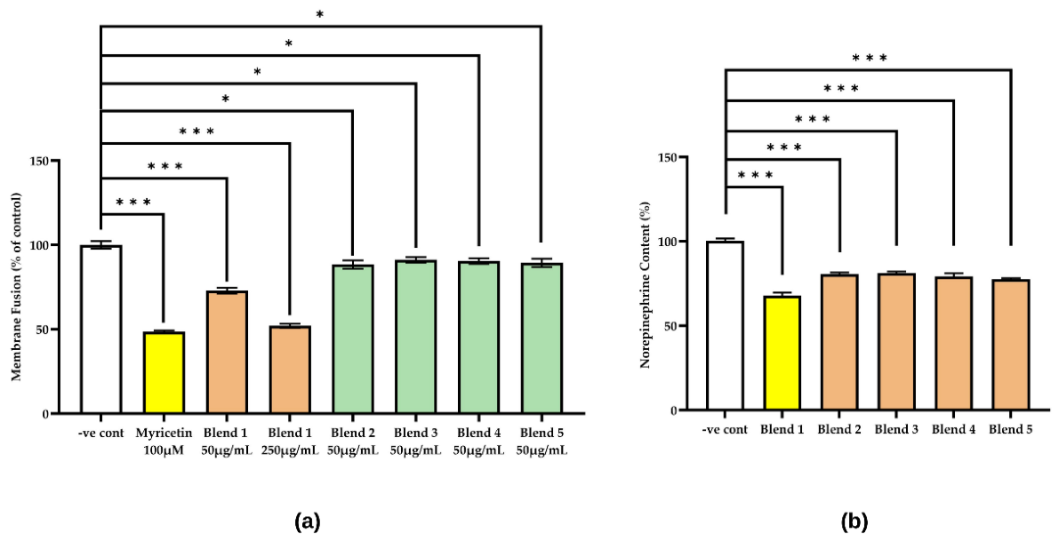
Figure 2.
Neurotoxins and natural extracts inhibit the contraction frequency of skeletal muscle fibers within a muscle cell/nerve cell co-culture. Skeletal muscle and nerve cells were cultured in the presence of the indicated treatment for (a) 2 h or (b) 24 h, after which spontaneous contractions of the muscle cells were counted over a 2 min recording. Data are shown as box-and-whisker plots; boxes represent the interquartile range, the horizontal line indicates the median, the + symbol indicates the mean, and whiskers indicate the data range. * denotes p < 0.05 and *** p < 0.005; n = 6, one-way ANOVA with Holm–Sidak multiple comparisons.
Figure 2.
Neurotoxins and natural extracts inhibit the contraction frequency of skeletal muscle fibers within a muscle cell/nerve cell co-culture. Skeletal muscle and nerve cells were cultured in the presence of the indicated treatment for (a) 2 h or (b) 24 h, after which spontaneous contractions of the muscle cells were counted over a 2 min recording. Data are shown as box-and-whisker plots; boxes represent the interquartile range, the horizontal line indicates the median, the + symbol indicates the mean, and whiskers indicate the data range. * denotes p < 0.05 and *** p < 0.005; n = 6, one-way ANOVA with Holm–Sidak multiple comparisons.
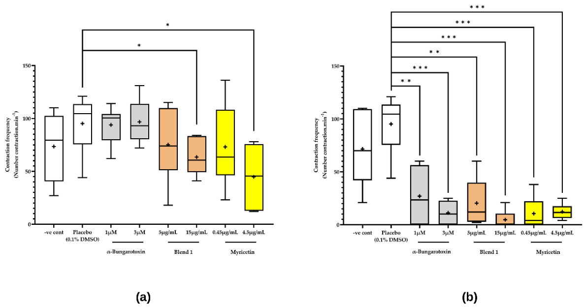
Figure 3.
Neurotoxins and natural extracts modulate ACh levels within ex vivo human skin explants. Skin explants were treated for either a single day (a) or three consecutive days (b) with the indicated treatment conditions, after which the whole skin explants were homogenized and assayed for ACh levels. All data represent the mean ± standard deviation normalized to ACh readings for untreated skin samples. * denotes p < 0.05 and *** p < 0.005; n = 9, one-way ANOVA with Holm–Sidak multiple comparisons.
Figure 3.
Neurotoxins and natural extracts modulate ACh levels within ex vivo human skin explants. Skin explants were treated for either a single day (a) or three consecutive days (b) with the indicated treatment conditions, after which the whole skin explants were homogenized and assayed for ACh levels. All data represent the mean ± standard deviation normalized to ACh readings for untreated skin samples. * denotes p < 0.05 and *** p < 0.005; n = 9, one-way ANOVA with Holm–Sidak multiple comparisons.
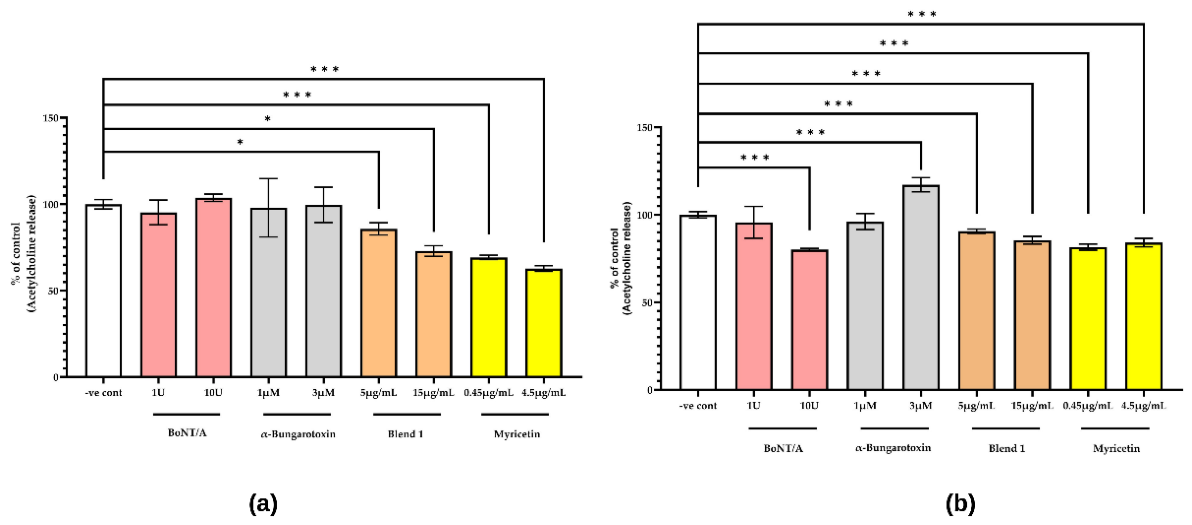
Figure 4.
Neurotoxins and natural extracts have overlapping transcriptomic profiles within human skin explants. Skin explants were treated with the indicated conditions for three consecutive days, after which they were analyzed using bulk transcriptomics. Volcano plots for (a) BoNT/A, (b) α-bungarotoxin, (c) myricetin and (d) Blend 1 treated samples. Genes significantly upregulated (log2FC > 0.585, adjusted p-value < 0.05) are marked in red, while those significantly downregulated (log2FC < −0.585, adjusted p-value < 0.05) are marked in blue. (e) UpSet plot for downregulated and (f) upregulated differentially expressed genes across treatment groups. (g) Heatmap of 18 representative differentially expressed genes across treatment groups.
Figure 4.
Neurotoxins and natural extracts have overlapping transcriptomic profiles within human skin explants. Skin explants were treated with the indicated conditions for three consecutive days, after which they were analyzed using bulk transcriptomics. Volcano plots for (a) BoNT/A, (b) α-bungarotoxin, (c) myricetin and (d) Blend 1 treated samples. Genes significantly upregulated (log2FC > 0.585, adjusted p-value < 0.05) are marked in red, while those significantly downregulated (log2FC < −0.585, adjusted p-value < 0.05) are marked in blue. (e) UpSet plot for downregulated and (f) upregulated differentially expressed genes across treatment groups. (g) Heatmap of 18 representative differentially expressed genes across treatment groups.
Disclaimer/Publisher’s Note: The statements, opinions and data contained in all publications are solely those of the individual author(s) and contributor(s) and not of MDPI and/or the editor(s). MDPI and/or the editor(s) disclaim responsibility for any injury to people or property resulting from any ideas, methods, instructions or products referred to in the content. |
© 2026 by the authors. Licensee MDPI, Basel, Switzerland. This article is an open access article distributed under the terms and conditions of the Creative Commons Attribution (CC BY) license (http://creativecommons.org/licenses/by/4.0/).
Copyright: This open access article is published under a Creative Commons CC BY 4.0 license, which permit the free download, distribution, and reuse, provided that the author and preprint are cited in any reuse.