

Submitted:
11 September 2026
Posted:
15 September 2026
You are already at the latest version
Abstract
Background/Objectives: T Lymphocyte-Secreted Protein I-309, also known as CC-motif Chemokine Ligand 1 (CCL-1), is a small glycoprotein produced by immune cells in response to infection or tissue injury. Increased circulating levels of CCL-1 have been reported in individuals with mild cognitive impairment and Alzheimer's disease (AD), suggesting a potential role in neuroinflammatory processes. However, the effects of CCL-1 on brain physiology remain poorly understood. Methods: Adeno-associated virus (AAV) vectors overexpressing CCL-1 or vector control were injected into the stratum radiatum (CA1) of 250 g Sprague- Dawley rats. Three weeks after inoculation, animals were euthanized or subjected to behavioral testing. BrdU was administered in a subset to label dividing cells. Hippocampal cell populations were assessed by immunohistochemistry using Iba-1, NeuN, GFAP and BrdU antibodies. Locomotion, learning and memory were evaluated using open field, novel object recognition and contextual fear conditioning. Results: CCL-1 overexpression resulted in a significant reduction in microglial population in stratum radiatum, and a significant increase in BrdU+ maturing neurons in the dentate. Neuronal and astrocyte populations and behavioral assays were not significantly affected by CCL-1 overexpression. Conclusions: These findings suggest that CCL-1 overexpression modulates hippocampal cellular composition, potentially exerting anti-inflammatory and neuroprotective effects without measurable changes in cognitive performance of learning and memory. This chemokine may therefore represent a potential mediator of neuroimmune interactions relevant to neurodegenerative processes.
Keywords:
CCL-1
; cognition
; hippocampus
; microglia
; neurogenesis
; neuroimmune modulation
1. Introduction
Neuroimmune signaling plays a critical regulator role in maintaining brain homeostasis and in the pathogenesis of neurodegenerative disorders such as Alzheimer’s disease (AD). Increasing evidence indicates that neuroinflammation contributes significantly to disease progression, influencing synaptic dysfunction, neuronal loss, and cognitive decline [1,2,3]. Microglia are the resident immune cells of the central nervous system (CNS), and are central mediators of neuroinflammatory processes [1,2,3]. Depending on the context, microglia activation can exert both protective and detrimental effects by regulating inflammatory signaling, phagocytosis, and interactions with neurons and other glial cells [4,5,6,7].
Among the molecular mediators involved in these processes, chemokines can regulate cell migration and activation, but also can play broader roles in the CNS, including modulation of synaptic transmission, neuronal survival, and glial activity [8,9,10,11,12,13]. One such chemokine, the T-lymphocyte-secreted protein I-309, also known as CC-motif chemokine ligand 1 (CCL-1), is produced by immune cells in response to infection or tissue injury [14,15,16]. CCL-1 primarily signals through its receptor CCR8, is known to attract monocytes, natural killer cells, immature T and B cells, and dendritic cells [17,18,19]. Activation of CCR8 can also induce intracellular calcium mobilization from monocytes, contributing to immune activation and chemotactic responses [20]. CCL-1 has been implicated in several biological and pathological contexts, where it can exert both inflammatory and homeostatic functions. In the peripheral immune response, CCL-1 participates in immune cells migration to inflammatory sites [21], while under physiological conditions it contributes to immune cells proliferation and regulation [22,23,24]. In addition, CCL-1 has been reported to promote cell survival and inhibit apoptosis in certain immune cell populations and cancer cells [25,26], participate in fibrosis [21,27,28] and allergic [29,30] processes. These diverse functions suggest that CCL-1 may have context-dependent roles in regulating cellular responses during tissue injury and inflammation. Within the CNS, CCR8 expression has been detected in several brain cell types, including microglia, astrocytes, and neurons [17,31]. Experimental evidence indicates that CCL-1 can influence neuronal and glial function. For example, CCL-1 has been shown to enhance glutamatergic synaptic transmission and to increase microglia proliferation, motility and activation [17]. Moreover, CCL-1 expression is upregulated following nerve injury in dorsal root ganglion [32] and has been implicated in nociceptive signaling and pain processing [13,33,34,35,36]. These findings support a role for CCL-1 as a mediator of neuroimmune communication in the nervous system.
Emerging evidence also links CCL-1 signaling to AD. Increased expression of CCL-1 and its receptor CCR8, has been reported in different transgenic mouse models of AD [37,38]. Studies have detected elevated levels of CCL-1 in cerebrospinal fluid and plasma of patients with AD, where its concentrations correlate with global cognitive function and functional vulnerability [39,40,41,42,43]. Other reports suggest that alterations in CCL-1 signaling may influence microglial functions such as amyloid-β phagocytosis, highlighting a potential role in AD-related neuroinflammatory processes [17].
Despite this observation, the direct effect of CCL-1 within the brain remains poorly understood. In particular, it is unclear how increased CCL-1 signaling influences hippocampal cellular dynamics and whether it alters neuronal and glial populations or cognitive function. Given the central role of hippocampus in memory processing and its vulnerability in AD, understanding how chemokine signaling modulates hippocampal neuroimmune interactions is of particular interest. In the present study, we investigated the effects of CCL-1 overexpression in the hippocampus using adeno-associated virus (AAV)–mediated gene delivery in rats. We examined the impact of increased CCL-1 signaling on hippocampal microglial, astrocytic, and neuronal populations using immunohistochemistry, and evaluated its functional consequences using hippocampus-dependent behavioral tests. By exploring the cellular and behavioral effects of CCL-1 in the hippocampus, this study aims to provide new insight into the role of chemokine signaling in neuroimmune regulation and its potential relevance to neurodegenerative disease.
2. Materials and Methods
2.1. Animals
Adult male and female Sprague-Dawley rats (250± 50 g) were housed under standard laboratory conditions with ad libitum access to food and water and maintained on a 12-hour light/12-hour dark cycle at controlled temperature and humidity. All experimental procedures were approved and conducted in accordance with the guidelines of the Institutional Animal Care and Use Committee (CICUA) of INDICASAT AIP (CICUA 19-006 and CICUA 19-007) and complied with national and institutional regulations for the care and use of laboratory animals.
2.2. Experimental Design
Two experimental designs were utilized for this study and are visually represented in Figure 1. For morphological studies, 16 males were handled for a minimum of three days, prior to AAV delivery. Animals for IHC were inoculated on day 0 and tissue collection occurred on day 21. BrdU labelling was achieved via one single intraperitoneal (i.p.) administration (150 mg/kg, Sigma). To analyze cell division, BrdU was administered one day prior to tissue collection (day 20), n= 8. To analyze new neuron maturation, BrdU was administered seven days after stereotaxic inoculation (day 7) and tissue collection was performed on day 21, n= 8.
For behavioral assays, 24 males and females were handled for a minimum of three days, prior to AAV delivery. Animals for behavioral assays remained in their homecages for three weeks post-inoculation. During week three, animals were handled a minimum of three days for 2 minutes daily to minimize experimenter induced stress during behavior. Behavioral assays utilized a within-animal design always following the same design: novel object recognition, followed by contextual fear conditioning. Brains were collected for visual verification of infusion sites.
2.3. AAV Delivery
Rats were anesthetized with an intraperitoneal injection of a ketamine-xylazine mixture and secured in a stereotaxic frame. A midline scalp incision was performed to expose the skull, and small burr holes were drilled bilaterally according to stereotaxic coordinates: AP-3.8, ML +/-2.0, DV -4.2 from skull [44]. A micro-syringe was used to deliver ~1.3 x 10³ GC/µl of either AAV2-CMV-mCherry (Control) or AAV-CMV-hCCL1-IRES-mCherry (CCL-1 overexpression (OE), Vector Biolabs) into the stratum radiatum of the hippocampal Cornu ammonis (CA) 1 region., n= 6 condition. After surgery, animals received standard postoperative care and were allowed to recover for one week. Animals were maintained for three weeks post-infusion to allow stable AAV-mediated transgene expression before tissue collection or behavioral testing. Viral expression was verified in a subset of animals by acute dissection of hippocampal tissue followed by standard western blotting procedures for mCherry expression.
2.4. Tissue Collection
Animals were deeply anesthetized with an overdose of ketamine/xylazine and transcardially perfused with phosphate-buffered saline (PBS, 1X), followed by 4% paraformaldehyde (PFA) in PBS, as previously described [REF], with minor modifications. Briefly, approximately 100-200 ml of PBS was perfused to clear blood, followed by 200-300 ml of 4% PFA for fixation. Brains were carefully removed and post-fixed in 4% PFA at 4°C for at least 12 hours. Samples were then cryoprotected in 20% sucrose prepared in 1X PBS containing 0.02% sodium azide (NaN3) for approximately 48 hours, or until sunk. Coronal brain sections (50 µm thick) were collected serially using a freezing microtome and stored in PBS+azide solution at 4°C until further processing.
2.5. Immunohistochemistry
Immunohistochemistry was performed following previously described protocols with minor modifications [45]. Free-floating sections were first washed in PBS and incubated in blocking solution containing 1% normal goat serum (NGS) in PBS to reduce nonspecific binding. For BrdU immunostaining, sections underwent three sequential pretreatments: (1) antigen retrieval in 0.01 M citric acid buffer (pH 6.0) at 95°C for 15 min; (2) membrane permeabilization with 0.1% trypsin prepared in 0.1 M Tris base containing 1% CaCl₂; and (3) DNA denaturation with 2 M HCl in 1X PBS for 30 min. Following pretreatment, sections were incubated for 1 h in blocking solution containing 2% BSA, 0.3% Triton X-100, and 0.02% NaN₃ in 1X PBS. Sections were then incubated overnight (~12–16 hour) at 4°C with the following primary antibodies diluted in 1% NGS in 1X PBS: Anti-BrdU mouse monoclonal (marker of proliferating cells; Sigma, cat. B2531), anti-NeuN rabbit monoclonal (neuronal marker; Cell Signaling Technologies, cat. 24307S), Iba-1 (microgial marker; CST, cat. 17198S) and GFAP (astrocyte marker; CST cat. 3670S). The following day, sections were washed in PBS and incubated for 1 hour at room temperature with appropriate secondary antibodies diluted in 1% NGS in PBS. After additional washes, sections were mounted onto slides, coverslipped with antifade mounting medium with 4',6-diamidino-2-phenylindole (DAPI), and stored at 4°C until imaging. For colorimetric IHC, sections were DAB stained using the rabbit- or mouse-specific HRP/DAB detection kit (Abcam, cat. ab64261and ab64259), following manufacturer’s protocols, and mounted onto slides with DPX mountant.
2.6. Image Acquisition
Fluorescent images were acquired using an Axioskop 2 plus fluorescence microscope (Zeiss) equipped with appropriate filter sets for FITC, Alexa Fluor 594, and DAPI. Images were collected using identical acquisition settings (laser intensity/exposure time, gain, and offset) across all experimental groups to ensure comparability. For colorimetric IHC, images were acquired on a Leica DM 3000 LED with a MC170 HD camera or an Olympus BX-53 microscope with an DP73 camera.
For each hemisphere, multiple coronal sections spanning the infusion site within the hippocampus were analyzed. Images were obtained from predefined regions of interest (ROIs) that were kept consistent across all sections and animals. These included ROIs in the stratum pyramidale, stratum radiatum, stratum oriens of the CA1 region and the suprapyramidal (top) blade of the dentate gyrus (DG), based on anatomical landmarks. All image acquisition was performed by an experimenter blinded to treatment conditions.
Quantitative analysis was performed using ImageJ/Fiji (NIH) software. Microglia (Iba-1⁺), neurons (NeuN⁺ or BrdU+), and astrocytes (GFAP⁺) were quantified separately. Cell counts were performed either manually or using semi-automated particle analysis following thresholding and segmentation. All analyses were conducted by investigators blinded to experimental groups.
2.7. Behavioral Evaluation
2.7.1. Novel Object Recognition (NOR)
Novel object recognition was used to assess recognition memory [39]. On day 1, animals were habituated to the behavioral testing area (1 m x 1 m open field) for 5 minutes. On day 2, the familiarization phase (T1) was conducted. Two identical objects (A1 and A2) were placed in opposite corners of the arena, approximately 35 cm from the wall and 70 cm apart. Each rat was placed in the arena facing a corner without objects and allowed to freely explore for 5 minutes. The time spent exploring each object was recorded and a minimum of 20 seconds of object exploration was used. Twenty-four hours after the familiarization phase, animals were returned to the open field for the second test (T2), in which one familiar object and one novel object were presented. Exploration time for each object was recorded for 5 minutes. Exploration was defined as directing the nose toward the object at close distance and/or touching it with the vibrissae. A Preference Index (PI) was calculated for each trial as: PI= T novel/(Tnovel+Tfamiliar), where Tnovel represents time spent exploring the novel object and Tfamiliar represents time spent exploring the familiar object. After each trial, animals were returned to their home cage.
2.7.2. Contextual Fear Conditioning (CFC)
Contextual fear conditioning was used to evaluate hippocampus-dependent memory as previously described [46]. Experiments were conducted in an operant conditioning chamber equipped with a grid floor capable of delivering controlled foot shocks (Graphic State, Coulbourn Instruments). During the training session, animals were placed in the chamber and allowed to explore freely for 120 seconds. Three shocks (0.5 mA, 1 sec) were delivered with 60 seconds between shocks, followed by an additional 20 seconds in the chamber before the animals were returned to their home cages. Twenty-four hours later, rats were returned to the same chamber for a 5-minute testing session without shock delivery. The percentage of time spent freezing, defined as the absence of all movement except respiration, was recorded as an index of contextual fear memory as described.
2.7.3. Statistical Analysis
All statistical analyses were performed using GraphPad Prism (version 8.0.1; GraphPad Software, USA). Data are presented as the mean ± standard error of the mean (SEM). For comparisons between two groups (vector control vs CCL-1 overexpression), unpaired two-tailed Student’s t-tests were used. For behavioral analyses, two-way analysis of variance (ANOVA) was used, followed by appropriate post hoc tests when necessary. A p value < 0.05 was considered statistically significant.
3. Results
3.1. Modulation of Hippocampal Cellular Composition by CCL-1 Overexpression
3.1.1. CCL-1 Expression Influences Neurogenesis in the Dentate Gyrus of the Hippocampus, but not Neuronal Populations
To evaluate the effects of CCL-1 on hippocampal cellular composition, cell densities were quantified in defined regions of interest (ROIs) following immunohistochemical staining for major neural cell populations. Quantification of NeuN⁺ cells for the neuronal population revealed no significant differences between control and CCL-1 OE groups in the CA1 region (p= 0.566; n= 8/ grp, Figure 2A) or the dentate gyrus (DG) (p = 0.146; n= 8, 7/ grp) (Figure 2B).
Neurogenesis was assessed. Division of new progenitor cells was quantified 24 hours after a single BrdU administration. No significant different were observed in CCL-1 OE relative to control (p= 0.302, n= 4/grp, Figure 2C). Maturation of newborn neurons was assessed by analyzing BrdU+ cells after two weeks of i.p. administration of BrdU. CCL-1 OE significantly increased the number of BrdU+ cells relative to control (p= 0.009, n= 4/grp, Figure 2D).
3.1.2. CCL-1 Overexpression Reduces Microglial Density in the CA1 Stratum Radiatum, but does not Modify Astrocyte Density
For microglia population, analysis of Iba-1⁺ cells showed CA1 region-specific effects of CCL-1 overexpression. No significant differences were observed in the stratum oriens between groups (p= 0.172; n= 6, 8/ grp, Figure 3A). In contrast, a significant cell number reduction in microglial population was detected in the stratum radiatum in CCL-1–overexpressing animals compared to controls (p= 0.039; n= 8, 12/ grp, Figure 3B). For the astrocyte population, quantification of GFAP⁺ cells revealed no significant differences between control and CCL-1 OE (p= 0.972; n= 7, 8/ grp, Figure 3C).
Together, these findings indicate that CCL-1 overexpression induces region-specific alterations in hippocampal cellular composition, characterized by reduced microglial density and increased neurogenesis in the dentate gyrus without affecting neurons and astrocytes.
Behavioral Assessment of Hippocampal Function Following CCL-1 Overexpression
To evaluate whether CCL-1 overexpression affects learning and memory behavior, animals were subjected to novel object recognition (NOR) and contextual fear conditioning (CFC) tests.
For NOR test, no significant differences were in gross locomotion during the habituation phase of the test (open field, F (1,20)= 0.8062, p= 0.379, n= 6/ grp Figure 4a). The preference index between control and CCL-1 OE groups was not significantly different (F (1,19)= 2.947, p= 0.102, n= 6/ grp). Both male and female animals exhibited comparable preference for the novel object, indicating no detectable impairment or improvement in recognition memory following CCL-1 overexpression (F (1,19)= 0.0002, p= 0.988; n = 6 per group; Figure 4b).
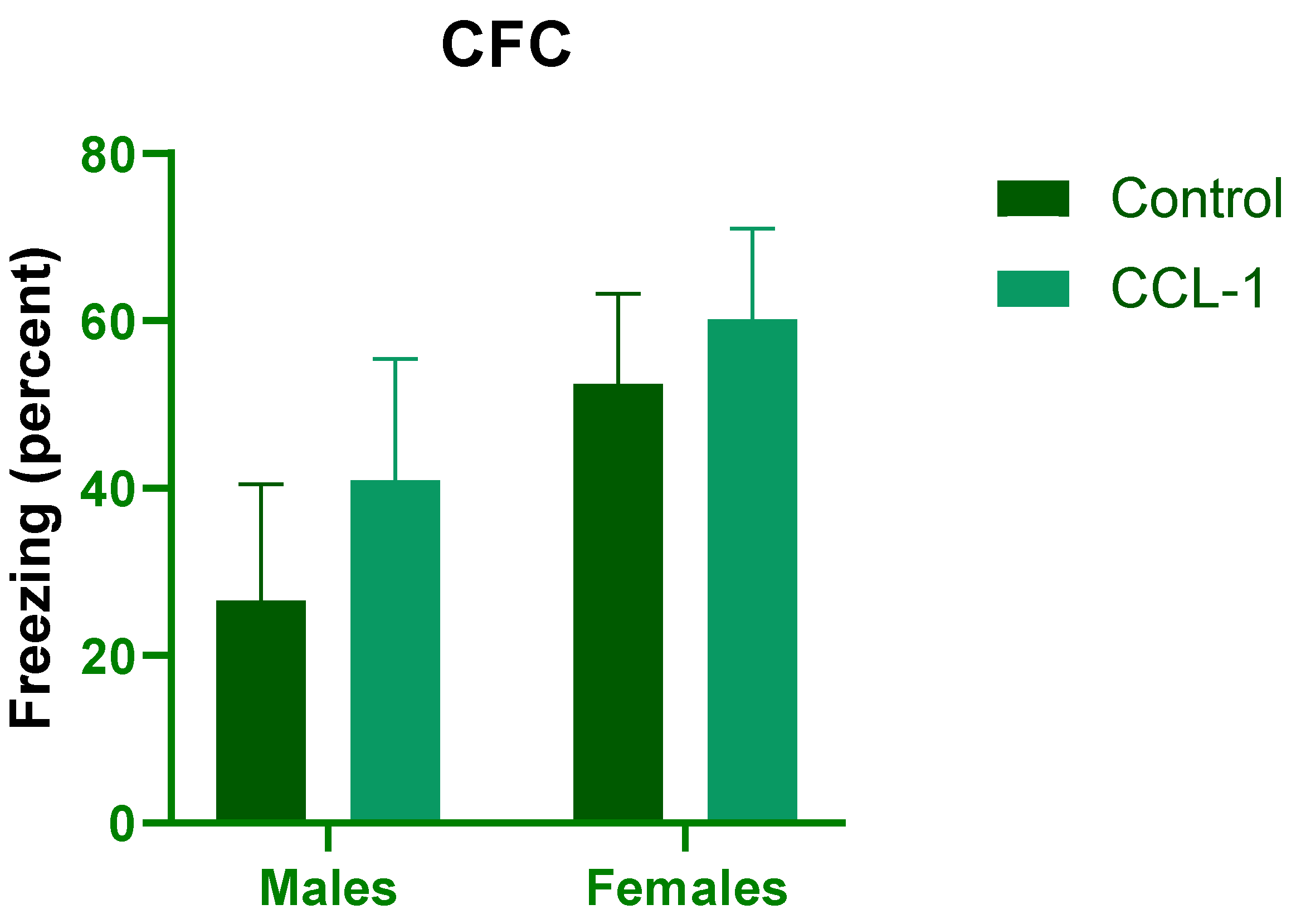
Similarly, no significant differences were observed in the CFC test between groups. The percentage of freezing behavior measured 24 hours after training was comparable between control and CCL-1 OE (F (1, 17)= 2.786, p= 0.113) and between males and females (F (1,17)= 0.664, p= 0.426; n=4–6 per group; Figure 5).
Together, these results indicate that CCL-1 overexpression does not significantly alter learning and memory in a hippocampus-dependent manner.
4. Discussion
In the present study, we demonstrate that hippocampal overexpression of CCL-1 induces region-specific changes in cellular composition, characterized by a reduction in microglial density in the CA1 stratum radiatum and an increase in neurogenesis in the dentate gyrus, without affecting astrocyte populations. Notably, these cellular alterations were not accompanied by changes in learning and memory performance. Together, these findings suggest that CCL-1 modulates neuroimmune homeostasis in the hippocampus without producing an evident behavioral effect under the conditions tested.
Our results indicate that chronic CCL-1 signaling may regulate microglial homeostasis rather than simply promoting inflammation. The observed reduction in microglial hippocampal population may reflect multiple, non-mutually exclusive mechanisms, including reduced activation, altered migration, or shifts in microglia phenotype. In humans, microglia undergo significant morphological and functional changes with aging and in Alzheimer’s disease, including reduced process length, decreased branching and arborized area [47]. In AD brains, microglia often exhibit a dystrophic phenotype [48,49,50,51], with altered Iba-1 expression, Iba-1-low populations associated with disease pathology and increased expression of immune related markers such as CD74, CD45, and ferritin [52]. Additionally, microglia surrounding amyloid plaques display high expression of activation markers such as HLA-DR but reduced Iba-1 labeling [48,49,50,51], suggesting that traditional markers may not fully capture functional states. In this context, the reduction in Iba-1+ cells observed in our study may reflect changes in microglia phenotype or activation state rather than a simple loss of cells. These findings are consistent with a role for chemokine signaling in modulating microglia dynamics. CCLI, through its receptor CCR8, has been implicated in immune cell recruitment and activation, but its function in the CNS remains to be elucidated. Our data support the idea that CCL1 may act as a regulator of microglial behavior, potentially influencing cell distribution, microglial activation states, or turnover. Rather than promoting a classical proinflammatory response, CCL-1 may contribute to a more shaded regulation of neuroimmune interactions, consistent with emerging evidence that chemokines can make context-dependent effects in the brain [9].
Notably, we observed a significant increase in neurogenesis in the dentate gyrus following CCL-1 overexpression. This finding suggests that elevated levels of CCL-1 may create a microenvironment that favors neuronal survival, as the effect was evident in maturation of newborn neurons, and not in division of progenitors. One possible explanation is that modulation of microglial activity leads to a reduction in inflammatory tone, thereby enhancing neuronal viability [53,54] or neuronal maintenance through interactions with glial cells [55,56] hereby influencing neurogenesis. Changes in DG neuron density can suggest off -target effects in that surrounding expression can affect neuronal populations. No change in CA1 neurons, which would be directly infected with AAV delivery may be due to a non-cell intrinsic effect for survival; suggesting that release and signaling-mediated changes may lead to changes in newborn neuronal populations. Although the mechanisms underlying this effect remain to be determined, our findings raise the possibility that CCL-1 has a neuroprotective or pro-survival effect within specific hippocampal subregions.
Despite CCL-1 mediated cellular changes, we did not observe changes in learning and memory performance in either object recognition or contextual fear conditioning tests. We also did not observe significant differences in locomotion. This apparent dissociation between cellular remodeling and behavioral outcomes suggests that the hippocampal network may be functionally resilient to the changes induced by CCL-1 overexpression, or at least within the time frame studied. Our findings are consistent with other studies evaluating the role of chemokines and their receptors in memory. Namely, studies with CCL2-/- [57,58], CCR2-/- [59], CCR3-/-[38], CCR5-/-[60], CXCR3-/-[61] have reported no change in memory assays, but rather a rescue of cognitive deficits in Alzheimer’s transgenic backgrounds, such as the APP/PS1 lines. Similarly, overexpression of CX3CL1 was shown to increase neurogenesis and neuronal signal in the hippocampus but displayed no change in working memory or contextual memory, suggesting a dissociation between neuronal survival and behavior in wild type backgrounds. Over expression of CX3CL1 did, however, rescue deficits in the PS19 Alzheimer’s disease model [62]. Future studies may evaluate the role of CCL-1 in Alzheimer’s disease models, as a modulator of neuroimmune homeostasis.
Other explanations may account for this dissociation between morphological and behavioral changes. First, the observed cellular changes may be compensatory to the viral over-expression approach employed. It is also possible that our single infusion stereotaxic protocol may be sufficient to alter local morphology but not sufficient to alter hippocampal function. Second, the three-weeks expression period may reflect an early stage of modulation that precedes functional consequences. Third, the behavioral test employed, while widely used, may not capture fine-scale changes associated to dorsal CA1 hippocampal function. Microglia have been increasingly recognized as active contributors to synaptic plasticity and memory processes [63], although their role remains incompletely defined. Recent evidence suggests that microglia can influence memory quality, synaptic remodeling, and the integration of newborn neurons, for example through IL-33 pathways [64]. However, the contribution of microglia to learning and memory appears to be more context-dependent and less established than other glial cell types [64]. It is therefore possible that the microglia changes observed in this study affect aspects of hippocampal function not captured by the behavioral assay used, or that more sensitive approaches are needed to detect their functional impact.
The role of chemokines in cognition remains incompletely understood, and identifying the molecular mechanisms through which they influence hippocampal function is of particular importance given the vulnerability of this region to neurodegeneration. Notably, CCL-1 levels in cerebrospinal fluid have been reported to correlate with the severity of cognitive impairment [40]. Elevated circulating levels of CCL-1 have also been associated with neuropsychiatric symptoms, cognitive decline [65], and increased risk of falls [39] and have been consistently observed in individuals with mild cognitive impairment and AD, suggesting a progressive upregulation across disease stages [41,42,43]. In addition, studies examining chemokine dynamics across transgenic mouse models of AD have reported higher cortical levels of CCL-1 in models incorporating tau pathology compared to APP/PS1 models alone, supporting a potential role for CCL-1 role in tau-related neurodegenerative processes [38]. Together, these observations highlight the relevance of CCL-1 in neurodegeneration. However, our findings suggest that its role may be more complex than previously appreciated. Rather than acting solely as a proinflammatory mediator, CCL-1 may contribute to both inflammatory regulation and cellular adaptation, potentially exerting context-dependent or even protective effects within the brain. Consistent with this interpretation, our previous work demonstrated that CCL-1 expression does not modify neuronal spine density, suggesting that key aspects of synaptic function remain preserved [66]. Taken together with the present findings, this supports the idea that CCL-1 modulates specific components of the neuroimmune environment without broadly disrupting neuronal architecture or cognitive function.
This study has several limitations. We did not assess microglial functional phenotypes or molecular signatures which would be necessary to better understand the mechanism underlying the observed changes. Future studies can incorporate detailed analyses of microglial activation states, as well as longer-term behavioral assessments to determine whether sustained CCL-1 signaling leads to functional consequences over the time. Furthermore, evaluation of the effects of CCL-1 in transgenic model of AD, will be important to establish its role in pathological contexts.
5. Conclusions
In conclusion, our findings demonstrate that CCL-1 overexpression induces region-specific changes in hippocampal cellular composition without altering cognitive performance, highlighting a potential role for this chemokine in modulating neuroimmune homeostasis. These results underscore the complexity of chemokine signaling in the brain and suggest that CCL-1 may play a context-dependent role in neurodegenerative processes.
Author Contributions
Study conceptualization, G.B.B., R.J., A.E.V. and M.B.C.; methodology, G.R., K.M., L.A., A.S.M., A.C.; validation and data curation, G.R., K.M. and M.B.C.; formal analysis, G.R. and M.B.C.; resources, G.B.B., A.C., A.E.V. and M.B.C..; writing—original draft preparation, G.R. and M.B.C..; writing—review and editing, K.M., R.J., A.C., A.E.V. G.B.B.; supervision, G.R., K.M. and M.B.C., funding acquisition, L.A., G.B.B. and M.B.C. All authors have read and agreed to the published version of the manuscript.
Funding
This research was funded by SENACYT, grant numbers NI2019A-10 and NI2018B-20, SNI support for A.C., A.E.V., G.B.B. and M.B.C. and the APC was funded by SNI and Cevaxin.
Institutional Review Board Statement
The animal study protocol was approved by the Institutional Review Board of INDICASAT AIP (protocol code CICUA 19-006, approval date July 8, 2019).
Informed Consent Statement
Not applicable.
Data Availability Statement
Data supporting reported results will be made available by authors upon request.
Acknowledgments
We thank the support of all personnel in the animal facilities that provide care and support to the colony and research staff. We would also like to acknowledge the support of the center of microscopy staff in guiding the acquisition and imaging data collection. Finally, we acknowledge the effort of student trainees, Heraclio Sanjur, Jennifer Escobar, Gabriel Rolla and Pedro Poveda in performing parts of the research that was collected for the purpose of these studies.
Conflicts of Interest
The authors declare no conflicts of interest.
Abbreviations
The following abbreviations are used in this manuscript:
| AD | Alzheimer’s disease |
| CCL-1 | C-C motif chemokine 1 |
| AAV | Adeno-associated virus |
| CNS | Central nervous system |
| CCR8 | C-C motif chemokine receptor 8 |
| CICUA | Institutional Committee for the care and use of animals |
| OE | Overexpression |
| CA1 | Cornus ammonis 1 |
| DG | Dentate gyrus |
| BrdU | 5-bromo-2'-deoxyuridine |
| i.p. | intraperitoneal |
| IHC | immuohistochemistry |
| NOR | Novel Object Recognition |
| CFC | Contextual Fear Conditioning |
References
- Twarowski, B.; Herbet, M. Inflammatory Processes in Alzheimer’s Disease-Pathomechanism, Diagnosis and Treatment: A Review. Int J Mol Sci 2023, 24, 6518. [CrossRef]
- Van Eldik, L.J.; Carrillo, M.C.; Cole, P.E.; Feuerbach, D.; Greenberg, B.D.; Hendrix, J.A.; Kennedy, M.; Kozauer, N.; Margolin, R.A.; Molinuevo, J.L.; et al. The Roles of Inflammation and Immune Mechanisms in Alzheimer’s Disease. Alzheimers Dement (N Y) 2016, 2, 99–109. [CrossRef]
- Heneka, M.T.; Carson, M.J.; El Khoury, J.; Landreth, G.E.; Brosseron, F.; Feinstein, D.L.; Jacobs, A.H.; Wyss-Coray, T.; Vitorica, J.; Ransohoff, R.M.; et al. Neuroinflammation in Alzheimer’s Disease. Lancet Neurol 2015, 14, 388–405. [CrossRef]
- Cai, Y.; Liu, J.; Wang, B.; Sun, M.; Yang, H. Microglia in the Neuroinflammatory Pathogenesis of Alzheimer’s Disease and Related Therapeutic Targets. Front Immunol 2022, 13, 856376. [CrossRef]
- Ebrahimi, R.; Shahrokhi Nejad, S.; Falah Tafti, M.; Karimi, Z.; Sadr, S.R.; Ramadhan Hussein, D.; Talebian, N.; Esmaeilpour, K. Microglial Activation as a Hallmark of Neuroinflammation in Alzheimer’s Disease. Metab Brain Dis 2025, 40, 207. [CrossRef]
- Müller, L.; Di Benedetto, S.; Müller, V. The Dual Nature of Neuroinflammation in Networked Brain. Front Immunol 2025, 16, 1659947. [CrossRef]
- Yassaghi, Y.; Nazerian, Y.; Ghasemi, M.; Nazerian, A.; Sayehmiri, F.; Perry, G.; Gholami Pourbadie, H. Microglial Modulation as a Therapeutic Strategy in Alzheimer’s Disease: Focus on Microglial Preconditioning Approaches. J Cell Mol Med 2024, 28, e18554. [CrossRef]
- Valiukas, Z.; Tangalakis, K.; Apostolopoulos, V.; Feehan, J. Microglial Activation States and Their Implications for Alzheimer’s Disease. J Prev Alzheimers Dis 2025, 12, 100013. [CrossRef]
- Wojcieszak, J.; Kuczyńska, K.; Zawilska, J.B. Role of Chemokines in the Development and Progression of Alzheimer’s Disease. J Mol Neurosci 2022, 72, 1929–1951. [CrossRef]
- Domingues, C.; da Cruz E Silva, O.A.B.; Henriques, A.G. Impact of Cytokines and Chemokines on Alzheimer’s Disease Neuropathological Hallmarks. Curr Alzheimer Res 2017, 14, 870–882. [CrossRef]
- Jorda, A.; Campos-Campos, J.; Iradi, A.; Aldasoro, M.; Aldasoro, C.; Vila, J.M.; Valles, S.L. The Role of Chemokines in Alzheimer’s Disease. Endocr Metab Immune Disord Drug Targets 2020, 20, 1383–1390. [CrossRef]
- Martin, E.; Delarasse, C. Complex Role of Chemokine Mediators in Animal Models of Alzheimer’s Disease. Biomed J 2018, 41, 34–40. [CrossRef]
- Pawlik, K.; Mika, J. Targeting Members of the Chemokine Family as a Novel Approach to Treating Neuropathic Pain. Molecules 2023, 28, 5766. [CrossRef]
- Atri, C.; Guerfali, F.Z.; Laouini, D. Role of Human Macrophage Polarization in Inflammation during Infectious Diseases. Int J Mol Sci 2018, 19, 1801. [CrossRef]
- Bajetto, A.; Bonavia, R.; Barbero, S.; Schettini, G. Characterization of Chemokines and Their Receptors in the Central Nervous System: Physiopathological Implications. J Neurochem 2002, 82, 1311–1329. [CrossRef]
- Ciechanowska, A.; Mika, J. CC Chemokine Family Members’ Modulation as a Novel Approach for Treating Central Nervous System and Peripheral Nervous System Injury-A Review of Clinical and Experimental Findings. Int J Mol Sci 2024, 25, 3788. [CrossRef]
- Akimoto, N.; Ifuku, M.; Mori, Y.; Noda, M. Effects of Chemokine (C–C Motif) Ligand 1 on Microglial Function. Biochemical and Biophysical Research Communications 2013, 436, 455–461. [CrossRef]
- Miller, M.D.; Krangel, M.S. The Human Cytokine I-309 Is a Monocyte Chemoattractant. Proc. Natl. Acad. Sci. U.S.A. 1992, 89, 2950–2954. [CrossRef]
- Gombert, M.; Dieu-Nosjean, M.-C.; Winterberg, F.; Bünemann, E.; Kubitza, R.C.; Da Cunha, L.; Haahtela, A.; Lehtimäki, S.; Müller, A.; Rieker, J.; et al. CCL1-CCR8 Interactions: An Axis Mediating the Recruitment of T Cells and Langerhans-Type Dendritic Cells to Sites of Atopic Skin Inflammation. J Immunol 2005, 174, 5082–5091. [CrossRef]
- White, G.E.; Iqbal, A.J.; Greaves, D.R. CC Chemokine Receptors and Chronic Inflammation—Therapeutic Opportunities and Pharmacological Challenges. Pharmacological Reviews 2013, 65, 47–89. [CrossRef]
- Liu, S.; Zhang, Z.; Wang, Y.; Zhang, Y.; Min, J.; Li, X.; Liu, S. The Chemokine CCL1 Facilitates Pulmonary Fibrosis by Promoting Macrophage Migration and M2 Polarization. International Immunopharmacology 2023, 120, 110343. [CrossRef]
- Knipfer, L.; Schulz-Kuhnt, A.; Kindermann, M.; Greif, V.; Symowski, C.; Voehringer, D.; Neurath, M.F.; Atreya, I.; Wirtz, S. A CCL1/CCR8-Dependent Feed-Forward Mechanism Drives ILC2 Functions in Type 2-Mediated Inflammation. J Exp Med 2019, 216, 2763–2777. [CrossRef]
- Iellem, A.; Mariani, M.; Lang, R.; Recalde, H.; Panina-Bordignon, P.; Sinigaglia, F.; D’Ambrosio, D. Unique Chemotactic Response Profile and Specific Expression of Chemokine Receptors CCR4 and CCR8 by CD4(+)CD25(+) Regulatory T Cells. J Exp Med 2001, 194, 847–853. [CrossRef]
- Kang, L.; Schmalzl, A.; Leupold, T.; Gonzalez-Acera, M.; Atreya, R.; Neurath, M.F.; Becker, C.; Wirtz, S. CCR8 Signaling via CCL1 Regulates Responses of Intestinal IFN-γ Producing Innate Lymphoid CelIs and Protects From Experimental Colitis. Front Immunol 2020, 11, 609400. [CrossRef]
- Van Snick, J.; Houssiau, F.; Proost, P.; Van Damme, J.; Renauld, J.C. I-309/T Cell Activation Gene-3 Chemokine Protects Murine T Cell Lymphomas against Dexamethasone-Induced Apoptosis. J Immunol 1996, 157, 2570–2576.
- Wang, M.; Qin, Z.; Wan, J.; Yan, Y.; Duan, X.; Yao, X.; Jiang, Z.; Li, W.; Qin, Z. Tumor-Derived Exosomes Drive Pre-Metastatic Niche Formation in Lung via Modulating CCL1+ Fibroblast and CCR8+ Treg Cell Interactions. Cancer Immunol Immunother 2022, 71, 2717–2730. [CrossRef]
- Liu, S.-S.; Liu, C.; Lv, X.-X.; Cui, B.; Yan, J.; Li, Y.-X.; Li, K.; Hua, F.; Zhang, X.-W.; Yu, J.-J.; et al. The Chemokine CCL1 Triggers an AMFR-SPRY1 Pathway That Promotes Differentiation of Lung Fibroblasts into Myofibroblasts and Drives Pulmonary Fibrosis. Immunity 2021, 54, 2042-2056.e8. [CrossRef]
- Diao, S.; Li, L.; Zhang, J.; Ji, M.; Sun, L.; Shen, W.; Wu, S.; Chen, Z.; Huang, C.; Li, J. Macrophage-Derived CCL1 Targets CCR8 Receptor in Hepatic Stellate Cells to Promote Liver Fibrosis through JAk/STAT Pathway. Biochem Pharmacol 2025, 237, 116884. [CrossRef]
- Chiu, T.-M.; Chen, C.-B.; Lu, C.-W.; Hui, R.C.-Y.; Chi, M.-H.; Chang, Y.-C.; Wu, J.; Chen, K.-Y.; Lin, Y.Y.-W.; Lo, P.-C.; et al. CCR8/CCL1 and CXCR3/CXCL10 Axis-Mediated Memory T-Cell Activation in Patients with Recalcitrant Drug-Induced Hypersensitivity. Br J Dermatol 2025, 192, 293–305. [CrossRef]
- Sha, J.; Yang, M.; Lei, Y.; Sun, L.; Meng, C.; Zhu, D. Interaction between Nasal Epithelial Cells and Tregs in Allergic Rhinitis Responses to Allergen via CCL1/CCR8. Front Immunol 2025, 16, 1526081. [CrossRef]
- Trebst, C.; Staugaitis, S.M.; Kivisäkk, P.; Mahad, D.; Cathcart, M.K.; Tucky, B.; Wei, T.; Rani, M.R.S.; Horuk, R.; Aldape, K.D.; et al. CC Chemokine Receptor 8 in the Central Nervous System Is Associated with Phagocytic Macrophages. Am J Pathol 2003, 162, 427–438. [CrossRef]
- Akimoto, N.; Honda, K.; Uta, D.; Beppu, K.; Ushijima, Y.; Matsuzaki, Y.; Nakashima, S.; Kido, M.A.; Imoto, K.; Takano, Y.; et al. CCL-1 in the Spinal Cord Contributes to Neuropathic Pain Induced by Nerve Injury. Cell Death Dis 2013, 4, e679–e679. [CrossRef]
- Wang, C.; Xu, R.; Wang, X.; Li, Q.; Li, Y.; Jiao, Y.; Zhao, Q.; Guo, S.; Su, L.; Yu, Y.; et al. Spinal CCL1/CCR8 Regulates Phosphorylation of GluA1-Containing AMPA Receptor in Postoperative Pain after Tibial Fracture and Orthopedic Surgery in Mice. Neurosci Res 2020, 154, 20–26. [CrossRef]
- García-Domínguez, M.; Aguirre, A.; Lastra, A.; Hidalgo, A.; Baamonde, A.; Menéndez, L. The Systemic Administration of the Chemokine CCL1 Evokes Thermal Analgesia in Mice Through the Activation of the Endocannabinoid System. Cell Mol Neurobiol 2019, 39, 1115–1124. [CrossRef]
- García-Domínguez, M.; González-Rodríguez, S.; Hidalgo, A.; Baamonde, A.; Menéndez, L. Kappa-opioid Receptor-mediated Thermal Analgesia Evoked by the Intrathecal Administration of the Chemokine CCL1 in Mice. Fundamemntal Clinical Pharma 2021, 35, 1109–1118. [CrossRef]
- Zychowska, M.; Rojewska, E.; Piotrowska, A.; Kreiner, G.; Nalepa, I.; Mika, J. Spinal CCL1/CCR8 Signaling Interplay as a Potential Therapeutic Target - Evidence from a Mouse Diabetic Neuropathy Model. Int Immunopharmacol 2017, 52, 261–271. [CrossRef]
- Jorda, A.; Cauli, O.; Santonja, J.M.; Aldasoro, M.; Aldasoro, C.; Obrador, E.; Vila, J.M.; Mauricio, M.D.; Iradi, A.; Guerra-Ojeda, S.; et al. Changes in Chemokines and Chemokine Receptors Expression in a Mouse Model of Alzheimer’s Disease. Int J Biol Sci 2019, 15, 453–463. [CrossRef]
- Sun, Y.; Xie, X.; Zou, X.; Zhou, F. Neuroinflammatory Chemokine Networks in Transgenic Models of Alzheimer’s Disease: A Comparative Multi-Compartmental Analysis. Hum Exp Toxicol 2025, 44, 9603271251348723. [CrossRef]
- Britton, G.B.; O’Bryant, S.E.; Johnson, L.A.; Hall, J.R.; Villarreal, A.E.; Oviedo, D.C.; Pérez-Lao, A.R.; Carreira, M.B. Inflammatory Biomarkers, Depressive Symptoms and Falls Among the Elderly in Panama. CAS 2019, 11, 236–241. [CrossRef]
- Hu, W.T.; Chen-Plotkin, A.; Arnold, S.E.; Grossman, M.; Clark, C.M.; Shaw, L.M.; Pickering, E.; Kuhn, M.; Chen, Y.; McCluskey, L.; et al. Novel CSF Biomarkers for Alzheimer’s Disease and Mild Cognitive Impairment. Acta Neuropathol 2010, 119, 669–678. [CrossRef]
- Villarreal, A.E.; O’Bryant, S.E.; Edwards, M.; Grajales, S.; Britton, G.B.; Panama Aging Research Initiative Serum-Based Protein Profiles of Alzheimer’s Disease and Mild Cognitive Impairment in Elderly Hispanics. Neurodegener. Dis. Manag. 2016, 6, 203–213. [CrossRef]
- Zhou, F.; Sun, Y.; Xie, X.; Zhao, Y. Blood and CSF Chemokines in Alzheimer’s Disease and Mild Cognitive Impairment: A Systematic Review and Meta-Analysis. Alzheimers Res Ther 2023, 15, 107. [CrossRef]
- Gupta, V.B.; Hone, E.; Pedrini, S.; Doecke, J.; O’Bryant, S.; James, I.; Bush, A.I.; Rowe, C.C.; Villemagne, V.L.; Ames, D.; et al. Altered Levels of Blood Proteins in Alzheimer’s Disease Longitudinal Study: Results from Australian Imaging Biomarkers Lifestyle Study of Ageing Cohort. Alzheimers Dement (Amst) 2017, 8, 60–72. [CrossRef]
- Paxinos, G.; Watson, C. The Rat Brain in Stereotaxic Coordinates; 4th ed.; Academic Press: San Diego, 1998; ISBN 978-0-12-547619-5.
- Noonan, M.A.; Choi, K.H.; Self, D.W.; Eisch, A.J. Withdrawal from Cocaine Self-Administration Normalizes Deficits in Proliferation and Enhances Maturity of Adult-Generated Hippocampal Neurons. J. Neurosci. 2008, 28, 2516–2526. [CrossRef]
- Cossio, R.; Carreira, M.B.; Vásquez, C.E.; Britton, G.B. Sex Differences and Estrous Cycle Effects on Foreground Contextual Fear Conditioning. Physiology & Behavior 2016, 163, 305–311. [CrossRef]
- Davies, D.S.; Ma, J.; Jegathees, T.; Goldsbury, C. Microglia Show Altered Morphology and Reduced Arborization in Human Brain during Aging and A Lzheimer’s Disease. Brain Pathology 2017, 27, 795–808. [CrossRef]
- Lier, J.; Streit, W.J.; Bechmann, I. Beyond Activation: Characterizing Microglial Functional Phenotypes. Cells 2021, 10, 2236. [CrossRef]
- Streit, W.J.; Braak, H.; Xue, Q.-S.; Bechmann, I. Dystrophic (Senescent) Rather than Activated Microglial Cells Are Associated with Tau Pathology and Likely Precede Neurodegeneration in Alzheimer’s Disease. Acta Neuropathol 2009, 118, 475–485. [CrossRef]
- Streit, W.J.; Khoshbouei, H.; Bechmann, I. Dystrophic Microglia in Late-onset Alzheimer’s Disease. Glia 2020, 68, 845–854. [CrossRef]
- Streit, W.J.; Braak, H.; Del Tredici, K.; Leyh, J.; Lier, J.; Khoshbouei, H.; Eisenlöffel, C.; Müller, W.; Bechmann, I. Microglial Activation Occurs Late during Preclinical Alzheimer’s Disease. Glia 2018, 66, 2550–2562. [CrossRef]
- Swanson, M.E.V.; Scotter, E.L.; Smyth, L.C.D.; Murray, H.C.; Ryan, B.; Turner, C.; Faull, R.L.M.; Dragunow, M.; Curtis, M.A. Identification of a Dysfunctional Microglial Population in Human Alzheimer’s Disease Cortex Using Novel Single-Cell Histology Image Analysis. acta neuropathol commun 2020, 8, 170. [CrossRef]
- Verduzco Espinoza, A.P.; Na, N.; Campanati, L.; Ngo, P.; Baldwin, K.K.; Cline, H.T. Microglia-to-Neuron Signaling Links APOE4 and Inflammation to Enhanced Neuronal Lipid Metabolism and Network Activity. Proc Natl Acad Sci U S A 2025, 122, e2516103122. [CrossRef]
- Goshi, N.; Morgan, R.K.; Lein, P.J.; Seker, E. A Primary Neural Cell Culture Model to Study Neuron, Astrocyte, and Microglia Interactions in Neuroinflammation. J Neuroinflammation 2020, 17, 155. [CrossRef]
- Zhao, S.; Umpierre, A.D.; Wu, L.-J. Tuning Neural Circuits and Behaviors by Microglia in the Adult Brain. Trends Neurosci 2024, 47, 181–194. [CrossRef]
- Akiyoshi, R.; Wake, H.; Kato, D.; Horiuchi, H.; Ono, R.; Ikegami, A.; Haruwaka, K.; Omori, T.; Tachibana, Y.; Moorhouse, A.J.; et al. Microglia Enhance Synapse Activity to Promote Local Network Synchronization. eNeuro 2018, 5, ENEURO.0088-18.2018. [CrossRef]
- Kiyota, T.; Gendelman, H.E.; Weir, R.A.; Higgins, E.E.; Zhang, G.; Jain, M. CCL2 Affects β-Amyloidosis and Progressive Neurocognitive Dysfunction in a Mouse Model of Alzheimer’s Disease. Neurobiology of Aging 2013, 34, 1060–1068. [CrossRef]
- Kiyota, T.; Morrison, C.M.; Tu, G.; Dyavarshetty, B.; Weir, R.A.; Zhang, G.; Xiong, H.; Gendelman, H.E. Presenilin-1 Familial Alzheimer’s Disease Mutation Alters Hippocampal Neurogenesis and Memory Function in CCL2 Null Mice. Brain, Behavior, and Immunity 2015, 49, 311–321. [CrossRef]
- Naert, G.; Rivest, S. CC Chemokine Receptor 2 Deficiency Aggravates Cognitive Impairments and Amyloid Pathology in a Transgenic Mouse Model of Alzheimer’s Disease. J. Neurosci. 2011, 31, 6208–6220. [CrossRef]
- Passos, G.F.; Figueiredo, C.P.; Prediger, R.D.S.; Pandolfo, P.; Duarte, F.S.; Medeiros, R.; Calixto, J.B. Role of the Macrophage Inflammatory Protein-1α/CC Chemokine Receptor 5 Signaling Pathway in the Neuroinflammatory Response and Cognitive Deficits Induced by β-Amyloid Peptide. The American Journal of Pathology 2009, 175, 1586–1597. [CrossRef]
- Krauthausen, M.; Kummer, M.P.; Zimmermann, J.; Reyes-Irisarri, E.; Terwel, D.; Bulic, B.; Heneka, M.T.; Müller, M. CXCR3 Promotes Plaque Formation and Behavioral Deficits in an Alzheimer’s Disease Model. J. Clin. Invest. 2015, 125, 365–378. [CrossRef]
- Fan, Q.; He, W.; Gayen, M.; Benoit, M.R.; Luo, X.; Hu, X.; Yan, R. Activated CX3CL1/Smad2 Signals Prevent Neuronal Loss and Alzheimer’s Tau Pathology-Mediated Cognitive Dysfunction. J. Neurosci. 2020, 40, 1133–1144. [CrossRef]
- Nguyen, P.T.; Dorman, L.C.; Pan, S.; Vainchtein, I.D.; Han, R.T.; Nakao-Inoue, H.; Taloma, S.E.; Barron, J.J.; Molofsky, A.B.; Kheirbek, M.A.; et al. Microglial Remodeling of the Extracellular Matrix Promotes Synapse Plasticity. Cell 2020, 182, 388-403.e15. [CrossRef]
- Alberini, C.M. Not Just Neurons: The Diverse Cellular Landscape of Learning and Memory. Neuron 2025, 113, 1664–1679. [CrossRef]
- Rabl, M.; Clark, C.; Dayon, L.; Popp, J. Neuropsychiatric Symptoms in Cognitive Decline and Alzheimer’s Disease: Biomarker Discovery Using Plasma Proteomics. J Neurol Neurosurg Psychiatry 2025, 96, 370–382. [CrossRef]
- Adames-Torres, L.L.; Madrid L, A.S.; Mata A., K.G.; Flores-Cuadra, J.A.; Britton, G.B.; Villarreal, A.E.; Carreira F., M.B. Efecto de La Sobreexpresión de CCL1 (I-309) En Las Espinas Dendríticas de Neuronas Del Hipocampo. Scientia 2025, 35, 71–85. [CrossRef]
Figure 1.
Experimental Design. Animals underwent stereotaxic surgery, followed by three weeks to allow for sustained expression of AAV viral vectors. For the morphological analysis, animals were either BrdU labelled on day 7 or day 20, then brains harvested on day 21. For behavioral assays, animals began behavior on day 21.
Figure 1.
Experimental Design. Animals underwent stereotaxic surgery, followed by three weeks to allow for sustained expression of AAV viral vectors. For the morphological analysis, animals were either BrdU labelled on day 7 or day 20, then brains harvested on day 21. For behavioral assays, animals began behavior on day 21.
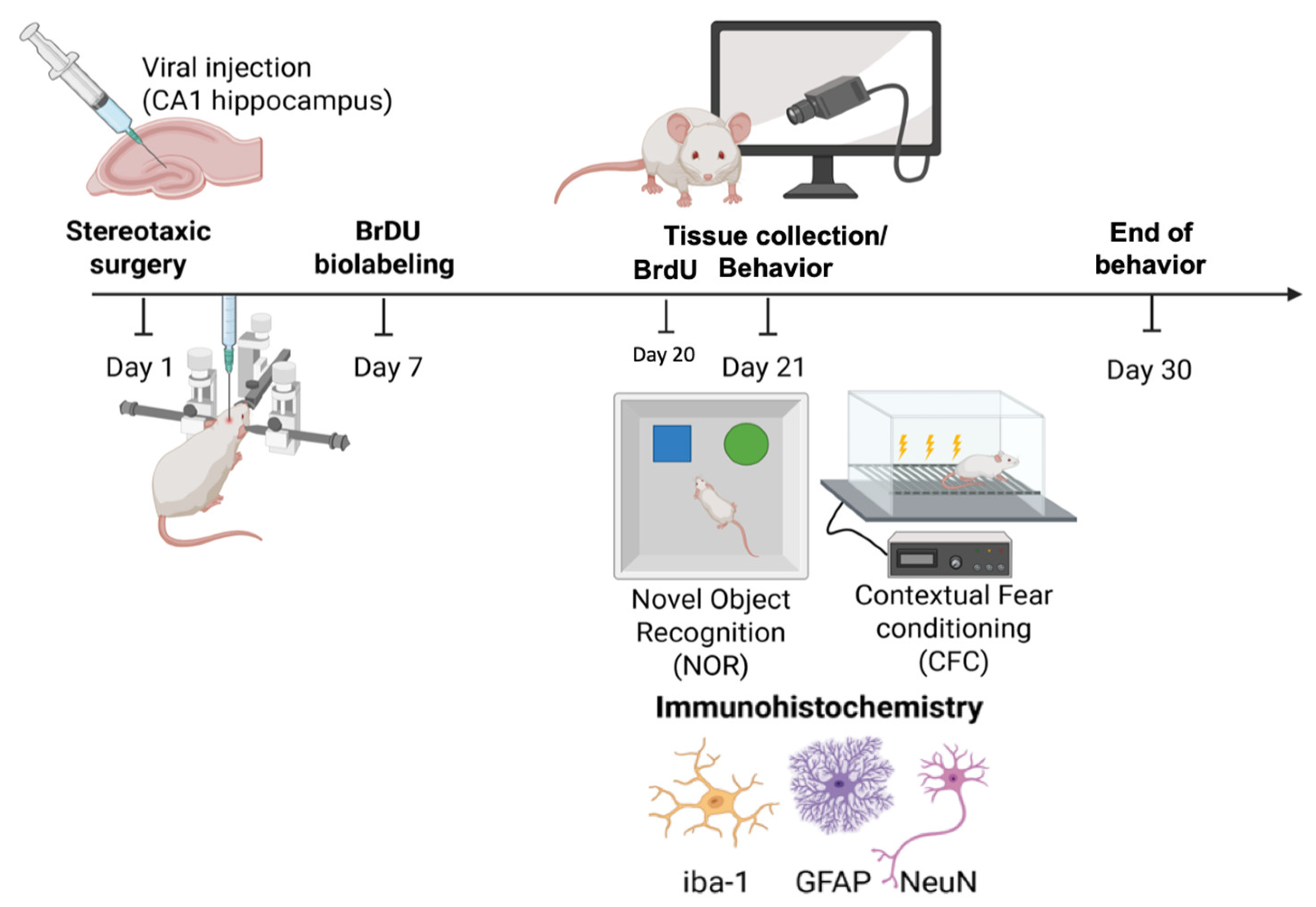
Figure 2.
CCL-1 expression influences neurogenesis in the dentate gyrus of the hippocampus, but not neuronal populations. NeuN+ cells in the CA1 s. pyramidale ((p= 0.566; n= 8/ grp, a) and in the suprapyramidal layer of the dentate gyrus (p = 0.146; n= 8, 7/ grp, b) show no significant differences in counts between control and CCL-1 overexpression. Newly divided BrdU+ cells were not significantly different in the subgranular zone of the dentate gyrus between control and CCL-1 OE (p= 0.302, n= 4/grp ,c) but maturing Brdu+ cells were significantly different between control and CCL-1 OE (p= 0.009, n= 4/grp, d).
Figure 2.
CCL-1 expression influences neurogenesis in the dentate gyrus of the hippocampus, but not neuronal populations. NeuN+ cells in the CA1 s. pyramidale ((p= 0.566; n= 8/ grp, a) and in the suprapyramidal layer of the dentate gyrus (p = 0.146; n= 8, 7/ grp, b) show no significant differences in counts between control and CCL-1 overexpression. Newly divided BrdU+ cells were not significantly different in the subgranular zone of the dentate gyrus between control and CCL-1 OE (p= 0.302, n= 4/grp ,c) but maturing Brdu+ cells were significantly different between control and CCL-1 OE (p= 0.009, n= 4/grp, d).
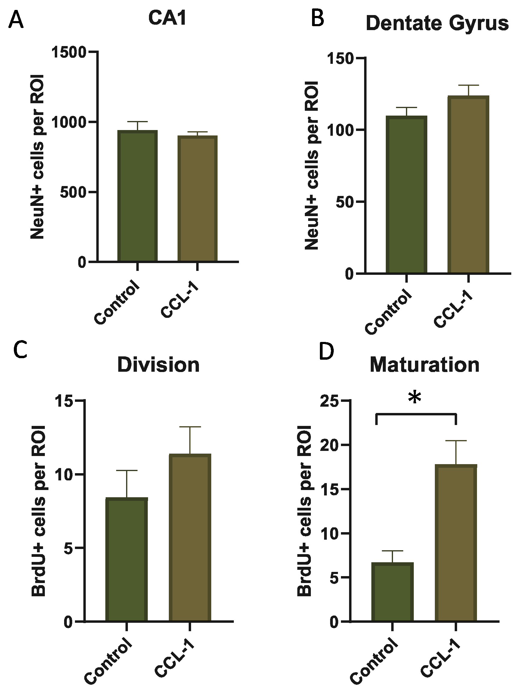
Figure 3.
CCL-1 overexpression reduces microglial density in the CA1 stratum radiatum, but does not modify astrocyte density. Iba-1+ cells in the s. oriens were not significantly different between control and CCL-1 OE (p= 0.172; n= 6, 8/ grp, a) while Iba-1+ counts in the s. radiatum were significantly different in the CCL-1 OE group relative to control (p= 0.039; n= 8, 12/ grp, b). GFAP+ cells in the s. radiatum were not significantly different between control and CCL-1 OE groups (p= 0.972; n= 7, 8/ grp, c).
Figure 3.
CCL-1 overexpression reduces microglial density in the CA1 stratum radiatum, but does not modify astrocyte density. Iba-1+ cells in the s. oriens were not significantly different between control and CCL-1 OE (p= 0.172; n= 6, 8/ grp, a) while Iba-1+ counts in the s. radiatum were significantly different in the CCL-1 OE group relative to control (p= 0.039; n= 8, 12/ grp, b). GFAP+ cells in the s. radiatum were not significantly different between control and CCL-1 OE groups (p= 0.972; n= 7, 8/ grp, c).
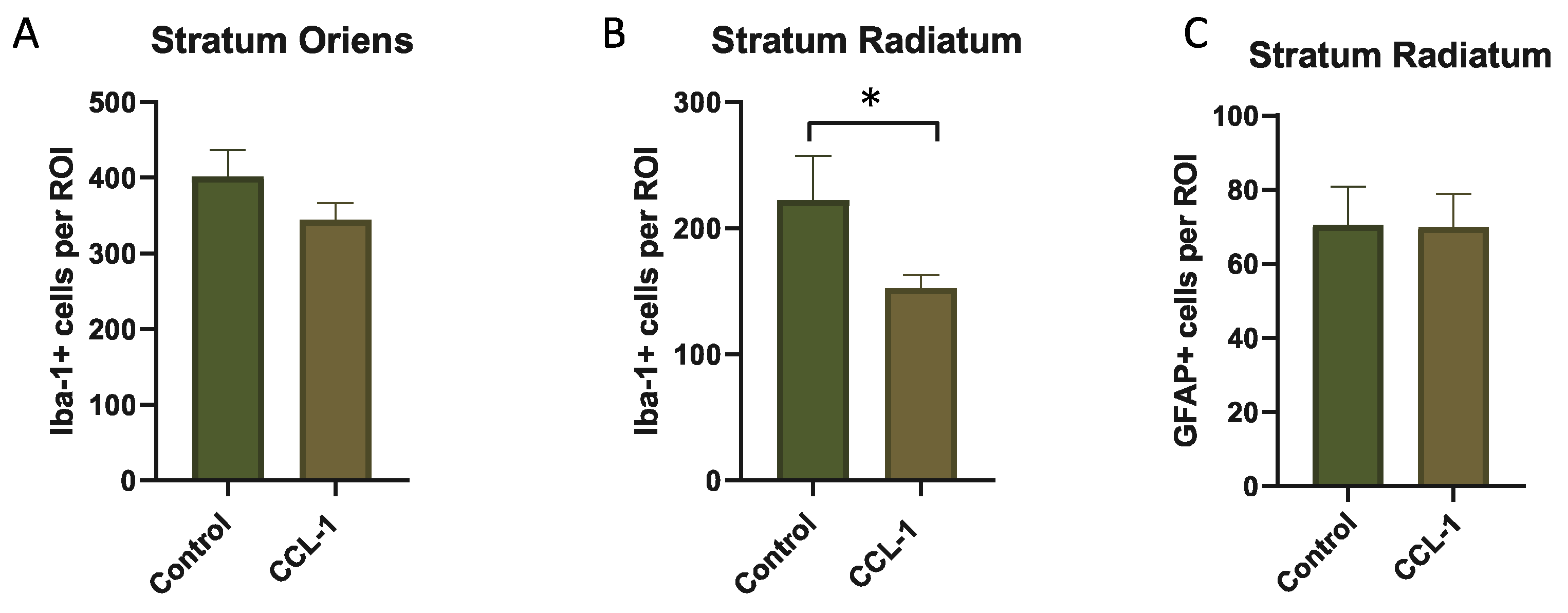
Figure 4.
CCL-1 overexpression did not modify locomotion or recognition memory. a) Mobility in the open field arena was not significantly different between control and CCL-1 OE (F (1,20)= 0.8062, p= 0.379) or between males and females (F (1,20)= 2.452, p= 0.133). b) Preference index in the NOR was not significantly different between control and CCL-1 OE (F (1,19)= 2.947, p= 0.102) or between males and females (F (1,19)= 0.0002, p= 0.988); n= 5-6/ grp. NOR, Novel Object Recognition.
Figure 4.
CCL-1 overexpression did not modify locomotion or recognition memory. a) Mobility in the open field arena was not significantly different between control and CCL-1 OE (F (1,20)= 0.8062, p= 0.379) or between males and females (F (1,20)= 2.452, p= 0.133). b) Preference index in the NOR was not significantly different between control and CCL-1 OE (F (1,19)= 2.947, p= 0.102) or between males and females (F (1,19)= 0.0002, p= 0.988); n= 5-6/ grp. NOR, Novel Object Recognition.
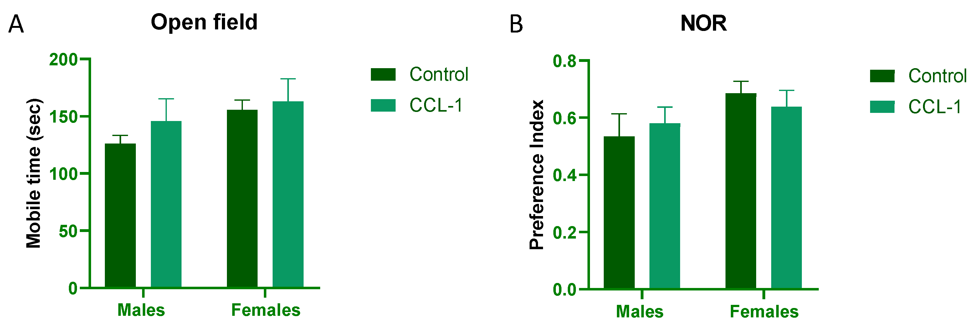
Figure 5.
CCL-1 overexpression did not modify associative learning. Contextual fear conditioning was tested 24h after training and no significant differences were observed between control and CCL-1 OE groups (F (1, 17)= 2.786, p= 0.113); n=4-6/grp. CFC, contextual fear conditioning.
Figure 5.
CCL-1 overexpression did not modify associative learning. Contextual fear conditioning was tested 24h after training and no significant differences were observed between control and CCL-1 OE groups (F (1, 17)= 2.786, p= 0.113); n=4-6/grp. CFC, contextual fear conditioning.
Disclaimer/Publisher’s Note: The statements, opinions and data contained in all publications are solely those of the individual author(s) and contributor(s) and not of MDPI and/or the editor(s). MDPI and/or the editor(s) disclaim responsibility for any injury to people or property resulting from any ideas, methods, instructions or products referred to in the content. |
© 2026 by the authors. Licensee MDPI, Basel, Switzerland. This article is an open access article distributed under the terms and conditions of the Creative Commons Attribution (CC BY) license (http://creativecommons.org/licenses/by/4.0/).
Copyright: This open access article is published under a Creative Commons CC BY 4.0 license, which permit the free download, distribution, and reuse, provided that the author and preprint are cited in any reuse.