
Submitted:
10 September 2026
Posted:
14 September 2026
You are already at the latest version
Abstract
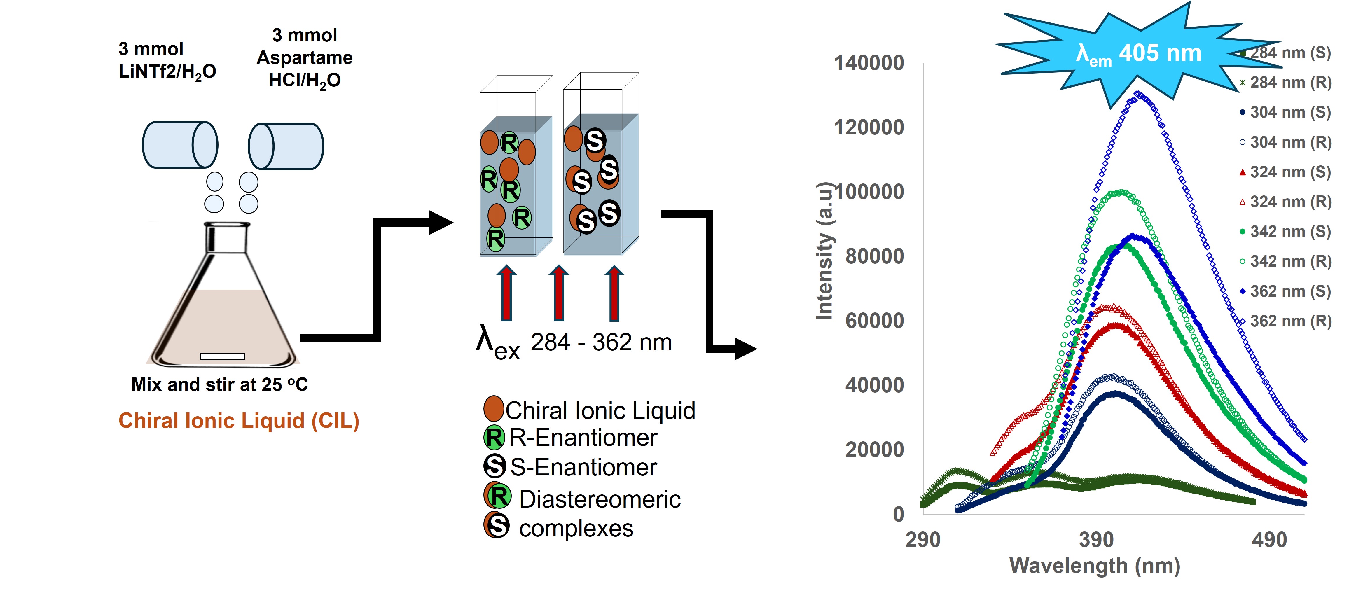
This study describes the synthesis, characterization, and evaluation of enantiomeric recognition properties of two fluorescent chiral ionic liquids (FCILs), L-aspartyl-L-phenylalanine methyl ester bis-(trifluoromethanesulfonyl) amide [ASP] [NTf2] and L-aspartyl-L-phenylalanine methyl ester bis-(perfluoroethylsulfonyl) imide [ASP] [BETI]. The two FCILs were synthesized by means of ion-exchange between L-aspartyl-L-phenylalanine methyl ester hydrochloride [ASP] [HCl] and the lithium salts of bis-(trifluoromethanesulfonyl) amide (LiNTf2) and bis-(perfluoroethylsulfonyl) imide (LiBETI). The products FCILs, [ASP] [NTf2] and [ASP] [BETI], were viscous liquids at room temperature and were stable up to 366 oC. UV and fluorescence studies indicated the FCILs were highly UV absorptive and had strong fluorescence backgrounds. Fluorescence studies demonstrated the FCILs ability to discriminate between the enantiomers of naproxen and propranolol indicating ability serving as effective fluorescent recognition agents for chiral pharmaceutical analytes.
Keywords:
fluorescent chiral ionic liquids
; L-aspartyl-L-phenylalanine methyl ester
; chiral recognition
; fluorescence
; UV spectroscopy
; FTIR spectroscopy
; thermal analysis
1. Introduction
Chiral recognition and chiral separation of enantiomeric compounds are of particular importance in the field of analytical chemistry. To this day, chiral discrimination continues to be a significant problem particularly in the pharmaceutical and agrochemical industries. This is because enantiomers of chiral drugs often differ in their biological and pharmaceutical effects because the molecules that they interact with in the human body are chiral. [1,2] A chemical and engineering news (C&EN) cover story report by Maureen Rouhi [3] and a review by Jessica Caramella et al [4] described many examples of how racemic drugs can cause issues due to differing biological effects and pharmacokinetics profiles. Over the years, scientists have investigated chiral separation and proposed a three-point interaction between the analyte and the chiral selector as a likely recognition process. [5,6,7]
The development of a fast, cost-effective, and straightforward assay for screening and quantifying enantiomeric purity would boost analytical lab testing, regulatory enforcement, and patient safety. There has been a substantial interest in the development of dual-purpose chiral selectors that function simultaneously as solvents and sensors in chiral resolution and spectroscopic detection. [8,9] Chiral ionic liquids (CILs) have emerged as powerful dual-functional agents, serving as reaction media and highly specific chiral selectors for enantiomeric resolution. CILs refer to a highly unique class of compounds known as ionic liquids (ILs) which contain a combination of a chiral cation and/or a chiral anion, or both the cation and anion can be chiral. [8,10] CILs possess the same attractive properties as ILs including high solubility power, negligible vapor pressure, good conductivity, high thermal stability, [11,12,13,14] in addition to having the ability to discriminate chiral molecules. CILs are beneficial because they offer dual solvation of both polar and nonpolar compounds and tunable chiral selectivity. To introduce chirality in CILs, researchers have relied on the use of precursors derived from amino acids, alkaloids, carbohydrates, nicotine, menthol and others. [8] CILs derived from such substituents are desirable because they are naturally occurring and thus considered environmentally safe, sustainable and renewable. Several reports have focused on the role of CILs in enantiodiscrimination using various analytical methods such as spectroscopy, chromatography, and electrophoresis. [15,16,17,18,19] CILs made using amino acids are well suited for enantiodiscrimination studies because they are biodegradable and therefore less toxic. [20,21] Amino acids have proved to be an excellent choice because they are abundant and typically are low cost and have molecular diversity.
Non-fluorescent amino acid-based CILs that serve as chiral selectors have been previously reported. For example, Bwambok et al previously reported the preparation of an alanine butyl ester-based CIL that demonstrated chiral discrimination ability of fluorescent enantiomers of, 1,1-binapthyl-2,2- diamine (BNA), 1,1′-binaphthalene-2,2′-diol (BNP) and naproxen. [22] Other nonfluorescent amino acid-based CILs including L- and D-alanine methyl ester bis(trifluoromethanesulfonimide), L-leucine methyl ester bis(trifluoromethanesulfonimide), L-proline methyl ester bis(trifluoromethanesulfonimide), and tetrabutylammonium L-alanate were described by Kroupa and colleagues. [23] In this study, a racemic mixture of europium complexes was dissolved in each of the five different amino acid-based CILs and circularly polarized luminescence was used to show handedness preferences matching specific L- and D-stereoisomers. Li and coworkers have also reported one fluorescent and various non-fluorescent amino acid ester-based magnetic CILs containing iron (III) that were used as solvents and chiral selectors. In this work, fluorescence spectroscopy was used in chiral discrimination of pure enantiomers of 1,1-binapthyl-2,2- diamine (BNA), 2,2,2-trifluoro-1-(9-anthryl) ethanol and naproxen. [24]
Although the mentioned amino acid based CILs have proven to be effective solvents and chiral selectors, the studies on chiral discrimination needed that the analytes be fluorescent. In response to this limitation, a class of fluorescent chiral ionic liquids (FCILs) for chiral discrimination of both fluorescent and nonfluorescent analytes are needed. Growth in this area has been slow and there are a limited number of instances. For example, a phenylalanine ethyl ester chiral ionic liquid that exhibited chiral discrimination for the pure enantiomers of serine, glucose and mannose was reported by Bwambok and colleagues. [20] More recently, we reported the synthesis of a fluorescent chiral ionic liquid based on glycine-L-histidine dipeptide that demonstrated chiral recognition for enantiomers of naproxen, propranolol and 2,2,2-trifluoro-1-(9-anthryl) ethanol. [18] Anum and coworker lately reported a FCIL from tryptophan amino acid ester that demonstrated enantiomeric recognition of pharmaceutical and environmental fluorescent and nonfluorescent chiral analytes. [25]
In this work, we present the synthesis and characterization of two FCILs derived from L-aspartyl-L-phenylalanine methyl ester also known as aspartame, a popular artificial sugar substitute used in food products. Aspartame contains a dipeptide of two naturally occurring amino acids, aspartic acid and phenylalanine. A hydrochloride salt of aspartame was prepared through ester hydrolysis in a solvent composed of a mixture of methanol and hydrochloric acid crystallizing out the salt. [26] The two CILs were synthesized through ion exchange of the isolated aspartame hydrochloride [ASP] [HCl] and the lithium salts of bis-(perfluoroethylsulfonyl) imide (BETI) and bis-(trifluoromethanesulfonyl) amide (NTf2). The obtained FCILs were characterized to determine purity, and their spectroscopic, thermal and chiral properties evaluated. We show that the two FCILs recognized the enantiomers of two drug analytes: naproxen and propranolol. This work introduces two new fluorescent CILs that can be used for enantiomeric recognition of fluorescent and nonfluorescent analytes.
2. Materials and Methods
2.1. Materials
L-aspartyl-L-phenylalanine methyl ester (aspartame) was purchased from TCI America. Deuterated dimethylsulfoxide (DMSO-d6) was obtained from Millipore Sigma. Lithium bis-(perfluoroethylsulfonyl) imide (LiBETI) and lithium bis-(trifluoromethanesulfonyl) amide (LiNTf2) were purchased from TCI America and Millipore Sigma Aldrich, respectively. (R)-(+)-Propranolol hydrochloride, (S)-(−)-Propranolol hydrochloride, (S)-(+)-6-methoxy-α-methyl-2-naphthaleneacetic acid (S)-naproxen) and (R)-(−)-6-Methoxy-α-methyl-2-naphthaleneacetic acid (R)-naproxen) were obtained from Millipore Sigma Aldrich. Concentrated hydrochloric acid, dichloromethane and methanol (HPLC grade) were bought from VWR and 200 proof ethanol was obtained from Lab Alley Essential Chemicals. All products were used as received.
2.2. Synthesis and Characterization of FCILs
The FCILs were synthesized using an ion exchange reaction between L-aspartyl-L-phenylalanine methyl ester hydrochloride cation, [ASP] [HCl] and two lithium-based perfluorinated anions; lithium bis-(perfluoroethylsulfonyl) imide (LiBETI) and lithium bis-(trifluormethanesulfonyl) amide (LiNTf2). [ASP] [HCl] was prepared by dissolving 8.0 g of L-aspartyl-L-phenylalanine methyl ester (ASP) in a mixture of 5% methanol and 31.5% hydrochloric acid, both made in distilled water. When left to sit without stirring, the aspartame hydrochloride crystallized out of this solution. The FCILs were prepared by combining separate equimolar (8.54 g, 0.0258 mol) aqueous solutions of [ASP] [HCl] with the anions: (7.41 g, 0.0258 mol) of LiNTf2 and (10.0 g, 0.0258 mol) of LiBETI and stirring the mixtures at room temperature until the ionic liquids had formed as shown in Scheme 1. The reaction mixtures separated into two layers the lower layer consisted of the formed FCIL products, [ASP] [NTf2] and [ASP] [BETI], while the upper phase comprised the aqueous lithium chloride (LiCl) byproduct. The lower layers were separated and extracted with three successive portions of dichloromethane to remove the lithium chloride byproduct, and each organic layer was subsequently washed with distilled water. The presence of residual LiCl in the distilled water extract was tested via the addition of an aqueous silver nitrate (AgNO3) solution. The resulting FCILs were dried for 24 hours under vacuum (0.1 Pa) at room temperature to remove water, upon which the NTf2-based FCIL became a thick viscous liquid and the BETI-based FCIL changed to a consistency of a sticky clear gel. The dried FCILs were stored in a desiccator for subsequent characterization using 1H NMR, 13C NMR, FTIR, and UV-Visible spectroscopy, circular dichroism, fluorescence, differential scanning calorimetry (DSC) and thermal gravimetric analysis (TGA).
2.3. Instrumentation
Structures of the synthesized FCILs were characterized by 1H and 13C NMR using a Bruker-AscendTM 400 MHz nuclear magnetic resonance spectrometer with tetramethyl silane (TMS) as an internal standard and DMSO-d6 was used as the solvent. FTIR measurements were acquired on a Nicolet iS10 FTIR spectrometer (Thermo Fisher Scientific) and solid sampling using an ATR accessory with an optical crystal (diamond) on the deck of the accessory and a metal anvil, attached to a metal arm assembly, that applies pressure to solid samples to create close contact with the optical crystal. FTIR measurements were set up for 16 scans, 2 cm-1 resolution, %Transmittance, ATR and background correction, and wavenumber range of 4000 cm-1 – 650 cm-1. A background spectrum was measured with nothing on the diamond crystal. Subsequently, small solid and viscous liquid samples were individually placed on the optical crystal and measured.
The absorption spectra of the two FCILs dissolved in methanol were acquired using a Cary 60 UV-Vis spectrophotometer (Agilent Technologies) equipped with a xenon arc lamp and a photomultiplier tube detector. Sample measurements were taken with a 10-mm quartz cuvette with 2 nm slit widths. A Jasco (J-815) CD spectrometer was used to obtain the CD spectra of the FCILs using a 10-mm quartz cuvette. Steady-state fluorescence measurements were taken using a Photon Technology International QuantaMaster Spectrofluorometer (QM-4 CW) equipped with a 75 W xenon lamp, dual monochromators and a photomultiplier tube detector. Sample measurements were taken with a 10-mm quartz cuvette with 0.25 mm slit widths for the entrance and emission monochromators for the FCILs dissolved in methanol, 0.75 mm for naproxen and 0.5 mm for propranolol chiral recognition studies.
Melting point and glass transition properties of the FCILs were investigated using a TA DSC Q20 Calorimeter under nitrogen. An empty standard aluminum sample pan served as the reference and the FCILs samples were placed on a similar pan. In this study, the temperature increased linearly from 0.0 to 400.0 °C at a heating rate of 10.00 °C/min and the difference in heat flow into samples and reference was measured. The synthesized FCILs were not supercooled due to lack of a liquid nitrogen cooling system. The thermal decomposition temperatures of the FCILs were determined using a TA instrument model, TGA Q50 V20.10 Build 36, operated from 25 to 500.0 °C at a rate of 10.00 °C/min under a nitrogen atmosphere with an open platinum pan.
2.4. Fluorescence Spectroscopic Studies
The fluorescence properties of the two FCILs presented in this work were studied using methanol as a solvent. Solutions of the FCILs were prepared by individually adding an appropriate amount of the neat FCIL to methanol at 15 % m/v concentration. Fluorescence measurements of the resulting FCILs solutions were recorded at an excitation wavelength of 270 nm and emission wavelength of 284 nm. Two commercially available chiral pharmaceuticals were used to investigate the chiral recognition ability of the FCILs. The chiral pharmaceuticals were naproxen, a non-steroidal drug used as anti-inflammatory and propranolol that is applicable to different therapies. Individual solutions of 2.5 µM of pure (R)- and (S)-naproxen in the presence of [ASP] [NTf2]) FCIL in ethanol and 1.69 µM pure (R) and (S)-propranolol in the presence of ASP] [BETI] FCIL in ethanol were investigated using fluorescence spectroscopy at multiple excitation wavelengths to investigate the chiral recognition of the two chiral drugs using the synthesized FCILs as chiral selectors.
2.5. Solution Preparation for Fluorescence-Based Enantiomeric Recognition Studies
Solutions of equal concentrations of pure enantiomers of naproxen and propranolol were prepared and the intensities of their fluorescent emission signals matched. For each pair of pure enantiomers, concentrations were adjusted to make sure they were the same by matching the fluorescence emission intensities of the solutions upon excitation at the maximum absorbance wavelengths. These solutions consisted of pure enantiomers of (R) and (S)-naproxen and (R) and (S)- propranolol dissolved in ethanol at concentrations of 2.5 µM and 1.69 µM, respectively. Pre-established volumes (3.0 mL) of the obtained solutions of 2.5 µM pure enantiomers of naproxen were separately added into empty vials and a stream of nitrogen was used to evaporate off the solvent and subsequently 2.5 mL of the neat [ASP] [NTf2] FCIL and 1.5 mL of ethanol were added to each of the vials that contained known masses of the naproxen enantiomers. Ethanol was added to the 2.5 mL neat [ASP] [NTf2] FCIL solutions to increase the transparency of the solutions as the neat FCIL was hazy. Because the neat [ASP] [BETI] FCIL was a gel it could not be used directly as a solvent. Solutions of enantiomeric recognition studies of 1.69 µM pure enantiomers of (R) and (S)-propranolol using [ASP] [BETI] FCIL were prepared by adding a pre-established volume (2.0 mL) of the obtained equal concentrations of the pure enantiomers of (R) and (S)-propranolol into vials containing 0.2 g of the neat [ASP] [BETI] FCIL. The vialed contents were mixed using a vortex for five minutes and subsequently the mixtures were stored in a desiccator and in the dark overnight to allow for total dissolution of the enantiomers and equilibration prior to fluorescence measurements. All naproxen and propranolol solutions were prepared in triplicate.
3. Results and Discussion
3.1. 1H and 13C NMR
The purity of the synthesized FCILs was determined by 1H and 13C NMR. Figure 1, Figure 2, Figure 3 and Figure 4 show the obtained 1H and 13C NMR spectra of the two FCILs. The spectra were acquired using deuterated dimethyl sulfoxide (DMSO-d6) with TMS as the internal standard. As the 1H and 13C NMR results show, the chemical shifts of protons and carbons are in good agreement with the chemical structures of the [ASP] [NTf2] and [ASP] [BETI] FCILs.
3.2. FTIR Analysis
The purity of the two FCILs was also evaluated using FTIR. Figure 5 shows that the IR spectra of the two FCILs ([ASP] [NTf2] and ([ASP] [BETI]) had a combination of peaks of the starting materials including the two lithium-based anions, LiNTf2, LiBETI and the aspartame hydrochloride [ASP] [HCl] cation. For example, characteristic IR absorption peaks corresponding to the functional groups, methyl ester carbonyl (~ 1720 cm-1), amide/peptide carbonyl (~ 1690 cm-1), and N-H/O-H stretching regions (~ 3317 cm-1) were observed in both the starting material [ASP] [HCl] and the two FCILs ([ASP] [NTf2] and ([ASP] [BETI]). Similarly, SO2 asymmetric stretching mode (~1320–1350 cm-1), the S–N stretching mode, (~746 cm-1), C–S stretching vibration (~750 cm-1) and the S–N–S stretching vibration ((~740 cm-1) commonly found in sulfonylimide anions) were observed in both the starting materials, lithium bis-(trifluoromethanesulfonyl) amide ([LiNTf2]), lithium bis-(perfluoroethylsulfonyl) imide ([LiBETI]), and the two FCILs ([ASP] [NTf2] and ([ASP] [BETI]).
3.3. UV Analysis
UV measurements of 15 % m/v FCIL to methanol (Figure 6) showed that the two FCILs had similar absorption spectra with an absorption peak at a maximum of 260 nm. This peak is well known to be the absorption characteristic of phenylalanine as documented by Lundblad and coworker in the handbook of biochemistry and molecular biology. [27]
3.4. Circular Dichroism Measurements
Circular dichroism occurs when chiral molecules absorb left and right circularly polarized light differently. The chiral integrity of the synthesized FCILS was determined using CD to check for any possibility of racemization following the ion exchange. The positive CD spectral bands of [ASP] [NTf2] and [ASP] [BETI] measured in methanol are shown in Figure 7. The positive signals indicate that the pure enantiomers of the dipeptide L-aspartyl-L-phenylalanine present in the synthesized [ASP] [NTf2] and [ASP] [BETI] FCILs absorb left-circularly polarized light confirming their retention of chiral configuration.
3.5. Fluorescence Spectra
Fluorescence spectra of 15 % m/v FCIL to methanol were collected at excitation maximum wavelength of 270 nm, respectively for the [ASP] [NTf2] and [ASP] [BETI] FCILs. Figure 8 illustrates the fluorescence characteristics of the two FCILs. The FCILs had similar fluorescence spectral profiles to the reported excitation and emission spectra of phenylalanine fluorophore. [28] The excitation scan of the FCIL derived from the NTf2 anion lacked vibrational structure as peaks at 257 and 264 nm were not observed which could imply stronger interactions of the excited state species with methanol. The two FCILs exhibited a strong fluorescence background with an emission centered at 284 nm. This characteristic makes the two FCILs useful as chiral selectors for enantiomeric recognition of both non-fluorescent and fluorescent analytes.
3.6. Fluorescence Enantiomeric Recognition Studies
Published work has demonstrated that fluorescence spectroscopy is an effective technique for analyzing chiral recognition between a chiral ionic liquid (CIL) and pure individual enantiomers of compounds. [16,18,19,20,22,24,25,29,30] The chiral recognition ability of the two synthesized FCILs, [ASP] [NTf2] and [ASP] [BETI] were evaluated using pure enantiomers of naproxen and propranolol. The pure enantiomers of naproxen and propranolol had emission maxima’s at 345 nm, and 329 nm upon excitation at 333 nm, and 293 nm, respectively. The fluorescence emission spectra of equal concentrations of the pure enantiomers of naproxen (2.5 µM) and propranolol (1.69 µM) dissolved in ethanol are shown in the supporting information. Without the FCILs, the variation in fluorescence intensity of these solutions was statistically insignificant. Figure 9 shows the emission spectra of the equimolar amounts of the pure enantiomers of naproxen (2.5 µM pure (R) and (S)-naproxen) in the presence of 62.5% v/v [ASP] [NTf2] FCIL in ethanol obtained at excitation wavelengths that exhibited largest difference in fluorescence intensity. The results showed that the R form of naproxen had a higher emission intensity than the S form. For a CIL to effectively serve as a chiral selector for enantiomeric recognition, the CIL must differentiate between the (R)- and (S)-enantiomers of a chiral compound and diastereomeric interactions must result in the emission intensity difference between the enantiomer pair. In the presence of [ASP] [NTf2] FCIL, the emission intensity difference observed between pure enantiomers of naproxen at multiple wavelengths confirmed the chiral discrimination ability this FCIL. The different behavior of the naproxen enantiomers with the FCIL is due to intermolecular interactions that depend on molecular structure, such as H-bonds from functional groups such as OH, NH, COOH and fluorine atoms as well as π-π interactions between the aromatic rings present in the FCIL and naproxen. The emission spectra of equimolar amounts of the pure enantiomers of naproxen in the presence of 62.5% v/v [ASP] [NTf2] FCIL in ethanol upon excitation at other wavelengths can be seen in the supporting information.
Figure 10 shows the emission spectra of equimolar amounts of the pure enantiomers of propranolol (1.69 µM pure (R) and (S)-propranolol) in the presence of 0.1 % m/v [ASP] [BETI] FCIL in ethanol obtained at excitation wavelengths that exhibited the largest separation in fluorescence intensity. (R)- propranolol displayed a higher emission intensity than (S)-propranolol. [ASP] [BETI] demonstrated chiral recognition properties for propranolol even in the presence of a substantially smaller concentration of the chiral ionic liquid. The observed emission intensity difference between (R) and (S)-propranolol confirmed the discrimination ability of this FCIL through intermolecular interactions involving hydrogen bonding and π electrons. The emission spectra of equimolar amounts of the pure enantiomers of propranolol in the presence of 0.1 % m/v [ASP] [BETI] FCIL in ethanol upon excitation at other wavelengths can be seen in the supporting information.
A red shifted broad emission peak was observed upon excitation at longer wavelengths in the fluorescence spectra of both pure enantiomers of naproxen and propranolol in the presence of [ASP] [NTf2] and [ASP] [BETI] CILs, respectively as displayed in Figure 9 and Figure 10. This can be attributed to different geometric orientations in the excited and ground states arising from π - π interactions of the phenylalanine fluorophore leading to the formation of excited dimer. [28]
We observed that the enantiomeric recognition processes of [ASP] [NTf2] and [ASP] [BETI] on fluorescent analytes exhibited both shared characteristics and notable deviations from other reported amino acid-based CILs. For example, the [ASP][BETI] has enantiomeric recognition properties on propranolol that is comparable to TrpC2NTf2 CIL [25] in which R-propranolol displayed higher intensity compared to S-propranolol after interaction with both ([ASP][BETI] and TrpC2NTf2) FCILs but the reverse was observed with the dipeptide [Gly-L-His] [NTf2] CIL. [18] On the contrary, the [ASP] [NTf2] CIL has enantiomeric recognition properties on naproxen in which R-naproxen displayed higher intensity compared to S-naproxen but the complete reverse was observed with the dipeptide [Gly-L-His] [NTf2], [18] and AlaOMeFeCl4 [24] CILs in which S-naproxen displayed higher intensity compared to R-naproxen. The noted similarities and contrasts in enantiomeric recognition show that the intermolecular interactions and emission intensity of the formed diastereomers depend on the chemical structures of both the CILs and the enantiomers.
3.7. Thermal Analysis
Differential scanning calorimetry (DSC) analysis results of the [NTf2] and [BETI]- based FCILs can be seen in Figure 11. The DSC curves displayed an endothermic shift in the baseline at around 150 °C and 200 °C respectively, due to the glass transition. This is due to an increase in heat capacity while heating the amorphous samples through the glass transition temperature, Tg. As the samples are heated above the Tg, there is high molecular mobility leading to alignment of the molecules and crystallization displayed as the exothermic peak around 250 °C in both figures. The melting transition for both samples can be seen centered at around 300 °C. This high melting point can be attributed to the aspartame moiety that is the bulk of the cation in this FCILs. Aspartame has a reported melting point of 246.5 °C. [31] As the temperature was ramped above the melting point, the (NTf2) and (BETI) -based FCILs began decomposing at 325 °C and 350 °C respectively.
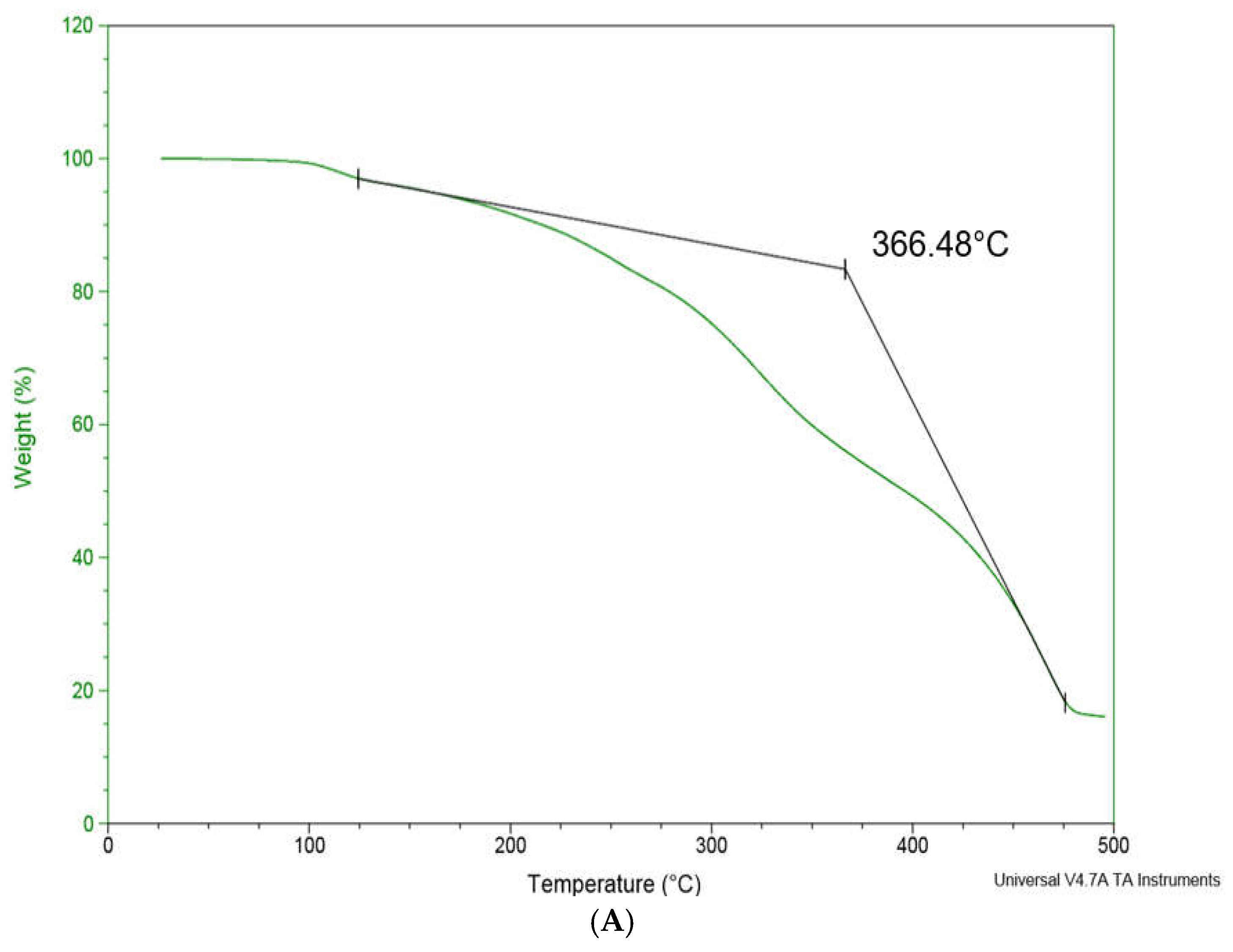
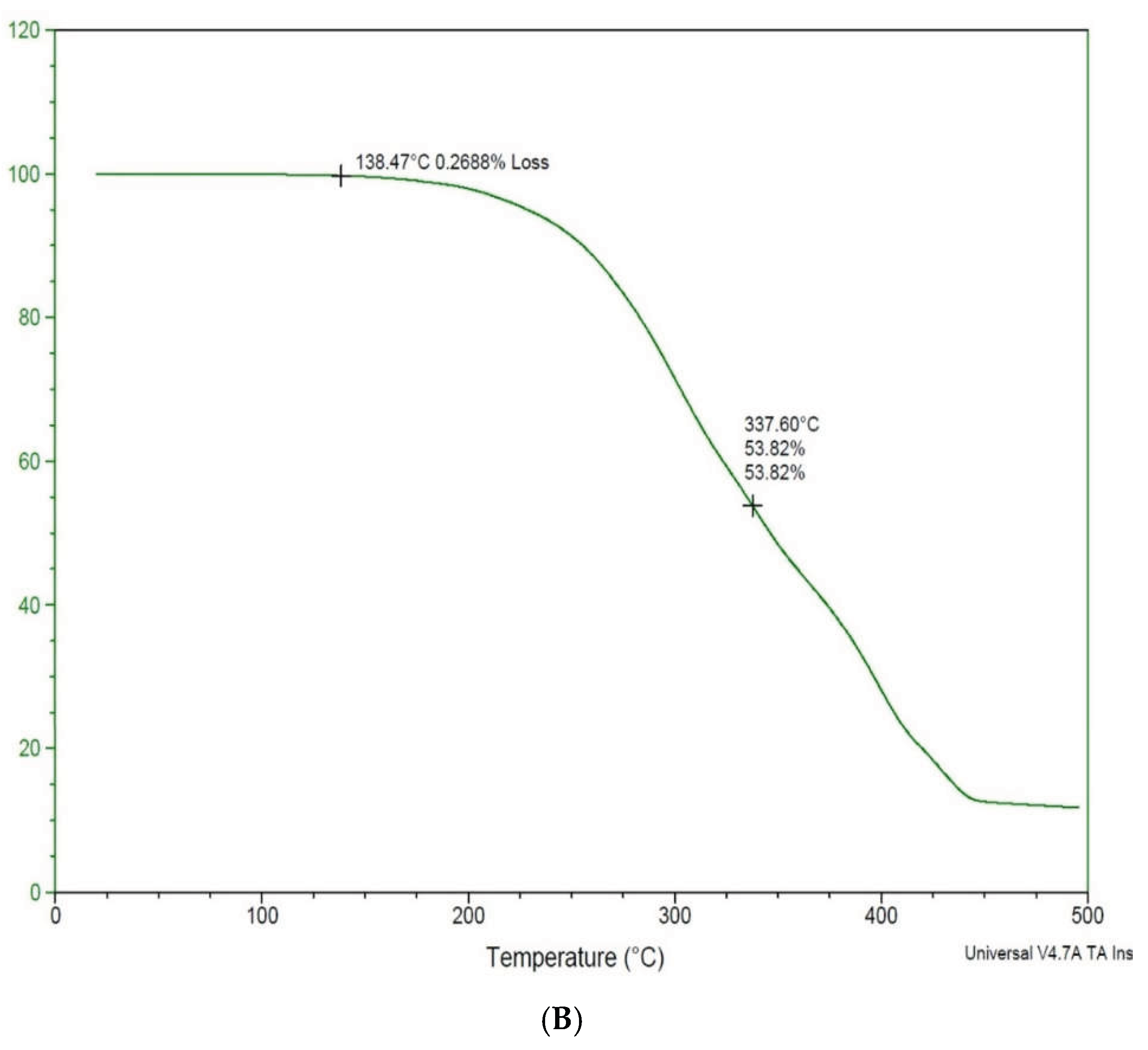
Thermal gravimetric analysis (TGA) results (Figure 12) showed that the two FCILs have good temperatures of decomposition up to 366 °C at a scan rate of 10 °C min-1. The onset temperature Tonset, of the two CILs was around 200 °C. The T50% temperature of the FCILs were 366 °C for the NTf2-based IL and 337 °C for the [BETI]-based IL indicating that the [NTf2]-based IL has a higher thermal stability. These results indicate that both [ASP] [NTf2] and [ASP] [BETI] have potential to serve as reliable chiral coatings in gas chromatography or as chiral selectors for applications and reactions at room and higher temperatures.
4. Conclusions
A series of new dipeptide-based FCILs derived from L-aspartyl-L-phenylalanine methyl ester were synthesized using an ion-exchange reaction. The FCILs were viscous liquid at room temperature. The CILs were characterized using NMR, IR, UV and fluorescence spectroscopy. 1H and 13C NMR analysis indicated that the synthesized FCILs were pure and FTIR spectra showed they had a combination of peaks of the cation (aspartame hydrochloride) and the lithium-based perfluorinated anions. The synthesized FCILs were found to be highly UV absorptive and possess strong fluorescence background that can be attributed to the phenylalanine moiety present in the dipeptide. Steady state fluorescence spectroscopy was used to confirm their chiral recognition ability towards two chiral drug molecules, naproxen and propranolol. Their strong fluorescence properties and ability to discriminate chiral analytes make them particularly useful for investigations that seek to analyze interactions between them and both nonfluorescent and fluorescent compounds. The two dipeptide-based FCILs had high melting points and were thermally stable up to 367 °C. These new FCILs with high thermal stability are expected to have potential as chiral selectors in applications such as high temperature reactions and or as suitable chiral coatings in gas chromatography. To our knowledge, this is the first report of dipeptide-based FCILs derived from L-aspartyl-L-phenylalanine methyl ester.
Supplementary Materials
The following supporting information can be downloaded at https://www.mdpi.com/article/doi/s1, Figure S1: Fluorescence emission spectra of pure enantiomers of naproxen (2.50 µM) in ethanol. Figure S2. Fluorescence emission spectra of pure enantiomers of propranolol (1.69 µM) in ethanol. Figure S3. Fluorescence emission spectra of equal concentrations of the pure enantiomers of naproxen (2.5 µM pure (R) and (S)-naproxen) in the presence of 62.5% v/v [ASP] [NTf2] FCIL in ethanol upon excitation at all the wavelengths that covered the excitation maxima of naproxen and [ASP] [NTf2] FCIL. Figure S4. Fluorescence emission spectra of 1.69 µM pure (R) and (S)-Propranolol in the presence of 0.1 % m/v [ASP] [BETI] FCIL in ethanol obtained at all the wavelengths that covered the excitation maxima of propranolol and [ASP] [BETI] FCIL.
Author Contributions
Irene W. Kimaru: conceptualization, methodology, investigation, resources, data curation, writing, and review and editing. Alex Zoey Slater: investigation, data curation, review and editing, and validation. Olivia Culberston and Emma Garn: investigation, data curation.
Funding
This research received no external funding.
Data Availability Statement
The data that support the findings of this study are available on request from the corresponding author. The data is not publicly available due to privacy or ethical restrictions.
Acknowledgments
The authors would like to acknowledge St. John Fisher University for supporting this work. We thank Dr. Dave Vivek for help with TGA and DSC measurements and Dr. Ruel McKnight from SUNY Geneseo for help with CD measurements.
Conflicts of Interest
The authors declare no conflicts of interest.
Abbreviations
The following abbreviations are used in this manuscript:
| CILs | Chiral ionic liquids |
| FCILs | Fluorescent chiral ionic liquids |
| [ASP] | L-aspartyl-L-phenylalanine methyl ester |
| [ASP] [HCl] | L-aspartyl-L-phenylalanine methyl ester hydrochloride |
| (CF3SO2)2N | Bis-(trifluoromethanesulfonyl) amide |
| NTf2 | Bis-(trifluoromethanesulfonyl) amide |
| (C2F5SO2)2N) | Bis-(perfluoroethylsulfonyl) imide |
| BETI | Bis-(perfluoroethylsulfonyl) imide |
| LiNTf2 | Lithium bis-(trifluoromethanesulfonyl) amide |
| LiBETI | Lithium bis-(perfluoroethylsulfonyl) imide |
| [ASP] [NTf2] | L-aspartyl-L-phenylalanine methyl ester bis-(trifluoromethanesulfonyl) amide |
| [ASP] [BETI] | L-aspartyl-L-phenylalanine methyl ester bis-(perfluoroethylsulfonyl) imide |
| UV | Ultraviolet |
| NMR | Nuclear magnetic resonance |
| TMS | Tetramethylsilane |
| FTIR | Fourier-Transform Infrared spectroscopy |
| ATR | Attenuated Total Reflectance |
| TGA | Thermal Gravimetric Analysis |
| DSC | Differential Scanning Calorimetry |
References
- Caldwell, J. Importance of stereospecific bioanalytical monitoring in drug development. J. Chromatogr. A 1996, 719, 3–13. [Google Scholar] [CrossRef] [PubMed]
- Calcaterra, A.; D'Acquarica, I. The market of chiral drugs: Chiral switches versus de novo enantiomerically pure compounds. J. Pharm. Biomed. Anal. 2018, 147(5), 323–340. [Google Scholar] [CrossRef] [PubMed]
- Rouhi, M.A. Chirality at Work. C&EN 2003, 81(18), 56–61. [Google Scholar] [CrossRef]
- Ceramella, J.; Iacopetta, D.; Franchini, A.; De Luca, M.; Saturnino, C.; Andreu, I.; Sinicropi, M.S.; Catalano, A. A Look at the Importance of Chirality in Drug Activity: Some Significant Examples. App. Sci. 2022, 12, 10909. [Google Scholar] [CrossRef]
- Dalgliesh, C.E. The Optical Resolution of Aromatic Amino-Acids on Paper Chromatograms. J. Chem. Soc. 1952, 3940–3942. [Google Scholar] [CrossRef]
- Pasutto, M.F. Mirror Images: The Analysis of Pharmeceutical Enantiomers. J. Clin. Pharmacol. 1992, 32, 917–924. [Google Scholar] [CrossRef] [PubMed]
- Berthod, A. Chiral Recognition Mechanisms. Anal. Chem. 2006, 78(7), 2093–2099. [Google Scholar] [CrossRef] [PubMed]
- Flieger, J.; Feder-Kubis, J.; Tatarczak-Michalewska, T. Chiral Ionic Liquids: Structural Diversity, Properties and Applications in Selected Separation Techniques. Int. J. Mol. Sci. 2020, 21(4253), 1–39. [Google Scholar] [CrossRef] [PubMed]
- Qian, H.L.; Xu, S.T.; Yan, X.P. Recent Advances in Separation and Analysis of Chiral Compounds. Anal. Chem. 2023, 95(1), 304–318. [Google Scholar] [CrossRef] [PubMed]
- Wang, T.; Yang, H.; Qiu, R.; Huang, S. Synthesis of novel chiral imidazolium stationary phases and their enantioseparation evaluation by high-performance liquid chromatography. Anal. Chim. Acta 2016, 944, 70–77. [Google Scholar] [CrossRef] [PubMed]
- Sun, P.; Armstrong, D.W. Ionic liquids in analytical chemistry. Anal. Chim. Acta 2010, 661(1), 1–16. [Google Scholar] [CrossRef] [PubMed]
- Welton, T. Room-Temperature Ionic Liquids. Solvents for Synthesis and Catalysis. Chem. Rev. 1999, 99, 2071. [Google Scholar] [CrossRef] [PubMed]
- Zhao, H.; Xia, S.; Ma, P. Use of ionic liquids as ‘green’ solvents for extractions. J. Chem. Technol. Biotechnol. 2005, 80, 1089. [Google Scholar] [CrossRef]
- Akolkar, H.N.; Shaikh, M.H.; Haghi, A.K. Ionic Liquids. Greener Solvents for Sustainable Chemistry (Synthesis Lectures on Sustainable Development); Springer: New York, NY, USA, 2025; pp. 1–32. [Google Scholar] [CrossRef]
- Singh, A.; Kaur, N.; Kumar Chopra, H. Chiral recognition methods in analytical chemistry: Role of the chiral ionic liquids. Crit. Rev. Anal. Chem. 2019, 49, 553–569. [Google Scholar] [CrossRef] [PubMed]
- Li, M.; Bwambok, D.K.; Fakayode, S.O.; Warner, I.M. Chiral ionic liquids in chromatographic separation and spectroscopic discrimination. In Chiral Recognition in Separation Methods; Berthod, A., Ed.; Springer: New York, NY, USA; Berlin/Heidelberg, Germany, 2010; p. 289. [Google Scholar]
- Kapnissi-Christodoulou, C.P.; Stavrou, I.J.; Mavroudi, M.C. Chiral ionic liquids in chromatographic and electrophoretic separations. J. Chromatogr. A 2014, 1363, 2–10. [Google Scholar] [CrossRef] [PubMed]
- Kimaru, I.W.; Maltese, L. Synthesis and Characterization of a Glycine-L-Histidine-Based Chiral Ionic Liquid and Enantioselectivity Evaluation by Fluorescence Spectroscopy. ChemistrySelect 2022, 7, 1–7. [Google Scholar] [CrossRef]
- Kimaru, I.W.; Morris, L.; Vassiliou, J.; Savage, N. Synthesis and Evaluation of L-Phenylalanine Ester-based Chiral Ionic Liquids for GC Stationary Phase Ability. J. Mol. Liq. 2017, 237, 193–200. [Google Scholar] [CrossRef]
- Bwambok, D.K.; Challa, S.K.; Lowry, M.; Warner, I.M. Amino acid-based fluorescent chiral ionic liquid for enantiomeric recognition. Anal. Chem. 2010, 82, 5028–5037. [Google Scholar] [CrossRef] [PubMed]
- Kirchhecker, S.; Esposito, D. Amino acid based ionic liquids: A green and sustainable perspective. Curr. Opin. Green Sustain. Chem. 2016, 2, 28–33. [Google Scholar] [CrossRef]
- Bwambok, D.K.; Marwani, H.M.; Fernand, V.E.; Fakayode, S.O.; Lowry, M.; Negulescu, I.; Strongin, R.M.; Warner, I.M. Synthesis and Characterization of Novel Chiral Ionic Liquids and Investigation of Their Enantiomeric Recognition Properties. Chirality 2008, 20(2), 151–158. [Google Scholar] [CrossRef] [PubMed]
- Kroupa, D.M.; Brown, C.J.; Heckman, L.M.; Hopkins, Todd A. Chiroptical Study of Chiral Discrimination by Amino Acid Based Ionic Liquids. J. Phys. Chem. B. 2012, 116(16), 4952–4958. [Google Scholar] [CrossRef] [PubMed]
- Li, M.; De Rooy, S.L.; Bwambok, D.K.; El-Zahab, B.; DiTusa, J.F.; Warner, I.M. Magnetic Chiral Ionic Liquids Derived from Amino Acids. Chem. Commun. 2009, 45, 6922. [Google Scholar] [CrossRef] [PubMed]
- Anum, D.; Bwambok, D.K. Synthesis of a chiral ionic liquid based on L-tryptophan ester and evaluation of enantiomeric recognition properties using fluorescence spectroscopy. J. Mol. Liq. 2023, 390, 123141. [Google Scholar] [CrossRef]
- Ozawa, Y.; Shinohara, E.; Eguchi, C.; Kishimoto, S.; Takemoto, T. Method of preparing alpha-l-aspartyl-l-phenylalanine methyl ester and its hydrochloride. 1984, 1–9. [Google Scholar] [PubMed]
- Lundblad, R.L.; Macdonald, F. Handbook of biochemistry and molecular biology; CRC Press: Boca Raton, FL, USA, 2010; p. 81. [Google Scholar]
- Lakowicz, J.R. Principles of Fluorescence Spectroscopy; Kluwer Academic/Plenum Publishers: New York, NY, USA, 1999; p. 452. [Google Scholar]
- Kaur, N.; Rahim, J.U.; Rai, R.; Chopra, H.K. Synthesis and Application of (S)-Nicotine-Based Chiral Ionic Liquids in Enantiomeric Recognition by Using Fluorescence Spectroscopy. ChemistrySelect. 2021, 6(23), 5685–5688. [Google Scholar] [CrossRef]
- De Rooy, S.L.; Li, M.; Bwambok, D.K.; El-Zahab, B.; Challa, S.; Warner, I.M. Ephedrinium-Based Protic Chiral Ionic Liquids for Enantiomeric Recognition. Chirality 2011, 23, 54–62. [Google Scholar] [CrossRef] [PubMed]
- Lide, D.R. CRC Handbook of Chemistry and Physics 88TH Edition 2007-2008; CRC Press, Taylor & Francis: Boca Raton, FL, USA, 2007; pp. 3–30. [Google Scholar]
Scheme 1.
Reaction scheme for the synthesis of L-aspartyl-L-phenylalanine methyl ester-based ionic liquids ([ASP] [Y]). [ASP = Aspartame] [Y = (CF3SO2)2N) or NTf2 and (C2F5SO2)2N) or BETI.
Scheme 1.
Reaction scheme for the synthesis of L-aspartyl-L-phenylalanine methyl ester-based ionic liquids ([ASP] [Y]). [ASP = Aspartame] [Y = (CF3SO2)2N) or NTf2 and (C2F5SO2)2N) or BETI.
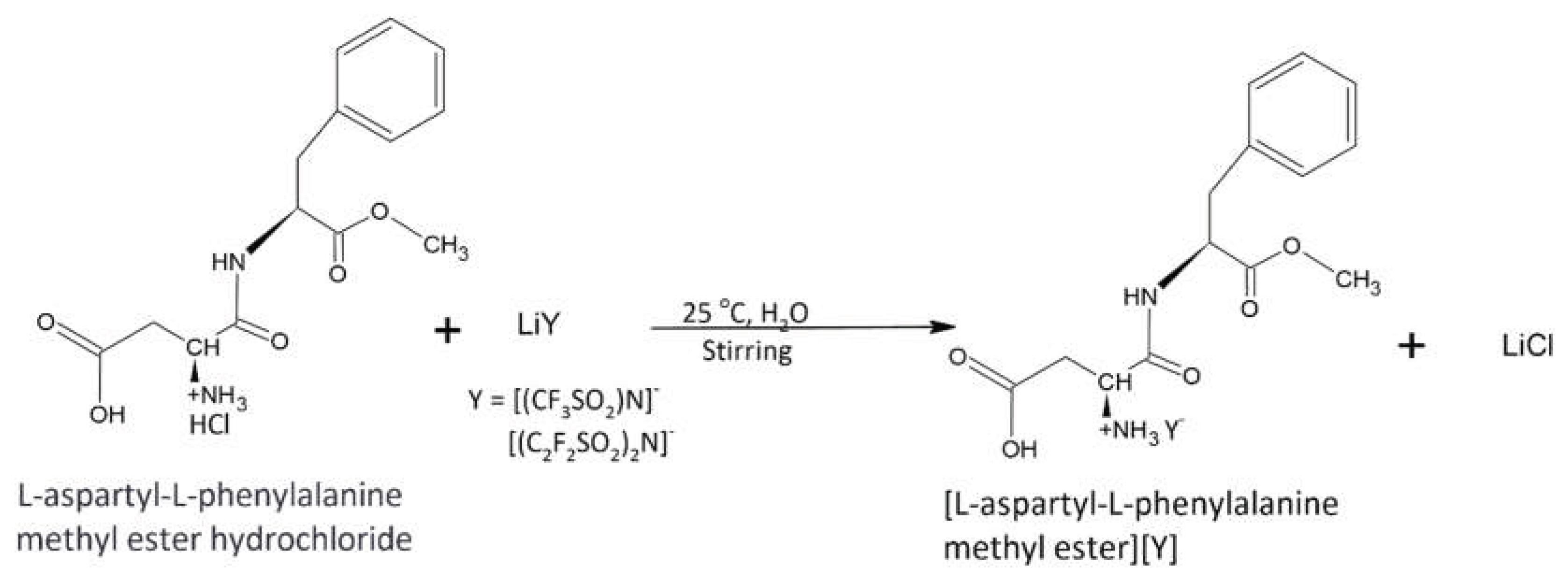
Figure 1.
1H NMR Spectrum of [ASP] [NTf2] FCIL. (The structure of the FCIL and peak chemical shift assignments are included in the inset).
Figure 1.
1H NMR Spectrum of [ASP] [NTf2] FCIL. (The structure of the FCIL and peak chemical shift assignments are included in the inset).
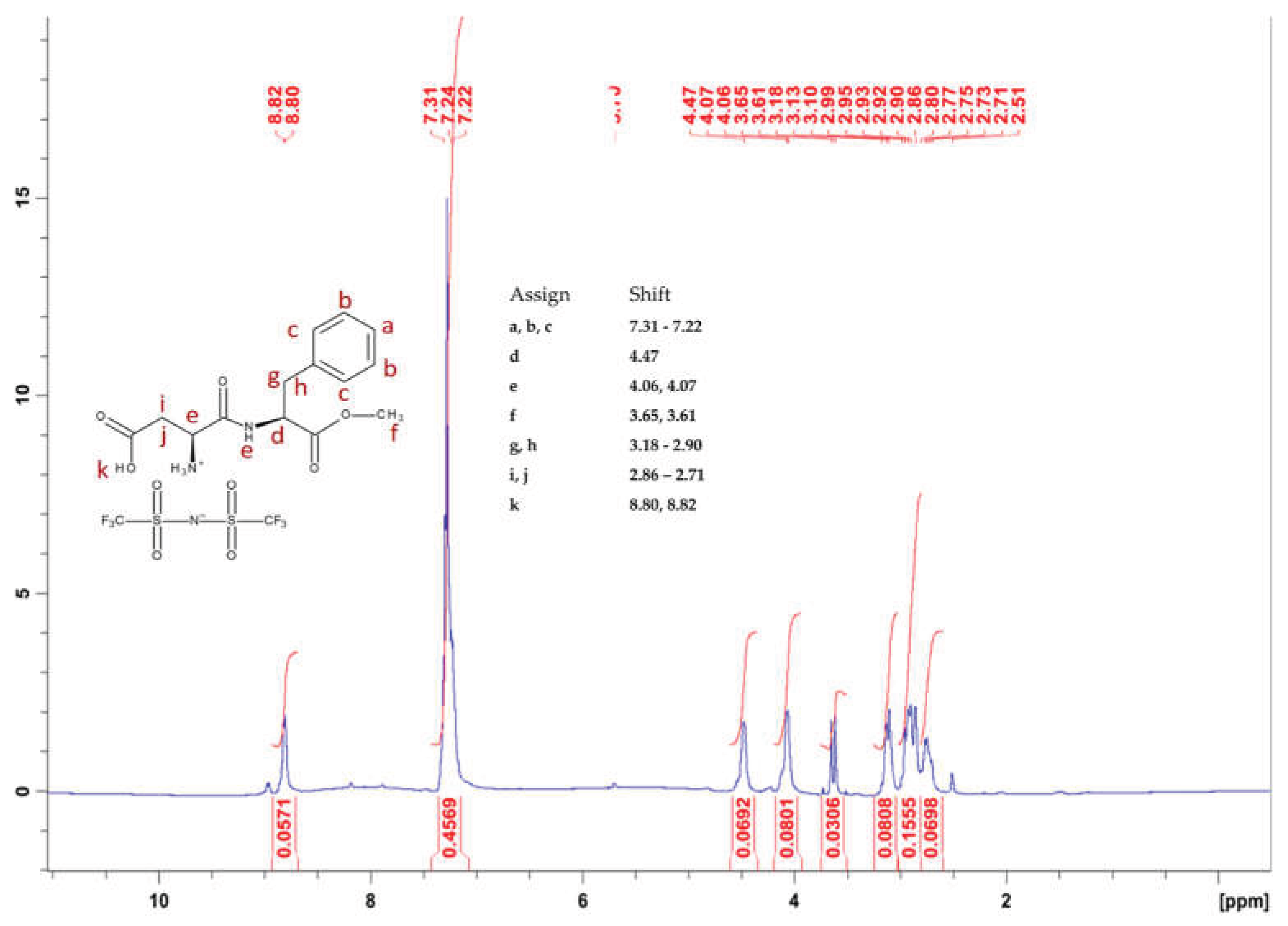
Figure 2.
1H NMR Spectrum of [ASP] [BETI] FCIL. (The structure of the FCIL and peak chemical shift assignments are included in the inset).
Figure 2.
1H NMR Spectrum of [ASP] [BETI] FCIL. (The structure of the FCIL and peak chemical shift assignments are included in the inset).
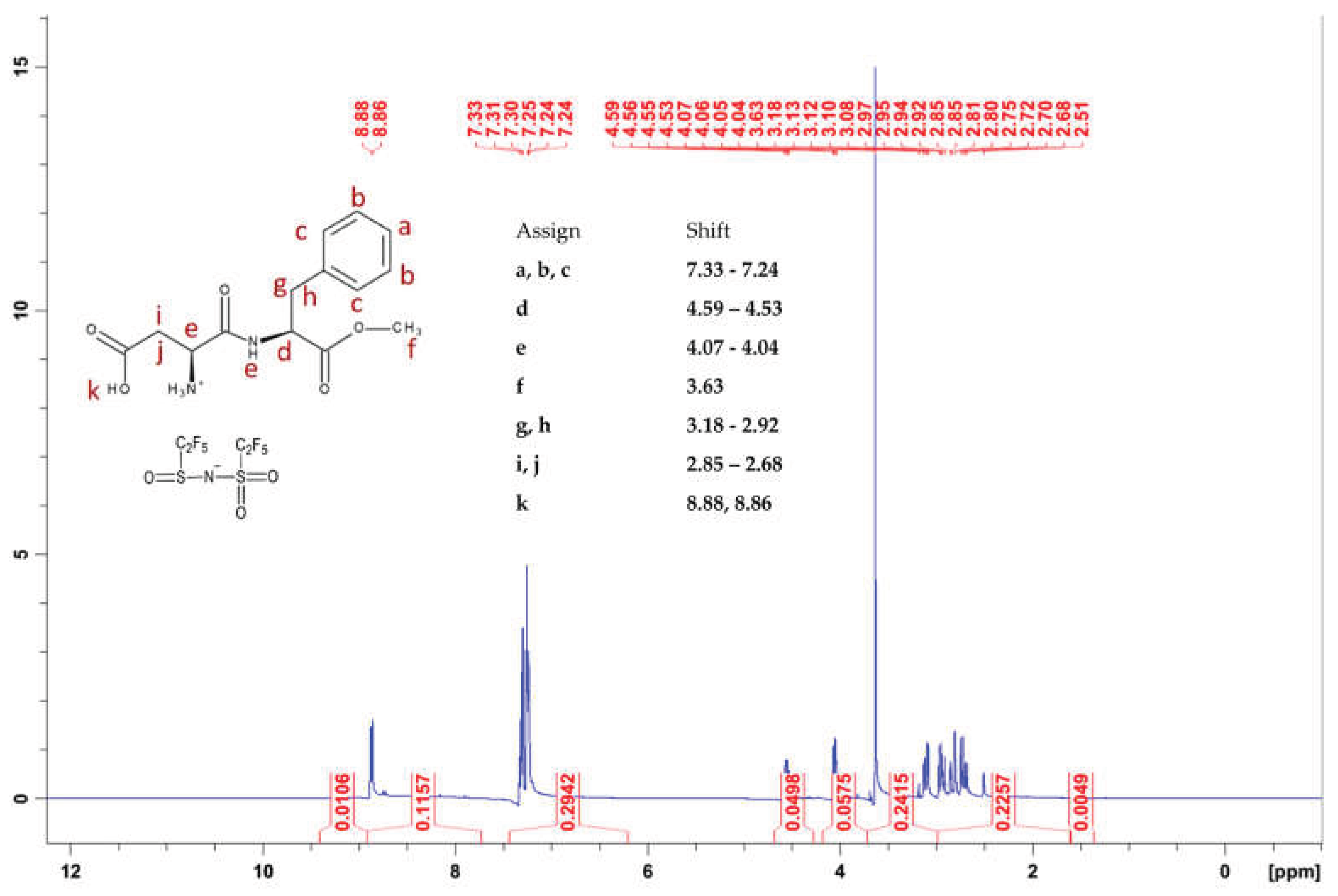
Figure 3.
13C NMR Spectrum of [ASP] [NTf2] FCIL. (The structure of the FCIL and peak chemical shift assignments are included in the inset).
Figure 3.
13C NMR Spectrum of [ASP] [NTf2] FCIL. (The structure of the FCIL and peak chemical shift assignments are included in the inset).
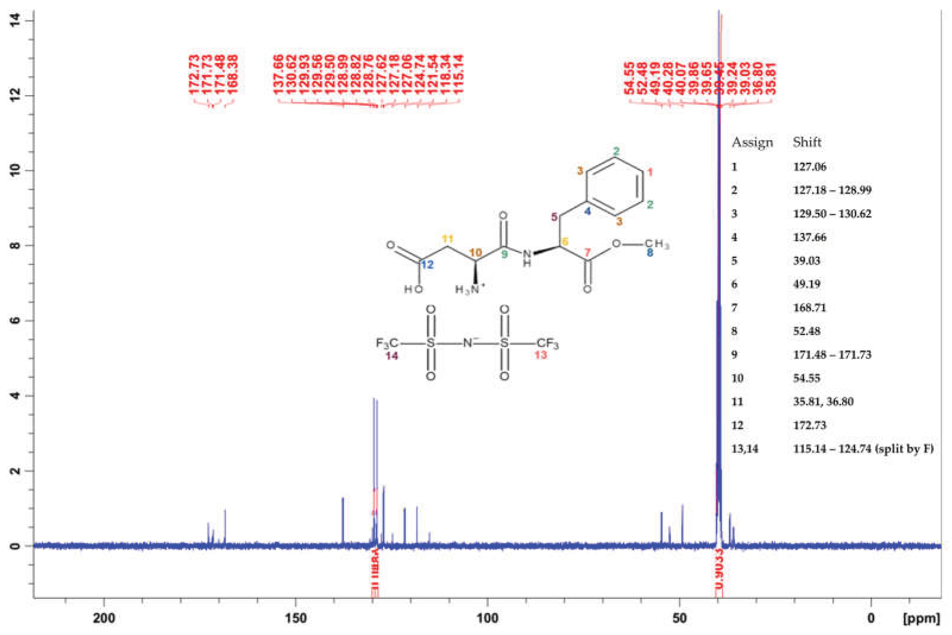
Figure 4.
13C NMR Spectrum of [ASP] [BETI] FCIL. (The structure of the FCIL and peak chemical shift assignments are included in the inset).
Figure 4.
13C NMR Spectrum of [ASP] [BETI] FCIL. (The structure of the FCIL and peak chemical shift assignments are included in the inset).
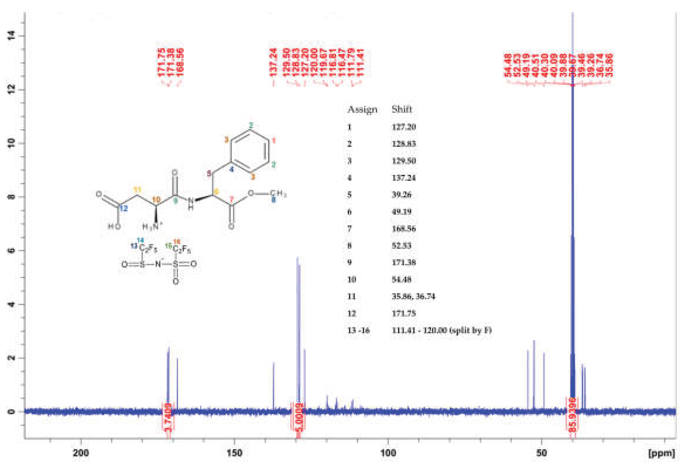
Figure 5.
FTIR spectra of (A) LiNTf2 (B) LiBETI, (C) [ASP] [HCl] (D) [ASP] [NTf2] FCIL and (E) [ASP] [BETI] FCIL.
Figure 5.
FTIR spectra of (A) LiNTf2 (B) LiBETI, (C) [ASP] [HCl] (D) [ASP] [NTf2] FCIL and (E) [ASP] [BETI] FCIL.
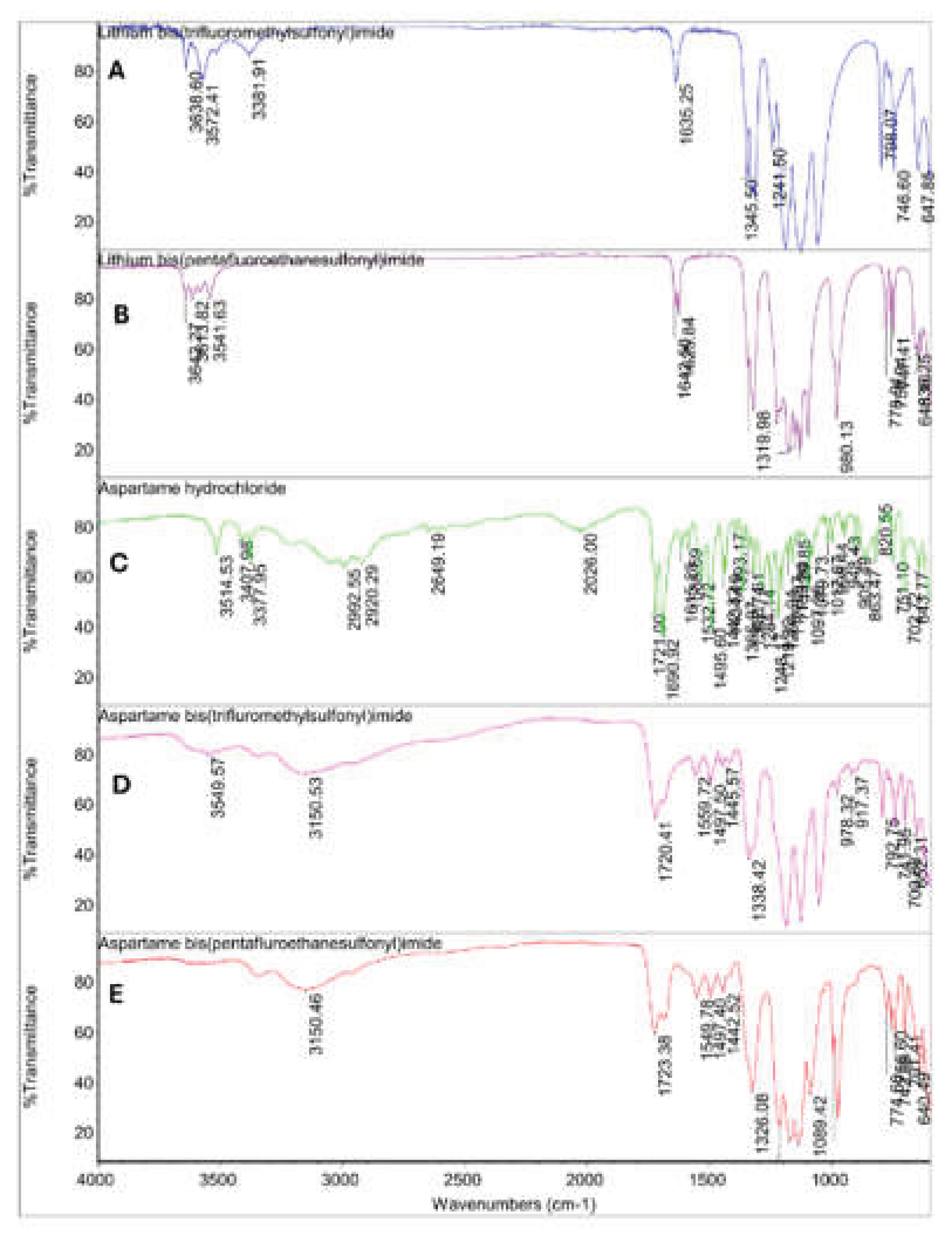
Figure 6.
Ultraviolet spectra of 15 % m/v [ASP] [NTf2] and [ASP] [BETI] in methanol.
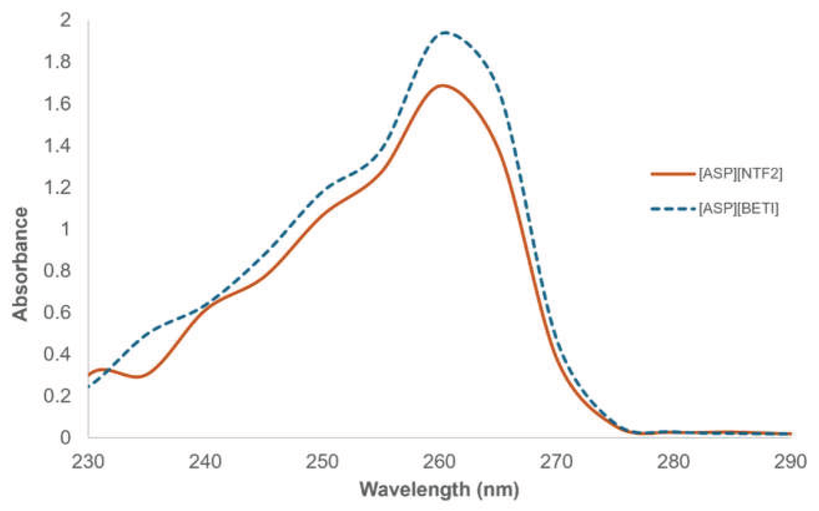
Figure 7.
Circular dichroism spectrum of 15 % m/v [ASP] [NTf2] and [ASP] [BETI] in methanol at room temperature.
Figure 7.
Circular dichroism spectrum of 15 % m/v [ASP] [NTf2] and [ASP] [BETI] in methanol at room temperature.
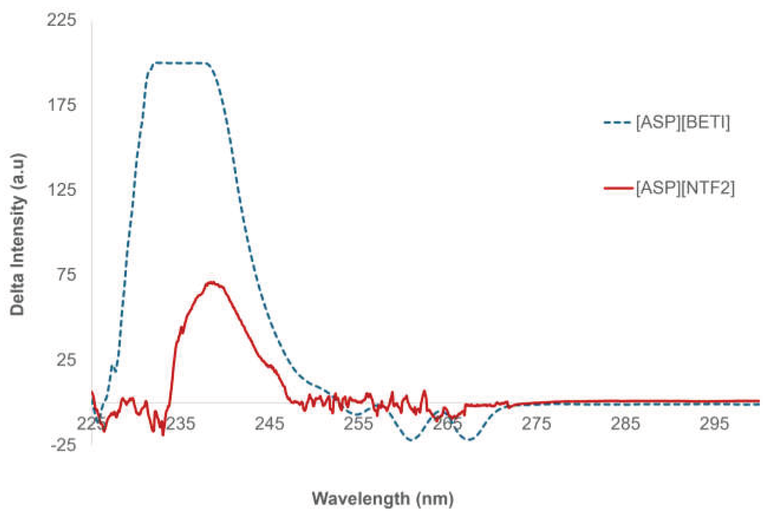
Figure 8.
Fluorescence spectra of 15 % m/v [ASP] [NTf2] and [ASP] [BETI] in methanol.
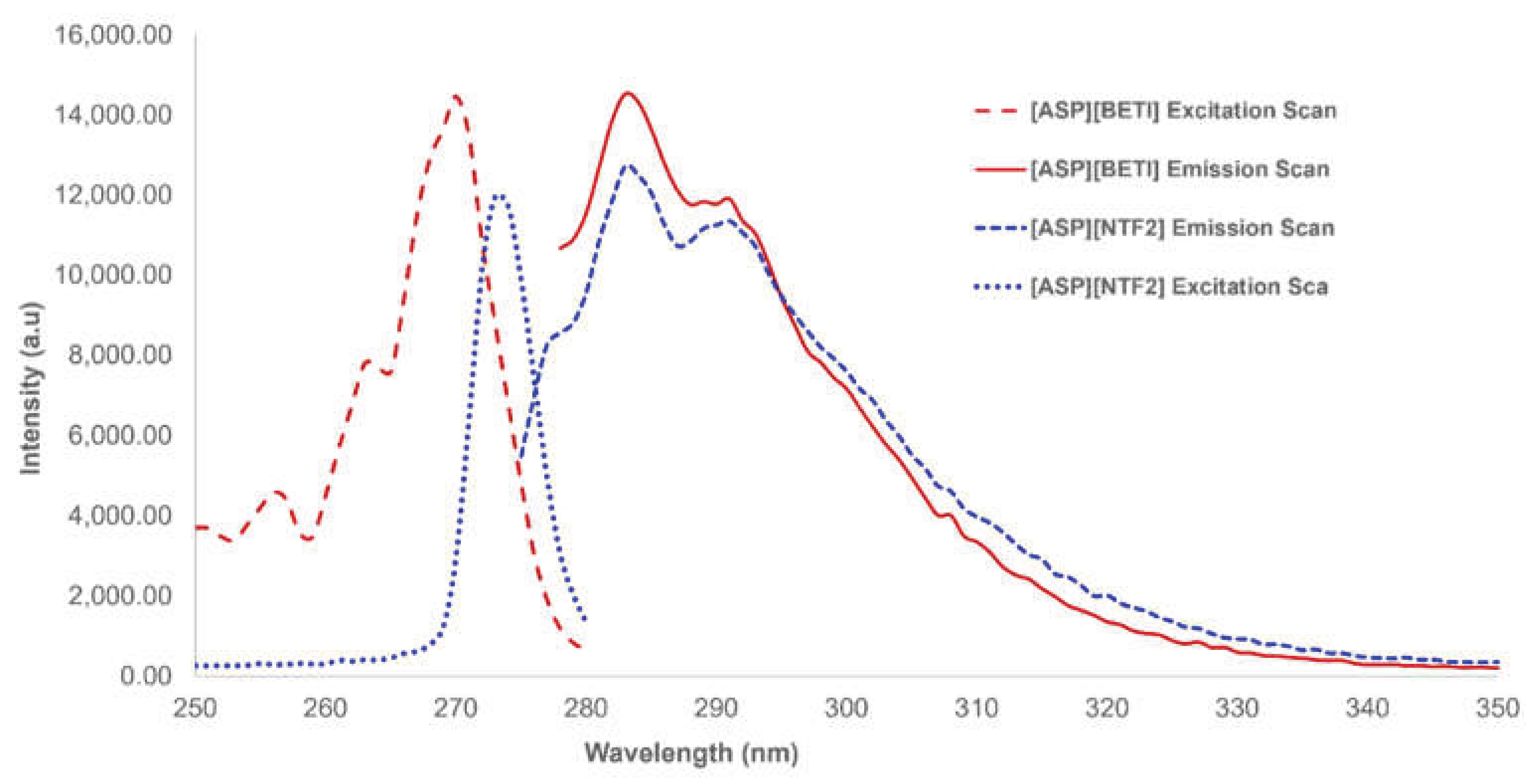
Figure 9.
Emission spectra of 2.5 µM pure (R) and (S)-naproxen in the presence of 62.5% v/v [ASP] [NTf2] FCIL in ethanol obtained at 284 – 362 nm excitation wavelengths (slit width = 0.5 mm). (The structure of naproxen is included in the inset).
Figure 9.
Emission spectra of 2.5 µM pure (R) and (S)-naproxen in the presence of 62.5% v/v [ASP] [NTf2] FCIL in ethanol obtained at 284 – 362 nm excitation wavelengths (slit width = 0.5 mm). (The structure of naproxen is included in the inset).
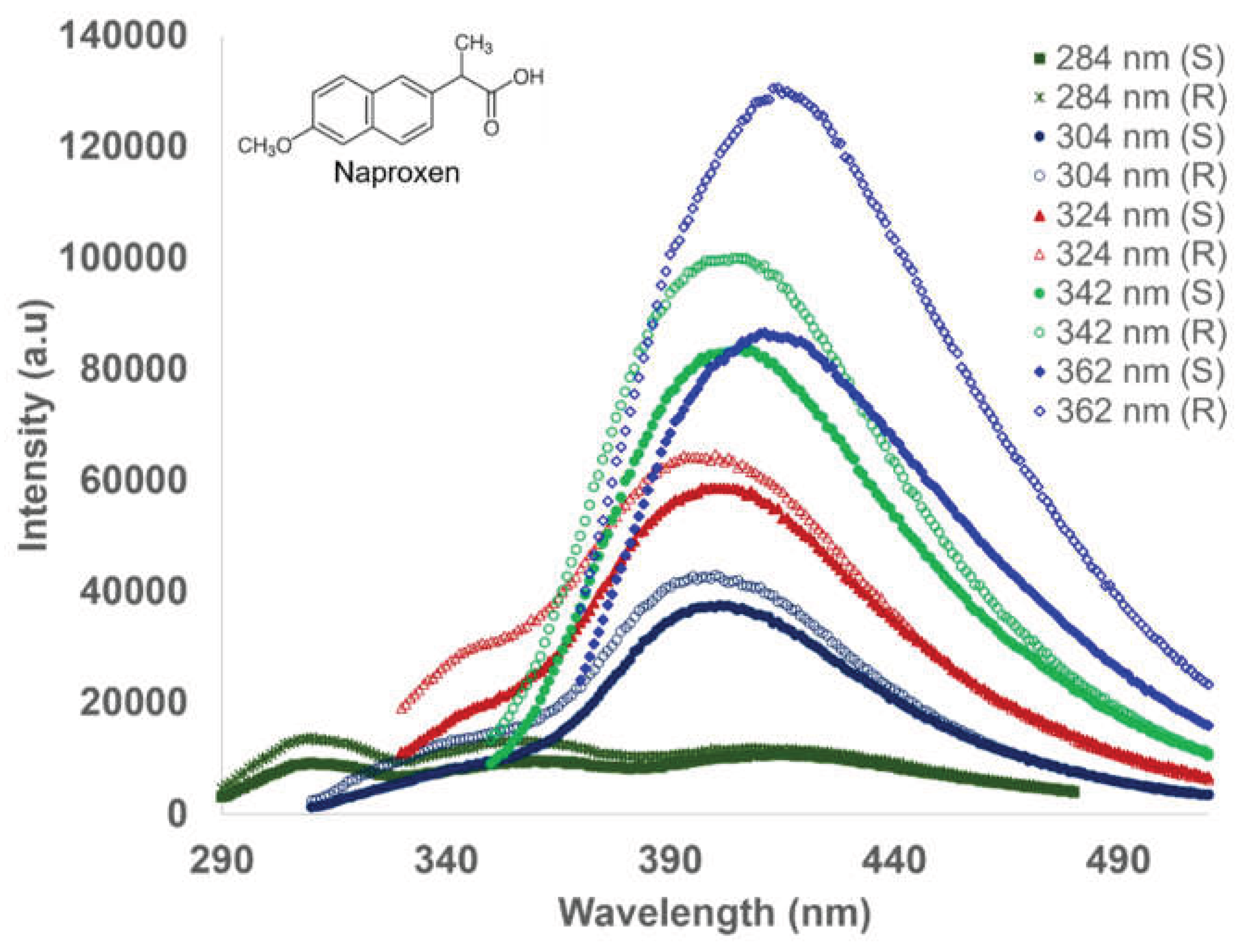
Figure 10.
Emission spectra of 1.69 µM pure (R) and (S)-Propranolol in the presence of 0.1 % m/v [ASP][BETI] FCIL in ethanol obtained at 324 – 344 nm excitation wavelengths. (The structure of propranolol is included in the inset).
Figure 10.
Emission spectra of 1.69 µM pure (R) and (S)-Propranolol in the presence of 0.1 % m/v [ASP][BETI] FCIL in ethanol obtained at 324 – 344 nm excitation wavelengths. (The structure of propranolol is included in the inset).
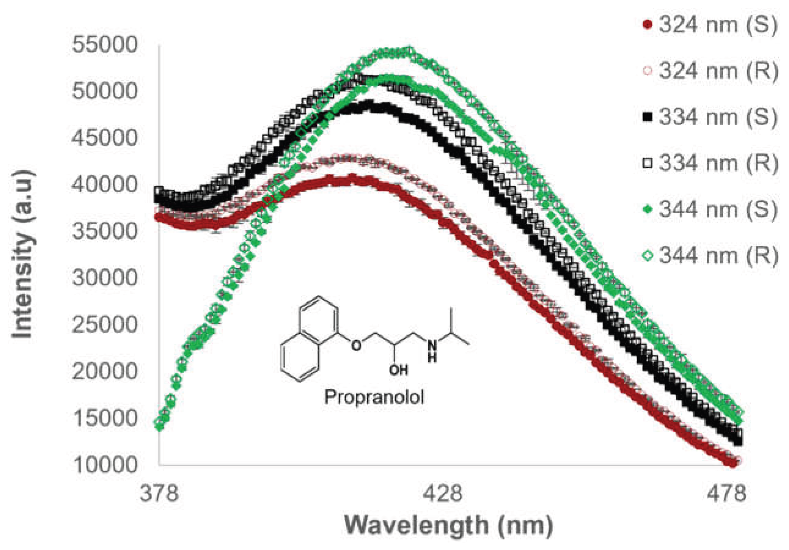
Figure 11.
DSC analysis of neat (A) [ASP] [NTf2] and (B) [ASP] [BETI] FCILs.
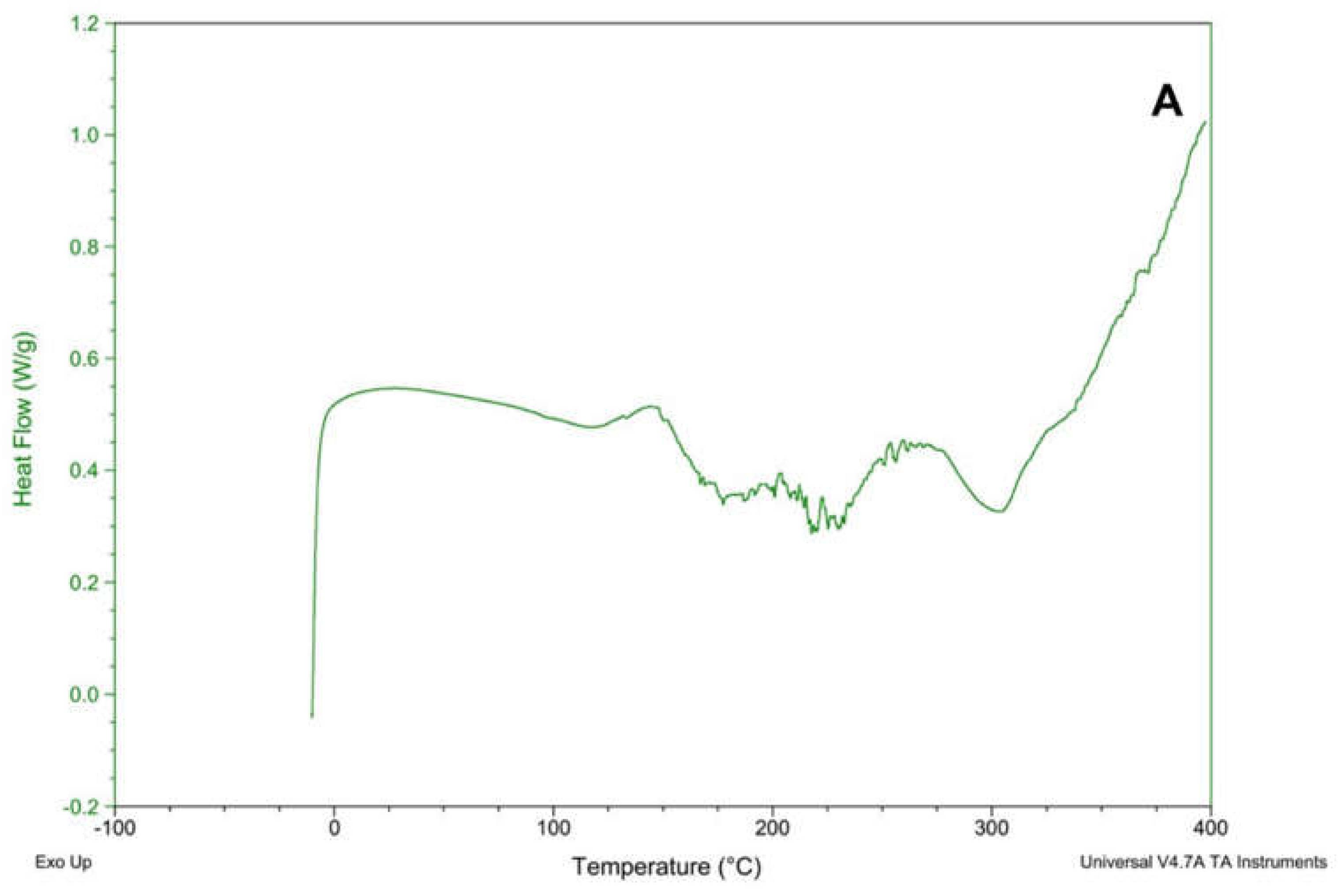
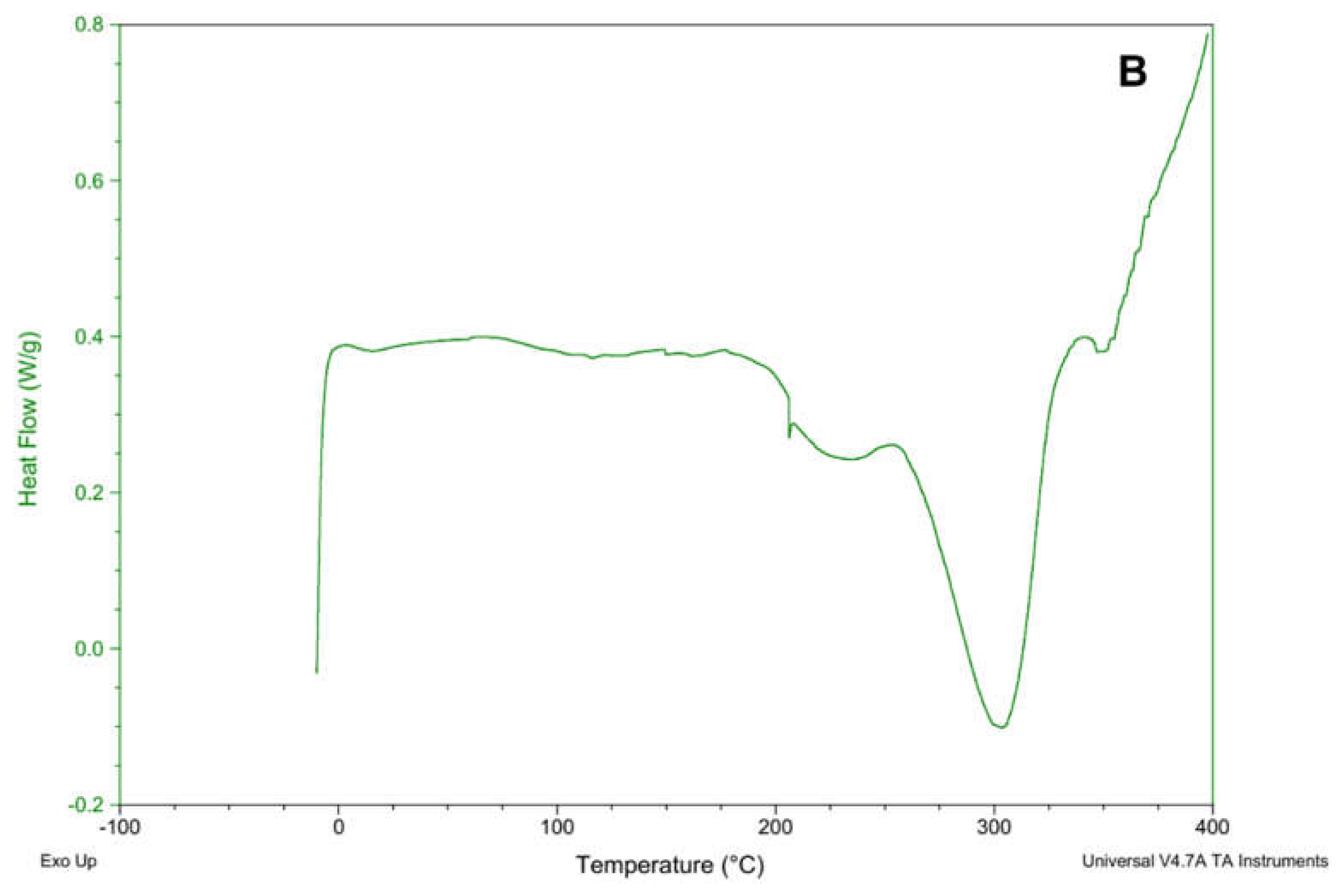
Figure 12.
TGA of neat (A) [ASP] [NTf2] and (B) [ASP] [BETI] FCILs.
Disclaimer/Publisher’s Note: The statements, opinions and data contained in all publications are solely those of the individual author(s) and contributor(s) and not of MDPI and/or the editor(s). MDPI and/or the editor(s) disclaim responsibility for any injury to people or property resulting from any ideas, methods, instructions or products referred to in the content. |
© 2026 by the authors. Licensee MDPI, Basel, Switzerland. This article is an open access article distributed under the terms and conditions of the Creative Commons Attribution (CC BY) license (http://creativecommons.org/licenses/by/4.0/).
Copyright: This open access article is published under a Creative Commons CC BY 4.0 license, which permit the free download, distribution, and reuse, provided that the author and preprint are cited in any reuse.