

Submitted:
06 September 2026
Posted:
11 September 2026
You are already at the latest version
Abstract
Research related to medicine has been centered around testing and finding a cure for diseases. Model organisms such as mice and monkeys have been key to the drug discovery process but the use of these organisms is becoming difficult due to ethical concerns. Tardigrades are a species of invertebrate organisms that could be widely used in drug discovery as they have a complex nervous system for their size, possess a transparent exoskeleton which makes observing neural activity accessible, and there are currently no ethical constraints regarding their use. Analyzing the movement of model organisms in response to stimuli is a very common method in drug discovery, previously used with another invertebrate organism C. elegans. This paper aims to analyze the movement response of tardigrades to electric stimuli to map the neuromuscular response, which can be used as a standard to measure the neurological effect of drug testing. This study shows that tardigrades respond to low-voltage electric fields in a microfluidic environment without long-lasting side effects. The tardigrades were shown to have a neuromuscular response to electric fields, which can be used to test the effects of drug candidates on the organism. The tardigrades were attracted toward the negative electrode in electric field intensities of 3-8 V/cm with speeds ranging from 154.90-259.00 μm/s. The neuromuscular response seen in tardigrades towards the electric field can be used in drug discovery by tracking its response before and after the drug has been administered. The use of tardigrades in medical research can provide an alternative solution to the ethical dilemma faced with the use of vertebrate organisms.
Keywords:
neuromuscular response
; electric fields
; neuroscience
; tardigrades
; electrotaxis
; drug discovery
; microfluidics
1. Introduction
1.1. Drug Discovery Process
Much of the research conducted in medicine is directed toward testing and finding a cure for diseases. Ethical concerns arise in the use of human subjects to test various drugs and are therefore being discontinued. Lately, the use of vertebrate animals such as mice, dogs, and monkeys in the field of clinical research faces public ethical concerns and may be discontinued in the future [1]. Without animal test subjects, drug discovery and safety testing will continue to grow in time, making the time horizon for the production of drugs to treat more modern diseases longer.
1.2. Current Invertebrate Model Organisms
Invertebrate model organisms such as C. elegans and fruit flies are a heavily researched alternative, although they both present disadvantages that prove they are not an efficient model to use for drug testing. C. elegans do not have the requisite neurological structures found in the human brain. The brain of C. elegans is composed of multiple neurons that do not have the formal organization that is seen in the human brain [2]. Due to this fact, neurological effects observed in tests with C. elegans may not be as translatable to humans as other model organisms. A study was performed on C. elegans where an electric field was applied to measure the neuromuscular response. This work takes inspiration from that study but instead performs it on a model organism with a more structured centralized nervous system [3].
On the other hand, fruit flies are shown to have many of the genes that cause diseases present in humans. The mature adult fruit flies cannot be tested for their neuromuscular response as they are not aquatic organisms and the tests done are not practical with airborne creatures. For this reason, the larvae of the fruit fly are used to test the response to electric fields, though this early stage of the fruit fly lacks the general complexity that is provided in a mature adult [4]. Other vertebrate model organisms like mice, rats, and monkeys have much more similarities to humans, though they do not have the transparent body that is seen in C. elegans and tardigrades. This unique feature seen in these organisms can be highly valuable to researchers, especially while studying the neuronal responses to external stimuli as direct observation of the neurons firing can be seen.
1.3. Tardigrades as a Model Organism
Tardigrades are model organisms that share a multitude of organ systems such as the nervous system and digestive system with humans, which have the potential to be widely used in the field of drug discovery. They also have numerous advantages such as small size, transparency, and a complex internal organization including a dorsal brain and ventral chain of nerve ganglia that provide a model for studying neural circuits in a small organism [5]. These features of tardigrades can be used in the field of drug discovery to test the effect of drugs on the nervous system without the use of human subjects. These unique features in tardigrades can make them an ideal model organism to be used in the field of drug discovery.
Tardigrades have been widely studied due to their neurological and morphological features. They possess limbs controlled for muscular movement and two eyes which provide visual input to a similarly structured brain. These similarities to humans, as well as their complex behaviors such as multi-limb walking, individual limb grasping, phototaxis, and response to various stimuli, make them an ideal candidate for model organism assays. Key questions can be answered when studying the tardigrade, such as how neural activity controls movement, how neural resilience and recovery occur under stress, and whether drugs may have neurological effects on humans [6].
The life cycle of a tardigrade was observed to have 4 physically different stages of active life. The first stage is stage 1 (S1). In this stage, the tardigrade is extremely small with an average length of about 50-70 µm. The tardigrades in this stage are also seen to move around the channel very quickly. The tardigrades are much more elongated in stage 2 (S2) and measured about 85 µm in length. They were also observed to be relatively active in movement. The tardigrades in the third stage (S3) were much more different from the previous two stages as they resembled mature tardigrades and did not engage in extensive movement. The final stage is the mature tardigrade (S4), which measures an average of 90-500 µm in length and does not move more than 50 µm from its original position. The stages of tardigrade development described here are not a global standard but were devised in this work to better describe the stage at which electrotaxis is observed.
Adult tardigrades are found to be in constant movement (∼89% of the time). They move an average of 0.23 body lengths per second in a natural scuttling motion. Tardigrades rarely move in a straight line pattern and often make loops or move diagonally. Additionally, it is also noted that newly hatched tardigrades and others in younger stages of life also move in a similar way to adult tardigrades [7].
1.4. Microfluidic Systems
Microfluidics is a field of research mainly centered on studying the behavior of small amounts of liquids. The use of microfluidics has been crucial in identifying model organisms that exhibit a reproducible neuromuscular response to an electric field, which can be used for drug discovery. Microorganisms such as C. elegans have been subject to these experiments and have demonstrated a neuromuscular response to electric fields [3]. This method was used in this work to observe the tardigrade’s response to an electric field in a controlled manner. Water is a good conductor of electricity and microfluidic applications are easy to observe for drug discovery purposes. For this reason, microfluidics and microfluidic devices were used in this work to observe the tardigrade under the influence of an electric field.
Microfluidic devices in conjunction with microscopes are one of the most effective ways to observe the effect of different stimuli on microorganisms [3]. The xurography method is a microfluidic fabrication technique which involves cutting films into stencils using motion-controlled razor blades. When using this method, Polyethylene Terephthalate (PET) sheets are usually used to make the devices as they are transparent and inert to chemical and electric activity, though other polymeric films can be utilized. The time length necessary to complete the device using the xurography method can vary but can usually be completed in less than an hour [8]. For this reason, these techniques were utilized to engineering a complex testing environment in which tardigrades will be tested in repeatedly.
In this work, assays were conducted on the tardigrade species Hypsibius exemplaris to identify response to an electric field and observe if the organisms show any directional movement. Through the findings, tardigrades can be determined to have the potential to be used in the drug discovery process.
2. Materials and Methods
2.1. Device Fabrication
The microfluidic device was designed using Cricut Design Space software (Cricut, Cricut Design Space, South Jordan, Utah), and the design was cut out using the Cricut cutting device on polyethylene terephthalate (PET) sheets, which are a thermoplastic polyester that provide excellent chemical resistance. These sheets were purchased from the company 3M. After retrieving the finished PET sheet and removing the cut areas (inlet/outlet and channel holes), the device can be assembled as shown in Figure 1. The device consisted of five PET sheets, each having different properties. The first (bottom) sheet is plain with no cuts and was used as the base. The middle three PET sheets have an adhesive coating on both sides, which also have the channel and inlet/outlet hole cuts. The inlet/outlet holes are where the tardigrade will be inserted into the microfluidic device for experimentation with electric fields. These layers are then sandwiched between that base PET sheet and a separate top PET sheet, which has holes for the inlet/outlet but no channel cut. This top layer also lacks an adhesive coating. Additionally, the two electrodes (copper tape) of length 3 cm each were placed so that the tape’s head is about 0.25 cm inside the inlet/outlet hole and had a distance of 1 cm between them. The electrodes have adhesive on one side and are folded at the end so that there was no exposed adhesive side. Finally, the layers were lined, assembled, and laminated by hand.
2.2. Tardigrade Species and Culturing
The tardigrades used in the experiment were of the Hypsibius exemplaris species. Tardigrades were grown at room temperature at 20 °C in containers used to ship them from the supplier (Ward’s Science, Rochester, New York). The plastic cap of the container was left unscrewed to allow airflow throughout the volume for the tardigrades to survive. The tardigrades were fed by replacing the top 4 ml of culture medium with 4 ml of food (green algae) obtained from a separate vial once every week from the same supplier. The tardigrade culture solution was bubbled using a pipette before experiments were conducted to ensure an even distribution of animals throughout the medium.
2.3. Electrotaxis Test Setup
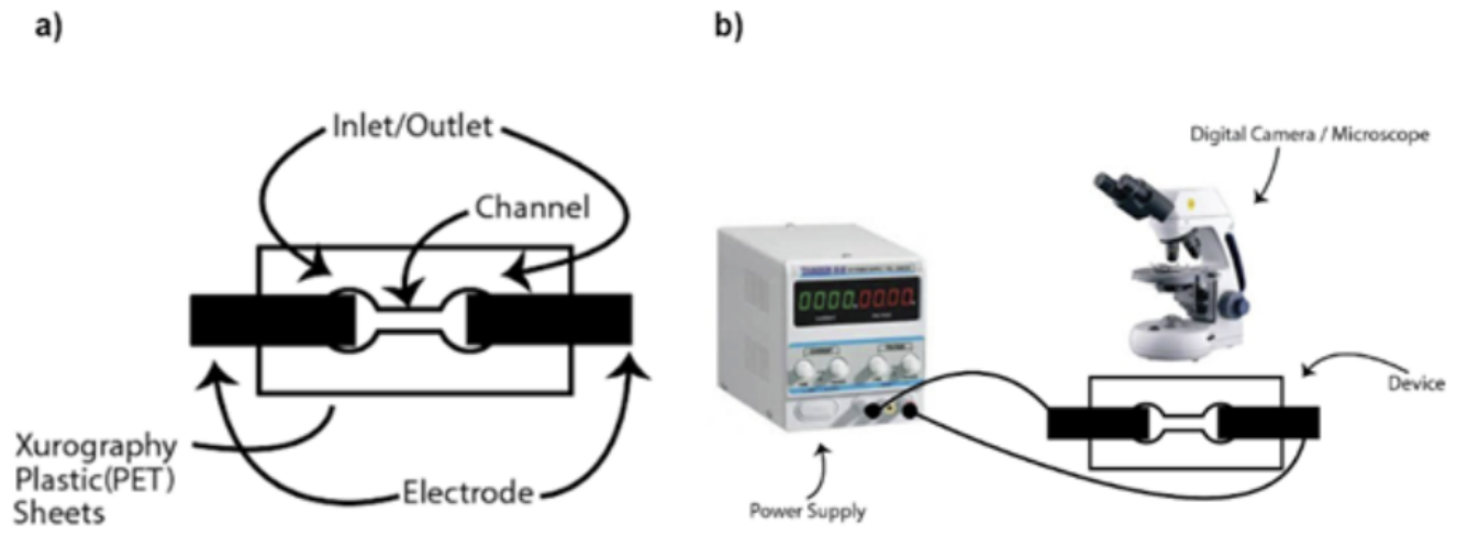
The experimental setup for studying the electrotaxis of tardigrades was comprised of three major components: a microfluidic device (microchannel and electrodes connected to the inlet and outlets), an electric field generator (power supply and connectors), and a monitoring system (microscope and camera). The microfluidic device used during the experiments was created using the xurography method with PET sheets as described above.
First, the culture solution was stirred by applying air using a pipette to create a homogenous mixture of tardigrades throughout. Subsequently, a petri dish was set aside to carefully extract 4 ml of the homogenous culture to be analyzed under a microscope (Leica S8 APO) at 10× magnification. When confirming the existence of multiple tardigrades in the petri dish, the microfluidic device as described before was attached to the stage of the microscope. After connecting the positive and negative connectors attached to the power supply (GW Instek GPS-3030DD) to the appropriate electrodes, as well as setting up the microscope/camera to focus at the channel of the device, a pipette was used to extract ml of culture from the petri dish to place in the inlet hole. When a tardigrade was found through the repetition of the process described, both the power supply and video camera were turned on. The raw footage was taken for later analysis of this study.
Figure 2.
(a) Diagram of xurographically cut microfluidic device; (b) Diagram of the experimental setup used to analyze the electrotaxis of tardigrades in the microfluidic device.
Figure 2.
(a) Diagram of xurographically cut microfluidic device; (b) Diagram of the experimental setup used to analyze the electrotaxis of tardigrades in the microfluidic device.
2.4. Data Analysis
Data analysis was carried out by recording videos of the movement of stage 1 (S1) tardigrades when exposed to the electric field. The raw videos were taken in the following format: the microscope is positioned to view the tardigrade; the video recording is started and the power supply is turned on. Once the tardigrade is outside of the microscope’s field of view and has moved an adequate distance, the power supply as well as the video recording are turned off. This process was repeated 10 times for each increase of intensity in the electric field (from 3-8 V/cm). The error value of the measurements for calculating the distance traveled was ± 2.95 µm.
Manual video analysis was conducted using pixel-based distance measurements calibrated against the known channel length (50 mm) to determine the conversion rate from pixels to µm. Equation 1 was used to calculate the distance traveled, where is the number of pixels from the screen to the head of the tardigrade after 3 seconds of the video have passed and is the number of pixels from the screen to the head of the tardigrade at the beginning of the video. The constant 2.95 is the conversion rate from pixels to micrometers at the magnification such that 1 px = 2.95 µm.
3. Results
3.1. Electric Field Induced Movement
This experiment aimed to test if the tardigrades respond to an electric stimulus and if any directional movement can be observed in response to the electric field. Two sets of experiments were completed as shown in Figure 3a and Figure 3b. The first was recording the natural movement of the S1 tardigrade without the effects of an electric field. Secondly, another set of experiments was completed to show the response the S1 tardigrades had to an electric field. The S1 tardigrades move randomly and change directions constantly when the electric field is not present. Unlike other stages of tardigrades, S1 tardigrades rarely stop for any reason including while eating algae or while encountering a barrier. The S1 tardigrades were also observed to move at an average speed of about 140 µm/s in a natural scuttling movement without exposure to an electric field [9]. When the electric field is applied, the tardigrade moves toward the negative electrode with a speed of 159.3 µm/s in a natural scuttling movement. The tardigrade was also observed to react almost immediately to the electric field as it ceased all other activities and moved towards the negative electrode after exposure. This shows that the electric field induces a robust response in the tardigrade. The presence of any debris or other particulate matter was observed to be able to halt the movement of the tardigrade when it came into contact. In this case, the tardigrade would attempt to move around the barrier by pushing against it, but depending on the size of the obstacle could be unsuccessful.
3.2. Effect of Electric Field on Different Life Stages of Tardigrades
Figure 4 shows the displacement of the two life stages of the tardigrades. A video taken where an electric field of 3 V/cm was applied across the channel was observed to see the different reactions of the S1 and S4 tardigrades. In this sketch, we can see that the S1 tardigrade moved in a straight line towards the negative pole, while the older S4 tardigrade had no significant change in its placement (inherently random movement). Through this figure, we can determine that only the S1 tardigrade is seen to display a robust and significant response to the electric field intensities applied. Similarly, other tardigrade stages were tested with similar effect. Through this experiment, only the S1 stage tardigrades show controlled movement to electric fields.
3.3. Movement Speed and Electric Field Response Thresholds
To test the speed of movement when exposed to an electric field, experiments were conducted to assess the electrotaxis in a controlled manner. A wide range of electric field intensities from 1-9 V/cm was applied through the channel to observe the movement of the tardigrade.
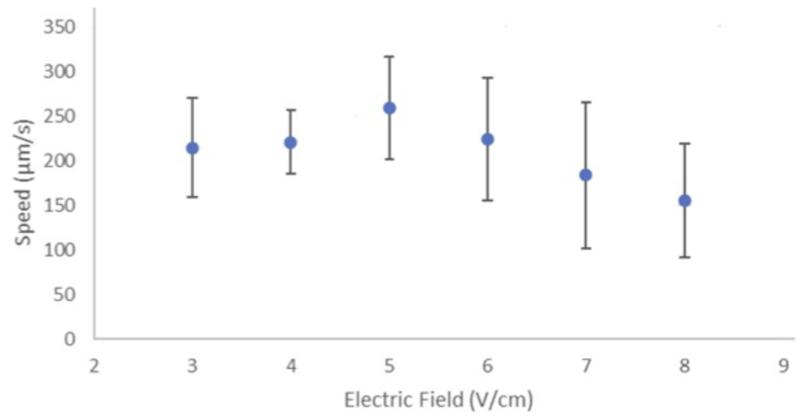
When the electric field was applied, there was a dramatic change in the movement of the S1 tardigrade. The tardigrades moved along the electric field instead of their normal movement as stated above when an electric field of 3-8 V/cm was applied. The tardigrades moved in this manner in all 60 experiments conducted with varying electric field intensities, showing the robustness of the response. When testing the electrotaxis of the S1 tardigrades, it was found that they continued to move in random directions when under electric fields lower than a threshold of 3 V/cm. It was also shown that a similar random movement continued if an electric field intensity stronger than the upper threshold of 8 V/cm was applied. Using the external scale bar, the distance traveled was calculated by analyzing the videos taken (see Section 2.4) to create Figure 5. It is clear that the S1 tardigrades respond to electric fields from a range of 3-8 V/cm, in which the average speed at each intensity is as follows: 3 V/cm (214.44 µm/s), 4 V/cm (220.47 µm/s), 5 V/cm (259.00 µm/s), 6 V/cm (224.45 µm/s), 7 V/cm (183.74 µm/s), and 8 V/cm (154.90 µm/s). The other stages of tardigrades were not able to respond to the electric stimuli and instead moved randomly. For example, it was noted that the S4 tardigrades moved an average of 22.43 µm in 25 seconds.
4. Discussion
4.1. Threshold Response Reasoning
In contrast to the tardigrade’s response to electric fields higher than 8 V/cm, C. elegans, an organism that has also been shown to respond to electric fields, continues to show a neuromuscular response until the electric stimuli cause mortality [5]. The tardigrade’s size most likely plays a role in its response to the electric field. It is possible that the potential drop across the organism is a similar signal to what its neurons are sending to the muscles, which is how we are able to observe this neuromuscular response. It is possible that after the 8 V/cm range, the potential drop is higher than the intensity of the signal that is given by the nerves, but not enough to cause mortality [10]. Further studies will need to be done in the future to research this possibility.
4.2. Statistical Differentiation of Speed at Different Electric Field Intensities
Although the averages are different for each speed, from Figure 5 it can be seen that the electrotaxis movements at each intensity are not statistically different from each other. The measured speeds at different intensities of the electric field seemed to overlap, and the neuromuscular response seems to be an on-and-off response.
The conclusion that only S1 tardigrades can respond robustly to the electric field of this range can potentially mean that the required electric stimuli are too small to induce a movement in any of the other stages of the lifecycle. Though, testing with the required electric field intensities to confirm this hypothesis can result in too dangerous a current that could be injurious to the experimenter. The movement induced by the electric field shows that the neuromuscular response can be controlled by electric stimuli and can have the potential to become a widely used model organism for the testing of drugs.
4.3. Electro-Osmotic Flow
The captured videos of the response to the electric field shown by the tardigrades were observed for movement due to the influence of electro-osmotic flow. The application of an electric field showed the movement of the tardigrade, while the particulate matter was stationary. Extraction of the fluid in the microfluidic channel, including the tardigrade and particulate matter, showed that the debris and algae were not secured to the channel in any way. This confirmed that electro-osmotic flow was not the reason for the tardigrade’s movement and it was a response to the electric stimuli by the organism itself. Additionally, if the electro-osmotic flow were to be the determining factor of the movement seen in the videos, the movement speed throughout the given range should continue to increase linearly and continue past the 8 V/cm intensity. As this was not observed, the conclusion can be made that the movement was due to a neuromuscular response in the tardigrade and not electro-osmotic flow.
4.4. Limitations and Future Work
This work presents a strong case for how electric fields can induce a neuromuscular response in tardigrades and shows an important effect that needs to be studied further. The current sample size of tests is still small and acts as a proof of concept to further research that may characterize this behavior further. Additionally, a single species of tardigrade was tested, and in the future more species as well as automatic tracking systems can be utilized for broader and more specific results. Joule heating was not investigated but will be included in a future study. As an electric field of different intensity is applied, the water in which the experiment is being conducted may heat. The temperature of the water in each experiment was not measured but could be a further analysis that will depict whether the response observed was due to heat or electric field. Since the direction of movement was consistent with the electric field, the most likely outcome is that the temperature of the water did not cause the movement, but this would be something to confirm in the future. Further research needs to be conducted to investigate how drug testing and validation can be applied to tardigrades, as well as neuronal activity monitoring.
5. Conclusion
Tardigrades are model microorganisms that have not been used widely in the field of drug discovery. The tardigrade has been classified as a model organism due to its complex dorsal brain, well-defined organ systems, and completely transparent body. These factors contribute to the tardigrade having the potential to be widely used in this field.
This study shows that tardigrades respond to low-voltage electric fields in a microfluidic environment without long-lasting side effects (the tardigrades were monitored for several days after experiments were concluded). Their movement can be controlled to attract them toward the negative electrode in a highly robust manner. In the experiments presented in Section 3, the tardigrade responds to the electric field by moving in a normal scuttling movement similar to the movement observed without the presence of the potential.
Experiments identified that amongst the age groups, S1 tardigrades responded to the electric field, while older stages (S2-S4) did not show significant movement to any electric field intensity. A threshold (minimum and maximum values) of 3-8 V/cm was identified where the S1 tardigrades exhibited the electrotaxis. Any intensities outside of this threshold proved to induce no such behavior even in the S1 tardigrade. Studies confirmed that the S1 tardigrades moved at an average speed ranging from 154.90-259.00 µm/s throughout the 6 different electric field intensities applied.
The reaction induced by the electric field indicated that S1 tardigrades have the ability to perform electrotaxis. The neuromuscular response to the electric field, as well as its biological composition, allows the tardigrade to be used in drug discovery as a model organism. The tardigrade’s electrotaxis movements can be tracked before and after the drug has been administered, to test whether the drug candidate affects the tardigrade on a neurological level, informing us of the effects of the substance. This research can be used to help provide an alternative to the vertebrate animal testing that is performed today.
Acknowledgments
This work was supported by the CAMEF Microfluidics lab at McMaster University. I also thank Dr. Nidhi Jain and Dr. Ravi Selvaganapathy for their help in this project.
Conflicts of Interest
The author declares no conflicts of interest.
References
- Badyal, D. K.; Desai, C. Animal use in pharmacology education and research: the changing scenario. Indian J. Pharmacol. 2014, 46(3), 257. [Google Scholar] [CrossRef]
- Hobert, O. Neurogenesis in the nematode Caenorhabditis elegans. In WormBook; 2010. [Google Scholar] [CrossRef]
- Rezai, P.; Siddiqui, A.; Selvaganapathy, P. R.; Gupta, B. P. Electrotaxis of Caenorhabditis elegans in a microfluidic environment. Lab A Chip 2009, 10(2), 220–226. [Google Scholar] [CrossRef]
- Strange, K. Drug discovery in fish, flies, and worms. ILAR J. 2016, 57(2), 133–143. [Google Scholar] [CrossRef]
- Persson, D. K.; Halberg, K. A.; Jørgensen, A.; Møbjerg, N.; Kristensen, R. M. Brain anatomy of the marine tardigrade Actinarctus doryphorus (Arthrotardigrada). J. Morphol. 2014, 275(2), 173–190. [Google Scholar] [CrossRef]
- Lyons, A. M.; Kato, S. The tardigrade as an emerging model organism for systems neuroscience. arXiv 2025, arXiv:2501.06606. [Google Scholar]
- Anderson, E. M.; et al. Comparative analysis of tardigrade locomotion across life stage, species, and disulfiram treatment. PLoS ONE 2024, 19(9), e0310738. [Google Scholar] [CrossRef]
- Singapore University of Technology and Design. New methods to create microfluidic devices with fluoropolymers. In ScienceDaily; 8 February 2019. [Google Scholar]
- Nirody, J. A.; et al. Tardigrades exhibit robust interlimb coordination across walking speeds and terrains. Proc. Natl. Acad. Sci. 2021, 118(35), e2107289118. [Google Scholar] [CrossRef]
- Lockery, S. R.; Goodman, M. B. The quest for action potentials in C. elegans neurons hits a plateau. Nat. Neurosci. 2009, 12(4), 377–378. [Google Scholar] [CrossRef]
Figure 1.
Exploded view of the xurographically cut microfluidic device. The device has dimensions of 50 mm in length, 27 mm in width, and 1 mm in height. The device consisted of a channel (5 mm in length, 2 mm in width, and 600 µm in height) with two circular inlet/outlet ports (each 7 mm in diameter).
Figure 1.
Exploded view of the xurographically cut microfluidic device. The device has dimensions of 50 mm in length, 27 mm in width, and 1 mm in height. The device consisted of a channel (5 mm in length, 2 mm in width, and 600 µm in height) with two circular inlet/outlet ports (each 7 mm in diameter).
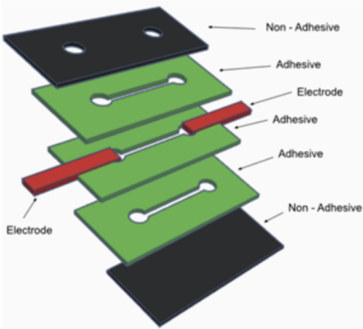
Figure 3.
(a) S1 tardigrade movement without exposure to an electric field; (b) S1 tardigrade moving towards the right side in a 3 V/cm electric field.
Figure 3.
(a) S1 tardigrade movement without exposure to an electric field; (b) S1 tardigrade moving towards the right side in a 3 V/cm electric field.
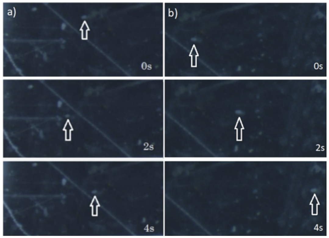
Figure 4.
The traced image of S1 (red line) and S4 (green line) tardigrade’s movement under an electric field over a duration of 10 seconds.
Figure 4.
The traced image of S1 (red line) and S4 (green line) tardigrade’s movement under an electric field over a duration of 10 seconds.
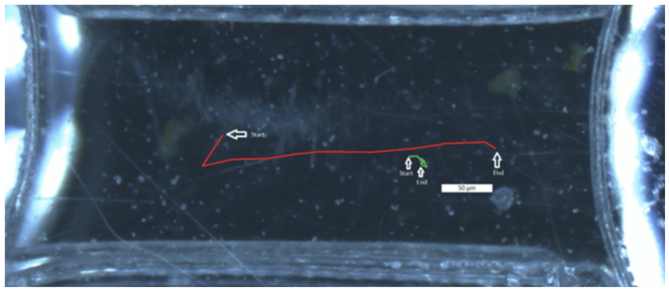
Figure 5.
Electrotaxis speed of S1 tardigrades when subjected to an electric field ranging from 3-8 V/cm.
Figure 5.
Electrotaxis speed of S1 tardigrades when subjected to an electric field ranging from 3-8 V/cm.
Disclaimer/Publisher’s Note: The statements, opinions and data contained in all publications are solely those of the individual author(s) and contributor(s) and not of MDPI and/or the editor(s). MDPI and/or the editor(s) disclaim responsibility for any injury to people or property resulting from any ideas, methods, instructions or products referred to in the content. |
© 2026 by the author. Licensee MDPI, Basel, Switzerland. This article is an open access article distributed under the terms and conditions of the Creative Commons Attribution (CC BY) license.
Copyright: This open access article is published under a Creative Commons CC BY 4.0 license, which permit the free download, distribution, and reuse, provided that the author and preprint are cited in any reuse.