

Submitted:
09 September 2026
Posted:
10 September 2026
You are already at the latest version
Abstract
Deregulation of transcriptional programs is an essential step in cancer development and progression, with many oncogenes and tumor suppressors being transcriptional regulators. The Mediator complex is a multi-subunit transcriptional coregulator that directly links DNA binding transcription factors to RNA polymerase enzymes. In cancer, Mediator is frequently dysregulated, as genes encoding Mediator subunits are frequently mutated or aberrantly expressed, with some subunits such as MED12 and CDK8 having particularly well characterized roles. Here, we critically review accumulating evidence that implicates another Mediator subunit as an emerging, important player in cancer, MED15. We revisit the rare incidences of MED15 mutation and more common events of MED15 misexpression, and consider pathways that may lead to the latter. We then discuss the important roles MED15 plays in several cellular signaling pathways that critically contribute to the development or progression of many cancer types, namely YAP/TAZ, TGF-β/SMAD, SREBP, and HIF. Identifying shared notes and themes, we highlight the mechanisms by which MED15 may promote cancer and speculate on how such mechanisms may apply in other contexts. We also evaluate the risk and promise of MED15 as a drug target for cancer treatment, focusing on its potential as a target for small molecules that interfere with specific transcription factor interactions. Finally, we pose some important questions regarding future research on MED15, speculating on its putative roles in other processes central to cancer, such as immune responses and stress adaptive pathways. Collectively our work highlights the emerging importance of MED15 in multiple aspects of cancer biology and cements it as a critical factor for further study.
Keywords:
mediator complex
; cancer
; EMT
; stress
; hypoxia
; HIF
; TGF-β
; YAP
1. Introduction
1.1. The Mediator Complex Is an Essential Regulator of Gene Expression in Eukaryotes
In eukaryotes, gene expression must remain under precise spatiotemporal control because the dysregulation of this process can result in pathological outcomes. Cancers, in particular, often arise from the deregulation of transcriptional regulatory circuits, for example the genome surveillance pathway centred on tumor protein 53 (TP53), the cell cycle progression pathway focused on Retinoblastoma (RB), or the growth-promoting c-Myc pathway. DNA-binding transcription factors are key players in these and many other cancer-promoting pathways, cementing the importance of these types of proteins in cancer initiation, development, and progression.
To maintain precise spatiotemporal control over their downstream targets, transcription factors engage in context-dependent interactions with a diverse group of proteins referred to as coregulators, typically classified as either coactivators or corepressors [1,2,3,4,5]. Such proteins and protein complexes can influence many activities critical for transcriptional regulation, including DNA accessibility; chromatin modification, structure, and conformation; transcription factor binding, stability and activity; and interaction with the RNA polymerase machinery, which synthesizes RNA molecules.
Amongst coregulators, the so-called Mediator complex stands out as a multi-protein machine that can directly link DNA-bound transcription factors to RNA polymerases and that is required for virtually all gene expression in humans [6,7,8,9,10,11]. Human Mediator is composed of ~30 protein subunits (MED genes/proteins), three of which feature mutually exclusive paralogs (MED12/MED12L, MED13/MED13L, cyclin dependent kinases 8 and 19(CDK8/CDK19)). Mediator directly influences RNA polymerase activity, including transcription initiation, elongation, and termination, as well as RNA splicing and processing, thus adopting a central role in the control of RNA synthesis [6,7,8,9,10,11]. Architecturally, Mediator is assembled into a complex and dynamic structure that features four modules: the head, middle, tail, and kinase [6,12,13,14,15]. The head and the middle modules interact with RNA polymerases, whereas the tail predominantly engages in dynamic and specific interactions with various transcription factors; together, these three modules form the stable so-called core Mediator. Core Mediator can reversibly associate with the kinase module whose enzymatic activity targets numerous transcriptional regulators, including Mediator subunits themselves as well as RNA polymerases and transcription factors [6,12,13,14,15]. In addition to kinase module-associated and core Mediator complexes, some Mediator subunits can moonlight and adopt entirely different functions outside the complex [16,17,18]. In sum, by virtue of its multi-subunit composition and dynamic interaction with numerous other players in gene regulation, Mediator represents a critical nexus in and integrator of the many cellular signaling pathways that control developmental and homeostatic processes and whose dysfunction can lead to various cancers.
1.2. Mediator Subunit Mutations Can Cause Cancers and Are Key Effectors and Possible Therapeutic Targets in Certain Cancer Types
Mediator dysfunction is now well understood to underlie several human pathologies. In particular, during the last 15 years, many Mediator subunits have emerged as key players in various types of cancer [19,20,21,22]. Mutations in some MED genes can directly cause certain tumors and cancers. For example, CDK8 gain of function by amplification or epigenetic deregulation can cause colon cancer and melanoma [19,23], whereas MED12 mutations are dominant drivers of several benign tumors, such as uterine leiomyomas, breast fibroadenomas, and others [20,24,25]. Moreover, despite not being subject to mutation, many other Mediator subunits are indirectly deregulated in or vital for various aspects of cancer establishment, progression, and/or metastasis [21,26,27,28].
In line with these roles, pharmacological inhibitors against oncogenic CDK8 are in clinical trials [29,30]. Moreover, some non-kinase Mediator subunits have also emerged as therapeutic targets because they physically bind transcription factors in a highly specific fashion. For example, MED1 interacts with the oncogenic fusion transcription factor E2 immunoglobulin enhancer-binding factor–Pre-B-cell leukemia homeobox 1 (E2A-PBX1), and this interaction promotes E2A-PBX1-driven leukemia [31]. Such Mediator-transcription factor interactions represent new therapeutic opportunities, as their interaction surfaces are promising new targets for precision medicine drugs. However, how the activities of individual Mediator subunits can influence cancer development and progression, and especially how differential expression of on individual subunit may alter pertinent signaling pathways, is not well understood and remains an area of active investigation.
1.3. Mediator Subunit MED15 Is an Emerging Player in Several Types of Cancer
Whereas Mediator subunits such as CDK8 and MED12 have well established roles in cancer and tumor formation is, recent evidence pinpoints MED15 as another emerging player in cancer development. MED15 locates to the tail module of the Mediator complex where it directly interacts with transcription factors. Functionally, MED15 was first described as a key regulator of several developmental and physiological processes that could underlie its relevance to cancer. First, MED15 is a coregulator for SMAD-type transcription factors that are governed by transforming growth factor β (TGF-β) signaling, which controls cell proliferation and migration as well as immune functions [32]; of note, TGF-β can both antagonize and promote cancer development [33]. Second, MED15 coactivates the lipogenic sterol regulatory element binding protein (SREBP) type transcription factors, and thus enhances de novo fatty acid synthesis, a process often activated by cancer cells to accommodate the anabolic demands of rapidly growing cells [34]. Finally, extensive evidence from studies in yeast and invertebrate organisms show that MED15 interacts with several different transcription factors to promote adaptation to stresses such as hypoxia and oxidative stress [35,36,37,38,39,40]. Because cancers experience these stresses, activating pertinent stress responses can help them survive and metastasize, representing another avenue for MED15 to participate in key signaling pathways for cancer development or progression.
Here, we review recent evidence on MED15 mutation and deregulation in cancer cells, integrate insights on functional effects of MED15 across cancer types, speculate on possible molecular functions of MED15 beyond identified mechanisms, and assess its possible value as a therapeutic target.
2. MED15 Is Subject to Sporadic Fusion Mutations and Expression Alterations in Cancers
Evidence available in cancer genomics databases suggests that MED15 itself is likely not subject to frequent somatic point mutations or other variations; this distinguishes it from CDK8 and MED12, which both feature recurrent and frequent mutations in specific cancer types. However, MED15 is mutated in some rare gene fusion events. Gene fusions are important drivers in the development of specific cancer types, and the knowledge on the molecular nature of these fusion events and the functions of their resulting proteins can inform cancer outcomes as well as treatment strategies. Here, we review information about the reported fusions that involve MED15, detail how MED15’s molecular function may impact cancer development, and highlight commonalities and differences between the reported MED15 fusions.
2.1. MED15 Gene Fusions Occur in Multiple Cancer Types
2.1.1. MED15::TFE3 Fusions Are Important Drivers of Rare Renal Cell Carcinomas
Fusions of Transcription Factor E3 (TFE3) and MED15 are established drivers of TFE3-rearranged renal cell carcinoma (TFE3-rRCC). TFE3-rRCC is a rare subtype of RCC caused by chromosomal rearrangements involving the gene encoding TFE3 [41,42,43]. In adults, TFE3-rRCCs make up only about 1-2% of all RCC cases; however, TFE3-rRCCs represent 40-50% of pediatric RCC cases [41,42,43,44]. TFE3 fusion genes are generated by chromosomal rearrangements that join a 5′ partner gene to the 3′ portion of TFE3, where TFE3 can fuse with several different gene partners. Genes encoding Alveolar Soft Part Sarcoma Critical Region 1 (ASPSCR1), Proline Rich Mitotic Checkpoint Control Factor (PRCC), Splicing factor, proline- and glutamine-rich (SFPQ), and Non-POU domain-containing octamer-binding (NONO) fusions account for approximately 80% of TFE3-rRCCs, whereas the remainder are fusions of TFE3 to MED15 and various other genes [41,42,43].
42 cases of MED15::TFE3 RCCs have been documented to date (Table 1). MED15::TFE3 cancers display varying architectural patterns, with most cases exhibiting a predominantly cystic architecture (33/38 cases with a defined architecture) [45,46,47,48,49,50,51,52,53,54,55,56]. A few cases entirely lack this characteristic cystic architecture (5/38 cases) and rather display a mixture of papillary, tubular, acinar, and solid components, with one case being entirely solid [45,46,57,58] (Table 1).
Within these MED15::TFE3 RCCs, several different fusion transcripts likely drive the development of this cancer (Table 1). Fusion transcripts connecting exon 5 or 6 of TFE3 to either exon 8 or exon 9 of MED15 are observed in MED15::TFE3 RCCS featuring a predominant cystic architecture [45,46,47,48,49,50,51,52,53,54,55,56]. In contrast, the transcripts in cases with minimal or no cystic component fuse TFE3 exon 5 or 6 to downstream MED15 exons, such as exon 11 and 13 [46,57,58]. The connection points of the fusion transcripts may, therefore, influence the phenotypes of the MED15::TFE3 RCCs, with certain connection points being associated with conventional (cystic) and non-conventional (minimal or non-cystic) phenotypes. This idea is further supported by the fact that two fusion transcripts are present in the one purely solid MED15::TFE3 RCC: one connecting exon 7 of TFE3 to exon 11 of MED15, and one connecting exon 6 of TFE3 to mid-intron 11 of MED15 [45]. In sum, various MED15::TFE3 fusions occur that may result in fusion proteins with slightly different molecular activities, ultimately being responsible for a certain degree of heterogeneity in tumor phenotypes. Molecular profiling strategies including transcriptomics, chromatin occupancy studies, and integration proteomics could help shed light on these nuances.
TFE3-rRCCs typically present at more advanced stages and with more frequent lymph node metastases than common RCCs, but the clinical presentation and prognosis of TFE3-rRCCs is highly influenced by the fusion partner [41,43]. For example, ASPSCR1::TFE3 RCCs, a common TFE3 fusion partner, is highly aggressive with frequent metastasis and poor overall survival [42,51]. Vice versa, PRCC::TFE3 and NONO::TFE3 RCCs display better outcomes and more favorable prognosis, although 20-25% of patients nonetheless develop metastases or feature recurrence [42]. Interestingly, despite featuring varying architectures and underlying fusion transcripts, MED15::TFE3 RCCs uniformly show low malignant potential and excellent prognosis and are thus commonly termed “indolent-behaving” [45,51]. Most patients with MED15::TFE3 fusion undergo partial or radical nephrectomy and show no evidence of recurrent or persistent disease during follow-ups, despite treatment being limited to surgery. Indeed, no patients with MED15::TFE3 RCC appear to have died from their cancer, and only one case of distant metastasis has been reported, occurring 15 years after initial diagnosis [45,46,47,48,49,50,51,52,53,54,55,56,57,58]. Additionally, only two cases showed regional lymph node metastasis, where both were present preoperatively at initial diagnosis and no recurrence was observed after undergoing treatment [51,55]. Mechanistically, one reason underlying the indolent nature of MED15::TFE3 RCCs may be their immune environment. Programmed Death-Ligand 1 (PD-L1) mRNA and protein levels were high in MED15::TFE3 tumors, whereas most other TFE3-rRCCs display an “immune ignorant” phonotype with low PD-L1 positivity and low Cluster of Differentiation 8 positive (CD8+) T cell infiltration [51]. Therefore, MED15::TFE3 RCCs may be responsive to immunotherapy, which could further enhance their excellent prognosis.
TFE3 rearrangements also exist in other tumors, including TFE3 rearrangement-associated perivascular epithelioid cell tumors (PEComa) [43]. A MED15::TFE3 gene fusion was identified in a PEComa that lacked melanotic features and morphologically resembled rRCCs [62]. The fusion transcript in this case connects exon 9 of MED15 to exon 4 of TFE3, a fusion type not observed in MED15::TFE3 RCCs [62]. This suggests that the specific fusion transcript is not only a determinant of phenotype within these rRCCs but may also contribute to specific tumor types. Finally, a MED15::TFE3 fusion was identified in one melanocytic neoplasm, also referred to as a TFE3-rearranged PEComa-like neoplasm [46]. These neoplasms are very rare, with less than 100 cases reported, and aggressive, with a 5-year overall survival rate of ~50% [66,67]. In line with this, the MED15::TFE3 rearranged PEComa-like neoplasm showed recurrence in the kidney as well as liver metastases after 2 years [46]. The fusion protein in this case connected exon 8 of MED15 to exon 6 of TFE3, a fusion previously detected in non-aggressive MED15::TFE3 RCCs with no recurrence [46]. This highlights a possibility for different roles of TFE3::MED15 fusions in different cell and tumor types (melanocytic versus non-melanocytic) resulting in different behaviours, pending further investigation.
How do MED15::TFE3 fusions promote cancer formation? Although specific fusion partners influence the clinical presentation and prognosis in TFE3-rRCCs, it is generally the TFE3 mutation that appears to drive the development of these cancers [41]. All TFE3 fusions deregulate TFE3, leading to its constitutive activation and strong induction of its downstream genes. In these fusion proteins, the DNA-binding domain, activation domain, and nuclear localization signal of TFE3 are retained, whereas the TFE3 N-terminus, which contains kinase-regulated sites and controls TFE3 subcellular localization and stability, is replaced by the fusion partner [41]. Loss of normal regulation provokes unrestrained TFE3 activity. Moreover, the addition of a new 5’ region from the fusion partner typically leads to high expression of the fusion transcript [41]. Collectively, this promotes constitutive fusion protein nuclear localization and transcriptional activity, leading to the development of RCC and other tumours.
2.1.2. MED15::ATF1 Fusions Promote the Development of Very Rare Tumors with Melanocytic Differentiation
MED15 is also a fusion partner for Activating Transcription Factor 1 (ATF1) in pediatric tumors with melanocytic differentiation, a broad category of neoplasms that produce melanin or share cellular features with melanocytes but arise from non-melanocytic origins [68]. ATF1 plays a major role in these tumors, specifically in dermal clear cell sarcomas (CCS) and cutaneous clear cell tumors with melanocytic differentiation. Molecularly, ATF1 regulates the expression of target genes involved in cell growth and survival and drives microphthalmia-associated transcription factor (MITF) expression, which induces melanocytic differentiation [63,64]. Notably, MED15::ATF1-rearranged (fusion) tumors were reported in five patients and showed evidence of melanocytic differentiation, expressing melanocytic markers such as SRY-box transcription factor 10 (SOX10), MITF, and Melan A [63,64]. MED15::ATF1 cancers thus share both cellular and gene expression features with other melanocytic tumors such as CCS and cAMP response element-binding protein (CREB)-regulated transcription coactivator 1 – Tripartite Motif Containing 11 (CRTC1::TRIM11) cutaneous tumors (CTCT) [63,64]. Compared to other dermal CCS, MED15::ATF1 tumors behave aggressively, although the low case number represents a caveat. Particularly, two patients displayed local lymph node metastasis at the time of diagnosis and one patient died from disease after recurrence and failure to respond to multiple therapies [63,64].
Additionally, a MED15::ATF1 fusion was identified in one pediatric patient with aggressive melanoma [65]. This tumor also featured mutations in other genes, including the known oncogene Telomerase Reverse Transcriptase (TERT), so the aggressiveness may reflect the presence of multiple mutations, not just the fusion protein [65].
The fusion transcripts in all MED15::ATF1 rearrangements involve the 5’ portion of MED15 and the 3’ end of ATF1, typically exons 3, 4, and 5 [63,64,65]. In these MED15::ATF1 gene fusions and others involving ATF1, the 5’ end of ATF1 is replaced by the gene fusion partner, resulting in loss of the cAMP-dependent N-terminus of ATF1 [63]. Because ATF1 activation is linked to elevated cAMP levels, loss of the N-terminus likely renders its activation cAMP independent, leading to the constitutive activation of ATF1 [63]. Meanwhile, the kinase-inducible and basic leucine zipper domains of ATF1, which are relevant for its transcriptional functions, are retained [65]. This allows constant transcriptional activation of MITF by ATF1, contributing to the development of these tumors. Ultimately, as is the case for TFE3::MED15 fusions in RCCs, the constitutive activation of the transcription factor fusion partner of MED15 appears to be the dominant driver of cancer development [63].
In sum, rare gene fusion events exist that involve MED15 and promote cancer development. The TFE3::MED15 and ATF1::MED15 fusions occur in different cancer types, have different levels of aggressiveness, and consist of different fusion transcripts, but share the overall mechanism driving cancer development, which is likely due to the constitutive activation of the fused transcription factor with oncogenic potential and not due to intrinsic oncogenic activity of MED15. The phenotypic variability observed in TFE3::MED15 RCCs may reflect the higher prevalence of reported cases compared to ATF1::MED15 fusion tumors, rather than a lack of heterogeneity of the ATF1::MED15 tumors. These rare MED15 fusion tumors, while important, thus represent a unique niche with distinct mechanistic involvement of MED15 when compared to other cancer types that feature aberrant MED15 expression, as discussed below.
2.2. MED15 Expression Alterations Occur in Many Cancer Types and Are Associated with Aggressive Tumor Features
Although MED15 does not appear to be a frequent target for somatic mutations, accumulating evidence indicates that MED15 overexpression occurs in many cancer types. Interestingly, this overexpression is associated with poor prognosis, more advanced tumor stage, and increased tumor aggressiveness in many cases. Cancer types with high MED15 expression include lung cancer, prostate cancer, clear cell renal cell carcinoma (ccRCC), hepatocellular carcinoma (HCC), breast cancer, bladder cancer, and head and neck squamous cell carcinoma (HNSCC) [69,70,71,72,73,74,75,76]. These cancer types are all solid tumors, and interestingly, many are part of or adjacent to the gastrointestinal system. MED15 is expressed in most or all tissues, so the potential tissue-preference of MED15’s influence on cancer aggressiveness and clinical outcomes may reflect the genes and processes it regulates.
In many of these cancers, MED15 overexpression is associated with epithelial–mesenchymal transition (EMT) and metastasis. In HCCs, MED15 overexpression correlates with increased levels of α-fetoprotein, a biomarker for increased risk of vascular invasion and metastasis [73]. In HNSCCs, MED15 overexpression is detected in ~one-third of primary tumors and even more recurrent tumors, where it is associated with increased mortality and poor clinical outcomes [76]. In ccRCCs, MED15 is transcriptionally upregulated by Hypoxia-Inducible Factor 2α (HIF-2α) and promotes tumor proliferation, metastasis, and fatty acid synthesis [72]. The connection between HIFs and MED15 may explain why MED15 overexpression is associated with poor survival in these solid tumors, which commonly feature hypoxia and HIF upregulation. In breast cancers, MED15 overexpression is correlated with phospho-SMAD3 overexpression and promotes metastasis through modulation of the TGF-β/SMAD signaling [74]. MED15 overexpression also correlates with tumor stage, lymph node metastasis and enhanced EMT in bladder cancer, through stabilization of Yes-associated protein (YAP) by interfering with TRIM11 activity [75]. In castration-resistant prostate cancer, MED15 is highly overexpressed, and elevated MED15 levels correlate with androgen receptor amplification, increased proliferative activity, and worse clinical outcomes [70]. MED15 overexpression is also associated with treatment resistance in this cancer type, where MED15 expression is induced during androgen deprivation therapy through activation of the Phosphoinositide 3-kinase (PI3K)/mechanistic target of rapamycin (mTOR) signaling pathway and can be attenuated by inhibiting the PI3K/mTOR axis [71]. In sum, MED15 overexpression occurs in many cancer types, likely via distinct mechanisms, and in turn promotes aggressive presentation via multiple pathways that crosstalk (see Section 3).
In line with the lack of apparent recurrent genetic change within the MED15 locus, epigenetic changes may contribute to MED15 overexpression. In HNSCCs, aberrant methylation of the MED15 promoter represents a potential biomarker, suggesting that epigenetic dysregulation may contribute to altered MED15 expression in this cancer type [77]. Additionally, TGF-β/SMAD signaling is associated with MED15 in breast cancer, and TGF-β secretion is considered a component of the senescence-associated secretory phenotype (SASP) [32,74,78]. Interestingly, MED15 T603 phosphorylation is a key control switch for SASP and may be associated with age-related diseases, including cancer [78]. Although there is no direct evidence of MED15 T603 being linked with cancer, further evaluation of this amino acid and its phosphorylation in cancer contexts will be interesting.
Collectively, it emerges that high MED15 expression is associated across multiple cancer types with poor patient survival, enhanced proliferation, metabolic reprogramming, invasion, metastasis, and therapeutic resistance. In vivo studies in mouse models agree with these clinical findings, as subcutaneous (SQ) lung adenocarcinoma, bladder cancer, colorectal cancer, and ccRCC cell xenograft mouse models display reduced tumor volume after MED15 loss [75,79,80] (Figure 1). Further, resembling the clinical presentation, MED15 overexpression promotes metastatic features of bladder cancer cells in a lung metastasis xenograft model, resulting in increased formation of lung metastatic nodules [75]. Conversely, MED15 loss in breast cancer cells inhibits lung metastasis [74], highlighting the importance of MED15 in cancer metastasis.
One puzzling aspect of MED15 overexpression concerns its mechanistic relevance when considering action within the Mediator complex as a whole. Could increased availability of one subunit, such as MED15, alter the composition, stability, or transcriptional activity? When considering actions within the complex, studies on mouse Mediator show that a tail-less Mediator lacking the subunits MED15, 16, 23, 24, and 25 is viable [81]. In line with this, MED15 appears dispensable for viability in B cells, T cells, and embryonic stem cells, indicating that the loss or gain of MED15 may not drastically alter the overall activity of the Mediator complex, unlike deletion of some essential subunits such as MED14 [80,81,82]. Alternatively, compensation within or outside of the Mediator complex may occur when MED15 is absent.
Another conceptual possibility is that MED15 overexpression could hint at roles for MED15 outside the Mediator complex, as indicated for other Mediator subunits that “moonlight” in such roles, including with cytoplasmic activities [16,17,18,82,83,84]. One such activity could be MED15 overexpression interfering with the E3 ubiquitin ligase TRIM11 [85], thus indirectly stabilizing YAP and promoting cancer progression [75]. Whether the high MED15 levels seen in cancer cells play similar disruptive roles in other pathways is not known at this time, but the idea of MED15 overexpression causing such interference in other pathways is worthy of exploration. Elucidating how MED15 overexpression influences the function of the intact Mediator complex, as well as outside of the complex, will therefore be essential for understanding its contribution to tumorigenesis and for evaluating its potential as a therapeutic target.
Finally, we note that not all Mediator subunits are equally deregulated in cancers. For example, of 85 prostate cancer tumor samples examined in one study, MED15 had the third most expression alterations (47%) in the Mediator complex, following MED12 (59%) and MED21 (51%), whereas some subunits showed less than 10% of changed expression (MED10, MED18), indicating subunit-specific roles in different cancer types and disease progression [18].
3. MED15 Participates in Multiple Distinct Pathways in Different Cancer Types
As described above, MED15 overexpression occurs in many cancer types, but does this contribute to cancer development and progression, and if so, how? Here we review literature demonstrating that MED15 is an integral player in several pathways with essential roles in cancer development and metastasis (summarized in Figure 1). In some scenarios, MED15 may serve as a link between two or more such circuits, potentiating its role in cancer development and elevating its importance to cellular transformation.
3.1. MED15 Is a Key Coregulator for YAP/TAZ in Oncogenic Programs
3.1.1. YAP/TAZ Are Key Phenotypic Drivers in Many Cancers
YAP and transcriptional coactivator with PDZ-binding motif (TAZ) are paralogous transcriptional coactivators in the Hippo pathway. In mammals, YAP/TAZ interact with Transcriptional Enhanced Associate Domain (TEAD) type transcription factors to activate target gene expression [86]. Hippo signaling negatively regulates YAP/TAZ by promoting their cytoplasmic retention and degradation, ultimately controlling their nuclear localization and abundance [86]. Notably, aberrant Hippo signaling causes increased YAP/TAZ expression and nuclear localization in many human cancers, including lung, breast, brain (glioblastoma), colorectal/gastric, and more [87]. Within these cancers, YAP/TAZ-activated transcriptional programs promote several key attributes of cancer cells, including proliferation, metastasis, EMT, immune evasion, and cancer treatment resistance [88,89].
3.1.2. MED15 Influences the Transcriptional Activities of YAP/TAZ via Several Molecular Mechanisms
MED15 plays an important role in YAP/TAZ-mediated phenotypes in cancer, acting via several distinct molecular mechanisms to enhance YAP/TAZ activity (Figure 1 and Figure 2). These mechanisms may cooperate and simultaneously increase YAP/TAZ protein levels and transcriptional output, highlighting MED15 as a critical gatekeeper whose dysfunction may promote runaway YAP/TAZ activity.
3.1.2.1. YAP/TAZ Physically Bind MED15
MED15 directly binds YAP/TAZ, with interactions between endogenous and exogenous proteins reported in multiple cell and cancer types [75,79,90]. Initially, MED15 and other Mediator subunits were identified as potential interactors of YAP/TAZ in human embryonic kidney (HEK) cells via proximity-dependent biotin identification assay (BioID) [79]. Co-immunoprecipitation (Co-IP) studies show that exogenously expressed YAP and TAZ bind MED15 in HEK cells [75,79,90], and also demonstrate interactions between endogenous TAZ and MED15 in mouse lung epithelial cells [90] and between endogenous YAP and MED15 in lung adenocarcinoma and bladder cancer cells [75,79]. Mapping studies demonstrate that multiple regions of TAZ promote interaction with MED15 [90], whereas the TAD domain of YAP was identified as the primary MED15 binding site; vice versa, the activator binding domain 3 (ABD3; residues 453–561) of MED15 constitutes the primary binding site for YAP [79]. This interaction site on MED15 is notable as it is distinct from other known regions of MED15 that mediate interaction with transcription factors, such as the KIX domain [72], suggesting that MED15 can engage in multiple molecular partnerships to modulate distinct transcriptional programs. MED15 binding to YAP is therefore one mechanism by which it can enhance YAP dependent transcriptional output.
3.1.2.2. MED15 Promotes Transcription Elongation of YAP Target Genes
YAP/TAZ control target gene transcription by modulating the elongation of paused Pol II [91], a poised state widely employed across metazoan genomes to rapidly activate genes involved in development, stress responses, cellular differentiation, and cancer [92]. An essential step in the transcriptional activation of YAP target genes is the recruitment of the Mediator complex to YAP-bound regulatory elements (YREs) [91]. Mediator then recruits elongation factors, including positive transcription elongation factor b (P-TEFb) and the super elongation complex (SEC) to mediate transcriptional pause release [91].
Specifically, the TADs of YAP recruit MED15 for the elongation of YAP target genes. The interaction mediates the positioning of coactivators at promoter regions, which promotes productive transcription initiation, elongation, and reinitiation through the direct recruitment of transcriptional machinery [93]. BioID and Co-IP experiments showed that MED15 interacts with numerous elongation factors, including RNA Polymerase II-Associated Factor 1 (PAF1), Suppressor of Ty 5 (SPT5), and CDK9 [79]. Thus, MED15 helps recruit elongation factors to YAP target genes, thereby contributing to the oncogenic transcriptional program regulated by YAP. Accordingly, MED15 loss resulted in increased Pol II occupancy at transcription start sites (TSS) and gene bodies of YAP/TAZ-responsive genes in lung adenocarcinoma cells, which corresponded with decreased target gene expression [79]. This Pol II accumulation phenocopies the effect of depleting positive elongation factors SPT5 and PAF1C, where impaired elongation causes Pol II accumulation within target genes [94,95], cementing MED15’s role as a key factor in YAP target gene elongation.
3.1.2.3. MED15 boosts YAP Condensate Formation
Many TADs form intracellular condensates that arise through liquid-liquid phase separation (LLPS) of proteins [96], which is driven by weak multivalent protein-protein interactions involving intrinsically disordered regions (IDRs) [97,98]. YAP and TAZ both undergo phase separation to form condensates on enhancers [99,100], where condensate formation is mediated by the coiled-coil (CC) domain of TAZ and by the TAD and CC domains of YAP [99,100]. These YAP/TAZ condensates form transcription hubs by recruiting various coactivators and regulatory factors to maximize target gene expression. Although MED15 is known to form nuclear condensates on its own [101], it is also recruited to and helps boost the formation of YAP condensates. HEK cells that co-express YAP with MED15, but not with MED16, MED23, or MED24, show an increase in YAP condensate size and number [79]. MED15 therefore plays an important role in the formation of YAP condensates and may also contribute to the formation of condensates with other oncogenic proteins, including those that form condensates with MED1, another Mediator subunit that co-localizes with YAP condensates in HEK cells [102]. Molecularly, formation of MED15 condensates requires its glutamine-rich intrinsically disorganized region (IDR) and a short sequence of hydrophobic amino acids [101], whereas MED15-YAP co-condensate formation is mediated by the ABD3 of MED15 [79], the same domain that mediates the MED15-YAP interaction. Thus, the hydrophobic interaction between the ABD3 of MED15 and the TAD of YAP acts as an additional driver of condensate formation beyond YAP’s typical CC domain-mediated mechanism. Notably, the co-condensation of MED15 and YAP may enhance the transcription of YAP target genes, where nascent transcriptional activity signals are observed in MED15-YAP/TAZ-TAD co-condensates, signifying the transcriptional activity within these regions [79].
3.1.2.4. MED15 Promotes the Transcriptional Activity of YAP by Enhancing Its Stability
Lastly, MED15 positively regulates YAP levels (Figure 2). Specifically, MED15 overexpression increases while MED15 knockdown decreases YAP protein levels, while YAP mRNA levels remain unaltered, hinting at the post transcriptional regulation of YAP by MED15 [75]. YAP is subject to degradation by the ubiquitin–proteasome system, and the reduced YAP expression due to MED15 knockdown can be restored with proteasome inhibitor MG132 treatment, identifying MED15 as a contributor to YAP degradation [75,103]. Mechanistically, MED15 binds TRIM11, an E3 ubiquitin ligase that regulates MED15 function and stability [85] (Figure 2). The interaction of TRIM11 and MED15 indirectly modulates the levels of YAP, which also directly interacts with TRIM11, as identified by Co-IP [75]. In normal conditions, TRIM11 destabilizes both YAP and MED15 via the ubiquitin-proteasome degradation pathway, thereby regulating their function and stability [75]. If, however, MED15 expression is elevated, more TRIM11 may be bound to MED15, resulting in a concomitantly reduced level of TRIM11-YAP interaction and ultimately an increase in YAP protein level. As MED15 is overexpressed in cancers, this mechanism may be responsible for YAP/TAZ pathway hyperactivation and cancer development (Figure 2).
The sequestration of TRIM11 by MED15 represents an intriguing mechanism whose impact may beyond liberating YAP/TAZ towards tumor promoting activities. In principle, other TRIM11 substrates could similarly escape degradation and promote tumor development and progression (although potentially also inhibition). For instance, TRIM11 also mediates the degradation of the transcription factor Paired Box 6 (PAX6) [104], which exhibits oncogenic properties in several cancer types [105,106,107]. MED15 overexpression induced TRIM11 sequestration may therefore enhance multiple parallel pathways that jointly promote the development or progression of cancer.
3.1.3. MED15 Influences YAP/TAZ Target Genes and Pathways to Promote Cancer Development and Progression
In line with a key role for MED15 in YAP/TAZ activity, tumors with high MED15 expression show an enrichment of Hippo and YAP signatures [75] (Figure 1). In lung cancer cells, MED15 deletion causes the downregulation of YAP target genes implicated in tumor initiation, progression, and immunosuppression [79]. The downregulation of similar immunosuppressive genes in cells lacking the TAD of YAP further implicates MED15 in the immunosuppressive phenotypes promoted by YAP [79]. Additionally, MED15 and YAP expression are both positively correlated with EMT and proliferation-related genes in a bladder cancer cohort [75]. MED15 directly influences the migratory capacity of bladder cancer cells, where MED15 overexpression enhances, and MED15 depletion reduces migratory capacity of these cells [75]. Interestingly, this phenotype was only partially YAP-dependent, where YAP overexpression in MED15 depleted cells only slightly increased migration, suggesting the involvement of other factors and pathways in the control of MED15 overexpression driven EMT. In sum, MED15 plays essential roles in driving YAP/TAZ transcriptional programs that promote tumor formation, particularly EMT, proliferation, and immunosuppressive processes critical for early tumorigenesis.
3.1.4. MED15 as a Link Between the Immune System and Cancer Development
Immunosuppression within the tumor microenvironment (TME) not only acts as a major driver of cancer development and progression, but also dampens the effectiveness of immunotherapies [108]. Therefore, elucidating the mechanisms responsible for regulating and promoting this immunosuppressive environment is essential in the optimization of treatment strategies. As discussed above, MED15 contributes to the immunosuppressive phenotypes promoted by YAP in lung cancer, in part by promoting the expression of certain genes involved in immunosuppression [79]. YAP plays similar roles in other cancer types, including acting as a transcriptional driver of various immunomodulatory factors, ultimately orchestrating the immunosuppressive microenvironment within pancreatic ductal adenocarcinoma (PDAC) [109]. At this time there is little knowledge on possible roles for MED15 in the immune system and in immunosuppression. However, based on its role in YAP transcriptional programs in lung cancer we speculate that MED15 may promote similar immunosuppressive phenotypes through YAP in other cancer types, including PDAC.
Because immunosuppression is driven by many factors, it is reasonable to believe that MED15 may also regulate immunosuppression through different mechanisms. In Caenorhabditis elegans, the MED15 ortholog MDT-15 plays a central role in the coordination of xenobiotic detoxification and innate immune responses, through the regulation of the p38 mitogen-activated protein kinase (MAPK) ortholog PMK-1 pathway. The PMK-1 pathway serves as the primary defense mechanism against various bacterial, fungal, and physical stressors in C. elegans, therefore acting as a conserved innate immune signaling cascade [110]. Although MED15 has not been linked to such mechanisms in mammals, this suggests an additional mechanism that MED15 may work through to promote immunosuppression, considering the importance of the MAPK pathway in cancer development/progression and generation of an immunosuppressive TME [111].
Yet another mechanism for MED15 to influence immune environment is the TGF-β pathway (see below). TGF-β signaling can act as an immunosuppressive factor by inhibiting the development, proliferation, and function of the adaptive and innate immune systems, thereby promoting cancer progression [33]. MED15 is a crucial regulator of the TGF-β pathway [32,74] (see next section), and this relationship may be involved in TGF-β-induced immunosuppressive properties, although this has yet to be investigated. Further investigation into the regulation of the immune system, particularly immunosuppression, by MED15 in cancer will further our knowledge into pathways governing these mechanisms.
3.2. MED15 Is a Critical Coregulator for TGF-β Signaling
3.2.1. TGF-β Plays Dual, Stage-Dependent Roles in Cancer Development
TGF-β signaling is essential for many biological processes, including embryonic development, wound healing, tissue homeostasis, and immune homeostasis [112]. In canonical TGF-β signaling, TGF-β ligands bind TGF-β type II receptor (TGF-βRII), which recruits and phosphorylates the TGF-β type I receptor (TGF-βRI) [112]. Phosphorylated TGF-βRI serves as a docking site for receptor-activated SMAD (R-SMAD) proteins, key effectors of TGF-β signaling. R-SMADs such as SMAD2 and SMAD3 are activated through phosphorylation by TGF-βRI, triggering their homo- or hetero-oligomerization [33,112]. Oligomerized R-SMADs then form trimeric complexes with their common partner SMAD4, enter the nucleus, and modulate target gene transcription [33,112].
TGF-β plays dual roles in cancer biology [33]. In early tumorigenesis, TGF-β signaling inhibits cell proliferation and induces differentiation and apoptosis, thus suppressing tumor development. Cancer cells, however, can escape this suppression, and TGF-β overexpression then promotes cancer cell proliferation and invasiveness and enhances metastatic potential. Context dependent SMAD driven gene regulation therefore remains of key interest to cancer research.
3.2.2. MED15 Binds SMAD Transcription Factors and Promotes Their Nuclear Localization
MED15 is an important transcriptional coregulator for SMADs (Figure 1). Molecularly, MED15 directly binds SMAD2/3 to regulate TGF-β signaling [32,74]. Co-IP identified the binding of MED15 (but not MED23) to SMAD2, SMAD3, and SMAD4 after TGF-β1 stimulation in human HEK cells [32,74]. Furthermore, MED15 promotes the association of the Mediator complex with the SMAD2/3–SMAD4 complex in TGF-β1 stimulated cells, where MED15 loss reduced Mediator complex association with SMAD2/3–SMAD4 complexes [32]. This interaction of MED15 with the SMADs occurs through the MH2 domain of SMAD proteins [32] and a specific domain of MED15, spanning amino acids 558-658 [74]. Interestingly, MED15 not only binds SMAD2/3 but also promotes their nuclear translocation. Specifically, TGF-β1 stimulation evoked almost complete nuclear SMAD2/3 localization in control cells, whereas MED15-deficient cells display largely cytoplasmic SMAD2/3 [74]. Mechanistically, MED15 dependent phosphorylation of SMAD2/3 in breast and lung cancer cells and in primary xenograft breast cancer tumors is linked to the nuclear import of these proteins [74]. This suggests that these two transcriptional regulators not only interact, but MED15 may promote feedback signaling leading to increased phosphorylation of SMAD2/3. Additionally, association with MED15 and thus the Mediator complex may also contribute to the enhanced nuclear import of SMAD2/3 via inherent nuclear localization of this molecular machine.
Finally, MED15 also promotes phospho-SMAD2 stability. The reduction in phospho-SMAD2 levels due to MED15 depletion can be restored by treatment with the proteasome inhibitor MG-132, suggesting that phospho-SMAD2 degradation occurs via the proteasome degradation pathway through MED15 [74]. This suggests that loss of MED15 leads to increased proteasome dependent degradation of PHOSPHO-SMAD2. It is tempting to speculate that a mechanistic relationship between SMAD degradation and MED15 may exist that resemble the YAP-MED15-TRIM11 scenario, and that increased MED15 levels may sequester the machinery that normally degrades SMADs; however, SMADs are not known to be degraded by TRIM11, so evidence for such a mechanism is yet lacking. Finally, we note that high phospho-SMAD3 expression correlates with high MED15 expression in clinical breast and lung cancer tissues [74], suggesting that the relationship between MED15 and PHOSPHO-SMAD levels may have clinical relevance (Figure 1).
3.2.3. MED15 Activates TGF-β-Induced Genes and Progression in Cancer Cells
In vitro, MED15 promotes TGF-β-induced gene expression (Figure 1). For example, immortalized human keratinocyte cells with MED15 depletion show reduction of TGF-β target genes Plasminogen activator inhibitor-1 (PAI-1) and p21 [74]. Additionally, analysis of TGF-β responses with luciferase reporters showed diminished activity in MED15 depleted cells [32]. Moreover, MED15 is critical for implementing the biological functions of TGF-β signaling, including EMT and proliferation. MED15 depletion renders cells partially resistant to the TGF-β inhibited growth in breast and lung cancer cells [74]. MED15 depletion also relieves TGF-β-induced EMT in breast and lung cancer cells, with increases in epithelial markers and decreases in EMT markers [74]. Thus, MED15’s involvement in TGF-β signaling is reflected in important aspects of cancer biology.
3.2.4. MED15 Regulates TAZ-Mediated EMT via TGF-β Signaling
As described, MED15 is a critical effector of both YAP/TAZ and TGF-β signaling in cancer cells. Interestingly, these two signaling circuits interact in several different cancer types, situating MED15 as a nexus between two critical pathways in cancer development.
YAP/TAZ promote cancerous properties via TGF-β signaling and through the expression of TGF-β target genes. For example, TAZ promotes EMT and invasion in neuroblastoma cells through connective tissue growth factor (CTGF), an important regulator of TGF-β-induced EMT [113]. Additionally, YAP inhibits TGF-β-induced apoptosis and indirectly induces EMT in non-transformed mammary epithelial cells by increasing the number of cells undergoing EMT [114]. YAP is therefore an important driver of the ability of cancer cells to escape TGF-β-induced apoptosis.
MED15 affects TGF-β signaling by interacting with YAP/TAZ, by a mechanism distinct from those discussed above [90]. TAZ is a mediator of SMAD nucleocytoplasmic shuttling. TAZ binds SMAD complexes and is required for their nuclear accumulation, which in turn depends on TAZ nuclear localization [90,115]. The cytoplasmic localization of TAZ is promoted by its phosphorylation and binding to 14-3-3 proteins [86], whereas binding to MED15 promotes and retains nuclear localization of TAZ [90]. Retaining TAZ in the nucleus also retains SMAD proteins that are bound to TAZ, with the TGF-β-dependent nuclear accumulation of SMAD2 only observed when TAZ and MED15 are co-expressed [90]. MED15 is, therefore, indirectly required for the nuclear accumulation of SMAD complexes, by retaining TAZ in the nucleus. Furthermore, TAZ couples SMADs to the general transcriptional machinery, specifically to MED15 and thus the Mediator complex, to activate downstream target genes. For example, chromatin immunoprecipitation (ChIP) experiments show that MED15 and SMAD2 bind the TGF-β response element, and that this binding requires TAZ, as these interactions were abolished by the loss of TAZ expression [90]. MED15, therefore plays an important role in TAZ mediated TGF-β signaling, highlighting a potential mechanism for TGF-β-related cancer development.
3.3. MED15 Binds SREBP to Regulate Lipid and Iron Metabolism in Cancer Cells
3.3.1. SREBPs Are Critical Regulators of Lipid Metabolism
Dysregulation of cellular metabolism is a hallmark of cancer, as the unrestrained proliferation of cancer cells imposes high energy demands [116]. Sterol regulatory element-binding proteins (SREBPs) are a family of membrane-bound transcription factors that serve as master regulators of cellular lipid and cholesterol metabolism [117]. The mammalian SREBP family consists of three isoforms, SREBP-1a, SREBP-1c, and SREBP-2, encoded by the SREBF1 and SREBF2 genes. Under low lipid conditions, SREBPs undergo proteolytic cleavage, releasing their active N-terminal domains, which translocate to the nucleus and promote the transcription of genes involved in fatty acid synthesis, cholesterol biosynthesis, and lipid uptake [117].
3.3.2. MED15 Is a Conserved Coactivator for SREBP Dependent Regulation of Lipid Metabolism
Evidence from multiple model organisms supports a conserved physical and functional interaction between SREBPs and MED15 in the regulation of lipid metabolism. In C. elegans, the SREBP homolog SBP-1 physically and functionally interacts with the MED15 homolog MDT-15 to coordinate the transcriptional regulation of genes involved in fatty acid synthesis and metabolic homeostasis [34,38]. Accordingly, MDT-15 loss leads to altered fat storage, lipid profiles, and accelerated aging [118]. Similarly, MED15 knockout zebrafish exhibit markedly reduced lipid deposition in the head, vasculature, and other tissues, accompanied by growth defects [80]. In mammals, MED15 and SREBP similarly regulate lipid synthesis and storage but also feature additional functions, for example in the control of iron homeostasis (Figure 1). SREBP-1a and SREBP-1c regulate the expression of the hepcidin antimicrobial peptide (HAMP) gene, which encodes hepcidin, an essential hormone controlling iron homeostasis [119]. Overall, these findings demonstrate that the conserved MED15–SREBP regulatory axis plays crucial roles in the regulation of lipid metabolism across species but also more broadly impacts physiology in mammals.
3.3.3. SREBP Physically Interacts with MED15
SREBPs and MED15 interact physically via an evolutionarily conserved mechanism. Molecularly, MED15 directly binds the activation domain of SREBP-1a, while SREBP-1c and SREBP-2 also bind MED15 but through weaker interactions, suggesting isoform-specific differences in transcriptional activation potential [34]. Within SREBP-1a, six amino acids (aa36–41) are conserved across multiple species, and mutating these residues abolished MED15 binding, highlighting their essential role in the SREBP–MED15 interaction [120]. The Kinase-Inducible Domain Interacting (KIX) domain of MED15, a three-helix bundle motif located at its N-terminus, is responsible for mediating interaction with SREBPs [34]. Interestingly, SREBPs also bind the KIX domain of another transcriptional coactivator, p300/CREB-binding protein (CBP), however, the lack of interaction between MED15 and other canonical p300/CBP-binding transcription factors such as CREB or c-Myb, suggests that MED15 may display distinct interaction specificity with transcription factors [34].
3.3.4. SREBPs and MED15 May Promote Cancer Development by Rewiring Lipid Metabolism
Aberrant lipid metabolism is a hallmark of cancer, with excessive lipid accumulation supporting tumor growth, invasion, and disease progression [116], and MED15’s role in the regulation of these processes may help promote cancer development (Figure 1). MED15 knockdown inhibits ccRCC proliferation, migration, and invasion, and MED15 overexpression enhances these phenotypes, supporting an oncogenic role of MED15 in ccRCC [72]. Mechanistically, MED15 depletion reduces, whereas MED15 overexpression increases the expression of both SREBP1 and SREBP2 [72]. Supporting action via SREBP and lipid metabolism, MED15 depletion decreases the expression of SREBP1 and SREBP2 and their target genes in melanoma cell lines [72]. Thus, in two cancer types, MED15 may not only function as a transcriptional coactivator of SREBPs but also control their expression through additional mechanisms, possibly engaging feedback loops.
One mechanism employed by MED15 to promote SREBP signaling is through the Akt pathway. The PI3K/Akt/mTOR pathway is a central regulator of cell growth, proliferation, survival, metabolism, and protein synthesis, and is one of the most frequently dysregulated signaling pathways in human cancers [121]. Akt-mediated oncogenesis is linked to SREBP signaling, as SREBP-2 promotes cell proliferation, migration, and invasion by activating the Akt pathway in non-small cell lung cancer (NSCLC) [121]. Mechanistically, MED15 promotes the transcriptional activity of polo-like kinase 1 (PLK1), which activates the PI3K/Akt/mTOR pathway to elevate SREBP signaling in prostate cancer [72,122]. Decreased PLK1 expression was observed in ccRCC cells lacking MED15, whereas cells overexpressing MED15 show increased PLK1 levels [72]. Additionally, the MED15 overexpression-mediated increase in SREBP expression was partially reversed by PLK1 knockdown, indicating the involvement of PLK1 in MED15-mediated SREBP expression [72]. Together, these studies suggest that MED15 may promote cancer progression through both direct coactivation of SREBP transcriptional activity and indirect enhancement of SREBP signaling via the AKT pathway.
Interestingly, inhibiting the PI3K/AKT/mTOR axis sensitizes cancer cells to ferroptosis, iron overload-induced cell death, in some cancer cells. In breast cancer cells, ferroptosis resistance is achieved by sustained activation of mTORC1, which leads to SREBP-1 induction [123]. Genetic or pharmacological ablation of SREBP-1 or stearoyl-CoA desaturase-1 (SCD1), an SREBP-1 target gene, sensitizes cells to ferroptosis, whereas overexpression of SREBP-1 or SCD1 restores ferroptosis resistance [123]. As noted above, MED15 may promote SREBP-1a-mediated HAMP induction, promoting iron homeostasis [119]. Because ferroptosis is driven by iron-dependent lipid peroxidation and thus reflects iron homeostasis, the convergence of PI3K/Akt signaling, SREBP-mediated lipid metabolism, and MED15-dependent regulation of HAMP suggests that these pathways may cooperatively influence ferroptosis sensitivity. Although direct evidence linking SREBP–MED15 to ferroptosis remains limited, the intersection of lipid metabolism, iron homeostasis, and PI3K/Akt signaling provides a compelling rationale for investigating the SREBP–MED15 axis as a novel regulator of ferroptosis in some cancer types.
3.4. Emerging Roles for MED15 in Hypoxia and HIF Signaling in Cancer
3.4.1. Upregulation of Hypoxia Adaptation Mechanisms Is Common in Many Cancer Types
Hypoxia, defined as reduced oxygen availability, is a common characteristic of the TME that arises as rapidly proliferating cancer cells outpace their blood and thus oxygen supply [124]. To adapt to oxygen deprivation, cells activate HIFs, a family of heterodimeric transcription factors composed of oxygen-sensitive HIF-α subunits (HIF-1α, HIF-2α, or HIF-3α) and the constitutively expressed HIF-1β subunit [125]. In normoxia, HIF-α is ubiquitinated and degraded, whereas in hypoxia, HIF-α translocates to the nucleus, dimerizes with HIF-1β, and activates the transcription of target genes. This contributes to angiogenesis, glucose metabolism, erythropoiesis, cell survival, invasion, and metastasis [125], thus promoting tumor reoxygenation via angiogenesis and cancer cell hypoxia adaptation by metabolic rewiring.
3.4.2. MED15 Directly Binds HIF-1 and Promotes Its Activity
Recent studies have identified MED15 as a critical regulator of the hypoxia response through physical interaction with HIFs. Evidence from zebrafish demonstrates that MED15 is essential for hypoxia adaptation, as MED15 knockout embryos and adult zebrafish exhibited reduced expression of HIF target genes under hypoxic conditions and reduced survival compared with wild-type embryos [80]. Mechanistic studies in mammalian cells demonstrate that MED15 functions as both a transcriptional target and a coactivator of HIF signaling. Analysis of the human MED15 promoter identified one specific hypoxia response element (HREs) that is responsive to hypoxia, and its mutation abolishes hypoxia-induced reporter activity, demonstrating that MED15 is a direct transcriptional target of HIF [80]. In addition, pull-down assays show that a region spanning the N-terminal and C-terminal TADs of HIF-1α directly interacts with MED15 and several other Mediator complex subunits [126]. Due to the implication of the TAD in the binding of MED15 to both HIF-1α and YAP/TAZ, MED15 may interact with other transcription factors through this domain. Additionally, MED15 knockout leads to decreased reporter activity driven by HIF-1α-TAD, showing that MED15 not only binds HIF-1α-TAD, but is required for its activity [80]. Finally, MED15 depletion in colorectal carcinoma cell lines decreases HIF-1α binding to promoters of hypoxia response genes such as Carbonic Anhydrase 9 (CA9), Vascular Endothelial Growth Factor A (VEGFA), and Carnitine Palmitoyltransferase 1A (CPT1A) [80]. Collectively, these results indicate that MED15 promotes HIF-1α transcriptional activity by both binding to the HIF-1α TAD domain and enhancing HIF-1α binding to HREs.
3.4.3. MED15 Regulates HIF-Target Gene Expression and Progression in Cancer
MED15 is emerging as an important mediator of HIF-driven tumor progression in multiple cancer types. Analysis of The Cancer Genome Atlas (TCGA) datasets from cholangiocarcinoma, HCC, thyroid carcinoma, and other malignancies revealed a significant positive correlation between MED15 and HIF-1α mRNA expression, as well as between MED15 and multiple HIF-1α target genes, indicating that MED15 expression is closely associated with HIF transcriptional output in several human cancers [80]. Indeed, colorectal cancer cells exposed to hypoxia show an increase in MED15 expression, yet a downregulation of most other Mediator complex subunits [80]. Similarly, HIF-1α overexpression, HIF-2α overexpression, or pharmacological HIF stabilization all induced MED15 transcript levels [80]. Collectively, these data suggest the existence of a positive feedback loop where HIF and MED15 promote each other’s expression and reinforce hypoxia resistance in evolving cancer cells (Figure 1).
Functionally, MED15 is required for HIF-mediated tumor growth and metabolic adaptation in cancers [80]. MED15 deficiency reduced colony formation and cell proliferation in both colorectal cancer and RCC lines in vitro, whereas MED15 knockout xenograft tumors exhibit markedly reduced tumor growth, decreased proliferation, and lower expression of the HIF target gene CA9, indicating impaired proliferative capacity and hypoxic adaptation in vivo [80] (Figure 1). Thus, MED15 inhibition may be a relevant treatment approach in cancers with strong hypoxia signatures.
3.4.4. SREBP and MED15 May Cooperate to Rewire Lipid Metabolism in Hypoxia
As described above, MED15 and SREBP regulate lipid metabolism. Interestingly, MED15 contributes to lipid metabolic reprogramming in hypoxic conditions. In colorectal cancer and RCC cells, loss of MED15 increases the expression of CPT1A, the rate-limiting enzyme for mitochondrial fatty acid transport and β-oxidation, thereby disrupting HIF-mediated suppression of fatty acid oxidation that normally promotes lipid droplet (LD) accumulation in hypoxia [80]. Consequently, MED15 deletion attenuates hypoxia-induced LD accumulation [80]. CPT1A overexpression suppresses proliferation, suggesting that MED15 promotes tumor growth, at least in part, by facilitating HIF-dependent lipid storage and metabolic adaptation to hypoxia [80]. Collectively, these findings establish MED15 as a critical downstream effector of HIF signaling that coordinates hypoxic transcriptional responses, lipid metabolism, and tumor progression.
3.4.5. MED15 as a Putative Link Between Hypoxia and Oxidative Stress Adaptation
Hypoxia and oxidative stress are closely interconnected features of the TME that together promote cancer progression, and MED15 may be relevant to this link. Cancer cells exhibit elevated levels of reactive oxygen species (ROS) due to increased metabolic activity, oncogenic mutations, and persistent hypoxia [127]. Mitochondrial ROS production transiently increases in hypoxia and is further amplified during reoxygenation, resulting in the coexistence of hypoxia and oxidative stress within tumors [128]. Importantly, oxidative stress can stabilize HIF-1α independently of oxygen availability, thereby reinforcing hypoxia-responsive transcriptional programs that promote tumor survival and adaptation [129]. In parallel, nuclear factor erythroid 2-related factor 2 (NRF2) serves as a master regulator of the oxidative stress response by inducing genes that protect against oxidative damage and enhance cell survival [130]. HIF and NRF2 signaling pathways are thus interconnected and cooperatively regulate key processes involved in tumor progression, including metabolic reprogramming, angiogenesis, metastasis, and cancer stemness [130].
Hypothetically, MED15 could represent a nexus between hypoxia and oxidative stress signaling. In C. elegans, the NRF2 ortholog skinhead (SKN-1) directly interacts with the MED15 homolog MDT-15, and MDT-15 is required for SKN-1-dependent transcriptional activation and organismal survival under chemically induced oxidative stress [37]. Although a direct interaction between mammalian NRF2 and MED15 has not been demonstrated, the conserved requirement of MED15 for HIF signaling, together with the interaction of MDT-15/MED15 with SKN-1/NRF2 in C. elegans, suggests that MED15 may serve as an important molecular bridge integrating hypoxia and oxidative stress responses. Further investigation into this regulatory axis may provide new insights into how cancer cells coordinate adaptation to fluctuating oxygen levels and oxidative stress within the TME.
3.4.6. Hypoxia, Ferroptosis and MED15 as a Potential Nexus in Cancer Development
Hypoxia also plays a complex role in ferroptosis, mainly through Egl-9 family prolyl hydroxylases (EGLNs) [131]. In hypoxia or upon iron depletion, EGLN activity is inhibited, resulting in HIF stabilization and activation of genes involved in iron metabolism, such as Heme Oxygenase 1 (HMOX1) and HAMP [131]. Notably, HIF isoforms exert opposing effects on ferroptosis: stabilization of HIF-1α by hypoxia or cobalt chloride suppresses ferroptosis, whereas HIF-2α promotes ferroptosis in certain cancer cell lines [132,133]. Although HIF signaling is clearly implicated in ferroptosis regulation, the downstream target genes and molecular mechanisms responsible for these divergent effects are not fully understood. Interestingly, beyond HIF, another MED15 interactor, YAP/TAZ, also affects ferroptosis in various cancer types. In particular, the overexpression of YAP in breast cancer, lung cancer, HCC, and ccRCC cells sensitizes them to ferroptosis, whereas YAP loss renders cells ferroptosis resistant [134,135]. Mechanistically, the expression of the YAP target genes arachidonate lipoxygenase 3 (ALOXE3), a lipoxygenase, and S-phase kinase-associated protein 2 (SKP2), an E3 ubiquitin ligase, contributes to YAP-induced ferroptosis [134,135]. There is currently no evidence for MED15 cooperating with HIFs or YAP to promote ferroptosis, but considering that MED15 regulates SREBP, HIF and YAP/TAZ, and their downstream targets, all of which have function in ferroptosis, we postulate that MED15 serves as a potential hub for different transcription factors in ferroptosis regulation.
4. Emerging Potential and Considerations for the Development of MED15 as a Drug Target in Cancer
Several characteristics make MED15 a promising target for cancer treatment. First, its high expression in numerous cancer types, especially aggressive cancers such as RCC, castration-resistant prostate cancer, breast cancer, and bladder cancer, is intriguing. Second, MED15’s established molecular role in several cancer-promoting pathways support its development as a drug target. Third, loss-of-function studies in cancerous and non-cancer cells offer additional support: MED15 mutation or knockdown in several cancer cell lines by hypothesis-driven (see above) or unbiased studies as part of the DepMap project [136,137] consistently causes reduced fitness in many cancer cell lines in vitro as well as in xenografts tumors derived from such lines in mouse models, establishing important preclinical proof of principle. Conversely, genetic ablation of MED15 in mouse models of tissue and organ development, i.e., in the context of untransformed cells, does not appear to impair cellular viability, although it does alter cell fates [138,139]; this nevertheless provides evidence that interfering with MED15 function may be possible within a therapeutic window. In sum, a wealth of data supports the concept that MED15 is ripe for development as a drug target in certain cancers.
How, then can this be achieved? Several studies have already taken substantial steps in this direction. Following the identification of the roles MED15-SREBP plays in the regulation of lipid metabolism, one group identified a small molecule that abrogates this interaction, and consequently impairs SREBP transcriptional activity in vitro, decreases SREBP target gene expression in cultured hepatocytes, and improves lipid homeostasis in an obese mouse model [140]. These findings suggest that targeting the SREBP-MED15 interface represents a promising therapeutic strategy for diseases characterized by dysregulated lipid metabolism, which may include cancer. More broadly, this study and an earlier report on a molecule capable of interfering with a MED15 transcription factor interaction in a pathogenic yeast [141] provide evidence that it is not only possible to interfere with MED15 but that a desirable specificity can be reached. Supporting this concept, a set of synthetic peptides was recently developed that disrupts YAP-MED15 interaction, and these peptides suppress the growth of organoids derived from primary lung adenocarcinoma [79]. Moreover, combined treatment of the organoids with these peptides plus an anti-PD-1 inhibitor resulted in a stronger suppressive growth effect, suggesting that treating cancer cells with an inhibitor of the MED15-YAP interactor sensitizes them to immunotherapy [79]. It is tempting to speculate that similar small molecules could be developed that interfere for example with e.g., the MED15-SMAD or the MED15-HIF interactions, which may also drive cancer development. Moreover, some compounds can be envisioned that target multiple interactions simultaneously, rendering them potentially more effective.
Although these studies are exciting and represent substantial steps towards developing MED15-transcription factor interfaces as drug targets, obstacles remain. Bioavailability of peptide-like molecules is not assured. Moreover, while several molecules have been developed that appear to target specific interactions, it may be desirable to ensure that MED15-trancsription factor interactions are not affected; for example, MED15 also physically interacts with the tumor suppressor p73 [142], and interfering with this interaction may be undesirable in the context of cancer therapy.
5. Outlook and Perspectives
Intensive work over two decades have established MED15 as an important player in cancer development and progression. Multifaceted studies have identified several normal developmental pathways in which MED15 acts, and identified mechanisms uniquely linked to the MED15 overexpression that occurs in cancers, all of which may promote cancer development. Building on this, emerging work has aimed to generate small molecules that interfere with specific molecular interactions to inhibit MED15 and hence slow tumor growth. Collectively, these studies highlight exciting avenues for future development.
Throughout this review we have speculated on additional functions of MED15, as well as considerations of the insights generated to date. Additional areas remain to be studied, as many aspects of MED15’s roles remain obscure. For example, although many studies collectively show that MED15 loss is associated with reduced cellular proliferation and tumor growth, the mechanisms underlying this effect are unclear. It will be important to delineate such effects and epically to clarify whether such MED15 dependent effects also affect primary, untransformed cells.
Similarly, several aspects of MED15’s high expression in cancer remain puzzling. Although some molecular mechanisms underlying this induction have been identified (Figure 1), whether they generally apply in multiple cancer cell types is unclear; novel mechanisms may emerge that lead to elevated MED15 expression. Also intriguing is the consideration that MED15 expression may occur independently of the simultaneous regulation of other Mediator subunits. Thus, it remains unclear whether overexpressed MED15 acts in canonical function via the Mediator complex, or non-canonically, perhaps in a dominant negative fashion by interfering with proteins such as TRIM11 (Figure 2). Such a moonlighting role via TRIM11, or other proteins, could be more common and release other cancer-promoting factors from restraint. These considerations are also important when considering emerging efforts to target MED15 interactions with small molecules. Finally, are the discoveries on stress and immune response regulatory roles for MED15 from lower models relevant to cancer biology? Hypoxia and oxidative stress adaptive pathways as well as immune signaling circuits play important roles in some cancers. MED15 is already linked to the hypoxia response, and its participation in other stress and/or immune response pathways may emerge as well. Future work in these may pinpoint promising new avenues whereby MED15’s expression and function in cancer cells can be harnessed towards therapeutically actionable outcomes.
Author Contributions
CS: Conceptualization, Visualization, Writing – original draft, Writing – review & editing. SPC: Conceptualization, Writing – original draft, Writing – review & editing. ST: Conceptualization, Supervision, Funding acquisition, Writing – original draft, Writing – review & editing.
Funding
This work was supported by grants from the Canadian Institutes of Health Research (CIHR; PJT-186144 to S.T.), the Natural Sciences and Engineering Research Council of Canada (NSERC; RGPIN-2024-06537 to S.T.), and the Cancer Research Society (Operating Grant 1458914). S.P.C. was supported by an NSERC CREATE GerOmics scholarship, C.S. by UBC and BC Children’s Hospital Research Institute (BCCHR) scholarships, and S.T. by a BCCHR Investigator Grant Award Program (IGAP) award. The funders had no role in decision to publish or preparation of the manuscript.
Conflicts of Interest
The authors declared that this work was conducted in the absence of any commercial or financial relationships that could be construed as a potential conflict of interest.
Generative AI statement
The authors declare that generative AI was not used in the creation of this manuscript.
Preprint
This manuscript has been deposited on Preprints.org.
References
- Dasgupta, S.; Lonard, D.M.; O’Malley, B.W. Receptor Coactivators: Master Regulators of Human Health and Disease. Annu Rev. Med. 2014, 65, 279–292. [Google Scholar] [CrossRef] [PubMed]
- Stallcup, M.R.; Poulard, C. Gene-specific actions of transcriptional coregulators facilitate physiological plasticity: Evidence for a physiological coregulator code. Trends Biochem Sci. 2020, 45, 497–510. [Google Scholar] [CrossRef] [PubMed]
- Cassim Bawa, F.N.; Lazar, M.A. Nuclear receptor coregulator interactions: beyond the switch. Genes Dev. 2026, 40, 146–159. [Google Scholar] [CrossRef] [PubMed]
- Mouchiroud, L.; Eichner, L.J.; Shaw, R.; Auwerx, J. Transcriptional coregulators: fine-tuning metabolism. Cell Metab. 2014, 20, 26–40. [Google Scholar] [CrossRef] [PubMed]
- Kornberg, R.D.; Feklistov, A. In Defense of Biochemistry: Studies of Transcriptional Regulation. J. Mol. Biol. 2025, 437, 169299. [Google Scholar] [CrossRef] [PubMed]
- Li, T.; Chao, T.-C.; Tsai, K.-L. Structures and Compositional Dynamics of Mediator in Transcription Regulation. Curr. Opin. Struct. Biol. 2024, 88, 102892. [Google Scholar] [CrossRef] [PubMed]
- Richter, W.F.; Nayak, S.; Iwasa, J.; Taatjes, D.J. The Mediator complex as a master regulator of transcription by RNA polymerase II. Nat. Rev. Mol. Cell Biol. 2022, 23, 732–749. [Google Scholar] [CrossRef] [PubMed]
- Verger, A.; Monté, D.; Villeret, V. Twenty years of Mediator complex structural studies. Biochem Soc. Trans. 2019, 47, 399–410. [Google Scholar] [CrossRef] [PubMed]
- Allen, B.L.; Taatjes, D.J. The Mediator complex: a central integrator of transcription. Nat. Rev. Mol. Cell Biol. 2015, 16, 155–166. [Google Scholar] [CrossRef] [PubMed]
- Soutourina, J. Transcription regulation by the Mediator complex. Nat. Rev. Mol. Cell Biol. 2018, 19, 262–274. [Google Scholar] [CrossRef] [PubMed]
- Chen, X.; Xu, Y. The molecular basis of transcription initiation by RNA polymerase II. Nat. Rev. Mol. Cell Biol. 2026, 1–18. [Google Scholar] [CrossRef] [PubMed]
- Chao, T.-C.; Chen, S.-F.; Kim, H.J.; Tang, H.-C.; Tseng, H.-C.; Xu, A.; Palao, L.; Khadka, S.; Li, T.; Huang, M.-F.; et al. Structural basis of the human transcriptional Mediator regulated by its dissociable kinase module. Mol. Cell 2024, 84, 3932–3949.e10. [Google Scholar] [CrossRef] [PubMed]
- Park, J.; Kim, D.-H.; Shah, S.R.; Kim, H.-N.; Kshitiz; Kim, P.; Quiñones-Hinojosa, A.; Levchenko, A. Switch-like enhancement of epithelial-mesenchymal transition by YAP through feedback regulation of WT1 and Rho-family GTPases. Nat. Commun. 2019, 10, 2797. [Google Scholar] [CrossRef] [PubMed]
- Tsai, K.-L.; Yu, X.; Gopalan, S.; Chao, T.-C.; Zhang, Y.; Florens, L.; Washburn, M.P.; Murakami, K.; Conaway, R.C.; Conaway, J.W.; et al. Mediator structure and rearrangements required for holoenzyme formation. Nature 2017, 544, 196–201. [Google Scholar] [CrossRef] [PubMed]
- Zhao, H.; Young, N.; Kalchschmidt, J.; Lieberman, J.; El Khattabi, L.; Casellas, R.; Asturias, F.J. Structure of mammalian Mediator complex reveals Tail module architecture and interaction with a conserved core. Nat. Commun. 2021, 12, 1355. [Google Scholar] [CrossRef] [PubMed]
- André, K.M.; Sipos, E.H.; Soutourina, J. Mediator Roles Going Beyond Transcription. Trends Genet 2021, 37, 224–234. [Google Scholar] [CrossRef] [PubMed]
- Verger, A.; Monté, D.; Villeret, V. Non-canonical subunit functions in multiprotein complexes: insights from the Mediator complex. Biochem Soc. Trans. 2026, 54, 781–791. [Google Scholar] [CrossRef] [PubMed]
- Weber, H.; Garabedian, M.J. The Mediator Complex in Genomic and Non-Genomic Signaling in Cancer. Steroids 2018, 133, 8–14. [Google Scholar] [CrossRef] [PubMed]
- Firestein, R.; Bass, A.J.; Kim, S.Y.; Dunn, I.F.; Silver, S.J.; Guney, I.; Freed, E.; Ligon, A.H.; Vena, N.; Ogino, S.; et al. CDK8 is a colorectal cancer oncogene that regulates β-catenin activity. Nature 2008, 455, 547–551. [Google Scholar] [CrossRef] [PubMed]
- Lim, W.K.; Ong, C.K.; Tan, J.; Thike, A.A.; Ng, C.C.Y.; Rajasegaran, V.; Myint, S.S.; Nagarajan, S.; Nasir, N.D.M.; McPherson, J.R.; et al. Exome sequencing identifies highly recurrent MED12 somatic mutations in breast fibroadenoma. Nat. Genet 2014, 46, 877–880. [Google Scholar] [CrossRef] [PubMed]
- Brägelmann, J.; Klümper, N.; Offermann, A.; von Mässenhausen, A.; Böhm, D.; Deng, M.; Queisser, A.; Sanders, C.; Syring, I.; Merseburger, A.S.; et al. Pan-Cancer Analysis of the Mediator Complex Transcriptome Identifies CDK19 and CDK8 as Therapeutic Targets in Advanced Prostate Cancer. Clin. Cancer Res. 2017, 23, 1829–1840. [Google Scholar] [CrossRef] [PubMed]
- Schiano, C.; Casamassimi, A.; Rienzo, M.; de Nigris, F.; Sommese, L.; Napoli, C. Involvement of Mediator complex in malignancy. Biochim Biophys. Acta 2014, 1845, 66–83. [Google Scholar] [CrossRef] [PubMed]
- Kapoor, A.; Goldberg, M.S.; Cumberland, L.K.; Ratnakumar, K.; Segura, M.F.; Emanuel, P.O.; Menendez, S.; Vardabasso, C.; LeRoy, G.; Vidal, C.I.; et al. The histone variant macroH2A suppresses melanoma progression through regulation of CDK8. Nature 2010, 468, 1105–1109. [Google Scholar] [CrossRef] [PubMed]
- Mäkinen, N.; Mehine, M.; Tolvanen, J.; Kaasinen, E.; Li, Y.; Lehtonen, H.J.; Gentile, M.; Yan, J.; Enge, M.; Taipale, M.; et al. MED12, the mediator complex subunit 12 gene, is mutated at high frequency in uterine leiomyomas. Science 2011, 334, 252–255. [Google Scholar] [CrossRef] [PubMed]
- Bhole, R.; Shinkar, J.; Labhade, S.; Karwa, P.; Kapare, H. MED12 dysregulation: insights into cancer and therapeutic resistance. Naunyn Schmiedebergs Arch. Pharmacol. 2025, 398, 10049–10069. [Google Scholar] [CrossRef] [PubMed]
- Iyer, S.; Ito, T.; Nakagawa, T.; Hattori, N. The Mediator Complex: From Transcriptional Regulation to Disease Pathogenesis. Int. J. Mol. Sci. 2026, 27, 2221. [Google Scholar] [CrossRef] [PubMed]
- Zhao, X.; Wang, Z.; Min, L.; Cao, D.; Chu, Y.; Li, C.; Cai, J.; Wang, G. Targeting MED23 inhibits hepatocellular carcinoma development by suppressing compensatory proliferation and facilitating ROS-mediated cell death. Cell Death Dis. 2025, 17, 131. [Google Scholar] [CrossRef] [PubMed]
- Syring, I.; Klümper, N.; Offermann, A.; Braun, M.; Deng, M.; Boehm, D.; Queisser, A.; von Mässenhausen, A.; Brägelmann, J.; Vogel, W.; et al. Comprehensive analysis of the transcriptional profile of the Mediator complex across human cancer types. Oncotarget 2016, 7, 23043–23055. [Google Scholar] [CrossRef] [PubMed]
- Clopper, K.C.; Taatjes, D.J. Chemical inhibitors of transcription-associated kinases. Curr. Opin. Chem. Biol. 2022, 70, 102186. [Google Scholar] [CrossRef] [PubMed]
- Zou, M.; Zou, K.; Li, Q.; Chen, Z. Discovery and research progress of CDK8 inhibitors. Biochem Pharmacol. 2026, 249, 117916. [Google Scholar] [CrossRef] [PubMed]
- Lee, Y.-L.; Ito, K.; Pi, W.-C.; Lin, I.-H.; Chu, C.-S.; Malik, S.; Cheng, I.-H.; Chen, W.-Y.; Roeder, R.G. Mediator subunit MED1 is required for E2A-PBX1-mediated oncogenic transcription and leukemic cell growth. Proc. Natl. Acad. Sci. U S A 2021, 118, e1922864118. [Google Scholar] [CrossRef] [PubMed]
- Kato, Y.; Habas, R.; Katsuyama, Y.; Näär, A.M.; He, X. A component of the ARC/Mediator complex required for TGF beta/Nodal signaling. Nature 2002, 418, 641–646. [Google Scholar] [CrossRef] [PubMed]
- Massagué, J. TGFβ in Cancer. Cell 2008, 134, 215–230. [Google Scholar] [CrossRef] [PubMed]
- Yang, F.; Vought, B.W.; Satterlee, J.S.; Walker, A.K.; Jim Sun, Z.-Y.; Watts, J.L.; DeBeaumont, R.; Saito, R.M.; Hyberts, S.G.; Yang, S.; et al. An ARC/Mediator subunit required for SREBP control of cholesterol and lipid homeostasis. Nature 2006, 442, 700–704. [Google Scholar] [CrossRef] [PubMed]
- Vozdek, R.; Long, Y.; Ma, D.K. The receptor tyrosine kinase HIR-1 coordinates HIF-independent responses to hypoxia and extracellular matrix injury. Sci. Signal 2018, 11, eaat0138. [Google Scholar] [CrossRef] [PubMed]
- Shomer, N.; Kadhim, A.Z.; Grants, J.M.; Cheng, X.; Alhusari, D.; Bhanshali, F.; Poon, A.F.-Y.; Lee, M.Y.Y.; Muhuri, A.; Park, J.I.; et al. Mediator subunit MDT-15/MED15 and Nuclear Receptor HIZR-1/HNF4 cooperate to regulate toxic metal stress responses in Caenorhabditis elegans. PLoS Genet 2019, 15, e1008508. [Google Scholar] [CrossRef] [PubMed]
- Goh, G.Y.S.; Martelli, K.L.; Parhar, K.S.; Kwong, A.W.L.; Wong, M.A.; Mah, A.; Hou, N.S.; Taubert, S. The conserved Mediator subunit MDT-15 is required for oxidative stress responses in Caenorhabditis elegans. Aging Cell 2014, 13, 70–79. [Google Scholar] [CrossRef] [PubMed]
- Taubert, S.; Hansen, M.; Van Gilst, M.R.; Cooper, S.B.; Yamamoto, K.R. The Mediator subunit MDT-15 confers metabolic adaptation to ingested material. PLoS Genet 2008, 4, e1000021. [Google Scholar] [CrossRef] [PubMed]
- Lallet, S.; Garreau, H.; Garmendia-Torres, C.; Szestakowska, D.; Boy-Marcotte, E.; Quevillon-Chéruel, S.; Jacquet, M. Role of Gal11, a component of the RNA polymerase II mediator in stress-induced hyperphosphorylation of Msn2 in Saccharomyces cerevisiae. Mol. Microbiol. 2006, 62, 438–452. [Google Scholar] [CrossRef] [PubMed]
- Kim, S.; Gross, D.S. Mediator recruitment to heat shock genes requires dual Hsf1 activation domains and mediator tail subunits Med15 and Med16. J. Biol. Chem. 2013, 288, 12197–12213. [Google Scholar] [CrossRef] [PubMed]
- Kmeid, M.; Akgul, M. TFE3 Rearrangement and Expression in Renal Cell Carcinoma. Int. J. Surg. Pathol. 2023, 31, 509–520. [Google Scholar] [CrossRef] [PubMed]
- Tretiakova, M.S. Chameleon TFE3-translocation RCC and How Gene Partners Can Change Morphology: Accurate Diagnosis Using Contemporary Modalities. Adv. Anat. Pathol. 2022, 29, 131–140. [Google Scholar] [CrossRef] [PubMed]
- Caliò, A.; Marletta, S.; Brunelli, M.; Antonini, P.; Martelli, F.M.; Marcolini, L.; Stefanizzi, L.; Martignoni, G. TFE3-Rearranged Tumors of the Kidney: An Emerging Conundrum. Cancers 2024, 16, 3396. [Google Scholar] [CrossRef] [PubMed]
- Geller, J.I.; Ehrlich, P.F.; Cost, N.G.; Khanna, G.; Mullen, E.A.; Gratias, E.J.; Naranjo, A.; Dome, J.S.; Perlman, E.J. Characterization of Adolescent and Pediatric Renal Cell Carcinoma, a Report from the Children’s Oncology Group Study AREN03B2. Cancer 2015, 121, 2457–2464. [Google Scholar] [CrossRef] [PubMed]
- Kwon, J.W.; Lammert, S.M.; Zullow, H.; Wang, P.; Tjota, M.Y.; Antic, T. Comprehensive analysis of six MED15::TFE3 renal cell carcinomas including two novel solid MED15::TFE3 renal cell carcinomas, highlighting their morphologic, immunohistochemical and molecular differences from their cystic counterparts. Virchows Arch. 2026. [Google Scholar] [CrossRef] [PubMed]
- Wang, X.; Xia, Q.; Ye, S.; Wang, X.; Li, R.; Fang, R.; Shi, S.; Zhang, R.; Tan, X.; Chen, J.; et al. RNA sequencing of Xp11 translocation-associated cancers reveals novel gene fusions and distinctive clinicopathologic correlations. Mod. Pathol. 2018, 31, 1346–1360. [Google Scholar] [CrossRef] [PubMed]
- Cai, Q.; Gagan, J.; Koduru, P.; Cadeddu, J.; Shah, R.B.; Kapur, P.; Palsgrove, D.N. Multicystic Clear Cell Renal Tumors With Low-grade Nuclear Features: Time to Include TFE3 Translocation-associated Carcinomas. Adv. Anat. Pathol. 2024, 31, 34–42. [Google Scholar] [CrossRef] [PubMed]
- Doytcheva, K.; Gallan, A.J.; Wang, P.; Wanjari, P.; Segal, J.; Antic, T. Cystic MED15::TFE3 translocation renal cell carcinoma: histologic mimicker of multilocular cystic renal neoplasm of low malignant potential with review of the literature. Hum. Pathol. 2023, 136, 25–33. [Google Scholar] [CrossRef] [PubMed]
- Song, Y.; Yin, X.; Xia, Q.; Zheng, L.; Yao, J.; Zeng, H.; Nie, L.; Gong, J.; Zhou, Q.; Chen, N. Xp11 translocation renal cell carcinoma with morphological features mimicking multilocular cystic renal neoplasm of low malignant potential: a series of six cases with molecular analysis. J. Clin. Pathol. 2021, 74, 171–176. [Google Scholar] [CrossRef] [PubMed]
- Argani, P.; Matoso, A.; Baraban, E.G.; Epstein, J.I.; Antonescu, C.R. MED15::TFE3 Renal Cell Carcinomas: Report of Two New Cases and Review of the Literature Confirming Nearly Universal Multilocular Cystic Morphology. Int. J. Surg. Pathol. 2023, 31, 409–414. [Google Scholar] [CrossRef] [PubMed]
- Sun, G.; Chen, J.; Liang, J.; Yin, X.; Zhang, M.; Yao, J.; He, N.; Armstrong, C.M.; Zheng, L.; Zhang, X.; et al. Integrated exome and RNA sequencing of TFE3-translocation renal cell carcinoma. Nat. Commun. 2021, 12, 5262. [Google Scholar] [CrossRef] [PubMed]
- Mladenovic, A.; Harik, L.R.; Deeb, K.K.; Genega, E.M.; Saeed, F.; Gandhi, J.S. TFE3/TFEB altered renal cell carcinomas in end-stage renal disease setting: A single institution clinicopathological study of 4 cases. Hum. Pathol. 2025, 158, 105783. [Google Scholar] [CrossRef] [PubMed]
- Yin, X.; Hu, X.; Xu, J.; Xiong, D.; Zhao, M. MED15::TFE3 fusion renal cell carcinoma with extensive cystic change: A clinicopathologic and molecular genetic study of 2 cases, with an emphasis on differential diagnosis. Am. J. Clin. Pathol. 2024, 162, 86–94. [Google Scholar] [CrossRef] [PubMed]
- Classe, M.; Malouf, G.G.; Su, X.; Yao, H.; Thompson, E.J.; Doss, D.J.; Grégoire, V.; Lenobin, J.; Fantoni, J.-C.; Sudour-Bonnange, H.; et al. Incidence, clinicopathological features and fusion transcript landscape of translocation renal cell carcinomas. Histopathology 2017, 70, 1089–1097. [Google Scholar] [CrossRef] [PubMed]
- Deng, Z.; Xu, Y.; Duan, E.; Zhang, X.; Dong, X.; Guo, H.; Gan, W. MED15-TFE3 rearranged renal cell carcinoma: a subtype of TFE3-rearranged renal cell carcinoma with unique clinicopathologic features and better prognosis. World J. Surg. Onc 2025, 23, 385. [Google Scholar] [CrossRef] [PubMed]
- Ye, H.; Qin, S.; Li, N.; Lin, M.; Xu, Y.; Li, X. A Rare Partner of TFE3 in the Xp11 Translocation Renal Cell Carcinoma: Clinicopathological Analyses and Detection of MED15-TFE3 Fusion. BioMed Res. Int. 2019, 2019, 5974089. [Google Scholar] [CrossRef] [PubMed]
- Melegh, Z.B.; Csernák, E.; Nyári, G.R.; Mikola, Z.; Kuthi, L. Renal Cell Carcinoma With MED15 Exon 13-TFE3 Exon 6 Fusion Lacks Cystic Architecture. Pathol. Int. 2025, 75, 635–637. [Google Scholar] [CrossRef] [PubMed]
- Liao, Y.; Wang, X.; Zhang, Z.; Shen, X.; Su, P.; Chong, D.; Jiang, Y.; Li, Y.; Zhang, W.; Yu, W. Renal cell carcinoma with biphasic morphology: A cohort showing similar morphology but distinct clinicopathological and molecular features. Pathol.-Res. Pract. 2026, 279, 156360. [Google Scholar] [CrossRef] [PubMed]
- Harada, S.; Caliò, A.; Janowski, K.M.; Morlote, D.; Pena, M.D.R.; Canete-Portillo, S.; Harbi, D.; DeFrank, G.; Magi-Galluzzi, C.; Netto, G.J.; et al. Diagnostic utility of one-stop fusion gene panel to detect TFE3/TFEB gene rearrangement and amplification in renal cell carcinomas. Mod. Pathol. 2021, 34, 2055–2063. [Google Scholar] [CrossRef] [PubMed]
- Achom, M.; Sadagopan, A.; Bao, C.; McBride, F.; Li, J.; Konda, P.; Tourdot, R.W.; Xu, Q.; Nakhoul, M.; Gallant, D.S.; et al. A genetic basis for sex differences in Xp11 translocation renal cell carcinoma. Cell 2024, 187, 5735–5752.e25. [Google Scholar] [CrossRef] [PubMed]
- Pei, J.; Cooper, H.; Flieder, D.B.; Talarchek, J.N.; Al-Saleem, T.; Uzzo, R.G.; Dulaimi, E.; Patchefsky, A.S.; Testa, J.R.; Wei, S. NEAT1-TFE3 and KAT6A-TFE3 renal cell carcinomas, new members of MiT family translocation renal cell carcinoma. Mod. Pathol. 2019, 32, 710–716. [Google Scholar] [CrossRef] [PubMed]
- Agaimy, A.; Acosta, A.M.; Cheng, L.; Collins, K.; Fridman, E.; Schubart, C.; Williamson, S.R.; Hartmann, A.; Trpkov, K. TFE3-rearranged nonmelanotic renal PEComa: a case series expanding their phenotypic and fusion landscape. Histopathology 2024, 85, 783–793. [Google Scholar] [CrossRef] [PubMed]
- Ko, J.S.; Lemahieu, J.; Billings, S.D.; Tirode, F.; Payton, D.; Boone, B.; Pissaloux, D.; de la Fouchardiere, A. MED15::ATF1-Rearranged Tumor: A Novel Cutaneous Tumor With Melanocytic Differentiation. Mod. Pathol. 2024, 37, 100438. [Google Scholar] [CrossRef] [PubMed]
- Sargen, M.R.; Fitzhugh, M.H.; Ko, J.S.; Close, A.G.; Horton, S.C.; Malik, F.; Furtado, L.V.; Bahrami, A.; Seynnaeve, B.K.N.; Pappo, A.S.; et al. Clinicopathologic Features, Management, and Outcomes Of Pediatric CRTC1::TRIM11 and MED15::ATF1 Tumors With Spitzoid Morphology: A Case Series. Pediatr. Blood Cancer 2025, 72, e31814. [Google Scholar] [CrossRef] [PubMed]
- Furtado, L.V.; Cardenas, M.; Santiago, T.; Ruiz, R.E.; Shi, Z.; Pappo, A.; Kacar, M. Novel MED15::ATF1 fusion in a pediatric melanoma with spitzoid features and aggressive presentation. Genes Chromosom. Cancer 2024, 63, e23230. [Google Scholar] [CrossRef] [PubMed]
- Argani, P.; Gross, J.M.; Baraban, E.; Rooper, L.M.; Chen, S.; Lin, M.-T.; Gocke, C.; Agaimy, A.; Lotan, T.; Suurmeijer, A.J.H.; et al. TFE3-Rearranged PEComa/PEComa-like Neoplasms: Report of 25 new cases expanding the clinicopathologic spectrum and highlighting its association with prior exposure to chemotherapy. Am. J. Surg. Pathol. 2024, 48, 777–789. [Google Scholar] [CrossRef] [PubMed]
- Chen, Z.-J.; Cai, J.; Li, L.; Ji, J.; Yan, X.-C.; Bian, X.-W.; Cao, M.-F.; Duan, G.-J. TFE3-rearranged PEComa-like neoplasm of the kidney with carcinoma-like morphology and rapid progression: a case report. World J. Surg. Oncol. 2025, 23, 206. [Google Scholar] [CrossRef] [PubMed]
- Ko, J.S. Fusion-Associated Cutaneous Tumors with Melanocytic Differentiation. Surg. Pathol. Clin. 2026, 19, 119–129. [Google Scholar] [CrossRef] [PubMed]
- Weiten, R.; Müller, T.; Schmidt, D.; Steiner, S.; Kristiansen, G.; Müller, S.C.; Ellinger, J.; Syring, I. The Mediator complex subunit MED15, a promoter of tumour progression and metastatic spread in renal cell carcinoma. Cancer Biomark. 2018, 21, 839–847. [Google Scholar] [CrossRef] [PubMed]
- Adler, D.; Menon, R.; Braun, M.; Offermann, A.; Queisser, A.; Boehm, D.; Vogel, W.; Rüenauver, K.; Ruiz, C.; Zellweger, T.; et al. MED15, encoding a subunit of the mediator complex, is overexpressed at high frequency in castration-resistant prostate cancer. Int. J. Cancer 2014, 135, 19–26. [Google Scholar] [CrossRef] [PubMed]
- Offermann, A.; Vlasic, I.; Syring, I.; Vogel, W.; Ruiz, C.; Zellweger, T.; Rentsch, C.A.; Hagedorn, S.; Behrends, J.; Nowak, M.; et al. MED15 overexpression in prostate cancer arises during androgen deprivation therapy via PI3K/mTOR signaling. Oncotarget 2016, 8, 7964–7976. [Google Scholar] [CrossRef] [PubMed]
- Hua, X.; Ge, S.; Zhang, L.; Jiang, Q.; Chen, J.; Xiao, H.; Liang, C. MED15 is upregulated by HIF-2α and promotes proliferation and metastasis in clear cell renal cell carcinoma via activation of SREBP-dependent fatty acid synthesis. Cell Death Discov. 2024, 10, 1–16. [Google Scholar] [CrossRef] [PubMed]
- Wang, K.; Duan, C.; Zou, X.; Song, Y.; Li, W.; Xiao, L.; Peng, J.; Yao, L.; Long, Q.; Liu, L. Increased mediator complex subunit 15 expression is associated with poor prognosis in hepatocellular carcinoma. Oncol. Lett. 2018, 15, 4303. [Google Scholar] [CrossRef] [PubMed]
- Zhao, M.; Yang, X.; Fu, Y.; Wang, H.; Ning, Y.; Yan, J.; Chen, Y.-G.; Wang, G. Mediator MED15 modulates transforming growth factor beta (TGFβ)/Smad signaling and breast cancer cell metastasis. J. Mol. Cell Biol. 2013, 5, 57–60. [Google Scholar] [CrossRef] [PubMed]
- Liu, Z.-X.; Shi, Y.-Y.; Zhou, D.-S.; Chen, S.-N.; Liu, X.-Q.; Huang, Y.-T.; Yang, Z.-Y.; Liang, Y.-T.; Huang, M.-N.; Yan, Y.; et al. Role of MED15 in Enhancing EMT and Metastasis in Bladder Cancer Through YAP1 Stabilization. FASEB J. 2026, 40, e71558. [Google Scholar] [CrossRef] [PubMed]
- Adler, D.; Offermann, A.; Halbach, R.; Vogel, W.; Braun, M.; Kristiansen, G.; Bootz, F.; Wenzel, J.; Mikut, R.; Lengerke, C.; et al. Clinical and Molecular Implications of MED15 in Head and Neck Squamous Cell Carcinoma. Am. J. Pathol. 2015, 185, 1114–1122. [Google Scholar] [CrossRef] [PubMed]
- Ovchinnikov, D.A.; Wan, Y.; Coman, W.B.; Pandit, P.; Cooper-White, J.J.; Herman, J.G.; Punyadeera, C. DNA Methylation at the Novel CpG Sites in the Promoter of MED15/PCQAP Gene as a Biomarker for Head and Neck Cancers. Biomark�Insights 2014, 9, BMI.S16199. [Google Scholar] [CrossRef] [PubMed]
- Li, H.; Zheng, Y.; Yuan, C.; Wang, J.; Zhao, X.; Yang, M.; Xiong, D.; Yang, Y.; Dai, Y.; Gao, Y.; et al. A phosphorylation switch in the Mediator MED15 controls cellular senescence and cognitive decline. Cell Discov. 2025, 11, 69. [Google Scholar] [CrossRef] [PubMed]
- Yu, M.; Wang, J.; Zhang, X.; Zhang, H.; Li, C.; Li, J.; Lin, J.; Zheng, J.; Huang, L.; Li, Y.; et al. The mechanism of YAP/TAZ transactivation and dual targeting for cancer therapy. Nat. Commun. 2025, 16, 3855. [Google Scholar] [CrossRef] [PubMed]
- Zhang, B.; Zhu, Y.; Tang, Y.; Liu, L.; Liu, Y.; Li, Y.; Yu, W.; Lu, L. The mediator subunit complex protein MED15 promotes lipid deposition and cancer progression during hypoxia. J. Biol. Chem. 2025, 301, 108296. [Google Scholar] [CrossRef] [PubMed]
- Jeronimo, C.; Langelier, M.-F.; Bataille, A.R.; Pascal, J.M.; Pugh, B.F.; Robert, F. Tail and Kinase Modules Differently Regulate Core Mediator Recruitment and Function In Vivo. Mol. Cell 2016, 64, 455–466. [Google Scholar] [CrossRef] [PubMed]
- Baris, Y.; Jabbar, J.; Yozgat, Y.; Dinccelik-Aslan, M.; Cigirgan, E.; Erden, M.; Bay, S.; Aslan, V.; Cevher, M.A. N-terminal half of MED14 is critical for Mediator-RNA polymerase II interaction and the resulting transcription. J. Biol. Chem. 2025, 301, 110837. [Google Scholar] [CrossRef] [PubMed]
- Tourigny, J.P.; Schumacher, K.; Saleh, M.M.; Devys, D.; Zentner, G.E. Architectural Mediator subunits are differentially essential for global transcription in Saccharomyces cerevisiae. Genetics 2021, 217, iyaa042. [Google Scholar] [CrossRef] [PubMed]
- Cevher, M.A.; Shi, Y.; Li, D.; Chait, B.T.; Malik, S.; Roeder, R.G. Reconstitution of active human core Mediator complex reveals a pivotal role of the MED14 subunit. Nat. Struct. Mol. Biol. 2014, 21, 1028–1034. [Google Scholar] [CrossRef] [PubMed]
- Ishikawa, H.; Tachikawa, H.; Miura, Y.; Takahashi, N. TRIM11 binds to and destabilizes a key component of the activator-mediated cofactor complex (ARC105) through the ubiquitin-proteasome system. FEBS Lett. 2006, 580, 4784–4792. [Google Scholar] [CrossRef] [PubMed]
- Hong, W.; Guan, K.-L. The YAP and TAZ transcription coactivators: key downstream effectors of the mammalian Hippo pathway. Semin Cell Dev. Biol. 2012, 23, 785–793. [Google Scholar] [CrossRef] [PubMed]
- Zanconato, F.; Cordenonsi, M.; Piccolo, S. YAP/TAZ at the Roots of Cancer. Cancer Cell 2016, 29, 783–803. [Google Scholar] [CrossRef] [PubMed]
- Piccolo, S.; Panciera, T.; Contessotto, P.; Cordenonsi, M. YAP/TAZ as master regulators in cancer: modulation, function and therapeutic approaches. Nat. Cancer 2023, 4, 9–26. [Google Scholar] [CrossRef] [PubMed]
- Moroishi, T.; Hansen, C.G.; Guan, K.-L. The emerging roles of YAP and TAZ in cancer. Nat. Rev. Cancer 2015, 15, 73–79. [Google Scholar] [CrossRef] [PubMed]
- Varelas, X.; Sakuma, R.; Samavarchi-Tehrani, P.; Peerani, R.; Rao, B.M.; Dembowy, J.; Yaffe, M.B.; Zandstra, P.W.; Wrana, J.L. TAZ controls Smad nucleocytoplasmic shuttling and regulates human embryonic stem-cell self-renewal. Nat. Cell Biol. 2008, 10, 837–848. [Google Scholar] [CrossRef] [PubMed]
- Galli, G.G.; Carrara, M.; Yuan, W.-C.; Valdes-Quezada, C.; Gurung, B.; Pepe-Mooney, B.; Zhang, T.; Geeven, G.; Gray, N.S.; de Laat, W.; et al. YAP Drives Growth by Controlling Transcriptional Pause Release from Dynamic Enhancers. Mol. Cell 2015, 60, 328–337. [Google Scholar] [CrossRef] [PubMed]
- Adelman, K.; Lis, J.T. Promoter-proximal pausing of RNA polymerase II: emerging roles in metazoans. Nat. Rev. Genet 2012, 13, 720–731. [Google Scholar] [CrossRef] [PubMed]
- Talukdar, P.D.; Chatterji, U. Transcriptional co-activators: emerging roles in signaling pathways and potential therapeutic targets for diseases. Signal Transduct. Target Ther. 2023, 8, 427. [Google Scholar] [CrossRef] [PubMed]
- Hou, L.; Wang, Y.; Liu, Y.; Zhang, N.; Shamovsky, I.; Nudler, E.; Tian, B.; Dynlacht, B.D. Paf1C regulates RNA polymerase II progression by modulating elongation rate. Proc. Natl. Acad. Sci. U S A 2019, 116, 14583–14592. [Google Scholar] [CrossRef] [PubMed]
- Aoi, Y.; Takahashi, Y.; Shah, A.P.; Iwanaszko, M.; Rendleman, E.J.; Khan, N.H.; Cho, B.-K.; Ah Goo, Y.; Ganesan, S.; Kelleher, N.L.; et al. SPT5 stabilization of promoter-proximal RNA polymerase II. Mol. Cell 2021, 81, 4413–4424.e5. [Google Scholar] [CrossRef] [PubMed]
- Boija, A.; Klein, I.A.; Sabari, B.R.; Dall’Agnese, A.; Coffey, E.L.; Zamudio, A.V.; Li, C.H.; Shrinivas, K.; Manteiga, J.C.; Hannett, N.M.; et al. Transcription Factors Activate Genes through the Phase-Separation Capacity of Their Activation Domains. Cell 2018, 175, 1842–1855.e16. [Google Scholar] [CrossRef] [PubMed]
- Shin, Y.; Brangwynne, C.P. Liquid phase condensation in cell physiology and disease. Science 2017, 357, eaaf4382. [Google Scholar] [CrossRef] [PubMed]
- Ryu, K.; Park, G.; Cho, W.-K. Emerging insights into transcriptional condensates. Exp. Mol. Med. 2024, 56, 820–826. [Google Scholar] [CrossRef] [PubMed]
- Cai, D.; Feliciano, D.; Dong, P.; Flores, E.; Gruebele, M.; Porat-Shliom, N.; Sukenik, S.; Liu, Z.; Lippincott-Schwartz, J. Phase separation of YAP reorganizes genome topology for long-term YAP target gene expression. Nat. Cell Biol. 2019, 21, 1578–1589. [Google Scholar] [CrossRef] [PubMed]
- Lu, Y.; Wu, T.; Gutman, O.; Lu, H.; Zhou, Q.; Henis, Y.I.; Luo, K. Phase separation of TAZ compartmentalizes the transcription machinery to promote gene expression. Nat. Cell Biol. 2020, 22, 453–464. [Google Scholar] [CrossRef] [PubMed]
- Shi, Y.; Chen, J.; Zeng, W.-J.; Li, M.; Zhao, W.; Zhang, X.-D.; Yao, J. Formation of nuclear condensates by the Mediator complex subunit Med15 in mammalian cells. BMC Biol. 2021, 19, 245. [Google Scholar] [CrossRef] [PubMed]
- Yu, M.; Peng, Z.; Qin, M.; Liu, Y.; Wang, J.; Zhang, C.; Lin, J.; Dong, T.; Wang, L.; Li, S.; et al. Interferon-γ induces tumor resistance to anti-PD-1 immunotherapy by promoting YAP phase separation. Mol. Cell 2021, 81, 1216–1230.e9. [Google Scholar] [CrossRef] [PubMed]
- Jin, Y.; Xue, Y.; Yao, J.; Xu, C.; Yu, R. FBXO9 mediated the ubiquitination and degradation of YAP in a GSK-3β-dependent manner. J. Biol. Chem. 2025, 301, 110652. [Google Scholar] [CrossRef] [PubMed]
- Tuoc, T.C.; Stoykova, A. Trim11 modulates the function of neurogenic transcription factor Pax6 through ubiquitin–proteosome system. Genes Dev. 2008, 22, 1972–1986. [Google Scholar] [CrossRef] [PubMed]
- Kiselev, Y.; Andersen, S.; Johannessen, C.; Fjukstad, B.; Standahl Olsen, K.; Stenvold, H.; Al-Saad, S.; Donnem, T.; Richardsen, E.; Bremnes, R.M.; et al. Transcription factor PAX6 as a novel prognostic factor and putative tumour suppressor in non-small cell lung cancer. Sci. Rep. 2018, 8, 5059. [Google Scholar] [CrossRef] [PubMed]
- Guo, X.-F.; Wang, L.-L.; Zheng, F.-M.; Li, H.-P. PAX6 enhances Nanog expression by inhibiting NOTCH signaling to promote malignant properties in small cell lung cancer cells. Heliyon 2025, 11. [Google Scholar] [CrossRef] [PubMed]
- Mascarenhas, J.B.; Young, K.P.; Littlejohn, E.L.; Yoo, B.K.; Salgia, R.; Lang, D. PAX6 Is Expressed in Pancreatic Cancer and Actively Participates in Cancer Progression through Activation of the MET Tyrosine Kinase Receptor Gene *. J. Biol. Chem. 2009, 284, 27524–27532. [Google Scholar] [CrossRef] [PubMed]
- Lu, C.; Rong, D.; Zhang, B.; Zheng, W.; Wang, X.; Chen, Z.; Tang, W. Current perspectives on the immunosuppressive tumor microenvironment in hepatocellular carcinoma: challenges and opportunities. Mol. Cancer 2019, 18, 130. [Google Scholar] [CrossRef] [PubMed]
- Murakami, S.; Shahbazian, D.; Surana, R.; Zhang, W.; Chen, H.; Graham, G.T.; White, S.M.; Weiner, L.M.; Yi, C. Yes-associated protein mediates immune reprogramming in pancreatic ductal adenocarcinoma. Oncogene 2017, 36, 1232–1244. [Google Scholar] [CrossRef] [PubMed]
- Alcantara, A.V.; Indong, R.A.; Yoon, K.; Kim, B.; Hashizume, T.; Higashibata, A.; Higashitani, A.; Szewczyk, N.J.; Etheridge, T.; Deane, C.S.; et al. MAPK/PMK-1 innate immune signaling protects the nematode Caenorhabditis elegans from increased intestinal colonization in an animal host-pathogen model in space. npj Microgravity 2026, 12, 63. [Google Scholar] [CrossRef] [PubMed]
- Terp, M.G.; Gammelgaard, O.L.; Vever, H.; Gjerstorff, M.F.; Ditzel, H.J. Sustained compensatory p38 MAPK signaling following treatment with MAPK inhibitors induces the immunosuppressive protein CD73 in cancer: combined targeting could improve outcomes. Mol. Oncol. 2021, 15, 3299–3316. [Google Scholar] [CrossRef] [PubMed]
- Deng, Z.; Fan, T.; Xiao, C.; Tian, H.; Zheng, Y.; Li, C.; He, J. TGF-β signaling in health, disease and therapeutics. Sig Transduct. Target Ther. 2024, 9, 61. [Google Scholar] [CrossRef] [PubMed]
- WANG, Q.; XU, Z.; AN, Q.; JIANG, D.; WANG, L.; LIANG, B.; LI, Z. TAZ promotes epithelial to mesenchymal transition via the upregulation of connective tissue growth factor expression in neuroblastoma cells. Mol. Med. Rep. 2015, 11, 982–988. [Google Scholar] [CrossRef] [PubMed]
- Liu, Y.; He, K.; Hu, Y.; Guo, X.; Wang, D.; Shi, W.; Li, J.; Song, J. YAP modulates TGF-β1-induced simultaneous apoptosis and EMT through upregulation of the EGF receptor. Sci. Rep. 2017, 7, 45523. [Google Scholar] [CrossRef] [PubMed]
- Grannas, K.; Arngården, L.; Lönn, P.; Mazurkiewicz, M.; Blokzijl, A.; Zieba, A.; Söderberg, O. Crosstalk between Hippo and TGFβ: Subcellular Localization of YAP/TAZ/Smad Complexes. J. Mol. Biol. 2015, 427, 3407–3415. [Google Scholar] [CrossRef] [PubMed]
- Hanahan, D. Hallmarks of Cancer: New Dimensions. Cancer Discov. 2022, 12, 31–46. [Google Scholar] [CrossRef] [PubMed]
- Eberlé, D.; Hegarty, B.; Bossard, P.; Ferré, P.; Foufelle, F. SREBP transcription factors: master regulators of lipid homeostasis. Biochimie 2004, 86, 839–848. [Google Scholar] [CrossRef] [PubMed]
- Lee, D.; Jeong, D.-E.; Son, H.G.; Yamaoka, Y.; Kim, H.; Seo, K.; Khan, A.A.; Roh, T.-Y.; Moon, D.W.; Lee, Y.; et al. SREBP and MDT-15 protect C. elegans from glucose-induced accelerated aging by preventing accumulation of saturated fat. Genes Dev. 2015, 29, 2490–2503. [Google Scholar] [CrossRef] [PubMed]
- Xiaoli, A.M.; Song, Z.; Yang, F. Lipogenic SREBP-1a/c transcription factors activate expression of the iron regulator hepcidin, revealing cross-talk between lipid and iron metabolisms. J. Biol. Chem. 2019, 294, 12743–12753. [Google Scholar] [CrossRef] [PubMed]
- Li, X.; Zhang, M.; Liu, M.; Liu, T.-H.; Hemba-Waduge, R.-U.-S.; Ji, J.-Y. Cdk8 attenuates lipogenesis by inhibiting SREBP-dependent transcription in Drosophila. Dis. Model Mech. 2022, 15, dmm049650. [Google Scholar] [CrossRef] [PubMed]
- You, W.; Su, L.; Weng, S.; Li, J.; Wang, X.; Liang, B.; Li, D.; Li, L.; Chen, H. SREBP-2 promotes cancer progression through the mevalonate-Akt pathway in non-small cell lung cancer. Sci. Rep. 2025, 15, 23103. [Google Scholar] [CrossRef] [PubMed]
- Zhang, Z.; Hou, X.; Shao, C.; Li, J.; Cheng, J.-X.; Kuang, S.; Ahmad, N.; Ratliff, T.; Liu, X. Plk1 inhibition enhances the efficacy of androgen signaling blockade in castration-resistant prostate cancer. Cancer Res. 2014, 74, 6635–6647. [Google Scholar] [CrossRef] [PubMed]
- Yi, J.; Zhu, J.; Wu, J.; Thompson, C.B.; Jiang, X. Oncogenic activation of PI3K-AKT-mTOR signaling suppresses ferroptosis via SREBP-mediated lipogenesis. Proc. Natl. Acad. Sci. 2020, 117, 31189–31197. [Google Scholar] [CrossRef] [PubMed]
- Chen, Z.; Han, F.; Du, Y.; Shi, H.; Zhou, W. Hypoxic microenvironment in cancer: molecular mechanisms and therapeutic interventions. Sig Transduct. Target Ther. 2023, 8, 70. [Google Scholar] [CrossRef] [PubMed]
- Semenza, G.L. Hypoxia-Inducible Factors in Physiology and Medicine. Cell 2012, 148, 399–408. [Google Scholar] [CrossRef] [PubMed]
- Galbraith, M.D.; Allen, M.A.; Bensard, C.L.; Wang, X.; Schwinn, M.K.; Qin, B.; Long, H.W.; Daniels, D.L.; Hahn, W.C.; Dowell, R.D.; et al. HIF1A employs CDK8-Mediator to stimulate RNAPII elongation. Cell 2013, 153, 1327–1339. [Google Scholar] [CrossRef] [PubMed]
- Nakamura, H.; Takada, K. Reactive oxygen species in cancer: Current findings and future directions. Cancer Sci. 2021, 112, 3945–3952. [Google Scholar] [CrossRef] [PubMed]
- Hermes-Lima, M.; Moreira, D.C.; Rivera-Ingraham, G.A.; Giraud-Billoud, M.; Genaro-Mattos, T.C.; Campos, É.G. Preparation for oxidative stress under hypoxia and metabolic depression: Revisiting the proposal two decades later. Free Radic. Biol. Med. 2015, 89, 1122–1143. [Google Scholar] [CrossRef] [PubMed]
- Lee, G.; Won, H.-S.; Lee, Y.-M.; Choi, J.-W.; Oh, T.-I.; Jang, J.-H.; Choi, D.-K.; Lim, B.-O.; Kim, Y.J.; Park, J.-W.; et al. Oxidative Dimerization of PHD2 is Responsible for its Inactivation and Contributes to Metabolic Reprogramming via HIF-1α Activation. Sci. Rep. 2016, 6, 18928. [Google Scholar] [CrossRef] [PubMed]
- Bae, T.; Hallis, S.P.; Kwak, M.-K. Hypoxia, oxidative stress, and the interplay of HIFs and NRF2 signaling in cancer. Exp. Mol. Med. 2024, 56, 501–514. [Google Scholar] [CrossRef] [PubMed]
- Chen, X.; Yu, C.; Kang, R.; Tang, D. Iron Metabolism in Ferroptosis. Front Cell Dev. Biol. 2020, 8, 590226. [Google Scholar] [CrossRef] [PubMed]
- Zou, Y.; Palte, M.J.; Deik, A.A.; Li, H.; Eaton, J.K.; Wang, W.; Tseng, Y.-Y.; Deasy, R.; Kost-Alimova, M.; Dančík, V.; et al. A GPX4-dependent cancer cell state underlies the clear-cell morphology and confers sensitivity to ferroptosis. Nat. Commun. 2019, 10, 1617. [Google Scholar] [CrossRef] [PubMed]
- Yang, W.S.; Kim, K.J.; Gaschler, M.M.; Patel, M.; Shchepinov, M.S.; Stockwell, B.R. Peroxidation of polyunsaturated fatty acids by lipoxygenases drives ferroptosis. Proc. Natl. Acad. Sci. U S A 2016, 113, E4966–E4975. [Google Scholar] [CrossRef] [PubMed]
- Qin, Y.; Pei, Z.; Feng, Z.; Lin, P.; Wang, S.; Li, Y.; Huo, F.; Wang, Q.; Wang, Z.; Chen, Z.-N.; et al. Oncogenic Activation of YAP Signaling Sensitizes Ferroptosis of Hepatocellular Carcinoma via ALOXE3-Mediated Lipid Peroxidation Accumulation. Front Cell Dev. Biol. 2021, 9. [Google Scholar] [CrossRef] [PubMed]
- Yang, W.-H.; Lin, C.-C.; Wu, J.; Chao, P.-Y.; Chen, K.; Chen, P.-H.; Chi, J.-T. The Hippo Pathway Effector YAP Promotes Ferroptosis via the E3 Ligase SKP2. Mol. Cancer Res. 2021, 19, 1005–1014. [Google Scholar] [CrossRef] [PubMed]
- McFarland, J.M.; Ho, Z.V.; Kugener, G.; Dempster, J.M.; Montgomery, P.G.; Bryan, J.G.; Krill-Burger, J.M.; Green, T.M.; Vazquez, F.; Boehm, J.S.; et al. Improved estimation of cancer dependencies from large-scale RNAi screens using model-based normalization and data integration. Nat. Commun. 2018, 9, 4610. [Google Scholar] [CrossRef] [PubMed]
- Arafeh, R.; Shibue, T.; Dempster, J.M.; Hahn, W.C.; Vazquez, F. The present and future of the Cancer Dependency Map. Nat. Rev. Cancer 2025, 25, 59–73. [Google Scholar] [CrossRef] [PubMed]
- Kadhim, A.Z.; Vanderkruk, B.; Mar, S.; Dan, M.; Zosel, K.; Xu, E.E.; Spencer, R.J.; Sasaki, S.; Cheng, X.; Sproul, S.L.J.; et al. Transcriptional coactivator MED15 is required for beta cell maturation. Nat. Commun. 2024, 15, 8711. [Google Scholar] [CrossRef] [PubMed]
- Spencer, R.J.; Dan, M.; Cristiano, S.P.; Verchere, C.B.; Lynn, F.C.; Taubert, S. Transcriptional coactivator MED15 is required to maintain β-cell maturity. (2026)2026.08.26.745805. [CrossRef]
- Zhao, X.; Xiaoli, null; Zong, H.; Abdulla, A.; Yang, E.S.T.; Wang, Q.; Ji, J.-Y.; Pessin, J.E.; Das, B.C.; Yang, F. Inhibition of SREBP transcriptional activity by a boron-containing compound improves lipid homeostasis in diet-induced obesity. Diabetes 2014, 63, 2464–2473. [Google Scholar] [CrossRef] [PubMed]
- Nishikawa, J.L.; Boeszoermenyi, A.; Vale-Silva, L.A.; Torelli, R.; Posteraro, B.; Sohn, Y.-J.; Ji, F.; Gelev, V.; Sanglard, D.; Sanguinetti, M.; et al. Inhibiting fungal multidrug resistance by disrupting an activator-Mediator interaction. Nature 2016, 530, 485–489. [Google Scholar] [CrossRef] [PubMed]
- Satija, Y.K.; Das, S. Tyr99 phosphorylation determines the regulatory milieu of tumor suppressor p73. Oncogene 2016, 35, 513–527. [Google Scholar] [CrossRef] [PubMed]
Figure 1.
MED15 acts in several distinct cellular signaling pathways to promote cancer development and progression. MED15 is involved in YAP/TAZ, TGF-β/SMAD, SREBP, and HIF signaling by directly binding to each respective transcription factor, as well as indirectly, for example by preventing ubiquitination of certain proteins (top). Loss of MED15 affects signaling via these transcription factors, consequently altering the gene expression and phenotypic profile of many different cancer cell lines in vitro as well as in vivo using xenograft models, where human cancer cell lines are injected into a mouse model host (bottom). EMT: epithelial-mesenchymal transition.
Figure 1.
MED15 acts in several distinct cellular signaling pathways to promote cancer development and progression. MED15 is involved in YAP/TAZ, TGF-β/SMAD, SREBP, and HIF signaling by directly binding to each respective transcription factor, as well as indirectly, for example by preventing ubiquitination of certain proteins (top). Loss of MED15 affects signaling via these transcription factors, consequently altering the gene expression and phenotypic profile of many different cancer cell lines in vitro as well as in vivo using xenograft models, where human cancer cell lines are injected into a mouse model host (bottom). EMT: epithelial-mesenchymal transition.
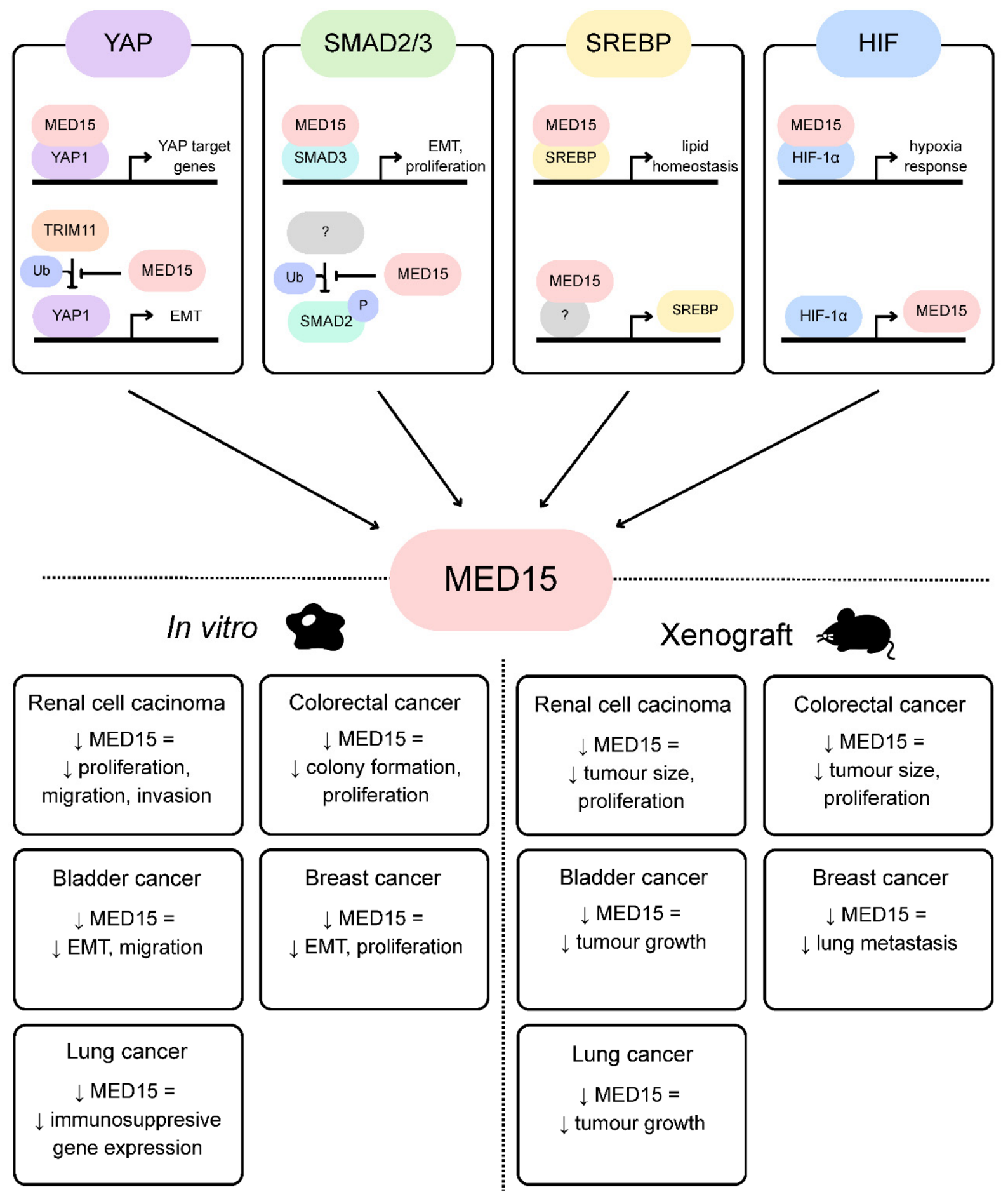
Figure 2.
High expression of MED15 prevents YAP ubiquitination by sequestering TRIM11. In conditions of normal MED15 expression, TRIM11 can ubiquitinate YAP, leading to its degradation (top). When MED15 expression is high, most TRIM11 binds and ubiquitinates MED15, which in turn frees YAP, allowing nuclear YAP accumulation and leading to increased YAP target gene expression, subsequently promoting EMT and immunosuppression.
Figure 2.
High expression of MED15 prevents YAP ubiquitination by sequestering TRIM11. In conditions of normal MED15 expression, TRIM11 can ubiquitinate YAP, leading to its degradation (top). When MED15 expression is high, most TRIM11 binds and ubiquitinates MED15, which in turn frees YAP, allowing nuclear YAP accumulation and leading to increased YAP target gene expression, subsequently promoting EMT and immunosuppression.
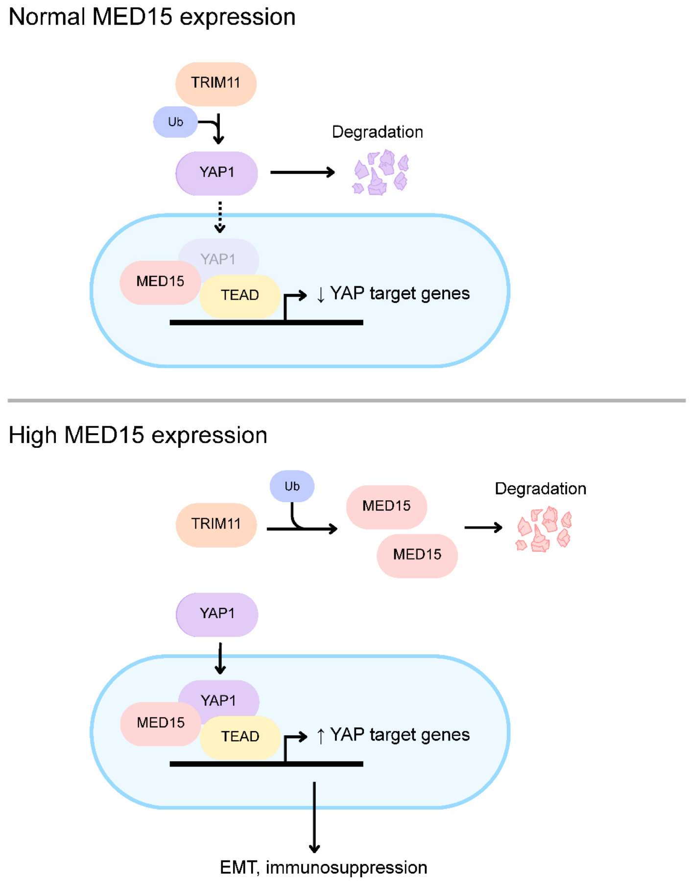

Table 1.
Summary of cancer cases featuring MED15 translocations, with notes on specific genetic changes and clinical presentation.
Table 1.
Summary of cancer cases featuring MED15 translocations, with notes on specific genetic changes and clinical presentation.
| Fusion | Tumor type | # of cases | Clinical presentation | Breakpoints | Ref. |
|---|---|---|---|---|---|
| MED15::TFE3 | Xp11 translocation RCC | 35 | No recurrence | (a) MED15 exon 8/9/11/13 to TFE3 exon 6 (b) MED15 exon 8/9 to TFE3 exon 5 (c) MED15 exon 6/13 to TFE3 exon 4 (d) MED15 exon 11 to TFE3 exon 7 |
[45,46,47,48,49,50,51,52,53,54,55,56,57,58] |
| 2 | Local metastasis | (a) MED15 exon 8 to TFE3 exon 5/6 | [51,55] | ||
| 1 | Distant metastasis | (a) MED15 exon 13 to TFE3 exon 4 | [46] | ||
| 4 | N/A | (a) MED15 exon 6 to TFE3 exon 4 (b) MED15 exon 8 to TFE3 exon 5 |
[59,60,61] | ||
| Melanotic Xp11 neoplasm | 1 | Distant metastasis | (a) MED15 exon 8 to TFE3 exon 6 | [46] | |
| PEComa | 1 | No recurrence | (a) MED15 exon 9 to TFE3 exon 4 | [62] | |
| MED15::ATF1 | Tumor with melanocytic differentiation | 2 | No recurrence | (a) MED15 exon 9 to ATF1 exon 3 | [63] |
| 2 | Local metastasis | (a) MED15 exon 9 to ATF1 exon 3 | [63,64] | ||
| 1 | Death | (a) MED15 exon 9 to ATF1 exon 3 | [64] | ||
| Melanoma | 1 | Death | (a) MED15 exon 8 to ATF1 exon 3 | [65] |
Disclaimer/Publisher’s Note: The statements, opinions and data contained in all publications are solely those of the individual author(s) and contributor(s) and not of MDPI and/or the editor(s). MDPI and/or the editor(s) disclaim responsibility for any injury to people or property resulting from any ideas, methods, instructions or products referred to in the content. |
© 2026 by the authors. Licensee MDPI, Basel, Switzerland. This article is an open access article distributed under the terms and conditions of the Creative Commons Attribution (CC BY) license (http://creativecommons.org/licenses/by/4.0/).
Copyright: This open access article is published under a Creative Commons CC BY 4.0 license, which permit the free download, distribution, and reuse, provided that the author and preprint are cited in any reuse.