

Submitted:
03 September 2026
Posted:
04 September 2026
You are already at the latest version
Abstract
Hyaluronan (HA) has traditionally been viewed as a structural component of the extracellular matrix, valued primarily for its viscoelastic, lubricating, and hydrating properties. However, accumulating evidence suggests that ultra-high molecular weight hyaluronan (UHMW-HA) functions far beyond passive tissue support, acting as a dynamic regulator of inflammation, mechanotransduction, immune homeostasis, cellular senescence, and regeneration.
In this narrative review, we examine HA and specifically focus upon UHMW-HA operating as a “living hydrogel”, an adaptive regenerative macromolecule that continuously interacts with its microenvironment to shape tissue function and resilience. We examine evidence spanning evolutionary biology, glycobiology, biomaterials science, and regenerative medicine to examine how polymer size encodes biological activity and how UHMW-HA influences stem cell behaviour, extracellular matrix remodelling, and responses to injury and ageing.
Attention is given to unique biological models, including the naked mole-rat, whose exceptional longevity and cancer resistance have been linked to abundant UHMW-HA, offering insights into the relationship between extracellular matrix architecture and organismal health.
We further discuss how advances in synthetic biology and biomaterials engineering are enabling the development of next-generation HA-based hydrogels that mimic these adaptive properties for therapeutic applications in tissue engineering, in addition to potential roles in dynamically optimising wound repair, and musculoskeletal rejuvenation, through precision medicine approaches.
Finally, we argue that reclassifying UHMW-HA as a living regenerative material rather than an inert scaffold provides a new concept for designing bioinspired therapeutics and understanding extracellular matrix function. This review demonstrates HA-adaptive macromolecules as critical entities in future regenerative strategies and highlights the specific and unique potential of UHMW-HA to combine material science developments with enhancement of preferable biological outcome.
Keywords:
ultra-high molecular weight hyaluronan (UHMW-HA)
; living hydrogel
; regenerative macromolecule
; extracellular matrix (ECM) signalling
; naked mole-rat model
1. Molecular Size as a Biological Signal: Why UHMW-HA is Different
1.1. The Hyaluronan Molecular Weight Spectrum
Hyaluronan (HA) exists in vivo not as a single, well-defined species but as a continuum of chain lengths, this continuum being functionally, not merely descriptively, significant. Specifically, four operational tiers are recognized. HA oligosaccharides and fragments below 20 kDa arise almost exclusively from enzymatic or oxidative degradation and are reported to remain at low levels in healthy, undamaged tissue. Low molecular weight HA (LMW-HA), spanning 20-100 kDa, and medium molecular weight HA (MMW-HA), spanning 100-1,000 kDa, can arise through the degradation of longer HA chains during tissue turnover, injury, inflammation, or active remodelling. High molecular weight HA (HMW-HA), conventionally defined as approximately 1-2 MDa, is commonly found in intact, non-inflamed connective tissue, synovial fluid, and skin, and is the form most extensively studied in the viscosupplementation and dermal filler literature. Beyond this lies ultra-high molecular weight HA (UHMW-HA), generally defined as chains exceeding 2-6 MDa and, in specialised biological contexts such as the naked mole-rat tissues, reaching 6-12 MDa or more. Each respective threshold corresponds to a measurable shift in receptor engagement, degradation kinetics, and biophysical behaviour [1,2,3,4].
Another important distinction is between native, linear UHMW-HA, a polydisperse unbranched polysaccharide synthesized by hyaluronan synthases (HASs), and chemically cross-linked HA hydrogels, including BDDE and methacrylate-cross-linked systems. In native HA, molecular size refers primarily to the length of individual polymer chains; in cross-linked HA, separate chains or chemically modified chain segments are covalently connected to form a three-dimensional network. Cross-linking can produce stronger and more persistent gel-like viscoelastic behaviour, but the resulting effective network size and mechanical response should not be equated with the molecular weight of a single uninterrupted HA molecule. This distinction matters because some of UHMW-HA’s biological effects may depend on the molecular size, conformation, and receptor-accessible properties of individual, exceptionally long, non-cross-linked chains, not solely on the bulk stiffness of a material produced by covalent network formation [5,6,7,8,9].
Consequently, the fundamental question becomes why changing HA molecular weight and chain architecture, rather than simply increasing HA concentration, can produce markedly different and, in some contexts, opposing biological responses.
1.2. How Molecular Size Shapes HA Biological Activity
HA is chemically uniform: unlike other glycosaminoglycans, it undergoes no template-directed sulfation or epimerisation during biosynthesis, and its core repeating disaccharide unit (β-D-glucuronic acid–[1→3]–β-D-N-acetylglucosamine) remains identical across chain lengths spanning from oligosaccharides to ultra-high-molecular-weight polymers, see Figure 1. Consequently, native HA does not encode biological information through dense combinatorial carbohydrate modifications, but rather primarily through polymer length, hydrodynamic volume, and multivalent receptor engagement. Cells, including elements of the innate immune and mechanotransductive systems, interpret these size differences through cell-surface receptors capable of discriminating fragment size and multivalent clustering [8,9,10].
Cluster of differentiation 44 (CD44), the primary cell-surface HA receptor, clearly demonstrates this size-dependent regulatory behaviour. Short HA oligomers bind CD44 monovalently with low intrinsic affinity and fail to promote receptor clustering. In contrast, HMW- and UHMW-HA chains present multiple tandem binding sites along a single continuous backbone, allowing simultaneous engagement of multiple CD44 ectodomains. This multivalent interaction can promote receptor clustering and increased functional avidity; a spatial reorganization that short fragments generally do not reproduce under comparable conditions. This mechanism differs fundamentally from classical ligand-receptor pharmacology: because clustering strictly requires a continuous molecular scaffold, increasing the concentration of short fragments cannot substitute for physical polymer length. A similar pattern occurs across immune regulation. Intact HMW- and UHMW-HA chains predominantly promote homeostatic and anti-inflammatory signalling (such as CD44/receptor for hyaluronan-mediated motility (RHAMM)-associated tissue stabilization and M2-like macrophage responses), whereas low-molecular-weight fragments generated during injury or catabolism have been associated, in some experimental systems, with Toll-like receptor (TLR) 2/TLR4-dependent pro-inflammatory cascades, nuclear factor kappa-light-chain-enhancer of activated B cells (NF-κB) activation, and cytokine release [8,9,11,12,13,14].
Therefore, identical repeating disaccharide units can convey different biological cues depending on chain length, degradation state, and multivalent presentation. These size-dependent interactions provide the molecular basis for HA’s capacity to support either tissue homeostasis or inflammatory and reparative signalling, depending on the surrounding cellular context.
1.3. Distinctive Physical Properties of UHMW-HA
The size-dependent signalling described above is closely linked to physical properties that become especially pronounced as HA chains reach the ultra-high-molecular-weight range. HMW-HA can also display some of these behaviours, but UHMW-HA forms larger, more persistent polymer domains and can exhibit these effects at lower concentrations and over longer timespans. Four properties are particularly relevant: chain entanglement, viscoelasticity, hydration, and biological persistence [2,15,16].
1.3.1. Chain Entanglement
As HA chains increase in length, they occupy larger hydrodynamic volumes and are more likely to overlap and interpenetrate with neighbouring chains. Once the concentration is sufficiently high, these interactions produce a transient network in which the chains restrict one another’s movement and redistribute mechanical forces. The concentration required for this behaviour decreases as molecular weight increases. Consequently, UHMW-HA can form physically interconnected polymer networks at lower concentrations than shorter HMW- or MMW-HA chains; the longer chains characteristic to ULMW-HA reach the entangled regime more readily and can maintain larger, more continuous polymer domains [15,16,17].
1.3.2. Viscoelasticity
Chain entanglement contributes substantially to the viscoelastic behaviour of concentrated HA solutions. Under slow deformation, HA chains have sufficient time to rearrange and release applied stress, allowing the material to flow. Under rapid or repeated loading, the chains have less time to reorganize, and the solution therefore exhibits a stronger elastic response, resisting deformation and partially recoiling when the load is removed. Longer HA chains generally take more time to rearrange and disengage than shorter chains. Accordingly, increasing molecular weight can prolong relaxation times and increase the elastic contribution of HA solutions at comparable concentrations. This may help sustain a mechanically supportive environment in HA-rich extracellular or pericellular matrices, although the biological significance of this effect remains context-dependent. The magnitude of the response depends not only on molecular weight but also on polymer concentration, ionic strength, chain organization, temperature, pH, and the rate and type of deformation [15,16,18].
1.3.3. Hydration
HA hydration depends on both direct interactions between water and the polymer’s carboxylate and hydroxyl groups and the collective osmotic behaviour of its negatively charged backbone. At physiological pH, the carboxyl groups of HA are negatively charged and interact with mobile counterions, contributing to the swelling and water retention of HA-rich polymer domains [1,2,3]. These effects become particularly relevant when HA chains are concentrated, confined, or associated with other components of the extracellular matrix (ECM). Longer HA chains occupy larger hydrodynamic volumes and can support more extended polymer domains than shorter chains. UHMW-HA may therefore contribute to the formation of hydrated, mechanically resilient matrix environments and may retain water more effectively than shorter HA [15,17,19].
1.3.4. Biological Persistence
Biological persistence refers to the time HA remains in the ECM before being degraded, internalized, or cleared. HA turnover involves several processes, including hyaluronidase-mediated cleavage, receptor-mediated uptake, lysosomal degradation, lymphatic transport, and further processing by liver endothelial cells. Very long HA chains must undergo successive cleavage and processing steps before they are reduced to fragments that can be efficiently internalized and cleared. Their effective residence time may therefore be greater than that of shorter HA species in some extracellular environments, although this depends on tissue localization, receptor expression, interactions with HA-binding proteins, chain organization, and the accessibility of the polymer to degrading enzymes. This relationship is particularly relevant to the naked mole-rat, whose tissues have been reported to contain abundant UHMW-HA and to exhibit altered HA synthesis and degradation. The resulting persistence of HA-rich ECM may help sustain protective HA-CD44 signalling and potentially limit the relative accumulation or effects of degradation fragments [4,20,21,22,23].
1.4. Emergent Properties: Why Longer Chain Length is Not Simply “More”
The properties described above point to a central conceptual distinction: as HA chains become sufficiently long and concentrated to overlap and interact, increasing chain length does not simply produce a linearly scaled-up version of HMW-HA biology. Instead, it can generate emergent behaviours that differ substantially from those of shorter chains at an equivalent total polymer mass, including changes in matrix organization, viscoelasticity, receptor presentation, and biological signalling. This distinction can be understood in terms of the changing physical organization of HA as chain length and concentration increase. At lower concentrations, HA chains behave more independently, whereas longer and/or more concentrated chains increasingly overlap and interact to form a transient, viscoelastic network. In UHMW-HA, these effects may be especially pronounced because exceptionally long chains can occupy larger hydrodynamic domains and influence the organization of water, mechanical forces, and receptor interactions within the matrix. The resulting structure can function as more than a soluble ligand or passive scaffold: it may act as a dynamic, hydrated, and continuously remodelled extracellular network that can be described conceptually as a “living hydrogel” [15,16,24].
This change in polymer organization can influence how HA is presented to cells and how HA receptors are organized at the cell surface. Native HMW-HA can promote CD44 clustering, whereas shorter HA oligosaccharides can reduce or disrupt this clustering, demonstrating that chain length affects receptor organization rather than simply increasing ligand concentration. At still greater molecular masses, UHMW-HA may produce a different mode of CD44 regulation: very-high-molecular-mass HA has been reported to suppress CD44 protein-protein interactions rather than simply intensifying the clustering promoted by shorter HMW-HA. These findings suggest that the physical state of the HA-rich matrix can shape receptor accessibility, multivalent interactions, and downstream signalling. The same principle may contribute to the contrasting inflammatory and vascular effects associated with intact HMW-HA and smaller HA fragments, although these outcomes remain dependent on concentration, degradation state, receptor expression, and cellular context [9,22,25].
Framed in this way, UHMW-HA should not be understood simply as “more HA,” or as a higher dose of the same signal. Rather, its exceptional chain length may confer distinct physical and biological properties, including larger hydrodynamic domains, altered viscoelastic behaviour, greater matrix persistence, and different modes of receptor organization. These features may influence how water, mechanical forces, and biochemical signals are distributed within the pericellular matrix. UHMW-HA may therefore be viewed as a candidate “living regenerative macromolecule.”

Table 1.
Comparative physical and biological properties of conventional HMW-HA and UHMW-HA.
| Dimension | Conventional HMW-HA | UHMW-HA |
|---|---|---|
| Lubrication and Hydration | Provides viscoelastic lubrication and shock absorption in synovial fluid, cartilage, and skin through steric excluded-volume effects and bulk water retention | Retains hygroscopic water-binding capacity while occupying significantly expanded hydrodynamic domains, establishing an extended, hydrated pericellular matrix (PCM) that modulates the local microenvironment |
| Signalling mode | Engages CD44 and RHAMM via multivalent binding sites to promote receptor clustering and homeostatic, anti-inflammatory signalling cascades | Modulates receptor spatial presentation and can selectively attenuate CD44-associated protein–protein interactions, generating distinct cytoprotective and stress-resistance responses |
| Mechanical role | Functions primarily as a viscoelastic structural component, mediating load dissipation and passive tissue compliance under shear and compression | Functions as an active mechanoregulatory network: forms an expanded, transiently entangled viscoelastic network capable of modulating mechanical force transmission and pericellular stress distribution across cell boundaries |
| Tissue residence and Turnover | Exhibits rapid fluid clearance (intra-articular half-life of hours to days) and progressive degradation by endogenous hyaluronidases (HYAL) into lower-MW fragments | Requires more extensive enzymatic cleavage events before degradation into short fragments, potentially prolonging high-mass matrix persistence and functional integrity |
| Conceptual framing | Regarded as a standard structural glycosaminoglycan whose functional capacity scales incrementally with molecular mass and solution concentration | Conceptualized as a dynamic “living hydrogel” whose distinct physical and biological traits emerge from macromolecular chain length, network organization, and cell-matrix interactions |
2. UHMW-HA as a Living Regenerative Macromolecule
HA has long been considered a glycosaminoglycan responsible only for tissue hydration and lubrication and organization of the ECM through selective interaction with receptors such as CD44, RHAMM and lymphatic vessel endothelial hyaluronan receptor-1 (LYVE-1) depending on the length of the molecule. More recent studies, however, have shown that HMW-HA and especially UHMW-HA behave as a dynamic extracellular network, capable of inducing mechanical, biochemical and cellular changes in certain tissues, properties that cannot be explained by simple interaction with receptors [26]. Due to its polymeric structure, tissue retention and water-binding capacity, UHMW-HA forms an active and dynamic PCM that modifies its properties depending on the local microenvironment, influencing the diffusion of mediators, intercellular communication or the matrix-cell interaction [27]. For a comparative view see Figure 2.
Even though the term "living hydrogel" refers especially to living microorganism-encapsulated hydrogels, the meaning of this term can be extended to biological systems that exhibit adaptive properties, which evolve in vivo, in response to oxidative stress, pro-inflammatory factors or extracellular enzymes, and the increase in the length of the HA polymer should be seen as a transition towards a living regenerative macromolecule capable of continuously sensing, integrating, and modulating biological information [28].
2.1. Mechanotransduction: Translating Polymer Architecture into Cellular Responses
Mechanotransduction is the property that may explain the relationship between the length of the HA polymer and its biological effects. Thus, while HMW-HA is normally described as a lubricant, osmotic buffer, or anti-inflammatory ligand, increasing the polymer length allows the generation of an extended, highly hydrated pericellular domain that modulates the information reaching the cell surface via CD44.
2.1.1. Formation of the Pericellular Matrix
The first step in HMW-HA-induced mechanotransduction is the formation of the PCM. HA synthesized in the cell membrane by HASs is extruded into the extracellular space where it binds to CD44 and can associate with hyaladherins, proteoglycans, and other matrix-binding proteins. Due to the large hydrodynamic volume of the HA polymer, this surface-associated network can diffuse considerably through the plasma membrane, thus forming a hydrated compartment that continuously associates new HA molecules [5].
The PCM should not be considered a passive hydrated shell but a living polymer that regulates the access of molecules to the plasma membrane, influences local osmotic pressure, compressive strength, transport of soluble mediators and spatial organization of membrane proteins. For example, the use of a hybrid hyaluronic acid–collagen scaffold loaded with either transforming growth factor-β3 (TGF-β3) or prednisolone in a rat osteochondral defect model, showed that the use of such a scaffold offered superior benefits in structural cartilage repair due to the formation of PCM [29].
Molecular weight is the main factor influencing mechanotransduction, thus CD44-expressing U937 leukocytes exposed to HMW-HA (~1000 kDa) or low molecular weight LMW-HA (~20 kDa) under physiological shear stress (0.1-1.0 dyne/cm²) showed that HMW-HA promoted significantly faster CD44 association. This was accompanied by stronger receptor-ligand interactions, enhanced CD44 clustering, increased CD44 and CD18 expression, and greater cell spreading under low shear stress. However, LMW-HA preferentially increased resistance to shear-induced detachment, indicating that HA molecular weight determines the mechanical organization and mechanotransductive properties of the CD44 signalling complex [30].
The large size of the HA molecule is an absolutely indispensable condition for PCM formation, as physiological HA polymers (10⁶-10⁷ Da) can extend several micrometres from the plasma membrane, and considering that native pericellular covers frequently exceed 20 μm indicates that long HA chains remain in an extended configuration through interactions with the proteoglycans aggrecan or versican [31].
Exogenous HA or aggrecan alone are not sufficient for PCM formation in chick and rat embryonic chondrocytes, both in monolayer and suspension cultures, whereas their combination rapidly restored a mature pericellular envelope within 30 min. Hyaluronan hexasaccharides (HA6) competitively inhibited PCM assembly by competing with HMW-HA-receptor interaction [32].
Published studies to date demonstrate that HMW-HA promotes CD44 receptor clustering and the formation of a mechanically active PCM, which modulates hydration, receptor organization, molecular diffusion, and mechanotransduction. It can be assumed that UHMW-HA would extend these functions to a more persistent and highly multivalent cell-surface polymer environment, however, there are no experimental data indicating glycocalyx thickness and permeability in cells exposed to HMW-HA versus UHMW-HA.
2.1.2. CD44 Organization and Receptor Clustering
CD44 is traditionally considered the main cell surface receptor for HA, but the interaction of HA with CD44 should not be seen in the classical sense of ligand-receptor bond formation. Depending on the length of the chains, the polymers can organize several CD44 molecules in membrane groups that together constitute signalling platforms. Thus, Takasugi et al. demonstrated that UHMW-HA (>6.1 MDa) is not a simple ligand of CD44 but completely reorganizes the cellular architecture by preventing the interactions of CD44 with neighbouring proteins and promoting their organization in the form of a dynamic mechanochemical signalling interface modulated by the polymer [22].
The molecular size of HA is essential in the interaction with receptors, for example, in CD44-transfected COS-7 cells together with HK-2 renal epithelial and BT-549 breast cancer cells, UHMW-HA demonstrated clustering and dimerization of CD44, by fluorescence resonance energy transfer (FRET). HA oligosaccharides on the other hand, only disrupted these receptor assemblies [9].
Also, HA with different molecular weights (1 MDa, 100 kDa, 40 kDa, and 10 kDa) incorporated in injectable HA-based microporous annealed particle scaffolds (HMAPS) influenced differently CD44 organization and endothelial cell behaviour in a study on human cerebral microvascular endothelial cells (HCMVECs), together with in vivo subcutaneous implantation in C57BL/6J mice. Thus, 1 MDa HA induced significantly greater CD44 clustering, reduced receptor membrane diffusion, enhanced endothelial cell survival, stabilized tube formation, and promoted vascularization of implanted scaffolds. Moreover, forced antibody-mediated CD44 clustering reproduced the pro-angiogenic effects of HMW-HA independently of polymer molecular weight, demonstrating that the spatial organization of the receptor is the determining factor of HA activity in vivo [33].
The efficacy of HMW-HA binding to CD44 clusters was demonstrated in inflammatory pain models in male and female Sprague-Dawley rats produced with prostaglandin E₂ (PGE₂) and in cell cultures of dorsal root ganglion neurons. This study showed that HMW-HA reduces hyperalgesia and neuronal excitability only when the phosphoinositide 3-kinase gamma (PI3Kγ)/ protein kinase B (AKT)/ mechanistic target of rapamycin (mTOR) pathway is functional. By successively blocking PI3Kγ with antisense ODN and the inhibitor AS605240, AKT with a specific inhibitor and mTOR with rapamycin, the protective effect of HMW-HA was almost completely lost [34].
All these studies actually show a conceptual change in HA pharmacodynamics, the binding of the UHMW-HA form to CD44 being viewed as a dynamic interaction, mediated by the polymer itself, which behaves as a living regenerative macromolecule.
2.1.3. CD44–Integrin Cooperation and Cytoskeletal Coupling
Mechanical cues generated within the HA-rich PCM are transmitted across the cell membrane and transformed into biochemical signals within the cell. The molecular weight of HA also influences CD44-mediated mechanotransduction. Thus, in the case of CD44-positive U937 leukocytes under physiological shear stress perfused with HMW-HA (~1000 kDa) or LMW-HA (~20 kDa), HMW-HA promoted faster CD44-mediated cell adhesion and stronger receptor-ligand interactions, and only HMW-HA increased the expression of both CD44 and the β2-integrin subunit CD18 [30].
Cytoskeletal adaptors such as ezrin/radixin/moesin (ERM) and ankyrin proteins play an essential role in the organization of CD44. Thus, it has been demonstrated that in murine leukocytes, K562 cells and ex vivo differentiated neutrophils, the binding of CD44 to ERM and ankyrin proteins organizes the receptor into actin-dependent clusters and limits its membrane mobility. Disruption of these interactions by mutagenesis or depolymerization of actin filaments reduced CD44 clustering, increased receptor mobility and diminished Src activation, demonstrating that the cytoskeletal organization of CD44 is essential for the transmission of mechanical signals [35].
Exogenous HA (220 kDa)-induced CD44 activation in CD44-positive MDA-MB-231 and Hs578T breast cancer cells or PC3 prostate cancer cells leads to the formation of a CD44-talin-β1-integrin complex, leading to β1-integrin activation, phosphorylation of Src and focal adhesion kinase (FAK; Tyr397 and Tyr861), and subsequent activation of the focal adhesion proteins cortactin and paxillin. Talin depletion abolished HA-induced β1-integrin activation and significantly reduced CD44–β1-integrin association, whereas inhibition of β1-integrin signalling attenuated cortactin phosphorylation, demonstrating that HA-bound CD44 initiates integrin-dependent focal adhesion signalling through a talin-mediated inside-out activation mechanism [36].
CD44 functions as a mechanosensitive signalling hub coupling HA signalling with integrin activation, focal adhesion assembly, and cytoskeletal remodelling.
2.1.4. Hippo Signalling and YAP/TAZ Localization: A Putative Downstream Mechanism of UHMW-HA Mechanoregulation
The Hippo signalling pathway represents a plausible downstream transcriptional mechanism by which CD44 clustering may regulate cell fate. The transcriptional co-activators Yes-associated protein (YAP) and transcriptional co-activator with PDZ-binding motif (TAZ) integrate multiple biomechanical signalling pathways. Activation of the Hippo kinase cascade results in LATS1/2-mediated phosphorylation of YAP/TAZ, leading to cytoplasmic sequestration, while increased intracellular tension promotes their nuclear translocation and activation of genes involved in proliferation, ECM remodelling, and tissue regeneration [37].
Tunable hydrogels of polyacrylamide and fully synthetic polyethylene glycol exhibit key properties similar to HA of different molecular weights in terms of hydration, elasticity, and cell-matrix adhesion. Modification of the ECM architecture of MCF10A mammary epithelial cells and U2OS osteosarcoma cells by these gels increased nuclear accumulation of YAP/TAZ with increasing matrix stiffness and hydrogel rigidity [38].
Hippo signalling as an endpoint of mechanotransduction was studied in nucleus pulposus cells using substrates with different stiffness to mimic the progressive mechanical changes that occur during intervertebral disc degeneration. Increasing matrix stiffness activated Hippo-associated mechanotransduction and promoted YAP nuclear translocation, leading to increased expression of acyl-CoA synthetase long-chain family member 4 (ACSL4) and ferroptosis. Pharmacological inhibition of YAP or N-cadherin overexpression prevented nuclear accumulation of YAP and significantly attenuated stiffness-induced cellular injury [39].
Even if experimental evidence directly linking HA to YAP/TAZ localization does not exist, however, based on data obtained from other studies linking ECM rigidity to the mechanical properties of some gels, it could be hypothesized that increasing HA molecular weight → enhanced CD44 clustering → stabilization of receptor-cytoskeleton interactions → modulation of focal adhesion dynamics and intracellular tension → context-dependent regulation of Hippo signalling and YAP/TAZ localization. This mechanism is presented in Figure 3.
2.2. Immune Homeostasis Rather Than Immune Suppression
The immunological properties of HA change progressively with increasing polymer length. In intact tissues, in the ECM, the HMW-HA form predominates, with a role in immune homeostasis. On the other hand, in damaged tissues, the HYAL 1 and 2 generate LMW-HA that functions as damage-associated molecular patterns (DAMPs), which amplify the immune response by binding to TLR2/TLR4 and RHAMM, promoting cytokine production, neutrophil recruitment, and pro-inflammatory macrophage activation [23].
Macrophages, dendritic cells, neutrophils, activated T lymphocytes and other cells involved in immunity all express CD44, as shown by experimental vaccination studies using different sizes of the polymer [40]. Based on these considerations, it is assumed that UHMW-HA should not be considered a simple suppressor of immune activation but a stabilizer of CD44 over larger membrane domains, which stabilizes the PCM and reduces the formation of LMW-HA fragments.
Macrophages are the most representative immune cell population regulated by HA. MMW-HA (750 kDa to 1 MDa) modulates macrophage polarization through RHAMM-dependent signalling. The use of a RHAMM-blocking peptide (P15-1) on THP-1-derived macrophages polarized with interferon-γ (IFN-γ) and lipopolysaccharide (LPS) leads to the blockade of HA-RHAMM interactions and the suppression of the release of M1-associated inflammatory mediators such as interleukin-1β (IL-1β), interleukin-6 (IL-6), tumour necrosis factor-α (TNF-α), inducible nitric oxide synthase (iNOS), TLR1, TLR4, cluster of differentiation 80 (CD80), cluster of differentiation 86 (CD86), and human leukocyte antigen-DR (HLA-DR), while amplifying M2-associated markers, including cluster of differentiation 163 (CD163), cluster of differentiation 206 (CD206), peroxisome proliferator-activated receptor-γ (PPARγ), and signal transducer and activator of transcription 6 (STAT6). This indicates that HA actively regulates the macrophage phenotype [41].
Macrophage polarization by a ~100 kDa HA-based dopamine hydrogel towards an M2 phenotype occurs through inhibition of the FAK/PI3K/AKT/NF-κB pathway, but in this case the presence of dopamine must also be taken into account, the molecular mass of HA being relatively low in this study [42].
Dendritic cells synthesize HA polymers of up to 10,000 disaccharide units via the hyaluronan synthase 2 (HAS2) pathway, which form a glycocalyx with a thickness of approximately 500 nm necessary for immune cell trafficking. Genetic deletion of LYVE-1 or CD44, enzymatic degradation of HA, and antibody-mediated blockade of either receptor significantly impaired dendritic cell docking and delayed their entry into afferent lymphatic vessels, demonstrating that the HA-CD44-LYVE-1 axis is required for efficient lymphatic trafficking [43].
On the other hand, enzymatically crosslinked HA fragmented with tyramine (HATyr) and mechanically fragmented granular hydrogels using 50 or 150 μm meshes functioned as endogenous DAMPs. They produced activation of TLR2 and TLR4 receptors, in human THP-1 monocytes leading to NF-κB and IRF signalling and increased expression of pro-inflammatory cytokines including IL-1β and IL-6, with the greater the degree of fragmentation [44].
The involvement of HA in maintaining immune equilibrium has an extremely important clinical application in the production of vaccines. Thus, among HA polymers of different molecular weights (15, 50, 200, and 500 kDa) chemically conjugated with protein antigens (including ovalbumin, tetanus toxoid, H5N1 hemagglutinin, rabies glycoprotein, and human growth hormone), 200 kDa HA generated the strongest antigen-specific adaptive immune response in female BALB/c, CD1, and C57BL/6 mice. This induced a persistent IgG production together with both IgG1 and IgG2a/IgG2b subclasses, indicative of a balanced Th2/Th1 response [40].
HMW-HA actively regulates immune responses through CD44-dependent signalling, macrophage polarization, leukocyte trafficking, and preservation of ECM integrity therefore it is expected that UHMW-HA will amplify these biological functions because of its unique physicochemical characteristics including greater multivalent receptor engagement, increased resistance to enzymatic degradation, prolonged tissue residence, and formation of highly hydrated viscoelastic networks. However, experimental studies comparing HMW-HA and native UHMW-HA remain remarkably scarce.
2.3. Control of Oxidative Stress and Cellular Senescence
Reactive oxygen species (ROS) are not only active inside the cell, but can remodel the ECM by cleaving long HA polymers. For example, HMW-HA exposed for 4 h to increasing concentrations of hydrogen peroxide (H₂O₂) or hypochlorous acid (HOCl) depolymerized to LMW-HA under the action of H₂O₂ and fragmented and formed chloramides under the action of HOCl [45].
HA can influence oxidative stress indirectly by maintaining mitochondrial quality control. For example, in a model of oxidative stress induced with tert-butyl hydroperoxide in primary human nucleus pulposus cells isolated from patients undergoing lumbar discectomy (46–65 years old) exposure to HMW-HA (1.8–2.2 MDa) enhanced mitophagy, increased LC3 and Beclin-1-associated responses, preserved mitochondrial membrane potential, reduced mitochondrial superoxide and total cellular ROS, restored ATP production, and decreased SA-β-gal-positive cells. Mitophagy was confirmed by treating cells with mitophagy inhibitor cyclosporin A or transfected with C1QBP siRNA to silence complement component 1q-binding protein, a mitochondrial HA-binding protein involved in mitochondrial quality control [46].
An ECM rich in HA can modify the reciprocal cause/effect relationship between senescence and inflammation. In an osteoarthritis model, induced by TNF-α in mouse chondrocytes, BDDE-crosslinked granular hyaluronic acid hydrogel reduced the number of SA-β-gal-positive senescent chondrocytes, suppressed the expression of the senescence markers p16^INK4a^ and p21, decreased oxidative stress by increasing superoxide dismutase (SOD) and reducing cyclooxygenase-2 (COX-2) expression, and significantly lowered IL-6 and IL-1β production. The basis of these effects was reduced TLR-2 expression and inhibition of NF-κB activation, together with decreased expression of the matrix-degrading enzymes matrix metalloproteinase (MMP) MMP3, MMP13, and ADAMTS-5, while preserving COL2A1, aggrecan expression, and ECM integrity. A limitation of this study is that, in addition to the molecular weight of HA, the porosity and local mechanical properties of the hydrogel must be considered [47].
The influence of HA molecular weight on senescence is still controversial. A study conducted on human amniotic epithelial stem cells (hAECs) undergoing replicative aging in vitro using HA preparations ranging from 300 to 1500 kDa showed that only 300-kDa HA reduced the proportion of SA-β-gal-positive cells, decreased the expression of the senescence markers p16 and p21, alleviated G2/M cell-cycle arrest, and preserved stemness. The same study showed that HAS2 knockdown or CD44 blockade abolished the anti-senescent phenotype, and inhibition of AKT with MK-2206 prevented AKT/mTOR activation and restored SA-β-gal positivity together with p16 and p21 expression, identifying the CD44s-AKT/mTOR axis as a main downstream mediator of HA-induced cytoprotection [48].
2.4. Evidence from the Naked Mole-Rat
The length of the HA polymer directly influences the resistance to stress-induced cell-cycle arrest. Thus, human IMR90 fibroblasts, mouse skin fibroblasts, and naked mole-rat fibroblasts were incubated for 6 hours with HA of different lengths (naked mole-rat UHMW-HA, HMM-HA, fragmented naked mole-rat HA, and a synthetic 1 MDa HA) before being exposed to oxidative stress induced with tert-butyl hydroperoxide, doxorubicin or ionizing irradiation. UHMW-HA protected cells from stress-induced death and cell-cycle arrest more effectively than HMM-HA, and this protection was progressively lost after enzymatic shortening of the polymer, demonstrating a clear polymer length-dependent effect [22].
The tissue distribution of UHMW-HA in naked mole-rat is not uniform, tissues such as skin, muscle, and particularly lymph nodes are abundant in UHMW-HA, while the content in the renal medulla is lower than in guinea pigs. The molecular weight of HA in these tissues also varies greatly, from 2.5 MDa in lymph nodes to 0.5 to 2.5 MDa in skin and kidney. Transcriptomic analysis showed that at the lymph node level there was an overexpression of Hyal3 (129-fold), Has1 (100-fold), and Tnfaip6 (58-fold), while HYAL1 expression and activity were reduced [23].
Compelling evidence that HA-mediated longevity mechanisms can be transferred across mammalian species was obtained by generating a tamoxifen-inducible transgenic mouse (nmrHas2 mice) overexpressing naked mole-rat Has2 under the CAG promoter. Both the abundance and molecular mass of HA were observed to increase in multiple tissues, including skeletal muscle, heart, kidney, and small intestine, although the increase remained moderate because of the higher endogenous HYAL activity in mice. nmrHas2 mice presented lower spontaneous cancer incidence, with only 49% of animals older than 27 months developing cancer compared with 83% of age-matched controls and were also protected against DMBA/TPA-induced skin papillomagenesis. nmrHas2 mice exhibited a 4.4% increase in median lifespan and a 12.2% increase in maximum lifespan [49].
3. Translating UHMW-HA into Next-Generation Biomaterials
The discovery that UHMW-HA actively regulates a plethora of processes including mechano-transduction, immune homeostasis, cellular senescence and tissue resilience, has fundamentally changed the way ECMs were viewed. UHMW-HA is now known not to be an inert structural polymer that can act as a dynamic biological interface capable of continuously sensing and responding to changes within the tissue microenvironment. These observations suggest that biomaterials could be engineered to reproduce dynamically adaptive scaffolds
The naked mole-rat provided the first proof-of-principle that the biological effects of UHMW-HA extended well beyond lubrication or mechanical cushioning. Its remarkable resistance to cancer, osteoarthritis, chronic inflammation and age-associated functional decline are inextricably associated with abundant tissue deposition of HA polymers frequently exceeding 6–12 MDa. Experimental studies showed that introduction of the naked mole-rat HAS2 gene into mice increased tissue HA abundance, whilst preventing spontaneous tumour formation. Transgenic mice expressing the naked mole-rat HAS2 gene resulted in enhanced tissue HA content and a shift towards higher molecular weight polymers in multiple organs. Additionally, the presence of this gene attenuated systemic inflammation, improved gut barrier integrity and significantly improved both lifespan and health span, demonstrating that these ECM adaptations were transferable between mammalian species and did not in fact represent species-specific evolutionary anomalies [49]. Similar observations showed a remarkable resistance of naked mole-rats to post-traumatic osteoarthritis induced by meniscal-ligamentous injury, suggested that UHMW-HA tissue conferred resilience through direct preservation of cartilage mechanics and cellular homeostasis. In contrast, mice undergoing the same procedure reached a terminal disease state at the 4-week endpoint of the experiment [50].
Recent work additionally demonstrated that elevated CD44 expression in the naked mole-rat correlated closely with mammalian longevity. Characterization of isolated oliogodendrocyte progenitor cells showed enhanced endoplasmic reticulum proteostasis through activation of ATF6 activity, but independently of HA binding. This suggested that evolutionary optimisation of the HA-CD44 signalling axis represented an integrated adaptive longevity mechanism rather than an isolated extracellular phenomenon [51]. Taken together, these studies and others have established a biological proof-of-principle that suggests that recreating UHMW-HA-rich extracellular environments may represent a feasible tissue/cellular regenerative strategy rather than merely being perhaps an unexpected but interesting evolutionary observation.
3.1. The Naked Mole-Rat as Proof-of-Principle for Adaptive Biomaterials
As mentioned earlier, the remarkable biology of the naked mole-rat fundamentally has now altered the understanding of HA biology and provided the first compelling evidence that UHMW-HA functions as far more than a structural ECM polymer. The exceptionally long HA polymers help to create a continuously adaptive extracellular environment that dictates preferable tissue homeostasis, resistance to chronic inflammation, cancer suppression and exceptional longevity. From an evolutionary standpoint, it is likely that the biological activity of UHMW-HA emerged from its ability to regulate cellular behaviour over prolonged periods rather than simply from its mechanical properties.
The molecular basis for this phenotype has now been extensively characterised. Naked mole-rats produce abundant HA polymers frequently exceeding 6–12 MDa (far longer than those seen in other species including humans) through increased activity of HAS2 together with reduced degradation by HYAL, leading to a continuous and persistent accumulation of UHMW-HA throughout multiple tissues throughout its whole lifespan. This ECM architecture has been correlated with enhanced skin elasticity, improved wound healing, attenuation of oxidative stress, resistance to spontaneous tumour development and preservation of tissue function during ageing, largely through sustained CD44-dependent signalling and maintenance of ECM integrity- reviewed recently and eloquently by Lagunas-Rangel [52].
As mentioned above, transfer of the HAS2 naked mole rat gene to mice caused significant positive health-span-related consequences for the animals. The work also studies transcriptomic changes associated with the gene transfer and showed that expression profiles shifted towards those observed in naturally longer-lived mammalian species, suggesting that elevated UHMW-HA influenced fundamental biological programmes associated with healthy ageing rather than isolated disease pathways [49]. Importantly, the beneficial effects were attributed to increased HMW/UHMW-HA itself rather than to species-specific properties of the naked mole-rat HAS2 enzyme, indicating that manipulation of extracellular HA biology may represent a potentially transferable therapeutic strategy.
These observations have important implications for biomaterials science. Conventional HA-based biomaterials have generally been designed to reproduce the viscoelastic or lubricating properties of native cartilage and ECM. The naked mole-rat studies instead suggested that successful regenerative materials should reproduce the dynamic biological functions of UHMW-HA, including sustained receptor organisation, mechanotransduction, immune homeostasis and adaptive extracellular signalling. In this context, therefore, we propose that UHMW-HA should not be considered as a higher molecular weight version of conventional HA, but as the biological blueprint for engineering biomaterials capable of continuously sensing and responding to changes within their surrounding microenvironment. Support for this concept was provided from studies examining the biological consequences of polymer integrity. Oxidative fragmentation of HMW-HA into lower molecular weight species profoundly altered its biological behaviour, reducing its capacity to support corneal epithelial cell proliferation while modifying wound healing responses This data demonstrated that preservation of polymer length was essential for maintaining regenerative function [45].
These findings suggest that maintaining or engineering persistent UHMW-HA architectures may be more important than simply increasing the total amount of HA delivered to tissues. The data provides the first biological proof-of-principle that the exceptional regenerative properties associated with UHMW-HA represent a transferable ECM that could potentially be recreated through biomaterials engineering. This indicates that instead of attempting to replace damaged tissues with inert scaffolds, future regenerative biomaterials may be able to reproduce the adaptive extracellular environment established naturally by UHMW-HA, and in this way control key elements of cellular behaviour, tissue homeostasis and regenerative repair.
It is interesting to hypothesise then whether synthetic or semi-synthetic materials could be engineered to reproduce these unique biological properties. This question has driven the recent development of engineered UHMW-HA hydrogels designed to emulate not only the physicochemical characteristics of native HA, but also its capacity to dynamically regulate cellular behaviour and tissue regeneration.
3.2. Engineering Living Hydrogels that Mimic UHMW-HA
Early generations of HA-based hydrogels were primarily developed to reproduce the physical characteristics of native ECM, including lubrication, hydration, viscoelasticity and shock absorption. These materials have proved valuable as injectable fillers, viscosupplements and temporary scaffolds, particularly in the field of orthopaedic medicine, yet their biological function remained largely passive, serving principally to provide mechanical support while gradually degrading within the surrounding tissue. Consequently, conventional HA hydrogels were designed to mimic the material properties of cartilage rather than the biological behaviour of living ECM.
Evidence presented throughout this review suggested that the biological activity of UHMW-HA cannot be explained solely by its rheological properties. It is now known that its exceptionally long polymer chains can generate highly hydrated pericellular matrices capable of re-organising CD44 signalling platforms, that can modulate mechanotransduction and promote stem-cell activity. The following examples of HA-based regenerative materials have been designed therefore, to retain water/resist compression, and to continuously and dynamically regulate receptor organisation, whilst controlling ECM remodelling, inflammatory signalling and cellular communication in response to changes within the surrounding microenvironment.
This transition has been accelerated by advances in hydrogel engineering, permitting increasingly sophisticated control over HA architecture and network organisation. Methacrylated HA (MeHA), interpenetrating polymer networks, supramolecular assemblies and dynamic cross-linking chemistries have all been developed to improve mechanical stability while preserving biological functionality. Unlike conventional non-bioactive covalently cross-linked hydrogels, these newer systems attempt to maintain the dynamic mobility and hydration characteristics that distinguish native UHMW-HA. A recent example of this strategy was demonstrated using a mussel-inspired interpenetrating hydrogel network composed of catechol-functionalised chitosan and ultra-high molecular weight methacrylated HA, that was designed specifically to recreate an adaptive osteochondral interface [53]. The presence of citric acid-mediated polymer interactions significantly enhanced the stiffness of the gel resulting in reduced excessive swelling, along with a notable improved tissue adhesion. Concomitantly, the authors identified a greater and more stable integration with three-dimensional printed osteochondral constructs whilst the constructs maintained full cytocompatibility and this was correlated with enhanced fibroblast viability and a significant increase in Young’s modulus. Importantly, the mechanical behavioural changes resulted from coordinated interactions between the polymer networks and not from simple increases in polymer concentration.
Similar design principles have also been applied to injectable HMAPS, where altering HA molecular weight profoundly influenced CD44 organisation and endothelial cell behaviour. Hydrogels incorporating approximately 1 MDa HA promoted significantly greater CD44 clustering and reduced receptor mobility in cultured human cerebral microvascular EC. This incorporation also enhanced endothelial survival and produced a more extensive stabilised vascular network formation compared with lower molecular weight formulations. Artificial antibody-mediated clustering of CD44 reproduced many of these biological effects independently of polymer size, demonstrating that spatial receptor organisation rather than ligand availability was the major determinant of HA-mediated regenerative activity [33]. These findings further supported the concept that future biomaterials should be engineered to organise signalling platforms that can modulate cell behaviour and tissue microenvironment according to changes in mechanical loading, oxidative stress, enzymatic degradation or local cytokine production rather than simply deliver bioactive molecules [28].
Within this broader biological context, UHMW-HA may represent the closest naturally occurring example of a living hydrogel. Unlike conventional synthetic matrices possessing fixed physicochemical characteristics, UHMW-HA continuously remodels its pericellular environment through multivalent receptor interactions, dynamic hydration, polymer entanglement and reciprocal communication with surrounding cells. Its biological activity therefore emerges from continuous adaptation rather than static material properties. This distinction represents a fundamental change in biomaterial design philosophy, shifting emphasis from constructing increasingly sophisticated scaffolds towards engineering extracellular environments capable of sensing, integrating and responding to biological information. This concept is particularly relevant for regenerative medicine because tissue repair depends upon dynamic interactions between resident stem cells, immune cells and ECM rather than simple structural replacement. Consequently, biomaterials capable of reproducing the adaptive behaviour of UHMW-HA may provide superior control over both local and systemic parameters critical for protection and tissue repair for example, macrophage polarisation, angiogenesis and stem-cell niche maintenance, when compared with conventional hydrogel systems.
These advances suggest that the future of HA-based biomaterials lies not in further optimisation of viscosity or mechanical strength alone, but in engineering matrices that emulate the adaptive biological behaviour of UHMW-HA. In this respect, UHMW-HA should be viewed not simply as a superior hydrogel constituent but as the biological blueprint for designing precision regenerative biomaterials capable of continuously integrating mechanical, biochemical and cellular information to restore tissue homeostasis.
3.3. Precision Regenerative Biomaterials: Towards Adaptive Extracellular Matrix Engineering
The development of UHMW-HA-inspired biomaterials has implications has recently extending beyond cartilage replacement or viscosupplementation.
Articular cartilage remains one of the most advanced clinical applications of HA-based biomaterials. Conventional viscosupplementation has historically focused upon improving lubrication and reducing joint loading, whereas increasing evidence suggests that higher molecular weight preparations may additionally influence inflammation, chondrocyte survival and ECM preservation. A randomized clinical trial comparing UHMW-HA with MMW-HA for hip osteoarthritis demonstrated that a single intra-articular injection of UHMW-HA produced comparable or superior long-term improvements in pain and disability despite requiring fewer injections [54]. More recently, a Level I Bayesian network meta-analysis including more than 9,800 patients showed that UHMW- and HMW-HA preparations produced the greatest reduction in pain at medium-term follow-up compared with lower molecular weight formulations, supporting the concept that higher polymer length correlated directly with therapeutic [55]. These observations further reinforce the biological importance of molecular weight in determining regenerative outcomes.
Successful regeneration of intervertebral disc tissue similarly depends upon restoration of a highly hydrated ECM capable of maintaining mechanical loading while preserving nucleus pulposus cell phenotype and mitochondrial homeostasis. Likewise, skin regeneration requires coordinated regulation of inflammation, angiogenesis and ECM remodelling, whereas corneal repair depends upon transparent hydrated matrices capable of supporting epithelial migration without excessive fibrosis. In the central nervous system, HA-rich ECMs regulate neural stem-cell behaviour, synaptic plasticity and neuroinflammatory responses, while vascular regeneration requires dynamic endothelial mechanotransduction together with controlled CD44-mediated angiogenic signalling [56]. Although these tissues differ considerably in structure and function, each depends upon adaptive ECM biology rather than simple mechanical support, suggesting that biomaterials capable of reproducing UHMW-HA function may provide broad regenerative applications across multiple organ systems.
These developments have simultaneously driven the emergence of precision biomaterials. Historically, biomaterial design has largely focused upon optimising individual physical characteristics including stiffness, porosity, degradation kinetics or mechanical strength. While these parameters remain important, they cannot fully account for the dynamic biological interactions that determine regenerative success. Future biomaterials will increasingly require engineering according to the biological characteristics of individual tissues and patients rather than universal material specifications. Variables including patient age, disease stage, inflammatory status, ECM composition, mechanical loading, receptor expression profiles and resident stem-cell phenotype may all influence the optimal biological properties of regenerative materials. For example, age-dependent differences in host immune responses can substantially alter the biological behaviour of implanted extracellular-matrix-based versus synthetic biomaterials, illustrating why biomaterial performance cannot necessarily be extrapolated between young and aged tissues. In a murine model (C57BL/6J; 4 months versus 18-month-old) comparison of ECM-derived urinary bladder matrix (UBM) and synthetic polypropylene mesh, both materials initially induced inflammation, however, UBM subsequently transitioned toward a pro-regenerative, tissue-remodelling response, whereas polypropylene elicited sustained pro-inflammatory activity. This difference was reflected in macrophage polarisation, with UBM associated predominantly with an anti-inflammatory M2-like phenotype and polypropylene with a persistent M1-like response. Importantly, ageing delayed the resolution of inflammation following UBM implantation and altered the associated transcriptional response, with TNF-α and IL-1β identified as important drivers of chronic inflammation in aged animals. These findings demonstrate that biomaterial performance is strongly influenced by the age-dependent immune microenvironment, and that ECM-based materials may retain regenerative potential even in the context of age-related immune dysregulation [57].
Cheng et al showed that a dynamic hydrogel incorporating pH-responsive imine crosslinks and glucose oxidase (GOx), promoted restoration of wound pH and accelerated wound closure in a diabetic mouse model. The model utilised the initially alkaline environment of diabetic wounds as the trigger for material activation and generation of biocatalytic acid as anti-fuel and demonstrated effective creation of a closed-loop interaction between the material and the tissue [58].
Consequently, future UHMW-HA-based hydrogels will use dynamic cross-linking chemistries, enzyme-responsive degradation, mechanically sensitive polymer architectures and programmable release systems. In effect these will be tuneable and will allow biomaterials to alter their biological activity in response to inflammatory cytokines, oxidative stress, mechanical loading or changes in cellular phenotype. Regenerative matrices will be tailored to the biological requirements of individual diseases and even individual patients, as described recently by de Kort et al regarding potential formulations of blood vessels and heart valves in cardiovascular tissue engineering [59]. For precision ECM engineering, UHMW-HA provides an ideal biological template because of its integrative regenerative activity and hence engineering biomaterials that reproduce these adaptive properties represent a logical progression towards personalised regenerative medicine, in which restoration of tissue homeostasis replaces structural tissue replacement as the principal therapeutic objective.
3.4. Future directions
Future regenerative biomaterials may therefore be designed not simply to replace damaged tissue but to recreate the adaptive extracellular environments characteristic of youthful tissues indicating a possible anti-ageing impact. UHMW-HA represents a biological blueprint for a new generation of precision regenerative biomaterials capable of continuously integrating mechanical, biochemical and cellular information. Such "living" biomaterials have the potential to redefine tissue engineering by shifting emphasis from structural replacement towards restoration of dynamic tissue function.
The next stage of UHMW-HA research should move beyond demonstrating its biological effects towards defining the specific molecular, mechanical and temporal properties that make exceptionally long HA polymers biologically distinct. Although evidence supports a relationship between HA molecular size and cellular responses, direct comparisons between native UHMW-HA, conventional HMW-HA and fragmented HA remain surprisingly limited. Future studies should therefore establish molecular-weight-dependent biological thresholds using rigorously characterised HA preparations, while simultaneously controlling for polymer concentration, polydispersity, crosslinking, tissue retention and degradation. Such studies will be essential to determine whether UHMW-HA possesses genuinely distinct biological functions or whether its effects represent an amplification of mechanisms already present with HMW-HA.
A second priority is to define how polymer architecture is translated into cellular signalling. The current evidence supports a model in which long HA polymers generate an expanded pericellular matrix, promote CD44 clustering and influence integrin-dependent mechanotransduction. However, the downstream consequences of these interactions remain incompletely understood. In particular, the proposed links between UHMW-HA, cytoskeletal tension and YAP/TAZ signalling require direct experimental validation. Future work combining live-cell imaging, single-cell transcriptomics, spatial proteomics and quantitative measurements of CD44 organisation, glycocalyx dimensions and matrix mechanics could establish how UHMW-HA simultaneously regulates biochemical and mechanical information at the cell surface. This would help transform the current conceptual model of UHMW-HA as a “living regenerative macromolecule” into a quantitatively testable biological framework.
An equally important direction is the development of adaptive UHMW-HA-based hydrogels that retain the dynamic properties of native polymer networks. Conventional crosslinking can improve mechanical stability but may also restrict polymer mobility and potentially compromise receptor accessibility and matrix remodelling. Future materials should therefore incorporate reversible, supramolecular or enzyme-responsive interactions that permit controlled changes in stiffness, hydration, degradation and molecular presentation in response to the surrounding tissue. Recent developments in engineered living hydrogels demonstrate that biomaterials can be designed to sense environmental changes and alter their behaviour, accordingly, providing a broader technological framework for UHMW-HA-inspired materials [60].
The concept of precision regenerative biomaterials represents another major opportunity. Rather than developing a single UHMW-HA formulation for all applications, future materials could be engineered according to the biological characteristics of the target tissue and individual patient. Age, inflammatory state, ECM composition, mechanical loading and receptor expression may all influence the response to an implanted material. Precision biomaterials research is increasingly moving towards this model, in which patient-specific biological information is incorporated into material design rather than being considered only after implantation. This approach may be particularly relevant to ageing, where impaired inflammatory resolution, altered matrix composition and reduced stem-cell function could require different UHMW-HA architectures from those appropriate for young or acutely injured tissues.
A further challenge will be determining whether UHMW-HA can provide long-term tissue resilience rather than simply accelerated repair. The naked mole-rat and nmrHAS2 mouse studies provide an important proof-of-principle that increasing tissue HA abundance and molecular weight can influence cancer susceptibility, inflammation, healthspan and lifespan. However, translating this observation into human regenerative medicine will require clarification of the appropriate molecular-weight range, tissue concentration, duration of exposure and potential consequences of sustained CD44 engagement. Long-term studies should therefore examine not only regeneration but also fibrosis, aberrant proliferation, immune surveillance and tissue-specific effects following repeated or persistent UHMW-HA exposure.
Finally, translation will require standardisation and clinical-scale manufacturing. HA molecular weight is often reported as a single value despite substantial differences in polydispersity, chain integrity and degradation behaviour between preparations. For UHMW-HA in particular, manufacturing conditions, sterilisation, storage and exposure to oxidative or enzymatic degradation may alter polymer length and consequently biological activity. Future clinical development should therefore incorporate molecular-weight distribution, polymer integrity, degradation kinetics and biological potency into product characterisation and release criteria. This will be particularly important if UHMW-HA is to be incorporated into injectable hydrogels, 3D-printed constructs or combination therapies [61].
Taken together, these developments suggest that the ultimate objective should not simply be to manufacture higher-molecular-weight HA hydrogels, but to reproduce the defining biological characteristics of the native UHMW-HA environment: persistent hydration, multivalent receptor organisation, mechanical adaptability, controlled degradation and reciprocal communication with resident cells. The emerging field of adaptive and living hydrogels demonstrates that such materials are becoming technically feasible. UHMW-HA therefore provides more than an improved biomaterial formulation; it offers a biological blueprint for engineering extracellular environments capable of sensing, adapting and restoring tissue homeostasis. The transition from static scaffolds to such adaptive regenerative materials may ultimately represent one of the most important directions for the next generation of personalised regenerative medicine.
Abbreviations
| ACSL4 | acyl-CoA synthetase long-chain family member 4 |
| AKT | protein kinase B |
| CD163 | cluster of differentiation 163 |
| CD206 | cluster of differentiation 206 |
| CD44 | cluster of differentiation 44 |
| CD80 | cluster of differentiation 80 |
| CD86 | cluster of differentiation 86 |
| COX-2 | cyclooxygenase-2 |
| DAMPs | damage-associated molecular patterns |
| ECM | extracellular matrix |
| ERM | ezrin/radixin/moesin |
| FAK | focal adhesion kinase |
| FRET | fluorescence resonance energy transfer |
| GOx | glucose oxidase |
| H₂O₂ | hydrogen peroxide |
| HA | Hyaluronan/Hyaluronic acid |
| HA6 | Hyaluronan hexasaccharide |
| hAECs | human amniotic epithelial stem cells |
| HAS | hyaluronan synthase |
| HAS2 | hyaluronan synthase 2 |
| HATyr | hyaluronan fragmented with tyramine |
| HCMVECs | human cerebral microvascular endothelial cells |
| HLA-DR | human leukocyte antigen-DR |
| HMAPS | HA-based microporous annealed particle |
| HMW-HA | high molecular weight HA |
| HOCl | hypochlorous acid |
| HYAL | hyaluronidases |
| IFN-γ | interferon-γ |
| IL-1β | interleukin-1β |
| IL-6 | interleukin-6 |
| iNOS | inducible nitric oxide synthase |
| LPS | lipopolysaccharide |
| LYVE-1 | lymphatic vessel endothelial hyaluronan receptor-1 |
| MeHA | Methacrylated hyaluronan |
| MMP | matrix metalloproteinase |
| MMW-HA | medium molecular weight hyaluronan |
| mTOR | mechanistic target of rapamycin |
| NF-κB | nuclear factor kappa-light-chain-enhancer of activated B cells |
| PCM | pericellular matrix |
| PGE₂ | prostaglandin E₂ |
| PI3Kγ | phosphoinositide 3-kinase gamma |
| PPARγ | peroxisome proliferator-activated receptor-γ |
| RHAMM | receptor for hyaluronan-mediated motility |
| ROS | reactive oxygen species |
| SOD | superoxide dismutase |
| STAT6 | signal transducer and activator of transcription 6 |
| TAZ | transcriptional co-activator with PDZ-binding motif |
| TGF-β3 | transforming growth factor-β3 |
| TLR2/TLR4 | Toll-like receptors |
| TNF-α | tumor necrosis factor-α |
| UBM | urinary bladder matrix |
| UHMW-HA | ultra high molecular weight hyaluronan |
| YAP | Yes-associated protein |
References
- Liu, Y.; Li, L.; Wang, L.; Lu, L.; Li, Y.; Huang, G.; Song, J. ‘Two-Faces’ of Hyaluronan, a Dynamic Barometer of Disease Progression in Tumor Microenvironment. Discov. Oncol. 2023, 14, 11. [Google Scholar] [CrossRef] [PubMed]
- Cowman, M.K.; Lee, H.-G.; Schwertfeger, K.L.; McCarthy, J.B.; Turley, E.A. The Content and Size of Hyaluronan in Biological Fluids and Tissues. Front. Immunol. 2015, 6, 261. [Google Scholar] [CrossRef] [PubMed]
- Wu, Y.-Z.; Huang, H.-T.; Ho, C.-J.; Shih, C.-L.; Chen, C.-H.; Cheng, T.-L.; Wang, Y.-C.; Lin, S.-Y. Molecular Weight of Hyaluronic Acid Has Major Influence on Its Efficacy and Safety for Viscosupplementation in Hip Osteoarthritis: A Systematic Review and Meta-Analysis. CARTILAGE 2021, 13, 169S–184S. [Google Scholar] [CrossRef] [PubMed]
- Tian, X.; Azpurua, J.; Hine, C.; Vaidya, A.; Myakishev-Rempel, M.; Ablaeva, J.; Mao, Z.; Nevo, E.; Gorbunova, V.; Seluanov, A. High-Molecular-Mass Hyaluronan Mediates the Cancer Resistance of the Naked Mole Rat. Nature 2013, 499, 346–349. [Google Scholar] [CrossRef] [PubMed]
- Maloney, F.P.; Kuklewicz, J.; Corey, R.A.; Bi, Y.; Ho, R.; Mateusiak, L.; Pardon, E.; Steyaert, J.; Stansfeld, P.J.; Zimmer, J. Structure, Substrate Recognition and Initiation of Hyaluronan Synthase. Nature 2022, 604, 195–201. [Google Scholar] [CrossRef] [PubMed]
- Itano, N.; Sawai, T.; Yoshida, M.; Lenas, P.; Yamada, Y.; Imagawa, M.; Shinomura, T.; Hamaguchi, M.; Yoshida, Y.; Ohnuki, Y.; et al. Three Isoforms of Mammalian Hyaluronan Synthases Have Distinct Enzymatic Properties. J. Biol. Chem. 1999, 274, 25085–25092. [Google Scholar] [CrossRef] [PubMed]
- La Gatta, A.; Bedini, E.; Aschettino, M.; Finamore, R.; Schiraldi, C. Hyaluronan Hydrogels: Rheology and Stability in Relation to the Type/Level of Biopolymer Chemical Modification. Polymers 2022, 14, 2402. [Google Scholar] [CrossRef] [PubMed]
- Cyphert, J.M.; Trempus, C.S.; Garantziotis, S. Size Matters: Molecular Weight Specificity of Hyaluronan Effects in Cell Biology. Int. J. Cell Biol. 2015, 2015, 1–8. [Google Scholar] [CrossRef] [PubMed]
- Yang, C.; Cao, M.; Liu, H.; He, Y.; Xu, J.; Du, Y.; Liu, Y.; Wang, W.; Cui, L.; Hu, J.; et al. The High and Low Molecular Weight Forms of Hyaluronan Have Distinct Effects on CD44 Clustering. J. Biol. Chem. 2012, 287, 43094–43107. [Google Scholar] [CrossRef] [PubMed]
- Stern, R.; Asari, A.A.; Sugahara, K.N. Hyaluronan Fragments: An Information-Rich System. Eur. J. Cell Biol. 2006, 85, 699–715. [Google Scholar] [CrossRef] [PubMed]
- Lesley, J. Hyaluronan Binding by Cell Surface CD44. J. Biol. Chem. 2000. [Google Scholar] [CrossRef] [PubMed]
- Rayahin, J.E.; Buhrman, J.S.; Zhang, Y.; Koh, T.J.; Gemeinhart, R.A. High and Low Molecular Weight Hyaluronic Acid Differentially Influence Macrophage Activation. ACS Biomater. Sci. Eng. 2015, 1, 481–493. [Google Scholar] [CrossRef] [PubMed]
- Jiang, D.; Liang, J.; Fan, J.; Yu, S.; Chen, S.; Luo, Y.; Prestwich, G.D.; Mascarenhas, M.M.; Garg, H.G.; Quinn, D.A.; et al. Regulation of Lung Injury and Repair by Toll-like Receptors and Hyaluronan. Nat. Med. 2005, 11, 1173–1179. [Google Scholar] [CrossRef] [PubMed]
- Weigel, P.H. Planning, Evaluating and Vetting Receptor Signaling Studies to Assess Hyaluronan Size-Dependence and Specificity. Glycobiology 2017, 27, 796–799. [Google Scholar] [CrossRef] [PubMed]
- Cowman, M.K.; Schmidt, T.A.; Raghavan, P.; Stecco, A. Viscoelastic Properties of Hyaluronan in Physiological Conditions. F1000Research 2015, 4, 622. [Google Scholar] [CrossRef] [PubMed]
- Oelschlaeger, C.; Cota Pinto Coelho, M.; Willenbacher, N. Chain Flexibility and Dynamics of Polysaccharide Hyaluronan in Entangled Solutions: A High Frequency Rheology and Diffusing Wave Spectroscopy Study. Biomacromolecules 2013, 14, 3689–3696. [Google Scholar] [CrossRef] [PubMed]
- Cowman, M.K.; Spagnoli, C.; Kudasheva, D.; Li, M.; Dyal, A.; Kanai, S.; Balazs, E.A. Extended, Relaxed, and Condensed Conformations of Hyaluronan Observed by Atomic Force Microscopy. Biophys. J. 2005, 88, 590–602. [Google Scholar] [CrossRef] [PubMed]
- Kobayashi, Y.; Okamoto, A.; Nishinari, K. Viscoelasticity of Hyaluronic Acid with Different Molecular Weights. Biorheology 1994, 31, 235–244. [Google Scholar] [CrossRef] [PubMed]
- Horkay, F.; Basser, P.J.; Londono, D.J.; Hecht, A.-M.; Geissler, E. Ions in Hyaluronic Acid Solutions. J. Chem. Phys. 2009, 131, 184902. [Google Scholar] [CrossRef] [PubMed]
- Fraser, J.R.E.; Laurent, T.C.; Laurent, U.B.G. Hyaluronan: Its Nature, Distribution, Functions and Turnover. J. Intern. Med. 1997, 242, 27–33. [Google Scholar] [CrossRef] [PubMed]
- Bourguignon, V.; Flamion, B. Respective Roles of Hyaluronidases 1 and 2 in Endogenous Hyaluronan Turnover. FASEB J. 2016, 30, 2108–2114. [Google Scholar] [CrossRef] [PubMed]
- Takasugi, M.; Firsanov, D.; Tombline, G.; Ning, H.; Ablaeva, J.; Seluanov, A.; Gorbunova, V. Naked Mole-Rat Very-High-Molecular-Mass Hyaluronan Exhibits Superior Cytoprotective Properties. Nat. Commun. 2020, 11, 2376. [Google Scholar] [CrossRef] [PubMed]
- Del Marmol, D.; Holtze, S.; Kichler, N.; Sahm, A.; Bihin, B.; Bourguignon, V.; Dogné, S.; Szafranski, K.; Hildebrandt, T.B.; Flamion, B. Abundance and Size of Hyaluronan in Naked Mole-Rat Tissues and Plasma. Sci. Rep. 2021, 11, 7951. [Google Scholar] [CrossRef] [PubMed]
- Laurent, T.C.; Laurent, U.B.; Fraser, J.R.E. The Structure and Function of Hyaluronan: An Overview. Immunol. Cell Biol. 1996, 74. [Google Scholar] [CrossRef] [PubMed]
- Tavianatou, A.G.; Caon, I.; Franchi, M.; Piperigkou, Z.; Galesso, D.; Karamanos, N.K. Hyaluronan: Molecular Size-dependent Signaling and Biological Functions in Inflammation and Cancer. FEBS J. 2019, 286, 2883–2908. [Google Scholar] [CrossRef] [PubMed]
- Gorbunova, V.; Takasugi, M.; Seluanov, A. Hyaluronan Goes to Great Length. Cell Stress 2020, 4, 227–229. [Google Scholar] [CrossRef] [PubMed]
- Valachová, K.; Šoltés, L. Hyaluronan as a Prominent Biomolecule with Numerous Applications in Medicine. Int. J. Mol. Sci. 2021, 22, 7077. [Google Scholar] [CrossRef] [PubMed]
- Mao, S.; Bai, B.; Ye, X.; Lin, Y. Living Hydrogels: Harnessing Microorganism–Material Synergy for Next-Generation Therapeutics. Adv. Sci. 2026, 13, e21766. [Google Scholar] [CrossRef] [PubMed]
- Mohammadi, F.; Tanideh, N.; Mohammadi Samani, S.; Ahmadi, F. Efficacy of a Hybrid System of Hyaluronic Acid and Collagen Loaded with Prednisolone and TGF-Β3 for Cartilage Regeneration in Rats. J. Drug Deliv. Sci. Technol. 2019, 51, 55–62. [Google Scholar] [CrossRef]
- Li, L.; Ding, Q.; Wu, Y.; Zheng, Z.; Zhang, X.; Zhang, M.; Long, M.; Lü, S. Binding of Different Hyaluronan to CD44 Mediates Distinct Cell Adhesion Dynamics under Shear Flow. FEBS J. 2023, 290, 4695–4711. [Google Scholar] [CrossRef] [PubMed]
- Evanko, S.; Tammi, M.; Tammi, R.; Wight, T. Hyaluronan-Dependent Pericellular Matrix☆. Adv. Drug Deliv. Rev. 2007, 59, 1351–1365. [Google Scholar] [CrossRef] [PubMed]
- Knudson, C.B. Hyaluronan Receptor-Directed Assembly of Chondrocyte Pericellular Matrix. J. Cell Biol. 1993, 120, 825–834. [Google Scholar] [CrossRef] [PubMed]
- Karam, J.; Singer, B.J.; Miwa, H.; Chen, L.H.; Maran, K.; Hasani, M.; Garza, S.; Onyekwere, B.; Yeh, H.-C.; Li, S.; et al. Molecular Weight of Hyaluronic Acid Crosslinked into Biomaterial Scaffolds Affects Angiogenic Potential. Acta Biomater. 2023, 169, 228–242. [Google Scholar] [CrossRef] [PubMed]
- Bonet, I.J.M.; Khomula, E.V.; Araldi, D.; Green, P.G.; Levine, J.D. PI3Kγ/AKT Signaling in High Molecular Weight Hyaluronan (HMWH)-Induced Anti-Hyperalgesia and Reversal of Nociceptor Sensitization. J. Neurosci. 2021, 41, 8414–8426. [Google Scholar] [CrossRef] [PubMed]
- Wang, Y.; Yago, T.; Zhang, N.; Abdisalaam, S.; Alexandrakis, G.; Rodgers, W.; McEver, R.P. Cytoskeletal Regulation of CD44 Membrane Organization and Interactions with E-Selectin. J. Biol. Chem. 2014, 289, 35159–35171. [Google Scholar] [CrossRef] [PubMed]
- McFarlane, S.; McFarlane, C.; Montgomery, N.; Hill, A.; Waugh, D.J.J. CD44-Mediated Activation of A5β1-Integrin, Cortactin and Paxillin Signaling Underpins Adhesion of Basal-like Breast Cancer Cells to Endothelium and Fibronectin-Enriched Matrices. Oncotarget 2015, 6, 36762–36773. [Google Scholar] [CrossRef] [PubMed]
- Hodgkinson, T.; Kelly, D.C.; Curtin, C.M.; O’Brien, F.J. Mechanosignalling in Cartilage: An Emerging Target for the Treatment of Osteoarthritis. Nat. Rev. Rheumatol. 2022, 18, 67–84. [Google Scholar] [CrossRef] [PubMed]
- Gandin, A.; Torresan, V.; Ulliana, L.; Panciera, T.; Contessotto, P.; Citron, A.; Zanconato, F.; Cordenonsi, M.; Piccolo, S.; Brusatin, G. Broadly Applicable Hydrogel Fabrication Procedures Guided by YAP/TAZ-Activity Reveal Stiffness, Adhesiveness, and Nuclear Projected Area as Checkpoints for Mechanosensing. Adv. Healthc. Mater. 2022, 11, 2102276. [Google Scholar] [CrossRef] [PubMed]
- Ke, W.; Liao, Z.; Liang, H.; Tong, B.; Song, Y.; Li, G.; Ma, L.; Wang, K.; Feng, X.; Li, S.; et al. Stiff Substrate Induces Nucleus Pulposus Cell Ferroptosis via YAP and N-Cadherin Mediated Mechanotransduction. Adv. Healthc. Mater. 2023, 12, 2300458. [Google Scholar] [CrossRef] [PubMed]
- Dalla Pietà, A.; Carpanese, D.; Grigoletto, A.; Tosi, A.; Dalla Santa, S.; Pedersen, G.K.; Christensen, D.; Meléndez-Alafort, L.; Barbieri, V.; De Benedictis, P.; et al. Hyaluronan Is a Natural and Effective Immunological Adjuvant for Protein-Based Vaccines. Cell. Mol. Immunol. 2021, 18, 1197–1210. [Google Scholar] [CrossRef] [PubMed]
- Bianchini, E.; Ashley Sin, Y.J.; Lee, Y.J.; Lin, C.; Anil, U.; Hamill, C.; Cowman, M.K.; Kirsch, T. The Role of Hyaluronan/Receptor for Hyaluronan-Mediated Motility Interactions in the Modulation of Macrophage Polarization and Cartilage Repair. Am. J. Pathol. 2024, 194, 1047–1061. [Google Scholar] [CrossRef] [PubMed]
- Pei, D.; Zeng, Z.; Geng, Z.; Cai, K.; Lu, D.; Guo, C.; Guo, H.; Huang, J.; Gao, B.; Yu, S. Modulation of Macrophage Polarization by Secondary Cross-Linked Hyaluronan-Dopamine Hydrogels. Int. J. Biol. Macromol. 2024, 270, 132417. [Google Scholar] [CrossRef] [PubMed]
- Johnson, L.A.; Banerji, S.; Lawrance, W.; Gileadi, U.; Prota, G.; Holder, K.A.; Roshorm, Y.M.; Hanke, T.; Cerundolo, V.; Gale, N.W.; et al. Dendritic Cells Enter Lymph Vessels by Hyaluronan-Mediated Docking to the Endothelial Receptor LYVE-1. Nat. Immunol. 2017, 18, 762–770. [Google Scholar] [CrossRef] [PubMed]
- Asadikorayem, M.; Weber, P.; Surman, F.; Puiggalí-Jou, A.; Zenobi-Wong, M. Foreign Body Immune Response to Zwitterionic and Hyaluronic Acid Granular Hydrogels Made with Mechanical Fragmentation. Adv. Healthc. Mater. 2025, 14, 2402890. [Google Scholar] [CrossRef] [PubMed]
- Lin, X.; Moreno, I.Y.; Nguyen, L.; Gesteira, T.F.; Coulson-Thomas, V.J. ROS-Mediated Fragmentation Alters the Effects of Hyaluronan on Corneal Epithelial Wound Healing. Biomolecules 2023, 13, 1385. [Google Scholar] [CrossRef] [PubMed]
- Zhang, F.; Wang, S.; Gao, M.; Li, B.; He, Z.; Tang, T.; Zhu, Z.; Liu, S.; Zhou, Z. Hyaluronic Acid Ameliorates Intervertebral Disc Degeneration via Promoting Mitophagy Activation. Front. Bioeng. Biotechnol. 2022, 10, 1057429. [Google Scholar] [CrossRef] [PubMed]
- Zhang, C.; Cheng, Z.; Zhou, Y.; Yu, Z.; Mai, H.; Xu, C.; Zhang, J.; Wang, J. The Novel Hyaluronic Acid Granular Hydrogel Attenuates Osteoarthritis Progression by Inhibiting the TLR -2/ NF-κB Signaling Pathway through Suppressing Cellular Senescence. Bioeng. Transl. Med. 2023, 8, e10475. [Google Scholar] [CrossRef] [PubMed]
- Yu, C.; Yuan, H.; Xu, Y.; Luo, Y.; Wu, Z.-H.; Zhong, J.-J.; Xiao, J.-H. Hyaluronan Delays Human Amniotic Epithelial Stem Cell Senescence by Regulating CD44 Isoform Switch to Activate AKT/mTOR Signals. Biomed. Pharmacother. 2024, 170, 116100. [Google Scholar] [CrossRef] [PubMed]
- Zhang, Z.; Tian, X.; Lu, J.Y.; Boit, K.; Ablaeva, J.; Zakusilo, F.T.; Emmrich, S.; Firsanov, D.; Rydkina, E.; Biashad, S.A.; et al. Increased Hyaluronan by Naked Mole-Rat Has2 Improves Healthspan in Mice. Nature 2023, 621, 196–205. [Google Scholar] [CrossRef] [PubMed]
- Taguchi, T.; Kotelsky, A.; Takasugi, M.; Chang, M.; Ke, Z.; Betancourt, M.; Buckley, M.R.; Zuscik, M.; Seluanov, A.; Gorbunova, V. Naked Mole-rats Are Extremely Resistant to Post-traumatic Osteoarthritis. Aging Cell 2020, 19, e13255. [Google Scholar] [CrossRef] [PubMed]
- Takasugi, M.; Ohtani, N.; Takemura, K.; Emmrich, S.; Zakusilo, F.T.; Yoshida, Y.; Kutsukake, N.; Mariani, J.N.; Windrem, M.S.; Chandler-Militello, D.; et al. CD44 Correlates with Longevity and Enhances Basal ATF6 Activity and ER Stress Resistance. Cell Rep. 2023, 42, 113130. [Google Scholar] [CrossRef] [PubMed]
- Lagunas-Rangel, F.A. Naked Mole-Rat Hyaluronan. Biochimie 2024, 220, 58–66. [Google Scholar] [CrossRef] [PubMed]
- Du, M.; Tronci, G.; Yang, X.B.; Brooker, C.; Wood, D.J. Citric Acid-Enhanced Chitosan/Hyaluronic Acid Interpenetrating Hydrogel Network as a Bioinspired Adhesive for Osteochondral Scaffold Fabrication. Int. J. Biol. Macromol. 2026, 373, 153122. [Google Scholar] [CrossRef] [PubMed]
- Clementi, D.; D’Ambrosi, R.; Bertocco, P.; Bucci, M.S.; Cardile, C.; Ragni, P.; Giaffreda, G.; Ragone, V. Efficacy of a Single Intra-Articular Injection of Ultra-High Molecular Weight Hyaluronic Acid for Hip Osteoarthritis: A Randomized Controlled Study. Eur. J. Orthop. Surg. Traumatol. 2018, 28, 915–922. [Google Scholar] [CrossRef] [PubMed]
- Migliorini, F.; Maffulli, N.; Nijboer, C.H.; Pappalardo, G.; Pasurka, M.; Betsch, M.; Kubach, J. Comparison of Different Molecular Weights of Intra-Articular Hyaluronic Acid Injections for Knee Osteoarthritis: A Level I Bayesian Network Meta-Analysis. Biomedicines 2025, 13, 175. [Google Scholar] [CrossRef] [PubMed]
- Peters, A.; Sherman, L.S. Diverse Roles for Hyaluronan and Hyaluronan Receptors in the Developing and Adult Nervous System. Int. J. Mol. Sci. 2020, 21, 5988. [Google Scholar] [CrossRef] [PubMed]
- Kulkarni, M.M.; Popovic, B.; Nolfi, A.L.; Skillen, C.D.; Brown, B.N. Distinct Impacts of Aging on the Immune Responses to Extracellular Matrix-Based versus Synthetic Biomaterials. Biomaterials 2025, 320, 123204. [Google Scholar] [CrossRef] [PubMed]
- Cheng, Y.; Wang, Y.; Wang, Y.; Tan, P.-C.; Yu, S.; Li, C.; Li, Z.-Y.; Li, Q.-F.; Zhou, S.-B.; Wang, C.; et al. Microenvironment-Feedback Regulated Hydrogels as Living Wound Healing Materials. Nat. Commun. 2025, 16, 6050. [Google Scholar] [CrossRef] [PubMed]
- De Kort, B.J.; Koch, S.E.; Wissing, T.B.; Krebber, M.M.; Bouten, C.V.C.; Smits, A.I.P.M. Immuno-Regenerative Biomaterials for in Situ Cardiovascular Tissue Engineering – Do Patient Characteristics Warrant Precision Engineering? Adv. Drug Deliv. Rev. 2021, 178, 113960. [Google Scholar] [CrossRef] [PubMed]
- Liu, X.; Inda, M.E.; Lai, Y.; Lu, T.K.; Zhao, X. Engineered Living Hydrogels. Adv. Mater. 2022, 34, 2201326. [Google Scholar] [CrossRef] [PubMed]
- Guzzi, E.A.; Tibbitt, M.W. Additive Manufacturing of Precision Biomaterials. Adv. Mater. 2020, 32, 1901994. [Google Scholar] [CrossRef] [PubMed]
Figure 1.
- Alternating β(1→3) and β(1→4) glycosidic linkages in the HA backbone.
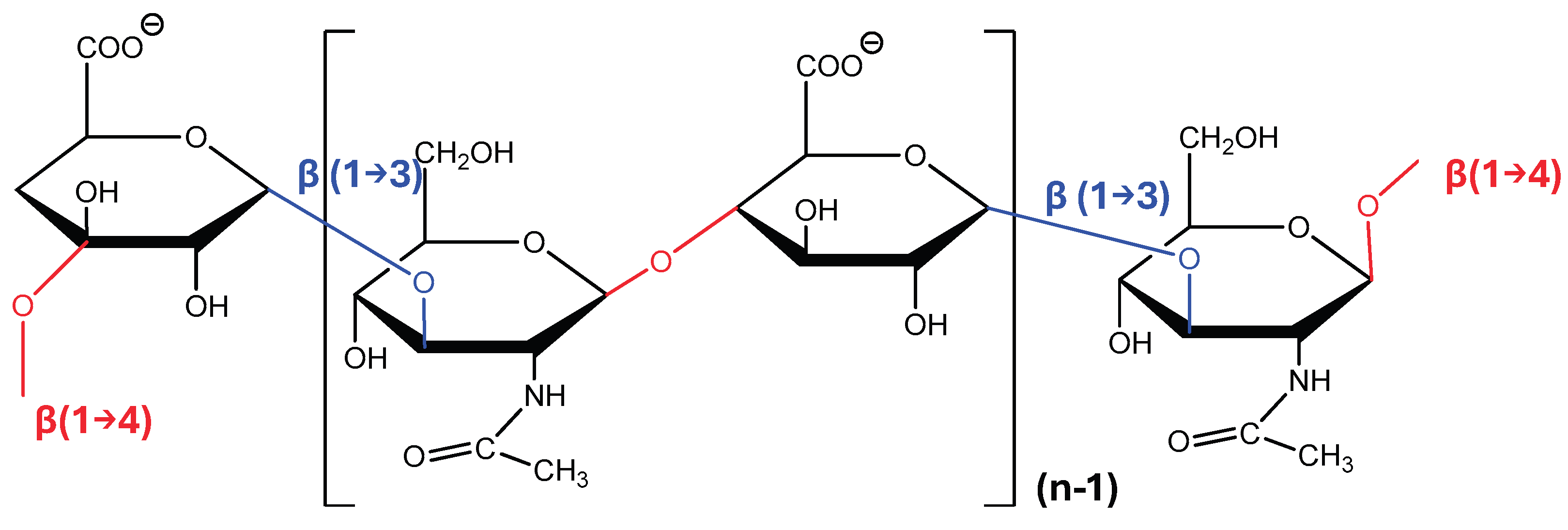
Figure 2.
Polymer Length Determines the Biological Activity of HA.
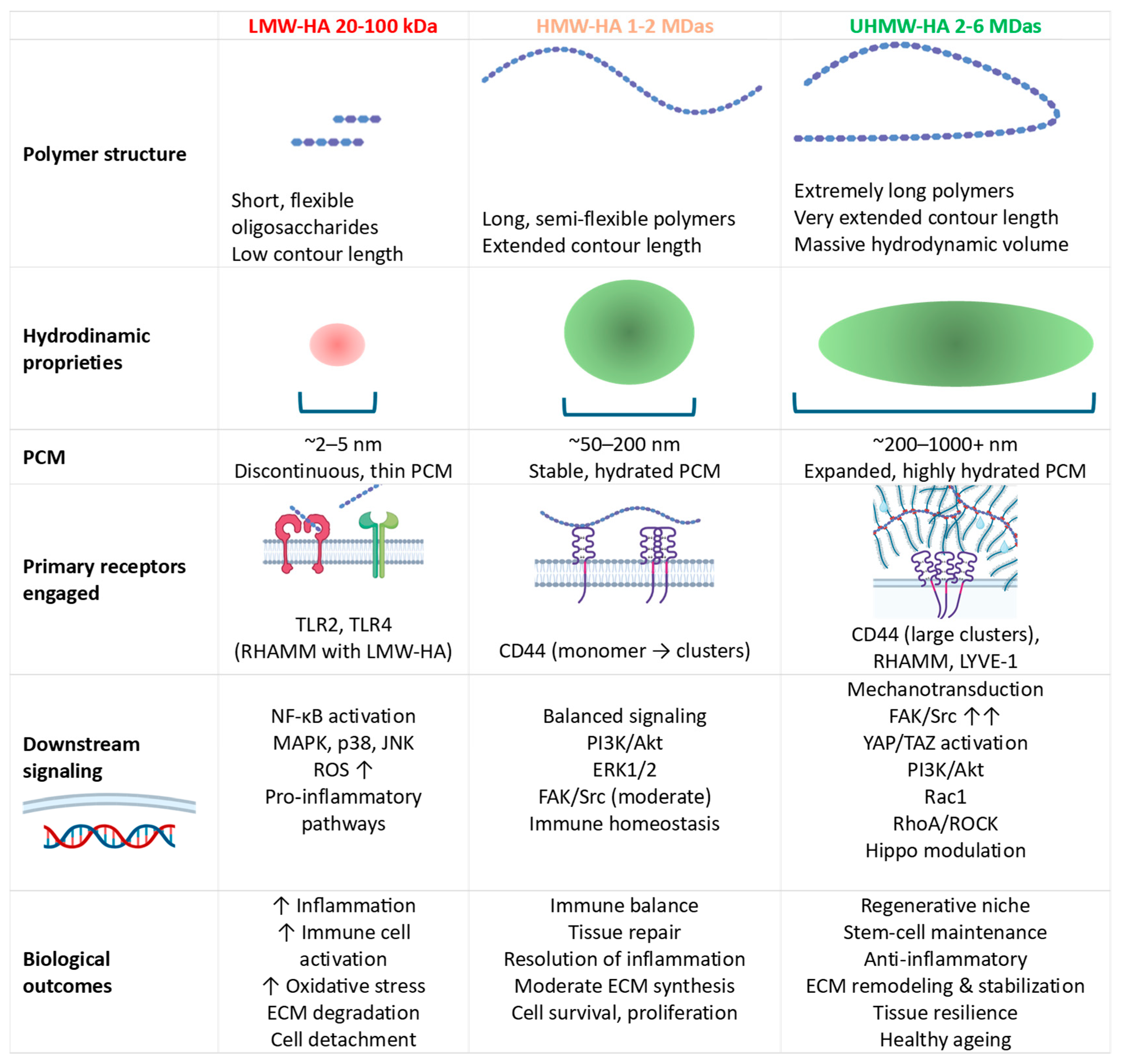
Figure 3.
Proposed mechanism by which UHMW-HA regulates mechanotransduction. UHMW-HA organizes an expanded hydrated PCM that promotes CD44 clustering and β1-integrin cooperation, leading to FAK/Src activation and downstream MAPK/ERK, PI3K/AKT, and RhoA/ROCK signalling. Increased actomyosin tension may subsequently modulate Hippo signalling and YAP/TAZ nuclear localization, promoting adaptive gene transcription involved in ECM remodelling, stem-cell maintenance, regenerative repair, and tissue homeostasis. Dashed lines indicate putative mechanisms that remain to be experimentally validated for UHMW-HA. Created in BioRender. Tero-Vescan, A. (2026) https://BioRender.com/nc9muvg.
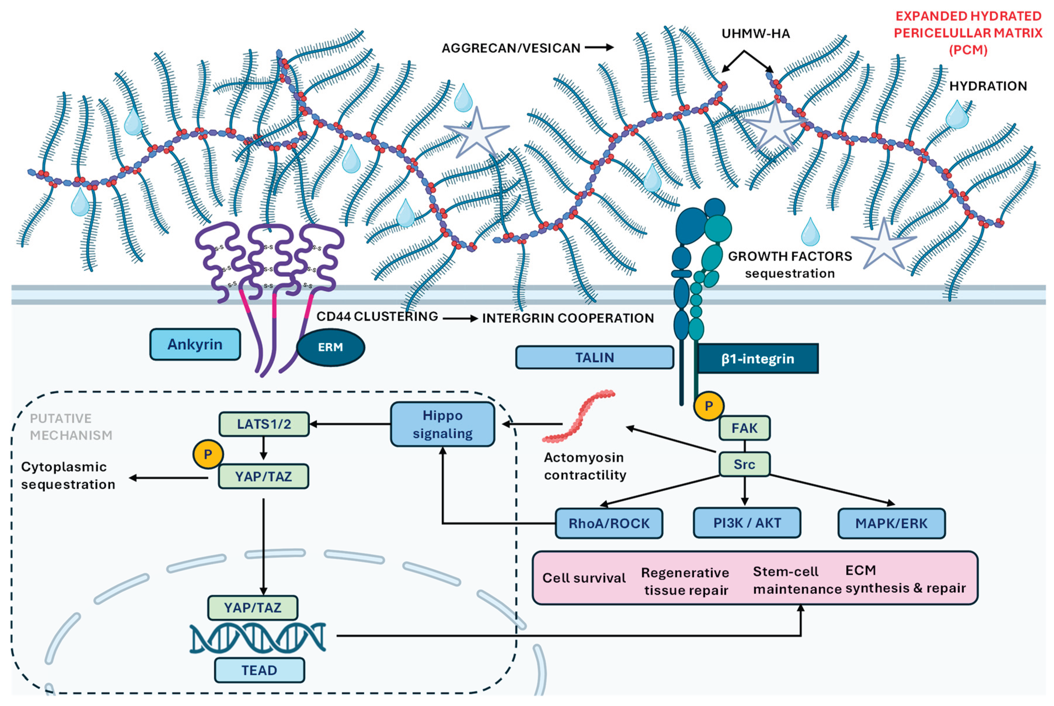
Disclaimer/Publisher’s Note: The statements, opinions and data contained in all publications are solely those of the individual author(s) and contributor(s) and not of MDPI and/or the editor(s). MDPI and/or the editor(s) disclaim responsibility for any injury to people or property resulting from any ideas, methods, instructions or products referred to in the content. |
© 2026 by the authors. Licensee MDPI, Basel, Switzerland. This article is an open access article distributed under the terms and conditions of the Creative Commons Attribution (CC BY) license (http://creativecommons.org/licenses/by/4.0/).
Copyright: This open access article is published under a Creative Commons CC BY 4.0 license, which permit the free download, distribution, and reuse, provided that the author and preprint are cited in any reuse.