

Submitted:
02 September 2026
Posted:
02 September 2026
You are already at the latest version
Abstract
Vagus nerve stimulation (VNS) is a versatile neuromodulatory modality with demonstrated efficacy across diverse central and peripheral disorders, yet a unifying mechanistic framework reconciling its central neuromodulatory and peripheral somatic actions remains elusive. This review proposes a dual‑axis regulatory paradigm centered on the nucleus of the solitary tract (NTS) as the core signal integration hub, synthesizing preclinical and clinical evidence to systematically unify VNS mechanisms across brain‑derived and systemic diseases. The ascending afferent‑dominant brain regulatory axis mediates therapeutic effects in drug‑resistant epilepsy, treatment‑resistant depression, and ischemic stroke via a conserved NTS‑mediated neurotransmitter‑cytokine cascade: it drives norepinephrine and serotonin release to rebalance neural excitatory‑inhibitory homeostasis, suppresses NF‑κB‑dependent neuroinflammation, and activates BDNF/TrkB signaling to promote neuroplasticity and neuronal survival. The descending efferent‑mediated visceral regulatory axis targets rheumatoid arthritis, functional dyspepsia, and atrial fibrillation (under low‑intensity stimulation paradigms) through a neural‑immune‑target organ synergistic axis, with core actions including cholinergic anti‑inflammatory pathway activation, sympathovagal balance restoration, and stabilization of tissue electrophysiological and structural homeostasis, exhibiting disease‑specific functional dominance. This dual‑axis framework resolves the mechanistic heterogeneity of VNS across central and peripheral pathologies, delineates hierarchical therapeutic circuits, and provides a mechanistic foundation for clinical indication expansion and precision neuromodulation strategies.
Keywords:
vagus nerve stimulation
; dual-axis regulatory framework nucleus of the solitary tract
; neuromodulation
; neuroplasticity
1. Introduction
Vagus nerve stimulation (VNS) is a clinically established neuromodulatory modality with proven efficacy across diverse central and peripheral disorders. Centered on the brainstem nucleus of the solitary tract (NTS) as the core signal integration hub, its regulatory system operates through two functionally specialized axes: the ascending afferent-dominant brain regulatory axis propagates signals rostrally to rebalance neural excitability, attenuate neuroinflammation and enhance neuroplasticity, addressing epilepsy, treatment-resistant depression and ischemic stroke; the descending efferent-mediated somatic regulatory axis routes integrated signals caudally to peripheral target organs, engaging anti-inflammatory pathways, restoring sympathovagal balance and stabilizing tissue homeostasis for rheumatoid arthritis, functional dyspepsia and atrial fibrillation. As the core conduit of the autonomic nervous system, the vagus nerve connects visceral organs to the NTS, which serves as the primary hub for integrating afferent sensory input and coordinating efferent regulatory output.
Despite its expanding clinical footprint and growing preclinical evidence, a unifying mechanistic framework reconciling VNS’s central neuromodulatory actions with peripheral somatic effects remains lacking. Current diseasespecific studies largely treat central and peripheral mechanisms as discrete pathways, creating a conceptual disconnect that obscures the hierarchical regulatory logic underlying VNS’s broad therapeutic spectrum. This fragmented understanding hinders rational parameter optimization, patient stratification, and evidence-based indication expansion, limiting advances in precision VNS neuromodulation.
To resolve this conceptual disconnect, this review proposes an NTS-centered dual-axis regulatory paradigm that unifies VNS mechanisms across central and peripheral disorders. We delineate the disease-specific functional dominance of each axis across six representative indications, synthesizing supporting preclinical and clinical evidence. This framework resolves the mechanistic heterogeneity of VNS, delineates its hierarchical therapeutic circuits, and provides a theoretical foundation for clinical indication expansion and precision neuromodulation development.
2. Functional Mechanisms and Clinical Evidence of VNS
Neuromodulation therapies employ implantable or nonimplantable devices to target and regulate nervous system function via electrical stimulation or chemical means, among which VNS is a prominent modality. VNS intervenes in or treats a variety of refractory diseases by modulating the afferent and efferent activities of the vagus nerve through electrical stimulation (Fang et al., 2023). As the core conduit of the autonomic nervous system, the vagus nerve (the tenth cranial nerve) extensively innervates visceral organs such as the heart, lungs, and gastrointestinal tract through its cervical branches, while its intracranial fibers project through the jugular foramen to the NTS, forming a critical information highway connecting the body and the brain (Jayaprakash et al., 2023) (Figure 1).
As introduced above, the NTS serves as the pivotal signal relay station for nearly all VNS effects, integrating stimulation signals before distributing them to downstream central nuclei or peripheral effector pathways (Forstenpointner et al., 2022). The physiological regulatory pathway of the vagus nerve originates at the visceral sensory terminals in somatic organs: visceral sensory signals are transmitted to the brain via vagal afferent fibers, first arriving at the NTS for primary integration (Chuyue et al.,2020), then the NTS projects information to the locus coeruleus (LC) and parabrachial nucleus. Ultimately, mediated by the noradrenergic (NE) output of the LC and the interplay between excitatory glutamatergic and inhibitory GABAergic neurons in the parabrachial nucleus, electrical signals are transmitted to the thalamus and limbic system, thereby regulating visceral states, emotions, and autonomic functions (Bowles et al., 2022).
When exogenous VNS is applied, it bidirectionally modulates brain and somatic functions through this NTScentered pathway, disrupting the pathological vicious cycle of "abnormal electrical activity → inflammation amplification → neural injury → abnormal electrical activity" (Bonaz et al., 2016). In line with the function-dominant dual-axis framework proposed in this work, the two circuits exert hierarchical therapeutic effects: in the ascending afferent-predominant brain regulatory axis, stimulation activates the NTS, which projects to the LC and triggers NE release to suppress neuronal hypersynchrony and modulate pain perception (Lai et al., 2023); in the descending efferent-mediated somatic regulatory axis, the brainstem integrates NTS-processed signals and transmits efferent output to peripheral target organs to regulate immune inflammation, electrophysiological homeostasis and tissue repair, which is the core mechanism for systemic somatic diseases like rheumatoid arthritis (RA) (Bonaz et al., 2016). This section systematically expands on the diseasespecific manifestations, functional dominance rules, and supporting preclinical and clinical evidence of these two regulatory axes across central and peripheral disorders.
2.1. Brain Circuit Regulation
The VNS modulates brain circuits through the afferent and efferent vagal pathways, regulating the synthesis and release of neurotransmitters, as well as the expression and secretion of cytokines within the CNS. By correcting imbalances in the excitability of the neural network, blocking secondary inflammatory damage, and maintaining neuronal structural and functional stability, the VNS exerts targeted therapeutic effects on DRE, TRD, and IS (Xiong et al., 2023).
A unified core mechanism is proposed. It is termed the “NTS-mediated neurotransmitter-cytokine regulatory axis”. VNS electrical signals reach the brainstem NTS via afferent fibers and initiate the central regulatory cascade. These signals then trigger two primary pathways. First, projections to the LC and raphe nucleus trigger the release of norepinephrine (NE) and 5-hydroxytryptamine (5-HT), respectively — these constitute the core neuromodulatory arm of the axis, suppressing epileptiform discharges, enhancing pain inhibition, and promoting synaptic repair after stroke (Lopes et al., 2016). Complementing this, the NTS–PBN GABAergic projection forms a secondary network-modulating branch that fine-tunes thalamocortical excitability, with particular relevance to seizure propagation control. Second, the anti-inflammatory pathway: VNS activates cholinergic pathways to inhibit the nuclear factor kappa B (NF-ĸB) signaling cascade, thereby reducing the release of pro-inflammatory cytokines and alleviating inflammation associated with these three conditions (Liu et al., 2024). Concurrently, VNS upregulates brain-derived neurotrophic factor (BDNF) and activates its high-affinity receptor tropomyosin receptor kinase B (TrkB), further promoting neurogenesis, synaptic plasticity, and neuronal survival across pathological states. This conserved signaling cascade unifies the neuromodulatory, anti-inflammatory, and pro-plasticity effects of VNS across DRE, treatment-resistant depression (TRD), and IS, establishing a common mechanistic framework for VNS therapy in brain-derived disorders (Figure 2).
Furthermore, VNS further modulates the pathological processes of multiple disorders through synergistic pathways, including the vagus nerve–LC–NE circuit. These mechanisms regulate inhibition of the thalamic reticular nucleus in epilepsy, excitability of the trigeminocervical complex in migraine, as well as the secretion of stroke-related factors and activation of antioxidative responses, thereby addressing distinct pathological requirements across diseases (Hulsey et al., 2019).
2.1.1. Drug-Resistant Epilepsy (DRE)
DRE is defined by the International League Against Epilepsy (ILAE) as the failure of adequate trials of two tolerated, appropriately chosen and used antiepileptic drug schedules (whether as monotherapies or in combination) to achieve sustained seizure freedom (French, 2007, Pe- rucca et al., 2023). The primary etiology involves irreversible neural network remodeling, triggered by specific pathologies such as hippocampal sclerosis, cortical dysplasia, or genetic mutations, which causes aberrant synaptic connectivity and forms self-sustaining epileptogenic networks (Kumar et al., 2022). Analogous to an aberrant circuit formed following a short circuit, pharmacotherapy is often incapable of repairing such structural damage. Concurrently, this process is accompanied by chronic inflammation that activates the overexpression of P-glycoprotein at the blood-brain barrier (BBB), leading to the efflux of antiepileptic drugs (e.g., carbamazepine, phenytoin). Furthermore, recurrent seizures induce dysfunction in drug targets (e.g., sodium channels, GABA receptors), reducing neuronal sensitivity to pharmacotherapy and ultimately culminating in DRE (Vezzani et al., 2019).
For DRE, a central nervous system-dominant disease characterized by abnormal synchronous firing of intracranial neurons as its core pathology, the ascending afferentpredominant brain regulatory circuit is the functionally dominant therapeutic pathway through which VNS exerts its anti-seizure effects, while the descending efferentmediated somatic circuit plays a synergistic therapeutic role by inhibiting neuroinflammation and regulating the drug efflux function of the blood-brain barrier.
First, VNS exerts its core acute anti-seizure effect by enhancing inhibitory control and rebalancing neurotransmitter homeostasis (Beste et al., 2016). Following stimulation of the vagus nerve, GABA concentrations in the hippocampus of epilepsy patients increase significantly, which attenuates glutamatergic excitation, directly inhibiting abnormal discharges at the epileptic focus to prevent seizures (Jong- kees et al., 2018). Clinical studies in patients with partial seizures have demonstrated that VNS directly activates noradrenergic neurons in the LC, suppressing hyperexcitabilityin limbic structures such as the amygdala and hippocampus, which effectively raises the seizure threshold and reduces seizure frequency (Berger et al., 2021).
Atthe network level, the well-validated NTS–LC–norepinephrine pathway serves as the primary circuit: it suppresses hyperexcitability in limbic structures such as the amygdala and hippocampus, and reduces functional connectivity within epileptogenic networks in treatment responders (Carron et al., 2023). As a complementary secondary circuit, the NTS-PBN-TRN pathway is postulated to gate seizure propagation. Built on confirmed anatomical projections and the established role of TRN as the inhibitory gatekeeper of thalamocortical synchrony, activation of this circuit may attenuate the spread of epileptiform discharges to the cortex, though direct causal evidence from pathway-specific VNS manipulation remains limited (Bowles et al., 2022).
At the neural network level, VNS projects from the NTS to the LC, inhibiting amygdala hyperexcitability and reducing limbic seizures; responders exhibit a significant decrease in functional connectivity (FC) during the stimulation ON phase (Carron et al., 2023). It may also enhance the activity of the thalamic reticular nucleus (an inhibitory nucleus) via the NTS-parabrachial nucleus-thalamus pathway. As the thalamus serves as a "relay station" for synchronous epileptic discharges, modulating its activity can block cortical abnormal oscillations and prevent seizure genesis (Bowles et al., 2022).
Second, regarding long-term synergistic anti-epileptogenic and inflammatory modulation, chronic VNS has been found to upregulate the cerebral expression of peroxisome proliferator-activated receptor alpha (PPAR“) and silent mating type information regulation 2 homolog 1 (SIRT1) in a small cohort of DRE patients, thereby promoting fatty acid oxidation and ketogenesis (Manca et al., 2024). While neurons traditionally rely on glucose for energy, under conditions of metabolic stress (e.g., inflammation, ischemia), neurons—and particularly astrocytes—accelerate fatty acid utilization. The oxidation of one molecule of palmitic acid yields 106 ATP, significantly exceeding that of glucose (Virtuoso et al., 2023). Ketone bodies (e.g., β-hydroxybutyrate) freely cross the BBB; upon neuronal uptake and oxidation, they generate more ATP while producing fewer reactive oxygen species (ROS) compared to glucose, thus reducing oxidative damage. Furthermore, ketone bodies inhibit the assembly of the NOD-like receptor pyrin domain-containing 3 (NLRP3) inflammasome, reducing the maturation and release of IL-1β (Youm et al., 2015).
In terms of clinical efficacy, a multicenter long-term cohort study involving 99 patients with DRE showed that the response rate to VNS therapy (x ≥ 50% reduction in seizure frequency) continued to increase with treatment duration: 33.4% at 1 year, 40.0% at 2 years, 51.4% at 3 years, and 55.1% at 5 years. Structural etiology emerged as an independent predictor of treatment failure, with multivariate logistic regression confirming that patients with structural etiologies had a 65% lower odds of achieving therapeutic response compared with non-structural cases (adjusted OR = 0.35, 95% CI 0.13–0.95, P = 0.039) (Tan et al., 2025).
2.1.2. Treatment-Resistant Depression (TRD)
VNS was approved by the U.S. Food and Drug Administration (FDA) for the treatment of TRD in 2005 (Aaron- son and van der Vaart, 2025). TRD is defined as the failure to achieve an adequate clinical response after at least two adequate trials of antidepressant medications, characterized by sufficient dosage, full treatment duration, and good patient treatment adherence (McIntyre et al., 2023). The core pathogenesis of TRD is not simply a deficiency of monoamine neurotransmitters, but rather multi-pathway pathological crosstalk among 5-HT mediated inhibition of dopaminergic and noradrenergic (NE) system transmission, neuroinflammatory cascade reactions, hypothalamicpituitary-adrenal (HPA) axis dysfunction, and glutamate-丫-aminobutyric acid (GABA) homeostasis dysregulation. Among these, the vicious cycle formed by dopaminergic dysfunction and neuroinflammation, as well as the impairment of the BDNF-TrkB signaling pathway, are the key hubs mediating treatment resistance to conventional antidepressants (Kamel et al.,2022). Pro-inflammatory cytokines interfere with the biosynthesis of 5-HT and DA/NE by activating indoleamine 2,3-dioxygenase (IDO) and depleting tetrahydrobiopterin (BH4), respectively. Meanwhile, they drive the shift of the kynurenine pathway toward neurotoxic quinolinic acid, exacerbate glutamatergic excitotoxicity and synaptic plasticity impairment, and further amplify monoaminergic system dysfunction and treatment resistance, which constitutes the molecular cascade underlying the onset and progression of TRD (Kajumba et al., 2024).
These interrelated, targetable core pathways in the pathological progression ofTRD form a precise pathophysiological link with the antidepressant therapeutic mechanisms of VNS, which is primarily mediated by the ascending afferent-predominant brain regulatory circuit. The differential regulation of the monoaminergic neurotransmitter system is the core molecular mechanism by which VNS reverses the pathological alterations of TRD. Electrophysiological and microdialysis studies have confirmed that acute VNS can immediately activate the spontaneous firing of noradrenergic neurons in the LC, and rapidly elevate NE levels in key brain regions across the whole brain (Kamel et al., 2022). Chronic VNS continuously enhances the firing frequency of serotonergic neurons in the dorsal raphe nucleus, and simultaneously modulates the mesolimbic dopaminergic system to targetedly ameliorate the core anhedonic symptoms ofTRD (Manta et al., 2013).
In addition, VNS reverses inflammation-mediated antidepressant resistance in TRD via the conserved cholinergic anti-inflammatory cascade described above, reducing circulating pro-inflammatory cytokines and correcting chronic low-grade neuroinflammation (Tyler, 2025). Ultimately, VNS achieves progressive and sustained remission of depressive symptoms by reshaping the functional connectivity of depression-related abnormal brain networks and restoring the functional balance between the default mode network and the executive control network (Guo et al., 2024).
VNS regulates central neurotransmitters and ameliorates depression-related neural circuits via electrical stimulation of the left vagus nerve, and its efficacy in the treatment of TRD has been fully validated by experimental studies and clinical trials. The most robust clinical evidence for VNS in TRD comes from a 5-year multicenter non-randomized observational registry of 795 patients with ≥ 4 failed prior treatment regimens. Using the Montgomery-Åsberg Depression Rating Scale (MADRS), with response defined as ≥ 50% reduction from baseline and remission as a score ≤ 9, the 5-year cumulative response (67.6%) and remission (43.3%) rates were significantly higher in the VNS plus treatment-as-usual (TAU) group than in the TAU-only group (40.9% and 25.7%, respectively) (Aaronson et al., 2017). Subgroup analysis showed VNS benefits in both prior responders and nonresponders to ECT, albeit with lower response rates in ECT nonresponders, supporting a unique neuromodulatory mechanism distinct from conventional antidepressants (Aaronson et al., 2017). A systematic review of 22 studies reported time-dependent improvements in VNS efficacy, with a 24-month response rate of 52.6% and a 12-month serious adverse event rate of 5.5%. VNS was associated with reduced suicidality and mortality, but high between-study heterogeneity and inconsistent adverse event definitions limit the interpretation of pooled estimates (Bottomley et al., 2020).
2.1.3. Ischemic Stroke (IS)
IS represents the predominant subtype of stroke, precipitated by the transient or permanent occlusion of cerebral blood vessels. This occlusion leads to cerebral ischemia and hypoxia, subsequently inducing cerebral infarction, tissue necrosis, and focal neuronal injury (Sommer, 2017).
The underlying pathological cascade proceeds from "intravascular thrombosis → cerebral ischemia/hypoxia → neuronal injury." Specific injury mechanisms include neuronal loss directly resulting from ischemic infarction, the overproduction of reactive oxygen species (ROS) triggered by vascular occlusion, and ischemia-induced inflammation accompanied by BBB disruption (Qin et al., 2022).
The therapeutic mechanism of VNS specifically targets these injury pathways. Addressing "neuronal loss caused directly by ischemic infarction," VNS activates the "vagus nerve-LC-NE" pathway, which promotes the transcription and secretion of factors such as BDNF and Growth Differentiation Factor 11 (GDF11) within the brain (Follesa et al., 2007). Upon binding to the tropomyosinrelated kinase B (TrkB) receptor on the neuronal surface, BDNF activates two core signaling pathways: phosphatidylinositol 3-kinase/protein kinase B (PI3K/Akt) and mitogen-activated protein kinase/extracellular regulated protein kinases (MAPK/ERK) pathways. First, this activation inhibits apoptosis-related proteins such as Caspase3, thereby reducing programmed neuronal death in the ischemic penumbra. Second, it promotes the expression of synaptosomal-associated protein 25 (presynaptic membrane) and N-methyl-D-aspartate (NMDA) receptors (postsynaptic membrane), enhancing synaptic stability. Furthermore, it stimulates the proliferation and differentiation of neural stem cells to generate new neurons (Yoshii and Constantine-Paton, 2010).
Regarding the "malignant cycle of inflammation and BBB disruption," VNS intervenes through two synergistic mechanisms: "anti-inflammatory action" and "BBB protection." In terms of anti-inflammatory effects, VNS stimulates vagal nerve endings to release acetylcholine (ACh), which binds to α7 nicotinic acetylcholine receptor (α7nAChR) on the surface of microglia and macrophages, directly inhibiting the nuclear translocation of the NF-ĸB pathway. As NF-ĸB is a transcriptional regulator for proinflammatory factors such as TNF-α, IL-1β, and IL-17A, the inhibition of its translocation reduces the mRNA transcription and protein secretion of these cytokines. Concurrently, α7nAChR activation promotes the expression of anti-inflammatory molecules like arginase-1 (Arg-1) and interleukin-10 (IL-10) in microglia, facilitating a phenotypic shift from the "pro-inflammatory M1 phenotype" (characterized by high iNOS and pro-inflammatory factor expression) to the "anti-inflammatory M2 phenotype." M2 microglia further reduce inflammatory stimuli by phagocytosing cellular debris (Chen et al., 2023). BBB protection is primarily manifested by the VNS-mediated inhibition of the NF-ĸB pathway, which reduces the gene expression of matrix metalloproteinase-2/9 (MMP-2/9) (enzymes that degrade type IV collagen and laminin in the BBB basement membrane). Non-invasive vagus nerve stimulation (nVNS) downregulates ischemia-induced upregulation of MMP-2 and MMP-9 in perivascular reactive astrocytes, attenuates MMP-mediated degradation of endothelial tight junction proteins to preserve BBB integrity, limits plasma protein extravasation into the brain parenchyma, and disrupts the vicious cycle of "inflammation → BBB disruption → secondary neuroinflammation" (Yang et al., 2018).
Addressing "ROS oxidative stress injury," the antiinflammatory effects of VNS reduce the infiltration of neutrophils, a primary source of ROS. Additionally, VNSupregulated BDNF activates the intrinsic antioxidant enzyme systems within neurons, enhancing their capacity to scavenge ROS, thereby indirectly mitigating oxidative stress-induced neuronal damage (Lai et al., 2019).
Furthermore, ischemia-induced disturbancesin neuronal energy metabolism lead to the excessive release of glutamate from the presynaptic membrane. This glutamate overactivates NMDA receptors on the postsynaptic membrane, triggering calcium-dependent proteases that degrade neuronal structural proteins and promote ROS generation, ultimately inducing neuronal apoptosis (Belov Kirdajova et al., 2020). VNS mitigates this mainly via the activation of the “vagus nerve-NTS-brainstem cholinergic” pathway. This modulation regulates M2 receptors on the presynaptic membrane, inhibits the opening of voltage-gated calcium channels, and reduces presynaptic Ca2+ influx, thereby lowering glutamate release (Fang et al., 2023). Simultaneously, VNS promotes the expression of glutamate transporters (such as glutamate transporter 1) in the synaptic cleft, accelerating glutamate reuptake, preventing NMDA receptor overactivation, and reducing the risk of excitotoxicity-induced apoptosis (Chen and Liu, 2025).
Clinically, IS results in limb functional impairment. In the pivotal VNS-REHAB trial (108 chronic ischemic stroke patients), paired invasive VNS yielded a 5.0-point withingroup Fugl-Meyer Assessment-Upper Extremity (FMAUE) improvement at 1 day post-therapy, with a betweengroup difference of 2.6 points versus controls (2.4 points). As a secondary endpoint, the 90-day FMA-UE clinical response rate reached 47% in the VNS group vs 24% in controls (Dawson et al., 2021).
Additionally, rodent VNS experiments by Noller et al. demonstrate that 0.5-2.5 mA current activates the inflammatory reflex in rodents a parameter not directly translatable to humans due to the 5-10-fold smaller vagus nerve diameter and thus disparate current density. A 10–30 Hz frequency with 250-1000 μ s pulse width balances therapeutic efficacy and tolerability, while chronic continuous 50–100 Hz stimulation induces axonal damage. Adverse events (cough, bradycardia) correlate positively with current and frequency, with no clinically relevant serious events in the FDA-approved human range (0.25–3.5 mA) (Noller et al., 2019). Both trials confirm the therapeutic potential of VNS in IS through the activation of CAP, noradrenergic pathways, and the promotion of BDNF secretion.
Taken together, these three CNS indications represent the full spectrum of brain disorders amenable to VNS, spanning acute to chronic, focal to network-wide, and functional to structural pathologies. Their shared underlying cascade provides a unified mechanistic basis for expanding VNS use in other neurological conditions.
2.2. Somatic Circuit Regulation
Distinct from the cerebral circuit modulation mode characterized by the "vagus nerve-brain nuclei" axis, which focuses on brain-derived disorders, the somatic circuit of VNS relies on a "vagus nerve-somatic target organ" signaling cascade, which is the core of the descending efferent-mediated somatic regulatory circuit in our dualaxis framework. It acts upon the immune-inflammatory and cardiovascular electrophysiological systems to regulate somatic immune dysregulation, autonomic imbalance, and tissue repair obstacles, thereby exerting targeted therapeutic effects on three categories of somatic diseases: RA, FD, and AF. The unified core mechanism is proposed as the “neural-immune-target organ synergistic axis”, with disease-specific functional dominance of two distinct downstream pathways.
First, an immune inflammation modulation pathway, serves as the core therapeutic mechanism for autoimmune and inflammatory somatic diseases including RA, and is primarily mediated by the CAP and the HPA axis. Second, a cardiovascular electrophysiological homeostasis regulation pathway, is the functionally dominant mechanism for malignant arrhythmias including AF, with antiinflammatory effects playing a secondary synergistic role. Furthermore, via pathways such as the splenic nerve-spleen and vagus nerve-spinal cord axes, VNS synergistically regulates immune cell polarization, myocardial signal transduction, and spinal cord repair, adapting to the specific requirements of different somatic diseases (Fan et al., 2019).
2.2.1. Rheumatoid Arthritis (RA)
RA is a chronic systemic autoimmune disease predominantly affecting the synovial joints. Its clinical hallmark is symmetrical joint involvement, which may be accompanied by extra-articular manifestations, such as pulmonary nodules (Smolen et al., 2016). The progression of RA is generally delineated into four distinct stages. Initially, a combination of factors, including genetic susceptibility (e.g., PTPN22 mutations) and environmental triggers, facilitates the production of anti-citrullinated protein antibodies (ACPA), thereby establishing risk. In the second stage, ACPA levels progressively increase within lymphoid tissues or bone marrow. This aberrant ACPA production represents the first definitive signal of the immune system’s failure to recognize self, directly disrupting the balance of immune tolerance via epitope spreading and T/B cell interactions. Subsequently, immune cells infiltrate the joint synovium and release pro-inflammatory cytokines (such as TNF-α and IL-6), leading to chronic synovitis(Petrovská et al., 2021). Finally, the disease enters a fulminant stage characterized by synovial hyperplasia, cartilage destruction, and systemic impacts, including cardiovascular disease (Figure 3).
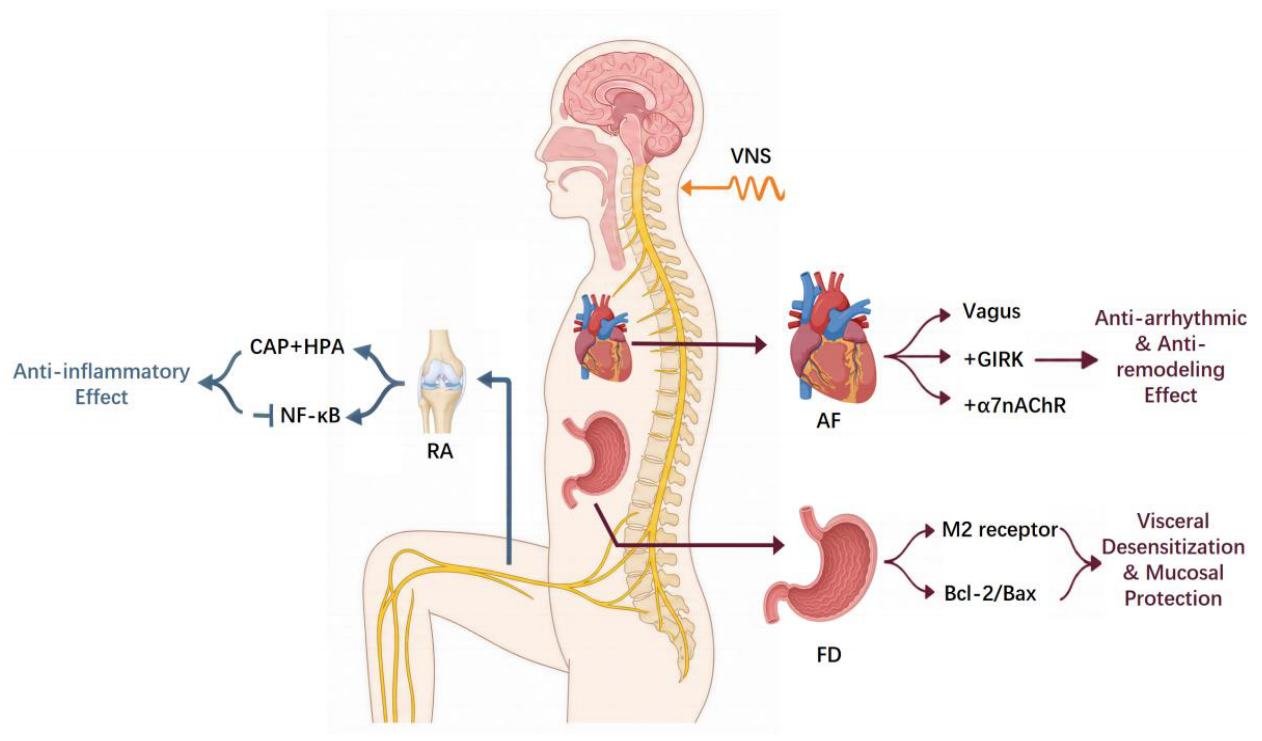
For RA, a systemic autoimmune disease dominated by peripheral inflammatory pathology, the descending efferent-mediated somatic regulatory circuit is the functionally dominant therapeutic pathway, with the ascending afferent circuit playing a synergistic role via central neuroimmune regulation. The therapeutic efficacy of VNS in RA is primarily mediated through the CAP, the core effector pathway of the descending somatic circuit. VNS stimulation signals activate NTS in the brainstem via ascending afferent fibers, thereby regulating sympathetic centers such as the rostral ventrolateral medulla (RVLM). Through preganglionic sympathetic neurons in the intermediolateral column of the spinal cord, these signals innervate the splenic nerve after relaying in the celiac ganglion, prompting the release of NE from the splenic nerve terminals (Kaestner et al., 2019).
Furthermore, data suggest the existence of an alternative complementary pathway: VNS modulation of cytokine release via the hypothalamus-pituitary-adrenal axis. Following vagal stimulation, electrical signals ascend along the vagus nerve to the NTS and are subsequently relayed to the hypothalamus. Upon receiving inflammatory signals, the hypothalamus initiates a cascade of CorticotropinReleasing Hormone → Adrenocorticotropic Hormone (ACTH) → adrenal cortex, stimulating the synthesis and release of glucocorticoids (primarily cortisol in humans). Circulating cortisol reaches systemic targets, including inflamed joints. As an endogenous anti-inflammatory hormone, cortisol enters the nuclei of immune cells and inhibits the gene transcription of multiple pro-inflammatory cytokines, thereby systemically suppressing the inflammatory response (Bonaz et al., 2016).
Together, these two complementary pathways—the peripheral cholinergic anti-inflammatory cascade and the central neuroendocrine HPA axis modulation—constitute the integrated neural-immune regulatory circuit underlying VNS-mediated therapeutic effects in RA, which alleviates synovial inflammation, joint destruction, and systemic immune dysregulation.
The application of VNS in the treatment of RA has yielded positive results in both preclinical animal models and human clinical trials, providing compelling mechanistic evidence. Animal studies-including pharmacological validation of CAP activation, α7nAChR gene knockout models, and direct VNS experiments—have demonstrated that VNS treatment significantly activates the CAP to attenuate the severity of arthritis (Koopman et al., 2017). Moreover, VNS significantly reduced ex vivo lipopolysaccharide-induced TNF-α, IL-6, and IL-1β release from whole blood in rheumatoid arthritis patients. These data provide mechanistic support that VNS engages the inflammatory reflex to modulate leukocyte cytokineproducing capacity, rather than directly lowering circulating cytokine levels in vivo. Intraoperative findings under anesthesia exclude placebo contributions but cannot be directly extrapolated to awake patients, given anesthesia’s confounding effects on autonomic tone and immune function (Koopman et al., 2016).
2.2.2. Functional Dyspepsia(FD)
FD is a highly prevalent chronic disorder of gut–brain interaction characterized by persistent epigastric symptoms including postprandial fullness, early satiation, epigastric pain and burning, with no identifiable organic pathology accounting for manifestations per Rome IV diagnostic criteria (Black et al., 2022, Ho et al., 2022). Its pathophysiology involves multilevel abnormalities spanning peripheral and central pathways: gastric sensorimotor dysfunction, duodenal mucosal barrier impairment coupled with lowgrade inflammation, dysregulated central processing of visceral afferent signals, and modifying factors such as Helicobacter pylori infection (Broeders et al., 2023, Wauters et al., 2021). As a gut–brain disorder dominated by peripheral gastrointestinal pathology, FD falls primarily under the descending efferent-mediated somatic regulatory axis of VNS, with low-level VNS (LL-VNS) and non-invasive transcutaneous auricular VNS (taVNS) serving as mechanistically rational therapeutic modalities.
The most proximal mechanism underlying VNS efficacy is restoration of sympathovagal balance. With its fiberselective safety profile, LL-VNS suppresses hyperactivity of intrinsic enteric ganglia, reduces spontaneous neural discharge frequency and amplitude, and eliminates spatial conduction heterogeneity driving functional gastrointestinal disturbances, with effects sustained by receptor desensitization and long-term depression-like synaptic plasticity (Yu et al.,2011). Non-invasive transcutaneous VNS targeting the auricular vagal branch achieves comparable autonomic recalibration via central relays such as the nucleus tractus solitarius, resetting global autonomic tone without surgical implantation (Li et al., 2015).
Beyond autonomic rebalancing, VNS attenuates lowgrade gastrointestinal inflammation via the conserved cholinergic anti-inflammatory pathway, suppressing NFĸB/AP-1 signaling and reducing pro-inflammatory cytokine secretion in gastric mucosal immune cells (Bazoukis et al., 2023, Sun et al., 2022). This peripheral action is augmented by central modulation of brainstem inflammatory signaling, which attenuates neurogenic inflammation and central sensitization of visceral pain pathways (Bergeon et al., 2026). Concurrently, LL-VNS upregulates specialized pro-resolving mediators, preserves mucosal barrier integrity via modulation of B-cell lymphoma-2/Bcl-2 associated X protein (Bcl-2/Bax)-mediated epithelial apoptosis, and reverses enteric nervous system remodeling through glial support and normalized neuromuscular signaling (Sun et al., 2022, Toumpourleka et al., 2025).
VNS further corrects core functional deficits by improving gastric electrophysiological coordination and motility. It maintains the expression and orderly distribution of connexin 40 and connexin 43 in gastric tissue, reducing electrical conduction heterogeneity between smooth muscle cells and enteric neurons and ameliorating delayed gastric emptying (Sun et al., 2022). The nitric oxide signaling pathway, a key downstream mediator of LL-VNS bioactivity, further facilitates smooth muscle relaxation and dampens visceral afferent transmission to reduce visceral hypersensitivity, a cardinal FD symptom (Kharbanda et al., 2022). Closed-loop LL-VNS systems, which deliver stimulation only upon detection of pathological patterns, enhance long-term safety and tolerability by avoiding continuous parasympathetic activation (Sun et al., 2022).
Converging preclinical and clinical evidence corroborates these mechanistic findings. In a neonatal iodoacetamide-induced FD rat model, auricular VNS attenuated visceral hypersensitivity, accelerated gastric emptying and relieved depression-like behaviors; these effects were abolished by subphrenic vagotomy, verifying the indispensable vago-vagal pathway (Hou et al., 2023). Another rodent study showed 25 Hz cervical VNS reversed colorectal distension-induced gastric slow wave dysrhythmia and delayed emptying, an effect blocked by atropine, confirming cholinergic pathway mediation (Li et al.,2025). An ongoing double-blind trial is further validating taVNS efficacy in elderly patients with comorbid insomnia and FD alongside dynamic biomarker changes (Zhou et al., 2025).
2.2.3. Atrial Fibrillation(AF)
AF is the most prevalent sustained cardiac arrhythmia worldwide. It affects approximately 1% to 2% of the general population, with over 52 million affected individuals globally, and its prevalence is projected to double over the next four decades (Panahi et al.,2026,Pellman and Sheikh, 2015). As an established independent risk factor for embolic stroke, heart failure, cognitive decline and dementia, AF contributes to substantial excess mortality and global disability burden. Its pathogenesis arises from the interplay of electrical, structural, inflammatory and neurohumoral remodeling. Reentrant wave propagation and enhanced ectopic automaticity form the core electrophysiological substrates of the arrhythmia, while progressive electrical remodeling, atrial fibrosis, NLRP3 inflammasome activation and autonomic imbalance collectively form a selfperpetuating vicious cycle that drives disease progression (Schotten et al., 2011, Karakasis et al., 2024).
VNS, including invasive low-level VNS (LLVNS) and noninvasive taVNS, exerts multifaceted therapeutic effects against AF by targeting these core pathophysiological abnormalities. It first restores sympathovagal balance by augmenting vagal outflow and suppressing excessive sympathetic activation, which reduces atrial ectopic triggering and stabilizes cardiac electrical dynamics. As noted above, this low-intensity modality confers these regulatory benefits without clinically significant bradycardia (Stavrakis et al., 2015, Bazoukis et al., 2023). The cholinergic antiinflammatory reflex forms a core mechanistic pillar: efferent vagal activation suppresses NF-ĸB-driven production of pro-remodeling mediators (TNF-α, IL-6, HMGB1) via α7nAChR signaling, slowing atrial structural and electrical degeneration (Bazoukis et al., 2023).
Beyond indirect inflammatory modulation, VNS directly counteracts AF-associated structural and electrophysiological remodeling to stabilize the arrhythmogenic substrate. It also stabilizes atrial connexin 40/43 distribution, maintaining intercellular coupling and reducing conduction heterogeneity that drives reentrant AF circuits. Preclinical data from canine models provide direct biological validation for these mechanisms, showing that chronic low-level VNS reduces AF inducibility by attenuating atrial oxidative stress and stabilizing connexin distribution (Bergeon et al.,2026).
Accumulating clinical evidence corroborates antiarrhythmic and anti-inflammatory efficacy of VNS across distinct delivery modalities. In a prospective randomized sham-controlled trial of 54 cardiac surgery patients, intraoperative epicardial LLVNS targeting caval-adjacent vagal preganglionic fibers was delivered for ≤ 72 h postoperatively, an approach not generalizable to conventional VNS. The intervention significantly reduced postoperative AF incidence, with only 12% of patients in the LLVNS group developing AF versus 36% in the control group. It also attenuated postoperative systemic inflammation, and no wire-placement complications were reported, supporting its safety in surgical populations (Stavrakis et al., 2017). For patients with paroxysmal AF, the double-blind, shamcontrolled TREAT AF trial enrolled 53 participants to evaluate noninvasive low-level tragus stimulation. Daily 1-hour 20 Hz stimulation for 6 months reduced median AF burden by 85% vs sham, with a 23% drop in serum TNF-α and favorable heart rate variability changes. This median effect masks marked interindividual heterogeneity, with some patients unresponsive. No device-related adverse events occurred in this cohort (Stavrakis et al., 2015).
A 2025 systematic review of three randomized controlled trials has further consolidated these findings, confirming the consistent efficacy of low-level tragus stimulation in reducing AF burden and inflammatory markers in paroxysmal AF populations over 6-month follow-up. Nevertheless, the review also noted that current evidence remains limited by small sample sizes, uniform stimulation parameters and short follow-up durations. Larger multicenter trials with optimized parameter designs are therefore warranted to validate the long-term therapeutic outcomes of VNS for AF management (Mejia et al., 2025).
Taken together, RA, FD and AF represent three prototypical peripheral disease categories responsive to VNS, covering immune, gastrointestinal and cardiac pathologies. Their shared neural-immune-target organ synergistic mechanism supports further translational exploration of VNS for other systemic disorders.
3. Conclusion
Built on the NTS as the core integration hub, the dualaxis framework reconciles VNS’s central neuromodulatory effects (for DRE, TRD and IS) and peripheral somatic actions (for RA, FD and AF) under a unified hierarchical regulatory logic, resolving the long-standing mechanistic heterogeneity across indications.
The descending efferent-mediated somatic regulatory axis targets rheumatoid arthritis, functional dyspepsia and atrial fibrillation through a neural-immune-target organ synergistic network. Its core actions include cholinergic anti-inflammatory pathway activation, sympathovagal balance restoration, and stabilization of tissue electrophysiological and structural homeostasis, with disease-specific functional dominance across indications.
This framework delineates the hierarchical therapeutic circuits of VNS and provides a mechanistic foundation for clinical indication expansion and precision neuromodulation. Future work should prioritize large-scale shamcontrolled trials, dissect cell-type-specific NTS subcircuits via advanced neurotechnologies, and validate predictive biomarkers to enable individualized parameter optimization. Closed-loop adaptive VNS systems also represent a promising direction to enhance therapeutic specificity and clinical outcomes.
Acknowledgments
This work was supported by the Brain Science and Brain-like Intelligence TechnologyNational Science and Technology Major Project (2022ZD0208500); the Sichuan Science and Technology Program (2024ZDZX0014). The funders had no role in study design, data collection and analysis, decision to publish, or preparation of the manuscript. No AI tool was used to generate any part of the paper.
References
- Aaronson, S.T.; Sears, P.; Ruvuna, F.; Bunker, M.; Conway, C.R.; Dougherty, D.D.; Reimherr, F.W.; Schwartz, T.L.; Zajecka, J.M. A 5-year observational study of patients with treatment-resistant depression treated with vagus nerve stimulation or treatment as usual: Comparison of response, remission, and suicidality. Am. J. Psychiatry 2017, 174, 640–648. [Google Scholar] [CrossRef] [PubMed]
- Aaronson, S.T.; van der Vaart, A. Vagal nerve stimulation for treatment-resistant depression. Ther. Treat.-Resist. Depress. 2025, 245–269. [Google Scholar]
- Bazoukis, G.; Stavrakis, S.; Armoundas, A.A. Vagus nerve stimulation and inflammation in cardiovascular disease: A state-of-the-art review. J. Am. Heart Assoc. 2023, 12, e030539. [Google Scholar] [CrossRef] [PubMed]
- Belov Kirdajova, D.; Kriska, J.; Tureckova, J.; Anderova, M. Ischemia-triggered glutamate excitotoxicity from the perspective of glial cells. Front. Cell. Neurosci. 2020, 14, 51. [Google Scholar] [CrossRef] [PubMed]
- Bergeon, J.; Chassagne, F.; Fanget, M.; Merlet, A.N.; Avril, S.; Féasson, L.; Roche, F.; Bäck, M.; Hupin, D. Preoperative transcutaneous vagus nerve stimulation as a novel strategy to prevent postoperative atrial fibrillation in calcific aortic valve disease: Mechanistic insights and translational perspectives. Front. Cardiovasc. Med. 2026, 12, 1625436. [Google Scholar] [CrossRef] [PubMed]
- Berger, A.; Vespa, S.; Dricot, L.; Dumoulin, M.; Iachim, E.; Doguet, P.; Vandewalle, G.; El Tahry, R. How is the norepinephrine system involved in the antiepileptic effects of vagus nerve stimulation? Front. Neurosci. 2021, 15, 790943. [Google Scholar] [CrossRef] [PubMed]
- Beste, C.; Steenbergen, L.; Sellaro, R.; Grigoriadou, S.; Zhang, R.; Chmielewski, W.; Stock, A.K.; Colzato, L. Effects of concomitant stimulation of the gabaergic and norepinephrine system on inhibitory control–a study using transcutaneous vagus nerve stimulation. Brain Stimul. 2016, 9, 811–818. [Google Scholar] [CrossRef] [PubMed]
- Black, C.J.; Paine, P.A.; Agrawal, A.; Aziz, I.; Eugenicos, M.P.; Houghton, L.A.; Hungin, P.; Overshott, R.; Vasant, D.H.; Rudd, S.; et al. British society of gastroenterology guidelines on the management of functional dyspepsia. Gut 2022, 71, 1697–1723. [Google Scholar] [CrossRef] [PubMed]
- Bonaz, B.; Sinniger, V.; Pellissier, S. Anti-inflammatory properties of the vagus nerve: Potential therapeutic implications of vagus nerve stimulation. J. Physiol. 2016, 594, 5781–5790. [Google Scholar] [CrossRef] [PubMed]
- Bottomley, J.M.; LeReun, C.; Diamantopoulos, A.; Mitchell, S.; Gaynes, B.N. Vagus nerve stimulation (vns) therapy in patients with treatment resistant depression: A systematic review and meta-analysis. Compr. Psychiatry 2020, 98, 152156. [Google Scholar] [CrossRef] [PubMed]
- Bowles, S.; Hickman, J.; Peng, X.; Williamson, W.R.; Huang, R.; Washington, K.; Donegan, D.; Welle, C.G. Vagus nerve stimulation drives selective circuit modulation through cholinergic reinforcement. Neuron 2022, 110, 2867–2885. [Google Scholar] [CrossRef] [PubMed]
- Broeders, B.; Carbone, F.; Balsiger, L.; Schol, J.; Raymenants, K.; Huang, I.; Verheyden, A.; Vanuytsel, T.; Tack, J. Functional dyspepsia—a gastric disorder, a duodenal disorder or a combination of both? Aliment Pharmacol. Ther. 2023, 57, 851–860. [Google Scholar] [CrossRef] [PubMed]
- Carron, R.; Roncon, P.; Lagarde, S.; Dibué, M.; Zanello, M.; Bartolomei, F. Latest views on the mechanisms of action of surgically implanted cervical vagal nerve stimulation in epilepsy. Neuromodulation Technol. At. Neural Interface 2023, 26, 498–506. [Google Scholar] [CrossRef] [PubMed]
- Chen, Y.; Zhang, Y.; Wang, J.; Li, S.; Wang, Y.; Zhang, Z.; Zhang, J.; Xin, C.; Wang, Y.; Rong, P. Anti-neuroinflammation effects of transcutaneous auricular vagus nerve stimulation against depression-like behaviors via hypothalamic α7nachr/jak2/stat3/nf-ĸb pathway in rats exposed to chronic unpredictable mild stress. CNS Neurosci. Ther. 2023, 29, 2634–2644. [Google Scholar] [CrossRef] [PubMed]
- Chen, Z.; Liu, K. Mechanism and applications of vagus nerve stimulation. Curr. Issues Mol. Biol. 2025, 47, 122. [Google Scholar] [CrossRef] [PubMed]
- Chuyue, D.Y.; Xu, Q.J.; Chang, R.B. Vagal sensory neurons and gut-brain signaling. Curr. Opin. Neurobiol. 2020, 62, 133–140. [Google Scholar] [CrossRef] [PubMed]
- Dawson, J.; Liu, C.Y.; Francisco, G.E.; Cramer, S.C.; Wolf, S.L.; Dixit, A.; Alexander, J.; Ali, R.; Brown, B.L.; Feng, W.; et al. Vagus nerve stimulation paired with rehabilitation for upper limb motor function after ischaemic stroke (vns-rehab): A randomised, blinded, pivotal, device trial. The lancet 2021, 397, 1545–1553. [Google Scholar] [CrossRef] [PubMed]
- Fan, J.J.; Shan, W.; Wu, J.P.; Wang, Q. Research progress of vagus nerve stimulation in the treatment of epilepsy. CNS Neurosci. Ther. 2019, 25, 1222–1228. [Google Scholar] [CrossRef] [PubMed]
- Fang, Y.T.; Lin, Y.T.; Tseng, W.L.; Tseng, P.; Hua, G.L.; Chao, Y.J.; Wu, Y.J. Neuroimmunomodulation of vagus nerve stimulation and the therapeutic implications. Front. Aging Neurosci. 2023, 15, 1173987. [Google Scholar] [CrossRef] [PubMed]
- Follesa, P.; Biggio, F.; Gorini, G.; Caria, S.; Talani, G.; Dazzi, L.; Puligheddu, M.; Marrosu, F.; Biggio, G. Vagus nerve stimulation increases norepinephrine concentration and the gene expression of bdnf and bfgf in the rat brain. Brain Res. 2007, 1179, 28–34. [Google Scholar] [CrossRef] [PubMed]
- Forstenpointner, J.; Maallo, A.M.S.; Elman, I.; Holmes, S.; Freeman, R.; Baron, R.; Borsook, D. The solitary nucleus connectivity to key autonomic regions in humans. Eur. J. Neurosci. 2022, 56, 3938–3966. [Google Scholar] [CrossRef] [PubMed]
- French, J.A. Refractory epilepsy: Clinical overview. Epilepsia 2007, 48, 3–7. [Google Scholar] [CrossRef] [PubMed]
- Guo, Z.P.; Liao, D.; Chen, L.; Wang, C.; Qu, M.; Lv, X.Y.; Fang, J.L.; Liu, C.H. Transcutaneous auricular vagus nerve stimulation modulating the brain topological architecture of functional network in major depressive disorder: An fmri study. Brain Sci. 2024, 14, 945. [Google Scholar] [CrossRef] [PubMed]
- Ho, L.; Chen, S.; Ho, F.F.; Wong, C.H.; Ching, J.Y.; Cheong, P.K.; Wu, I.X.; Liu, X.; Leung, T.H.; Wu, J.C.; et al. Comparing diagnostic performance of cantonese-chinese version of rome iv criteria and a short reference standard for functional dyspepsia in china. BMC Gastroenterol. 2022, 22, 432. [Google Scholar] [CrossRef] [PubMed]
- Hou, L.; Rong, P.; Yang, Y.; Fang, J.; Wang, J.; Wang, Y.; Zhang, J.; Zhang, S.; Zhang, Z.; Chen, J.D.; et al. Auricular vagus nerve stimulation improves visceral hypersensitivity and gastric motility and depression-like behaviors via vago-vagal pathway in a rat model of functional dyspepsia. Brain Sci. 2023, 13, 253. [Google Scholar] [CrossRef] [PubMed]
- Hulsey, D.R.; Shedd, C.M.; Sarker, S.F.; Kilgard, M.P.; Hays, S.A. Norepinephrine and serotonin are required for vagus nerve stimulation directed cortical plasticity. Exp. Neurol. 2019, 320, 112975. [Google Scholar] [CrossRef] [PubMed]
- Jayaprakash, N.; Song, W.; Toth, V.; Vardhan, A.; Levy, T.; Tomaio, J.; Qanud, K.; Mughrabi, I.; Chang, Y.C.; Rob, M.; et al. Organand function-specific anatomical organization of vagal fibers supports fascicular vagus nerve stimulation. Brain Stimul. 2023, 16, 484–506. [Google Scholar] [CrossRef] [PubMed]
- Jongkees, B.J.; Immink, M.A.; Finisguerra, A.; Colzato, L.S. Transcutaneous vagus nerve stimulation (tvns) enhances response selection during sequential action. Front. Psychol. 2018, 9, 1159. [Google Scholar] [CrossRef] [PubMed]
- Kaestner, C.L.; Smith, E.H.; Peirce, S.G.; Hoover, D.B. Immunohistochemical analysis of the mouse celiac ganglion: An integrative relay station of the peripheral nervous system. J. Comp. Neurol. 2019, 527, 2742–2760. [Google Scholar] [CrossRef] [PubMed]
- Kajumba, M.M.; Kakooza-Mwesige, A.; Nakasujja, N.; Koltai, D.; Canli, T. Treatment-resistant depression: Molecular mechanisms and management. Mol. Biomed. 2024, 5, 43. [Google Scholar] [CrossRef] [PubMed]
- Kamel, L.Y.; Xiong, W.; Gott, B.M.; Kumar, A.; Conway, C.R. Vagus nerve stimulation: An update on a novel treatment for treatmentresistant depression. J. Neurol. Sci. 2022, 434, 120171. [Google Scholar] [CrossRef] [PubMed]
- Karakasis, P.; Theofilis, P.; Vlachakis, P.K.; Korantzopoulos, P.; Patoulias, D.; Antoniadis, A.P.; Fragakis, N. Atrial fibrosis in atrial fibrillation: Mechanistic insights, diagnostic challenges, and emerging therapeutic targets. Int. J. Mol. Sci. 2024, 26, 209. [Google Scholar] [CrossRef] [PubMed]
- Kharbanda, R.K.; van der Does, W.F.; van Staveren, L.N.; Taverne, Y.J.; Bogers, A.J.; de Groot, N.M. Vagus nerve stimulation and atrial fibrillation: Revealing the paradox. Neuromodulation Technol. At. Neural Interface 2022, 25, 356–365. [Google Scholar] [CrossRef] [PubMed]
- Koopman, F.; Van Maanen, M.; Vervoordeldonk, M.J.; Tak, P. Balancing the autonomic nervous system to reduce inflammation in rheumatoid arthritis. J. Intern. Med. 2017, 282, 64–75. [Google Scholar] [CrossRef] [PubMed]
- Koopman, F.A.; Chavan, S.S.; Miljko, S.; Grazio, S.; Sokolovic, S.; Schuurman, P.R.; Mehta, A.D.; Levine, Y.A.; Faltys, M.; Zitnik, R.; et al. Vagus nerve stimulation inhibits cytokine production and attenuates disease severity in rheumatoid arthritis. Proc. Natl. Acad. Sci. 2016, 113, 8284–8289. [Google Scholar] [CrossRef] [PubMed]
- Kumar, P.; Lim, A.; Hazirah, S.N.; Chua, C.J.H.; Ngoh, A.; Poh, S.L.; Yeo, T.H.; Lim, J.; Ling, S.; Sutamam, N.B.; et al. Single-cell transcriptomics and surface epitope detection in human brain epileptic lesions identifies pro-inflammatory signaling. Nat. Neurosci. 2022, 25, 956–966. [Google Scholar] [CrossRef] [PubMed]
- Lai, S.K.; Wu, K.L.K.; Ma, C.W.; Ng, K.P.; Hu, X.Q.; Tam, K.W.; Yung, W.H.; Wang, Y.T.; Wong, T.P.; Shum, D.K.Y.; et al. Timely insertion of ampa receptor in developing vestibular circuits is required for manifestation of righting reflexes and effective navigation. Prog. Neurobiol. 2023, 221, 102402. [Google Scholar] [CrossRef] [PubMed]
- Lai, Y.; Deng, J.; Wang, M.; Wang, M.; Zhou, L.; Meng, G.; Zhou, Z.; Wang, Y.; Guo, F.; Yin, M.; et al. Vagus nerve stimulation protects against acute liver injury induced by renal ischemia reperfusion via antioxidant stress and anti-inflammation. Biomed. Pharmacother. 2019, 117, 109062. [Google Scholar] [CrossRef] [PubMed]
- Li, S.; Ye, F.; Zhang, S.; Liu, Y.; Chen, J.D. Effect and mechanism of vagal nerve stimulation on gastric motility: A preliminary rodent study. Neuromodulation Technol. At. Neural Interface 2025, 28, 767–774. [Google Scholar] [CrossRef] [PubMed]
- Li, S.; Zhou, X.; Yu, L.; Jiang, H. Low level non-invasive vagus nerve stimulation: A novel feasible therapeutic approach for atrial fibrillation. Int. J. Cardiol. 2015, 182, 189–190. [Google Scholar] [CrossRef] [PubMed]
- Liu, T.T.; Chen, S.P.; Wang, S.J.; Yen, J.C. Vagus nerve stimulation inhibits cortical spreading depression via glutamate-dependent trkb activation mechanism in the nucleus tractus solitarius. Cephalalgia 2024, 44, 03331024241230466. [Google Scholar] [CrossRef] [PubMed]
- Lopes, L.T.; Patrone, L.G.A.; Li, K.Y.; Imber, A.N.; Graham, C.D.; Gar-gaglioni, L.H.; Putnam, R.W. Anatomical and functional connections between the locus coeruleus and the nucleus tractus solitarius in neonatal rats. Neuroscience 2016, 324, 446–468. [Google Scholar] [CrossRef] [PubMed]
- Manca, C.; Coa, R.; Murru, E.; Carta, G.; Pinna, G.; Sanfilippo, R.; Polizzi, L.; Pistis, M.; Follesa, P.; Puligheddu, M.; et al. Identification of metabolic biomarkers of chronic vagus nerve stimulation (vns) in subjects with drug-resistant epilepsy (dre). Epilepsia Open 2024, 9, 432–438. [Google Scholar] [CrossRef] [PubMed]
- Manta, S.; El Mansari, M.; Debonnel, G.; Blier, P. Electrophysiological and neurochemical effects of long-term vagus nerve stimulation on the rat monoaminergic systems. Int. J. Neuropsychopharmacol. 2013, 16, 459–470. [Google Scholar] [CrossRef] [PubMed]
- McIntyre, R.S.; Alsuwaidan, M.; Baune, B.T.; Berk, M.; Demyttenaere, K.; Goldberg, J.F.; Gorwood, P.; Ho, R.; Kasper, S.; Kennedy, S.H.; et al. Treatment-resistant depression: Definition, prevalence, detection, management, and investigational interventions. World Psychiatry 2023, 22, 394–412. [Google Scholar] [CrossRef] [PubMed]
- Mejia, C.A.U.; Flores, C.S.A.; Radilla, S.G.G.; Salinas, M.A.L.; Alvarez, D.A.M.; Vargas, E.C.; Borbolla, M.D.; Barajas, J.M.M.; Frutos, D.J.; Perez, G.A.M.; et al. Efficacy of transcutaneous vagus nerve stimulation to suppress paroxysmal atrial fibrillation: A systematic review. Cureus 2025, 17. [Google Scholar] [CrossRef] [PubMed]
- Noller, C.M.; Levine, Y.A.; Urakov, T.M.; Aronson, J.P.; Nash, M.S. Vagus nerve stimulation in rodent models: An overview of technical considerations. Front. Neurosci. 2019, 13, 911. [Google Scholar] [CrossRef] [PubMed]
- Panahi, B.; Dababneh, S.; Fadaei, S.; Babini, H.; Singh, S.; Prondzynski, M.; Akbari, M.; Backx, P.H.; Andrade, J.G.; Rose, R.A.; et al. Versatile hipsc models and bioengineering platforms for investigation of atrial fibrosis and fibrillation. Cells 2026, 15, 187. [Google Scholar] [CrossRef] [PubMed]
- Pellman, J.; Sheikh, F. Atrial fibrillation: Mechanisms, therapeutics, and future directions. Compr. Physiol. 2015, 5, 649–665. [Google Scholar] [CrossRef]
- Perucca, E.; Perucca, P.; White, H.S.; Wirrell, E.C. Drug resistance in epilepsy. Lancet Neurol. 2023, 22, 723–734. [Google Scholar] [CrossRef] [PubMed]
- Petrovská, N.; Prajzlerová, K.; Vencovsk, J.; Šenolt, L.; Filková, M. The pre-clinical phase of rheumatoid arthritis: From risk factors to prevention of arthritis. Autoimmun. Rev. 2021, 20, 102797. [Google Scholar] [CrossRef] [PubMed]
- Qin, C.; Yang, S.; Chu, Y.H.; Zhang, H.; Pang, X.W.; Chen, L.; Zhou, L.Q.; Chen, M.; Tian, D.S.; Wang, W. Signaling pathways involved in ischemic stroke: Molecular mechanisms and therapeutic interventions. Signal Transduct. Target. Ther. 2022, 7, 215. [Google Scholar] [CrossRef] [PubMed]
- Schotten, U.; Verheule, S.; Kirchhof, P.; Goette, A. Pathophysiological mechanisms of atrial fibrillation: A translational appraisal. Physiol. Rev. 2011, 91, 265–325. [Google Scholar] [CrossRef] [PubMed]
- Smolen, J.S.; Aletaha, D.; McInnes, I.B. Rheumatoid arthritis. The Lancet 2016, 388, 2023–2038. [Google Scholar]
- Sommer, C.J. Ischemic stroke: Experimental models and reality. Acta Neuropathol. 2017, 133, 245–261. [Google Scholar] [CrossRef] [PubMed]
- Stavrakis, S.; Humphrey, M.B.; Scherlag, B.; Iftikhar, O.; Parwani, P.; Abbas, M.; Filiberti, A.; Fleming, C.; Hu, Y.; Garabelli, P.; et al. Low-level vagus nerve stimulation suppresses post-operative atrial fibrillation and inflammation: A randomized study. JACC Clin. Electrophysiol. 2017, 3, 929–938. [Google Scholar] [CrossRef] [PubMed]
- Stavrakis, S.; Humphrey, M.B.; Scherlag, B.J.; Hu, Y.; Jackman, W.M.; Nakagawa, H.; Lockwood, D.; Lazzara, R.; Po, S.S. Low-level transcutaneous electrical vagus nerve stimulation suppresses atrial fibrillation. J. Am. Coll. Cardiol. 2015, 65, 867–875. [Google Scholar] [CrossRef] [PubMed]
- Sun, Y.; Chao, S.; Ouyang, H.; Zhang, W.; Luo, W.; Nie, Q.; Wang, J.; Luo, C.; Ni, G.; Zhang, L.; et al. Hybrid nanogenerator based closed-loop self-powered low-level vagus nerve stimulation system for atrial fibrillation treatment. Sci. Bull. 2022, 67, 1284–1294. [Google Scholar] [CrossRef] [PubMed]
- Tan, B.; Liu, Y.; Gong, M.; Meng, F.; Yang, A.; Zhang, K.; Sang, L.; Zhang, J. Long-term efficacy and predictors of vagus nerve stimulation in drug-resistant epilepsy: A multicenter cohort study. Acta Epileptol. 2025, 7, 40. [Google Scholar] [CrossRef] [PubMed]
- Toumpourleka, M.; Venkatesan, T.; Morris, L.; Whyte, S.; Farhat, K.; Elkholey, K.; Harveille, L.; Stavrakis, S. Atrial fibrillation-induced autonomic remodeling is reversed by noninvasive vagus nerve stimulation. Europace 2025, 27, euaf085–795. [Google Scholar] [CrossRef]
- Tyler, W.J. Transcutaneous auricular vagus nerve stimulation for treating emotional dysregulation and inflammation in common neuropsychiatric disorders. Brain Sci. 2025, 16, 8. [Google Scholar] [CrossRef] [PubMed]
- Vezzani, A.; Balosso, S.; Ravizza, T. Neuroinflammatory pathways as treatment targets and biomarkers in epilepsy. Nat. Rev. Neurol. 2019, 15, 459–472. [Google Scholar] [CrossRef] [PubMed]
- Virtuoso, A.; De Luca, C.; Korai, S.A.; Papa, M.; Cirillo, G. Neuroinflammation and glial activation in the central nervous system: A metabolic perspective. Neural Regen. Res. 2023, 18, 1025–1026. [Google Scholar] [CrossRef] [PubMed]
- Wauters, L.; Dickman, R.; Drug, V.; Mulak, A.; Serra, J.; Enck, P.; Tack, J.; Consensus Group, E.F.; Accarino, A.; Barbara, G.; et al. United european gastroenterology (ueg) and european society for neurogastroenterology and motility (esnm) consensus on functional dyspepsia. Neurogastroenterol. Motil. 2021, 33, e14238. [Google Scholar] [CrossRef] [PubMed]
- Xiong, H.; Tang, F.; Guo, Y.; Xu, R.; Lei, P. Neural circuit changes in neurological disorders: Evidence from in vivo two-photon imaging. Ageing Res. Rev. 2023, 87, 101933. [Google Scholar] [CrossRef] [PubMed]
- Yang, Y.; Yang, L.Y.; Orban, L.; Cuylear, D.; Thompson, J.; Simon, B.; Yang, Y. Non-invasive vagus nerve stimulation reduces bloodbrain barrier disruption in a rat model of ischemic stroke. Brain Stimul. 2018, 11, 689–698. [Google Scholar] [CrossRef] [PubMed]
- Yoshii, A.; Constantine-Paton, M. Postsynaptic bdnf-trkb signaling in synapse maturation, plasticity, and disease. Dev. Neurobiol. 2010, 70, 304–322. [Google Scholar] [CrossRef] [PubMed]
- Youm, Y.H.; Nguyen, K.Y.; Grant, R.W.; Goldberg, E.L.; Bodogai, M.; Kim, D.; D’agostino, D.; Planavsky, N.; Lupfer, C.; Kanneganti, T.D.; et al. The ketone metabolite β-hydroxybutyrate blocks nlrp3 inflammasome–mediated inflammatory disease. Nat. Med. 2015, 21, 263–269. [Google Scholar] [CrossRef] [PubMed]
- Yu, L.; Scherlag, B.J.; Li, S.; Sheng, X.; Lu, Z.; Nakagawa, H.; Zhang, Y.; Jackman, W.M.; Lazzara, R.; Jiang, H.; et al. Low-level vagosympathetic nerve stimulation inhibits atrial fibrillation inducibility: Direct evidence by neural recordings from intrinsic cardiac ganglia. J. Cardiovasc. Electrophysiol. 2011, 22, 455–463. [Google Scholar] [CrossRef] [PubMed]
- Zhou, H.; Tang, X.; Wang, D.; Huang, Z.; Zeng, Y.; Liu, S.; Wang, C. Neuroregulatory and clinical efficacy of auricular vagus nerve stimulation in elderly patients with chronic insomnia comorbid with functional dyspepsia: Protocol for a randomized controlled trial. Front. Med. 2025, 12, 1537515. [Google Scholar] [CrossRef] [PubMed]
Figure 1.
NTS-centered dual-axis regulatory framework unifying central and peripheral therapeutic mechanisms of vagus nerve stimulation. This schematic illustrates the core anatomical and functional architecture of the proposed dualaxis regulatory paradigm for VNS, with the brainstem NTS as the central signal integration hub. Exogenous VNS activates vagal afferent fibers to propagate electrical signals to the NTS, which coordinates two parallel, functionally specialized regulatory axes: the ascending afferent-dominant brain regulatory axis, in which NTS projections target the LC and RN as core effectors to drive NE and 5-HT release, with the PBN GABAergic circuit serving as a complementary modulatory branch, collectively rebalancing neural excitatory-inhibitory homeostasis, suppressing neuroinflammation, and promoting neuroplasticity to mediate therapeutic effects on DRE, TRD, and IS; and the descending efferent-mediated somatic regulatory axis, in which NTS-integrated signals are transmitted via vagal efferent pathways to peripheral target organs including arthritic joints, the heart, and the stomach, activating cholinergic anti-inflammatory pathways, restoring sympathovagal balance, and stabilizing tissue electrophysiological homeostasis to support the efficacy of VNS in RA, AF, and FD. Abbreviations: 5-HT=5-hydroxytryptamine (serotonin), AF=atrial fibrillation, DRE=drug-resistant epilepsy, FD=functional dyspepsia, GABA=丫-aminobutyric acid, IS=ischemic stroke, LC=locus coeruleus, NE=norepinephrine, NTS=nucleus of the solitary tract, PBN=parabrachial nucleus, RA=rheumatoid arthritis, RN=raphe nucleus, TRD=treatment-resistant depression, VNS=vagus nerve stimulation.
Figure 1.
NTS-centered dual-axis regulatory framework unifying central and peripheral therapeutic mechanisms of vagus nerve stimulation. This schematic illustrates the core anatomical and functional architecture of the proposed dualaxis regulatory paradigm for VNS, with the brainstem NTS as the central signal integration hub. Exogenous VNS activates vagal afferent fibers to propagate electrical signals to the NTS, which coordinates two parallel, functionally specialized regulatory axes: the ascending afferent-dominant brain regulatory axis, in which NTS projections target the LC and RN as core effectors to drive NE and 5-HT release, with the PBN GABAergic circuit serving as a complementary modulatory branch, collectively rebalancing neural excitatory-inhibitory homeostasis, suppressing neuroinflammation, and promoting neuroplasticity to mediate therapeutic effects on DRE, TRD, and IS; and the descending efferent-mediated somatic regulatory axis, in which NTS-integrated signals are transmitted via vagal efferent pathways to peripheral target organs including arthritic joints, the heart, and the stomach, activating cholinergic anti-inflammatory pathways, restoring sympathovagal balance, and stabilizing tissue electrophysiological homeostasis to support the efficacy of VNS in RA, AF, and FD. Abbreviations: 5-HT=5-hydroxytryptamine (serotonin), AF=atrial fibrillation, DRE=drug-resistant epilepsy, FD=functional dyspepsia, GABA=丫-aminobutyric acid, IS=ischemic stroke, LC=locus coeruleus, NE=norepinephrine, NTS=nucleus of the solitary tract, PBN=parabrachial nucleus, RA=rheumatoid arthritis, RN=raphe nucleus, TRD=treatment-resistant depression, VNS=vagus nerve stimulation.
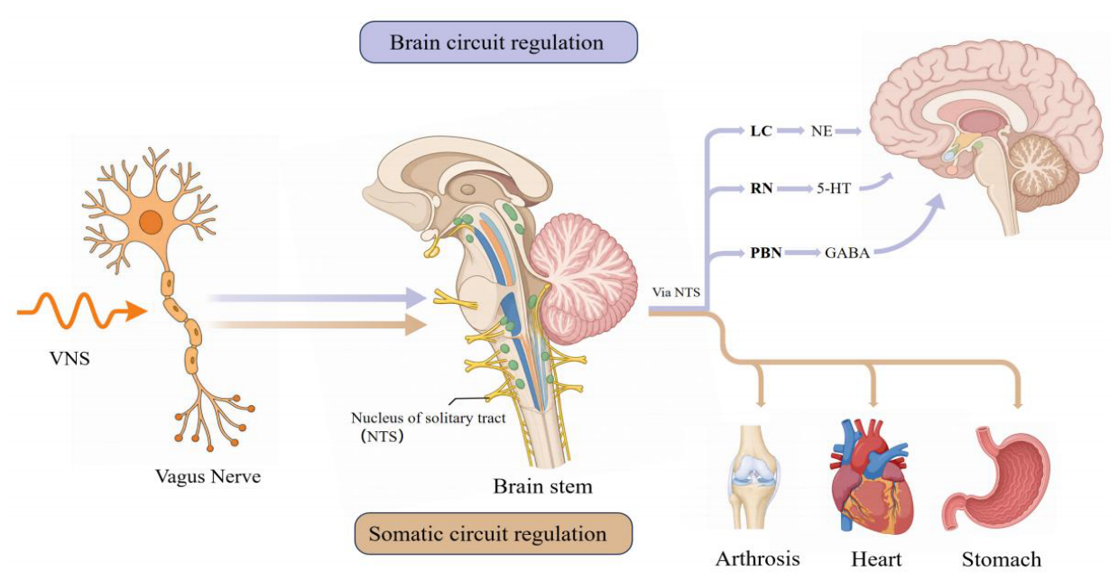
Figure 2.
Central neuromodulatory mechanisms of VNS underlying therapeutic efficacy in brain-derived disorders: the unified NTS-mediated neurotransmitter-cytokine regulatory axis. This schematic depicts the conserved central signaling cascade activated by VNS for treating DRE, TRD, and IS. Afferent vagal signals reach the brainstem NTS the core integration hub driving two parallel therapeutic pathways: (1) The neuromodulatory pathway triggers NE release from the LC and 5-HT from the RN, rebalancing GABA-glutamate homeostasis to suppress epileptiform hyperexcitability, restore mood networks, and stabilize synapses. (2) The anti-inflammatory pathway inhibits NFĸB signaling, lowering pro-inflammatory cytokines TNF-α and IL-1β to attenuate neuroinflammation. VNS also upregulates BDNF and activates TrkB, promoting neurogenesis, synaptic plasticity, and neuronal survival. Abbreviations: 5HT=5-hydroxytryptamine (serotonin), BDNF=brain-derived neurotrophic factor, DRE=drug-resistant epilepsy, GABA=丫aminobutyric acid, IL-1β=interleukin-1β, IS=ischemic stroke, LC=locus coeruleus, NE=norepinephrine, NF-ĸB=nuclear factor-ĸB, NTS=nucleus of the solitary tract, RN=raphe nucleus, TNF-α=tumor necrosis factor-α, TrkB=Tropomyosin receptor kinase B, TRD=treatment-resistant depression, VNS=vagus nerve stimulation.
Figure 2.
Central neuromodulatory mechanisms of VNS underlying therapeutic efficacy in brain-derived disorders: the unified NTS-mediated neurotransmitter-cytokine regulatory axis. This schematic depicts the conserved central signaling cascade activated by VNS for treating DRE, TRD, and IS. Afferent vagal signals reach the brainstem NTS the core integration hub driving two parallel therapeutic pathways: (1) The neuromodulatory pathway triggers NE release from the LC and 5-HT from the RN, rebalancing GABA-glutamate homeostasis to suppress epileptiform hyperexcitability, restore mood networks, and stabilize synapses. (2) The anti-inflammatory pathway inhibits NFĸB signaling, lowering pro-inflammatory cytokines TNF-α and IL-1β to attenuate neuroinflammation. VNS also upregulates BDNF and activates TrkB, promoting neurogenesis, synaptic plasticity, and neuronal survival. Abbreviations: 5HT=5-hydroxytryptamine (serotonin), BDNF=brain-derived neurotrophic factor, DRE=drug-resistant epilepsy, GABA=丫aminobutyric acid, IL-1β=interleukin-1β, IS=ischemic stroke, LC=locus coeruleus, NE=norepinephrine, NF-ĸB=nuclear factor-ĸB, NTS=nucleus of the solitary tract, RN=raphe nucleus, TNF-α=tumor necrosis factor-α, TrkB=Tropomyosin receptor kinase B, TRD=treatment-resistant depression, VNS=vagus nerve stimulation.
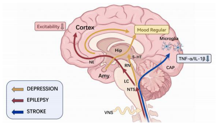
Figure 3.
Somatic regulatory axis of vagus nerve stimulation: organ-specific therapeutic mechanisms across peripheral disorders. This schematic illustrates the descending efferent-mediated somatic regulatory axis, the peripheral effector branch of the NTS-centered dual-axis framework for VNS. Operating via a unified neural-immune-target organ synergistic network, this axis exerts context-dependent therapeutic effects on three representative peripheral pathologies. In RA, VNS elicits systemic anti-inflammatory effects through two convergent pathways: activation of the CAP and engagement of the HPA axis, which jointly suppress pro-inflammatory NF-ĸB signaling to attenuate synovial inflammation and joint damage. In AF, VNS confers integrated anti-arrhythmic and anti-remodeling benefits via three complementary mechanisms: direct vagal efferent modulation of myocardial electrophysiology, activation of GIRK channels to stabilize cardiac excitability, and α7nAChR-dependent inhibition of inflammatory structural remodeling. In FD, VNS mediates visceral desensitization and mucosal protection by regulating gastric M2 receptors to restore gastrointestinal sensorimotor homeostasis, and modulating the Bcl-2/Bax signaling axis to preserve mucosal barrier integrity and restrain epithelial apoptosis. Abbreviation: α7nAChR=α7 nicotinic acetylcholine receptor, AF=atrial fibrillation, CAP=cholinergic anti-inflammatory pathway, FD=functional dyspepsia, GIRK=G protein-gated inwardly rectifying potassium, HPA=hypothalamic-pituitary-adrenal axis, NF-ĸB=nuclear factor-ĸB, NTS=nucleus of the solitary tract, RA=rheumatoid arthritis, VNS=vagus nerve stimulation.
Figure 3.
Somatic regulatory axis of vagus nerve stimulation: organ-specific therapeutic mechanisms across peripheral disorders. This schematic illustrates the descending efferent-mediated somatic regulatory axis, the peripheral effector branch of the NTS-centered dual-axis framework for VNS. Operating via a unified neural-immune-target organ synergistic network, this axis exerts context-dependent therapeutic effects on three representative peripheral pathologies. In RA, VNS elicits systemic anti-inflammatory effects through two convergent pathways: activation of the CAP and engagement of the HPA axis, which jointly suppress pro-inflammatory NF-ĸB signaling to attenuate synovial inflammation and joint damage. In AF, VNS confers integrated anti-arrhythmic and anti-remodeling benefits via three complementary mechanisms: direct vagal efferent modulation of myocardial electrophysiology, activation of GIRK channels to stabilize cardiac excitability, and α7nAChR-dependent inhibition of inflammatory structural remodeling. In FD, VNS mediates visceral desensitization and mucosal protection by regulating gastric M2 receptors to restore gastrointestinal sensorimotor homeostasis, and modulating the Bcl-2/Bax signaling axis to preserve mucosal barrier integrity and restrain epithelial apoptosis. Abbreviation: α7nAChR=α7 nicotinic acetylcholine receptor, AF=atrial fibrillation, CAP=cholinergic anti-inflammatory pathway, FD=functional dyspepsia, GIRK=G protein-gated inwardly rectifying potassium, HPA=hypothalamic-pituitary-adrenal axis, NF-ĸB=nuclear factor-ĸB, NTS=nucleus of the solitary tract, RA=rheumatoid arthritis, VNS=vagus nerve stimulation.
Disclaimer/Publisher’s Note: The statements, opinions and data contained in all publications are solely those of the individual author(s) and contributor(s) and not of MDPI and/or the editor(s). MDPI and/or the editor(s) disclaim responsibility for any injury to people or property resulting from any ideas, methods, instructions or products referred to in the content. |
© 2026 by the authors. Licensee MDPI, Basel, Switzerland. This article is an open access article distributed under the terms and conditions of the Creative Commons Attribution (CC BY) license.
Copyright: This open access article is published under a Creative Commons CC BY 4.0 license, which permit the free download, distribution, and reuse, provided that the author and preprint are cited in any reuse.