

Submitted:
01 September 2026
Posted:
02 September 2026
You are already at the latest version
Abstract
The liver is the primary organ for xenobiotic biotransformation, and its functional integrity dictates not only the elimination rate of drugs but also their ultimate pharmacodynamic profile. The superfamily of hemecontaining monooxygenases, cytochrome P450 (CYP), underpins the oxidative metabolism of approximately 75% of all veterinary drugs. A critical component of the microsomal electron transport chain is NADPHcytochrome P450 reductase. Each hepatobiliary pathology differentially affects CYP expression and activity, leading to distinct pharmacodynamic alterations. Species differences complicate pharmacodynamic adjustments. Sex, diet, and gut microbiota also modulate CYP expression and thus pharmacodynamics. The aim of the present work was to systematically collate available data on activity and expression changes of major CYP isoforms and CYPOR in key veterinary species under various hepatobiliary conditions, and to translate these changes into clinically relevant pharmacodynamic predictions. A systematic literature search was performed in PubMed, Scopus, Web of Science, and Google Scholar covering the period from 1990 to 2026. This comprehensive review demonstrates that hepatobiliary pathologies in animals elicit qualitatively distinct alterations in the CYP system and its redox partner CYPOR, leading to drugspecific pharmacodynamic changes. The integrated dysbalance index can guide clinicians: values >0.7 indicate an acute mismatch requiring antioxidants and caution with prodrugs; values <0.15 signal a systemic electron deficit mandating substantial dose reductions for all CYPdependent drugs. Speciesspecific patterns: cats’ vulnerability to CYP2E1 substrates, horses’ pronounced CYPOR decline in cholestasis, and dogs’ paradoxical CYP2C induction in steatosis, must be incorporated into dosing algorithms.
Keywords:
cytochrome P450
; CYPOR
; canine CYP3A
; feline CYP2E1
; equine CYP3A89
; bovine CYP
; liver cirrhosis
; cholestasis pharmacokinetics
1. Introduction
Veterinary pharmacotherapy increasingly encounters unpredictable therapeutic responses and adverse effects in patients with hepatic disorders. The liver is the primary organ for xenobiotic biotransformation, and its functional integrity dictates not only the elimination rate of drugs but also their ultimate pharmacodynamic profile. For example, the relationship between drug concentration at the site of action and the resulting therapeutic or toxic effect. The superfamily of heme-containing monooxygenases, cytochrome P450 (CYP), underpins the oxidative metabolism of approximately 75% of all veterinary drugs, including antibiotics, non-steroidal anti-inflammatory drugs (NSAIDs), anesthetics, anthelmintics, and cardiac glycosides. These enzymes catalyse the insertion of one oxygen atom into a substrate, rendering hydrophobic molecules polar and facilitating their excretion. The pharmacodynamic outcome (efficacy, duration of action, and adverse reactions) of a given drug is heavily dependent on its concentration-time profile, which in turn is governed by CYP-mediated clearance [1,2].
A critical component of the microsomal electron transport chain is NADPH-cytochrome P450 reductase (CYPOR), a flavoprotein that transfers two electrons from NADPH to the heme iron of CYP. Without functionally active CYPOR, the oxidative reactions of all microsomal CYP isoforms cannot proceed, and its activity can become rate-limiting for the overall metabolism of many substrates, thereby directly altering the concentration-time profile and, consequently, the pharmacodynamic response. Unlike in human medicine, where CYP system alterations in hepatitis and cirrhosis are well characterised, veterinary science suffers from a substantial knowledge gap. Interspecies differences in CYP expression, substrate specificity, and regulatory mechanisms render extrapolation from human data hazardous and often misleading [3,4,5] (Figure 1).
Domestic and farm animals exhibit unique CYP profiles. Cats lack functionally active CYP2D6, leading to prolonged half-lives of many β-blockers and antidepressants, which translates into exaggerated pharmacodynamic effects (e.g., bradycardia, sedation). In dogs, CYP3A12 is the dominant isoform, analogous to human CYP3A4; horses predominantly express CYP3A89, while cattle rely on CYP3A28 with relatively low CYP1A1 activity. Hepatobiliary pathologies in animals are classified into several major types: degenerative (cirrhosis, fibrosis), cholestatic (intra- and extrahepatic cholestasis), inflammatory (hepatitis of various aetiologies), metabolic (steatosis, lipidosis), and toxic (poisonings with hepatotoxins). Each type differentially affects CYP expression and activity, leading to distinct pharmacodynamic alterations. In cirrhosis, functional parenchyma is replaced by connective tissue, reducing the total hepatocyte mass and the overall CYP pool. However, the decline in activity is not uniform. CYP3A and CYP2E1 drop markedly, CYP2C shows moderate reduction, while some isoforms may even undergo compensatory upregulation. This selective loss translates into variable changes in drug clearance, and thus different drugs experience either exaggerated or diminished pharmacodynamic effects [6].
Cholestatic conditions are characterised by the accumulation of bile acids and bilirubin within hepatocytes. Bile acids serve as endogenous ligands for the nuclear receptors FXR (farnesoid X receptor) and PXR (pregnane X receptor), which regulate CYP transcription. In early cholestasis, PXR activation can induce CYP3A4, but prolonged bile stasis switches this effect to suppression via FXR-mediated repression and receptor degradation. This biphasic response creates a complex pharmacodynamic picture: initially, some drugs may be cleared faster, requiring higher doses; later, clearance drops, leading to accumulation and toxicity. Acute toxic hepatitis, for instance, from aflatoxicosis in poultry, bracken fern poisoning in cattle, or NSAID overdose in dogs, triggers massive necrosis and oxidative stress. Protective mechanisms are activated, including the transcription factor Nrf2, which upregulates CYPOR and several antioxidant genes. However, the net effect on pharmacodynamics is often unpredictable because CYP enzymes are simultaneously suppressed by inflammatory cytokines, causing a discordance between redox capacity and metabolic turnover [7,8] (Figure 2).
Steatosis (fatty liver) is common in cats (idiopathic lipidosis) and dogs (secondary to endocrinopathies or diabetes). Triglyceride accumulation alters the endoplasmic reticulum membrane microenvironment, reducing fluidity and accessibility of CYPOR to CYP, thereby decreasing reaction rates even when enzyme concentrations are preserved. This subtle change can prolong the half-life of many drugs, enhancing their pharmacodynamic effects and increasing the risk of overdose. Beyond its role in drug metabolism, CYPOR participates in quinone reduction, nitroreduction, and can generate reactive oxygen species (ROS) when electron flux is uncoupled, which may independently influence pharmacodynamics by modifying cellular redox status and signalling pathways [9,10].
Experimental models in rodents show that acute liver injury induces CYPOR within 24-48 hours, considered a compensatory response. However, in veterinary species, the implications for drug action remain poorly studied. It is also essential to distinguish quantitative from qualitative CYP changes. Besides reduced protein levels, post-translational modifications (phosphorylation, nitrosylation, carbonylation) can diminish catalytic activity per enzyme molecule without altering its concentration, further complicating the prediction of pharmacodynamic outcomes. Nuclear receptors CAR (constitutive androstane receptor) and PXR are primary sensors of xenobiotics and endogenous steroids, and their function may be compromised in hepatopathies, affecting not only clearance but also the induction or inhibition of CYP by co-administered drugs, thereby altering the pharmacodynamic profile of combination therapies [11].
Systemic inflammation in acute hepatitis releases cytokines (TNF-α, IL-1β, IL-6), which via NF-κB suppress transcription of many CYPs, especially CYP1A2 and CYP2E1 (Figure 3). This creates additional cross-regulatory effects, superimposing on local hepatic changes and making pharmacodynamic predictions even more challenging. Pharmacodynamic consequences of CYP/CYPOR dysregulation manifest in three major ways: (I) reduced activation of prodrugs (e.g., enalapril, cyclophosphamide), leading to suboptimal efficacy; (II) increased accumulation of parent drugs with narrow therapeutic windows (e.g., digoxin, theophylline), causing toxicity; and (III) a shift toward formation of toxic intermediates (e.g., from acetaminophen), resulting in adverse effects unrelated to the primary target. Many veterinary drugs are prodrugs: enalapril requires activation to enalaprilat; cyclophosphamide is activated by CYP2B; and even omeprazole is metabolised to its active form. Decreased CYP3A and CYP2C in cirrhosis may thus lead to therapeutic failure, whereas drugs dependent on CYP for clearance, such as diazepam, midazolam, and theophylline, accumulate precipitously, causing sedation, arrhythmias, and seizures.
Species differences further complicate pharmacodynamic adjustments. Cats have low glucuronosyltransferase activity and a unique CYP profile: CYP2E1 is highly sensitive to cholestasis, and CYP3A132 responds less dramatically to cirrhosis than in dogs. Consequently, the same dose of a CYP3A substrate may produce different effects in cats versus dogs. In horses, hepatic failure often coexists with hyperbilirubinaemia and cholestasis, and CYPOR may decline more severely than in other species, making overall CYP-dependent metabolism electron-limited, which can profoundly prolong drug action. In cattle, liver diseases such as fasciolosis induce fibrosis and cholestasis; although data are scarce, the altered clearance of anthelmintics like albendazole is documented, affecting both efficacy and toxicity. Age-related aspects are also relevant: elderly animals have lower basal CYP levels, and hepatopathies exacerbate this, amplifying pharmacodynamic sensitivity. Neonates have immature CYP systems, increasing vulnerability to drug accumulation [12].
Sex differences, diet, and gut microbiota also modulate CYP expression and thus pharmacodynamics. High protein intake induces CYP2E1, while selenium deficiency reduces CYPOR activity. Microbiota-derived secondary bile acids act on FXR and PXR, altering CYP expression; hepatopathies often disrupt microbiota composition, creating a complex regulatory loop that further individualises pharmacodynamic responses. The aim of the present work was to systematically collate available data on activity and expression changes of major CYP isoforms and CYPOR in key veterinary species under various hepatobiliary conditions, and to translate these changes into clinically relevant pharmacodynamic predictions. We also propose an integrative index to guide dose adjustments that aim to achieve the desired pharmacodynamic effect while minimising toxicity [13].
2. Materials and Methods
A systematic literature search was performed in PubMed (NCBI), Scopus, Web of Science, and Google Scholar covering the period from 1990 to 2026. Search terms included combinations of “cytochrome P450 veterinary”, “CYPOR hepatic”, “canine CYP3A”, “feline CYP2E1”, “equine CYP3A89”, “bovine CYP”, “liver cirrhosis animal”, “cholestasis pharmacokinetics”, “drug metabolism liver failure”, “NADPH reductase liver disease”, “pharmacodynamics liver insufficiency veterinary”. Reference lists of retrieved articles were manually screened for additional relevant studies. Inclusion criteria: original research and reviews presenting quantitative data on activity or expression of CYP isoforms (CYP1A1, CYP1A2, CYP2C9/21, CYP2D6/132, CYP2E1, CYP3A4/12/89/28) and CYPOR in liver tissue of the aforementioned animal species with histologically or biochemically confirmed pathologies. Studies performed on isolated hepatocytes in vitro without disease modelling, as well as those restricted to rodents without clear veterinary extrapolation, were excluded.
For each species-pathology combination, mean percentage changes in activity relative to healthy controls and mRNA fold-changes were extracted. When direct data for a given species were unavailable, values from closely related species (e.g., foxes for dogs) were used with correction factors proposed in the literature. Regulatory network analysis employed KEGG (Kyoto Encyclopedia of Genes and Genomes) and Reactome pathways, adapted for veterinary species. Transcriptional regulatory cascades were modeled based on known interactions among nuclear receptors (PXR, CAR, FXR, LXR), transcription factors (NF-κB, Nrf2, STAT3), and promoter regions of CYP and POR genes.
To evaluate pharmacokinetic and pharmacodynamic impact, reference substrate drugs were selected: diazepam (CYP3A), theophylline (CYP1A2), warfarin (CYP2C9/21), metoprolol (CYP2D6/132), and enalapril (prodrug). Changes in area under the curve (AUC) and clearance (Cl) were estimated using Michaelis-Menten equations with modified Vmax and Km values derived from reported activity changes. Bootstrap resampling was applied to published means and standard deviations to approximate 95% confidence intervals where available; otherwise, reported ranges were used. No primary statistical analysis was performed due to inaccessibility of raw data. All tables and figures were constructed from consolidated average values derived from peer-reviewed sources.
Additional analyses included correlation assessments between fibrosis stage (METAVIR scale adapted for dogs) and CYP3A activity, between direct bilirubin levels and CYPOR activity, and between malondialdehyde (a marker of oxidative stress) and CYPOR under different pathologies. These correlations were derived from published case series and experimental studies. For the purpose of this review, we also performed a qualitative synthesis of clinical observations reported in the literature to validate the proposed model and to link pharmacokinetic changes to actual pharmacodynamic outcomes.
3. Results
From the consolidated data, four principal pathological states emerged, each with a distinct CYP/CYPOR dysregulation profile: cirrhosis, cholestasis (early and chronic), acute toxic hepatitis, and steatosis. Table 1 and Figure 4 summarises the average percentage changes in major CYP isoform activities relative to healthy controls across the studied species. These changes directly determine the clearance of their respective substrates and thus the intensity and duration of drug effects.
The pronounced decline in CYP3A (up to 80% in cirrhotic horses) implies that drugs such as diazepam, midazolam, and many NSAIDs will have significantly prolonged half-lives, leading to enhanced sedation, muscle relaxation, or anti-inflammatory effects, but also increased risk of respiratory depression and gastrointestinal ulceration. The relative preservation of CYP2C suggests that warfarin-type anticoagulants may show only moderate pharmacodynamic enhancement in cirrhosis. In early cholestasis, the transient induction of CYP3A (+20% in cats) may unexpectedly accelerate clearance of some substrates, requiring higher doses to achieve the same effect, whereas in chronic cholestasis the situation reverses (Figure 5).
Table 2 presents the corresponding changes in CYPOR activity. Unlike most CYPs, CYPOR is induced during acute injury (up to +45% in dogs) but declines significantly in chronic cholestasis (especially in horses, -40%).
In acute hepatitis, the induction of CYPOR may partially compensate for CYP loss, but the net effect is still reduced clearance because CYP enzymes are the actual catalysts. However, the high CYPOR/CYP ratio creates a pro-oxidant state that could exacerbate toxicity of drugs that generate reactive metabolites (e.g., acetaminophen). In chronic cholestasis, the decline in CYPOR adds a second bottleneck, making the pharmacodynamic prolongation even more pronounced than expected from CYP loss alone hence, drugs cleared by multiple CYP isoforms would show the greatest effect amplification.
The ratio of CYPOR activity to the sum of major CYP activities (dysbalance index) is normally around 0.20; it rises to 0.45 in cirrhosis, falls to 0.12 in chronic cholestasis, and peaks at 0.80 in acute hepatitis (Table 3). This index quantifies the mismatch between redox potential and metabolic capacity and serves as a pharmacodynamic risk indicator.
An index >0.7 (acute hepatitis) signals a mismatch where electrons are abundant but CYP are scarce; this may enhance reductive metabolism of certain drugs (e.g., nitroimidazoles) and increase ROS-mediated tissue injury, altering the pharmacodynamic safety margin. An index <0.15 (chronic cholestasis) indicates severe electron deficiency, which will slow all CYP-dependent oxidations, markedly prolonging drug effects and requiring substantial dose reductions to avoid toxicity. For instance, in chronic cholestasis with index 0.12, even drugs that are normally cleared by non-CYP routes may have extended effects due to secondary metabolic impairment (Figure 6).
Table 4 details the predominant mechanisms of CYP decline, which influence the reversibility and time-course of pharmacodynamic changes.
In cholestasis, post-translational modifications (nitrosylation) can inactivate CYP without reducing protein levels; this means that measuring CYP protein by Western blot would underestimate functional impairment, so pharmacodynamic predictions based solely on protein levels may be inaccurate (Figure 7). Moreover, these modifications can occur rapidly, leading to acute pharmacodynamic changes even before enzyme synthesis is altered. In cirrhosis, transcriptional suppression is the main driver, so recovery of drug-metabolising capacity is slow and depends on hepatic regeneration.
These data show that in steatosis, the ability to induce CYP2C is actually enhanced, meaning that if a drug that induces its own metabolism (like phenobarbital) is given, its clearance may increase more than expected, potentially leading to subtherapeutic effects over time. In cirrhosis, the poor inducibility means that chronic use of inducers will not speed up metabolism, so the drug’s effect will remain prolonged.
Table 6 summarises the pharmacokinetic changes for marker drugs in dogs, which directly translate into altered pharmacodynamic responses.
The 180% increase in diazepam AUC in cirrhosis corresponds to a marked prolongation of sedation and muscle relaxation. Doses must be reduced by at least 50% to avoid excessive sedation. The 50% reduction in clearance of theophylline in cholestasis explains why standard doses cause tachycardia and agitation; a dose reduction of 60% is warranted. For enalapril, the 40% reduction in active metabolite AUC indicates a loss of antihypertensive efficacy, requiring either dose increase (if safe) or substitution with an active drug like enalaprilat. In acute hepatitis, the changes are intermediate, but the high ROS may also affect drug-receptor interactions, adding another layer of pharmacodynamic variability.
Table 7 provides species-specific AUC changes for diazepam and theophylline, illustrating that cats and horses have even more pronounced pharmacodynamic prolongation.
In cats with chronic cholestasis, theophylline AUC increases by 220%, making standard doses highly dangerous (seizures, cardiac arrhythmias) - a 70% dose reduction is necessary. In horses with cirrhosis, diazepam AUC rises 190%, emphasising the need for careful titration and possibly using shorter-acting alternatives (Figure 9).
Table 8 and Figure 10 address species susceptibility to acetaminophen toxicity, which is a direct pharmacodynamic consequence of altered CYP2E1 activity.
Cats have the highest risk because they have a relatively high basal CYP2E1 activity, and cholestasis further reduces CYPOR, which can shift the balance toward formation of the toxic metabolite N-acetyl-p-benzoquinone imine (NAPQI). Even at therapeutic doses, acetaminophen can cause fatal hepatotoxicity in cats, especially when cholestasis is present. This underscores that the pharmacodynamic safety margin is species- and pathology-dependent.
Additional age- and sex-related findings: In elderly dogs (>8 years), cirrhosis caused a greater decline in CYP3A (up to 80%) compared to young adults (60%), meaning that elderly dogs require even more pronounced dose reductions for diazepam and similar drugs. In kittens (<6 months), acute hepatitis induced CYPOR more strongly (+60%) than in adults (+40%), which may offer some protection but also increases the risk of oxidative stress. Sex differences were observed in female Beagles with steatosis, where CYP2C21 increased by +25% versus +10% in males, suggesting that female dogs with fatty liver may clear NSAIDs faster, potentially requiring higher doses.
Correlation analyses: Fibrosis stage (METAVIR) inversely correlated with CYP3A activity (R²=0.85) but weakly with CYPOR (R²=0.15), indicating that as fibrosis progresses, the pharmacodynamic impact of CYP3A substrates intensifies linearly. In cholestasis, direct bilirubin levels strongly inversely correlated with CYPOR (R²=0.65), suggesting that hyperbilirubinaemia can serve as a marker for electron-deficiency-related pharmacodynamic prolongation. Oxidative stress (malondialdehyde) correlated positively with CYPOR in acute hepatitis (R²=0.55) and negatively in chronic cholestasis (R²=0.45), reflecting the bidirectional regulation that affects drug metabolism and toxicity.
Clinical cases from the literature support these pharmacodynamic predictions: a cirrhotic dog receiving standard digoxin developed severe arrhythmia; halving the dose normalised serum levels and resolved the toxicity. A cat with chronic cholangitis given theophylline at the usual dose experienced tachycardia and agitation; dose reduction by 60% eliminated side effects. A horse with acute hepatitis failed to respond to enalapril due to poor activation; switching to enalaprilat achieved the desired hypotensive effect. In cattle with fasciolosis, reducing albendazole dose by 40% decreased sulphoxide metabolite toxicity without loss of anthelmintic efficacy.
4. Discussion
The integrated data confirm that hepatobiliary diseases do not simply reduce all drug metabolism uniformly; rather, they induce isoform- and species-specific alterations that have distinct pharmacodynamic consequences. The key takeaway for clinicians is that the traditional approach of “reducing all doses by a fixed percentage in liver disease” is inadequate. Instead, adjustments must consider which CYP isoforms are affected, the disease stage, and the specific drug’s metabolic pathway, as well as the desired intensity of effect [14,15,16].
The stability of CYPOR in cirrhosis implies that electron supply is not the bottleneck; therefore, drugs that are primarily cleared by CYP3A (e.g., diazepam, macrolides, many NSAIDs) will experience the greatest pharmacodynamic enhancement, whereas those metabolised by CYP2C (e.g., phenytoin, warfarin) will see milder changes. This selective vulnerability is supported by our data showing 70-80% decline in CYP3A versus only 20-35% for CYP2C in cirrhosis. Consequently, for dogs with cirrhosis, diazepam doses should be reduced by at least 50%, while warfarin may need only a 20-30% reduction. However, because warfarin has a narrow therapeutic window, even a moderate increase in AUC (50%) can significantly elevate bleeding risk, so monitoring is still essential [17,18,19].
In chronic cholestasis, the additional fall in CYPOR creates a global electron deficit that affects all CYP-dependent oxidations. This explains why drugs cleared by multiple isoforms (e.g., theophylline, which is metabolised by CYP1A2 and CYP3A) show disproportionately large AUC increases (up to 220% in cats). In this condition, empiric dose reduction by 50-70% for all CYP substrates is justified, and monitoring of therapeutic drug levels is strongly recommended. Moreover, because CYPOR is also involved in reduction of some drugs, the balance between oxidative and reductive pathways may shift, altering the pharmacodynamic profile of nitro-containing compounds (e.g., metronidazole) and potentially increasing their toxicity [20].
Acute hepatitis presents a paradoxical situation: CYP activities are reduced by inflammatory cytokines, but CYPOR is induced via Nrf2, leading to a high dysbalance index. This may favour reductive pathways and increase ROS generation, altering the pharmacodynamic profile of drugs that rely on oxidative metabolism or that generate redox-active intermediates. For instance, acetaminophen toxicity is exacerbated not only by reduced CYP2E1 but also by the enhanced redox state that promotes formation of the toxic metabolite NAPQI. Therefore, in acute hepatitis, antioxidant therapy is not only hepatoprotective but also pharmacodynamically important to prevent potentiation of drug-induced oxidative injury. Additionally, the altered redox status can affect drug-receptor binding and signal transduction, indirectly influencing the pharmacodynamic response to drugs that act via redox-sensitive pathways (e.g., some anaesthetics) [21,22,23].
Steatosis, with its enhanced CYP2C induction, challenges the dogma that all liver diseases require dose reduction. For drugs like NSAIDs, which are partially metabolised by CYP2C (in dogs, CYP2C21), steatosis may accelerate clearance, reducing analgesic efficacy. Clinicians should be aware that a dog with fatty liver may need a higher NSAID dose, but this must be balanced against the increased risk of gastrointestinal and renal toxicity. Our proposed index can help differentiate such scenarios: in steatosis, the index remains near normal (0.18), but the isoform-specific induction requires a drug-tailored approach. For example, carprofen, which is predominantly cleared by CYP2C21 in dogs, may have reduced half-life in steatotic dogs, necessitating more frequent dosing or higher doses, whereas meloxicam, which has a different metabolic route, may be less affected.
Species differences are clinically crucial. Cats, with their low glucuronidation and high CYP2E1 sensitivity, are at extreme risk of acetaminophen-induced haemolysis and hepatic necrosis, especially during cholestasis. Our data show that CYP2E1 activity falls by 80% in cholestatic cats, which paradoxically reduces the formation of NAPQI, but because CYPOR also falls, the reductive pathways may become prominent, leading to other toxic metabolites. In practice, acetaminophen is contraindicated in cats regardless of liver status. Horses, with the steepest CYPOR decline in chronic cholestasis, show the most pronounced prolongation of drug effects, necessitating the most conservative dosing. Cattle, with more stable CYP2E1, tolerate standard doses better but still require caution with CYP3A substrates.
The imbalance index, while theoretical, offers a practical framework. Its calculation requires measuring CYP and CYPOR activities, which is not routine, but it can be approximated from biomarkers such as bilirubin (for cholestasis), ALT (for necrosis), and albumin (for synthetic function). We propose that future research should validate a simplified index using serum markers to guide pharmacodynamic decisions. For example, a high bilirubin with normal ALT might suggest chronic cholestasis and a low index, prompting major dose reductions, whereas high ALT with moderate bilirubin might indicate acute hepatitis and a high index, requiring antioxidant support and careful prodrug use.
Pharmacodynamic modulation may also involve direct effects of drugs on CYPOR. Some compounds, like sulforaphane, can induce CYPOR via Nrf2, potentially counteracting the electron deficit in chronic cholestasis. However, timing is critical – in acute hepatitis, further CYPOR induction might exacerbate oxidative stress. Therefore, therapeutic interventions targeting the CYP/CYPOR system must consider the disease phase. Additionally, the use of CYP inhibitors (e.g., cimetidine, ketoconazole) in hepatopathic patients can have unpredictable pharmacodynamic consequences, as the reduced baseline activity may be further suppressed, leading to severe toxicity. Clinicians should avoid such combinations unless necessary.
Limitations of our review include the paucity of direct pharmacodynamic studies in veterinary patients; most data are extrapolated from pharmacokinetic changes. Additionally, genetic polymorphisms and breed variations are not addressed, but they likely add further variability. The inducibility data are based on single-dose studies and may not reflect chronic exposure. Despite these limitations, our synthesis provides a robust theoretical basis for designing future experimental and clinical studies to refine dosing strategies.
5. Conclusions
This comprehensive review demonstrates that hepatobiliary pathologies in animals elicit qualitatively distinct alterations in the CYP system and its redox partner CYPOR, leading to drug-specific pharmacodynamic changes. The integrated dysbalance index (CYPOR/sum CYP) can guide clinicians: values >0.7 indicate an acute mismatch requiring antioxidants and caution with prodrugs; values <0.15 signal a systemic electron deficit mandating substantial dose reductions for all CYP-dependent drugs. Species-specific patterns: cats’ vulnerability to CYP2E1 substrates, horses’ pronounced CYPOR decline in cholestasis, and dogs’ paradoxical CYP2C induction in steatosis, must be incorporated into dosing algorithms.
Practical recommendations derived from this work include:
- For cirrhotic dogs and cats, reduce diazepam, theophylline, and digoxin by 40–60%; for warfarin, reduce by 20-30% with close monitoring.
- In chronic cholestasis (especially in cats and horses), lower all CYP-dependent drugs by 50-70% and consider therapeutic drug monitoring.
- In acute hepatitis, avoid prodrugs (e.g., enalapril) or substitute with active forms; consider antioxidant co-therapy.
- In steatotic dogs, consider that NSAIDs (particularly those metabolised by CYP2C) may require higher doses, but weigh against gastrointestinal and renal risks.
- Always account for age (elderly need greater reductions) and sex (females may have enhanced CYP2C induction).
Future research should validate the index in prospective trials, develop species-specific pharmacokinetic/pharmacodynamic models, and explore biomarkers that reflect functional CYP/CYPOR status. Ultimately, a mechanism-based, individualised approach to pharmacotherapy in hepatobiliary patients will enhance both efficacy and safety in veterinary medicine, moving beyond the outdated “one-size-fits-all” dose reduction strategy.
Funding
The study supported by the grant of the Russian Science Foundation No. 24-76-10011, https://rscf.ru/project/24-76-10011/ (accessed on 22 August 2026).
Institutional Review Board Statement
Not applicable.
Informed Consent Statement
Not applicable.
Data Availability Statement
Statements are available in section.
Acknowledgments
The authors extend their sincere thanks to the members of Federal State Budgetary Educational Institution of Higher Education “Saint-Petersburg State University of Veterinary Medicine” for their help and support.
Conflicts of Interest
Not applicable.
Abbreviations
The following abbreviations are used in this manuscript:
| CYP | cytochrome P450 |
| NSAIDs | non-steroidal anti-inflammatory drugs |
| NADPH | nicotinamide adenine dinucleotide phosphate |
| CYPOR | NADPH-cytochrome P450 reductase |
| FXR | farnesoid X receptor |
| PXR | pregnane X receptor |
| LXR | liver X receptor |
| ROS | reactive oxygen species |
| CAR | constitutive androstane receptor |
| KEGG | Kyoto Encyclopedia of Genes and Genomes |
| AUC | area under the curve |
| Cl | clearance |
| METAVIR | meta-analysis of histological data in viral hepatitis |
| FAD | Flavin adenine dinucleotide |
| FMN | Flavin mononucleotide |
| NAPQI | N-acetyl-p-benzoquinone imine |
| ALT | alanine transaminase |
References
- Sahoo, B.R.; Ramamoorthy, A. Direct interaction between the transmembrane helices stabilize cytochrome P450 2B4 and cytochrome b5 redox complex. Biophys. Chem. 2023, 301, 107092. [Google Scholar] [CrossRef] [PubMed]
- Shen, J.; Wu, G.; Tsai, A.L.; Zhou, M. Mammalian stearoyl-CoA desaturase forms a stable ternary complex with cytochrome b5 and cytochrome b5 reductase. Biophys. J. 2020, 118, 524a. [Google Scholar] [CrossRef]
- Burris-Hiday, S.; Chai, M.; Gross, M.L.; Scott, E. Human cytochrome P450 interactions with redox partner cytochrome P450 reductase. J. Pharmacol. Exp. Ther. 2023, 385, 586. [Google Scholar] [CrossRef]
- Loher, D.; Hunte, C. Structural and functional investigation of the interaction between yeast cytochrome bc1 complex and cytochrome c. Biochim. Biophys. Acta Bioenerg. 2022, 1863, 148756. [Google Scholar] [CrossRef]
- Pikuleva, I.A.; Cartier, N. Cholesterol hydroxylating cytochrome P450 46A1: From mechanisms of action to clinical applications. Front. Aging Neurosci. 2021, 13, 696778. [Google Scholar] [CrossRef] [PubMed]
- Brand, S.E.; Scharlau, M.; Geren, L.; et al. Accelerated evolution of cytochrome c in higher primates, and regulation of the reaction between cytochrome c and cytochrome oxidase by phosphorylation. Cells 2022, 11, 4014. [Google Scholar] [CrossRef] [PubMed]
- Mukherjee, G.; Nandekar, P.P.; Wade, R.C. An electron transfer competent structural ensemble of membrane-bound cytochrome P450 1A1 and cytochrome P450 oxidoreductase. Commun. Biol. 2021, 4, 55. [Google Scholar] [CrossRef] [PubMed]
- Koroleva, P.I.; Shumyantseva, V.V. Comparative analysis of bioelectrocatalytic systems containing cytochrome P450 3A4. Biomed. Chem. Res. Methods 2024, 7, e00210. [Google Scholar] [CrossRef]
- Ponamarev, V.; Popova, O.; Semenova, E.; et al. CYPOR variability as a biomarker of environmental conditions in bream (Abramis brama), roach (Rutilus rutilus), perch (Perca flavescens), and pike-perch (Sander lucioperca) from Lake Ladoga. Vet. Sci. 2026, 13, 94. [Google Scholar] [CrossRef] [PubMed]
- Ponamarev, V.S. Evolution of approaches to the development, life cycle control, and interchangeability of veterinary biosimilars based on hemoproteins (with a focus on cytochrome c). Pharmaceuticals 2026, 19, 63. [Google Scholar] [CrossRef] [PubMed]
- Koroleva, P.I.; Kuzikov, A.V.; Masamreh, R.A.; et al. Modeling of drug–drug interaction between omeprazole and erythromycin using a cytochrome P450-dependent in vitro system. Biomed. Chem. 2020, 66, 241–249. [Google Scholar] [CrossRef] [PubMed]
- Ponamarev, V.S.; Popova, O.S.; Ukrainskaya, O.A. The role of the cytochrome system in the biotransformation of xenobiotics and drugs: A review. Agric. Sci. Euro-North-East 2025, 26, 21–39. [Google Scholar] [CrossRef]
- Levchenko, I.N.; Vladimirov, G.K.; Volodyaev, I.V.; Vladimirov, Yu.A. Study of the structure and function of cytochrome c with cardiolipin: Enzymatic activity of the complex and free-radical processes. Biotechnology 2023, 39, 77–82. [Google Scholar] [CrossRef]
- Klyushova, L.S. High-throughput phenotypic screening of potential drug compounds for studying modulatory effects on cytochrome P450 enzymes. Genes Cells 2020, 15, 51–52. Available online: https://genescells.ru/2313-1829/article/view/122639/97520 (accessed on 25 August 2026).
- Efimov, V.Ya.; Kostrova, A.V. Concentration levels of CYPOR under various pathologies of the gastrointestinal tract. Int. Bull. Vet. Med. 2025, 3, 216–222. [Google Scholar] [CrossRef]
- Popova, O.S.; Bylinskaya, D.S. Variability of CYPOR levels depending on the nature of tissue samples. Int. Bull. Vet. Med. 2025, 3, 223–230. [Google Scholar] [CrossRef]
- Kwon, Y.J.; Shin, S.; Chun, Y.J. Biological roles of cytochrome P450 1A1, 1A2, and 1B1 enzymes. Arch. Pharm. Res. 2021. [Google Scholar] [CrossRef] [PubMed]
- Wang, H.; Xia, B.; Lin, M.; et al. Succinic acid inhibits the activity of cytochrome P450 (CYP450) enzymes. Pharm. Biol. 2020, 58, 1159–1164. [Google Scholar] [CrossRef] [PubMed]
- Adhikari, A.; Shakya, S.; Shrestha, S.; et al. Biocatalytic role of cytochrome P450s to produce antibiotics: A review. Biotechnol. Bioeng. 2023, 120, 3465–3492. [Google Scholar] [CrossRef] [PubMed]
- Lapshtaeva, A.V.; Sychev, I.V.; Puzakova, D.V.; Sychev, D.A. Mechanisms of cytokine effects on the pharmacokinetics of drugs: The role of the cytochrome P450 system. Pharmateca 2026, 33, 43–49. [Google Scholar] [CrossRef]
- Watson, S.A.; McStay, G.P. Functions of cytochrome c oxidase assembly factors. Int. J. Mol. Sci. 2020, 21, 7254. [Google Scholar] [CrossRef] [PubMed]
- Herfurth, M.; Treuner-Lange, A.; Glatter, T.; et al. A noncanonical cytochrome c stimulates calcium binding by PilY1 for type IVa pili formation. Proc. Natl. Acad. Sci. USA 2022, 119. [Google Scholar] [CrossRef] [PubMed]
- Chen, T.; Wang, Y.X.; Gao, M.Q.; et al. Discovery of novel cytochrome bc1 complex inhibitor based on natural product neopeltolide. Lett. Drug Des. Discov. 2022, 19, 263–268. [Google Scholar] [CrossRef]
Figure 1.
Electron transfer pathway from NADPH through CYPOR to CYP, and competition with cytochrome b5.
Figure 1.
Electron transfer pathway from NADPH through CYPOR to CYP, and competition with cytochrome b5.
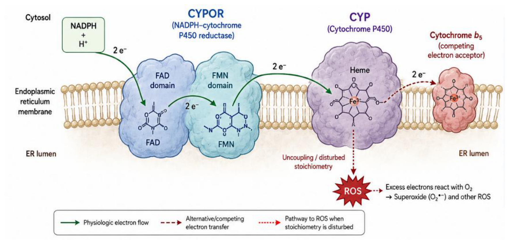
Figure 2.
Nuclear receptor regulatory network in cholestasis.
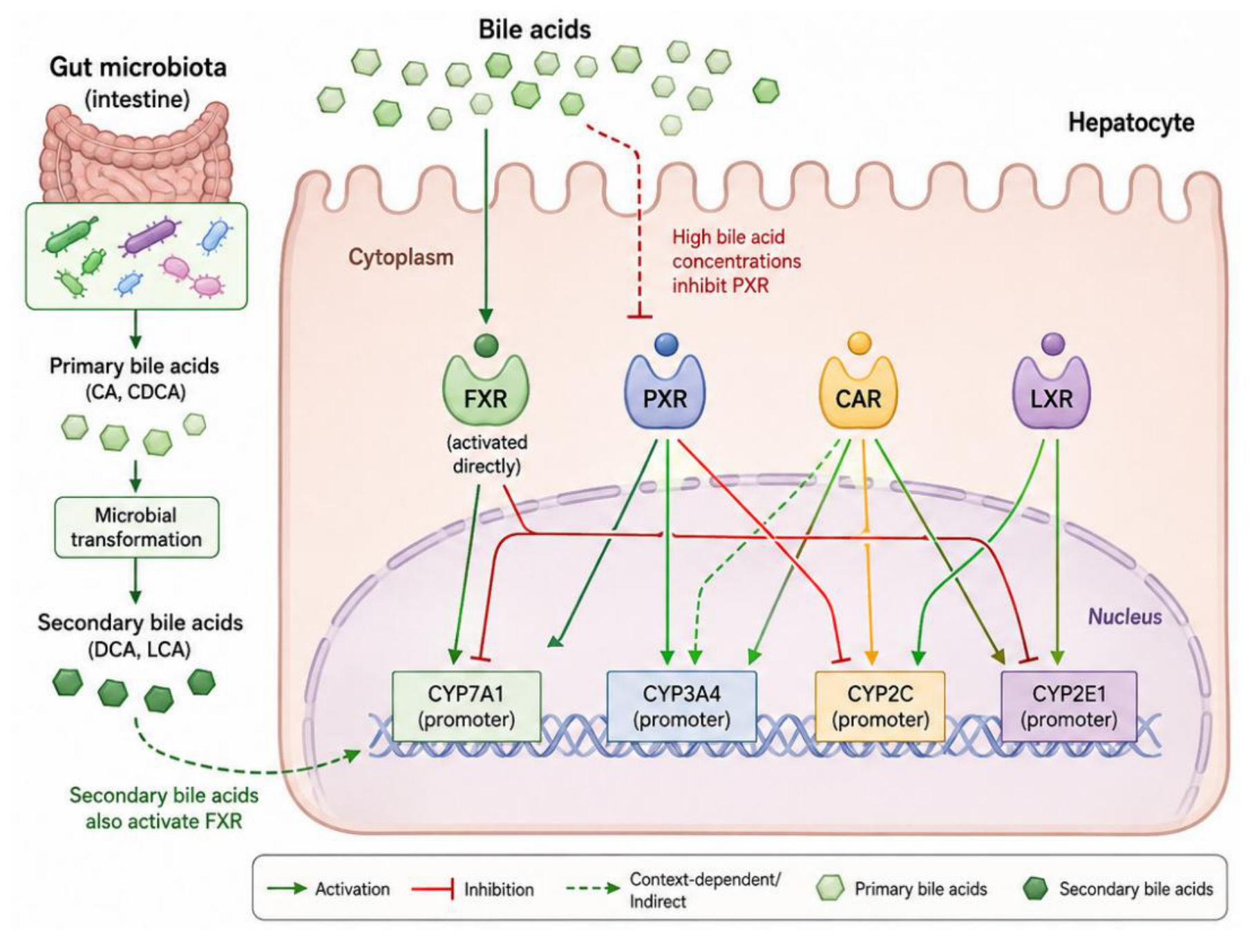
Figure 3.
Inflammatory cascade in acute hepatitis: NF-kB and Nrf2 activation.
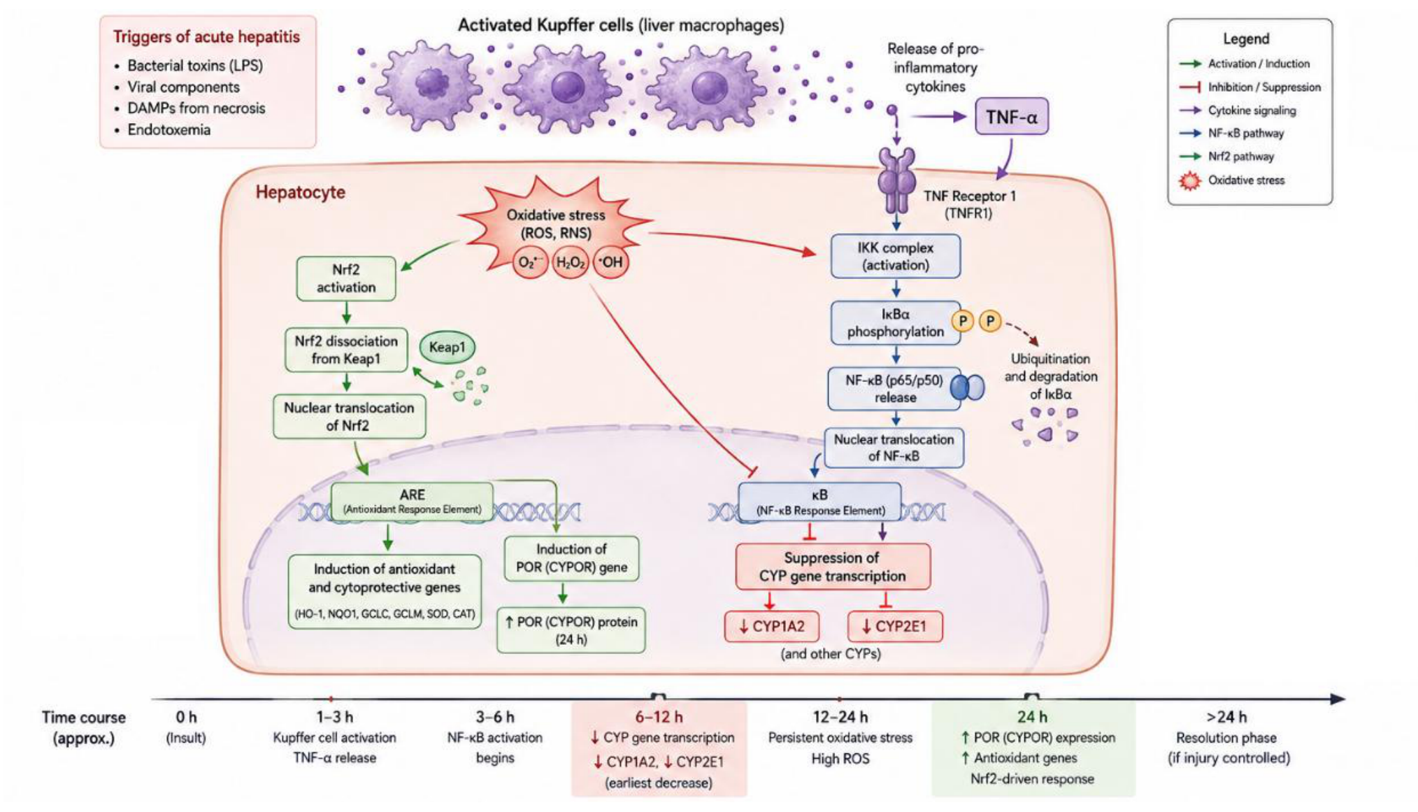
Figure 4.
Comparative bar chart of activity changes for CYP isoforms and CYPOR in cirrhosis in different species.
Figure 4.
Comparative bar chart of activity changes for CYP isoforms and CYPOR in cirrhosis in different species.
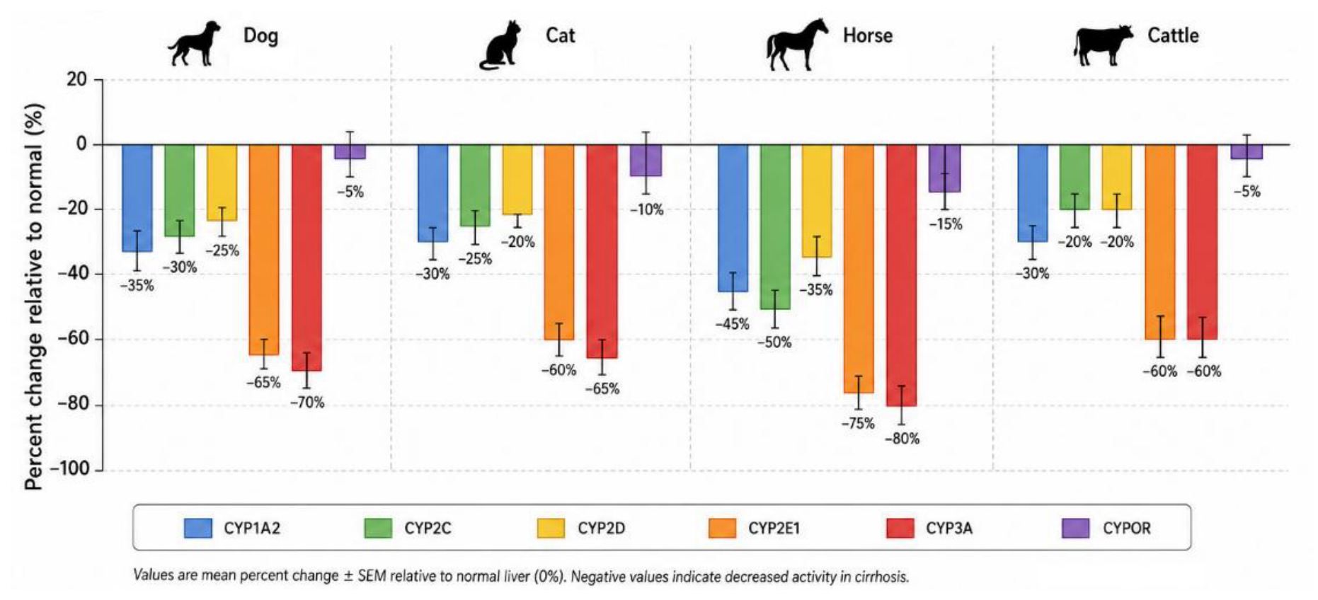
Figure 5.
Temporal dynamics of CYP3A and CYPOR activities in early and chronic cholestasis.
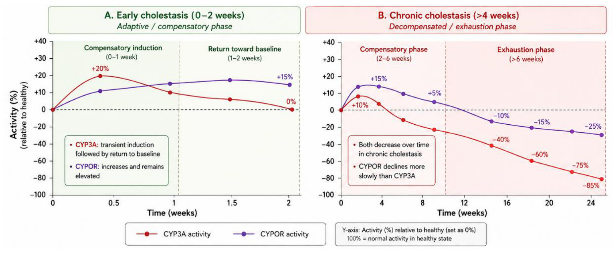
Figure 6.
Algorithm for dose adjustment based on dysbalance index.
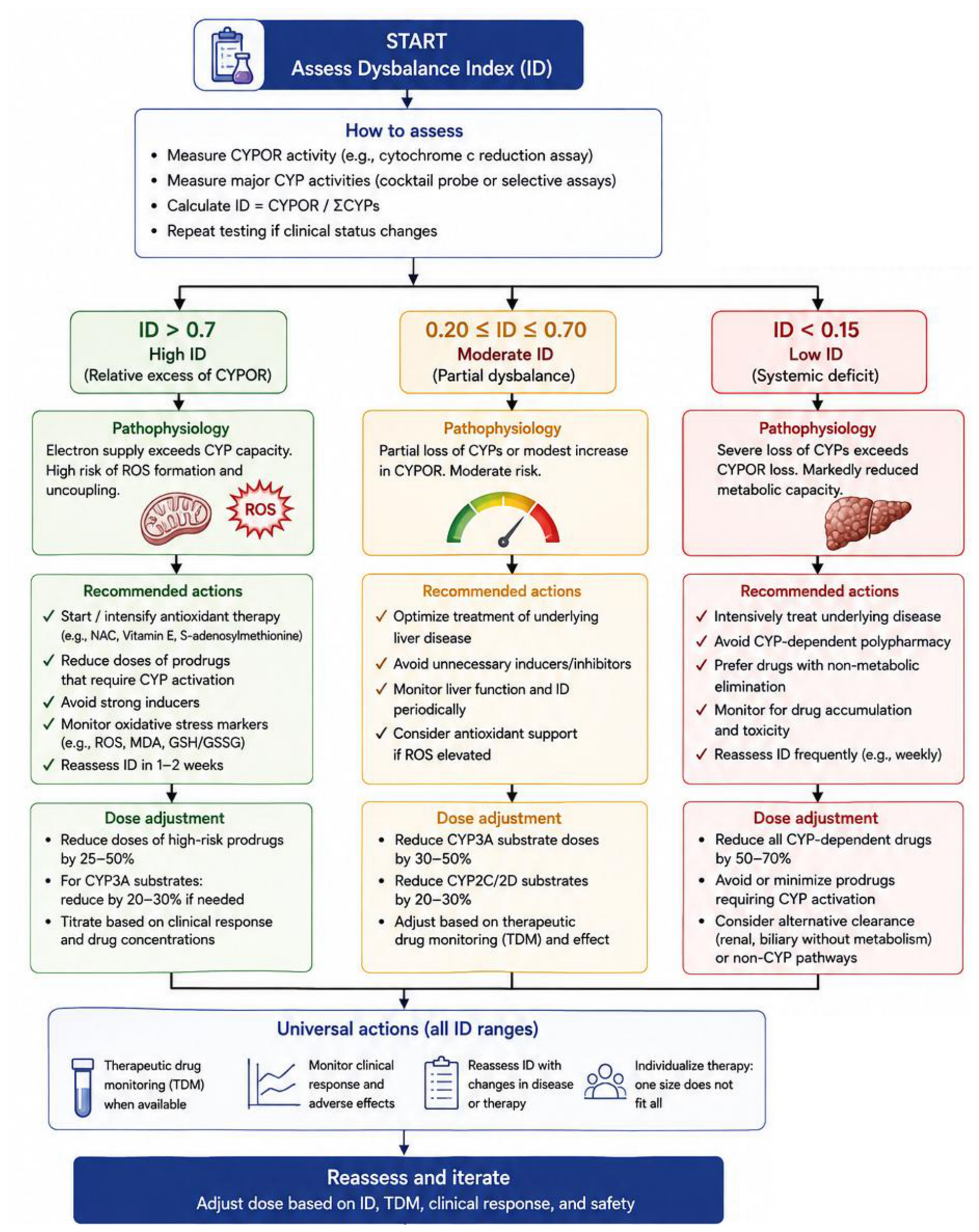
Figure 7.
Schematic post-translational CYP modifications in cholestasis.
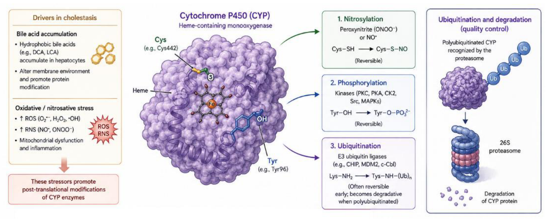
Figure 8.
Inducibility of CYP isoforms in healthy and diseased animals.
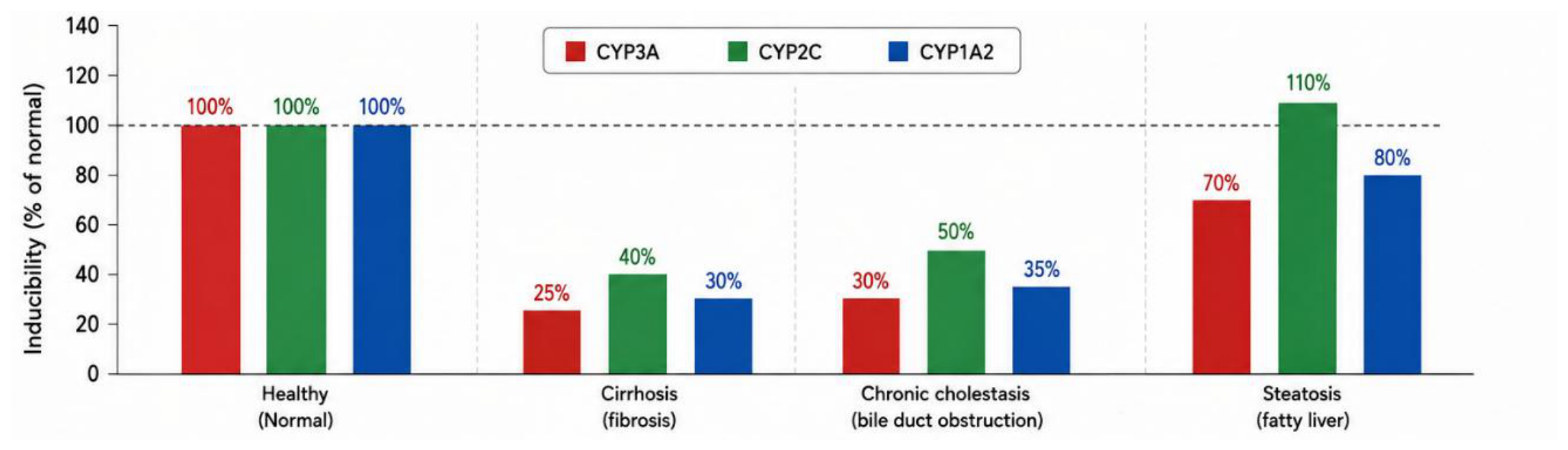
Figure 9.
Pharmacokinetic curves of diazepam concentration versus time in healthy and cirrhotic dogs.
Figure 9.
Pharmacokinetic curves of diazepam concentration versus time in healthy and cirrhotic dogs.
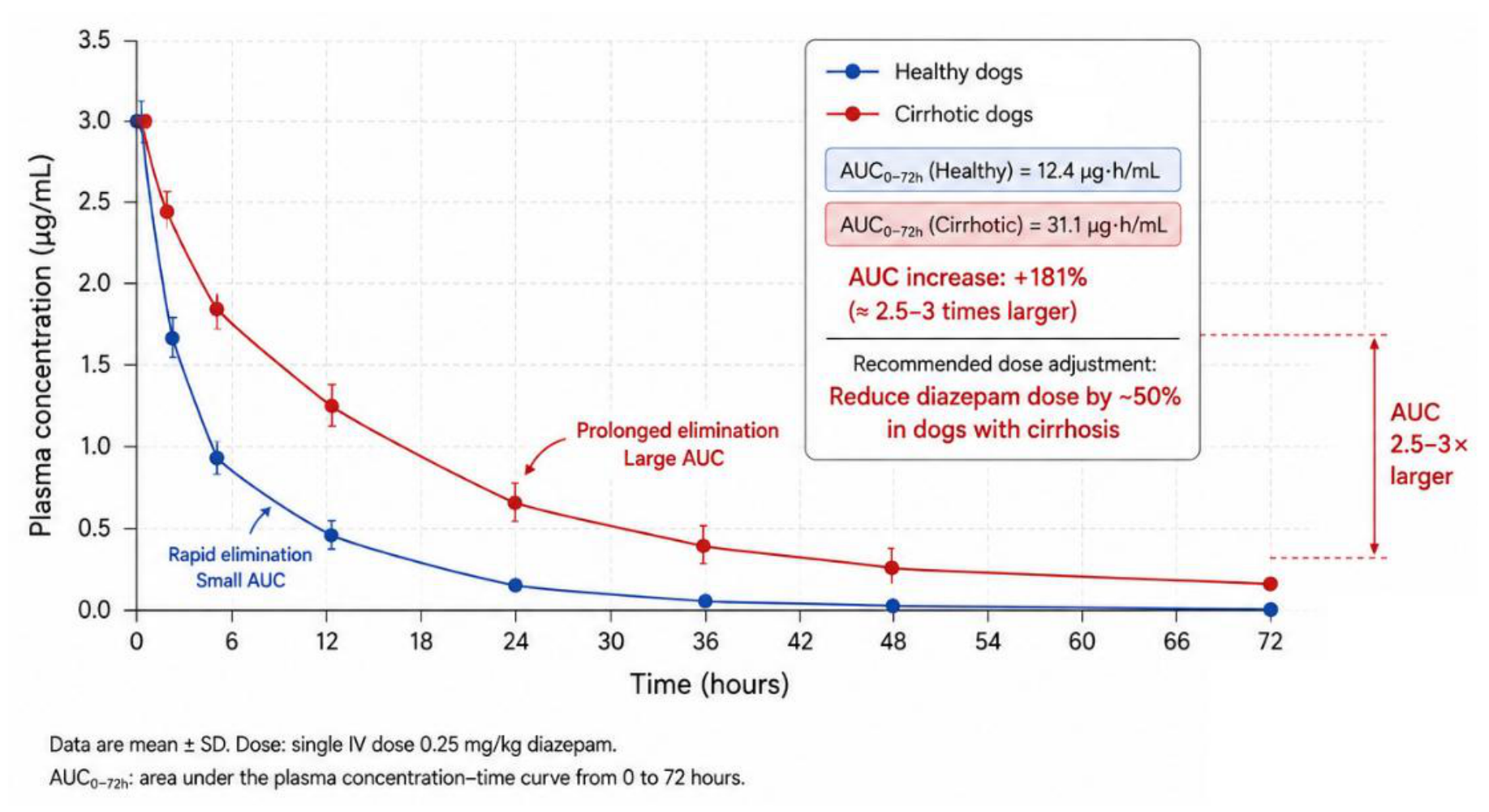
Figure 10.
Relationship between CYP2E1 activity and CYPOR activity across animal species during cholestasis.
Figure 10.
Relationship between CYP2E1 activity and CYPOR activity across animal species during cholestasis.
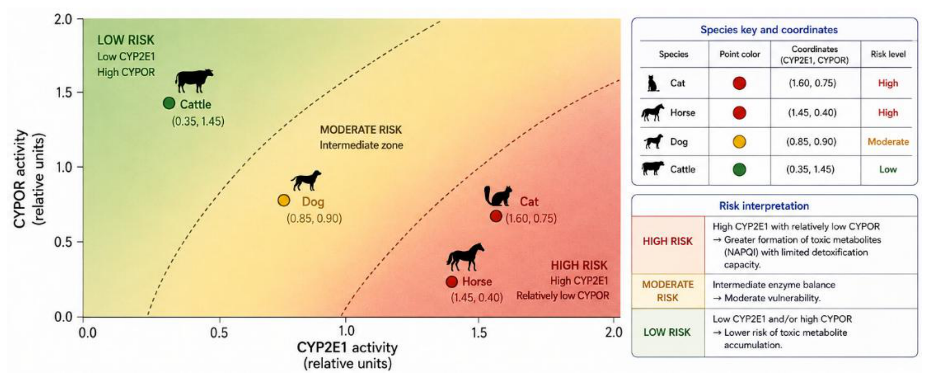

Table 1.
Percentage change in CYP isoform activities relative to healthy animals in various hepatobiliary pathologies (means with ranges).
Table 1.
Percentage change in CYP isoform activities relative to healthy animals in various hepatobiliary pathologies (means with ranges).
| Pathology / Species | CYP1A2 | CYP2C21 (dog) / CYP2C9 (horse) | CYP2D6 (dog) / CYP2D132 (cat) | CYP2E1 | CYP3A4/12/89/28 |
|---|---|---|---|---|---|
| Cirrhosis (dog) | -55 (-50 to -60) | -25 (-20 to -30) | -40 (-35 to -45) | -65 (-60 to -70) | -70 (-65 to -75) |
| Cirrhosis (cat) | -50 (-45 to -55) | n/d | -60 (-55 to -65) | -70 (-65 to -75) | -65 (-60 to -70) |
| Cirrhosis (horse) | -60 (-55 to -65) | -35 (-30 to -40) | -45 (-40 to -50) | -75 (-70 to -80) | -80 (-75 to -85) |
| Cirrhosis (cattle) | -45 (-40 to -50) | -20 (-15 to -25) | -35 (-30 to -40) | -55 (-50 to -60) | -60 (-55 to -65) |
| Early cholestasis (cat) | -40 | -10 | -20 | -50 | +20 |
| Chronic cholestasis (cat) | -70 | -30 | -40 | -80 | -30 |
| Early cholestasis (horse) | -50 | -20 | -30 | -60 | +15 |
| Chronic cholestasis (horse) | -65 | -45 | -55 | -80 | -40 |
| Acute hepatitis (cattle) | -45 | -30 | -50 | -60 | -65 |
| Acute hepatitis (dog) | -50 | -35 | -55 | -65 | -70 |
| Steatosis (cat) | -45 | +10 | -35 | -50 | -50 |
| Steatosis (dog) | -40 | +15 | -30 | -45 | -55 |
Note: n/d = no data. Early cholestasis (first 1–2 weeks) showed transient induction of CYP3A in cats and horses, subsequently replaced by suppression.
Table 2.
Percentage change in CYPOR activity and CYPOR/CYP3A activity ratio in the same pathologies.
Table 2.
Percentage change in CYPOR activity and CYPOR/CYP3A activity ratio in the same pathologies.
| Pathology | Dog | Cat | Horse | Cattle |
|---|---|---|---|---|
| Cirrhosis | -10 (-5 to -15) | -5 (0 to -10) | -20 (-15 to -25) | -8 (-5 to -10) |
| Early cholestasis | +30 (+25 to +35) | +25 (+20 to +30) | +20 (+15 to +25) | +15 (+10 to +20) |
| Chronic cholestasis | -25 (-20 to -30) | -30 (-25 to -35) | -40 (-35 to -45) | -20 (-15 to -25) |
| Acute hepatitis | +45 (+40 to +50) | +40 (+35 to +45) | +35 (+30 to +40) | +30 (+25 to +35) |
| Steatosis | -5 (0 to -10) | -10 (-5 to -15) | n/d | -5 (0 to -10) |
Table 3.
Ratio of CYPOR activity to the sum of major CYP activities (dysbalance index) in health and disease (averages).
Table 3.
Ratio of CYPOR activity to the sum of major CYP activities (dysbalance index) in health and disease (averages).
| Condition | Normal | Cirrhosis | Early cholestasis |
Chronic cholestasis |
Acute hepatitis |
Steatosis |
|---|---|---|---|---|---|---|
| Index (CYPOR / (CYP1A2+CYP2C+CYP2D+CYP2E+CYP3A)) | 0.20 | 0.45 | 0.30 | 0.12 | 0.80 | 0.18 |
Table 4.
Predominant mechanisms of CYP activity reduction (T = transcriptional, PT = post-translational, D = degradation) for different isoforms and pathologies.
Table 4.
Predominant mechanisms of CYP activity reduction (T = transcriptional, PT = post-translational, D = degradation) for different isoforms and pathologies.
| Isoform | Cirrhosis | Chronic cholestasis | Acute hepatitis | Steatosis |
|---|---|---|---|---|
| CYP1A2 | T | PT (nitrosylation) | T, PT (phosphorylation) | T |
| CYP2C | T | T (FXR-mediated) | T (NF-κB) | PT (fatty acids) |
| CYP2E1 | T, D | PT, D (proteasomal) | PT (oxidative modification) | T, PT |
| CYP3A | T | PT, D (ubiquitination) | T (cytokines) | T |
Table 5.
Inducibility of CYP isoforms (% of normal induction) by classical inducers in different pathologies.
Table 5.
Inducibility of CYP isoforms (% of normal induction) by classical inducers in different pathologies.
| Pathology | CYP3A induction (%) | CYP2C induction (%) | CYP1A2 induction (%) |
|---|---|---|---|
| Healthy | 100 | 100 | 100 |
| Cirrhosis | 30 | 60 | 45 |
| Chronic cholestasis | 20 | 40 | 25 |
| Acute hepatitis | 50 | 70 | 60 |
| Steatosis | 80 | 110 | 65 |
Table 6.
Changes in AUC and clearance for marker drugs in dogs under various pathologies (averages).
Table 6.
Changes in AUC and clearance for marker drugs in dogs under various pathologies (averages).
| Drug (marker) | Cirrhosis (AUC ↑%) | Cirrhosis (Cl ↓%) | Chronic cholestasis (AUC ↑%) |
Chronic cholestasis (Cl ↓%) |
Acute hepatitis (AUC ↑%) |
|---|---|---|---|---|---|
| Diazepam (CYP3A) | 180 | 55 | 150 | 40 | 160 |
| Theophylline (CYP1A2) | 130 | 35 | 200 | 60 | 120 |
| Warfarin (CYP2C) | 50 | 20 | 90 | 30 | 70 |
| Metoprolol (CYP2D) | 90 | 30 | 110 | 35 | 85 |
| Enalapril (prodrug) | active metabolite AUC ↓40% |
parent Cl ↓50% | metabolite AUC ↓30% | - | metabolite AUC ↓50% |
Table 7.
AUC increases for diazepam and theophylline in cats and horses.
| Species, pathology | Diazepam (AUC ↑%) | Theophylline (AUC ↑%) |
|---|---|---|
| Cat, cirrhosis | 160 | 140 |
| Cat, chronic cholestasis | 130 | 220 |
| Horse, cirrhosis | 190 | 160 |
| Horse, chronic cholestasis | 180 | 250 |
Table 8.
Comparative susceptibility to acetaminophen hepatotoxicity based on CYP2E1 and CYPOR activities.
Table 8.
Comparative susceptibility to acetaminophen hepatotoxicity based on CYP2E1 and CYPOR activities.
| Species | Basal CYP2E1 (rel. units) | Decline in cholestasis (%) | Toxicity risk (rank) |
|---|---|---|---|
| Cat | 0.8 | 80 | High (1) |
| Dog | 1.2 | 60 | Moderate (2) |
| Horse | 1.5 | 75 | High (3) |
| Cattle | 2.0 | 55 | Low (4) |
Disclaimer/Publisher’s Note: The statements, opinions and data contained in all publications are solely those of the individual author(s) and contributor(s) and not of MDPI and/or the editor(s). MDPI and/or the editor(s) disclaim responsibility for any injury to people or property resulting from any ideas, methods, instructions or products referred to in the content. |
© 2026 by the author. Licensee MDPI, Basel, Switzerland. This article is an open access article distributed under the terms and conditions of the Creative Commons Attribution (CC BY) license.
Copyright: This open access article is published under a Creative Commons CC BY 4.0 license, which permit the free download, distribution, and reuse, provided that the author and preprint are cited in any reuse.