
Submitted:
01 September 2026
Posted:
02 September 2026
You are already at the latest version
Abstract
Microalgae are thought to offer attractive biological platforms for the sustainable synthesis of biodegradable polymers, especially polyhydroxybutyrate (PHB), which belongs to the polyhydroxyalkanoate (PHA) family. Rapid and accurate screening techniques for finding high-PHB-producing microalgal strains are needed due to growing environmental issues related to traditional plastics. For the initial identification of intracellular polymer buildup, staining-based methods provide an economical and effective substitute for traditional analytical procedures. This study as first one achieved in Yemen aimed to evaluate and compare the performance of three staining approaches—Nile Red, Nile Blue A, and Sudan Black B—for the detection of PHB accumulation in microalgae. Nile Red was used for rapid visualization of intracellular hydrophobic compounds, while Nile Blue A was applied for more selective identification of PHB granules under optimized staining conditions. Sudan Black B provided complementary qualitative assessment of lipid and polymeric inclusions using light microscopy. In accordance to results reads above showed that the stain Nile Blue A dissolved in DMSO showed 100% to be the highest ratio of positive read followed by 98.2% of Nile Red dissolved in DMSO of positive read then 92.79% of Nile Blue A dissolved in Acetone of positive read after that 88.29% of Nile Red dissolved in acetone that showed as orange granules and finally 40.54% of Sudan Black B dissolved in ethanol as blue black granules to be the less ratio amongst all ones stains. However, as seen in the appendix's images, the negative findings revealed pink or red granules and cells. In calculating reads of samples the results showed out of 37 samples 32 samples are positive with 86.49% for biopolymer accumulating inside algal tested cells. These samples were selected to be used for other biotechnological application.
Keywords:
microalgae
; PHB
; Nile Red
; Nile Blue A
; Sudan Black B
; biopolymer
; screening techniques
1. Introduction
The increasing environmental concerns associated with petroleum-based plastics have intensified the search for sustainable and biodegradable alternatives [1,2]. Among the most promising substitutes are polyhydroxyalkanoates (PHAs), a group of microbial biopolymers synthesized as intracellular carbon and energy storage compounds [3,4,5]. One of the most widely studied PHAs is poly-β-hydroxybutyrate (PHB), which possesses thermoplastic properties similar to conventional plastics while remaining biodegradable and biocompatible [6]. These characteristics make PHB highly attractive for applications in packaging, agriculture, and biomedical industries [2].
Although PHB production has been extensively investigated in bacteria, microalgae have recently emerged as a potential alternative source due to their rapid growth rates, ability to fix atmospheric carbon dioxide, and capacity to thrive in diverse and extreme environments [7]. Microalgae can accumulate PHB under stress conditions such as nutrient limitation, high salinity, or light intensity variations [7]. Their cultivation does not compete directly with food crops and can be integrated with wastewater treatment systems, making them environmentally and economically promising candidates for sustainable bioplastic production [8].
Accurate and rapid detection of PHB accumulation in algal cells is essential for screening high-producing strains. Conventional methods for PHB quantification, including solvent extraction and chromatographic analysis, are reliable but time-consuming, labor-intensive, and costly [9]. Therefore, staining techniques have been widely adopted as rapid screening tools for detecting intracellular PHB granules. Lipophilic dyes such as Nile Red and Sudan Black B are commonly used due to their affinity for hydrophobic polymer inclusions [10]. Nile Red is particularly useful in fluorescence microscopy because it emits strong fluorescence when bound to intracellular lipid or polymer bodies, whereas Sudan Black B is typically applied in light microscopy for visualizing dark-stained granules within cells. [11].
Among these approaches, fluorescent dyes such as Nile Red and Nile Blue A, as well as the histochemical stain Sudan Black B, are widely used for visualizing intracellular lipid inclusions and PHA granules [12] Nile Red is extensively applied for the detection of neutral lipids in microalgae, with fluorescence intensity frequently correlated to intracellular lipid content, enabling rapid phenotypic screening [13,14,15]. Despite the widespread use of these staining methods, variations in sensitivity, specificity, clarity of visualization, cost, and ease of application have been reported [16]. Moreover, most comparative studies have focused on bacterial systems, with limited research addressing algal isolates, particularly those originating from unique environmental conditions such as those found in Yemen. Yemen encompasses diverse ecological niches, including marine coastal areas, freshwater bodies, and arid regions, which may harbor novel algal strains with potential for PHB production.
To date, there is limited information regarding PHB-producing algae isolated from Yemeni environments and the efficiency of different staining techniques in detecting intracellular PHB granules in these isolates. Therefore, a comparative evaluation of commonly used staining methods is necessary to determine the most reliable and practical approach for rapid screening.
The present study aims to isolate algae from various Yemeni environments and to comparatively evaluate different staining techniques for the detection of PHB accumulation. By assessing the sensitivity, clarity, and practicality of each staining method, this study seeks to identify the most effective approach for preliminary screening of PHB-producing algal strains and to contribute to the development of sustainable bioplastic research in the region.
2. Materials and Methods
2.1. Study Area and Sample Collection
Environmental samples were collected from diverse habitats across Yemen, including freshwater bodies, wastewater and agricultural irrigation canals wastewater sites. Sampling was conducted using sterile 500 mL glass bottles. Surface water samples (0–50 cm depth) were collected and transported to the laboratory in insulated containers at 4 °C for immediate processing within 24 h.
Physicochemical parameters including temperature, pH, and salinity were measured in situ using a portable multiparameter probe.
2.2. Isolation and Cultivation of Algal Strains
2.2.1. Isolation
Samples were serially diluted and inoculated onto BG-11 medium (for freshwater isolates) and modified f/2 medium (for marine isolates). Cultures were incubated under controlled laboratory conditions at 25 ± 2 °C, with a 12:12 h light–dark photoperiod and light intensity of approximately 60–80 µmol photons m−2 s−1. Individual colonies were purified by repeated streaking and microscopic examination to obtain unialgal cultures [17,18,19,20,21].
2.2.2. Biomass Production and PHB Induction
To enhance intracellular PHB accumulation, algal isolates were cultured under nutrient stress conditions (nitrogen limitation) by transferring exponentially growing cells into nitrogen-depleted BG-11 medium 1% glucose. Cultures were incubated for 7–14 days under the same environmental conditions. Biomass was harvested by centrifugation at 6000 rpm for 10 min and washed twice with sterile distilled water prior to staining [22,23,24].
2.3. Preparation of Staining Solutions
2.3.1. Nile Blue A Staining Solution
2.3.2. Nile Red Staining Solutions
Nile Red stock solution (1 mg mL−1) was prepared separately in:Ethanol (analytical grade), Acetone (analytical grade), Dimethyl sulfoxide (DMSO)
All solutions were prepared under low-light conditions and stored at 4 °C. Working solutions (10 µg mL−1) were prepared by dilution in the respective solvent immediately before use.
2.3.3. Sudan Black B Staining Solution
2.4. Staining Procedures for PHB Detection
2.4.1. Nile Blue A Staining
Approximately 1 mL of algal suspension was smeared onto a clean glass slide and heat-fixed. Slides were flooded with 0.02% Nile Blue A solution and incubated at 55 °C for 10 min. After cooling, slides were rinsed gently with distilled water and air-dried.For fluorescence visualization, stained samples were examined under a fluorescence microscope using an excitation wavelength of 460–490 nm. Intracellular PHB granules appeared as bright orange fluorescence [15,16].
2.4.2. Nile Red Staining
For each solvent system (ethanol, acetone, and DMSO), 1 mL of concentrated algal suspension was mixed with 10 µL of Nile Red working solution. Samples were incubated in the dark at room temperature for 10 min to allow dye penetration.
Stained cells were examined using fluorescence microscopy with excitation at 530–550 nm and emission detection at ≥575 nm. PHB granules were identified as bright yellow–orange fluorescent inclusions within the cytoplasm.
The efficiency of Nile Red staining was evaluated comparatively among the three solvents based on fluorescence intensity, background interference, and staining uniformity.
2.4.3. Sudan Black B Staining
Algal smears were heat-fixed and flooded with 0.3% Sudan Black B solution for 15 min. Slides were rinsed gently with 70% ethanol to remove excess stain and counterstained with 0.5% safranin for 30 s to enhance contrast.
Slides were observed under a light microscope at 1000× magnification using oil immersion. PHB granules appeared as dark blue to black intracellular inclusions.
2.5. Microscopic Analysis and Imaging
All stained samples were examined using a fluorescence microscope equipped with appropriate filter sets and a digital imaging system. For each isolate and staining method, at least 10 random microscopic fields were analyzed. Images were captured under identical exposure settings to allow comparative evaluation of fluorescence intensity [15,16,27].
3. Results
3.1. Isolation and Selection of PHB Producing Algae
3.1.1. Frequency and Percentage of Positive Productivity Samples Using Different Stains with Various Solvents
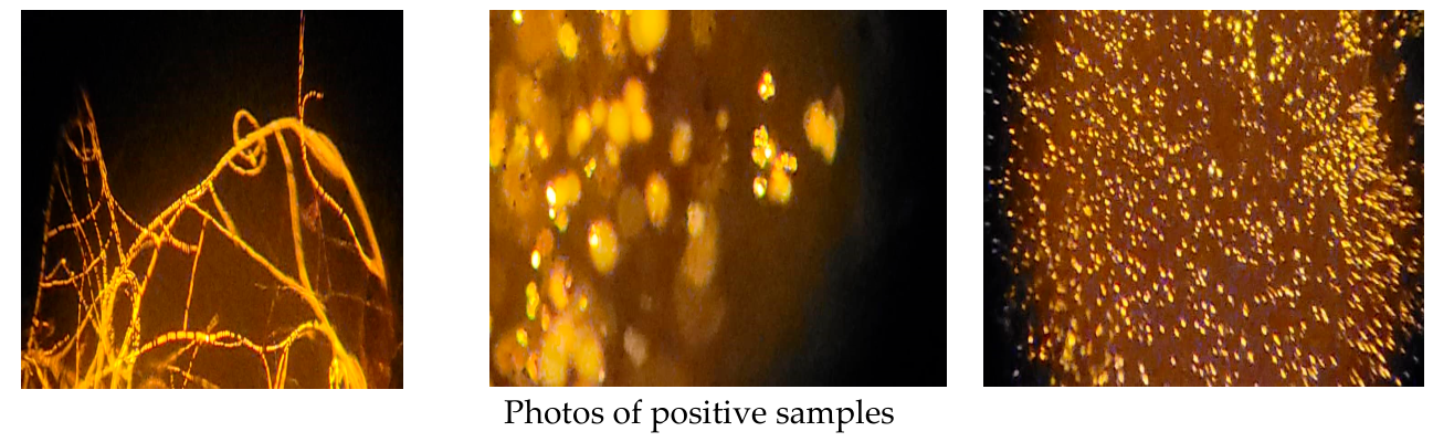
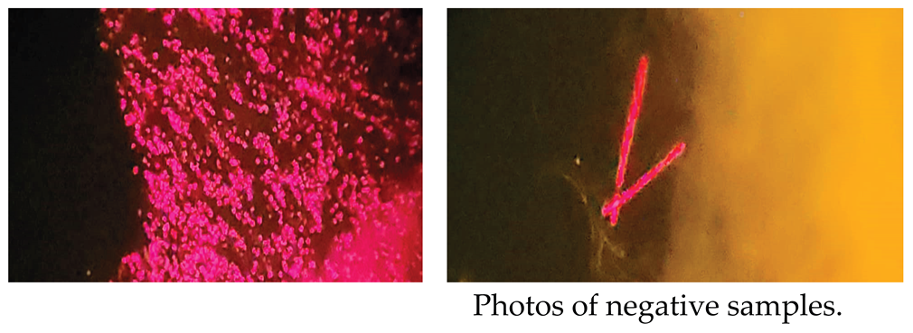
Thirty seven (37) selected samples collected from different localities were used for isolation of the PHB producing test from algae according to the method of Bormann et al. 1998a,b with modification of in 120-150 rpm overnight when adding stains in liquid media 1% glucose BG11 against control growth media BG11and 1% glucose of BG11 with no stain till read as wet preparation under fluorescent microscope under 450-490nm using different stains with various solvents as primary experiments of screening tested samples for biopolymer producing cells under fluorescent microscope among wavelength 450-590nm light to show the following positive results with Sudan Black B dissolved in ethanol as blue black granules; Nile Blue A dissolved in Acetone showed as orange granules; Nile Blue A dissolved in dimethyalsulfuxide (DMSO) showed as strong orange granules; Nile Red dissolved in acetone that showed as orange granules and Nile Red dissolved in DMSO that showed as strong orange granules as illustrated in photos in the Appendix A. And the results showed that:
In three replicates, out of 111 samples were tested by Sudan Black B with 70% ethanol 45 samples were positive showed blue black spot against negative that showed as control samples, to show 40.54% observed positively results. Followed informed by using Nile blue A with Acetone and DMSO solvents that showed out of 111 samples 103 and 111 samples were positive in 100% and 92.79% respectively, against control samples to be the best stains of observing positive algal biopolymer accumulating inside cells. For some negative samples saw in Nile blue A that were informed by Nile red with Acetone and DMSO that observed positive against control samples; out of 111 samples read 98 and 109 were positive including negative samples under using to be 98.2& and 88.29% respectively as observed under fluorescent microscope.
In calculating reads of samples the results showed out of 37 samples 32 samples are positive with 86.49% for biopolymer accumulating inside algal tested cells. These samples were selected to be used for other biotechnological application.
In accordance to results reads above showed that the stain Nile Blue A dissolved in DMSO showed 100% to be the highest ratio of positive read followed by 98.2% of Nile Red dissolved in DMSO of positive read then 92.79% of Nile Blue A dissolved in Acetone of positive read after that 88.29% of Nile Red dissolved in acetone that showed as orange granules and finally 40.54% of Sudan Black B dissolved in ethanol as blue black granules to be the less ratio amongst all ones stains. However, as seen in the appendix's images, the negative findings revealed pink or red granules and cells. As shown down in Table 1.
In calculating reads of samples the results showed out of 37 samples 32 samples are positive with 86.49% for biopolymer accumulating inside algal tested cells. These samples were selected to be used for other biotechnological application. As shown down in Figure 1.
3.2. Analysis Data of Above Experiments
PC1 (Component 1): Eigenvalue = 1.512. Percentage of Variance: Explains 37.8% of the total variance. Cumulative Total: 37.8%. Note: Only one component was extracted because the second component (0.956) is less than 1 (Kaiser's criterion). As shown down in Table 2.
Principal Component Analysis was conducted on four quantitative staining variables. The first principal component (PC1) emerged as the dominant axis, explaining 37.8% of the total variance (Eigenvalue = 1.512).The component loadings revealed that Nile Blue A with Acetone (0.738) and Nile Red with Acetone (0.661) were the primary contributors to this component, indicating their superior discriminatory power in differentiating PHB-producing isolates. As shown down in Table 3.
Nile Blue A. with Acetone: Its weight is 0.738 (highest). This means it contributes more to the formation of this axis, Nile Red Acetone: 0.661, Nile Red DMSO: 0.603 and Sudan Black: 0.409 (least potent). As shown down in Table 3.
3.3. Statistical Analysis
All experiments were conducted in triplicate. Statistical analyses were performed using Excel for percentages; Principal Component Analysis (PCA) followed by correlation - matrix to determine significant differences among staining methods and solvent systems. Differences were considered statistically significant at p < 0.05. As shown in Table 1, Table 2 and Table 3
4. Discussion
The current work shows that microalgae isolated from Yemeni settings have a significant potential for producing biodegradable biopolymers, especially polyhydroxybutyrate (PHB). Scientific interest in renewable and environmentally friendly substitutes like polyhydroxyalkanoates (PHAs) has increased due to the growing global concern over environmental contamination caused by petroleum-based plastics [28]. In this regard, using microalgae as biological factories for PHB synthesis is a cost-effective and environmentally friendly strategy.
The study's findings unequivocally show that staining-based techniques are useful instruments for quickly identifying microalgae that produce PHB. With a 100% positive reading, Nile Blue A dissolved in DMSO had the best detection performance among the stains under investigation. This observation is in line with the results of [29], who found that Nile Blue A produces strong fluorescence signals that enable precise detection and is highly selective for intracellular PHB granules. Nile Blue A's capacity to pierce algal cell walls and interact selectively with hydrophobic polymer granules may be connected to its strong affinity for PHB inclusions.
Nile Red also demonstrated remarkable efficiency, especially when dissolved in acetone (88.29%) and DMSO (98.2%). According to earlier research, Nile Red's high fluorescence response in nonpolar settings makes it a popular tool for the detection of intracellular lipids and hydrophobic substances [30]. Thus, the current results validate Nile Red's dependability as a quick and sensitive stain for initial PHB screening in microalgal isolates. Sudan Black B, on the other hand, contributed to qualitative visualization under light microscopy despite having the lowest detection rate (40.54%). This reduced sensitivity is consistent with earlier findings that Sudan Black B is less selective than fluorescent dyes and may result in a poorer contrast when viewed under a microscope [31].
An important outcome of this study is that 32 out of 37 tested samples (86.49%) showed positive accumulation of intracellular biopolymers. This remarkably high percentage suggests that Yemeni aquatic ecosystems may contain rich and unexplored microalgal diversity capable of synthesizing valuable biodegradable polymers. Such biodiversity could serve as a foundation for future industrial biotechnology applications including sustainable bioplastic production, wastewater bioremediation, and carbon capture technologies [32].
Moreover, the study highlights the practical importance of using low-cost staining techniques for the preliminary screening of PHB-producing microorganisms, especially in developing countries where advanced analytical instruments may not always be available. Conventional methods such as gas chromatography and nuclear magnetic resonance are highly accurate but require sophisticated equipment and trained personnel [33]. Therefore, rapid staining techniques offer a practical alternative for large-scale screening programs.
Overall, the findings confirm that Nile Blue A and Nile Red are highly effective stains for detecting intracellular PHB accumulation in microalgae. The study also emphasizes the untapped biotechnological potential of Yemeni microalgae and supports future investigations focused on molecular characterization, optimization of culture conditions, and enhancement of PHB productivity for commercial applications.
5. Conclusions and Recommendation
This study represents the first research conducted in Yemen on the detection of polyhydroxybutyrate (PHB) accumulation in microalgae isolated from Yemeni environments using different chemical stains. The findings confirmed that Yemeni microalgal isolates possess significant potential for biodegradable biopolymer production, highlighting the environmental and biotechnological value of local microbial resources. Among the tested methods, Nile Blue A in DMSO showed the highest efficiency for PHB detection, demonstrating its suitability as a rapid and cost-effective screening technique.
The study also emphasizes the importance of developing strategic plans for the sustainable exploitation of Yemen’s diverse environmental resources through the establishment of national microalgae research programs, creation of microbial culture collections, and support for green biotechnology initiatives. Furthermore, integrating academic research with industrial applications could promote the production of eco-friendly bioplastics and reduce dependence on conventional petroleum-based plastics.
It is recommended that future studies focus on isolating additional microalgal strains from different Yemeni ecosystems, optimizing cultivation and staining conditions, and applying molecular and biochemical analyses to improve PHB productivity. Supporting environmental biotechnology research and encouraging investment in renewable biological resources will contribute to sustainable development and environmental protection in Yemen.
Acknowledgments
Authors are thankful to Yemeni standardization Organization and Quality Control represent by General Manger and all laboratories employees and all thanks to wastewater treatment plant represents with General Manger and employees located in Sana'a and Ibb city. The first author is grateful to New Scan Laboratory for permission to use its Fluorescent microscope. And great thanks to Mr. Mohammed Al-Ghail who imported the chemical stains and standard materials from out of Yemen.
Conflicts of Interest
The authors declare no conflicts of interest.
Abbreviations
The following abbreviations are used in this manuscript:
| MDPI | Multidisciplinary Digital Publishing Institute |
| PHB | Polyhydroxybutrate |
| PHA | Polyhudroxyalkanuate |
| DMSO | Dimethyl sulfoxide |
| PCA | Principle component analysis |
Appendix A
References
- Narayanan, M.; Kandasamy, S.; Kumarasamy, S.; Gnanavel, K.; Ranganathan, M.; Kandasamy, G. Screening of polyhydroxybutyrate producing indigenous bacteria from polluted lake soil. Heliyon 2020, 6(10), 1–8. [Google Scholar] [CrossRef] [PubMed]
- Mascarenhas, J.; Aruna, K. Screening of Polyhydroxyalkonates (Pha) accumulating bacteria from diverse habitats. J. Glob. Biosci. 2017, 6(3), 4835–4848. Available online: http://www.mutagens.co.in/jgb/vol.06/3/060304.pdf[Ref.
- Calero, M.; Godoy, V.; Quesada, L.; Martín-Lara, M.Á. Green strategies for microplastics reduction. Curr. Opin. Green Sustain. Chem. 2021, 28, 1–6. [Google Scholar] [CrossRef]
- Meereboer, K.W.; Misra, M.; Mohanty, A.K. Review of recent advances in the biodegradability of polyhydroxyalkanoate (PHA) bioplastics and their composites. Green Chem. 2020, 22(17), 5519–5558. [Google Scholar] [CrossRef]
- Marjadi, D.; Dharaiya, N. Bioplastic: A better alternative for sustainable future. Everyman’s Sci. 2010, 45(2), 90–92. [Google Scholar]
- TANAMOOL, Varavut; IMAI, Tsuyoshi; DANVIRUTAI, Paiboon; KAEWKANNETRA, Pakawadee. Biopolymer generation from sweet sorghum juice: screening, isolation, identifcation, and fermentative polyhydroxyalkanoate production by Bacillus aryabhattai. Turk. J. Biol. 2013, 37, 259–264. Available online: http://journals.tubitak.gov.tr/biology/Turkish. [CrossRef]
- Moshood, T. D.; Nawanir, G.; Mahmud, F.; Mohamad, F.; Ahmad, M. H.; AbdulGhani, A. Sustainability of biodegradable plastics: New problem or solution to solve the global plastic pollution? Curr. Res. Green Sustain. Chem. 2022, 100273. [Google Scholar] [CrossRef]
- Awasthi, A.K.; Tan, Q.; Li, J. Biotechnological potential for microplastic waste. Trends Biotechnol. 2020, 38(11), 1196–1199. [Google Scholar] [CrossRef] [PubMed]
- Puttagunta, M.; Ravi, S. Medical image analysis based on deep learning approach Multimed Tools. Appl 2021, 80(16), 24365–24398. [Google Scholar] [CrossRef] [PubMed]
- Asgharnejad, H.; Sarrafzadeh, M.-H. Development of digital image processing as an innovative method for activated sludge biomass quantification. Front Microbiol. 2020, 11(2020), 574966. [Google Scholar] [CrossRef] [PubMed]
- P, Y. R.; Das, M.; Maiti, S.K. Recent progress and challenges in cyanobacterial autotrophic production of polyhydroxybutyrate (PHB), a bioplastic. J. Environ. Chem. Eng. 2021, 9, 2213–3437. [Google Scholar] [CrossRef]
- Jendrossek, D.; Pfeiffer, D. New insights in the formation of polyhydroxyalkanoate granules (carbonosomes) and novel functions of poly(3-hydroxybutyrate). Environ. Microbiol. 2014, 16(8), 2357–2373. [Google Scholar] [CrossRef] [PubMed]
- Selvaraj, K.; Vishvanathan, N.; Dhandapani, R. Screening, optimization and characterization of poly hydroxybutrate from fresh water microalgal isolates. Internatiobal J. Biobased Plast. 2021, 3(1), 139–162. [Google Scholar] [CrossRef]
- Cooksey, K. E.; Guckert, J. B.; Williams, S. A.; Callis, P. R. Fluorometric determination of neutral lipids in microalgae using Nile Red. J. Microbiol. Methods 1987, 6(6), 333–345. [Google Scholar] [CrossRef]
- Ansari, S.; Fatma, T. Cyanobacterial Polyhydroxybutyrate (PHB): Screening, Optimization and Characterization. PLoS ONE 2016, 11(6), e0158168. [Google Scholar] [CrossRef] [PubMed]
- Jendrossek, D.; Selchow, O.; Hoppert, M. Poly(3-hydroxybutyrate) granules at the early stages of formation are localized close to the cytoplasmic membrane in caryophanon latum. Appl. Environ. Microbiol. 2007, 73(2), 586–593. [Google Scholar] [CrossRef] [PubMed]
- Lourenço, S.O.; Vieira, A.A.H. Culture collec–tions of microalgae in Brazil: progress and constraints. Nova Hedwig. 2004, 79(1–2), 149–173. [Google Scholar] [CrossRef]
- Takács, G.; Gergely, I.; Molnár, Z.; Ördög, V. Plant biostimulating effects of the green alga chlamydopodium fusiforme on winter wheat in field experiments. In in Book of abstract, 9th symposium on microalgae and seaweed products in Plant/Soil systems; Mosonmagyaróvár, Hungary, 2019; p. 34. [Google Scholar]
- Kumar, J.; Ramla, A.; Mallick, D.; Mishra, V. An overview of some biopesticides and their importance in plant protection for commercial CrossRef. Plants 2021, 10, 1185. [Google Scholar] [CrossRef] [PubMed]
- Robert, Rebecca; Iyer, Priya R. Isolation and Optimization of PHB (Poly-β-hydroxybutyrate) Based Biodegradable Plastics from Chlorella vulgaris. J Bioremediat Biodegrad, an open access journal. ISSN: 2155-6199. Volume 9 • Issue 2 • 1000433; Ref for BBM; 2018; Volume BG11. [Google Scholar]
- WangY; GuoW; YenH, W.; HoS, H.; LoY, C.; ChengC, C. Cultivation of Chlorella vulgaris JSC-6 with swine wastewater for simultaneous nutrient/COD removal and carbohydrate production. Bioresour. Technol. 2015, 198, 619–625. [Google Scholar] [CrossRef] [PubMed]
- Bormann, E. J.; Leibner, M.; Beer, B. Growth-associated production of poly(hydroxybutyric acid) by Azotobacter beijerinckii from organic nitrogen substrates. In Applied Microbiology and Biotechnology; Springer, 1998a; Volume 49, 1, pp. 84–88. [Google Scholar] [CrossRef]
- Bormann, E. J.; Leibner, M.; Roth, M.; Beer, B.; Metzner, K. Production of polyhydroxybutyrate by Ralstonia eutropha from protein hydrolysates. In Applied Microbiology and Biotechnology; Springer, 1998b; Volume 50, pp. 604–607. [Google Scholar] [CrossRef]
- Mercan, N.; Aslim, B.; Yüksekda˘g, Z.N.U.R.; Beyatli, Y. Production of Poly-b Hydroxybutyrate (PHB) by Some Rhizobium Bacteria. Turk.J.Biol. 2002, 26, 215–219. [Google Scholar]
- Schlegel, H.G.; Lafferty, R.; Krauss, I. The isolation of mutants not accumulating Poly-B-hydroxybutyrate. Arch. Microbiol. 1970, 71, 283–294. [Google Scholar] [CrossRef] [PubMed]
- Mahitha, G.; Madhuri, R. Purification and Characterization of Polyhydroxybutyrate Produced from Marine Bacteria. Int.J.Sci.Eng.Res. 2015, 6, 71–75. [Google Scholar]
- Bhagowati, P.; Pradhan, S.; Dash, H.R.; Das, S. Production, Optimization and Characterization of Polyhydroxybutyrate, a Biodegradable Plastic by Bacillus spp. Biosci.Biotechnol.Biochem. 2015, 79, 1454–1463. [Google Scholar] [CrossRef] [PubMed]
- Anderson, A. J.; Dawes, E. A. Occurrence, metabolism, metabolic role, and industrial uses of bacterial polyhydroxyalkanoates. Microbiol. Rev. 1990, 54(4), 450–472. [Google Scholar] [CrossRef]
- Ostle, A. G.; Holt, J. G. Nile blue A as a fluorescent stain for poly-β-hydroxybutyrate. Appl. Environ. Microbiol. 1982, 44(1), 238–241. [Google Scholar] [CrossRef] [PubMed]
- Spiekermann, P.; Rehm, B. H.; Kalscheuer, R.; Baumeister, D.; Steinbüchel, A. A sensitive, viable-colony staining method using Nile red for direct screening of bacteria that accumulate polyhydroxyalkanoic acids. Arch. Microbiol. 1999, 171, 73–80. [Google Scholar] [CrossRef] [PubMed]
- Koller, M.; Salerno, A.; Dias, M.; Reiterer, A.; Braunegg, G. Modern biotechnological polymer synthesis: a review. Food Technol. Biotechnol. 2010, 48(3), 255–269. [Google Scholar]
- Sudesh, K.; Abe, H.; Doi, Y. Synthesis, structure and properties of polyhydroxyalkanoates: biological polyesters. Prog. Polym. Sci. 2000, 25(10), 1503–1555. [Google Scholar] [CrossRef]
- Verlinden, R. A.; Hill, D. J.; Kenward, M. A.; Williams, C. D.; Radecka, I. Bacterial synthesis of biodegradable polyhydroxyalkanoates. J. Appl. Microbiol. 2007, 102(6), 1437–1449. [Google Scholar] [CrossRef] [PubMed]
Figure 1.
The positive percentage of algal selected samples isolated for PHB production according to stain reading.
Figure 1.
The positive percentage of algal selected samples isolated for PHB production according to stain reading.
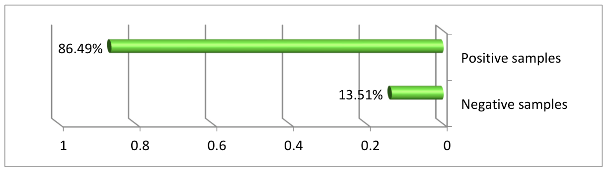

Table 1.
The positive percentage of algal selected samples isolated for PHB production according to stain reading.
Table 1.
The positive percentage of algal selected samples isolated for PHB production according to stain reading.
| Stain treatment | Positive read % |
|---|---|
| Nile Red_DMSO | 100 |
| Nile Red_Acetone | 92.79 |
| Nile Blue Acetone | 88.29 |
| Sudan Black B | 40.54 |
Table 2.
Principle component analysis PCA summary.
| Total Variance Explained | ||||||
|---|---|---|---|---|---|---|
| Extraction Sums of Squared Loadings | Initial Eigenvalues | Component | ||||
| Cumulative % | % of Variance | Total | Cumulative % | % of Variance | Total | |
| 37.8 | 37.8 | 1.512 | 37.8 | 37.8 | 1.512 | 1 |
| 61.691 | 23.89 | 0.956 | 2 | |||
| 82.696 | 21.005 | 0.84 | 3 | |||
| 100 | 17.304 | 0.692 | 4 | |||
Table 3.
Detection Method: Principal Component Analysis.
| Component Matrixa | |
|---|---|
| Component | |
| 1 | |
| 0.603 | Nile Red.DMSO/1% glucose BG11 with less N, much C |
| 0.661 | Nile Red. Aceton/1% glucose BG11 with less N,, much C |
| 0.738 | Nile blue A. with Aceton/1% glucose BG11 with less N,, much C |
| 0.409 | Sudan Black B with 70% ethanol |
Disclaimer/Publisher’s Note: The statements, opinions and data contained in all publications are solely those of the individual author(s) and contributor(s) and not of MDPI and/or the editor(s). MDPI and/or the editor(s) disclaim responsibility for any injury to people or property resulting from any ideas, methods, instructions or products referred to in the content. |
© 2026 by the authors. Licensee MDPI, Basel, Switzerland. This article is an open access article distributed under the terms and conditions of the Creative Commons Attribution (CC BY) license (http://creativecommons.org/licenses/by/4.0/).
Copyright: This open access article is published under a Creative Commons CC BY 4.0 license, which permit the free download, distribution, and reuse, provided that the author and preprint are cited in any reuse.