

Submitted:
31 August 2026
Posted:
02 September 2026
You are already at the latest version
Abstract
Background: Lactylation and glycolytic reprogramming of TCA cycle proteins for epigenetic and proteomic reorganization to trigger cuproptosis, in human cancer and multisystem degenerative diseases are here reviewed. Convergent signaling axes of different types of cancer and of different types of multisystem degeneration are presented, as well as divergent disease-specific pathways. The goal of this review is to provide a theoretical framework for cross-disease precision medicine. Methods: This review retrieved relevant studies published from 2022 to 2026, and the search databases included PubMed, web of science, and Scopus. Results: This paper hierarchically combed the change characteristics of common and disease-specific signaling pathways in cancer and multisystem degenerative diseases. In human tumors, there is a general damage amplification pathway: this pathway relies on exosomes to transport lactated modified proteins, thereby achieving the transmission of paracrine signals. By lactylation, cancer cells become resistant to cuproptosis to allow for their survival. In contrast, cells of patients with multisystem degenerative diseases undergo copper-induced mitochondrial damage leading to irreversible tissue damage. Here, we contrast two opposing functions: exosomal transfer of lactylated proteins to reorganize the tumor environment and to spread copper toxicity to lesions of degenerative diseases. Conclusion: This review develops a unified “convergence-divergence” model for the two functions of lactylation. It proposes shared targets as well as subtype-specific therapeutic strategies for the two diseases. Finally, suitable translational biomarkers are screened, and an integrated framework for comparative precision medicine in the two fields of oncology and geriatric metabolic diseases is established.
Keywords:
lactylation
; cuproptosis
; cancer
; degenerative disorder
; convergence-divergence
; framework
1. Introduction
Metabolic epigenetic remodeling of lysine lactylation and copper-dependent mitochondrial cuproptosis forms the core of two pathological axes involved in cancer and in age-related organ degeneration. Initially considered as merely a byproduct of glycolysis to be removed by lactate transport, lactate has recently emerged as a signaling metabolite. Lysine lactylation (Kla) is a reversible post-translational modification that affects chromatin, RNA, and enzyme activities and thus determines cellular redox states and metal ion homeostasis [1]. Very recently, cuproptosis, a distinct form of mitochondrial cell death, was identified in 2022 [2]. Importantly, it proceeds in the absence of caspase activation and of lipid peroxidation. Notably, it is triggered by the copper-dependent reduction of FDX1 followed by the aggregation of lipoylated proteins of the tricarboxylic acid cycle (TCA) cycle. Mechanistic studies have identified lactylation as an upstream master switch that determines cellular cuproptosis sensitivity [3,4,5]. In tissues of tumors and of degenerative diseases, identical core molecular hubs are expressed. However, the corresponding glycolysis-lactate transport microenvironment determines opposing cell fate decisions. Studies on lactylation–cuproptosis signaling in individual tumor types as well as in individual models of degenerative diseases are predominantly represented in the current literature [6,7,8]. A cross-disease study on conserved convergent as well as on divergent signaling pathways expressed in individual cancer types as well as in individual degenerative diseases is, however, lacking. Also, lacking are the bidirectional feedbacks established in malignant as well as in senescent cells.
2. Materials and Methods
We conducted an integrative narrative review on lysine lactylation and cuproptosis signaling pathways, which converge into and then diverge from cancer versus degenerative disease-associated molecular hubs to develop a convergence–divergence theoretical framework and to compare shared and disease-specific signaling pathways. Relevant studies published from 2022 to 2026 were retrieved from three major databases (PubMed, web of science, and Scopus) using a Boolean search strategy, supplemented by manual reverse snowballing of key studies. A total of 38 relevant publications were finally included, covering two areas: malignant tumors and multisystem degenerative diseases.
3. Results
3.1. Molecular Hubs Linking Lactylation and Cuproptosis
These three core hub pathways of convergent lactylation and cuproptosis signaling in tumor and in degenerative parenchymal cells, in addition to histone modification by lactylation, also include non-histone lactylation substrates [3,7,9]. All three core hub pathways of convergent lactylation and cuproptosis signaling in tumor and in degenerative parenchymal cells are influenced by three critical, inter-related parameters: intracellular concentration of lactate, flux through the mitochondrial TCA cycle, and the cell’s total antioxidant reserve. The correlations between the three core hub pathways of convergent lactylation and cuproptosis signaling in tumor and in degenerative parenchymal cells are detailed below. These correlations will serve as a framework for cross-disease comparisons in subsequent sections (Figure 1).
3.1.1. FDX1 RNA Modification Axis
FDX1 is the rate-limiting enzyme for the cuproptosis pathway. Lactylation of transcripts for FDX1, the rate-limiting enzyme for the cuproptosis pathway, is controlled by two RNA-binding proteins, NUDT21 and METTL16,which are ubiquitously expressed in all human tissues [10]. NUDT21 is hyper-lactylated on lysine 23 and this enhances the binding of NUDT21 to CPSF6 to extend 3’ untranslated region of FDX1 mRNAs. These mRNAs are then degraded in the cytoplasm to suppress the levels of FDX1 protein. METTL16 is hyper-lactylated on lysine 229 and this m6A modification to FDX1 mRNAs promotes their translation into FDX1 protein. This way the levels of FDX1 protein are increased [9,11,12]. The balance between NUDT21 and METTL16 therefore controls the levels of FDX1 protein and thus the levels of the cuproptosis pathway. A high level of glycolysis and a balanced level of lactate in tumors favor hyper-lactylation of NUDT21, which suppresses the levels of FDX1 protein and thus renders tumor cells resistant to the cuproptosis pathway. High levels of lactate and hyper-lactylation of METTL16 on the other hand promote the levels of FDX1 protein, which renders cells sensitive to the cuproptosis pathway. These identical sets of molecular events thus control the two opposing disease outcomes, i.e., cancer and degenerative diseases. Therefore, these identical sets of molecular events form a key convergent point for the two opposing disease outcomes.
3.1.2. NAT10-DLAT Lipoylation Stabilization Axis
DLAT, the rate limiting enzyme for the aggregation of copper by damaged cells, is also a protein whose mRNA stability is influenced by the histone modification NAT10 [13]. In fact, the RNA acetyltransferase activity of NAT10, a conserved histone modification present in all tissues, is influenced by the lactylation of a single lysine residue (K426) in the protein. Lactylation of NAT10 therefore increases the RNA acetyltransferase activity of NAT10 and, as a consequence, increases the stability of DLAT mRNAs and the resulting protein, DLAT, as well as other lipoylated, copper-sensitive proteins of the mitochondria. The increased concentration of these proteins in both tumor and degenerative cells therefore increases the risk of cuproptosis in these cells [9,12,13]. SIRT1 and SIRT3, the delactylating, deacetylating Sirtuins, therefore decrease the expression of DLAT by reversing NAT10 modification. In tumor cells, the moderate levels of lactate in these cells restrict NAT10 modification. As a consequence, there is little effect on DLAT mRNAs in these cells. In degenerative cells, however, the high, sustained levels of lactate in these cells inhibit SIRT1 and SIRT3 activity [4,8]. As a consequence, there is a marked, increasing effect on DLAT mRNAs in these cells, and, hence, on the concentration of DLAT protein in these cells.
3.1.3. G6PD-Pentose Phosphate Pathway Antioxidant Copper Buffering Axis
G6PD-lactylation in PPP pathway: a universal cellular copper detoxification system [9]. In tumor cells, the PPP/G6PD pathway can be up-regulated by G6PD-lactylation to increase NADPH production, which in turn increases GSH production for copper ion chelation. In addition, by down-regulating the TCA cycle flux, the PPP/G6PD pathway in tumor cells can provide a second layer of protection against cuproptosis [9]. In contrast, the same antioxidant hub in degenerative cells is not effective, because the cells harbor an intact mitochondrial lipoylation pathway that cannot be effectively counterbalanced by the above-mentioned antioxidant hub for the reasons outlined above.
3.2. Exosome-Mediated Paracrine Transmission of Lactylated Cargo
In addition to transfer of intracellular information via signaling pathways, cells can also transfer information contained in histone and non-histone lactylation via exosomes (secretory vesicles) [15,16]. Cells that are damaged in tissue of cancer patients as well as in patients with degenerative diseases package histone and non-histone lactylation products into exosomes. The exosomes that are secreted by damaged cells can cross tissue and organ barriers. For example, exosomes secreted by tumor cells carry lactylated NUDT21 protein into adjacent normal epithelial cells. These normal epithelial cells can then transfer cuproptosis-resistance information to other cells. In other words, the information contained in the histone and non-histone modification of tumor cells can be transferred to other cells in the tumor microenvironment. These modified cells can in turn be recognized by the immune system and therefore killed by the immune system. The information contained in exosomes secreted by damaged cells in patients with degenerative diseases is transmitted via secreted exosomes in the interstitial spaces[17]. These exosomes are then taken up by recipient parenchymal cells, where the information contained in the histone and non-histone modification, such as METTL16/Tau/HSPA, is used to upregulate FDX1, a cuproptosis-sensitive protein. These results in a progressive atrophy of affected tissues.
3.3. Epithelial Solid Tumors
The majority of human tumors of clinical interest are of epithelial origin. Within the class of epithelial tumors, individual tumor subtypes differ in their use of the three shared convergent hubs for lactylation to varying degrees of resistance to cuproptosis. For each of the shared convergent hubs, the corresponding section on the use of the corresponding hub for the treatment of various degenerative diseases in this review will be contrasted with the corresponding section for epithelial tumors (Figure 2).
3.3.1. Esophageal Squamous Cell Carcinoma (ESCC)
The description of a three-tier defense cascade were established in ESCC to prevent cuproptosis, a distinct form of oxidative stress-induced cell death that is not found in degenerative diseases [18]. First, in the pentose phosphate pathway (PPP) antioxidant axis, elevated G6PD, in part lactylated, in ESCC cells leads to increased intracellular NADPH and glutathione, allowing these cells to form stable copper-glutathione complexes to prevent cuproptosis. Second, in the FDX1 RNA regulatory axis, high MCT4 activity in ESCC leads to high levels of lactate that activate AARS1 to lactylate NUDT21 at lysine 23. Lactylated NUDT21 is an elongator that elongates the 3’UTR of FDX1 mRNAs, which are then degraded by the exosome to decrease FDX1 protein levels [3]. Importantly, this decrease in sensitivity to cuproptosis is in sharp contrast to what is found in degenerative diseases where METTL16 lactylation of FDX1 increases the expression of FDX1 to increase the sensitivity of cells to cuproptosis. Third, in the NAT10-DLAT axis, the high levels of lactate in ESCC activate SIRT2 delactylase to constrain NAT10 lactylation, leading to impaired NAT10 activity [13]. This results in the destabilization of DLAT mRNAs and the decreased expression of the lipoylated form of DLAT, a mitochondrial copper-sensitive protein, to prevent cuproptosis. Importantly, ESCC tumors contain high levels of anti-cuproptosis factors that are delivered to the tumor stroma by exosomes [16]. Exosomes secreted by cancer cells, such as those containing lactylated NUDT21, are taken up by stroma-resident fibroblasts. These fibroblasts contain high levels of MCT4, which leads to the remodeling of the tumor stroma into a anti-cupoprotosis factor-rich environment, preventing cuproptosis. In sharp contrast, exosomes secreted by cells of degenerative diseases, such as those that contain lactylated cargo, are delivered to adjacent parenchymal cells where they cause upregulation of FDX1 leading to persistent copper-dependent cell death and atrophy of the tissue, or progressive atrophy of the tissue. Importantly, these data highlight a fundamental difference in the mechanism of action of malignant tumors and degenerative diseases.
3.3.2. Hepatocellular Carcinoma (HCC)
HCC exhibits a unique bidirectional regulation of the three shared convergent hubs by the two types of lactylation present in cancer cells and in cancer-associated fibrobllasts (CAFs) [19]. The levels of NUDT21 and METTL16 lactylation in cancer cells are higher than in degenerative tissues. In particular, within the shared FDX1 RNA regulatory convergent hub, absolute predominance of NUDT21 lactylation is found in cancer cells as opposed to absolute predominance of METTL16 lactylation found in degenerative tissues. Within the shared PPP antioxidant hub, the PPP pathway is activated by sustained G6PD lactylation and moderate SIRT activity limits NAT10 lactylation to reduce DLAT expression similar to that found in ESCC [18]. Therefore, sustained G6PD lactylation reinforces PPP antioxidant activity and copper buffering in cancer cells as opposed to SIRT suppression found in degenerative diseases. As a result, mitochondrial copper targets are rendered susceptible to copper toxicity to a marginally higher extent than in the case of ESCC, thereby enabling direct comparison with the functionality of the three convergent hubs in degenerative diseases. HCC exosomes carry mixed NUDT/METTL16 lactylated proteins that transfer cancer cells’ metabolic states to transfer the cancer cells’ metabolic states to the appropriate hepatic microenvironmental cells in marked contrast to exosomes secreted by degenerative diseases that carry only single types of lactylated proteins.
3.3.3. Triple-Negative Breast Cancer (TNBC)
TNBC can be adapted to use the cancer-associated fibroblast (CAF) secreted lactate to regulate the three convergent hubs[20]. Within the shared FDX1 RNA regulatory convergent hub, for instance, TNBC uses the lactate to downregulate the FDX1 protein by means of the epigenetic modification of histone H3K18 by lactate. Within the shared NAT10-DLAT convergent axis, moderate levels of NUDT21-lactylation are observed within the RNA regulatory hub. This results in the activation of the G6PD-PPP hub to generate high levels of NADPH and S-adenosylmethionine for the subsequent generation of glutathione for copper chelation. MCT4 is highly expressed in TNBC to prevent toxic levels of lactate to generate NUDT21-lactylated histones, which in turn are used to activate the G6PD-PPP hub to generate NADPH and S-adenosylmethionine. Within the shared PPP antioxidant hub, SIRT2-mediated delactylation of NAT10 results in the decreased lipoylation of DLAT to form the mitochondrial-targeted copper-specific DLAT lipid to prevent copper-induced mitochondrial aggregation. Finally, TNBC utilize exosomes that contain the H3K18la-lactylated histones to activate endothelial cells to remodel the copper homeostasis to form a protective anti-cuproptosis state, which results in the formation of a tumor-protective, anti-cuproptosis-based tumor-vasculature. In marked contrast, endothelial cells from degenerative diseases undergo cuproptosis to form vascular leakages in response to the uptake of the pathological exosomal lactylated cargo.
3.3.4. Hematological Malignancies
Hematological Tumors: Similar Resistance but by Different Means. The three shared convergent hubs are rewired by metabolic enzyme-centric non-histone lactylation to confer similar but distinct resistance to cuproptosis in hematological tumors (melanoma, gastric cancer) [8,21]. These are compared to the operation of the convergent hub in degenerative diseases in this section.
BRAF inhibitor-resistant melanoma suppresses TFRC copper uptake peripherally to the G6PD antioxidant hub through LSD lactylation. Gastric cancer primarily utilizes METTL16 lactylation within the FDX1 convergent hub to slightly upregulate FDX1 in cancer cells, while maintaining strong NUDT21 lactylation and highly active G6PD-PPP antioxidant signaling. Thus, gastric cancer, which is an epithelial tumor type, exhibits mild levels of cuproptosis resistance, a property that is distinctly different from degenerative diseases and aligns with that of other epithelial tumors. Therefore, gastric cancer can be classified as a ‘transitional’ tumor type, which can serve as a very useful model to study the cross-disease functionality of the convergent hubs. In addition, hematological tumors release exosomes that carry lactylated metabolic enzymes to alter the copper metabolism in immune cells to evade immune surveillance. Exosomes from degenerative diseases on the other hand carry single-substrate lactylated proteins. Upon uptake by parenchymal cells by these exosomes, a cascade of cell deaths is triggered [15,16].
3.4. Subtype-Specific Divergent Mechanisms of Lactylation-Driven Cuproptosis in Degenerative Diseases
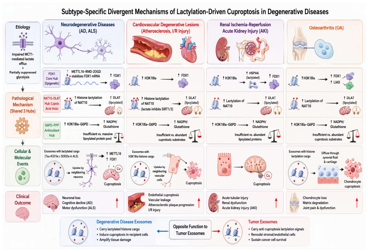
Similar three shared convergent molecular hubs are impaired in the four types of degenerative diseases in a totally different manner than in the corresponding malignant tumor subtypes. Impaired MCT1-mediated efflux of lactic acid, as well as partially suppressed glycolysis, leads to a shift in the activation balance of the three shared axes towards copper-dependent death of mitochondrial in degenerative cells. The detailed characteristics of the four types of degenerative diseases, namely neurodegenerative, cardiovascular, renal, and articular degenerative lesions, are compared to the corresponding malignant tumor subtypes [22]. For each of the four types of degenerative diseases identical three shared convergent molecular hubs are used as in the corresponding malignant tumor subtypes, but in a totally different manner due to impaired MCT1-mediated efflux of lactic acid, as well as partially suppressed glycolysis. Therefore, the three shared convergent molecular hubs are used in a totally opposite, that is, divergent, manner in degenerative cells as compared to malignant tumor cells (Figure 3).
3.4.1. Neurodegenerative Diseases (Alzheimer’s Disease [AD] and Amyotrophic Lateral Sclerosis [ALS])
The core issue in neurodegenerative diseases is impaired MCT1 mediated lactate efflux from neurons [23]. Lactate accumulation in degenerative neurons reverses the activation balance of all three shared axes of the three convergent hubs of cancer cells to an opposite direction, activating all three axes in a fully opposing manner to the activation of the same three axes in cancer cells (shared mechanisms in cancer and degenerative diseases). Within the shared epigenetic convergent axis of the FDX1 core convergent hub of cancer cells,while degenerative neurons lack hyper-NUDT21 (Gm18891, MML1) specific histone lactylation to suppress FDX1 expression. Instead, histone lysine 229 (K229) specific METTL16 (METTL6) modified RNA: 2-oxoglutarate-dependent dioxygenases (RMD-2OGD) directly stabilize FDX1 mRNAs and consequently lead to drastically elevated expression levels of FDX1 in degenerative neurons. FDX1 specific METTL16-mediated modified RNA:2-oxoglutarate-dependent dioxygenases (RMD-2OGD) of degenerative neurons have no equivalent in cancer cells to repress FDX1 expression. The corresponding subunit of the shared NAT10-DLAT convergent axis in degenerative neurons, in turn, is SIRT1/3 delactylase. Whereas SIRT1/3 delactylases in cancer cells remove lactylation from NAT10, thereby downregulating NAT10 specific DLAT lipoylated protein abundance to suppress cuproptosis in cancer cells, impaired lactate efflux from degenerative neurons triggers hyper-NAT10 specific histone lactylation to stabilize DLAT mRNAs in an alternative manner, thereby increasing DLAT lipoylated protein abundance to activate cuproptosis [24]. Similarly, within the shared G6PD-PPP antioxidant convergent hub of cancer cells, the corresponding subunit of the G6PD-PPP antioxidant convergent hub in degenerative neurons, for instance, is H3K18la (H3K18) modified G6PD. Despite of the corresponding H3K18la (H3K18) modified G6PD of degenerative neurons generating high levels of NADPH/glutathione to activate PPP antioxidant metabolism, the corresponding massive reserves of lipoylated DLAT and FDX1 protein-containing TCA cycle intermediates, in turn, that are produced by intact mitochondrial TCA cycles of degenerative neurons far exceed corresponding copper-buffering capacity of the same antioxidant convergent hub of cancer cells [25]. As a consequence, the same antioxidant convergent hub of degenerative neurons is completely and helplessly ineffective to counteract corresponding massive lipoylated protein pool in these cells. This subunit-specific difference in the same G6PD-PPP antioxidant convergent hub between cancer and degenerative cells constitutes a key difference between cancer and degenerative diseases. This difference is additional to the previously mentioned differences in the corresponding three subunits of the FDX1 core convergent hub between cancer and degenerative cells. Exosome-mediated paracrine transmission of lactylated cargo of identical modified histones between degenerative neurons and between neurons and surrounding glial cells constitutes a shared convergent damage-amplifying cascade between both disease categories. However, opposite to tumor exosomes that continuously remodel stromal and endothelial cells of tumor microenvironments to sustain long-term anti-cuprotosis resistance to prevent death of cancer cells, pathological exosomal lactylated cargo of degenerative neurons is transmitted between degenerative neurons and between degenerative neurons and surrounding glial cells to induce cuproptosis in recipient cells. For instance, AD patients exhibit high levels of cerebrospinal fluid (CSF)-derived exosomes carrying lysine 331 (K331) specific lactylated Tau (Tau) proteins in CSF of AD patients [26]. These exosomal Tau proteins are internalized by adjacent healthy neurons in AD brains to upregulate corresponding METTL16 and FDX1 expression levels in recipient neurons, thereby triggering subsequent FDX1-driven neuronal cuproptosis and progressive cognitive decline in these patients. ALS patients exhibit similar pathological exosomal Tau protein-mediated paracrine cuproptosis of recipient healthy neurons. However, ALS patients in addition exhibit superimposed non-histone specific lactylation of SOD2. SOD2 specific lactylation of ALS patients in addition to Tau-specific lactylation of AD patients is internalized by spinal cord neurons of ALS patients to trigger subsequent neuronal cuproptosis in these patients [27].
3.4.2. Cardiovascular Degenerative Lesions (Atherosclerosis, Myocardial Ischemia-Reperfusion Injury)
Senescent and ischemic vascular cells in senescent and ischemic tissues lose their ability to export lactate from their cytoplasm by losing MCT1 transporters. Consequently, the same epigenetic regulatory branch of the FDX1 hub that in cancer cells results in down-regulation of FDX1 via histone H3K4me3 and H3K9me3 and subsequent suppression of mitochondrial copper sensitive TCA cycle substrates results in up-regulation of FDX1 via histone H3K18la in senescent and ischemic vascular cells [23]. As a consequence of the RNA regulatory branch of the FDX1 hub, METTL16 catalyzed RNA modification of FDX mRNAs results in stabilization of the same mRNAs. As described above for the neurodegenerative subtypes of cancer, lactate, a by-product of glycolysis, inhibits SIRT1/3 delactylases. Consequently, NAT10 is lactylated leading to increased levels of lipoylated DLAT, the mitochondrial copper target. Although G6PD is lactylated in senescent and ischemic vascular cells, the resulting boost of PPP-antioxidant-synthesis does not compensate for the decreased function of the shared G6PD-PPP-antioxidant convergent hub in these cells, given the presence of abundant copper-sensitive mitochondrial TCA cycle substrates. Senescent and ischemic vascular cells release exosomes that carry pathological H3K18la histone modification cargo and thus travel in the syntheses of the vascular cells to induce cuproptosis in neighboring cells, including endothelial cells, leading to vascular leakage in the context of atherosclerosis plaques [28]. In marked contrast to tumor-derived exosomes that remodel endothelial cells to sustain microvascular survival, exosomes derived from senescent and ischemic vascular cells induce cuproptosis in endothelial cells [23].
3.4.3. Renal Ischemia-Reperfusion Acute Kidney Injury (AKI)
The histone modifications in kidney cells have been subjected to ischemia/reperfusion injury are similar to those found in the neurons of individuals that suffer from neurodegenerative diseases [29]. The H3K18la histone modification is upregulated and this modification is used for the H3K18la-mediated HSPA6 lactylation. The HSPA6-lactylated HSPA6 protein subsequently amplifies FDX1 transcription. Similar to other degenerative cells, hyper-lactylated NAT10 protein in these cells can stabilize DLAT mRNA, leading to increased amounts of mitochondrial copper targets. In contrast, however, the G6PD-PPP antioxidant convergent hub found in other degenerative cells is not able to neutralize the resulting copper toxicity, because the intact mitochondrial lipoylation cascades in these cells produce increased amounts of lipoylated proteins.
3.4.4. Osteoarthritis (OA)
Chondrocytes in OA have a fixed defect in lactate efflux which results in a reversal of function of the three shared convergent molecular axes for the induction of chondrocyte cuproptosis [9,30]. The shared epigenetic convergent axis for FDX1 and LIAS expression, as mentioned before, is a histone modification axis where histone H3K18la lactylation increases FDX1 and LIAS expression. In addition, a hyper-NAT10 lactylation increases the lipoylated DLAT protein pool. The G6PD-PPP-antioxidant-convergent hub is not able to counteract copper proteotoxicity in intact chondrocyte mitochondria. Chondrocyte exosomes, which contain histone lactylation-cargo, are released into the synovial fluid and diffuse through the cartilage tissue to induce widespread chondrocyte cuproptosis and subsequent matrix degradation. Therefore, chondrocyte exosomes contain histone lactylation-cargo, which is in sharp contrast to tumor exosomes that contain anti-cuproptosis-lactylation signals for remodeling of stromal and endothelial cells for establishment of cancer cell-protective microenvironment, i.e. in sharp contrast to function of degenerative-disease exosomes.
4. Discussion
4.1. Integrated Convergence-Divergence Comparative Model of the Lactylation-Cuproptosis Axis (Core Cross-Disease Linkage)
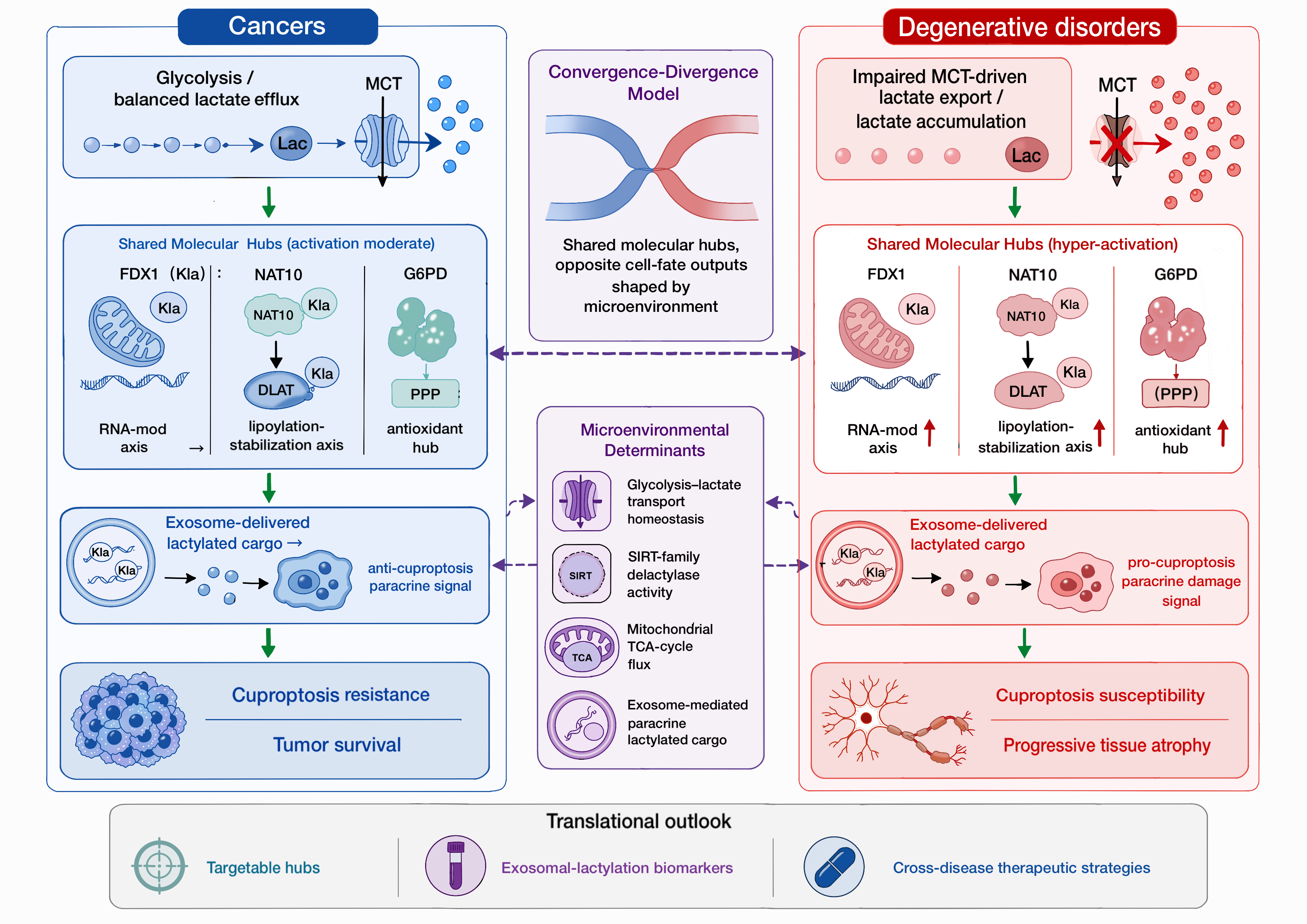
In this section, we describe an integrated framework for the three core molecular axes, which can be used to describe cancers and degenerative disorders. In particular, the same core molecular axes can produce completely opposite pathological outcomes, depending on four key microenvironmental determinants. The first cross-disease determinant is the glycolysis–lactate transport homeostasis that in turn determines the right substrate for modification within the FDX1-centered RNA regulatory hub (Figure 6). In solid tumors, within the FDX1-centered RNA regulatory hub for example, there is a bias towards NUDT21-mediated lactylation to repress FDX1 expression due to high glycolytic rate and high MCT4-mediated lactate transport [18]. In contrast, in degenerative diseases, there is impaired glycolytic capacity and down-regulation of MCT1/MCT4-mediated lactate transporters. This leads to toxic levels of intracellular lactate that shift the FDX1-centered RNA regulatory hub towards high levels of METTL16-mediated lactylation and high levels of FDX1 expression [8]. Thus, the glycolysis–lactate transport homeostasis is the primary upstream determinant of the divergent disease phenotypes between the two classes of diseases [31]. SIRT-family delactylases are the second cross-disease variable. While in tumors SIRT1/3 enzymes are in a balance with the lactate level, preventing NAT10 from being lactylated (see above) and thus preventing high levels of lipoylated DLAT to be present in degenerative cells, causing high levels of cuproptosis in these cells [13]. Thus, the NAT10-DLAT axis is suppressed in tumor cells but highly activated in degenerative cells [13]. The third variable for cross-disease regulation of shared convergent axes refers to the TCA-cycle flux in the mitochondria. This variable is a hub-dependent cell fate determinant, and it determines whether tumor cells or degenerative cells will die of cuproptosis [32]. Cancer cells with high rates of glycolysis have a partially down-regulated TCA-cycle. This results in low levels of lipoylated FDX1 and DLAT protein substrates, which, when processed by delactylases, do not give rise to toxic amounts of copper for the tumor cells, as the combined antioxidant capacity of the three convergent hubs of the core molecular axis is sufficient to counteract this toxicity [15,24]. However, in degenerative cells, the TCA-cycle is up-regulated and, consequently, high levels of lipoylated protein substrates are generated. Importantly, the combined antioxidant capacity of the three convergent hubs of the core molecular axis is not sufficient to counteract the toxicity of the processing of the lipoylated protein substrates by the delactylases. Therefore, in degenerative cells, cuproptosis can become a high-mitochondrial-mass based form of cell death [33]. A fourth cross-disease determinant that affects the same three cross-disease convergent hubs to produce different disease-specific divergent downstream branches is exosome-mediated paracrine transfer of lactylated cargo [16]. Senescent, ischemic or stressed cancer cells in tumors package in exosomes histones, and other proteins, such as Tau, METTL16, and HSPA family members, that have been lactylated [3,22,30]. These exosomes are then transferred to other cells within the same tumor or to endothelial cells in distant organs, thus establishing anti-cuproptosis signals in tissues containing these tumors. In contrast, in degenerative diseases, exosomes carry the same type of pathological lactylated cargo that, after transfer to adjacent parenchymal cells, can induce cell death in neurodegenerative, cardiovascular, and renal diseases, leading to atrophy of affected tissues [22,31,32,33,34]. In summary, exosome transfer of lactylated cargo between cells is an evolutionarily conserved intercellular bridge that translates intracellular lactylation imbalances into corresponding copper-dependent cell-death responses at the tissue level. In summary, four external factors, determined by the three convergent axes of intracellular regulation and by exosome-mediated intercellular signaling, can give rise to opposite decisions regarding cell fate in tumors versus in degenerative diseases, thus setting up a cross-disease theoretical framework that applies to all tumors as well as to all degenerative diseases of aging [28].
4.2. Hierarchical Cross-Disease Therapeutic Strategies Targeting Shared Convergent Hubs
Therapeutic strategies for the three molecular hubs, FDX1-related RNA regulation, NAT10-DLAT axis and G6PD-PPP antioxidant pathway, are presented for two different disease groups, cancers and degenerative diseases. Both disease groups display fundamentally different lactylation-dependent pathological features that need to be reversed by distinct intervention strategies [25].
In summary, tumor cells that have been protected by lactylation of tumor cell proteins can now be reversed and the cells induced to die by cuproptosis by anti-tumor strategies that affect the homeostasis of the glycolysis-lactate transport pathway. These strategies affect FDX1-related RNA regulation, the NAT10-DLAT axis, and the G6PD-PPP-antioxidant pathway to cause multiple anti-tumor effects by disrupting the G6PD-driven-antioxidant-copper-buffering in malignant cells [25,29]. The anti-tumor strategies that affect the homeostasis of the glycolysis-lactate transport pathway would be particularly effective for the NUDT21-lactylation-signature positive subset of esophageal squamous cell carcinomas that can be treated with a combination of LDHA- and HDAC2-inhibitors, plus elesclomol. For the hepatocellular carcinomas with balanced NUDT21/METTL16 profiles, in addition to the inhibition of NUDT21-lactylation, inhibition of METTL16, of lactate-transport and of copper-delivery would be required [11,15].
In addition to reversing tumor-protective lactylation, hyper-lactylation and its consequences, including cuproptosis of a variety of cell types, can be prevented and reversed. Inhibiting abnormal exosomal protein lactylation in core disease hubs, and in tissues affected by disease, upregulating the expression of MCT transporters by SIRT2/3 to decrease the concentration of toxic lactate, and to decrease the levels of FDX1 and DLAT mitochondrial proteins to decrease mitochondrial copper sensitivity, can prevent and reverse hyper-lactylation [12,19,21,28]. Tissue-specific strategies are likely to be required to treat the various subtypes of degenerative diseases. Thus, preventing abnormal lactylation of NAT10 by hyper-lactylation in chondrocytes, for example, by upregulating MCT transporters, even cartilage-specific MCT modulators, to prevent osteoarthritis, is likely to require a cartilage-specific strategy [9]. In addition to preventing and reversing hyper-lactylation, low-dose copper chelators can be used as adjuvants to prevent and reverse hyper-lactylation. Exosomal lactylated proteins in core disease hubs can serve as biomarkers for disease risk stratification by liquid-biopsy-based disease risk assessment [33]. Levels of exosomal lactylated proteins (SRLPs) can be measured using SERS-based microfluidic detection platforms to predict individual disease risk at early stages of the disease [25,34].
4.3. Unresolved Cutting-Edge Cross-Disciplinary Research Directions
There are several critical knowledge gaps in the cross-disease research area of pan-metabolic modulators for cancer prevention that need to be addressed. First, most pan-metabolic modulators lack tissue selectivity [28]. Therefore, most non-selective interventions for multiple organ systems will have off-target effects. Importantly, inter-tissue signal propagation by exosomal, lactylated cargo as well as effects on cellular cuproptosis sensitivity have not yet been fully characterized [36]. Notably, a defined threshold for lactate concentration that distinguishes between fluctuating, physiologic concentrations and pathologic, toxic concentrations does not yet exist [37,38]. To develop precise, therapeutic interventions for the lactylation–cuproptosis signaling pathway, both mechanistic as well as translational studies are required to address the several knowledge gaps identified for the cross-disease research area of pan-metabolic modulators [27,38].
5. Conclusions
In this review, we review current evidence linking lysine lactylation to cuproptosis in human cancer and in age-related multisystem degenerative disorders. We have developed a unified convergence-divergence framework for the three conserved molecular hubs (FDX1-centered RNA-modification, NAT10-DLAT-mediated lipoylation-stabilization and G6PD-driven pentose-phosphate-pathway antioxidant copper-buffering) that in a unified post-translational control fashion determine levels of cuproptosis-sensitive modified proteins in cancer versus degenerative lesion cells. While core intracellular signaling pathways of the three hubs are similar in cancer and degenerative disease cells, four tumor versus degenerative lesion microenvironment determinants produce identical molecular machinery to yield opposite cell type specific outcomes. Thus, in cancer cells, moderate lactate production and efficient efflux of excess lactate protects tumor cells from cuproptosis whereas in degenerative parenchymal cells, sustained intracellular lactate accumulation triggers excessive hub activation leading to cuproptosis-mediated progressive atrophy of affected tissues.
While cancer studies are mainly performed within the frame of single cancer types or even in different models for studies on degenerative disorders, here for the first time data from individual studies on lactylation and cuproptosis were integrated in cross-disease comparative studies. Thus, for the first time cross-disease comparative studies can be conducted on studies on different types of cancer as well as on studies on different degenerative disorders in order to derive new cancer-type-specific as well as disease-type-specific concepts for the development of specific, so-called “precision” therapies. In contrast to cancer studies on degenerative disorders are focused on the excessive hyper-lactylation of proteins and the excessive cuproptosis-induced tissue damage, which have to be curbed. We focused on hub molecules shared by different signaling pathways. The exosomal proteins modified by lactation can be used as early biomarkers of various diseases in liquid biopsy. Although the existing evidence collated based on this review has the feasibility of carrying out cross disease research, there are still many reasons that need more experimental data as support. Therefore, more studies within more tumor types as well as within more degenerative diseases are required for cross-disease comparative studies. Also, the complete characterization of the signaling networks that are lactylation-regulated within several tumor types as well as within several degenerative diseases is a task for future studies. Several apparently contrasting views on similar issues were presented by the individual studies reviewed here. These discrepancies have to be clarified in more detail within future studies. Most of the therapeutic approaches that were suggested within the individual studies reviewed here were tested within in-vitro systems as well as within preclinical animal models. However, these types of studies are severely hampered by the problem of unspecific tissue distribution. Therefore, reliable concentration thresholds of intracellular as well as of extracellular lactate have to be established within future studies in order to distinguish between adaptive, physiological levels of lactate and toxic, pathological levels of lactate.
Testing the outlined framework in cross-disease studies will be necessary in future. Sensitive detection systems for exosomal lactylation-related biomarkers as well as improved tissue-specificity of current metabolic modulators will be required for these studies. The major challenge that remains to be addressed is to determine the effects of exosome-mediated lactylation signal propagation in the system. As this review highlighted the key role of post-translational modification by metabolites in the control of copper-dependent cell death, studies from the fields of oncology and geriatric metabolic medicine will be required.
Author Contributions
Conceptualization, X.W. and S.C.; literature retrieval and data integration, X.K.; manuscript drafting, X.W.; critical revision, S.C.; figure and table design, X.K. All authors read and approved the final submitted manuscript. All authors have read and agreed to the published version of the manuscript.” Please turn to the CRediT taxonomy for the term explanation. Authorship must be limited to those who have contributed substantially to the work reported.
Funding
This research received no external funding support.
Institutional Review Board Statement
None.
Informed Consent Statement
None.
Data Availability Statement
None.
Acknowledgments
Thank you to all the researchers whose studies we reviewed for gathering evidence for this review. We would also like to thank the editors of this journal and the anonymous reviewers for guidance on the review and for their very constructive comments on the review.
AI Availability Statement
All the schematic figures (Figure 1, Figure 2 and Figure 3) were initially generated in the online platform Picdoc.cn during the manuscript preparation. The raw AI-generated drafts were completely and manually rewritten, redrawn and verified by the authors for accuracy of the molecular representations, transporter identities and the corresponding cuproptosis signaling cascades as presented in the relevant literature. The authors fully and completely endorse and take responsibility for the graphical figures presented in this manuscript for their correctness, integrity and interpretation.
Conflicts of Interest
No conflicts of interest were declared.
Abbreviations
Abbreviations used in this manuscript are as follows:
| AKI | Acute Kidney Injury |
| ALS | Amyotrophic Lateral Sclerosis |
| AD | Alzheimer’s Disease |
| AARS1 | Alanyl-tRNA Synthetase 1 |
| CAFs | Cancer-Associated Fibroblasts |
| CSF | Cerebrospinal Fluid |
| CPSF6 | Cleavage And Polyadenylation Specific Factor 6 |
| DLAT | Dihydrolipoamide S-Acetyltransferase |
| ESCC | Esophageal Squamous Cell Carcinoma |
| FDX1 | Ferredoxin 1 |
| G6PD | Glucose-6-Phosphate Dehydrogenase |
| GSH | Glutathione |
| HCC | Hepatocellular Carcinoma |
| HSPA6 | Heat Shock Protein Family A (Hsp70) Member 6 |
| Kla | Lysine Lactylation |
| LDHA | Lactate Dehydrogenase A |
| LIAS | Lipoic Acid Synthetase |
| LSD | Lysine Specific Demethylase |
| MCT1/4 | Monocarboxylate Transporter 1/4 |
| METTL16 | Methyltransferase Like 16 |
| NADPH | Nicotinamide Adenine Dinucleotide Phosphate |
| NAT10 | N-Acetyltransferase 10 |
| NUDT21 | Nudix Hydrolase 21 |
| OA | Osteoarthritis |
| PPP | Pentose Phosphate Pathway |
| RMD-2OGD | RNA:2-Oxoglutarate-Dependent Dioxygenase |
| SIRT1/2/3 | Sirtuin 1/2/3 |
| SOD2 | Superoxide Dismutase 2 |
| TCA | Tricarboxylic Acid Cycle |
| TNBC | Triple-Negative Breast Cancer |
| TFRC | Transferrin Receptor |
| Tau | Microtubule Associated Protein Tau |
| 3’UTR | 3’ Untranslated Region |
References
- Zhang, D.; Tang, Z.; Huang, H.; et al. Metabolic regulation of gene expression by histone lactylation. Nature 2019, 574, 575–580. [Google Scholar] [CrossRef] [PubMed]
- Tsvetkov, P.; Coy, S.; Petrova, B.; et al. Copper induces cell death by targeting lipoylated TCA cycle proteins. Science 2022, 375, 1254–1261. [Google Scholar] [CrossRef] [PubMed]
- Li, K.; Wei, Y.; Li, Y.; Dong, Y.; et al. EP300-Mediated MTF1-K218 Lactylation Buffers AR-Driven Copper Overload to Suppress Cuproptosis in Castration-Resistant Prostate Cancer. Adv. Sci. 2026, e77110. [Google Scholar] [CrossRef] [PubMed]
- Hadian, K.; Stockwell, B.R. The therapeutic potential of targeting regulated non-apoptotic cell death. Nat. Rev. Drug Discov. 2023, 22, 723–742. [Google Scholar] [CrossRef] [PubMed]
- Sui, M.; Fu, J.; Li, Y.; et al. Lactate promotes cuproptosis in acute kidney injury by activating H3K18 lactylation-dependent upregulation of HSPA6 expression. Life Sci. 2026, 389, 124244. [Google Scholar] [CrossRef] [PubMed]
- Zhu, B.; Wang, S.; Wang, R.; et al. Identification of molecular subtypes and a six-gene risk model related to cuproptosis for triple negative breast cancer. Front. Genet. 2022, 13, 1022236. [Google Scholar] [CrossRef] [PubMed]
- Zhu, Y.; Lei, C.; Jiang, Q.; et al. DSF/Cu induces antitumor effect against diffuse large B-cell lymphoma through suppressing NF-κB/BCL6 pathways. Cancer Cell Int. 2022, 22, 236. [Google Scholar] [CrossRef] [PubMed]
- Sun, L.; Zhang, Y.; Yang, B.; et al. Lactylation of METTL16 promotes cuproptosis via m⁶A-modification on FDX1 mRNA in gastric cancer. Nat. Commun. 2023, 14, 6523. [Google Scholar] [CrossRef] [PubMed]
- Nong, J.; Lu, G.; Huang, Y.; et al. Identification of cuproptosis-related subtypes, characterization of immune microenvironment infiltration, and development of a prognosis model for osteoarthritis. Front. Immunol. 2023, 14, 1178794. [Google Scholar] [CrossRef] [PubMed]
- Di, H.; Wang, X.; Hao, M.; et al. Mutational and transcriptional profiling of cuproptosis-associated genes in amyotrophic lateral sclerosis. Genes Dis. 2024, 12, 101208. [Google Scholar] [CrossRef] [PubMed]
- Yang, M.; Wang, Y.; He, L.; et al. Comprehensive bioinformatics analysis reveals the role of cuproptosis-related gene Ube2d3 in myocardial infarction. Front. Immunol. 2024, 15, 1353111. [Google Scholar] [CrossRef] [PubMed]
- Lin, J.; Yin, Y.; Cao, J.; et al. NUDT21 lactylation reprograms alternative polyadenylation to promote cuproptosis resistance. Cell Discov. 2025, 11, 52. [Google Scholar] [CrossRef] [PubMed]
- Yang, W.D.; Lu, M.R.; Shen, Q.; et al. Lactylated NAT10 contributes to elesclomol-triggered cuproptosis via the NAT10/ac4C-DLAT-mRNA/DLAT positive feedback loop in CRC. Proc. Natl. Acad. Sci. U.S.A. 2026, 123, e2501185123. [Google Scholar] [CrossRef] [PubMed]
- Zhang, X.; Yuan, Y.; Tang, L.; et al. Epigenetic regulation of cuproptosis in cancer: mechanisms, microenvironment, and therapeutic implications. Front. Cell Dev. Biol. 2026, 14, 1814142. [Google Scholar] [CrossRef] [PubMed]
- D'Agnelli, S.; Gerra, M.C.; Bignami, E.; et al. Exosomes as a new pain biomarker opportunity. Mol. Pain. 2020, 16, 1744806920957800. [Google Scholar] [CrossRef] [PubMed]
- Sun, H.; Zou, Y.; Chen, Z.; et al. Nanodrug-Engineered Exosomes Achieve a Jointly Dual-Pathway Inhibition on Cuproptosis. Adv. Sci. 2025, 12, e2413408. [Google Scholar] [CrossRef] [PubMed]
- Xu, S.; Zeng, Y.; Tan, X.; et al. Targeted Delivery of Exosome-Derived miRNA-185-5p Inhibitor via Liposomes Alleviates Apoptosis and Cuproptosis in Dilated Cardiomyopathy. Int. J. Nanomed. 2025, 20, 9407–9425. [Google Scholar] [CrossRef] [PubMed]
- Li, L.; Wang, Y.; Tian, R.; et al. TRIM21-Mediated K11-Linked Ubiquitination of ID1 Suppresses Tumorigenesis and Promotes Cuproptosis in Esophageal Squamous Cell Carcinoma. Adv. Sci. 2025, 12, e02501. [Google Scholar] [CrossRef] [PubMed]
- Yao, L.; Ye, S.; Xu, D. UCHL3 augments cuproptosis via PKM2 deubiquitination in hepatocellular carcinoma. Free Radic. Biol. Med. 2025, 237, 65–75. [Google Scholar] [CrossRef] [PubMed]
- Sun, Z.; Xu, H.; Lu, G.; et al. AKT1 Phosphorylates FDX1 to Promote Cuproptosis Resistance in Triple-Negative Breast Cancer. Adv. Sci. 2025, 12, e2408106. [Google Scholar] [CrossRef] [PubMed]
- Yang, R.; Deng, F.; Gao, Z.; et al. Melanoma MHC-I-membrane-encapsulated Cu@ferrihydrite induces ferroptosis/cuproptosis and systematic immunity against tumor. J. Control. Release. 2025, 388, 114281. [Google Scholar] [CrossRef] [PubMed]
- Chen, S.; Chen, T.; Xu, C.; et al. Iron overload exaggerates renal ischemia-reperfusion injury by promoting tubular cuproptosis via interrupting function of LIAS. Redox Biol. 2025, 86, 103795. [Google Scholar] [CrossRef] [PubMed]
- Sun, P.; Chen, G.; Guo, Y. Regulatory mechanisms of age-related degenerative diseases: Insights from the gut microbiota-cellular senescence interaction network. Ageing Res. Rev. 2026, 119, 103152. [Google Scholar] [CrossRef] [PubMed]
- Huang, Y.; Zhao, E.; Zhao, G.; et al. H3K18 lactylation-mediated SPHK1-SIRT1 feedback loop accelerates pyroptosis of tubular epithelial cells in sepsis-associated acute kidney injury. Theranostics 2026, 16, 4768–4786. [Google Scholar] [CrossRef] [PubMed]
- Wang, Q.; Duan, Y.; Xu, Y.; et al. Linking Parkinson's disease and melanoma: the impact of copper-driven cuproptosis and related mechanisms. npj Park. Dis. 2025, 11, 74. [Google Scholar] [CrossRef] [PubMed]
- Faridar, A.; Gamez, N.; Li, D.; et al. Low-dose interleukin-2 in patients with mild to moderate Alzheimer's disease: a randomized clinical trial. Alzheimers Res. Ther. 2025, 17, 146. [Google Scholar] [CrossRef] [PubMed]
- Zhu, Z.; Song, M.; Ren, J.; et al. Copper homeostasis and cuproptosis in central nervous system diseases. Cell Death Dis. 2024, 15, 850. [Google Scholar] [CrossRef] [PubMed]
- Chen, X.; Cai, Q.; Liang, R.; et al. Copper homeostasis and copper-induced cell death in the pathogenesis of cardiovascular disease and therapeutic strategies. Cell Death Dis. 2023, 14, 105. [Google Scholar] [CrossRef] [PubMed]
- Fu, X.; Chen, B.X.; Wang, J.; et al. Nitroxyl relieves acute kidney injury by suppressing SLC31A1-mediated cuproptosis in renal tubular epithelial cells. Life Sci. 2026, 401, 124502. [Google Scholar] [CrossRef] [PubMed]
- Jiang, Z.; Li, J.; Shi, M.; et al. Hypoxia, cuproptosis, and osteoarthritis: Unraveling the molecular crosstalk. Redox Biol. 2025, 85, 103757. [Google Scholar] [CrossRef] [PubMed]
- Guan, Y.; Bai, D.; Bu, N.; et al. Programming regulated cell death to engineer whole-tumor-cell vaccines for cancer immunotherapy. Biochim. Biophys. Acta Rev. Cancer. 2026, 1881, 189640. [Google Scholar] [CrossRef] [PubMed]
- Lu, K.; Wijaya, C.S.; Yao, Q.; et al. Cuproplasia and cuproptosis, two sides of the coin. Cancer Commun. 2025, 45, 505–524. [Google Scholar] [CrossRef] [PubMed]
- Zhu, G.; Xie, Y.; Wang, J.; et al. Multifunctional Copper-Phenolic Nanopills Achieve Comprehensive Polyamines Depletion to Provoke Enhanced Pyroptosis and Cuproptosis for Cancer Immunotherapy. Adv. Mater. 2024, 36, e2409066. [Google Scholar] [CrossRef] [PubMed]
- Zhou, S.; Liu, J.; Wan, A.; et al. Epigenetic regulation of diverse cell death modalities in cancer: a focus on pyroptosis, ferroptosis, cuproptosis, and disulfidptosis. J. Hematol. Oncol. 2024, 17, 22. [Google Scholar] [CrossRef] [PubMed]
- Schneider, L.; Chalmers, D.; O'Beirn, S.; et al. Premorbid beta blockade in sepsis is associated with a lower risk of a lactate concentration above the lactate threshold, a retrospective cohort study. Sci. Rep. 2022, 12, 20843. [Google Scholar] [CrossRef] [PubMed]
- Zhao, P.; Wang, Z.; Liu, K.; et al. A self-amplifying cuproptosis nanomedicine to overcome immunosuppression by blocking tumor-derived exosomes for enhancing lung cancer immunotherapy. J. Control. Release. 2026, 391, 114593. [Google Scholar] [CrossRef] [PubMed]
- Cao, L.; Li, P.; Ma, Y.; et al. Adipose-derived mesenchymal stem cell exosomes ameliorate copper metabolism dysregulation and reduce cuproptosis caused by liver IRI. Front. Vet. Sci. 2026, 13, 1895340. [Google Scholar] [CrossRef] [PubMed]
- Chi, C.; Xu, M.; Zhang, J.; et al. CAF-Secreted Exosomes Deliver BMP4 to Confer Radiotherapy Resistance in Cervical Cancer Through a Novel Mechanism Linking Nrf2 Activation to Cuproptosis Inhibition. FASEB J. 2026, 40, e71838. [Google Scholar] [CrossRef] [PubMed]
Figure 1.
Schematic overview of molecular hubs linking lactylation and cuproptosis, as well as exosome-mediated paracrine transmission of lactylated cargo.
Figure 1.
Schematic overview of molecular hubs linking lactylation and cuproptosis, as well as exosome-mediated paracrine transmission of lactylated cargo.
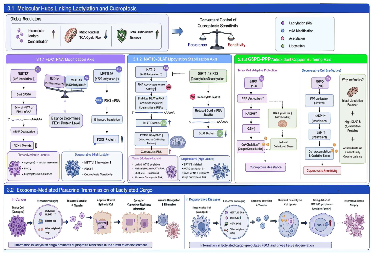
Figure 2.
Disease-specific profiles of lactylation-driven cuproptosis regulatory hubs across multiple human malignancies versus degenerative diseases.
Figure 2.
Disease-specific profiles of lactylation-driven cuproptosis regulatory hubs across multiple human malignancies versus degenerative diseases.
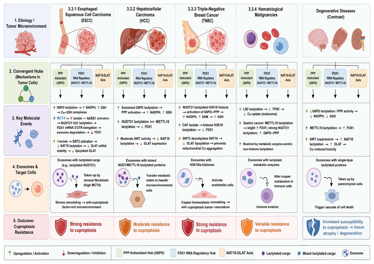
Figure 3.
Subtype-specific divergent mechanisms of lactylation-driven cuproptosis across degenerative diseases.
Figure 3.
Subtype-specific divergent mechanisms of lactylation-driven cuproptosis across degenerative diseases.
Disclaimer/Publisher’s Note: The statements, opinions and data contained in all publications are solely those of the individual author(s) and contributor(s) and not of MDPI and/or the editor(s). MDPI and/or the editor(s) disclaim responsibility for any injury to people or property resulting from any ideas, methods, instructions or products referred to in the content. |
© 2026 by the authors. Licensee MDPI, Basel, Switzerland. This article is an open access article distributed under the terms and conditions of the Creative Commons Attribution (CC BY) license (http://creativecommons.org/licenses/by/4.0/).
Copyright: This open access article is published under a Creative Commons CC BY 4.0 license, which permit the free download, distribution, and reuse, provided that the author and preprint are cited in any reuse.