

Submitted:
01 September 2026
Posted:
02 September 2026
You are already at the latest version
Abstract
Oxidative stress is a key regulator of epidermal homeostasis, inflammation, and wound repair. In pediatric skin, where epidermal barrier function, innate immunity, and antioxidant defenses continue to mature after birth, disruption of physiological redox homeostasis may increase susceptibility to infection and impaired wound healing. Rather than representing simply an excess of reactive oxygen species, oxidative stress reflects a disruption of redox homeostasis that reprograms keratinocyte function. However, the mechanisms linking these alterations to epidermal barrier failure and microbial infection have not been synthesized into a unified mechanistic framework. This review examines how oxidative stress alters intracellular signaling networks in keratinocytes, with particular emphasis on the NRF2, MAPK, NF-κB, and PI3K–Akt–mTOR pathways. We discuss how dysregulation of these pathways impairs tight junctions, intercellular adhesion complexes, cornified envelope formation, epidermal lipids, antimicrobial peptides, and the skin microbiota, thereby promoting barrier dysfunction, microbial dysbiosis, and infection. We further summarize the unique developmental characteristics of pediatric skin and their implications for wound healing, chronic inflammation, and fibrosis. Finally, we review emerging therapeutic strategies that target restoration of physiological redox homeostasis through modulation of antioxidant signaling, epidermal barrier repair, innate immunity, and the skin microbiota. Collectively, available evidence supports oxidative stress–driven keratinocyte dysfunction as an integrative mechanistic framework linking epidermal barrier failure with infection in pediatric skin and provides a basis for future redox-targeted therapeutic approaches.
Keywords:
keratinocyte
; oxidative stress
; redox homeostasis
; pediatric skin
; epidermal barrier
; skin infection
; wound healing
; reactive oxygen species
1. Introduction
Pediatric skin serves as the first line of defense against environmental insults and microbial invasion, relying on a highly coordinated epidermal barrier to maintain epidermal homeostasis. Disruption of this barrier is a hallmark of numerous pediatric conditions, including atopic dermatitis (AD), traumatic wounds, burn injuries, and surgical site complications, where impaired barrier integrity substantially increases the risk of microbial colonization and infection. Beyond its immediate impact on wound healing and disease severity, barrier failure contributes to prolonged inflammation, repeated tissue injury, increased healthcare utilization, and reduced quality of life in affected children. Despite advances in antimicrobial therapy and wound management, infectious complications remain a significant clinical challenge, underscoring the need for a deeper understanding of the cellular and molecular mechanisms that preserve epidermal integrity and host defense during skin injury and inflammation.
Considerable progress has been made in elucidating the molecular mechanisms governing epidermal barrier formation, cutaneous immunity, and host–microbe interactions. Accumulating evidence has further established oxidative stress as a critical regulator of inflammation, wound healing, and skin homeostasis. However, these areas have largely evolved as parallel fields of investigation, with relatively limited integration of redox biology, keratinocyte function, epidermal architecture, and infection susceptibility into a unified mechanistic framework. This gap is particularly evident in pediatric skin, where epidermal barrier function and cutaneous immunity continue to mature during postnatal development. Compared with adult skin, the developing epidermis is characterized by a thinner stratum corneum, an immature skin microbiota, evolving lipid composition, and immature antioxidant and immune defenses, all of which may modify redox homeostasis, keratinocyte function, and susceptibility to infection. Consequently, the mechanisms by which oxidative stress interacts with the developing epidermis to orchestrate keratinocyte dysfunction, barrier failure, and microbial colonization remain incompletely understood.
Addressing this knowledge gap requires a better understanding of keratinocyte biology. Keratinocytes, the predominant cell type of the epidermis, function not only as structural components of the skin barrier but also as active regulators of innate immunity and tissue repair. They establish the physical barrier through the coordinated formation of tight junctions (TJ), cell–cell adhesion complexes, and the cornified envelope (CE) [1,2], while simultaneously regulating innate immune responses through the production of antimicrobial peptides (AMPs) and the secretion of inflammatory cytokines [3]. Together, these complementary functions preserve epidermal homeostasis while supporting host defense against microbial infection and facilitating wound repair.
In addition to external factors such as mechanical injury, hypoperfusion/reperfusion, and Ultraviolet (UV) exposure, superimposed inflammatory stimulation induces the generation of reactive oxygen species (ROS) in keratinocytes [4]. ROS function as essential signaling molecules that regulate cell proliferation, differentiation, wound healing, and immune responses [4,5]. However, when ROS production exceeds the capacity of endogenous antioxidant defenses, oxidative stress is triggered, resulting in oxidative damage to lipids, proteins, and DNA, together with disruption of redox signaling homeostasis [6]. Growing evidence indicates that dysregulation of these redox signaling pathways have functional consequences extending beyond intracellular stress responses, ultimately compromising multiple components of the epidermal barrier [7,8].
Recent mechanistic studies have demonstrated that oxidative stress not only disrupts the tight-junction constituent proteins claudin-1/4, occludin, and ZO-1 [9,10], but also exerts broad effects on adherens-junction components such as E-cadherin, desmosomes, and hemidesmosomes, as well as on CE formation, which is composed of involucrin, loricrin, and small proline-rich proteins (SPRR) [11]. Moreover, ROS-mediated peroxidation of cutaneous surface lipids and dysregulated AMP expression have been suggested to alter the composition and function of the skin microbiota, thereby promoting colonization by pathogenic microorganisms and the establishment of infection [12,13]. Accordingly, oxidative stress is increasingly recognized not merely as a source of cellular damage but as a central regulator of keratinocyte dysfunction that integrates the pathological cascade of barrier architecture disruption, microbial dysbiosis, and infection.
Pediatric skin should not simply be regarded as a miniature version of adult skin but rather as a dynamically developing biological system in which epidermal barrier function, cutaneous immunity, antioxidant defenses, and microbial communities continue to mature after birth [14,15]. These developmental characteristics may modify redox homeostasis and keratinocyte responses to oxidative stress, thereby increasing susceptibility to barrier dysfunction, microbial colonization, infection, delayed wound healing, and pathological scarring [16]. Despite these unique biological features, mechanistic studies directly comparing redox regulation in pediatric and adult skin remain limited.
This review examines how oxidative stress disrupts keratinocyte function and thereby links epidermal barrier failure with microbial dysbiosis, infection, and impaired wound healing in pediatric skin. Specifically, we first summarize the major sources of ROS and the signaling pathways governing keratinocyte redox responses. Subsequently, we examine how redox imbalance compromises TJ, cell adhesion structures, CE formation, antimicrobial peptide function, and skin microbiota, ultimately promoting barrier dysfunction and infection. Finally, we discuss the unique developmental characteristics of pediatric skin, oxidative stress in wounds and burns, and innovative redox-targeted therapeutic strategies.
2. Sources and Triggers of ROS in Keratinocytes
Because keratinocytes reside at the interface between the host and the external environment, they are continuously exposed to a wide variety of endogenous and exogenous stimuli that induce intracellular ROS generation. Understanding these sources of oxidative stress provides the foundation for appreciating how redox imbalance initiates keratinocyte dysfunction and subsequent epidermal barrier disruption.
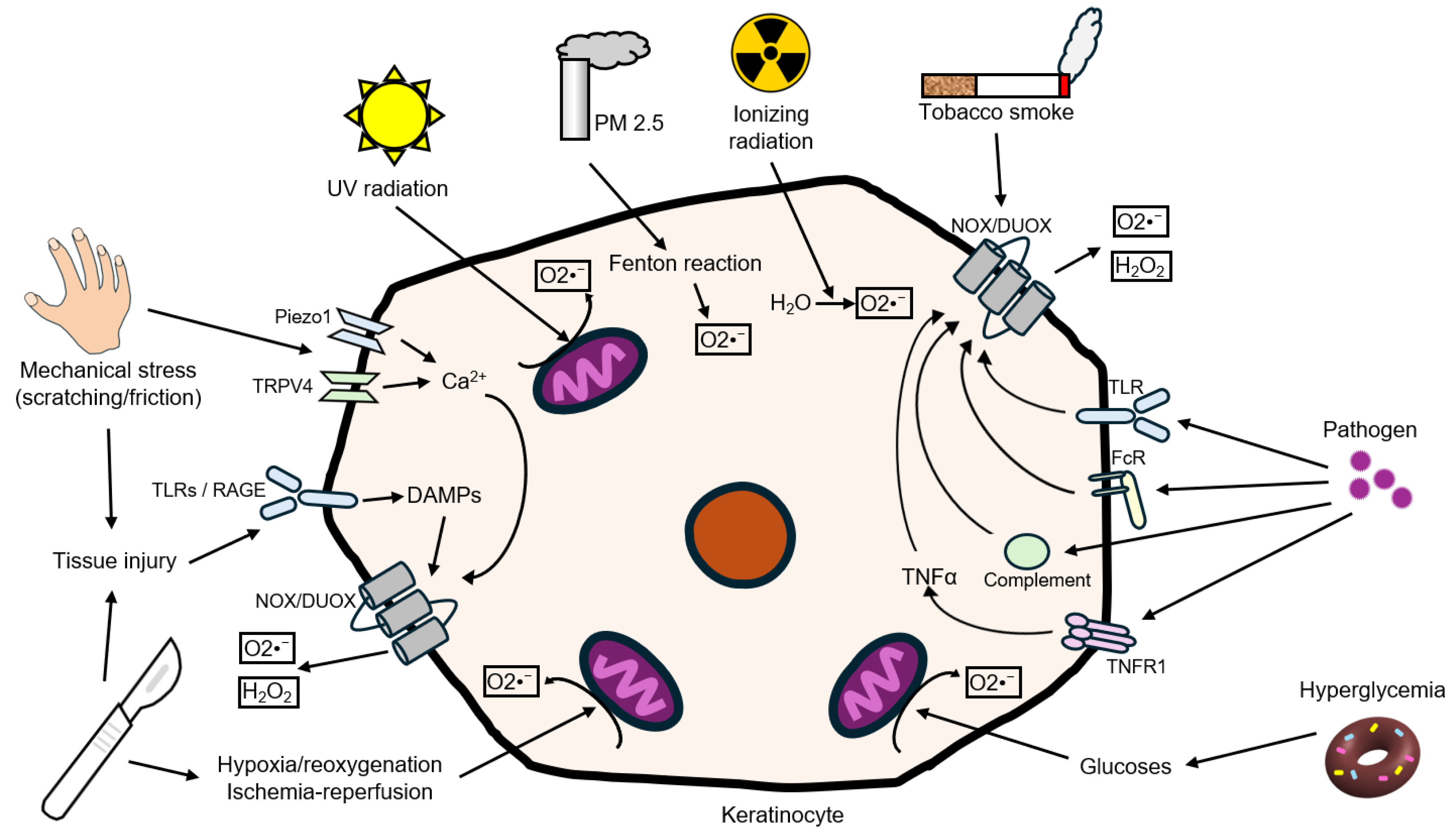
Environmental factors constitute major initiators of ROS production in keratinocytes. UV radiation induces mitochondrial ROS generation in human keratinocytes [17], whereas ionizing radiation directly generates free radicals through radiolysis of water molecules [18]. Particulate matter (PM) promotes redox cycling through Fenton reactions mediated by transition metals such as copper and iron (Cu/Fe), thereby enhancing ROS generation and oxidative stress [19,20]. Similarly, tobacco smoke activates keratinocyte NADPH oxidase (NOX), leading to increased intracellular ROS production [21]. During infection and inflammation, ROS generation is further amplified through activation of enzymatic oxidant-producing systems. Pathogen invasion stimulates NOX1 and dual oxidase 1 via Toll-like receptors (TLR2/4) [22,23], complement/Fc receptors [24], and pro-inflammatory cytokines such as TNF-α and IL-1β [25], representing major mechanisms of ROS generation in keratinocytes [26].
Tissue injury itself represents an additional source of redox signaling through the release of damage-associated molecular patterns (DAMPs) from injured or necrotic cells. DAMPs such as high-mobility group box 1 (HMGB1), extracellular nucleic acids, and ATP activate pattern-recognition receptors, including Toll-like receptors, thereby promoting inflammatory signaling and ROS generation within the wound microenvironment. This mechanism may be particularly relevant following traumatic or surgical injury, in which cellular disruption precedes and amplifies local oxidative and inflammatory responses.
Likewise, hypoxia/reoxygenation and ischemia–reperfusion associated with surgical wounds trigger rapid bursts of mitochondrial ROS originating from complexes I and III of the electron transport chain [27]. Mechanical stress, including scratching, activates mechanosensitive channels such as Piezo1 and TRPV4, resulting in intracellular Ca²⁺ influx [28,29], which subsequently promotes DUOX1-dependent ROS generation [26]. Although direct evidence in keratinocytes remains limited, studies in other epithelial and mesenchymal cells suggest that friction-associated increases in integrin tension amplify NOX-dependent oxidative stress, indicating that a similar mechanism may contribute to barrier disruption in keratinocytes [30].
Metabolic disorders also contribute substantially to oxidative stress. Hyperglycemia, through advanced glycation end-product (AGE)–receptor for advanced glycation end-products (RAGE) signaling, increases the intracellular ROS burden in keratinocytes within diabetic wounds while impairing keratinocyte migration and differentiation [31,32].
Although these stimuli differ considerably in origin and mechanism, they converge on a common biological outcome: disruption of keratinocyte redox homeostasis. The magnitude, duration, and subcellular localization of ROS production ultimately determine whether redox signaling promotes physiological adaptation or progresses to oxidative stress, keratinocyte dysfunction, and epidermal barrier impairment. These mechanisms may be particularly relevant to pediatric skin, in which epidermal barrier function and antioxidant defenses continue to mature after birth [14,15,16]. However, because most mechanistic studies of keratinocyte ROS generation have been performed using primary keratinocytes of varying donor origin, immortalized keratinocyte lines, or experimental animal models, whether the magnitude and regulation of these ROS-generating pathways differ across developmental stages remains poorly defined. This convergence provides the mechanistic basis for the redox-sensitive signaling pathways discussed in the following section.
Figure 1.
Major endogenous and exogenous sources of ROS in keratinocytes during skin infection, inflammation, and tissue injury. Keratinocytes generate ROS from multiple endogenous and environmental sources. Endogenous ROS arise primarily from mitochondrial electron transport, NADPH oxidases (NOX1 and DUOX1/2), inflammatory receptor signaling, and Fenton chemistry. Environmental and pathological stimuli—including UV radiation, ionizing radiation, PM2.5, tobacco smoke, mechanical or tissue injury, ischemia–reperfusion, microbial infection, and hyperglycemia—further enhance intracellular ROS production. Tissue injury and cell damage promote the release of damage-associated molecular patterns (DAMPs), including HMGB1, extracellular ATP, and nucleic acids, which activate pattern-recognition receptors such as TLRs and RAGE and amplify inflammatory and ROS-generating pathways. In parallel, mechanical stress activates mechanosensitive ion channels such as Piezo1 and TRPV4, promoting Ca²⁺ influx and DUOX activation. Excessive ROS production disrupts redox homeostasis and initiates signaling pathways that contribute to keratinocyte dysfunction, impaired barrier integrity, inflammatory responses, and tissue injury.
Figure 1.
Major endogenous and exogenous sources of ROS in keratinocytes during skin infection, inflammation, and tissue injury. Keratinocytes generate ROS from multiple endogenous and environmental sources. Endogenous ROS arise primarily from mitochondrial electron transport, NADPH oxidases (NOX1 and DUOX1/2), inflammatory receptor signaling, and Fenton chemistry. Environmental and pathological stimuli—including UV radiation, ionizing radiation, PM2.5, tobacco smoke, mechanical or tissue injury, ischemia–reperfusion, microbial infection, and hyperglycemia—further enhance intracellular ROS production. Tissue injury and cell damage promote the release of damage-associated molecular patterns (DAMPs), including HMGB1, extracellular ATP, and nucleic acids, which activate pattern-recognition receptors such as TLRs and RAGE and amplify inflammatory and ROS-generating pathways. In parallel, mechanical stress activates mechanosensitive ion channels such as Piezo1 and TRPV4, promoting Ca²⁺ influx and DUOX activation. Excessive ROS production disrupts redox homeostasis and initiates signaling pathways that contribute to keratinocyte dysfunction, impaired barrier integrity, inflammatory responses, and tissue injury.
3. Major Redox-Sensitive Signaling Pathways in Keratinocytes
3.1. NRF2–ARE Pathway
The nuclear factor erythroid 2-related factor 2 (NRF2)–antioxidant response element (ARE) pathway is a major adaptive defense mechanism that enables keratinocytes to maintain antioxidant capacity and restore redox homeostasis during transient oxidative stress. Under basal conditions, the Kelch-like ECH-associated protein 1 (Keap1)–Cullin 3 (Cul3)–RING-box protein 1 (Rbx1) complex ubiquitinates NRF2, targeting it for proteasomal degradation [33]. In keratinocytes, oxidative stress modifies cysteine residues on Keap1, thereby suppressing ubiquitin-mediated degradation of NRF2 [34,35]. Stabilized NRF2 subsequently undergoes phosphorylation, including by extracellular signal-regulated kinase (ERK), and translocates into the nucleus [36]. There, NRF2 forms heterodimers with small Maf proteins, binds to AREs [37], and induces antioxidant and detoxification genes, including heme oxygenase-1 (HO-1), NAD(P)H:quinone oxidoreductase 1 (NQO1), glutathione S-transferase (GST), UDP-glucuronosyltransferase (UGT), catalase (CAT), and superoxide dismutase (SOD), thereby promoting redox homeostasis [38,39,40].
NRF2 activity is further regulated through an additional mechanism involving p62/SQSTM1-mediated autophagy. Phosphorylated p62/SQSTM1 binds Keap1 with high affinity and promotes its autophagic degradation [41,42], thereby reducing NRF2 degradation and enhancing NRF2 signaling. This mechanism links oxidative stress, autophagy, and antioxidant defense.
The magnitude of oxidative stress critically influences the protective capacity of the NRF2 pathway. Under moderate ROS conditions, nuclear accumulation of NRF2 progressively increases, whereas the induction of NFE2L2 mRNA, which encodes NRF2, remains relatively modest. In contrast, under excessive or sustained oxidative stress, NRF2 protein levels decline despite increased NFE2L2 mRNA expression [43,44]. Similar ROS intensity-dependent effects have been reported for downstream antioxidant genes. Many studies demonstrate reduced HO-1 mRNA expression under excessive or sustained oxidative stress, whereas findings for NQO1, GST, and glutamate-cysteine ligase remain inconsistent, with some reports showing decreased expression and others showing little or no change [45,46]. These discrepancies likely reflect differences in experimental models, ROS sources (e.g., H₂O₂, UVB, dtBHQ, and sulforaphane), exposure conditions, and the severity of oxidative stress across studies.
Collectively, NRF2–ARE signaling provides an adaptive response to transient oxidative stress, whereas oxidative stress exceeding NRF2 protective capacity shifts the cellular response toward inflammation, altered proliferation, autophagy, and cell death.
3.2. MAPK Pathway
The mitogen-activated protein kinase (MAPK) pathway is a major redox-sensitive signaling network that enables keratinocytes to translate oxidative stress into inflammatory responses, proliferation, differentiation, or apoptosis, depending on the magnitude and duration of ROS exposure. Among its three principal branches, ERK, c-Jun N-terminal kinase (JNK), and p38 MAPK play distinct yet coordinated roles in determining keratinocyte fate.
ROS activate MAP3K (mitogen-activated protein kinase kinase kinase) [46], initiating signaling through both the p38/JNK and ERK1/2 pathways [47]. Activated p38/JNK phosphorylates c-Jun and activating transcription factor 2 (ATF2), whereas ERK phosphorylates c-Fos [46,48,49]. These transcription factors assemble into the activator protein-1 (AP-1) complex, which binds to the TPA-response element (TRE) and induces the expression of inflammatory mediators, including MMP1 (matrix metalloproteinase 1) [50], IL-1, IL-6, IL-8, TNF [51,52], and cyclooxygenase-2 (COX-2) [53]. In addition to regulating inflammatory responses, AP-1 has also been implicated in controlling keratinocyte differentiation-associated proteins such as involucrin and filaggrin [54], highlighting the broad influence of MAPK signaling on epidermal homeostasis.
MAPK responses are strongly influenced by ROS intensity. Moderate oxidative stress preferentially favors ERK signaling associated with keratinocyte proliferation and survival [55], whereas severe or sustained ROS exposure increasingly activates p38/JNK stress signaling and apoptosis [46]. However, these pathways are not mutually exclusive and may be activated concurrently depending on cellular context.
It should also be noted that most published studies have investigated MAPK activation using UV irradiation as the primary oxidative stimulus. Accordingly, whether different ROS sources—including inflammatory, metabolic, or mechanical oxidative stress—activate MAPK signaling in an equivalent manner remains incompletely understood. Furthermore, the duration and timing of ROS exposure are likely to influence pathway activation and downstream cellular responses.
Collectively, MAPK signaling links ROS intensity and duration to keratinocyte fate, with prolonged or excessive oxidative stress increasingly engaging p38/JNK and NF-κB-associated inflammatory responses.
3.3. NF-κB Pathway
The nuclear factor-κB (NF-κB) pathway is a major redox-sensitive regulator of inflammatory and stress responses in keratinocytes. ROS-dependent activation of inhibitor of κB kinase (IKKα/β) initiates phosphorylation and degradation of IκB, allowing nuclear translocation of the p65/p50 NF-κB complex and activation of downstream inflammatory transcriptional programs [56,57]. Target genes include IL-1, IL-6, IL-8, TNF [58], hypoxia-inducible factor-1 (HIF-1) [59], COX-2 [60], inducible nitric oxide synthase (iNOS) [61], and intercellular adhesion molecule-1 (ICAM-1) [62], collectively promoting inflammation, cell migration, and survival during tissue injury.
NF-κB responses also depend on ROS magnitude and duration. Moderate ROS levels are associated with adaptive ERK-linked NF-κB responses supporting survival and proliferation, whereas sustained or severe oxidative stress increasingly engages JNK/p38-associated stress and apoptotic signaling [63,64,65]. Thus, NF-κB should be viewed as a dynamic component of the redox response rather than as uniformly activated or suppressed by ROS.
A key feature of NF-κB signaling is its reciprocal regulation with the NRF2 pathway. Activation of NRF2 increases expression of HO-1, thereby reducing intracellular ROS levels and suppressing ROS-dependent IKK activation and IκB degradation, ultimately attenuating NF-κB signaling [66,67]. Furthermore, metabolites generated by HO-1 activity, including carbon monoxide, have also been reported to suppress inflammatory signaling [68]. Conversely, activated p65 inhibits NRF2 transcriptional activity by competing for transcriptional coactivators such as CBP/p300 [69]. Accordingly, transient or moderate oxidative stress may favor NRF2-mediated antioxidant defense, whereas prolonged or excessive oxidative stress may shift the balance toward sustained NF-κB-driven inflammation.
Additional crosstalk further integrates these signaling networks. Phosphorylated p62/SQSTM1 promotes autophagic degradation of Keap1 and enhances NRF2 activation; however, because p62 expression is itself influenced by inflammatory and metabolic status, this regulatory node may also indirectly modulate NF-κB activity [70]. More broadly, NRF2/NF-κB and ERK/p38/JNK signaling interact according to ROS intensity, duration, and cellular context to balance antioxidant defense, inflammation, proliferation, stress adaptation, and apoptosis.
Collectively, NF-κB functions as an important component of the integrated redox network in keratinocytes. When NRF2-mediated antioxidant defenses become insufficient, sustained NF-κB activation may amplify inflammatory responses and contribute to cutaneous barrier dysfunction and chronic wound pathology. Keratinocyte survival, autophagy, and cell death are further influenced by phosphoinositide 3-kinase (PI3K)–Akt signaling, as discussed in the following section.
3.4. PI3K–Akt–mTOR Signaling
The PI3K–Akt–mechanistic target of rapamycin (mTOR) pathway serves as a central regulator of keratinocyte survival, metabolic adaptation, and autophagy during oxidative stress. Unlike the NRF2, MAPK, and NF-κB pathways, which primarily coordinate antioxidant and inflammatory responses, PI3K–Akt–mTOR signaling determines whether keratinocytes recover from oxidative injury or progress toward autophagy, senescence, or apoptosis.
The activity of this pathway is strongly influenced by the magnitude of oxidative stress. Under moderate ROS conditions, Akt is activated and subsequently stimulates mTORC1 (mechanistic target of rapamycin complex 1), whereas AMP-activated protein kinase (AMPK) antagonizes mTORC1 activity, thereby regulating autophagy and maintaining cellular metabolic homeostasis [71]. In contrast, under severe or sustained oxidative stress, mTORC2 promotes cellular senescence through autophagy-related mechanisms, while ROS-mediated dissociation of the Bcl-2–Beclin-1 complex releases Beclin-1, leading to autophagy or apoptotic cell death [71,72].
The biological outcome of PI3K–Akt signaling is further modulated by epidermal growth factor receptor (EGFR) activity. Transient H₂O₂ generated through the EGFR–NOX axis oxidizes downstream redox-sensitive targets such as phosphatase and tensin homolog (PTEN), thereby activating PI3K–Akt signaling and promoting keratinocyte survival and proliferation [73]. In contrast, oxidative stress occurring in the absence of EGFR signaling fails to activate Akt efficiently, favoring autophagy or apoptosis. Thus, EGFR signaling functions as an important contextual regulator that influences how keratinocytes interpret oxidative stress.
Although these findings provide a mechanistic framework for understanding keratinocyte fate decisions, many studies have been performed in immortalized cell lines, and direct evidence in primary human keratinocytes remains limited. Nevertheless, these signaling mechanisms may help explain delayed wound healing, excessive tissue remodeling, and impaired epidermal regeneration observed under conditions of persistent oxidative stress.
3.5. Integrated Redox Signaling Determines Keratinocyte Fate
Although the NRF2, MAPK, NF-κB, and PI3K–Akt–mTOR pathways are often described individually, they function in vivo as components of an integrated redox-sensitive signaling network rather than as independent signaling cascades. In keratinocytes, these pathways continuously communicate with one another to coordinate antioxidant defense, inflammatory activation, proliferation, autophagy, senescence, and apoptosis according to the magnitude, duration, and cellular context of oxidative stress. Consequently, keratinocyte fate is determined not by activation of a single pathway but by the dynamic balance among multiple signaling networks.
During the early phase of oxidative stress, transient ROS primarily activates the NRF2–ARE pathway, enhancing antioxidant and detoxification capacity while restoring intracellular redox homeostasis. If oxidative stress persists, MAPK signaling increasingly interprets ROS intensity and duration, directing keratinocytes toward adaptive proliferation through ERK or toward stress responses through the p38/JNK pathways. Simultaneously, sustained ROS promotes activation of NF-κB, amplifying inflammatory gene expression that contributes to recruitment of immune cells and propagation of local inflammation. Finally, the PI3K–Akt–mTOR pathway integrates these upstream signals with metabolic status and growth factor signaling to determine whether keratinocytes recover, undergo autophagy, enter senescence, or proceed to apoptosis [36,46,56,63,64,65,66,67,68,69,70,71,72,73].
Importantly, these signaling pathways are extensively interconnected rather than sequential. NRF2 suppresses NF-κB activation by reducing intracellular ROS through induction of antioxidant enzymes such as HO-1, whereas activated NF-κB can attenuate NRF2 transcriptional activity through competition for transcriptional coactivators. Likewise, MAPK signaling modulates both NRF2 activation and NF-κB-mediated inflammatory responses, while p62/SQSTM1-mediated autophagy provides an additional regulatory node linking oxidative stress, autophagy, and antioxidant signaling. The PI3K–Akt–mTOR pathway further integrates these responses by coupling redox signaling with cellular energy metabolism and survival pathways [36,41,42,66,67,68,69,70,71,72,73].
Taken together, oxidative stress should not be viewed simply as a trigger for isolated signaling pathways but rather as a dynamic regulator of an integrated signaling network that continuously determines keratinocyte behavior. When this regulatory network functions appropriately, keratinocytes restore redox homeostasis and preserve epidermal barrier integrity. Conversely, persistent or excessive oxidative stress shifts the balance toward chronic inflammation, impaired differentiation, defective wound healing, cellular senescence, and apoptosis, thereby contributing to epidermal barrier dysfunction and the pathogenesis of pediatric inflammatory and infectious skin disorders. From a pediatric perspective, the core redox-sensitive pathways described above are likely conserved, but their developmental regulation in pediatric keratinocytes remains incompletely characterized. Much of the mechanistic evidence derives from immortalized keratinocyte cell lines, primary keratinocytes of varying donor origin, or experimental animal models, with relatively few studies directly comparing redox signaling across human developmental stages. Because epidermal barrier function and cutaneous immune responses undergo substantial postnatal maturation [74,75,76], the cellular context in which NRF2-, MAPK-, NF-κB-, and PI3K–Akt–mTOR-mediated responses operate may vary with age and developmental stage. Defining these age-dependent differences represents an important area for future investigation. The downstream consequences of this coordinated signaling network on epidermal barrier function are discussed in the following sections.
4. Oxidative Stress Disrupts Epidermal Barrier Architecture
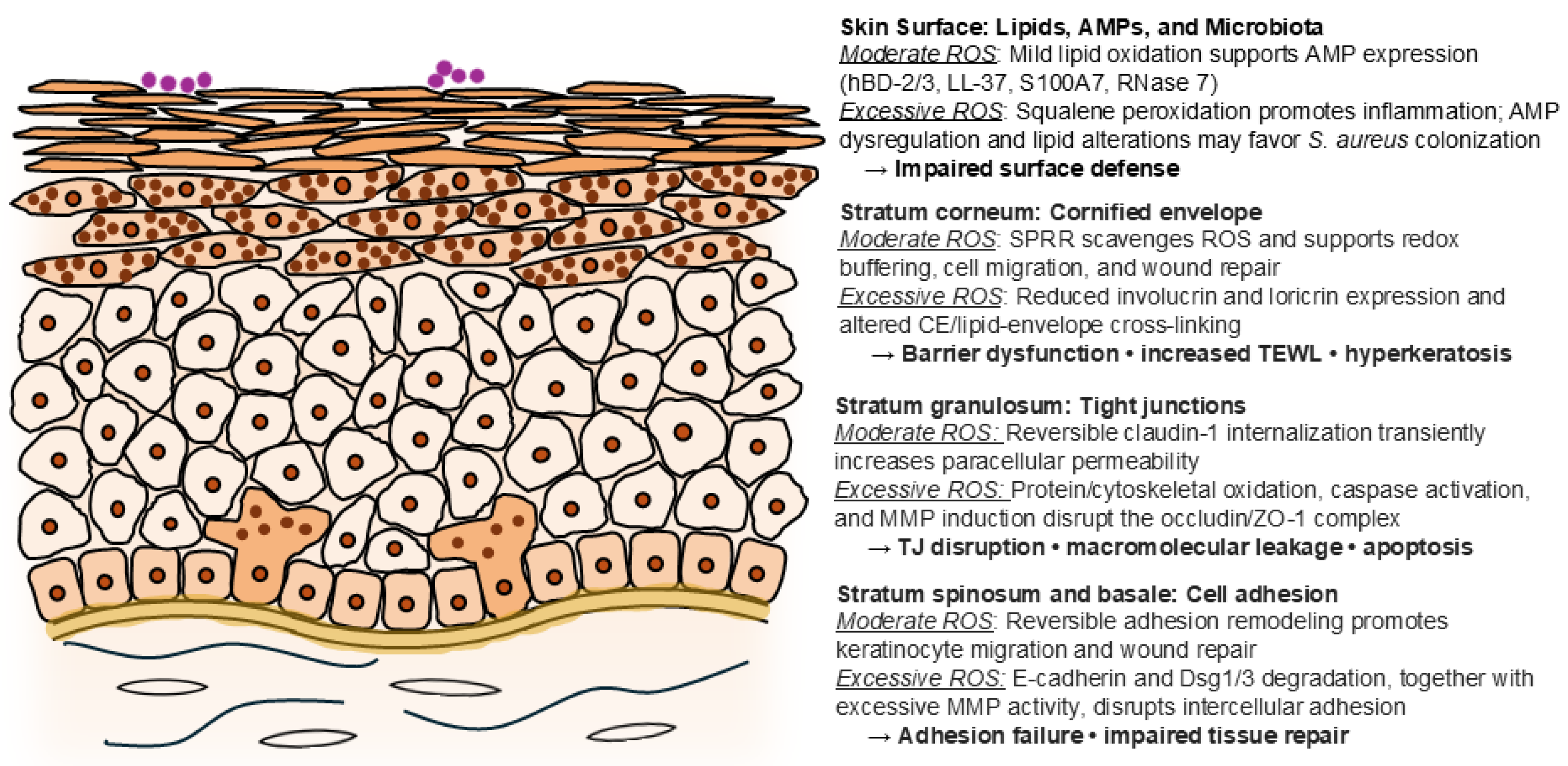
The signaling pathways described in the previous section ultimately converge on structural and functional components of the epidermal barrier. As summarized in Figure 2, disruption of this integrated redox signaling network progressively impairs barrier architecture through coordinated effects on TJ, intercellular adhesion complexes, and CE formation.
4.1. Oxidative Stress and Tight Junction Dysfunction
Keratinocyte TJ, localized primarily within the stratum granulosum, constitute the outermost intercellular barrier of the viable epidermis. Together with the stratum corneum, they regulate paracellular permeability, transepidermal water loss (TEWL), cell polarity, and the epidermal Ca²⁺ gradient, thereby maintaining skin barrier integrity. Claudin-1 serves as the principal sealing protein of epidermal TJ, while claudin-4 fine-tunes ion permeability, and zonula occludens-1 (ZO-1) anchors claudins and occludin to the actin cytoskeleton [2,77]. The critical role of claudin-1 is demonstrated by the fatal barrier defects observed in claudin-1-deficient mice [78]. TJ are highly dynamic structures that undergo continuous remodeling through endocytosis in response to cytokines, growth factors, viral infection, mechanical stress, and oxidative stimuli [79]. Their assembly and maintenance are regulated by the aPKC (atypical protein kinase C)/Par polarity complex and MAPK signaling, rendering TJ particularly susceptible to redox-dependent regulation [80,81].
Oxidative stress disrupts TJ integrity primarily by altering the localization and function of junctional proteins rather than simply reducing their expression. In human keratinocytes, UV-induced oxidative stress causes delocalization of claudin-1 and claudin-4 from the plasma membrane, resulting in reduced TJ function and increased paracellular permeability [9,10]. Oxidative stress has also been reported, mainly in intestinal and other epithelial models, to dissociate the occludin-ZO-1 complex, promoting phosphorylation, redistribution, and endocytosis of TJ proteins, thereby increasing barrier permeability [78,80,81,82]. These findings suggest that post-translational modification and intracellular trafficking of TJ proteins, rather than loss of total protein abundance, represent major mechanisms underlying oxidative stress-induced TJ dysfunction [9].
The extent of TJ dysfunction depends largely on the intensity and duration of oxidative stress, paralleling the redox-sensitive signaling responses described in the previous section. Mild-to-moderate ROS stimulation induces reversible TJ remodeling characterized by transient claudin-1 internalization and increased paracellular permeability. Proposed mechanisms include Ca²⁺ influx, nitric oxide synthase activation, actomyosin contraction, and dynamic regulation by RhoA/ROCK (Rho-associated coiled-coil kinase), aPKC/Par, and MAPK pathways [9]. In contrast, severe or sustained oxidative stress causes irreversible TJ disruption through protein oxidation, actin cytoskeletal damage, p38/JNK hyperactivation, MMP-mediated degradation of junctional proteins, mitochondrial dysfunction, and excessive Ca²⁺ influx, ultimately promoting keratinocyte apoptosis.
Taken together, oxidative stress impairs tight junction integrity through both reversible junctional remodeling and irreversible structural damage. While transient ROS-mediated TJ plasticity may facilitate physiological adaptation during wound repair, prolonged ROS exposure promotes barrier breakdown, increased epithelial permeability, and loss of epidermal homeostasis. These changes represent one of the earliest manifestations of oxidative stress-induced barrier dysfunction and precede disruption of the intercellular adhesion complexes discussed in the following section.
4.2. Oxidative Stress and Intercellular Adhesion Complexes
Intercellular adhesion complexes, extending from the basal layer through the stratum spinosum, provide both mechanical cohesion and signaling platforms that maintain epidermal integrity. These structures include adherens junctions, desmosomes, and hemidesmosomes, each of which is vulnerable to oxidative stress. Adherens junctions (AJ), formed by E-cadherin-β/α-catenin complexes linked to the actin cytoskeleton, preserve tissue architecture. Oxidative stress induced by UV irradiation promotes protease-mediated degradation and cleavage of E-cadherin [83], while H₂O₂ activates the transcription factor Snail-1, leading to decreased E-cadherin expression and increased expression of epithelial-mesenchymal transition markers such as α-smooth muscle actin (α-SMA) [84]. Desmosomes, composed of desmogleins (Dsg), desmocollins (Dsc), plakophilin, plakoglobin, and desmoplakin anchored to keratin 1/10 (K1/K10), are similarly disrupted by oxidative stress. UV irradiation reduces the expression of Dsg1, Dsc1, and K1/K10, while ROS-induced caspase activation promotes selective degradation of Dsg1/3 [85,86,87]. Hemidesmosomes anchor basal keratinocytes to the basement membrane through integrin α6β4, BP180, plectin, and BPAG-1 [88]. Oxidative stress decreases integrin α6β4 and plectin expression, weakening keratinocyte attachment and contributing to altered epidermal homeostasis and pigmentation [89].
Like TJ, intercellular adhesion complexes are highly dynamic and are regulated by multiple redox-sensitive signaling pathways. Assembly of AJ and desmosomes depends on the Ca²⁺-dependent "calcium switch," in which Ca²⁺ signaling together with PLC (phospholipase C)/PKC (protein kinase C), Rho-family GTPases, PI3K, EGFR, and Src-family kinases stabilizes the E-cadherin-β-catenin complex and regulates junctional turnover [90,91]. Desmosome assembly likewise requires cadherin function and Ca²⁺ elevation [92,93], whereas EGFR signaling and ADAM (a disintegrin and metalloproteinase)-mediated cleavage regulate desmosomal remodeling [94]. Under oxidative stress, activation of the MAPK/p38 pathway promotes internalization of Dsg, resulting in desmosome disassembly and acantholysis [95]. PI3K signaling may further influence desmosomal integrity through regulation of cell survival and expression of desmosomal components [96]. In hemidesmosomes, phosphorylation of integrin α6β4 promotes its dissociation from the cell membrane and activation of this pathway, although the precise mechanisms linking oxidative stress to this process remain incompletely understood [97].
As with TJ, the biological outcome depends on the intensity and duration of oxidative stress. Mild-to-moderate ROS stimulation promotes reversible adhesion remodeling that facilitates keratinocyte migration and wound re-epithelialization, whereas persistent oxidative stress induces irreversible oxidation and degradation of adhesion molecules, excessive MMP activity, cytoskeletal disruption, and loss of intercellular cohesion. These responses parallel the redox-sensitive signaling pathways described in Section 3, with moderate ROS favoring adaptive MAPK/ERK-mediated remodeling and severe or sustained ROS promoting p38/JNK activation, structural failure, and keratinocyte injury.
Taken together, oxidative stress progressively weakens epidermal cohesion by disrupting AJ, desmosomes, and hemidesmosomes through both reversible remodeling and irreversible structural damage. Loss of these adhesion complexes compromises tissue stability and amplifies barrier dysfunction beyond the tight junction layer, ultimately impairing keratinocyte differentiation and promoting the CE abnormalities discussed in the following section.
4.3. Oxidative Stress and Cornified Envelope (CE) Formation
Corneocytes are the terminally differentiated, dead cells of the stratum corneum produced by keratinocyte differentiation. Having lost their nucleus and organelles, they are filled with keratin filaments aggregated by filaggrin, and their outer surface is covered by a flattened, plate-like structure comprising the CE and an outer lipid envelope. The CE is initiated by transglutaminase-mediated cross-linking of involucrin as a precursor, forming a protein scaffold composed mainly of loricrin and small proline-rich proteins (SPRR); this scaffold is bound to an outer lipid envelope formed predominantly of long-chain ω-hydroxyceramides (acylceramides) derived from lamellar granules, which act as an adhesive linking the lipid envelope to the protein scaffold [98,99].
The CE is the structure most heavily exposed to oxidative stress in the skin, functioning as both a physical and a chemical barrier against ROS. SPRR proteins in particular are upregulated in response to stress and contribute to CE flexibility and antioxidant function; being cysteine-rich, SPRR proteins have been suggested to directly scavenge ROS and to promote cell migration and wound healing [100,101]. In vivo proteomic and functional analyses further support the notion that SPRR proteins function not merely as structural components but as redox-buffering factors [102]. However, the effects of severe or sustained ROS stimulation exceed these detoxification capacities. Environmental pro-oxidants also alter the profile of CE precursor proteins, changing CE composition and cross-linking patterns [103]. In vivo, expression of involucrin/loricrin is regulated by AP-1 and other factors and is reduced by oxidative stress and other stimuli, indicating suppression of keratinocyte differentiation [104]. These changes result in disordered CE composition and cross-linking, which can produce epidermal functional impairment, including increased permeability of the stratum corneum barrier, reduced water retention, and abnormal cornification (hyperkeratosis or immature CE formation).
Collectively, these findings demonstrate that oxidative stress progressively disrupts both the structural and functional components of the epidermal barrier, thereby compromising cutaneous homeostasis and creating a permissive environment for microbial colonization and persistent inflammation. These barrier-disruptive mechanisms may have particular relevance to pediatric skin because epidermal barrier structure and function continue to mature after birth. Compared with adult skin, infant skin exhibits developmental differences in stratum corneum structure, epidermal thickness, water-handling properties, and molecular markers of barrier formation [74,75,76]. These developmental characteristics may modify the functional consequences of oxidative disruption of TJs, intercellular adhesion complexes, and CE formation. However, direct studies comparing oxidative stress-induced alterations in these barrier structures between pediatric and adult skin remain scarce. Thus, although the molecular mechanisms described above are largely derived from adult human skin, keratinocyte models, and experimental animals, their impact may depend on developmental stage.
5. Oxidative Stress Alters Barrier Surface Defense
5.1. Lipid Barrier Remodeling
The lipid barrier of the stratum corneum constitutes the first chemical defense against environmental insults and microbial invasion. Sebum-derived lipids, including squalene, together with epidermal ceramides, cholesterol, and free fatty acids (FFA), maintain epidermal architecture, limit transepidermal water loss, and regulate interactions between keratinocytes and the skin surface microbiota. Besides their structural role, several lipid components possess intrinsic antimicrobial and antioxidant properties, thereby contributing to epidermal homeostasis [105,106].
Persistent oxidative injury disrupts this protective lipid barrier primarily through lipid peroxidation. Peroxidation of squalene and acylceramides impairs stratum corneum barrier function and promotes inflammatory responses characteristic of acne- and seborrheic dermatitis-like skin disorders. Squalene peroxides activate NF-κB and lipoxygenase signaling in keratinocytes, inducing IL-6 and other pro-inflammatory cytokines while altering the biochemical environment that supports microbial homeostasis [107,108,109,110]. Likewise, oxidative modification or impaired synthesis of acylceramides compromises the lipid seal of the stratum corneum, facilitating barrier dysfunction and colonization by opportunistic pathogens such as Staphylococcus aureus (S. aureus) [105,106].
The biological consequences of lipid oxidation depend on the balance between oxidative injury and local antioxidant defense. Vitamin E (α-tocopherol), a major lipid-soluble antioxidant delivered to the skin surface through sebaceous secretion, suppresses squalene peroxidation and partially protects against lipid-mediated inflammation [111,112,113]. However, because skin surface lipid levels remain relatively low before puberty and increase markedly during pubertal development [114], sebaceous delivery of vitamin E to the skin surface may also differ with age, potentially modifying local antioxidant protection against lipid peroxidation.
Surface lipids also actively shape antimicrobial defense. Lipid-metabolizing microorganisms such as Cutibacterium acnes (C. acnes) and Malassezia hydrolyze sebaceous triglycerides to generate FFA, while oxidized lipid metabolites, including hydroxy fatty acids, possess antimicrobial activity and inhibit bacterial adhesion and biofilm formation [115,116,117]. Thus, oxidative stress alters not only the structural integrity of the lipid barrier but also its antimicrobial and immunomodulatory functions. More broadly, the composition and abundance of skin surface lipids change substantially throughout postnatal development [114]. These developmental differences may influence the consequences of lipid oxidation in pediatric skin; however, direct comparative evidence linking age-dependent lipid composition to oxidative damage and cutaneous host defense remains limited. Persistent lipid oxidation therefore creates a biochemical environment that favors barrier dysfunction and modifies the cutaneous defense system, providing the basis for the antimicrobial peptide dysregulation discussed in the following section.
5.2. Oxidative Stress and Antimicrobial Peptide Dysregulation
Keratinocytes constitute an important component of the skin's innate immune defense by producing a broad spectrum of AMPs, including human β-defensins (hBD-1/2/3), cathelicidin (hCAP18/LL-37), the S100 family (S100A7/8/9), and RNase 7. These AMPs are induced by microbial products and inflammatory cytokines through pattern-recognition receptors, including TLRs, and function not only as direct antimicrobial effectors but also as immunomodulatory molecules that regulate cutaneous immune homeostasis [118,119].
AMP expression is tightly regulated by redox-sensitive signaling pathways. Infection, inflammation, and mild oxidative stress activate NF-κB and MAPK (particularly p38 and JNK), promoting transcription and secretion of hBD-2/3, LL-37, S100A7, and RNase 7 [120]. Similarly, environmental oxidative stimuli such as ultraviolet B (UVB) radiation, ozone, and PM transiently increase intracellular ROS and have been reported to enhance AMP expression under certain experimental conditions [121,122,123]. However, the effects of oxidative stress are highly dependent on its intensity and duration. Chronic or excessive ROS exposure impairs keratinocyte differentiation, promotes persistent inflammatory cytokine production, cellular senescence, and cytotoxicity, and disrupts NRF2-mediated antioxidant responses, collectively reducing the capacity of keratinocytes to maintain appropriate AMP production [122,124]. Consistent with this mechanism, activation of the NRF2 pathway has been shown to restore AMP expression suppressed by IL-4/IL-13 and other inflammatory stimuli, suggesting that impaired antioxidant defense contributes to AMP dysregulation under sustained oxidative stress [125].
Along with regulating AMP expression, oxidative stress also alters AMP function through post-translational modification. ROS-mediated oxidation and increased protease activity—including bacterial proteases, neutrophil elastase, kallikreins, and matrix metalloproteinases (MMPs)—promote cleavage or degradation of LL-37 and other AMPs, thereby reducing antimicrobial activity [126,127]. Oxidative modification further influences the biological activity of individual AMPs. For example, hBD-1 exhibits markedly enhanced antimicrobial activity in its reduced form, whereas oxidation diminishes its activity. Conversely, LL-37 undergoes oxidative modification associated with functional loss, while oxidation of RNase 7 and members of the S100 family, particularly S100A8/A9 (calprotectin), alters their antimicrobial and immunomodulatory properties, including metal-chelating activity essential for bacterial growth restriction [118,127,128].
Thus, oxidative stress affects antimicrobial defense at multiple levels by regulating AMP expression, stability, and biological activity. Rather than simply increasing or decreasing total AMP levels, different redox conditions selectively alter the composition and function of the AMP repertoire, thereby reshaping the antimicrobial environment at the skin surface. Together with oxidative remodeling of surface lipids, these changes disturb microbial homeostasis and favor the expansion of oxidative stress-tolerant pathogens such as S. aureus and Corynebacterium, ultimately promoting chronic inflammation and barrier dysfunction [129].
Developmental differences in cutaneous AMP expression may be particularly relevant in pediatric skin. Studies in neonates have demonstrated that antimicrobial peptide levels vary with gestational maturity and perinatal factors, indicating that cutaneous innate defense is developmentally regulated [130]. These differences may influence how oxidative stress-induced changes in AMP expression and function affect antimicrobial defense early in life. However, direct studies comparing redox-dependent AMP regulation between pediatric and adult keratinocytes remain limited.
Together, these redox-dependent alterations in antimicrobial defense provide a mechanistic basis for the microbiota dysbiosis discussed in the following section.
5.3. Oxidative Stress and Skin Microbiota Dysbiosis
The skin microbiota constitutes an integral component of the epidermal barrier, contributing to colonization resistance, immune maturation, and maintenance of cutaneous homeostasis. In healthy adults, microbial composition varies according to the local skin environment. Sebaceous sites are predominantly colonized by C. acnes, with lower abundances of Staphylococcus, Corynebacterium, and Malassezia, whereas moist sites are enriched with Corynebacterium and Staphylococcus. In contrast, dry sites harbor more diverse microbial communities, including members of the Proteobacteria and Bacteroidetes phyla [131,132,133,134]. These microbial communities remain relatively stable under physiological conditions but are influenced by host factors, environmental exposure, and lifestyle.
The pediatric skin microbiota differs substantially from that of adults because of developmental differences in sebaceous gland activity, skin pH, epidermal maturation, and immune function. Before puberty, reduced sebum production limits colonization by lipophilic microorganisms such as C. acnes and Malassezia, while Staphylococcus epidermidis, Streptococcus, and Corynebacterium are relatively more abundant [135,136,137]. Children also exhibit higher carriage rates of S. aureus, particularly in the presence of atopic predisposition, and the microbial communities of dry skin and the diaper area remain more dynamic and less stable than those of adults [135,136,138,139,140]. These developmental characteristics may influence how the pediatric skin microbiota responds to oxidative and inflammatory disturbances, although direct evidence linking developmental microbiota differences to redox-dependent dysbiosis remains limited.
Oxidative stress disrupts microbial homeostasis through multiple interconnected mechanisms. As discussed in the preceding sections, lipid peroxidation alters the biochemical composition of the skin surface, while dysregulation and oxidative modification of AMPs impair innate immune defense. Together with oxidative injury to keratinocytes and barrier structures, these changes create a selective environment that favors microorganisms capable of tolerating oxidative stress or evading antimicrobial peptide activity, including S. aureus and Corynebacterium [105,109,118,127,129]. Persistent colonization by these organisms further amplifies oxidative stress through activation of inflammatory signaling pathways and production of ROS, establishing a self-perpetuating cycle of barrier dysfunction, inflammation, and microbial imbalance.
Taken together, oxidative stress compromises the integrated surface defense of the epidermis by disrupting lipid homeostasis, antimicrobial peptide function, and the skin microbiota, thereby weakening cutaneous host defense and promoting persistent microbial colonization. These alterations may be particularly relevant in pediatric skin, where the microbiota, epidermal barrier, and cutaneous immune environment undergo substantial developmental changes after birth. Such developmental differences may modify the consequences of redox-driven dysbiosis for microbial colonization and inflammation, although direct pediatric evidence remains limited. Collectively, these changes establish the biological basis for the impaired wound healing and increased susceptibility to skin infection discussed in the following section.
Figure 3.
Oxidative stress–driven epidermal barrier dysfunction promotes microbial dysbiosis and impaired wound repair. Moderate ROS participate in physiological redox signaling that supports keratinocyte adaptation, barrier remodeling, and tissue repair. In contrast, excessive or persistent ROS disrupt tight junctions, intercellular adhesion, cornified envelope formation, surface lipid homeostasis, and antimicrobial peptide-mediated host defense. These alterations weaken epidermal integrity and surface defense, promoting microbial dysbiosis and persistent colonization, which may further amplify inflammatory signaling and ROS generation. The resulting self-reinforcing cycle of oxidative stress, barrier dysfunction, and inflammation may ultimately impair wound repair. In pediatric skin, postnatal development of the epidermal barrier, innate immunity, surface lipids, and skin microbiota may modify these interactions, although direct pediatric mechanistic evidence remains limited. Abbreviations: AMP, antimicrobial peptide; CE, cornified envelope; Dsg, desmoglein; hBD, human β-defensin; MMP, matrix metalloproteinase; ROS, reactive oxygen species; SPRR, small proline-rich protein; TEWL, transepidermal water loss; TJ, tight junction; ZO-1, zonula occludens-1.
Figure 3.
Oxidative stress–driven epidermal barrier dysfunction promotes microbial dysbiosis and impaired wound repair. Moderate ROS participate in physiological redox signaling that supports keratinocyte adaptation, barrier remodeling, and tissue repair. In contrast, excessive or persistent ROS disrupt tight junctions, intercellular adhesion, cornified envelope formation, surface lipid homeostasis, and antimicrobial peptide-mediated host defense. These alterations weaken epidermal integrity and surface defense, promoting microbial dysbiosis and persistent colonization, which may further amplify inflammatory signaling and ROS generation. The resulting self-reinforcing cycle of oxidative stress, barrier dysfunction, and inflammation may ultimately impair wound repair. In pediatric skin, postnatal development of the epidermal barrier, innate immunity, surface lipids, and skin microbiota may modify these interactions, although direct pediatric mechanistic evidence remains limited. Abbreviations: AMP, antimicrobial peptide; CE, cornified envelope; Dsg, desmoglein; hBD, human β-defensin; MMP, matrix metalloproteinase; ROS, reactive oxygen species; SPRR, small proline-rich protein; TEWL, transepidermal water loss; TJ, tight junction; ZO-1, zonula occludens-1.
6. Oxidative Stress in Pediatric Wound Healing and Infection
The barrier abnormalities described in the preceding sections have particular clinical relevance in pediatric skin. Because the epidermis, innate immune system, and antioxidant defenses continue to mature during postnatal development, disruption of redox homeostasis may have particularly important consequences for wound healing, susceptibility to infection, chronic inflammation, and pathological scar formation in pediatric skin.
6.1. Developmental Characteristics of Pediatric Skin and Redox Vulnerability
Pediatric skin should be regarded as a dynamically developing biological system rather than simply a thinner version of adult skin. Throughout infancy and childhood, cutaneous barrier structure, lipid composition, innate immune function, antioxidant capacity, and the cutaneous microbiota undergo progressive maturation. Accordingly, keratinocyte responses to oxidative stress differ quantitatively and qualitatively from those of adult skin, although direct molecular comparisons remain limited [14,15,75,76].
Structurally, neonatal and infant skin is characterized by a thinner stratum corneum and epidermis, smaller corneocytes, evolving lipid composition, and relatively higher TEWL, particularly in preterm infants [14,74,75,141]. Although the permeability barrier matures rapidly after birth in full-term neonates, components of the epidermal barrier—including TJ, intercellular adhesion complexes, and the CE—continue to undergo postnatal adaptation [14,76,142]. Direct comparative studies examining age-dependent expression of claudin-1, occludin, ZO-1, Dsg, Dsc, or other adhesion molecules remain scarce. Therefore, current evidence suggests developmental remodeling of these barrier structures rather than a simple reduction in their expression or function.
Redox homeostasis likewise changes during development. Physiological levels of ROS are required for normal keratinocyte proliferation, differentiation, and wound repair [4,5]. However, antioxidant reserve may be relatively limited during early life, particularly in preterm infants, in whom CAT, glutathione peroxidase, SOD, and glutathione levels have been reported to be lower than in adults [143,144,145]. Whether keratinocyte NRF2 responsiveness differs substantially between pediatric and adult skin remains unclear, as direct comparative studies are currently lacking. As a result, developing skin may possess a narrower therapeutic window between physiological ROS signaling and pathological oxidative stress.
The cutaneous innate immune system also undergoes maturation after birth. Expression patterns of AMPs, including hBD, LL-37, RNase 7, and S100 proteins, differ from those observed in adults and evolve according to gestational age, microbial colonization, and environmental exposure [140]. Simultaneously, the skin microbiota progressively develops under the influence of delivery mode, feeding, antibiotic exposure, anatomical site, and sebaceous gland maturation [135,136,146]. These parallel developmental processes establish reciprocal interactions between keratinocytes, AMPs, and resident microorganisms, ultimately shaping cutaneous immune homeostasis during childhood.
Collectively, these developmental characteristics suggest that oxidative stress interacts with a barrier that is still undergoing structural and immunological maturation. Although many mechanistic insights regarding redox signaling have been derived from adult skin or immortalized keratinocyte models, relatively few studies have directly examined these pathways in pediatric skin. This knowledge gap provides an important rationale for understanding how oxidative stress contributes to wound healing, infection susceptibility, and pathological scarring in children, which are discussed in the following sections [14,16,75].
6.2. Physiological Roles of ROS During Wound Repair
Wound healing is a highly coordinated biological process involving hemostasis, inflammation, proliferation, and tissue remodeling. Although excessive oxidative stress contributes to tissue injury, controlled production of ROS is indispensable for normal wound repair. Rather than functioning solely as cytotoxic molecules, physiological ROS act as signaling mediators that coordinate keratinocyte behavior, inflammatory responses, angiogenesis, ECM remodeling, and restoration of epidermal architecture [4,5,16,147]. Successful wound healing therefore depends on maintaining an appropriate balance between ROS generation and antioxidant defense.
During the early inflammatory phase, neutrophils and macrophages generate ROS primarily through activation of NOX as part of the respiratory burst [5,148,149]. These ROS contribute directly to microbial killing while simultaneously promoting removal of cellular debris and activation of innate immune signaling pathways [5,147,148,149]. Together with their antimicrobial activity, physiological ROS regulate cytokine production, leukocyte recruitment, and communication between inflammatory cells and keratinocytes, thereby coordinating the transition from inflammation to tissue repair [4,5,147].
The redox environment may also influence the transition from inflammation to tissue repair by modifying the biological activity of DAMPs. High-mobility group box 1 (HMGB1) provides a well-characterized example, as its extracellular functions are tightly regulated by the redox state of its cysteine residues. Fully reduced (all-thiol) HMGB1 primarily promotes chemotactic cell recruitment, whereas disulfide HMGB1 exhibits cytokine-inducing activity and promotes pro-inflammatory signaling. Further oxidation can render HMGB1 functionally inactive. Thus, changes in the wound redox environment may dynamically regulate DAMP signaling and influence the balance between inflammatory and reparative responses during wound healing [150,151].
Other DAMP–TLR pathways may similarly contribute to tissue repair. Endogenous noncoding RNAs released from damaged keratinocytes can activate TLR3 and promote epidermal barrier-repair responses, as demonstrated in primary human keratinocytes and Tlr3-deficient mouse models; consistent with this reparative role, poly(I:C), a synthetic TLR3 ligand, promoted collective migration of HaCaT keratinocytes in vitro, partly through IL-8-dependent signaling [152,153].
As inflammation resolves, ROS continue to function as essential signaling molecules during the proliferative phase of wound healing. Mild-to-moderate ROS levels stimulate keratinocyte proliferation and migration, promote fibroblast activation, and facilitate angiogenesis through regulation of growth factors including vascular endothelial growth factor (VEGF) [4,5,16,147]. These responses support re-epithelialization, ECM deposition, and formation of granulation tissue. As discussed in Section 3, these responses are coordinated by interconnected redox-sensitive signaling pathways, including NRF2, MAPK, NF-κB, and PI3K–Akt, illustrating how controlled ROS signaling regulates cellular behavior during tissue repair [36,46,67,73,147,154,155,156,157].
During the remodeling phase, ROS production gradually declines as antioxidant systems restore redox homeostasis. Resolution of oxidative signaling contributes to normalization of matrix metalloproteinase (MMP) activity, collagen remodeling, keratinocyte differentiation, and recovery of epidermal defense function [4,16,147,158,159]. Appropriate termination of ROS signaling is therefore as important as its initial activation, ensuring successful completion of wound healing while limiting excessive tissue remodeling and fibrosis.
Taken together, physiological ROS function as indispensable regulators of wound repair rather than merely as mediators of oxidative damage. The outcome depends largely on the magnitude, duration, and spatial localization of ROS production [4,5,147]. When ROS generation remains tightly controlled, redox signaling promotes effective tissue repair and restoration of epidermal function. Conversely, persistent or excessive ROS production overwhelms antioxidant defenses, shifting wound healing toward chronic inflammation, impaired re-epithelialization, infection, and pathological tissue remodeling, as discussed in the following section [16,147,158,159].
6.3. Infection and Chronic Inflammation
Under physiological conditions, oxidative stress is tightly controlled by endogenous antioxidant systems, allowing inflammatory responses to resolve and wound healing to progress toward tissue repair. However, when ROS production becomes excessive or persistent, antioxidant defenses are overwhelmed, disrupting redox homeostasis and shifting the wound microenvironment from adaptive repair toward chronic inflammation and infection [4,5,16,147].
Persistent oxidative stress progressively reprograms keratinocyte function. Excessive ROS impair keratinocyte proliferation, migration, and terminal differentiation while promoting mitochondrial dysfunction, DNA damage, lipid peroxidation, and apoptosis [6,8,46,71,72,124,147]. As discussed in Section 3 and Section 4, sustained activation of MAPK/p38, JNK, and NF-κB signaling together with impaired NRF2-mediated antioxidant responses amplifies inflammatory cytokine production, disrupts TJ, intercellular adhesion complexes, and CE formation, ultimately compromising epidermal barrier integrity.
Barrier disruption further facilitates microbial colonization, particularly by opportunistic pathogens such as S. aureus. In turn, microbial products activate TLRs and other pattern-recognition receptors on keratinocytes, promoting NOX-dependent ROS generation and activation of NF-κB and MAPK signaling pathways [22,23,26,51,52,56]. Concurrent dysregulation and oxidative modification of AMPs, together with lipid peroxidation, impair cutaneous antimicrobial defense and promote microbial dysbiosis and persistent colonization [105,118,127,129]. Consequently, oxidative stress and infection establish a self-amplifying cycle in which barrier dysfunction promotes microbial invasion, while microbial inflammation further enhances oxidative injury.
This vicious cycle is particularly relevant in pediatric wounds. Developmental immaturity of the epidermal barrier, antioxidant defenses, and cutaneous immune system may reduce the capacity of pediatric skin to restore redox homeostasis following injury. Persistent inflammation therefore prolongs neutrophil and macrophage activation, resulting in continued ROS production, delayed re-epithelialization, excessive protease activity, ECM degradation, and impaired wound closure [16,143,144,145,147,158,159,160,161,162]. Clinically, these processes contribute to delayed healing, recurrent infection, and progression toward chronic wounds or excessive scar formation.
Taken together, infection should not be regarded merely as a consequence of oxidative stress but also as an important amplifier of oxidative injury. Persistent activation of inflammatory cells, keratinocytes, and microbial signaling continuously reinforces ROS generation, producing a self-perpetuating cycle of oxidative stress, barrier failure, microbial dysbiosis, and chronic inflammation. Interrupting this cycle represents a major therapeutic objective in pediatric wound management and provides the rationale for the emerging redox-targeted interventions discussed in the following section.
6.4. Fibrosis and Hypertrophic Scar Formation
Successful wound healing requires timely resolution of inflammation and restoration of tissue homeostasis. When oxidative stress and inflammation persist, however, the wound healing program shifts from physiological tissue regeneration toward excessive extracellular matrix (ECM) deposition and pathological scar formation. This transition is particularly relevant in pediatric patients, in whom prolonged inflammation following burns, surgical wounds, or severe skin injury is associated with an increased risk of hypertrophic scar formation [158,159,160,161,162,163,164].
Unresolved oxidative injury promotes fibrosis through sustained activation of fibroblasts and dysregulation of keratinocyte–fibroblast communication. Excessive ROS promote profibrotic signaling, particularly through transforming growth factor-β (TGF-β)-dependent pathways, thereby stimulating fibroblast proliferation, myofibroblast differentiation, and excessive collagen synthesis [158,159,165,166,167]. At the same time, prolonged ROS exposure alters keratinocyte differentiation and barrier restoration, resulting in continued release of inflammatory mediators that further sustain fibroblast activation and matrix remodeling.
Redox-sensitive signaling pathways play central roles in this process. Persistent activation of MAPK, NF-κB, and PI3K–Akt signaling, together with impaired NRF2-mediated antioxidant responses, maintains a profibrotic microenvironment characterized by chronic inflammation, oxidative DNA damage, mitochondrial dysfunction, and imbalance between MMPs and their tissue inhibitors (TIMPs) [46,56,57,58,59,60,61,62,63,64,65,66,67,68,69,70,71,72,73,158,159,165,166,167,168]. These molecular alterations impair normal ECM turnover and favor excessive collagen accumulation, reduced scar remodeling, and increased tissue stiffness.
The consequences of these processes may be particularly important in pediatric wound healing. Children generally exhibit robust regenerative capacity; however, extensive burns, deep wounds, repeated infection, or prolonged inflammation may overwhelm physiological repair mechanisms and promote hypertrophic scar formation. Because pediatric skin is still undergoing structural and immunological maturation, chronic redox dysregulation may prolong inflammatory signaling, delay re-epithelialization, and increase the duration of fibroblast activation, thereby increasing the likelihood of pathological scar formation [143,144,145,161,162,163,164]. Although the precise contribution of oxidative stress to pediatric fibrosis remains incompletely defined, accumulating experimental and clinical evidence supports its important role in abnormal wound remodeling.
Collectively, fibrosis and hypertrophic scar formation represent the long-term consequences of unresolved oxidative stress and chronic inflammation. Rather than reflecting isolated fibroblast abnormalities, pathological scarring should be viewed as the culmination of persistent keratinocyte dysfunction, barrier failure, microbial dysbiosis, and sustained inflammatory signaling established throughout the wound healing process. These observations highlight oxidative stress not only as a mediator of tissue injury but also as a potential therapeutic target for improving wound healing outcomes and minimizing pathological scar formation in children.
7. Emerging Redox-Targeted Approaches
Understanding the integrated redox signaling network described throughout this review provides the mechanistic rationale for the innovative redox-targeted therapeutic strategies discussed in this section. Rather than focusing exclusively on infection control or tissue replacement, advancing interventions increasingly aim to restore physiological redox homeostasis and thereby interrupt the molecular events driving keratinocyte dysfunction, barrier failure, chronic inflammation, and impaired wound repair.
Although conventional wound management—including surgical debridement, infection control, moisture-balanced dressings, and antimicrobial therapy—remains the cornerstone of pediatric wound care, these approaches primarily address the consequences of tissue injury rather than the molecular mechanisms that drive disease progression. As discussed throughout this review, oxidative stress influences virtually every stage of wound healing by regulating keratinocyte function, cutaneous integrity, innate immunity, microbial homeostasis, inflammation, and tissue remodeling. These observations suggest that restoration of redox homeostasis represents a promising therapeutic strategy capable of interrupting the pathological cascade before irreversible barrier failure and chronic inflammation become established. Accordingly, increasing attention has focused on the development of redox-targeted interventions that not only reduce oxidative injury but also preserve physiological ROS signaling required for normal tissue repair [4,5,16,147,154].
Rather than acting through a single mechanism, innovative redox-targeted therapies aim to modulate multiple levels of the oxidative stress response, including suppression of excessive ROS generation, activation of endogenous antioxidant pathways, restoration of epidermal barrier function, regulation of cutaneous defense, and remodeling of the skin microbiota. Because these modalities target different stages of the pathogenic cascade, they should be viewed as complementary rather than mutually exclusive approaches. The following sections summarize current evidence according to their principal biological targets within the redox network.
Importantly, the objective of redox-targeted therapy is not to eliminate ROS indiscriminately but to restore redox homeostasis, thereby preserving the beneficial signaling functions of ROS while preventing the oxidative damage associated with persistent or excessive ROS production. This principle provides the conceptual framework for the therapeutic strategies discussed below.
7.1. Restoring Redox Homeostasis
Restoration of physiological redox homeostasis has emerged as a promising approach for improving wound healing, with the goal of preserving beneficial ROS signaling while preventing oxidative damage associated with persistent or excessive ROS production. Accordingly, therapeutic strategies increasingly focus on strengthening endogenous antioxidant defenses and limiting pathological ROS generation [147].
Among these approaches, activation of the NRF2 pathway has attracted considerable interest because it induces endogenous antioxidant and cytoprotective enzymes, including HO-1, NQO1, glutathione-related enzymes, catalase, and SOD [38,39,40]. Experimental studies indicate that pharmacological activation of NRF2 can attenuate oxidative stress, enhance cytoprotective responses, and promote tissue repair in preclinical models [168,169,170]. Although evidence specifically in pediatric wound healing remains limited, these findings support NRF2 modulation as a promising strategy for restoring redox balance while preserving physiological redox signaling.
Complementary strategies aim to limit excessive ROS production at its source. Pharmacological modulation of NOX activity and mitochondria-targeted antioxidants, including MitoQ and related compounds, have shown potential to reduce pathological oxidative stress in experimental settings [26,148,171]. Because excessive ROS may arise from multiple intracellular sources, future approaches combining enhancement of endogenous antioxidant defenses with targeted suppression of pathological ROS generation may offer advantages over nonspecific antioxidant therapy.
7.2. Promoting Epidermal Barrier Repair
Restoration of cutaneous integrity represents another fundamental therapeutic objective in pediatric wound management. As discussed throughout this review, oxidative stress compromises multiple structural and functional components of the epidermis, including TJ, intercellular adhesion complexes, the CE, epidermal lipids, and antimicrobial peptide production. Consequently, therapeutic strategies that promote keratinocyte differentiation and barrier repair may help restore epidermal integrity, reduce transepidermal water loss, strengthen host defense against microbial colonization, and support wound healing [78,105,106,115,147].
Among current approaches, topical barrier-repair formulations containing physiological lipids—including ceramides, cholesterol, and FFA—have demonstrated beneficial effects on epidermal barrier restoration. These formulations replenish stratum corneum lipids, improve barrier permeability, reduce transepidermal water loss, and decrease cutaneous inflammation, particularly in inflammatory skin diseases such as AD [105,106,111,112,113,115,116,117,171,172]. Because oxidative stress promotes lipid peroxidation and disrupts ceramide metabolism, restoration of epidermal lipid composition may not only improve barrier competence but also help re-establish redox homeostasis at the skin surface.
Experimental studies indicate that preservation or restoration of differentiation-associated structural proteins—including filaggrin, loricrin, involucrin, claudins, and other junctional proteins—may support epidermal integrity following oxidative injury [9,10,54,86,90,98]. Recent evidence that combined loss of the vitamin D receptor and calcium-sensing receptor in keratinocytes impairs oxidative stress responses, DNA repair, and epidermal differentiation further supports the close relationship between differentiation state and redox competence [173].
Beyond restoration of structural barrier components, successful wound healing also requires maintenance of redox homeostasis throughout the inflammatory, proliferative, and remodeling phases. Rather than completely eliminating ROS, emerging evidence indicates that therapeutic strategies should attenuate excessive oxidative stress while preserving the physiological ROS signaling required for host defense, keratinocyte migration, angiogenesis, and tissue remodeling [147].
Vitamin D and calcium signaling also contribute to keratinocyte differentiation, innate immune defense, and wound repair. Vitamin D-dependent signaling enhances injury-induced TLR2 function and antimicrobial peptide expression, while VDR and calcium-sensing receptor (CaSR) signaling contributes to keratinocyte migration and re-epithelialization during wound repair [174,175]. Consistent with these earlier findings, recent evidence that combined loss of VDR and CaSR in keratinocytes impairs oxidative stress responses, DNA repair, and epidermal differentiation further supports the close relationship between differentiation state and redox competence [173].
Recent developments in biomaterial science have further expanded opportunities for barrier-directed therapy. Modern wound dressings are increasingly designed not only to provide physical protection but also to regulate the wound microenvironment by maintaining appropriate moisture, reducing oxidative stress, delivering bioactive agents, and supporting tissue repair and re-epithelialization [154,155,156,157]. Recent advances in ROS-scavenging nanomaterials have further strengthened this concept by enabling targeted antioxidant delivery, minimizing off-target effects, and providing spatiotemporal control of oxidative stress within the wound microenvironment [154]. Novel hydrogel-, nanofiber-, and bioactive scaffold-based dressings capable of controlled release of redox-modulating agents represent promising next-generation platforms for pediatric wound care because they integrate structural support with molecular regulation of wound healing [155,156,157].
Collectively, these approaches extend beyond simple replacement of damaged barrier components. Instead, they aim to restore the coordinated biological functions of keratinocytes, thereby re-establishing epidermal integrity, limiting oxidative injury, and promoting physiological wound repair. Such approaches are likely to complement redox-targeted therapies discussed in the previous section and provide an important foundation for modulation of innate immunity and the skin microbiota.
7.3. Modulating Innate Immunity and the Skin Microbiota
The cutaneous microbiota has emerged as an important therapeutic target because oxidative stress disrupts keratinocyte function, epidermal barrier integrity, and the balance between host defense and resident microbial communities. As discussed in Section 5 and Section 6, persistent oxidative stress alters AMP expression and activity, promotes lipid peroxidation, and impairs keratinocyte differentiation, collectively favoring colonization by opportunistic pathogens such as Staphylococcus aureus [105,118,129]. These alterations establish a self-perpetuating cycle in which microbial dysbiosis amplifies inflammation and ROS production, further aggravating barrier dysfunction [129,176]. Consequently, interventions that restore microbial homeostasis may interrupt this pathogenic cycle while complementing conventional antimicrobial therapy [176,177].
Increasing attention has therefore focused on microbiota-directed interventions that promote beneficial host–microbe interactions rather than indiscriminately eliminating skin microorganisms. Topical commensal bacteriotherapy, probiotic preparations, and postbiotic products have shown potential to suppress S. aureus colonization, enhance epidermal barrier function, and modulate inflammatory responses in experimental models and early clinical studies [178,179,180,181]. Likewise, bacterial lysates and microbial-derived metabolites (postbiotics) have been reported to modulate keratinocyte immune responses, support epidermal barrier function, and promote microbial homeostasis [178,182]. Although clinical evidence remains limited, these approaches suggest that modulation of the skin microbiota may enhance host defense while reducing excessive inflammatory activation.
Future approaches are likely to combine microbiota modulation with advances in redox biology and biomaterials. Engineered AMPs and AMP-based delivery systems are being developed to enhance antimicrobial efficacy and improve targeted delivery in wound environments [183,184], whereas bacteriophage and microbiome-based therapeutic approaches offer opportunities for more selective targeting of pathogenic microorganisms while limiting disruption of beneficial microbial communities [185,186]. Integration of these approaches with antioxidant therapies, barrier-repair formulations, and controlled drug-delivery systems may provide synergistic benefits by simultaneously restoring epidermal integrity, redox homeostasis, innate immunity, and microbial balance. Rather than acting as isolated interventions, microbiota-directed therapies may ultimately become important components of integrated redox-targeted strategies for wound healing and skin infection, including future applications in pediatric care.
7.4. Future Perspectives: Toward Precision Redox Medicine
The growing understanding of redox biology has shifted therapeutic development from nonspecific antioxidant supplementation toward mechanism-based modulation of oxidative stress. Future strategies are expected to emphasize restoration of redox homeostasis rather than indiscriminate elimination of ROS, recognizing that controlled ROS signaling is indispensable for normal wound healing, immune defense, and tissue regeneration [4,147,187,188]. Achieving this balance will require therapeutic approaches capable of adapting to the dynamic redox environment of individual wounds rather than applying uniform treatment strategies.
An important step toward this goal is the identification of reliable biomarkers that accurately reflect wound redox status. Several oxidative stress markers—including 8-hydroxy-2′-deoxyguanosine (8-OHdG), malondialdehyde (MDA), 4-hydroxynonenal (4-HNE), protein carbonyls, glutathione redox balance, and NRF2-related measures—have been investigated as indicators of oxidative injury and redox status [189,190,191]. However, their translation into routine clinical practice remains limited, and standardized biomarker panels capable of distinguishing physiological redox signaling from pathological oxidative stress have yet to be established [190]. Development of standardized biomarker panels may facilitate patient stratification, treatment selection, and therapeutic monitoring, thereby supporting personalized redox-targeted interventions. Integration of oxidative stress biomarkers with transcriptomic, proteomic, metabolomic, and microbiome profiling may further improve individualized assessment of wound status, disease progression, and therapeutic response [192,193].
Rapid advances in systems biology are likewise expected to transform the understanding of wound healing. Integration of transcriptomic, proteomic, metabolomic, lipidomic, and microbiome datasets with single-cell and spatial transcriptomic technologies is providing increasingly detailed insights into the cellular and molecular heterogeneity of skin and the dynamic processes underlying wound repair [192,193,194,195,196]. These multidimensional approaches may facilitate identification of novel therapeutic targets and biomarkers and help explain interindividual differences in wound-healing responses, thereby supporting the development of precision wound medicine [192,193].
Simultaneously, progress in biomaterials and drug-delivery technologies is creating opportunities for intelligent wound management. ROS-responsive hydrogels, nanoparticle-based delivery systems, and bioactive wound dressings are being developed to modulate excessive oxidative stress and enable controlled delivery of therapeutic agents within the wound microenvironment [154,155,156,157]. Such approaches may permit more dynamic regulation of local wound conditions than conventional nonspecific treatment, with the potential to limit oxidative injury while preserving physiological redox signaling.
These emerging therapeutic approaches and their integration within a precision redox medicine framework are summarized in Figure 4.
Ultimately, future pediatric wound management is likely to integrate molecular diagnostics, biomarker-guided therapy, advanced biomaterials, microbiota modulation, and redox-targeted pharmacological interventions within a precision medicine framework. Because pediatric skin undergoes continuous structural and immunological maturation, age-specific therapeutic strategies should be developed and validated rather than extrapolated directly from adult studies. Successful translation of these emerging approaches into clinical practice will require continued interdisciplinary collaboration among dermatologists, pediatric surgeons, immunologists, microbiologists, biomaterial scientists, and redox biologists.
8. Conclusions
Pediatric skin infection and impaired wound healing should not be regarded simply as consequences of excessive ROS production, but rather as manifestations of disrupted redox homeostasis that progressively impairs keratinocyte function, epidermal architecture, cutaneous defense, and tissue repair. This review proposes oxidative stress–driven keratinocyte dysfunction as an integrative mechanistic framework linking epidermal barrier failure, microbial dysbiosis, chronic inflammation, and pathological wound healing in pediatric skin. Understanding this integrated redox network provides a mechanistic framework for developing more effective therapeutic strategies.
Accordingly, the objective of future redox-targeted therapies should not be to eliminate ROS indiscriminately, but to restore redox homeostasis, thereby preserving the essential signaling functions of ROS while preventing the oxidative damage associated with persistent or excessive ROS production. Such mechanism-based approaches have the potential to improve infection control, promote wound repair, and reduce pathological scar formation in children. Ultimately, the future of pediatric wound management may depend not on how effectively we eliminate ROS, but on how precisely we restore redox homeostasis.
Author Contributions
Conceptualization, Q.D.T., S.K.-A. and S.U.; writing—original draft preparation, K.H. and Q.D.T.; writing—review and editing, K.H., Q.D.T., N.T.K.P., K.T., D.B., S.U. and S.K.-A.; supervision, S.U. and S.K.-A. All authors have read and agreed to the published version of the manuscript.
Funding
This research received no external funding.
Institutional Review Board Statement
Not applicable.
Informed Consent Statement
Not applicable.
Data Availability Statement
No new data were created or analyzed in this study.
Acknowledgments
During the preparation of this manuscript, the authors used OpenAI ChatGPT (GPT-5.5) to assist with English language editing, scientific writing, text refinement, and improvement of manuscript organization and coherence. All AI-generated content was critically reviewed, revised, and verified by the authors, who take full responsibility for the accuracy, interpretation, and content of this publication.
Conflicts of Interest
The authors declare no conflicts of interest.
Abbreviations
The following abbreviations are used in this manuscript:
| ROS | Reactive oxygen species |
| AD | Atopic dermatitis |
| TJ | Tight junction |
| CE | Cornified envelope |
| AMPs | Antimicrobial peptides |
| UV | Ultraviolet |
| ZO-1 | Zonula occludens-1 |
| PM | Particulate matter |
| Cu | Copper |
| Fe | Iron |
| NOX | NADPH oxidase |
| TLR | Toll-like receptor |
| NRF2 | Nuclear factor erythroid 2-related factor 2 |
| ARE | Antioxidant response element |
| Keap1 | Kelch-like ECH-associated protein 1 |
| HO-1 | Heme oxygenase-1 |
| NQO1 | NAD(P)H:quinone oxidoreductase 1 |
| GST | Glutathione S-transferase |
| CAT | Catalase |
| SOD | Superoxide dismutase |
| MAPK | Mitogen-activated protein kinase |
| ERK | Extracellular signal-regulated kinase |
| JNK | c-Jun N-terminal kinase |
| AP-1 | Activator protein-1 |
| MMP | Matrix metalloproteinase |
| NF-κB | Nuclear factor kappa B |
| PI3K | Phosphoinositide 3-kinase |
| mTOR | Mechanistic target of rapamycin |
| mTORC1 | Mechanistic target of rapamycin complex 1 |
| EGFR | Epidermal growth factor receptor |
| TEWL | Transepidermal water loss |
| Dsg | Desmoglein |
| Dsc | Desmocollin |
| K1/K10 | Keratin 1/10 |
| FFA | Free fatty acids |
| S. aureus | Staphylococcus aureus |
| C. acnes | Cutibacterium acnes |
| hBD | Human β-defensins |
| ECM | Extracellular matrix |
| α-SMA | α-smooth muscle actin |
References
- Bazzoni, G., and E. Dejana. "Keratinocyte Junctions and the Epidermal Barrier: How to Make a Skin-Tight Dress." J Cell Biol 156, no. 6 (2002): 947-9.
- Brandner, J. M., M. Zorn-Kruppa, T. Yoshida, I. Moll, L. A. Beck, and A. De Benedetto. "Epidermal Tight Junctions in Health and Disease." Tissue Barriers 3, no. 1-2 (2015): e974451. [CrossRef]
- Simmons, J., and R. L. Gallo. "The Central Roles of Keratinocytes in Coordinating Skin Immunity." J Invest Dermatol 144, no. 11 (2024): 2377-98. [CrossRef]
- Hunt, Matthew, Monica Torres, Etty Bachar-Wikstrom, and Jakob D. Wikstrom. "Cellular and Molecular Roles of Reactive Oxygen Species in Wound Healing." Communications Biology 7, no. 1 (2024): 1534. [CrossRef]
- Andre-Levigne, D., A. Modarressi, M. S. Pepper, and B. Pittet-Cuenod. "Reactive Oxygen Species and Nox Enzymes Are Emerging as Key Players in Cutaneous Wound Repair." Int J Mol Sci 18, no. 10 (2017). [CrossRef]
- Li Pomi, F., L. Gammeri, F. Borgia, M. Di Gioacchino, and S. Gangemi. "Oxidative Stress and Skin Diseases: The Role of Lipid Peroxidation." Antioxidants (Basel) 14, no. 5 (2025). [CrossRef]
- Frantz, M. C., R. Rozot, and L. Marrot. "Nrf2 in Dermo-Cosmetic: From Scientific Knowledge to Skin Care Products." Biofactors 49, no. 1 (2023): 32-61. [CrossRef]
- Khiar-Fernández, I., N. Khiar-Fernández, J. J. Pereyra-Rodríguez, and I. Fernández. "Nrf2 as a Therapeutic Target in Dermatological Disorders: Mechanisms and Molecules." Pharmaceuticals (Basel) 19, no. 3 (2026). [CrossRef]
- Kobayashi, M., S. Shu, K. Marunaka, T. Matsunaga, and A. Ikari. "Weak Ultraviolet B Enhances the Mislocalization of Claudin-1 Mediated by Nitric Oxide and Peroxynitrite Production in Human Keratinocyte-Derived Hacat Cells." Int J Mol Sci 21, no. 19 (2020). [CrossRef]
- Marunaka, K., M. Kobayashi, S. Shu, T. Matsunaga, and A. Ikari. "Brazilian Green Propolis Rescues Oxidative Stress-Induced Mislocalization of Claudin-1 in Human Keratinocyte-Derived Hacat Cells." Int J Mol Sci 20, no. 16 (2019). [CrossRef]
- Komine, Mayumi, Mamitaro Ohtsuki, Razib M. D. Hossain, Tuba M. Ansary, Miho Sashikawa-Kimura, Hidetoshi Tsuda, Jin Meijuan, and Shin-ichi Tominaga. "Keratinocytes in Skin Disorders: The Importance of Keratinocytes as a Barrier." In Keratinocyte Biology - Structure and Function in the Epidermis, edited by Mayumi Komine. London: IntechOpen, 2022.
- Chen, J., Y. Liu, Z. Zhao, and J. Qiu. "Oxidative Stress in the Skin: Impact and Related Protection." Int J Cosmet Sci 43, no. 5 (2021): 495-509. [CrossRef]
- Wang, S., G. Peng, A. Abudouwanli, M. Yang, Q. Sun, W. Zhao, A. Ikeda, Y. Tan, L. Ma, H. Ogawa, K. Okumura, and F. Niyonsaba. "The Interaction between the Skin Microbiome and Antimicrobial Peptides within the Epidermal Immune Microenvironment: Bridging Insights into Atopic Dermatitis." Allergol Int 75, no. 1 (2026): 42-51. [CrossRef]
- Choi, E. H. "Skin Barrier Function in Neonates and Infants." Allergy Asthma Immunol Res 17, no. 1 (2025): 32-46. [CrossRef]
- Rahma, A., and M. E. Lane. "Skin Barrier Function in Infants: Update and Outlook." Pharmaceutics 14, no. 2 (2022). [CrossRef]
- Chen, R., H. Xu, X. Li, J. Dong, S. Wang, J. Hao, and G. Liang. "Role of Oxidative Stress in Post-Burn Wound Healing." Burns Trauma 13 (2025): tkaf040. [CrossRef]
- Fan, S., L. Lopez Llorens, F. P. Perona Martinez, and R. Schirhagl. "Quantum Sensing of Free Radical Generation in Mitochondria of Human Keratinocytes During Uvb Exposure." ACS Sens 9, no. 5 (2024): 2440-46. [CrossRef]
- Uchiyama, H., Q. L. Zhao, M. A. Hassan, G. Andocs, N. Nojima, K. Takeda, K. Ishikawa, M. Hori, and T. Kondo. "Epr-Spin Trapping and Flow Cytometric Studies of Free Radicals Generated Using Cold Atmospheric Argon Plasma and X-Ray Irradiation in Aqueous Solutions and Intracellular Milieu." PLoS One 10, no. 8 (2015): e0136956. [CrossRef]
- Cervellati, F., M. Benedusi, F. Manarini, B. Woodby, M. Russo, G. Valacchi, and M. C. Pietrogrande. "Proinflammatory Properties and Oxidative Effects of Atmospheric Particle Components in Human Keratinocytes." Chemosphere 240 (2020): 124746. [CrossRef]
- Pambianchi, E., F. Ferrara, A. Pecorelli, M. Benedusi, H. Choudhary, J. P. Therrien, and G. Valacchi. "Deferoxamine Treatment Improves Antioxidant Cosmeceutical Formulation Protection against Cutaneous Diesel Engine Exhaust Exposure." Antioxidants (Basel) 10, no. 12 (2021). [CrossRef]
- Sticozzi, C., G. Belmonte, A. Pecorelli, B. Arezzini, C. Gardi, E. Maioli, C. Miracco, M. Toscano, H. J. Forman, and G. Valacchi. "Cigarette Smoke Affects Keratinocytes Srb1 Expression and Localization Via H2o2 Production and Hne Protein Adducts Formation." PLoS One 7, no. 3 (2012): e33592. [CrossRef]
- Mempel, M., V. Voelcker, G. Kollisch, C. Plank, R. Rad, M. Gerhard, C. Schnopp, P. Fraunberger, A. K. Walli, J. Ring, D. Abeck, and M. Ollert. "Toll-Like Receptor Expression in Human Keratinocytes: Nuclear Factor Kappab Controlled Gene Activation by Staphylococcus Aureus Is Toll-Like Receptor 2 but Not Toll-Like Receptor 4 or Platelet Activating Factor Receptor Dependent." J Invest Dermatol 121, no. 6 (2003): 1389-96. [CrossRef]
- Song, P. I., Y. M. Park, T. Abraham, B. Harten, A. Zivony, N. Neparidze, C. A. Armstrong, and J. C. Ansel. "Human Keratinocytes Express Functional Cd14 and Toll-Like Receptor 4." J Invest Dermatol 119, no. 2 (2002): 424-32. [CrossRef]
- Abu-Humaidan, A. H., M. Elven, A. Sonesson, P. Garred, and O. E. Sorensen. "Persistent Intracellular Staphylococcus Aureus in Keratinocytes Lead to Activation of the Complement System with Subsequent Reduction in the Intracellular Bacterial Load." Front Immunol 9 (2018): 396. [CrossRef]
- Aufiero, B., M. Guo, C. Young, Z. Duanmu, H. Talwar, H. K. Lee, and G. J. Murakawa. "Staphylococcus Aureus Induces the Expression of Tumor Necrosis Factor-Alpha in Primary Human Keratinocytes." Int J Dermatol 46, no. 7 (2007): 687-94. [CrossRef]
- Choi, H., J. Y. Park, H. J. Kim, M. Noh, T. Ueyama, Y. Bae, T. R. Lee, and D. W. Shin. "Hydrogen Peroxide Generated by Duox1 Regulates the Expression Levels of Specific Differentiation Markers in Normal Human Keratinocytes." J Dermatol Sci 74, no. 1 (2014): 56-63. [CrossRef]
- Shi, Q., and Z. Wei. "Mechanisms of Mitochondrial Dysfunction and Protective Strategies in Skin Flap Ischemia-Reperfusion Injury." Front Pharmacol 17 (2026): 1865091. [CrossRef]
- Chen, Y., Q. Fang, Z. Wang, J. Y. Zhang, A. S. MacLeod, R. P. Hall, and W. B. Liedtke. "Transient Receptor Potential Vanilloid 4 Ion Channel Functions as a Pruriceptor in Epidermal Keratinocytes to Evoke Histaminergic Itch." J Biol Chem 291, no. 19 (2016): 10252-62. [CrossRef]
- Mikesell, A. R., O. Isaeva, F. Moehring, K. E. Sadler, A. D. Menzel, and C. L. Stucky. "Keratinocyte Piezo1 Modulates Cutaneous Mechanosensation." Elife 11 (2022). [CrossRef]
- Zeller, K. S., A. Riaz, H. Sarve, J. Li, A. Tengholm, and S. Johansson. "The Role of Mechanical Force and Ros in Integrin-Dependent Signals." PLoS One 8, no. 5 (2013): e64897. [CrossRef]
- Rizwan, H., S. Pal, S. Sabnam, and A. Pal. "High Glucose Augments Ros Generation Regulates Mitochondrial Dysfunction and Apoptosis Via Stress Signalling Cascades in Keratinocytes." Life Sci 241 (2020): 117148. [CrossRef]
- Wang, Z. H. "Anti-Glycative Effects of Asiatic Acid in Human Keratinocyte Cells." Biomedicine (Taipei) 4, no. 3 (2014): 19.
- Shibata, T., T. Ohta, K. I. Tong, A. Kokubu, R. Odogawa, K. Tsuta, H. Asamura, M. Yamamoto, and S. Hirohashi. "Cancer Related Mutations in Nrf2 Impair Its Recognition by Keap1-Cul3 E3 Ligase and Promote Malignancy." Proc Natl Acad Sci U S A 105, no. 36 (2008): 13568-73. [CrossRef]
- Kansanen, E., H. K. Jyrkkanen, and A. L. Levonen. "Activation of Stress Signaling Pathways by Electrophilic Oxidized and Nitrated Lipids." Free Radic Biol Med 52, no. 6 (2012): 973-82. [CrossRef]
- Natsch, A. "The Nrf2-Keap1-Are Toxicity Pathway as a Cellular Sensor for Skin Sensitizers--Functional Relevance and a Hypothesis on Innate Reactions to Skin Sensitizers." Toxicol Sci 113, no. 2 (2010): 284-92. [CrossRef]
- Schafer, M., and S. Werner. "Nrf2--a Regulator of Keratinocyte Redox Signaling." Free Radic Biol Med 88, no. Pt B (2015): 243-52. [CrossRef]
- Kimura, M., T. Yamamoto, J. Zhang, K. Itoh, M. Kyo, T. Kamiya, H. Aburatani, F. Katsuoka, H. Kurokawa, T. Tanaka, H. Motohashi, and M. Yamamoto. "Molecular Basis Distinguishing the DNA Binding Profile of Nrf2-Maf Heterodimer from That of Maf Homodimer." J Biol Chem 290, no. 17 (2015): 10644. [CrossRef]
- McMahon, M., K. Itoh, M. Yamamoto, S. A. Chanas, C. J. Henderson, L. I. McLellan, C. R. Wolf, C. Cavin, and J. D. Hayes. "The Cap'n'collar Basic Leucine Zipper Transcription Factor Nrf2 (Nf-E2 P45-Related Factor 2) Controls Both Constitutive and Inducible Expression of Intestinal Detoxification and Glutathione Biosynthetic Enzymes." Cancer Res 61, no. 8 (2001): 3299-307.
- Reisman, S. A., C. Y. Lee, C. J. Meyer, J. W. Proksch, and K. W. Ward. "Topical Application of the Synthetic Triterpenoid Rta 408 Activates Nrf2 and Induces Cytoprotective Genes in Rat Skin." Arch Dermatol Res 306, no. 5 (2014): 447-54. [CrossRef]
- Zhu, H., Z. Jia, L. Zhang, M. Yamamoto, H. P. Misra, M. A. Trush, and Y. Li. "Antioxidants and Phase 2 Enzymes in Macrophages: Regulation by Nrf2 Signaling and Protection against Oxidative and Electrophilic Stress." Exp Biol Med (Maywood) 233, no. 4 (2008): 463-74. [CrossRef]
- Ichimura, Y., S. Waguri, Y. S. Sou, S. Kageyama, J. Hasegawa, R. Ishimura, T. Saito, Y. Yang, T. Kouno, T. Fukutomi, T. Hoshii, A. Hirao, K. Takagi, T. Mizushima, H. Motohashi, M. S. Lee, T. Yoshimori, K. Tanaka, M. Yamamoto, and M. Komatsu. "Phosphorylation of P62 Activates the Keap1-Nrf2 Pathway During Selective Autophagy." Mol Cell 51, no. 5 (2013): 618-31. [CrossRef]
- Kageyama, S., T. Saito, M. Obata, R. H. Koide, Y. Ichimura, and M. Komatsu. "Negative Regulation of the Keap1-Nrf2 Pathway by a P62/Sqstm1 Splicing Variant." Mol Cell Biol 38, no. 7 (2018). [CrossRef]
- Kokot, A., D. Metze, N. Mouchet, M. D. Galibert, M. Schiller, T. A. Luger, and M. Bohm. "Alpha-Melanocyte-Stimulating Hormone Counteracts the Suppressive Effect of Uvb on Nrf2 and Nrf-Dependent Gene Expression in Human Skin." Endocrinology 150, no. 7 (2009): 3197-206. [CrossRef]
- Pensabene, K. M., J. LaMorte, A. E. Allender, J. Wehr, P. Kaur, M. Savage, and A. L. Eggler. "Acute Oxidative Stress Can Paradoxically Suppress Human Nrf2 Protein Synthesis by Inhibiting Global Protein Translation." Antioxidants (Basel) 12, no. 9 (2023). [CrossRef]
- Kleszczynski, K., I. M. Ernst, A. E. Wagner, N. Kruse, D. Zillikens, G. Rimbach, and T. W. Fischer. "Sulforaphane and Phenylethyl Isothiocyanate Protect Human Skin against Uvr-Induced Oxidative Stress and Apoptosis: Role of Nrf2-Dependent Gene Expression and Antioxidant Enzymes." Pharmacol Res 78 (2013): 28-40. [CrossRef]
- Van Laethem, A., K. Nys, S. Van Kelst, S. Claerhout, H. Ichijo, J. R. Vandenheede, M. Garmyn, and P. Agostinis. "Apoptosis Signal Regulating Kinase-1 Connects Reactive Oxygen Species to P38 Mapk-Induced Mitochondrial Apoptosis in Uvb-Irradiated Human Keratinocytes." Free Radic Biol Med 41, no. 9 (2006): 1361-71. [CrossRef]
- Guyton, K. Z., Y. Liu, M. Gorospe, Q. Xu, and N. J. Holbrook. "Activation of Mitogen-Activated Protein Kinase by H2o2. Role in Cell Survival Following Oxidant Injury." J Biol Chem 271, no. 8 (1996): 4138-42. [CrossRef]
- Derijard, B., M. Hibi, I. H. Wu, T. Barrett, B. Su, T. Deng, M. Karin, and R. J. Davis. "Jnk1: A Protein Kinase Stimulated by Uv Light and Ha-Ras That Binds and Phosphorylates the C-Jun Activation Domain." Cell 76, no. 6 (1994): 1025-37. [CrossRef]
- Gupta, S., D. Campbell, B. Derijard, and R. J. Davis. "Transcription Factor Atf2 Regulation by the Jnk Signal Transduction Pathway." Science 267, no. 5196 (1995): 389-93. [CrossRef]
- Oh, J. H., Y. H. Joo, F. Karadeniz, J. Ko, and C. S. Kong. "Syringaresinol Inhibits Uva-Induced Mmp-1 Expression by Suppression of Mapk/Ap-1 Signaling in Hacat Keratinocytes and Human Dermal Fibroblasts." Int J Mol Sci 21, no. 11 (2020). [CrossRef]
- Dai, X., H. Okazaki, Y. Hanakawa, M. Murakami, M. Tohyama, Y. Shirakata, and K. Sayama. "Eccrine Sweat Contains Il-1alpha, Il-1beta and Il-31 and Activates Epidermal Keratinocytes as a Danger Signal." PLoS One 8, no. 7 (2013): e67666. [CrossRef]
- Shi, X., L. Wang, J. D. Clark, and W. S. Kingery. "Keratinocytes Express Cytokines and Nerve Growth Factor in Response to Neuropeptide Activation of the Erk1/2 and Jnk Mapk Transcription Pathways." Regul Pept 186 (2013): 92-103. [CrossRef]
- Keum, Y. S., H. G. Kim, A. M. Bode, Y. J. Surh, and Z. Dong. "Uvb-Induced Cox-2 Expression Requires Histone H3 Phosphorylation at Ser10 and Ser28." Oncogene 32, no. 4 (2013): 444-52. [CrossRef]
- Tan, Q., H. Yang, E. Liu, and H. Wang. "P38/Erk Mapk Signaling Pathways Are Involved in the Regulation of Filaggrin and Involucrin by Il-17." Mol Med Rep 16, no. 6 (2017): 8863-67. [CrossRef]
- Kim, C., H. C. Ryu, and J. H. Kim. "Low-Dose Uvb Irradiation Stimulates Matrix Metalloproteinase-1 Expression Via a Blt2-Linked Pathway in Hacat Cells." Exp Mol Med 42, no. 12 (2010): 833-41. [CrossRef]
- Beak, S. M., Y. S. Lee, and J. A. Kim. "Nadph Oxidase and Cyclooxygenase Mediate the Ultraviolet B-Induced Generation of Reactive Oxygen Species and Activation of Nuclear Factor-Kappab in Hacat Human Keratinocytes." Biochimie 86, no. 7 (2004): 425-9. [CrossRef]
- Lawrence, T. "The Nuclear Factor Nf-Kappab Pathway in Inflammation." Cold Spring Harb Perspect Biol 1, no. 6 (2009): a001651.
- Liu, Ting, Lingyun Zhang, Donghyun Joo, and Shao-Cong Sun. "Nf-Κb Signaling in Inflammation." Signal Transduction and Targeted Therapy 2, no. 1 (2017): 17023.
- Rius, J., M. Guma, C. Schachtrup, K. Akassoglou, A. S. Zinkernagel, V. Nizet, R. S. Johnson, G. G. Haddad, and M. Karin. "Nf-Kappab Links Innate Immunity to the Hypoxic Response through Transcriptional Regulation of Hif-1alpha." Nature 453, no. 7196 (2008): 807-11. [CrossRef]
- Yamamoto, K., T. Arakawa, N. Ueda, and S. Yamamoto. "Transcriptional Roles of Nuclear Factor Kappa B and Nuclear Factor-Interleukin-6 in the Tumor Necrosis Factor Alpha-Dependent Induction of Cyclooxygenase-2 in Mc3t3-E1 Cells." J Biol Chem 270, no. 52 (1995): 31315-20. [CrossRef]
- Xie, Q. W., Y. Kashiwabara, and C. Nathan. "Role of Transcription Factor Nf-Kappa B/Rel in Induction of Nitric Oxide Synthase." J Biol Chem 269, no. 7 (1994): 4705-8. [CrossRef]
- Ledebur, H. C., and T. P. Parks. "Transcriptional Regulation of the Intercellular Adhesion Molecule-1 Gene by Inflammatory Cytokines in Human Endothelial Cells. Essential Roles of a Variant Nf-Kappa B Site and P65 Homodimers." J Biol Chem 270, no. 2 (1995): 933-43. [CrossRef]
- Oeckinghaus, A., M. S. Hayden, and S. Ghosh. "Crosstalk in Nf-Kappab Signaling Pathways." Nat Immunol 12, no. 8 (2011): 695-708.
- Papa, S., C. Bubici, F. Zazzeroni, C. G. Pham, C. Kuntzen, J. R. Knabb, K. Dean, and G. Franzoso. "The Nf-Kappab-Mediated Control of the Jnk Cascade in the Antagonism of Programmed Cell Death in Health and Disease." Cell Death Differ 13, no. 5 (2006): 712-29. [CrossRef]
- Perkins, N. D. "Integrating Cell-Signalling Pathways with Nf-Kappab and Ikk Function." Nat Rev Mol Cell Biol 8, no. 1 (2007): 49-62. [CrossRef]
- Han, E. J., I. P. S. Fernando, H. S. Kim, D. S. Lee, A. Kim, J. G. Je, M. J. Seo, Y. H. Jee, Y. J. Jeon, S. Y. Kim, and G. Ahn. "(-)-Loliolide Isolated from Sargassum Horneri Suppressed Oxidative Stress and Inflammation by Activating Nrf2/Ho-1 Signaling in Ifn-Gamma/Tnf-Alpha-Stimulated Hacat Keratinocytes." Antioxidants (Basel) 10, no. 6 (2021). [CrossRef]
- Jayasinghe, A. M. K., E. J. Han, Kgis Kirindage, I. P. S. Fernando, E. A. Kim, J. Kim, K. Jung, K. N. Kim, S. J. Heo, and G. Ahn. "3-Bromo-4,5-Dihydroxybenzaldehyde Isolated from Polysiphonia Morrowii Suppresses Tnf-Alpha/Ifn-Gamma-Stimulated Inflammation and Deterioration of Skin Barrier in Hacat Keratinocytes." Mar Drugs 20, no. 9 (2022). [CrossRef]
- Otterbein, L. E., F. H. Bach, J. Alam, M. Soares, H. Tao Lu, M. Wysk, R. J. Davis, R. A. Flavell, and A. M. Choi. "Carbon Monoxide Has Anti-Inflammatory Effects Involving the Mitogen-Activated Protein Kinase Pathway." Nat Med 6, no. 4 (2000): 422-8. [CrossRef]
- Liu, G. H., J. Qu, and X. Shen. "Nf-Kappab/P65 Antagonizes Nrf2-Are Pathway by Depriving Cbp from Nrf2 and Facilitating Recruitment of Hdac3 to Mafk." Biochim Biophys Acta 1783, no. 5 (2008): 713-27. [CrossRef]
- Shah, P., E. Trinh, L. Qiang, L. Xie, W. Y. Hu, G. S. Prins, J. Pi, and Y. Y. He. "Arsenic Induces P62 Expression to Form a Positive Feedback Loop with Nrf2 in Human Epidermal Keratinocytes: Implications for Preventing Arsenic-Induced Skin Cancer." Molecules 22, no. 2 (2017). [CrossRef]
- Redza-Dutordoir, M., and D. A. Averill-Bates. "Interactions between Reactive Oxygen Species and Autophagy: Special Issue: Death Mechanisms in Cellular Homeostasis." Biochim Biophys Acta Mol Cell Res 1868, no. 8 (2021): 119041.
- Klotz, L. O., C. Sanchez-Ramos, I. Prieto-Arroyo, P. Urbanek, H. Steinbrenner, and M. Monsalve. "Redox Regulation of Foxo Transcription Factors." Redox Biol 6 (2015): 51-72. [CrossRef]
- Weng, M. S., J. H. Chang, W. Y. Hung, Y. C. Yang, and M. H. Chien. "The Interplay of Reactive Oxygen Species and the Epidermal Growth Factor Receptor in Tumor Progression and Drug Resistance." J Exp Clin Cancer Res 37, no. 1 (2018): 61. [CrossRef]
- Stamatas, G. N., J. Nikolovski, M. C. Mack, and N. Kollias. "Infant Skin Physiology and Development During the First Years of Life: A Review of Recent Findings Based on in Vivo Studies." Int J Cosmet Sci 33, no. 1 (2011): 17-24. [CrossRef]
- Visscher, M. O., A. N. Carr, J. Winget, T. Huggins, C. C. Bascom, R. Isfort, K. Lammers, and V. Narendran. "Biomarkers of Neonatal Skin Barrier Adaptation Reveal Substantial Differences Compared to Adult Skin." Pediatr Res 89, no. 5 (2021): 1208-15.
- Visscher, M. O., P. Hu, A. N. Carr, C. C. Bascom, R. J. Isfort, K. Creswell, R. Adams, J. P. Tiesman, K. Lammers, and V. Narendran. "Newborn Infant Skin Gene Expression: Remarkable Differences Versus Adults." PLoS One 16, no. 10 (2021): e0258554. [CrossRef]
- Kirschner, N., R. Rosenthal, M. Furuse, I. Moll, M. Fromm, and J. M. Brandner. "Contribution of Tight Junction Proteins to Ion, Macromolecule, and Water Barrier in Keratinocytes." J Invest Dermatol 133, no. 5 (2013): 1161-9. [CrossRef]
- Furuse, M., M. Hata, K. Furuse, Y. Yoshida, A. Haratake, Y. Sugitani, T. Noda, A. Kubo, and S. Tsukita. "Claudin-Based Tight Junctions Are Crucial for the Mammalian Epidermal Barrier: A Lesson from Claudin-1-Deficient Mice." J Cell Biol 156, no. 6 (2002): 1099-111.
- Stamatovic, S. M., A. M. Johnson, N. Sladojevic, R. F. Keep, and A. V. Andjelkovic. "Endocytosis of Tight Junction Proteins and the Regulation of Degradation and Recycling." Ann N Y Acad Sci 1397, no. 1 (2017): 54-65. [CrossRef]
- Aono, S., and Y. Hirai. "Phosphorylation of Claudin-4 Is Required for Tight Junction Formation in a Human Keratinocyte Cell Line." Exp Cell Res 314, no. 18 (2008): 3326-39. [CrossRef]
- Minakami, M., N. Kitagawa, H. Iida, H. Anan, and T. Inai. "P38 Mitogen-Activated Protein Kinase and C-Jun Nh2-Terminal Protein Kinase Regulate the Accumulation of a Tight Junction Protein, Zo-1, in Cell-Cell Contacts in Hacat Cells." Tissue Cell 47, no. 1 (2015): 1-9. [CrossRef]
- Rao, R. "Oxidative Stress-Induced Disruption of Epithelial and Endothelial Tight Junctions." Front Biosci 13 (2008): 7210-26. [CrossRef]
- Hung, C. F., H. S. Chiang, H. M. Lo, J. S. Jian, and W. B. Wu. "E-Cadherin and Its Downstream Catenins Are Proteolytically Cleaved in Human Hacat Keratinocytes Exposed to Uvb." Exp Dermatol 15, no. 4 (2006): 315-21. [CrossRef]
- Fukawa, T., H. Kajiya, S. Ozeki, T. Ikebe, and K. Okabe. "Reactive Oxygen Species Stimulates Epithelial Mesenchymal Transition in Normal Human Epidermal Keratinocytes Via Tgf-Beta Secretion." Exp Cell Res 318, no. 15 (2012): 1926-32. [CrossRef]
- Dusek, R. L., S. Getsios, F. Chen, J. K. Park, E. V. Amargo, V. L. Cryns, and K. J. Green. "The Differentiation-Dependent Desmosomal Cadherin Desmoglein 1 Is a Novel Caspase-3 Target That Regulates Apoptosis in Keratinocytes." J Biol Chem 281, no. 6 (2006): 3614-24. [CrossRef]
- Johnson, J. L., J. L. Koetsier, A. Sirico, A. T. Agidi, D. Antonini, C. Missero, and K. J. Green. "The Desmosomal Protein Desmoglein 1 Aids Recovery of Epidermal Differentiation after Acute Uv Light Exposure." J Invest Dermatol 134, no. 8 (2014): 2154-62. [CrossRef]
- Weiske, J., T. Schoneberg, W. Schroder, M. Hatzfeld, R. Tauber, and O. Huber. "The Fate of Desmosomal Proteins in Apoptotic Cells." J Biol Chem 276, no. 44 (2001): 41175-81. [CrossRef]
- Walko, G., M. J. Castanon, and G. Wiche. "Molecular Architecture and Function of the Hemidesmosome." Cell Tissue Res 360, no. 2 (2015): 363-78.
- Coelho, S. G., J. C. Valencia, L. Yin, C. Smuda, A. Mahns, L. Kolbe, S. A. Miller, J. Z. Beer, G. Zhang, P. L. Tuma, and V. J. Hearing. "Uv Exposure Modulates Hemidesmosome Plasticity, Contributing to Long-Term Pigmentation in Human Skin." J Pathol 236, no. 1 (2015): 17-29. [CrossRef]
- Bikle, D. D., Z. Xie, and C. L. Tu. "Calcium Regulation of Keratinocyte Differentiation." Expert Rev Endocrinol Metab 7, no. 4 (2012): 461-72. [CrossRef]
- Kehrberg, R. J., and K. A. DeMali. "E-Cadherin: A Conductor of Cellular Signaling." Curr Opin Cell Biol 95 (2025): 102559. [CrossRef]
- Lewis, J. E., P. J. Jensen, and M. J. Wheelock. "Cadherin Function Is Required for Human Keratinocytes to Assemble Desmosomes and Stratify in Response to Calcium." J Invest Dermatol 102, no. 6 (1994): 870-7. [CrossRef]
- Watt, F. M., D. L. Mattey, and D. R. Garrod. "Calcium-Induced Reorganization of Desmosomal Components in Cultured Human Keratinocytes." J Cell Biol 99, no. 6 (1984): 2211-5. [CrossRef]
- Klessner, J. L., B. V. Desai, E. V. Amargo, S. Getsios, and K. J. Green. "Egfr and Adams Cooperate to Regulate Shedding and Endocytic Trafficking of the Desmosomal Cadherin Desmoglein 2." Mol Biol Cell 20, no. 1 (2009): 328-37. [CrossRef]
- Mavropoulos, A., T. Orfanidou, C. Liaskos, D. S. Smyk, V. Spyrou, L. I. Sakkas, E. I. Rigopoulou, and D. P. Bogdanos. "P38 Mapk Signaling in Pemphigus: Implications for Skin Autoimmunity." Autoimmune Dis 2013 (2013): 728529. [CrossRef]
- Johnson, J. L., N. A. Najor, and K. J. Green. "Desmosomes: Regulators of Cellular Signaling and Adhesion in Epidermal Health and Disease." Cold Spring Harb Perspect Med 4, no. 11 (2014): a015297. [CrossRef]
- Alt, A., M. Ohba, L. Li, M. Gartsbein, A. Belanger, M. F. Denning, T. Kuroki, S. H. Yuspa, and T. Tennenbaum. "Protein Kinase Cdelta-Mediated Phosphorylation of Alpha6beta4 Is Associated with Reduced Integrin Localization to the Hemidesmosome and Decreased Keratinocyte Attachment." Cancer Res 61, no. 11 (2001): 4591-8.
- Kalinin, A. E., A. V. Kajava, and P. M. Steinert. "Epithelial Barrier Function: Assembly and Structural Features of the Cornified Cell Envelope." Bioessays 24, no. 9 (2002): 789-800. [CrossRef]
- Roop, D. "Defects in the Barrier." Science 267, no. 5197 (1995): 474-5.
- Vermeij, W. P., A. Alia, and C. Backendorf. "Ros Quenching Potential of the Epidermal Cornified Cell Envelope." J Invest Dermatol 131, no. 7 (2011): 1435-41. [CrossRef]
- Vermeij, W. P., and C. Backendorf. "Skin Cornification Proteins Provide Global Link between Ros Detoxification and Cell Migration During Wound Healing." PLoS One 5, no. 8 (2010): e11957. [CrossRef]
- Vermeij, W. P., B. I. Florea, S. Isenia, A. Alia, J. Brouwer, and C. Backendorf. "Proteomic Identification of in Vivo Interactors Reveals Novel Function of Skin Cornification Proteins." J Proteome Res 11, no. 6 (2012): 3068-76. [CrossRef]
- Lin, L. W., B. P. Durbin-Johnson, D. M. Rocke, M. Salemi, B. S. Phinney, and R. H. Rice. "Environmental Pro-Oxidants Induce Altered Envelope Protein Profiles in Human Keratinocytes." Toxicol Sci 197, no. 1 (2023): 16-26. [CrossRef]
- Han, B., E. A. Rorke, G. Adhikary, Y. C. Chew, W. Xu, and R. L. Eckert. "Suppression of Ap1 Transcription Factor Function in Keratinocyte Suppresses Differentiation." PLoS One 7, no. 5 (2012): e36941. [CrossRef]
- Berdyshev, E. "Skin Lipid Barrier: Structure, Function and Metabolism." Allergy Asthma Immunol Res 16, no. 5 (2024): 445-61. [CrossRef]
- Maeng, J., S. Jeong, and H. Kim. "Skin Barrier Dysfunction in Contact and Atopic Dermatitis: A Comparative Review." Allergy Asthma Immunol Res 18, no. 2 (2026): 173-81. [CrossRef]
- Condro, G., R. Sciortino, and P. Perugini. "Squalene Peroxidation and Biophysical Parameters in Acne-Prone Skin: A Pilot "in Vivo" Study." Pharmaceuticals (Basel) 16, no. 12 (2023). [CrossRef]
- Deng, L., X. Ling, L. Li, Y. He, and M. Guo. "Advances in Scalp Microbiome Research: Molecular Insights into the Metabolism-Inflammation-Barrier Axis and Dandruff Pathogenesis." Molecules 31, no. 12 (2026). [CrossRef]
- Jourdain, R., A. Moga, P. Vingler, C. El Rawadi, F. Pouradier, L. Souverain, P. Bastien, N. Amalric, and L. Breton. "Exploration of Scalp Surface Lipids Reveals Squalene Peroxide as a Potential Actor in Dandruff Condition." Arch Dermatol Res 308, no. 3 (2016): 153-63. [CrossRef]
- Ottaviani, M., T. Alestas, E. Flori, A. Mastrofrancesco, C. C. Zouboulis, and M. Picardo. "Peroxidated Squalene Induces the Production of Inflammatory Mediators in Hacat Keratinocytes: A Possible Role in Acne Vulgaris." J Invest Dermatol 126, no. 11 (2006): 2430-7. [CrossRef]
- Joshi, M., P. Hiremath, J. John, N. Ranadive, K. Nandakumar, and J. Mudgal. "Modulatory Role of Vitamins a, B3, C, D, and E on Skin Health, Immunity, Microbiome, and Diseases." Pharmacol Rep 75, no. 5 (2023): 1096-114. [CrossRef]
- Ottaviani, M., E. Camera, and M. Picardo. "Lipid Mediators in Acne." Mediators Inflamm 2010 (2010). [CrossRef]
- Thiele, J. J., S. U. Weber, and L. Packer. "Sebaceous Gland Secretion Is a Major Physiologic Route of Vitamin E Delivery to Skin." J Invest Dermatol 113, no. 6 (1999): 1006-10. [CrossRef]
- Akutsu, N., M. Ooguri, T. Onodera, Y. Kobayashi, M. Katsuyama, N. Kunizawa, T. Hirao, J. Hosoi, Y. Masuda, S. Yoshida, M. Takahashi, T. Tsuchiya, and H. Tagami. "Functional Characteristics of the Skin Surface of Children Approaching Puberty: Age and Seasonal Influences." Acta Derm Venereol 89, no. 1 (2009): 21-7. [CrossRef]
- Douglas, E. J. A., N. Palk, E. R. Rudolph, and M. Laabei. "Anti-Staphylococcal Fatty Acids: Mode of Action, Bacterial Resistance and Implications for Therapeutic Application." Microbiology (Reading) 171, no. 5 (2025). [CrossRef]
- Khalil, S. B., and C. H. Kowalski. "The Cutaneous Microbiome: Microbial Remodeling of the Skin Lipid Landscape." mSphere 11, no. 6 (2026): e0064825. [CrossRef]
- Yuyama, K. T., M. Rohde, G. Molinari, M. Stadler, and W. R. Abraham. "Unsaturated Fatty Acids Control Biofilm Formation of Staphylococcus Aureus and Other Gram-Positive Bacteria." Antibiotics (Basel) 9, no. 11 (2020). [CrossRef]
- Chessa, C., C. Bodet, C. Jousselin, M. Wehbe, N. Leveque, and M. Garcia. "Antiviral and Immunomodulatory Properties of Antimicrobial Peptides Produced by Human Keratinocytes." Front Microbiol 11 (2020): 1155. [CrossRef]
- Jiang, Y., L. C. Tsoi, A. C. Billi, N. L. Ward, P. W. Harms, C. Zeng, E. Maverakis, J. M. Kahlenberg, and J. E. Gudjonsson. "Cytokinocytes: The Diverse Contribution of Keratinocytes to Immune Responses in Skin." JCI Insight 5, no. 20 (2020). [CrossRef]
- Wehkamp, K., L. Schwichtenberg, J. M. Schroder, and J. Harder. "Pseudomonas Aeruginosa- and Il-1beta-Mediated Induction of Human Beta-Defensin-2 in Keratinocytes Is Controlled by Nf-Kappab and Ap-1." J Invest Dermatol 126, no. 1 (2006): 121-7.
- Glaser, R., F. Navid, W. Schuller, C. Jantschitsch, J. Harder, J. M. Schroder, A. Schwarz, and T. Schwarz. "Uv-B Radiation Induces the Expression of Antimicrobial Peptides in Human Keratinocytes in Vitro and in Vivo." J Allergy Clin Immunol 123, no. 5 (2009): 1117-23. [CrossRef]
- Paik, K., J. I. Na, C. H. Huh, and J. W. Shin. "Particulate Matter and Its Molecular Effects on Skin: Implications for Various Skin Diseases." Int J Mol Sci 25, no. 18 (2024). [CrossRef]
- Pambianchi, E., Z. Hagenberg, A. Pecorelli, A. Pasqui, J. P. Therrien, and G. Valacchi. "Tension as a Key Factor in Skin Responses to Pollution." Sci Rep 13, no. 1 (2023): 16013. [CrossRef]
- Zhang, Y., C. Han, H. Yuan, L. A. DiPietro, and L. Chen. "Response of Oral and Skin Keratinocytes to Oxidative Stress." Cells 15, no. 2 (2026). [CrossRef]
- Dai, X., Y. Mizukami, K. Watanabe, T. Tsuda, M. Shidahara, S. Yoshida, K. Yatsuzuka, K. Shiraishi, H. Mori, M. Murakami, R. Kawakami, T. Imamura, Y. Fujisawa, and J. Muto. "Trehalose Prevents Il-4/Il-13-Induced Skin Barrier Impairment by Suppressing Il-33 Expression and Increasing Nrf2 Activation in Human Keratinocytes in Vitro." J Invest Dermatol 145, no. 6 (2025): 1422-32 e10. [CrossRef]
- Eissa, A., V. Amodeo, C. R. Smith, and E. P. Diamandis. "Kallikrein-Related Peptidase-8 (Klk8) Is an Active Serine Protease in Human Epidermis and Sweat and Is Involved in a Skin Barrier Proteolytic Cascade." J Biol Chem 286, no. 1 (2011): 687-706. [CrossRef]
- Potempa, J., and R. N. Pike. "Corruption of Innate Immunity by Bacterial Proteases." J Innate Immun 1, no. 2 (2009): 70-87.
- Rademacher, F., M. Simanski, and J. Harder. "Rnase 7 in Cutaneous Defense." Int J Mol Sci 17, no. 4 (2016): 560. [CrossRef]
- Brzoza, P., U. Godlewska, A. Borek, A. Morytko, A. Zegar, P. Kwiecinska, B. A. Zabel, A. Osyczka, M. Kwitniewski, and J. Cichy. "Redox Active Antimicrobial Peptides in Controlling Growth of Microorganisms at Body Barriers." Antioxidants (Basel) 10, no. 3 (2021). [CrossRef]
- Humberg, A., L. Neuenburg, H. Boeckel, M. I. Fortmann, C. Hartel, E. Herting, H. Hinrichs, F. Rademacher, and J. Harder. "Antimicrobial Skin Peptides in Premature Infants: Comparison with Term Infants and Impact of Perinatal Factors." Front Immunol 14 (2023): 1093340. [CrossRef]
- Findley, K., J. Oh, J. Yang, S. Conlan, C. Deming, J. A. Meyer, D. Schoenfeld, E. Nomicos, M. Park, N. I. H. Intramural Sequencing Center Comparative Sequencing Program, H. H. Kong, and J. A. Segre. "Topographic Diversity of Fungal and Bacterial Communities in Human Skin." Nature 498, no. 7454 (2013): 367-70. [CrossRef]
- Grice, E. A., and J. A. Segre. "The Skin Microbiome." Nat Rev Microbiol 9, no. 4 (2011): 244-53.
- Lee, H. J., S. E. Jeong, S. Lee, S. Kim, H. Han, and C. O. Jeon. "Effects of Cosmetics on the Skin Microbiome of Facial Cheeks with Different Hydration Levels." Microbiologyopen 7, no. 2 (2018): e00557. [CrossRef]
- Moskovicz, V., A. Gross, and B. Mizrahi. "Extrinsic Factors Shaping the Skin Microbiome." Microorganisms 8, no. 7 (2020). [CrossRef]
- Casterline, B. W., and A. S. Paller. "Early Development of the Skin Microbiome: Therapeutic Opportunities." Pediatr Res 90, no. 4 (2021): 731-37. [CrossRef]
- Murphy, B., M. Hoptroff, D. Arnold, A. Cawley, E. Smith, S. E. Adams, A. Mitchell, M. J. Horsburgh, J. Hunt, B. Dasgupta, N. Ghatlia, S. Samaras, A. MacGuire-Flanagan, and K. Sharma. "Compositional Variations between Adult and Infant Skin Microbiome: An Update." Microorganisms 11, no. 6 (2023). [CrossRef]
- Oh, J., and A. Y. Voigt. "The Human Skin Microbiome: From Metagenomes to Therapeutics." Nat Rev Microbiol 23, no. 12 (2025): 771-87. [CrossRef]
- Hoeger, P. H., W. Lenz, A. Boutonnier, and J. M. Fournier. "Staphylococcal Skin Colonization in Children with Atopic Dermatitis: Prevalence, Persistence, and Transmission of Toxigenic and Nontoxigenic Strains." J Infect Dis 165, no. 6 (1992): 1064-8. [CrossRef]
- Hu, P., A. N. Carr, M. Parlov, D. Swift, J. P. Tiesman, N. Ramji, J. J. Schoch, and A. G. Teufel. "Metagenomics Investigation on Baby Diaper Area Microbiome and Its Association with Skin Ph and Dermatitis in the Diapered Area." Microorganisms 13, no. 11 (2025). [CrossRef]
- Teufel, A., B. Howard, P. Hu, and A. N. Carr. "Characterization of the Microbiome in the Infant Diapered Area: Insights from Healthy and Damaged Skin." Exp Dermatol 30, no. 10 (2021): 1409-17. [CrossRef]
- Fluhr, J. W., R. Darlenski, N. Lachmann, C. Baudouin, P. Msika, C. De Belilovsky, and J. P. Hachem. "Infant Epidermal Skin Physiology: Adaptation after Birth." Br J Dermatol 166, no. 3 (2012): 483-90. [CrossRef]
- Fluhr, J. W., R. Darlenski, A. Taieb, J. P. Hachem, C. Baudouin, P. Msika, C. De Belilovsky, and E. Berardesca. "Functional Skin Adaptation in Infancy - Almost Complete but Not Fully Competent." Exp Dermatol 19, no. 6 (2010): 483-92. [CrossRef]
- Georgeson, G. D., B. J. Szony, K. Streitman, I. S. Varga, A. Kovacs, L. Kovacs, and A. Laszlo. "Antioxidant Enzyme Activities Are Decreased in Preterm Infants and in Neonates Born Via Caesarean Section." Eur J Obstet Gynecol Reprod Biol 103, no. 2 (2002): 136-9. [CrossRef]
- Jain, A., T. Mehta, P. A. Auld, J. Rodrigues, R. F. Ward, M. K. Schwartz, and J. Martensson. "Glutathione Metabolism in Newborns: Evidence for Glutathione Deficiency in Plasma, Bronchoalveolar Lavage Fluid, and Lymphocytes in Prematures." Pediatr Pulmonol 20, no. 3 (1995): 160-6. [CrossRef]
- Lavoie, J. C., and P. Chessex. "Gender and Maturation Affect Glutathione Status in Human Neonatal Tissues." Free Radic Biol Med 23, no. 4 (1997): 648-57. [CrossRef]
- Capone, K. A., S. E. Dowd, G. N. Stamatas, and J. Nikolovski. "Diversity of the Human Skin Microbiome Early in Life." J Invest Dermatol 131, no. 10 (2011): 2026-32. [CrossRef]
- Goncalves, R. V., M. B. Freitas, and D. Esposito. "Cellular and Molecular Mechanisms of Oxidative Stress in Wound Healing." Oxid Med Cell Longev 2022 (2022): 9785094. [CrossRef]
- Bedard, K., and K. H. Krause. "The Nox Family of Ros-Generating Nadph Oxidases: Physiology and Pathophysiology." Physiol Rev 87, no. 1 (2007): 245-313. [CrossRef]
- Tocu, G., B. I. Stefanescu, L. Stavar Matei, and L. Tocu. "Phagocyte Nadph Oxidase Nox2-Derived Reactive Oxygen Species in Antimicrobial Defense: Mechanisms, Regulation, and Therapeutic Potential-a Narrative Review." Antioxidants (Basel) 15, no. 1 (2025). [CrossRef]
- Tang, Daolin, Timothy R. Billiar, and Michael T. Lotze. "A Janus Tale of Two Active High Mobility Group Box 1 (Hmgb1) Redox States." Molecular Medicine 18, no. 10 (2012): 1360-62. [CrossRef]
- Venereau, E., M. Casalgrandi, M. Schiraldi, D. J. Antoine, A. Cattaneo, F. De Marchis, J. Liu, A. Antonelli, A. Preti, L. Raeli, S. S. Shams, H. Yang, L. Varani, U. Andersson, K. J. Tracey, A. Bachi, M. Uguccioni, and M. E. Bianchi. "Mutually Exclusive Redox Forms of Hmgb1 Promote Cell Recruitment or Proinflammatory Cytokine Release." J Exp Med 209, no. 9 (2012): 1519-28. [CrossRef]
- Borkowski, A. W., I. H. Kuo, J. J. Bernard, T. Yoshida, M. R. Williams, N. J. Hung, B. D. Yu, L. A. Beck, and R. L. Gallo. "Toll-Like Receptor 3 Activation Is Required for Normal Skin Barrier Repair Following Uv Damage." J Invest Dermatol 135, no. 2 (2015): 569-78. [CrossRef]
- Takada, Kazuhide, Shihoko Komine-Aizawa, Naoko Hirohata, Quang Duy Trinh, Atsuyoshi Nishina, Hirokazu Kimura, and Satoshi Hayakawa. "Poly I:C Induces Collective Migration of Hacat Keratinocytes Via Il-8." BMC Immunology 18, no. 1 (2017): 19. [CrossRef]
- Joorabloo, Alireza, and Tianqing Liu. "Recent Advances in Reactive Oxygen Species Scavenging Nanomaterials for Wound Healing." Exploration 4, no. 3 (2024): 20230066. [CrossRef]
- Pu, Minju, Huan Cao, Hengjie Zhang, Tianyou Wang, Yiwen Li, Shimeng Xiao, and Zhipeng Gu. "Ros-Responsive Hydrogels: From Design and Additive Manufacturing to Biomedical Applications." Materials Horizons 11, no. 16 (2024): 3721-46. [CrossRef]
- Wang, Peng, Feiyu Cai, Yu Li, Xuekang Yang, Rongqin Feng, He Lu, Xiaozhi Bai, and Juntao Han. "Emerging Trends in the Application of Hydrogel-Based Biomaterials for Enhanced Wound Healing: A Literature Review." International Journal of Biological Macromolecules 261 (2024): 129300. [CrossRef]
- Zivari-Ghader, T., M. R. Rashidi, and M. Mehrali. "Biological Macromolecule-Based Hydrogels with Antibacterial and Antioxidant Activities for Wound Dressing: A Review." Int J Biol Macromol 279, no. Pt 2 (2024): 134578. [CrossRef]
- Fang, X., Y. Wang, H. Chen, Z. Yan, S. Jin, Y. Wu, F. Shu, and S. Xiao. "Hypertrophic Scarring and Keloids: Epidemiology, Molecular Pathogenesis, and Therapeutic Interventions." MedComm (2020) 6, no. 10 (2025): e70381. [CrossRef]
- Mony, M. P., K. A. Harmon, R. Hess, A. H. Dorafshar, and S. H. Shafikhani. "An Updated Review of Hypertrophic Scarring." Cells 12, no. 5 (2023). [CrossRef]
- Williams, H., S. Suda, S. Dervish, Y. T. Yap, A. J. A. Holland, and H. J. Medbury. "Monocyte M1/M2 Profile Is Altered in Paediatric Burn Patients with Hypertrophic Scarring." Wound Repair Regen 29, no. 6 (2021): 996-1005. [CrossRef]
- Chipp, E., L. Charles, C. Thomas, K. Whiting, N. Moiemen, and Y. Wilson. "A Prospective Study of Time to Healing and Hypertrophic Scarring in Paediatric Burns: Every Day Counts." Burns Trauma 5 (2017): 3. [CrossRef]
- Cubison, T. C., S. A. Pape, and N. Parkhouse. "Evidence for the Link between Healing Time and the Development of Hypertrophic Scars (Hts) in Paediatric Burns Due to Scald Injury." Burns 32, no. 8 (2006): 992-9. [CrossRef]
- Barone, S., E. Bao, S. Rothberg, J. F. Palacios, I. T. Smith, N. Tanna, and N. Bastidas. "Scar Management in Pediatric Patients." Medicina (Kaunas) 61, no. 4 (2025).
- Vinaik, R., J. Fish, and M. G. Jeschke. "Burn Hypertrophic Scar in Pediatric Patients: Clinical Case." In Textbook on Scar Management: State of the Art Management and Emerging Technologies, edited by L. Teot, T. A. Mustoe, E. Middelkoop and G. G. Gauglitz, 517-21. Cham (CH), 2020.
- Barcellos-Hoff, M. H., T. A. Dix, and Author New Collective. "Redox-Mediated Activation of Latent Transforming Growth Factor-Beta 1." Mol Endocrinol 10, no. 9 (1996): 1077-83. [CrossRef]
- Huang, Y., Y. Wang, X. Wang, L. Lin, P. Wang, J. Sun, and L. Jiang. "The Effects of the Transforming Growth Factor-Beta1 (Tgf-Beta1) Signaling Pathway on Cell Proliferation and Cell Migration Are Mediated by Ubiquitin Specific Protease 4 (Usp4) in Hypertrophic Scar Tissue and Primary Fibroblast Cultures." Med Sci Monit 26 (2020): e920736. [CrossRef]
- Wang, R., A. Ghahary, Q. Shen, P. G. Scott, K. Roy, and E. E. Tredget. "Hypertrophic Scar Tissues and Fibroblasts Produce More Transforming Growth Factor-Beta1 Mrna and Protein Than Normal Skin and Cells." Wound Repair Regen 8, no. 2 (2000): 128-37. [CrossRef]
- Braun, S., C. Hanselmann, M. G. Gassmann, U. auf dem Keller, C. Born-Berclaz, K. Chan, Y. W. Kan, and S. Werner. "Nrf2 Transcription Factor, a Novel Target of Keratinocyte Growth Factor Action Which Regulates Gene Expression and Inflammation in the Healing Skin Wound." Mol Cell Biol 22, no. 15 (2002): 5492-505. [CrossRef]
- Barakat, M., C. Han, L. Chen, B. P. David, J. Shi, A. Xu, K. J. Skowron, T. Johnson, R. A. Woods, A. Ankireddy, S. P. Reddy, T. W. Moore, and L. A. DiPietro. "Non-Electrophilic Nrf2 Activators Promote Wound Healing in Human Keratinocytes and Diabetic Mice and Demonstrate Selective Downstream Gene Targeting." Sci Rep 14, no. 1 (2024): 25258. [CrossRef]
- Schafer, M., H. Farwanah, A. H. Willrodt, A. J. Huebner, K. Sandhoff, D. Roop, D. Hohl, W. Bloch, and S. Werner. "Nrf2 Links Epidermal Barrier Function with Antioxidant Defense." EMBO Mol Med 4, no. 5 (2012): 364-79. [CrossRef]
- Kelso, G. F., C. M. Porteous, C. V. Coulter, G. Hughes, W. K. Porteous, E. C. Ledgerwood, R. A. Smith, and M. P. Murphy. "Selective Targeting of a Redox-Active Ubiquinone to Mitochondria within Cells: Antioxidant and Antiapoptotic Properties." J Biol Chem 276, no. 7 (2001): 4588-96.
- Zhu, Y., M. Luo, X. Bai, J. Li, P. Nie, B. Li, and P. Luo. "Ss-31, a Mitochondria-Targeting Peptide, Ameliorates Kidney Disease." Oxid Med Cell Longev 2022 (2022): 1295509. [CrossRef]
- Oda, Y., M. B. Meyer, I. Yeh, Z. Lin, C. T. Wong, D. Oh, and D. D. Bikle. "Deletion of Vitamin D Receptor with Calcium Sensing Receptor in Keratinocytes Promotes Epidermal Tumorigenesis by Limiting Dna Repair and Oxidative Stress Response Genes." J Steroid Biochem Mol Biol 260 (2026): 106993. [CrossRef]
- Schauber, J., R. A. Dorschner, A. B. Coda, A. S. Büchau, P. T. Liu, D. Kiken, Y. R. Helfrich, S. Kang, H. Z. Elalieh, A. Steinmeyer, U. Zügel, D. D. Bikle, R. L. Modlin, and R. L. Gallo. "Injury Enhances Tlr2 Function and Antimicrobial Peptide Expression through a Vitamin D-Dependent Mechanism." J Clin Invest 117, no. 3 (2007): 803-11. [CrossRef]
- Bikle, D. D. "Role of Vitamin D and Calcium Signaling in Epidermal Wound Healing." J Endocrinol Invest 46, no. 2 (2023): 205-12. [CrossRef]
- Byrd, A. L., Y. Belkaid, and J. A. Segre. "The Human Skin Microbiome." Nat Rev Microbiol 16, no. 3 (2018): 143-55.
- Nakatsuji, T., and R. L. Gallo. "The Role of the Skin Microbiome in Atopic Dermatitis." Ann Allergy Asthma Immunol 122, no. 3 (2019): 263-69. [CrossRef]
- De Almeida, C. V., E. Antiga, and M. Lulli. "Oral and Topical Probiotics and Postbiotics in Skincare and Dermatological Therapy: A Concise Review." Microorganisms 11, no. 6 (2023). [CrossRef]
- Knackstedt, R., T. Knackstedt, and J. Gatherwright. "The Role of Topical Probiotics in Skin Conditions: A Systematic Review of Animal and Human Studies and Implications for Future Therapies." Exp Dermatol 29, no. 1 (2020): 15-21. [CrossRef]
- Myles, I. A., N. J. Earland, E. D. Anderson, I. N. Moore, M. D. Kieh, K. W. Williams, A. Saleem, N. M. Fontecilla, P. A. Welch, D. A. Darnell, L. A. Barnhart, A. A. Sun, G. Uzel, and S. K. Datta. "First-in-Human Topical Microbiome Transplantation with Roseomonas Mucosa for Atopic Dermatitis." JCI Insight 3, no. 9 (2018). [CrossRef]
- Nakatsuji, T., T. R. Hata, Y. Tong, J. Y. Cheng, F. Shafiq, A. M. Butcher, S. S. Salem, S. L. Brinton, A. K. Rudman Spergel, K. Johnson, B. Jepson, A. Calatroni, G. David, M. Ramirez-Gama, P. Taylor, D. Y. M. Leung, and R. L. Gallo. "Development of a Human Skin Commensal Microbe for Bacteriotherapy of Atopic Dermatitis and Use in a Phase 1 Randomized Clinical Trial." Nat Med 27, no. 4 (2021): 700-09. [CrossRef]
- Heo, Y. M., D. G. Lee, S. Mun, M. Kim, C. Baek, H. Lee, S. K. Yun, S. Kang, and K. Han. "Skin Benefits of Postbiotics Derived from Micrococcus Luteus Derived from Human Skin: An Untapped Potential for Dermatological Health." Genes Genomics 46, no. 1 (2024): 13-25. [CrossRef]
- Ahmad, A., J. M. Khan, and A. Bandy. "A Systematic Review of the Design and Applications of Antimicrobial Peptides in Wound Healing." Cureus 16, no. 4 (2024): e58178. [CrossRef]
- Firdous, S. O., M. M. H. Sagor, and M. T. Arafat. "Advances in Transdermal Delivery of Antimicrobial Peptides for Wound Management: Biomaterial-Based Approaches and Future Perspectives." ACS Appl Bio Mater 7, no. 8 (2024): 4923-43. [CrossRef]
- Liu, K., C. Wang, X. Zhou, X. Guo, Y. Yang, W. Liu, R. Zhao, and H. Song. "Bacteriophage Therapy for Drug-Resistant Staphylococcus Aureus Infections." Front Cell Infect Microbiol 14 (2024): 1336821. [CrossRef]
- Madaan, T., K. Doan, A. Hartman, D. Gherardini, A. Ventrola, Y. Zhang, and N. Kotagiri. "Advances in Microbiome-Based Therapeutics for Dermatological Disorders: Current Insights and Future Directions." Exp Dermatol 33, no. 12 (2024): e70019. [CrossRef]
- Sies, H., V. V. Belousov, N. S. Chandel, M. J. Davies, D. P. Jones, G. E. Mann, M. P. Murphy, M. Yamamoto, and C. Winterbourn. "Defining Roles of Specific Reactive Oxygen Species (Ros) in Cell Biology and Physiology." Nat Rev Mol Cell Biol 23, no. 7 (2022): 499-515. [CrossRef]
- Sies, H., and D. P. Jones. "Reactive Oxygen Species (Ros) as Pleiotropic Physiological Signalling Agents." Nat Rev Mol Cell Biol 21, no. 7 (2020): 363-83. [CrossRef]
- Bellezza, I., I. Giambanco, A. Minelli, and R. Donato. "Nrf2-Keap1 Signaling in Oxidative and Reductive Stress." Biochim Biophys Acta Mol Cell Res 1865, no. 5 (2018): 721-33. [CrossRef]
- Dragojevic, I. M., B. Kisic, D. Miric, A. Ilic, J. T. Todic, M. Kostic, Z. Andelkovic, L. Popovic, L. Subaric, A. Subaric, and N. S. Dordevic. "Oxidative Stress Biomarkers in Oral Mucosal Wound Healing and Photobiomodulation: Biochemical Pathways, Experimental Models, and Translational Perspectives." Int J Mol Sci 27, no. 13 (2026). [CrossRef]
- Moseley, R., J. R. Hilton, R. J. Waddington, K. G. Harding, P. Stephens, and D. W. Thomas. "Comparison of Oxidative Stress Biomarker Profiles between Acute and Chronic Wound Environments." Wound Repair Regen 12, no. 4 (2004): 419-29. [CrossRef]
- Jing, S. L., E. J. Suh, K. X. Huang, M. F. Griffin, D. C. Wan, and M. T. Longaker. "Understanding and Advancing Wound Healing in the Era of Multi-Omic Technology." Bioengineering (Basel) 13, no. 1 (2025). [CrossRef]
- Valsami, Eleftheria-Angeliki, Guangyu Chu, Ming Guan, Jessica Gilman, Georgios Theocharidis, and Aristidis Veves. "The Role of Omics Techniques in Diabetic Wound Healing: Recent Insights into the Application of Single-Cell Rna Sequencing, Bulk Rna Sequencing, Spatial Transcriptomics, and Proteomics." Advances in Therapy 42, no. 7 (2025): 3089-110. [CrossRef]
- Houser, Aubrey E., Abiha Kazmi, Arjun K. Nair, and Andrew L. Ji. "The Use of Single-Cell Rna-Sequencing and Spatial Transcriptomics in Understanding the Pathogenesis and Treatment of Skin Diseases." JID Innovations 3, no. 4 (2023): 100198. [CrossRef]
- Tekkela, S., G. Theocharidis, J. A. McGrath, and A. Onoufriadis. "Spatial Transcriptomics in Human Skin Research." Exp Dermatol 32, no. 6 (2023): 731-39. [CrossRef]
- Theocharidis, G., S. Tekkela, A. Veves, J. A. McGrath, and A. Onoufriadis. "Single-Cell Transcriptomics in Human Skin Research: Available Technologies, Technical Considerations and Disease Applications." Exp Dermatol 31, no. 5 (2022): 655-73. [CrossRef]
Figure 2.
Integrated redox-sensitive signaling network regulating keratinocyte fate under oxidative stress. ROS activate multiple interconnected signaling pathways that collectively determine keratinocyte responses to oxidative stress. Moderate ROS levels preferentially activate the NRF2–ARE pathway, promoting antioxidant defense through induction of cytoprotective enzymes such as HO-1, NQO1, GST, and related detoxification proteins. Concurrently, activation of ERK and PI3K–Akt signaling supports keratinocyte survival, proliferation, differentiation, and wound repair. In contrast, persistent or excessive oxidative stress shifts signaling toward stress-responsive MAPK (p38/JNK) and NF-κB pathways, resulting in inflammatory cytokine production, matrix metalloproteinase (MMP) expression, apoptosis, senescence, and autophagy. Crosstalk among the NRF2, MAPK, NF-κB, and PI3K–Akt–mTOR pathways ultimately determines whether keratinocytes maintain redox homeostasis or progress toward barrier dysfunction, chronic inflammation, and impaired wound healing.
Figure 2.
Integrated redox-sensitive signaling network regulating keratinocyte fate under oxidative stress. ROS activate multiple interconnected signaling pathways that collectively determine keratinocyte responses to oxidative stress. Moderate ROS levels preferentially activate the NRF2–ARE pathway, promoting antioxidant defense through induction of cytoprotective enzymes such as HO-1, NQO1, GST, and related detoxification proteins. Concurrently, activation of ERK and PI3K–Akt signaling supports keratinocyte survival, proliferation, differentiation, and wound repair. In contrast, persistent or excessive oxidative stress shifts signaling toward stress-responsive MAPK (p38/JNK) and NF-κB pathways, resulting in inflammatory cytokine production, matrix metalloproteinase (MMP) expression, apoptosis, senescence, and autophagy. Crosstalk among the NRF2, MAPK, NF-κB, and PI3K–Akt–mTOR pathways ultimately determines whether keratinocytes maintain redox homeostasis or progress toward barrier dysfunction, chronic inflammation, and impaired wound healing.
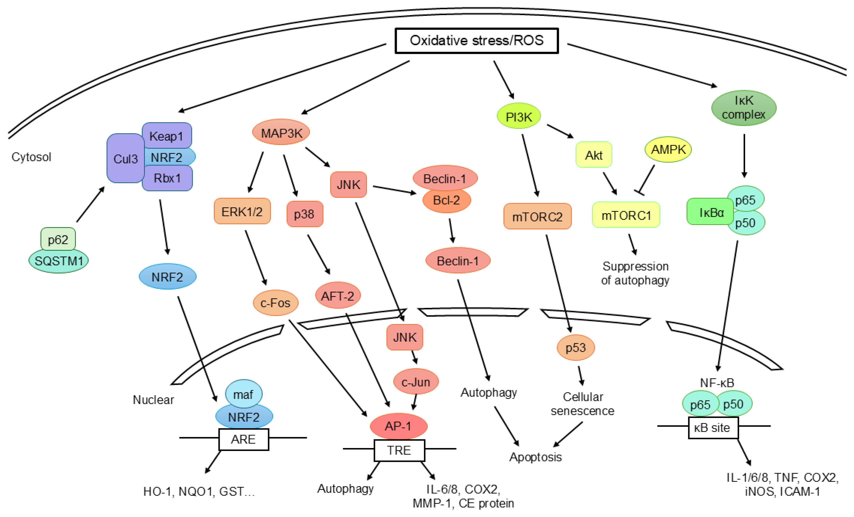
Figure 4.
Emerging redox-targeted therapeutic strategies for improving wound healing in pediatric skin. Therapeutic approaches aim to restore physiological redox homeostasis through modulation of antioxidant defenses and ROS production, epidermal barrier repair, regulation of innate immunity and the skin microbiota, and emerging precision redox medicine strategies. Abbreviations: AMP, antimicrobial peptide; NRF2, nuclear factor erythroid 2-related factor 2; NOX, NADPH oxidase; ROS, reactive oxygen species.
Figure 4.
Emerging redox-targeted therapeutic strategies for improving wound healing in pediatric skin. Therapeutic approaches aim to restore physiological redox homeostasis through modulation of antioxidant defenses and ROS production, epidermal barrier repair, regulation of innate immunity and the skin microbiota, and emerging precision redox medicine strategies. Abbreviations: AMP, antimicrobial peptide; NRF2, nuclear factor erythroid 2-related factor 2; NOX, NADPH oxidase; ROS, reactive oxygen species.
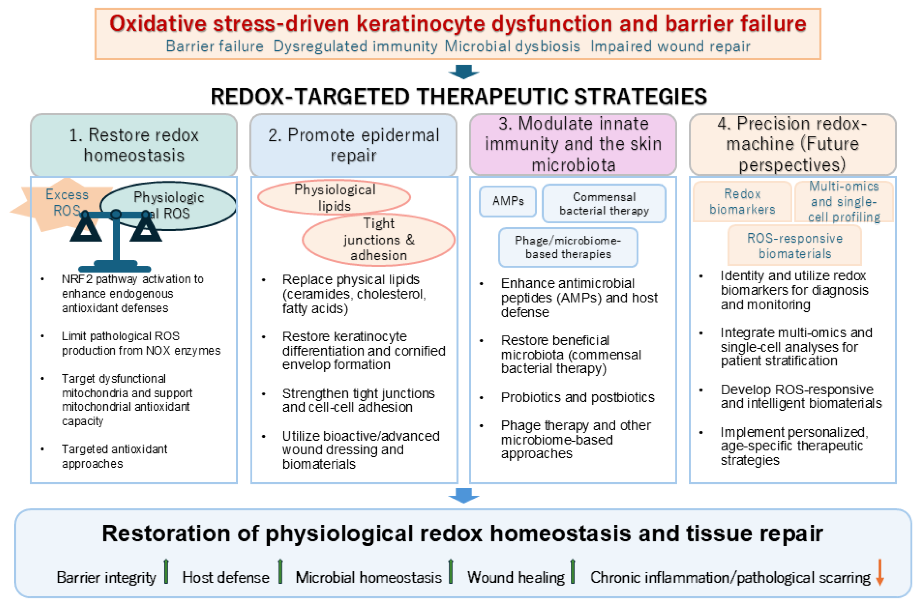
Disclaimer/Publisher’s Note: The statements, opinions and data contained in all publications are solely those of the individual author(s) and contributor(s) and not of MDPI and/or the editor(s). MDPI and/or the editor(s) disclaim responsibility for any injury to people or property resulting from any ideas, methods, instructions or products referred to in the content. |
© 2026 by the authors. Licensee MDPI, Basel, Switzerland. This article is an open access article distributed under the terms and conditions of the Creative Commons Attribution (CC BY) license.
Copyright: This open access article is published under a Creative Commons CC BY 4.0 license, which permit the free download, distribution, and reuse, provided that the author and preprint are cited in any reuse.