

Submitted:
01 September 2026
Posted:
01 September 2026
You are already at the latest version
Abstract
Recycling drainage solutions (DS) in closed-loop soilless culture systems reduces fertilizer and water consumption in greenhouse production while preventing environmental pollution caused by nitrogen and phosphorus emissions. However, recycling DS increases the risk of spreading root- infective pathogens through the irrigation system, making disinfection necessary prior to reuse. In this study, two oxygen-containing oxidizing agents, hydrogen peroxide (H₂O₂) and chlorine dioxide (ClO₂) were evaluated as nutrient solution (NS) disinfectants against Verticillium dahliae. The experiment was conducted in a closed-loop soilless pepper crop grown in a greenhouse during the spring–summer period. All treatments received nutrient solutions (NS) of identical composition. Three treatments were applied with three replicates each: (i) non-treated NS (control), (ii) NS supplemented with 2 ppm ClO₂, and (iii) NS supplemented with 2 ppm H₂O₂. Six-week-old pepper plants were inoculated with V. dahlia strain 402V. Both disinfectants significantly improved pepper fruit yield and quality compared with the non-disinfected control. ClO₂ resulted in the highest number of marketable fruits and total fruit yield, while both ClO₂ and H₂O₂ reduced unmarketable fruits and increased average fruit weight. Pathogen biomass quantification by qPCR demonstrated that ClO₂ significantly reduced pathogen presence in the hydroponic system, achieving approximately 63% lower biomass compared with the untreated control. In contrast, H₂O₂ showed limited efficacy and pathogen levels comparable to the control treatment. Application of H₂O₂ and ClO₂ significantly enhanced mineral accumulation in pepper tissues, increasing macronutrient (P, K, Mg, Ca) and micronutrient (Fe, Zn, Cu) concentrations compared with the control treatment.
Keywords:
closed-loop soilless culture
; Verticillium dahliae
; disinfectants
; chlorine dioxide
; hy-drogen peroxide
; pepper crops
1. Introduction
Soilless culture is an intensive cropping method applied mainly in greenhouses that can substantially contribute to the needs for food security and sustainability [1,2] by increasing yield and quality while using less water and fertilizers [3,4,5,6]. To reduce water and nutrient use and avoid aquifer pollution from NO₃⁻ and P emissions, it is essential to adopt circular soilless cropping systems with zero discharge of fertigation effluents [7]. However, both recycling the fertigation effluents in substrate-grown crops and recirculation of the nutrient solution in water culture systems are associated with a high risk for pathogen spread [8,9]. Therefore, to avoid the spread of root pathogens in closed-loop soilless systems, it is important to disinfect the drainage solution before reuse [6,7,9,10,11,12,13,14].
Vascular wilts are among the most dangerous diseases that affect vegetable crops [9,15,16]. Plant vascular system infections are most frequently caused by well-known hemi-biotrophic soil-borne fungus such as Verticillium dahliae [15,17,18,19,20]. V. dahliae is a xylem-invading fungal pathogen that causes vascular wilt and defoliation in a wide range of angiosperms [19], causing significant losses in Solanaceous and Cucurbitaceous crops cultivated in Mediterranean greenhouses [18]. V. dahliae infects more than 200 host plant species such as pepper, tomato, lettuce, potato, cauliflower, strawberry, eggplant, cotton, sunflower, and olive trees, causing significant yield losses annually [15,16,18,19,21,22,23,24]. V. dahliae penetrates the roots and crosses the endodermis to gain access to the vascular tissue, particularly the xylem vessels. After entering the xylem, the fungus produces conidia that are transported upward by the xylem sap and become trapped at vessel end walls. The germinating conidia produce hyphae that grow toward adjacent pit membranes, where the fungus secretes hydrolytic enzymes, including pectin-degrading enzymes, to degrade the pectin-rich pit membranes and facilitate movement between vessels. During colonization, V. dahliae spreads through the vascular system and may later colonize intercellular spaces and the middle lamella. Progressive fungal growth together with the accumulation of mycelium, spores, polysaccharides, and plant-derived gels and gums leads to vessel occlusion, disrupting water transport and ultimately causing wilt symptoms in infected plants. The symptoms of the disease in infected plants include vein clearing, chlorosis, dwarfing, foliar wilting and necrosis [15,18,23,24,25]. Over the past 60 years, two main mechanisms have been proposed to explain wilt symptoms: the vascular occlusion and the toxin involvement [21,26]. As observed in microscopic studies of infected plants, yellow vein patterning and leaf curling symptoms are associated with the trans-differentiation of bundle sheath and xylem parenchyma cells into functional xylem elements [21,27,28,29]. The pathogen persists in the soil for years in the form of microsclerotia, which constitutes the primary source of infection in the field. Plant losses reaching 90% can sometimes be caused even with inoculum densities as low as 1 to 2 microsclerotia (MS) per gram of soil [2,5,16,28,30].
Given the widespread pathogen and its high damage potential, including a disinfection system in closed-loop soilless cultivations to treat the recycled or recirculating NS is crucial. UV-based systems are widely used but costly and challenging in continuous recirculation [31,32], while chemical treatment may be a cheaper alternative if effective and residue-free. Currently, the most common chemical disinfectant applied in soilless cropping systems is NaClO (chlorine), ClO₂, O3, and H2O2 [9,31,33]. These substances, which are frequently used for disinfecting tap and wastewater, decompose into non-harmful byproducts such as O2 and Cl-, and therefore they are considered suitable for multiple applications [16].
Hydrogen peroxide (H2O2) is commonly applied even in medicine as an antibacterial agent and is considered environmentally safer, with particular interest due to its stability, moderate reactivity, and transmembrane mobility [34] than chlorine-based bleach because it could break down into oxygen and water [35]. Its effectiveness against pathogens is based either on reducing or on oxidizing actions and was discovered in 1818 by Louis Jacques Thénard [34]. It is commonly used for cleaning, disinfecting and sterilizing surgical instruments and surfaces [36]. In agriculture, especially soilless systems, properly diluted H₂O₂ is used in irrigation to promote strong root growth, control pests, and prevent oxygen-related root rot. It is a broad-spectrum disinfectant effective against bacteria, spores, fungi, and viruses, and has also been used in aquaculture to reduce microorganism-related mortality [37,38]. Studies show that applying H₂O₂ through irrigation in olive plants lowers V. dahliae levels in the soil [39]. Likewise, peroxygen-based treatments reduce pathogen persistence and help prevent Verticillium wilt in young olive plants [16,26,28,39]. Even low concentrations around 3 ppm of H2O2 can reduce spore populations, while higher concentrations and longer contact times are even more effective [39,40,41]. For general disinfectant use, 0.1 ppm to 1 ppm concentrations are typical, increasing to 1 ppm for antifungal effects. Plant leaves can resist up to 3 ppm concentration without risk [40,42].
Chlorine dioxide (ClO₂) is a disinfectant and oxidant that produces fewer toxic byproducts than chlorine[43]. Widely used across water treatment, industry, food production, and medical sterilization, it replaced chlorine for tap water disinfection in Brussels in 1956 [44]. ClO2 is a drastic oxidant that reduces fungal viruses and bacterial infections. It could block bacterial cellular processes [43]. ClO2 solutions react with amino acids and RNA, affecting the cell membrane and protein production [45]. Regarding viruses, ClO2 reacts with peptone, blocking protein formation more effectively than chlorine or ozone [46]. It works best at pH values between 3.5 and 6 and can reduce the quantity of organochlorine chemicals generated [44,47]. ClO2 is also superior to chlorine in managing biofilms in water distribution systems, above PH7 [48], and in the presence of amines and ammonia [49].
Taking this knowledge gap into consideration, the present study was designed to estimate the disinfection efficacy of ClO2 and H2O2 against V. dahliae in closed-loop soilless pepper cultivation [50].
2. Materials and Methods
Experimental Design, Plant Material, and Growth Conditions
The experiment was conducted from April to August 2022 in a glasshouse of the Laboratory of Vegetable Production at the Agricultural University of Athens (AUA: N 37◦59΄10΄΄, Е 23◦42΄29΄΄, altitude 24 m). A glasshouse compartment covering an area of 75 m2 was used for the current experiment. The experimental plant was a sweet pepper cultivar of Charleston type (Sammy RZ F1, Rijk Zwaan https://www.rijkzwaan.gr/home, accessed on 10 January 2022), which is widely cultivated in Greece. Sammy RZ F1 has a horn shape and is cultivated to produce green peppers.
The pepper plants were cultivated in a soilless cropping system that comprised 9 channels in total, with 3 channels per treatment. The experimental treatments were a) nutrient solution (NS) with no application of any disinfection agent, set as control; b) application of ClO2 to the NS at a concentration of 2 ppm; c) application of H2O2 to the NS at a concentration of 2 ppm. The channels used in the current experiment were constructed from stainless steel by Elastron S.a, Athens, Greece. The pepper plants were grown in bags containing mineral wool slabs provided by GRODAN S.A., Denmark (https://www.grodan.com/global/).
On each channel, there were 6 mineral wool slabs placed into bags, with 3 plants per slab, totaling 18 pepper plants per replication and 54 plants per treatment. The seedlings had been grown in mineral wool cubes (7.5 × 6 × 7.5 cm). Sowing took place on 02/08/2022 (M/D/Y), and the seedlings were transplanted onto the mineral wool slabs on 04/10/2022 (DAY 0) at the stage of 3–4 true leaves. The dimensions of the mineral wool slabs were 120 × 20 × 7.5 cm, corresponding to a volume of 18 L per slab, with a mean water content of 60% (v/v) after full irrigation (i.e., at container capacity). The plant density was 2.4 plants m−2. In all treatments, the entire amount of drainage solution was collected and recycled, and thus the crop was operated as a closed-loop soilless cropping system. The volume of the recirculating solution (RS) per plant was 1.8 L. In the morning, when drainage solution (DS) samples were collected, a mean DS amount of 1.15 L per plant was stored in the tank before being reused. Thus, the total volume of RS plus DS (Vd) was 2.95 L per plant.
The plants were pruned into two stems after the first flower every week until the end of the cultivation. Fruit harvesting commenced on May 22 (DAY 42). Plant pruning (leaves and stem parts) was collected weekly until DAY 65 and every second day until the end of the cultivation. The experiment was terminated on August 10 (DAY 120).
The supply of nutrients needed to replenish the DS prior to its recycling was calculated using the NUTRISENSE Decision Support System (https://nutrisense.online/). To optimize nutrition, DS samples were collected and analysed at fortnightly intervals to determine their mineral composition, and the results were introduced to the NUTRISENSE DSS to update the nutrient supply formula. The electrical conductivity (EC) of the NS supplied to the plants was 2.20 dS m-1 and the pH was 5. 50. The target composition of the NS in the root zone of the crop was according to Voogt and Sonneveld (2009). The electrical conductivity of the raw water used to prepare NS was 0.32 dS m-1 and the pH was 7.30. Before planting, all slabs were watered with NS to saturation, while after planting, the bottom of the bags was slit to allow for free drainage. The EC in the slabs and the outgoing drainage solution was 2.80 dS/m and the pH ranged from 5.30 to 6.45. The drainage solution was collected at the end of each channel, mixed with raw water up to the final volume of NS intended to be supplied to the plants, and supplied with concentrated stock solutions of fertilizers and HNO3 (for pH adjustment) to rates achieving the desired electrical conductivity (EC 2.20) and pH (5.50) to the outgoing supply solution (Table 1).
Greenhouse conditions:
The air temperature (◦C) and relative humidity (%) were monitored at 15-minute intervals throughout the cultivation period using data loggers. The minimum day and night temperatures were set at 20 and 17 ◦C, respectively, as suggested by Grimstad and Frimanslund (1993). The humidity ranged from 39 to 70.6%. The EC and pH levels in the DS, as well as the drainage fraction, were recorded every day.
Application and Quantification of Hydrogen Peroxide and Chlorine Dioxide in the Drainage Solution
H₂O₂ was applied using a commercial liquid formulation (Chemco, 35% w/w). ClO₂ was generated from Dutrion® powder, a transportable, non-explosive two-component system that reacts with a defined volume of water to produce a stabilized ClO₂ solution with a nominal concentration of 0.2–0.4% (w/v), according to the manufacturer. Disinfectant concentrations in the drainage solution were measured twice daily (approximately 09:00 and 18:00 h) in each treatment line, including the replenishment tank, emitters, and drainage outflow. Concentrations of both disinfectants were determined using a PoolLAB photometer equipped with an interchangeable cuvette for direct immersion sampling. The instrument measurement range was 0.0–11.4 mg L⁻¹ for ClO₂ and 0.00–2.90 mg L⁻¹ for H₂O₂. Additionally, H₂O₂ concentrations were cross-validated using peroxide test strips with a measurement range of 0–100 mg L⁻¹.
Pathogenicity assay:
The pathogen:
V. dahliae isolate 402V, isolated from pepper plants in the greenhouse, was obtained from the Laboratory of Plant Protection, AUA, Greece. However, no information was available regarding V. dahliae infection in plants grown in hydroponic systems and, therefore, a special protocol was developed. Ten-week-old pepper plants (DAY 75) were inoculated with a spore suspension prepared in the lab. As it concerns the fungal plate growth assay, potato dextrose agar plates (PDA) (Sigma, St. Louis, IL) were inoculated with 5μl of a fungal spore solution at a concentration of 5 × 10^7 spores/μl and incubated at 25 °C. The diameter of the colonies was measured at 7 and 14 days.
A conidial concentration of 10^7 conidia/mL were injected into the first slab of each line in the greenhouse. Disease symptoms such as vascular discoloration and foliage wilting were recorded almost two months later (DAY 120). A macroscopic approach in the greenhouse was used to evaluate the pathogenicity and aggressiveness of Verticillium isolates.
Sampling and Fungal Isolation:
Samples were collected at the end of the experiment (10/08/2022; Day 120), and disease progression was expressed as the percentage of infected leaves relative to total foliage. Stem segments from the base were excised and visually inspected for internal wood symptoms. Samples were transported to the Plant Pathology Laboratory of the AUA, where they were sectioned longitudinally and transversely, the phloem was removed, and wood fragments (~10 cm) were surface-sterilized (93% ethanol followed by flaming, repeated three times). Xylem chips from both symptomatic and asymptomatic tissues were aseptically plated onto acidified potato dextrose agar (PDA; Merck, Darmstadt, Germany) and incubated at 24 ± 0.2 °C in the dark for 5 days, with daily observations. Emerging fungal colonies were examined visually and microscopically, then subculture onto fresh PDA plates and stored at 4 °C for short-term preservation. The remaining tissue was immediately frozen at −80 °C for long-term storage, and fungal biomass was quantified by qPCR. DNA extraction was performed from stem sections (~10 cm above slab level) collected from 10 plants per treatment.
Morphological and Cultural Characterization of the collected samples
To estimate the mycelial growth of fungal isolates, mycelial-colonized PDA agar discs (5 mm in diameter) were transferred into the center of new PDA plates with a diameter of 92 mm (one disc per plate, three per isolate). The plates were incubated at 24 ◦C in the dark, and the diameter of the colonies was measured periodically for up to three weeks, or earlier if the fungus completely covered the plate surface. The growth rate of fungal isolates was expressed in mm/day. At the end of the incubation period, colony characteristics (color, mycelium, colony texture, and shape) were observed. Dimensions of reproductive structures and hyphal features (color, shape, presence or absence of septum, clump, and chlamydospores) were also recorded.
DNA Extraction & Quantification of fungal biomass in infected plant tissues:
Tissues were ground to a fine powder in the presence of dry ice or liquid nitrogen, and total DNA was extracted as described by Triantafyllopoulou et al. (2021). Briefly, 400 μL of cell lysis solution was added to each tube, followed by rapid homogenization by vortexing for 10–20 s and gently tapping and inverting the tubes. The samples were then incubated at room temperature for 5 min on a rotating Intelli-Mixer to facilitate cell lysis. Subsequently, 100 μL of pre-cooled protein–DNA precipitation solution was added, and the samples were homogenized by gently tapping and inverting the tubes. The homogenized samples were incubated on ice at 4 °C for 10 min and then centrifuged at 14,000 rpm for 20–25 min at 4 °C. Approximately 300 μL of the supernatant was transferred to a new tube. When tissue debris remained in the supernatant, the samples were centrifuged again at 14,000 rpm for 5 min, and the resulting supernatant was transferred to a new tube.
An equal volume of isopropanol was added to the supernatant, and the samples were gently homogenized. The samples were then centrifuged at 14,000 rpm for 10 min at 4 °C. The supernatant was discarded, and the resulting DNA pellet was washed with 300 μL of 70% ethanol. The samples were centrifuged at 16,000 rpm for 1 min, after which the supernatant was carefully removed using a small-volume pipette to minimize residual liquid. When necessary, the samples were briefly centrifuged again to remove any remaining supernatant. The pellet was washed once more with 300 μL of 70% ethanol and allowed to air-dry at room temperature for 5 min. The DNA pellet was subsequently resuspended in 40 μL of 10 mM Tris-HCl buffer (pH 8.0).
The concentration and quality of the extracted DNA were determined using a Q5000 UV–Vis Spectrophotometer (Quawell, San Jose, CA, USA). The DNA concentration of each isolate was subsequently adjusted to 20 ng μL⁻¹ and stored at −20 °C until further use. Isolates obtained from pepper plants maintained at the AUA laboratory were used as positive and negative controls and for the preparation of serial dilutions used to generate the standard curves [25].
PCR amplification:
The targeted amplicon was 121 bp and the conditions to generate this amplicon were 0.4 μM each hybrid primer, forward Vd-F929-947: (CGTTTCCCGTTACTCTTCT), reverse: Vd-R1076-1094: (GGATTTCGGCCCAGAAACT) (Synthesized at Integrated DNA Technologies, Inc., Coralville, IA.), underlined sequences represent the V. dahliae primer sequences. TaqMan Probe: Vdhrc FAM: [5′ 6-FAM] CACCGCAAGCAGACTCTTGAAAGCCA [3′ BHQ1] (Synthesized at Biosearch Technologies, Inc., Novato, CA) [25]. As the internal standard for normalizing the differences in plant DNA amounts. All PCR assays were carried out in a KAPA PROBE FastGene® by Merck
The amplification master mix contained 1.0 μM each of the species-specific primer pair, 0.4 μM KAPA PROBE FAST qPCR Master mix, 0.4 μM PROBE and 0.4 μM with Rox 0.2 (5 Prime; Fisher Scientific, Waltham, MA) with PCR cycling conditions set to generate the IC amplicon were 3 min initial denaturation at 95 oC and 40 cycles consisted of denaturation for 3 sec at 95 oC and primer and probe hybridization/chain elongation for 20 sec at 60 oC [20,25,51].
Figure 1.
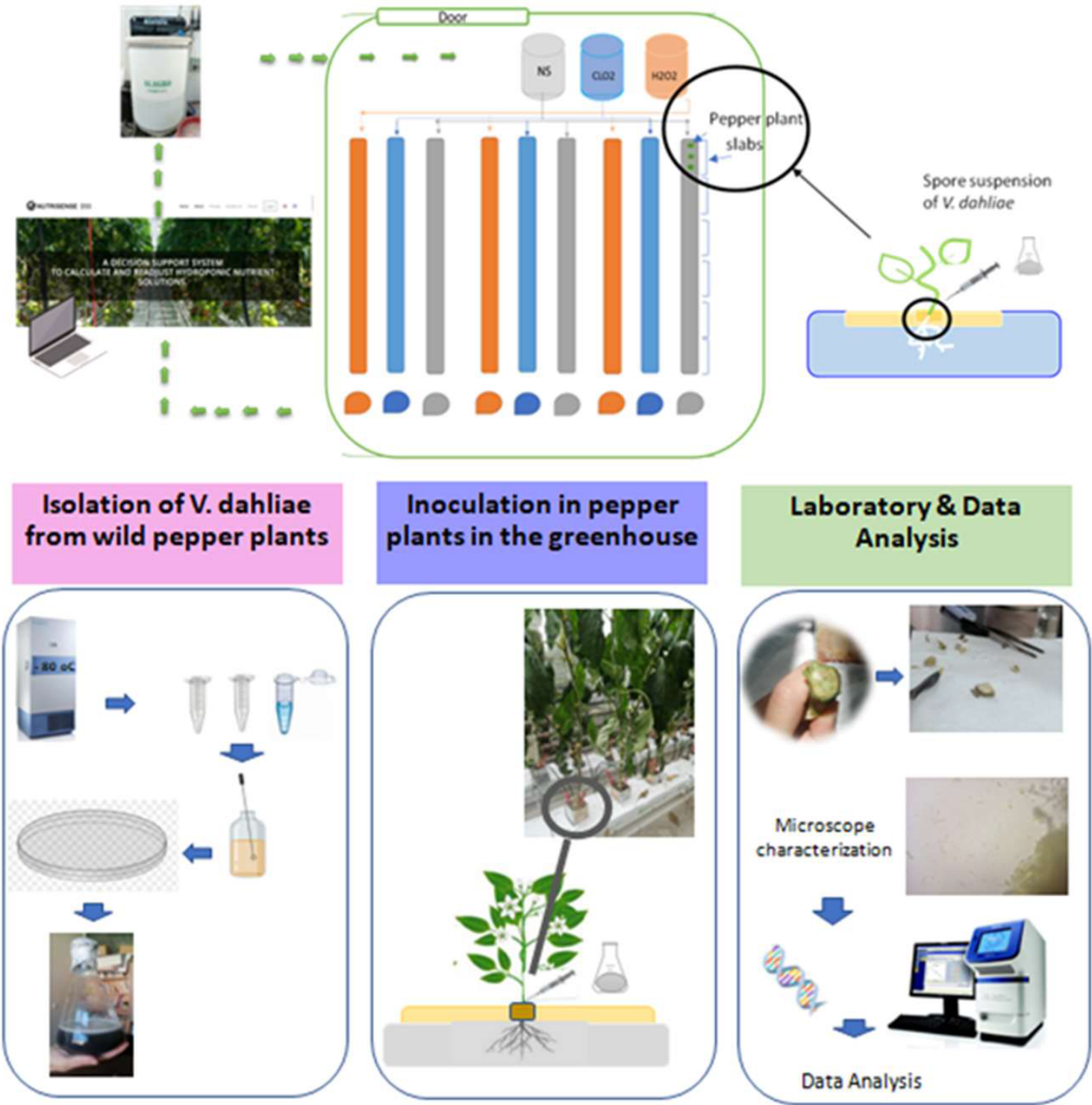
1. Spore suspension, that was prepared in the lab, was injected into six-week-old pepper plants. The concentration of conidia was adjusted to 107 conidia/mL using injection technique into of every first slab of each line. The plants were then incubated in the greenhouse for a week. 2. Symptoms of foliage wilting at the plants. 3. Vascular discoloration on the stem base.
Figure 1.
1. Spore suspension, that was prepared in the lab, was injected into six-week-old pepper plants. The concentration of conidia was adjusted to 107 conidia/mL using injection technique into of every first slab of each line. The plants were then incubated in the greenhouse for a week. 2. Symptoms of foliage wilting at the plants. 3. Vascular discoloration on the stem base.
Sampling and Quantification of Nutrient Concentrations in plant tissue:
Macro- and micronutrient concentrations were determined in pooled, oven-dried plant tissues of pepper plants grown in a circular soilless cropping system. Throughout the cropping cycle, all plants were supplied with nutrient solutions having identical mineral composition, with the exception of the respective disinfectant treatments.
The three treatments that were evaluated consisted of three rotated lines: (i) a control treatment supplied only with the nutrient solution, (ii) a treatment supplied with the nutrient solution supplemented with H₂O₂, and (iii) a treatment supplied with the nutrient solution supplemented with ClO₂ as a disinfectant. Measurements were performed at five cropping stages: the seedling stage (0 days after treatment, DAT), following disinfectant applications (40 DAT), the fruiting stage (45 and 71 DAT), the full reproductive stage (86 DAT), and at the end of the experiment (122 DAT). All plant tissues (leaves and fruits) were placed in an oven at 65 °C (STF-N 400, FALC Instruments S.L.R, Treviglio, Italia) for 5 days to dry to a constant weight. At the end of the experiment, all dried plant tissue samples were finely ground, and 0.5 g of each sample was transferred into a porcelain cup. The cups containing the samples were placed in a chamber furnace (LM-112, Linn High Therm, Hirschbach, Germany) and incinerated at 550 °C for 8 h until complete ashing was achieved. The resulting ash was subsequently extracted with 0.25 M HCl solution and filtered through 125 mm Whatman 42 filter paper. The filtrates were collected in 100 mL volumetric flasks and brought to a final volume of 100 mL with distilled water. The resulting aqueous extracts were then subjected to chemical analysis for the determination of K+, Ca2+, Mg2+, NO3- -N, P, Fe, Mn, Zn & Cu concentrations by various methods, depending on the element.
The concentrations of Ca2+, Mg2+, Fe, Mn, Zn, and Cu were measured using an Atomic Absorption Spectrophotometer (Shimadzu AA-7000, Kyoto, Japan). The measurements were performed after setting the acetylene gas flow and vacuum pressure in the AA-700 to 1.5 L/min and 3.5 bar, respectively. Potassium was measured by flame photometry, using a Flame Photometer (Sherwood Model 410, Cambridge, UK). The concentrations of phosphorus (P) were measured photometrically using a 96-position microplate spectrophotometer (Anthos Zenyth 200; Biochrom, Holliston MA, USA) at 540 nm [52] and 880 nm [30] respectively. The organically bound nitrogen content in the leaves and fruit tissue samples was measured by applying the Kjeldahl method [53]. The digestion and distillation were performed using a Labtec DT 220 with a Scrubber Labtec SR 210 and a Tecator Kjeltec 8200 (FOSS A/S, Hillerod, Denmark). Organically bound N was determined after manually titrating each distilled sample by measuring the volume (mL) of HCl solution (0.05 M) required to change the color of the solution from green to pink. The NO3-N concentrations in the aqueous extracts of the plant tissue samples were determined colorimetrically after nitrating salicylic acid and measuring the amount of nitrate at 410 nm using the Anthos Zenyth 200 spectrophotometer. The total N content in the plant tissue samples was obtained by adding the concentrations of organically bound N and NO3-N measured in their aqueous extracts.
Statistical analysis:
The current experiment was set up as a randomized design with three treatments (control as nutrient solution, ClO2 + nutrient solution and H2O2 + nutrient solution) corresponding to the one plant species (pepper). To statistically analyze the results, three rotated replicates were set for each treatment. The differences in nutrient concentrations found between the three treatments were statistically evaluated by applying one-way ANOVA using the STATISTICA software package, version 12.5 for Windows 9.0 (https://statistica.software.informer.com/9.0/) (Tulsa, OK, USA).
Data were analyzed separately for each sampling date using one-way analysis of variance (ANOVA) with treatment as the fixed factor. When the ANOVA was significant for a measured parameter, mean separation was performed using Duncan’s multiple range tests (p ≤ 0.05). Duncan’s multiple range test was used to separate the means between the three treatments. When the interaction between the treatments was significant according to the ANOVA, the same test was used to separate the means between treatments and cultivation period.
3. Results
Quantification of fungal biomass in infected plant tissues:
The pathogen biomass was quantified using a standard curve-based qPCR assay, which showed high linearity across six orders of magnitude (R2= 0.9977). Treatment with chlorine dioxide (ClO2) resulted in the most significant reduction of pathogen presence, yielding a mean biomass of 30.96 ± 0.35 pg/µl—a reduction of approximately 63% relative to the untreated control (83.14 ±3.03 pg/µl). In contrast, treatment with hydrogen peroxide (H2O2) showed similar to control pathogen load, resulting in a mean biomass of 118.30 ±24.40 pg/µl. The lack of efficacy for H2O2, supported by high intra-treatment variance (SE = 24.40), suggests that under these specific experimental conditions, the oxidative stress provided by H2O2 was insufficient to inhibit pathogen proliferation or may have been neutralized by the sample matrix.
While the R2 high value confirms the precision and reproducibility of the dilution series and pipetting technique, the calculated amplification efficiency was 76.78% (derived from a standardized slope of -4.04. This efficiency is below the ideal 90–110% range, which may indicate the presence of co-extracted inhibitors or sub-optimal primer annealing kinetics. However, because the standard curve remained highly linear across the entire dynamic range, the relative comparisons between treatment groups remain valid and accurate. These findings suggest that ClO2 is a significantly more potent agent for biomass reduction in this hydroponic system compared to H2O2, providing a clear statistical distinction (p < 0.05) as indicated by the mean separation analysis (Figure 5).
2. Pathogen Biomass Quantification
Quantification of fungal biomass in infected plant tissues
Fruit yield and quality:
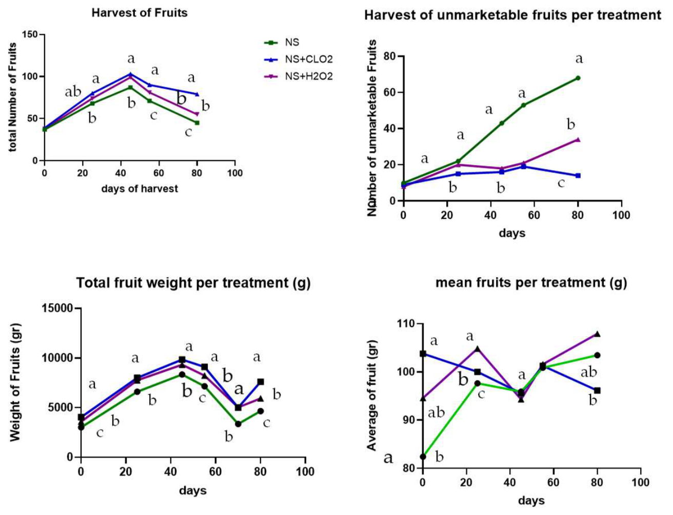
As concerns the fruit yield and quality were assessed by measuring the number of marketable fruits, the total harvested fruit weight per treatment, the mean fruit weight as the average weight of a single fruit, and the number of unmarketable fruits. These measurements summarize the overall production performance at the end of the experimental period Figure xx and during the harvesting period Figure 3.
The number of marketable fruits was significantly affected by treatment (F(2,78) = 4.41, p = 0.015). Plants treated with ClO₂ produced the highest number of marketable fruits (25.33 means), followed by H₂O₂ (23.22 means), while the control (NS) showed the lowest value (20.48 means). According to Duncan’s multiple range test (α = 0.05), ClO₂ differed significantly from the control, whereas H₂O₂ showed an intermediate response and did not differ from either treatment.
Number of unmarketable fruits was significantly affected by treatment (F(2,78) = 21.68, p < 0.001). The control (NS) produced the highest number of unmarketable fruits (12.37 means), whereas both oxidative treatments significantly reduced this value. Plants treated with ClO₂ showed the lowest number of unmarketable fruits (4.89 means), followed by H₂O₂ (6.52 means). According to Duncan’s multiple range test (α = 0.05), ClO₂ and H₂O₂ did not differ from each other and were grouped together, but both differed significantly from the control.
Total fruit weight was significantly affected by treatment (F (2,78) = 5.73, p = 0.0048). The highest total fruit weight was recorded in plants treated with ClO₂ (2524 means), followed by H₂O₂ (2351 means), while the control (NS) showed the lowest value (1988 g). According to Duncan’s multiple range test (α = 0.05), both oxidative treatments produced significantly higher total fruit weight than the control but did not differ from each other.
The average single fruit weight was significantly influenced by treatment (F(2,78) = 3.65, p = 0.030). The lowest mean fruit weight was observed in the control (NS: 96.85 means), while higher values were recorded in the oxidative treatments, with H₂O₂ showing the highest mean fruit weight (101.32 means), followed by ClO₂ (100.03 means). According to Duncan’s multiple range test (α = 0.05), H₂O₂ differed significantly from the control, whereas ClO₂ showed an intermediate response and did not differ from either treatment.
Figure 4.
Effect of treatments on a. the total number of marketable fruits at the end of the experiment, b. unmarketable harvested fruits, c. the total weight of the harvested fruits (g) and d. average weight of a single fruit (g). NS represents the control, while ClO₂ and H₂O₂ indicate the oxidative treatments. Bars represent total fruit number per treatment during the whole period of the experiment (Day 0 till Day 80).
Figure 4.
Effect of treatments on a. the total number of marketable fruits at the end of the experiment, b. unmarketable harvested fruits, c. the total weight of the harvested fruits (g) and d. average weight of a single fruit (g). NS represents the control, while ClO₂ and H₂O₂ indicate the oxidative treatments. Bars represent total fruit number per treatment during the whole period of the experiment (Day 0 till Day 80).
Plant tissue analysis:
Three treatments were applied: three rotated lines receiving only the nutrient solution (control), three rotated lines receiving the nutrient solution supplemented with H₂O₂, and three rotated lines receiving the nutrient solution supplemented with ClO₂ disinfectant. Measurements were conducted at five cropping stages: seedling stage (0 DAT), after disinfectant application (40 DAT), fruiting stage (45, 71 DAT), full reproductive stage (86 DAT), and at the end of the experiment (122 DAT). At each different treatment, different letters indicate significant differences among treatments according to Duncan’s multiple range test (p = 0.05). Macro and Micro- nutrient concentrations were determined in dry pooled plant tissues of pepper plants grown in a closed hydroponic system and supplied with nutrient solutions of identical mineral composition throughout the cropping cycle (Figure 2 and Figure 3).
According to Duncan’s multiple range test (α = 0.05), for all Macronutrient concentrations, either oxidative treatment formed a homogeneous group and differed significantly from the control after Day 40. Whereas no significant difference was detected between H₂O₂ and ClO₂ for Ca and K. P did not differ significantly between the three different treatments till the Day 71. Significant differences in Mg concentration were observed among all treatments at the beginning of disinfectant use.
The concentrations of the metallic micronutrients in the leaves of the three tested fruit vegetables are shown in 3. The concentration of Fe in plant tissues differed significantly among the three treatments at the fruiting stage (86 DAT). In contrast, Zn concentration showed significant differences among treatments at all cultivation stages following the onset of disinfectant application. Copper concentration was significantly higher in both disinfectant treatments compared with the control after the fruiting stage. Manganese concentration did not show any significant differences among treatments at any of the cultivation stages.
The application of the disinfectants H₂O₂ and ClO₂ significantly affected macronutrient concentrations in pepper plant tissues. Concentrations of P, K, Mg, and Ca were consistently higher and homogeneous statistical group in plants grown under both disinfectant treatments compared with the non-disinfected control.

Table 3.
Effect of the three different treatments control: nutrient solution, disinfection with H₂O₂ and ClO₂ on macronutrient concentrations in pepper plant tissues. Values are expressed as mg g-1 dry weight and represent means (n = 22). Within each row, different letters indicate significant differences according to Duncan’s multiple range test (P ≤ 0.05).
Table 3.
Effect of the three different treatments control: nutrient solution, disinfection with H₂O₂ and ClO₂ on macronutrient concentrations in pepper plant tissues. Values are expressed as mg g-1 dry weight and represent means (n = 22). Within each row, different letters indicate significant differences according to Duncan’s multiple range test (P ≤ 0.05).
| Macronutrient | control | H2O2 | CLO2 |
|---|---|---|---|
| (mg g-1) | means | ||
| P | 7.45 | 8.02 | 7.89 |
| K | 47.41 | 52.23 | 51.45 |
| Mg | 5.55 | 5.85 | 5.79 |
| Ca | 30.32 | 34.50 | 34.06 |
| significant | |||
| P | b | a | a |
| K | b | a | a |
| Mg | b | a | a |
| Ca | b | a | a |
As it concerns the metallic micronutrients concentrations in pepper plant tissues also responded to disinfectant application. The use of H₂O₂ and ClO₂ resulted in higher concentrations of Fe, Zn, and Cu compared with the control treatment, although the magnitude of the response varied among elements. Iron showed an intermediate response under H₂O₂, while Zn and Cu were significantly increased under either disinfectant treatment. In contrast, Mn concentration was not significantly affected by the three different treatments.
Figure 3.
Concentrations of metallic micronutrients in dry pooled plant tissues of pepper plant grown in a closed hydroponic system and supplied with a nutrient solution of the same mineral composition at cropping stages. The 1st treatment was the 3 rotated lines of the control treatment supplied only by the nutrient solution, the 3 rotated lines of the 2nd treatment supplied by the nutrient solution and 2ppm H2O2 and the 3 rotated lines of the 3rd treatment supplied by the nutrient solution and 2ppm CLO2 disinfectant. The cropping stages that measured were the seedling stage (0 DAT), the plant stage at the start of disinfectants use (40 DAT), Fruit stage (71 DAT), at full reproductive stage (86 DAT) and at the end of the experiment (122 DAT). At each cropping stage, different letters indicate significant differences between the three different treatments according to Duncan’s multiple range test (p = 0.05).
Figure 3.
Concentrations of metallic micronutrients in dry pooled plant tissues of pepper plant grown in a closed hydroponic system and supplied with a nutrient solution of the same mineral composition at cropping stages. The 1st treatment was the 3 rotated lines of the control treatment supplied only by the nutrient solution, the 3 rotated lines of the 2nd treatment supplied by the nutrient solution and 2ppm H2O2 and the 3 rotated lines of the 3rd treatment supplied by the nutrient solution and 2ppm CLO2 disinfectant. The cropping stages that measured were the seedling stage (0 DAT), the plant stage at the start of disinfectants use (40 DAT), Fruit stage (71 DAT), at full reproductive stage (86 DAT) and at the end of the experiment (122 DAT). At each cropping stage, different letters indicate significant differences between the three different treatments according to Duncan’s multiple range test (p = 0.05).
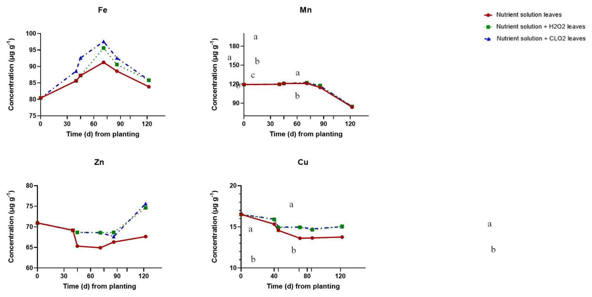
Table 4.
Effect of the three different treatments control: nutrient solution, disinfection with H₂O₂ and ClO₂ on micronutrient concentrations in pepper plant tissues. Values are expressed as μg g-1 dry weight and represent means (n = 22). Within each row, different letters indicate significant differences according to Duncan’s multiple range test (P ≤ 0.05).
Table 4.
Effect of the three different treatments control: nutrient solution, disinfection with H₂O₂ and ClO₂ on micronutrient concentrations in pepper plant tissues. Values are expressed as μg g-1 dry weight and represent means (n = 22). Within each row, different letters indicate significant differences according to Duncan’s multiple range test (P ≤ 0.05).
| Micronutrient | control | H2O2 | ClO2 |
| (μg g-1) | means | ||
| Fe | 86.53 | 87.67 | 89.62 |
| Mn | 112.66 | 113.43 | 113.42 |
| Zn | 66.40 | 69.44 | 69.59 |
| Cu | 14.54 | 15.31 | 15.33 |
| significant | |||
| Fe | b | ab | a |
| Mn | ns | ns | ns |
| Zn | b | a | a |
| Cu | b | a | a |
The application of the disinfectants H₂O₂ and ClO₂ in the closed-loop hydroponic system was associated with enhanced mineral accumulation in pepper plant tissues. Both disinfectant treatments resulted in significantly higher concentrations of the macronutrients P, K, Mg, and Ca compared with the control, with no differences between H₂O₂ and ClO₂. A similar trend was observed for the micronutrients Fe, Zn, and Cu, which were generally increased under disinfectant treatments, whereas Mn concentration was not affected. Overall, the use of H₂O₂ or ClO₂ consistently promoted higher concentrations relative to the non-disinfected control.
4. Discussion
Recirculation of drainwater in closed-loop soilless systems [4,7] may facilitate the spread of root-infecting pathogens. Therefore, a cleaning process must be applied to the drainwater before its reuse in the same crop [9]. Since there is no single, completely effective treatment against verticillium wilt, the best approach is to try multiple measures in concert while considering the specific circumstances of each field, area, and location. As chemical plant protection solutions become less available or obsolete, biological agents become more viable and environmentally safer than synthetic fungicides [54]. Plant protection should use more biological control techniques for the soilborne fungus, such as V. dahlia [55]. The efficiency is far lower, though. As a result, it is best to use various intricate defense strategies simultaneously to combat this infection [16].
The present study demonstrated that the application of oxidative disinfectants in the closed-loop hydroponic system improved nutrient accumulation, productivity, and fruit quality compared with the non-disinfected control. Nutrient responses were generally similar between H₂O₂ and ClO₂, suggesting comparable effects despite their different modes of action, so either treatment increased the concentrations of key macronutrients (P, K, Mg, and Ca) and micronutrients (Fe, Zn, and Cu), indicating enhanced nutrient uptake and translocation, while Mn did not show a significant effect. Compared with the control, both disinfectants improved yield by increasing marketable fruit production, reducing unmarketable fruits, and increasing average fruit weight. Among the treatments, ClO₂ showed the strongest effect, producing the highest number of marketable fruits and total fruit weight, whereas H₂O₂ showed intermediate improvements. Overall, oxidative disinfection not only supported pathogen control but also enhanced plant nutritional status and production efficiency, with ClO₂ providing the greatest overall benefit compared with the control.
The qPCR analysis revealed that ClO₂ was the most effective treatment for pathogen suppression, significantly reducing pathogen biomass relative to both the control and H₂O₂ treatments. The substantial reduction observed under ClO₂ indicates strong antimicrobial activity and highlights its potential for disease management in recirculating hydroponic systems. In contrast, H₂O₂ did not effectively suppress pathogen proliferation, exhibiting pathogen loads comparable to the control and greater variability within the treatment. This response suggests that the oxidative effect of H₂O₂ under the applied conditions may have been insufficient, evaporated, or partially neutralized within the hydroponic matrix. The qPCR assay demonstrated high reliability, as indicated by the strong linearity of the standard curve (R² = 0.9977), confirming accurate biomass quantification across the tested range. Although amplification efficiency (76.78%) was below the optimal range, the high linearity supports the validity of relative treatment comparisons. Overall, the results emphasize the superior efficacy of ClO₂ for pathogen control under these studied conditions. The lack of efficacy for H2O2, supported by high intra-treatment variance (SE = 24.40), suggests that under these specific experimental conditions, the oxidative stress provided by H2O2 was insufficient to inhibit pathogen proliferation or may have evaporated.
Hence, disinfection of the NS by applying chlorine is a common method of NS disinfection both in closed-loop soilless crops and in open soilless crops in the Mediterranean greenhouses, nevertheless, the safety of chlorine application is still an open question, as there are limited data about the possible accumulation of chlorate/perchlorate residues in harvested vegetables originating from soilless cultivations treated with chlorine [9,56]. Chlorine, as an anion, can be beneficial to plants by substituting for nitrates in vacuoles and positively impacting photosynthesis [9,57]. Therefore, some researchers used chlorine to replace part of the nitrates in the NS in tomato [9,45] and tobacco [58]. Relatively high concentrations have been found in tomato (0.2 mg kg-1) and carrot (0.3 mg kg-1) samples, which exceeded the MRLs set by Reg. 2020/749 (0.1 mg kg-1 and 0.15 mg kg-1, respectively) [1,9].
Recently, the use of ClO2 for water disinfection as an alternative to chlorine has been due to multiple advantages [43,59,60]. ClO2 is less harmful for the reduction of some pathogens, such as bacteria, water-containing viruses, and protozoa-like parasites [55,60,61,62]. It is less effective for the inactivation of rotaviruses and E. coli bacteria [62]. Furthermore, ClO2 eliminates and prevents biofilm formation without causing other odor issues. It also effectively eradicates phenols, addressing odor and taste concerns. Compared to chlorine, ClO2 is more efficient at removing iron and manganese, especially when they are present in complex substances. ClO2 is not negatively impacted by pH, so it does not lose efficacy over time. It also has not been negatively impacted by silica and phosphates, which could be used in the NS [63]. Compared to chlorine, ClO2 is effective at a pH between 5 and 10. Higher pH values enhance efficiency, whereas the active forms of chlorine are significantly affected by pH [63,64,65]. Unlike H2O2, ClO2 typically doesn’t hydrolyze, resulting in distinct outcomes. Consequently, its oxidation potential remains high, and its disinfection capacity remains unaffected by pH [40,41,42]. Water temperature and alkalinity do not affect the effectiveness of ClO2 for soilless culture agriculture. At disinfection concentrations, ClO2 isn’t corrosive and is more water-soluble than chlorine [64,65].
When hypochlorous acid forms through the reaction of sodium chlorite in solution, one of the byproducts is sodium hydroxide [66]. As sodium hydroxide is a common stabilizer of sodium chlorite feedstock, it can raise the pH of the mixture too high. The use of ClO2 in water treatment leads to the formation of the by-product chlorite [65,67], a high pH slows down the formation of chlorine dioxide and encourages less efficient chlorate-forming reactions. This process reflects the reaction of chlorite and hypochlorite ions in tap water, which form chlorate ions [44,64]. Chlorine-loop generators operate most effectively at high capacity since the chlorite ion is most abundant in this production mode. The World Health Organization recommends a 1 ppm concentration on food products [1]. ClO2 can also be used to fumigate fruits like blueberries, raspberries, and strawberries to eliminate molds and yeast [1,68]. At the moment, ClO2 is the most significant bleaching technique used globally [63]. The amount of ClO2 that is dozed depends upon the contact time, the pH, the temperature and the amount of pollution that is present in the water [58,61,68]. ClO2’s reaction to bacteria and other substances occurs in two steps and at the same time it produces disinfection byproducts that persist in the water. In the beginning, ClO2 forms chlorite (ClO3) by accepting an electron. Then, in the second time, it receives four electrons and transforms them into chloride (Cl-).
Chlorate and chlorite are oxidizing agents and may also be present in the water; free chlorine resulting from this process can react with organic matter, leading to the formation of harmful halogenated disinfection byproducts [61]. On the other hand, ClO2 is highly unstable and decomposes upon exposure to sunlight. Another disadvantage is the significant formation of chlorine during its production process [67].
While H2O2 is commonly used as a disinfectant, it is easily decomposed in higher pH or temperature [69]. Additionally, H2O2 may become unstable in alkaline environments too. The pH at the H2O2 environment influences its electrochemical properties, acting as an effective oxidant in acidic solutions and a reductant in alkaline solutions. Furthermore, when H2O2 acts as a reducing agent, it facilitates the production of oxygen gas. For instance, H2O2 can be used to reduce compounds like potassium permanganate and sodium hypochlorite [69,70]. Essentially, the H2O2 undergoes a chemical reaction where it donates electrons to the potassium permanganate or sodium hypochlorite, resulting in the formation of oxygen gas along with other byproducts [34,64,71].
To prevent dilution or evaporation, it’s recommended to store H2O2 in a cool, dry, and well-ventilated area, away from any flammable or combustible materials. It’s best to use containers made of non-reactive materials like stainless steel or glass, although certain plastics and aluminum alloys may also suffice [70]. Due to its rapid decomposition when exposed to light, it’s advisable to store it in an opaque container. Peroxisomes are structural components found in nearly all eukaryotic cells [71,72]. They serve various functions, including the breakdown of polyamines, D-amino acids, branched-chain fatty acids, and very long-chain fatty acids. In addition, through the action of the enzyme catalase, peroxisomes convert excess H2O2 accumulated within the cell into water, thus preventing cellular damage: H2O2 → 0.5 O2 + H2O. Finally, H2O2, whether pure or diluted, presents various risks, with the primary one being the formation of explosive mixtures upon contact with organic compounds [72].
Overall, the assumption of any effective disinfection methods using substances is essential for the long-term sustainable soilless agriculture, enabling growers to produce healthy crops while minimizing environmental impact.
5. Conclusions
By reducing the need for chemical interventions such as fungicides, the study highlights the potential for sustainable agricultural practices that are both environmentally friendly and economically viable. Daily disinfection of the nutrient solution in closed soilless cropping systems using 2 ppm doses of H2O2 and ClO2 did not cause phytotoxicity in pepper plants, which is crucial for production. However, ClO2 is more effective against the fungus because H2O2 can evaporate more easily at inconvenient temperatures and pH levels. In every soilless culture, the most appropriate form and dose of disinfectant should be used for the plants.
Nonetheless, to fully optimize the use of ClO2 in agriculture and gain a deeper understanding of its underlying mechanisms, further research through additional studies and experimentation is warranted. These efforts could help refine application techniques, dosage levels, and exposure durations, ensuring maximum efficacy while minimizing potential adverse effects. Moreover, comprehensive investigations into the mode of action of ClO2 within plant-pathogen interactions can provide invaluable insights into its potential benefits and limitations. Ultimately, this ongoing research is essential for developing evidence-based strategies that support the widespread adoption of ClO2 as a sustainable solution for disease management in agriculture.
Author Contributions
Conceptualization, D.S., S.E.T., E.J.P and G.N.; methodology, D.S., S.E.T., E.J.P and G.N.; software, D.S. and E.X.; investigation, E.X., C.T., and V.D.; resources, D.S., S.E.T., E.J.P.; data curation, D.S., E.X. and C.T.; writing—original draft preparation, D.S. and E.X.; writing—review and editing, D.S.; visualization, C.T. and D.S.; supervision, D.S.; project administration, D.S.; funding acquisition, D.S.
Data Availability Statement
The data is contained within the manuscript.
Conflicts of Interest
The authors declare no conflict of interest.
References
- FAO. https://openknowledge.fao.org/server/api/core/bitstreams/6fa56152-c232-4bc6-8eee- 7d413261ad64/content, doi:https://www.fao.org/family-farming/detail/en/c/1613700/. 2022.
- Jayachandran Research Scholar; A.; Jain, S.; Saini, S.; Subhasmita, S.; Kumar Shukla Research Scholar; K.; Jayachandran, A.; Maurya, P.; Kumar Shukla, K.; Kiran, B. Hydroponics: An Art of Soil Less Farming. 1049 Pharma Innov. J. 2022, 1049–1053. [Google Scholar]
- Savvas, D.; Gruda, N. Application of Soilless Culture Technologies in the Modern Greenhouse Industry - A Review. Eur. J. Hortic. Sci. 2018, 83, 280–293. [Google Scholar] [CrossRef]
- Xaxiri, E.; Darivakis, E.; Karavidas, I.; Ntatsi, G.; Savvas, D. Comparing the Nutritional Needs of Two Solanaceae and One Cucurbitaceae Species Grown Hydroponically under the Same Cropping Conditions. Plants 2023, 12. [Google Scholar] [CrossRef] [PubMed]
- Sharma, R.; Barnwal, P.; Vaishnav, T. Das; Mishra, S.; Ekka, S.K.; Kushwaha, A. A Concept of Hydroponic System in Horticultural Crops. Asian J. Biol. 2024, 20, 1–6. [Google Scholar] [CrossRef]
- Savvas, D.; Giannothanasis, E. Simulation Study to Assess the Efficiency of the Software NUTRISENSE to Optimise Nutrient Supply Using Ion Selective Electrodes in Closed-Loop Soilless Cropping Systems. Mol. Sci. Appl. 2025, 5, 6–18. [Google Scholar] [CrossRef]
- Savvas, D.; Giannothanasis, E.; Ntanasi, T.; Karavidas, I.; Drakatos, S.; Panagiotakis, I.; Neocleous, D.; Ntatsi, G. Improvement and Validation of a Decision Support System to Maintain Optimal Nutrient Levels in Crops Grown in Closed-Loop Soilless Systems. Agric. Water Manag. 2023, 285. [Google Scholar] [CrossRef]
- Savvas, D.; Gruda, N. Application of Soilless Culture Technologies in the Modern Greenhouse Industry - A Review. Eur. J. Hortic. Sci. 2018, 83, 280–293. [Google Scholar] [CrossRef]
- Lykogianni, M.; Bempelou, E.; Karavidas, I.; Anagnostopoulos, C.; Aliferis, K.A.; Savvas, D. Impact of Sodium Hypochlorite Applied as Nutrient Solution Disinfectant on Growth, Nutritional Status, Yield, and Consumer Safety of Tomato (Solanum Lycopersicum L.) Fruit Produced in a Soilless Cultivation. Horticulturae 2023, 9. [Google Scholar] [CrossRef]
- Runia’, W.T. View of Disinfection of Drainwater from Soilless Cultures by Heat Treatment_. 1988. [Google Scholar] [CrossRef]
- EPA Guidance Manual Alternative Disinfectants and Oxidants 4-1. 1999.
- Riggio, G.M.; Jones, S.L.; Gibson, K.E. Risk of Human Pathogen Internalization in Leafy Vegetables during Lab-Scale Hydroponic Cultivation. Horticulturae 2019, 5. [Google Scholar] [CrossRef]
- Sela Saldinger, S.; Rodov, V.; Kenigsbuch, D.; Bar-Tal, A. Hydroponic Agriculture and Microbial Safety of Vegetables: Promises, Challenges, and Solutions. Horticulturae 2023, 9. [Google Scholar] [CrossRef]
- Topalcengiz, Z.; Chandran, S.; Gibson, K.E. A Comprehensive Examination of Microbial Hazards and Risks during Indoor Soilless Leafy Green Production. Int. J. Food Microbiol. 2024, 411. [Google Scholar] [CrossRef] [PubMed]
- TJAMOS, E.C.; PAPLOMATAS, E.J. Effect of Soil Solarization on the Survival of Fungal Antagonists of Verticillium Dahliae. EPPO Bull. 1987, 17, 645–653. [Google Scholar] [CrossRef]
- Kowalska, B. Management of the Soil-Borne Fungal Pathogen – Verticillium Dahliae Kleb. Causing Vascular Wilt Diseases. J. Plant Pathol. 2021, 103, 1185–1194. [Google Scholar] [CrossRef]
- Sanogo, S. Interactive Effects of Two Soilborne Pathogens, Phytophthora Capsici and Verticillium Dahliae, on Chile Pepper. Phytopathology 2007, 97, 37–43. [Google Scholar] [CrossRef] [PubMed]
- Paplomatas, E.J.; Elena, K.; Tsagkarakou, A. Screening Tomato and Cucurbit Rootstocks for Resistance to Verticillium Dahliae. EPPO Bull. 2000, 30, 239–242. [Google Scholar] [CrossRef]
- Triantafyllopoulou, A.; Tzima, A.K.; Chronopoulou, E.G.; Labrou, N.E.; Kang, S.; Paplomatas, E.J. Overexpression of an NLP Protein Family Member Increases Virulence of Verticillium Dahliae. Plant Pathol. 2024, 73, 1264–1275. [Google Scholar] [CrossRef]
- Tsoukas, C.; Venieraki, A.; Savvas, D.; Paplomatas, E. First Report of Pythium Root Rot of Hydroponic Lettuce (Lactuca Sativa) in Greece, Caused by Pythium Cluster B2a Sp. Phytopathol. Mediterr. 2023, 62, 355–359. [Google Scholar] [CrossRef]
- Bhat, R.G.; Subbarao, K. V Genetics and Resistance Host Range Specificity in Verticillium Dahliae; 1999. [Google Scholar]
- Bilodeau, G.J.; Koike, S.T.; Uribe, P.; Martin, F.N. Development of an Assay for Rapid Detection and Quantification of Verticillium Dahliae in Soil. Phytopathology 2012, 102, 331–343. [Google Scholar] [CrossRef] [PubMed]
- Tzima, A.K.; Paplomatas, E.J.; Tsitsigiannis, D.I.; Kang, S. The G Protein β Subunit Controls Virulence and Multiple Growth- and Development-Related Traits in Verticillium Dahliae. Fungal Genet. Biol. 2012, 49, 271–283. [Google Scholar] [CrossRef] [PubMed]
- Zhu, Y.; Zhao, M.; Li, T.; Wang, L.; Liao, C.; Liu, D.; Zhang, H.; Zhao, Y.; Liu, L.; Ge, X.; et al. Interactions between Verticillium Dahliae and Cotton: Pathogenic Mechanism and Cotton Resistance Mechanism to Verticillium Wilt. Front. Plant Sci. 2023, 14. [Google Scholar] [CrossRef] [PubMed]
- Triantafyllopoulou, A.; Tzanaki, A.; Balomenou, O.I.; Jiménez-Díaz, R.M.; Tzima, A.; Paplomatas, E. Development of a Robust, VdNEP Gene-Based Molecular Marker to Differentiate between Pathotypes of Verticillium Dahliae. Plant Pathol. 2022, 71, 1404–1416. [Google Scholar] [CrossRef]
- Zhang, D.D.; Dai, X.F.; Klosterman, S.J.; Subbarao, K. V.; Chen, J.Y. The Secretome of Verticillium Dahliae in Collusion with Plant Defence Responses Modulates Verticillium Wilt Symptoms. Biol. Rev. 2022, 97, 1810–1822. [Google Scholar] [CrossRef] [PubMed]
- Inderbitzin, P.; Subbarao, K. V. Verticillium Systematics and Evolution: How Confusion Impedes Verticillium Wilt Management and How to Resolve It. Phytopathology 2014, 104, 564–574. [Google Scholar] [CrossRef] [PubMed]
- Gómez-Gálvez, F.J.; Vega-Macías, V.; Hidalgo-Moya, J.C.; Hidalgo-Moya, J.J.; Rodríguez-Jurado, D. Application to Soil of Disinfectants through Irrigation Reduces Verticillium Dahliae in the Soil and Verticillium Wilt of Olive. Plant Pathol. 2020, 69, 272–283. [Google Scholar] [CrossRef]
- Reusche, M.; Truskina, J.; Thole, K.; Nagel, L.; Rindfleisch, S.; Tran, V.T.; Braus-Stromeyer, S.A.; Braus, G.H.; Teichmann, T.; Lipka, V. Infections with the Vascular Pathogens Verticillium Longisporum and Verticillium Dahliae Induce Distinct Disease Symptoms and Differentially Affect Drought Stress Tolerance of Arabidopsis Thaliana. Environ. Exp. Bot. 2014, 108, 23–37. [Google Scholar] [CrossRef]
- Melo, V.F.; Uchôa, S.C.P.; Senwo, Z.N.; Amorim, R.J.P. Phosphorus Adsorption of Some Brazilian Soils in Relations to Selected Soil Properties. Open J. Soil Sci. 2015, 05, 101–109. [Google Scholar] [CrossRef]
- Kruithof, J.C.; Kamp, P.C.; Martijn, B.J. UV/H2O2 Treatment: A Practical Solution for Organic Contaminant Control and Primary Disinfection. Ozone Sci. Eng. 2007, 29, 273–280. [Google Scholar] [CrossRef]
- Dotson, A.D.; Keen, V.S.; Metz, D.; Linden, K.G. UV/H2O2 Treatment of Drinking Water Increases Post-Chlorination DBP Formation. Water Res. 2010, 44, 3703–3713. [Google Scholar] [CrossRef] [PubMed]
- Scarlett, K.; Collins, D.; Tesoriero, L.; Jewell, L.; van Ogtrop, F.; Daniel, R. Efficacy of Chlorine, Chlorine Dioxide and Ultraviolet Radiation as Disinfectants against Plant Pathogens in Irrigation Water. Eur. J. Plant Pathol. 2016, 145, 27–38. [Google Scholar] [CrossRef]
- Bienert, G.P.; Schjoerring, J.K.; Jahn, T.P. Membrane Transport of Hydrogen Peroxide. Biochim. Biophys. Acta Biomembr. 2006, 1758, 994–1003. [Google Scholar] [CrossRef] [PubMed]
- Abdelshafy, A.M.; Neetoo, H.; Al-Asmari, F. Antimicrobial Activity of Hydrogen Peroxide for Application in Food Safety and COVID-19 Mitigation: An Updated Review. J. Food Prot. 2024, 87. [Google Scholar] [CrossRef] [PubMed]
- McEvoy, B.; Rowan, N.J. Terminal Sterilization of Medical Devices Using Vaporized Hydrogen Peroxide: A Review of Current Methods and Emerging Opportunities. J. Appl. Microbiol. 2019, 127, 1403–1420. [Google Scholar] [CrossRef] [PubMed]
- Paul Gerard Thomas, by; Panel Assoc Surya Bhattarai, S.P.; Walsh, K.B.; Balsys, R.J. Optim. Stabilised Hydrog. Peroxide Use Drip Irrig. 2021. [CrossRef]
- Lakhiar, I.A.; Yan, H.; Syed, T.N.; Zhang, C.; Shaikh, S.A.; Rakibuzzaman, M.; Vistro, R.B. Soilless Agricultural Systems: Opportunities, Challenges, and Applications for Enhancing Horticultural Resilience to Climate Change and Urbanization. Horticulturae 2025, 11. [Google Scholar] [CrossRef]
- Gómez-Gálvez, F.J.; Hidalgo-Moya, J.C.; Vega-Macías, V.; Hidalgo-Moya, J.J.; Rodríguez-Jurado, D. Reduced Introduction of Verticillium Dahliae through Irrigation Systems and Accumulation in Soil by Injection of Peroxygen-Based Disinfectants. Plant Pathol. 2019, 68, 116–126. [Google Scholar] [CrossRef]
- Malik, D.J.; Shaw, C.M.; Rielly, C.D.; Shama, G. The Inactivation of Bacillus Subtilis Spores at Low Concentrations of Hydrogen Peroxide Vapour. J. Food Eng. 2013, 114, 391–396. [Google Scholar] [CrossRef]
- Mickelsen, R.L.; Wood, J.; Calfee, M.W.; Serre, S.; Ryan, S.; Touati, A.; Delafield, F.R.; Aslett, L.D. Low-Concentration Hydrogen Peroxide Decontamination for Bacillus Spore Contamination in Buildings. Remediation 2019, 30, 47–56. [Google Scholar] [CrossRef] [PubMed]
- Orlikowski, L.; Sas-Paszt, L.; Wojdyła, A.; Orlikowska, T. The Use of Hydrogen Peroxide and Silver Nanoparticles in Horticulture. J. Hortic. Res. 2023, 31, 1–22. [Google Scholar] [CrossRef]
- Silverj, R.; Di Palma, L.; Ermini, A.; Macchia, A. Toward Sustainable Chlorine Dioxide: Managing and Minimizing By-Product Generation 2025. [CrossRef]
- Agency for Toxic Substances and Disease Registry TOXICOLOGICAL PROFILE FOR CHLORINE DIOXIDE AND CHLORITE. 2004.
- Sharma, V.K.; Sohn, M. Reactivity of Chlorine Dioxide with Amino Acids, Peptides, and Proteins. Environ. Chem. Lett. 2012, 10, 255–264. [Google Scholar] [CrossRef]
- Mechanistic Insight into Virus Disinfection: Influence of Virus Characteristics, Solution Composition and Surface Interactions Anne Thérèse SIGSTAM. [CrossRef]
- Wilson, S.C.; Wu, C.; Andriychuk, L.A.; Martin, J.M.; Brasel, T.L.; Jumper, C.A.; Straus, D.C. Effect of Chlorine Dioxide Gas on Fungi and Mycotoxins Associated with Sick Building Syndrome. Appl. Environ. Microbiol. 2005, 71, 5399–5403. [Google Scholar] [CrossRef] [PubMed]
- Kastner, J.R.; Das, K.C.; Hu, C.; Mc Clendon, R. Effect of PH and Temperature on the Kinetics of Odor Oxidation Using Chlorine Dioxide. J. Air Waste Manag. Assoc. 2003, 53, 1218–1224. [Google Scholar] [CrossRef] [PubMed]
- Savvas, D.; Savva, A.; Ntatsi, G.; Ropokis, A.; Karapanos, I.; Krumbein, A.; Olympios, C. Effects of Three Commercial Rootstocks on Mineral Nutrition, Fruit Yield, and Quality of Salinized Tomato. J. Plant Nutr. Soil Sci. 2011, 174, 154–162. [Google Scholar] [CrossRef]
- Coşkun, F.; Alptekin, Y.; Demir, S. Reaction of Different Pepper (Capsicum Annuum L.) Cultivars to Isolates of Verticillium Dahliae Kleb. from Various Hosts. Yuz. Yil Univ. J. Agric. Sci. 2021, 31, 838–846. [Google Scholar] [CrossRef]
- Schnetger, B.; Lehners, C. Determination of Nitrate plus Nitrite in Small Volume Marine Water Samples Using Vanadium(III)Chloride as a Reduction Agent. Mar. Chem. 2014, 160, 91–98. [Google Scholar] [CrossRef]
- Sáez-Plaza, P.; Michałowski, T.; Navas, M.J.; Asuero, A.G.; Wybraniec, S. An Overview of the Kjeldahl Method of Nitrogen Determination. Part I. Early History, Chemistry of the Procedure, and Titrimetric Finish. Crit. Rev. Anal. Chem. 2013, 43, 178–223. [Google Scholar] [CrossRef]
- Testempasis, S.I.; Markakis, E.A.; Tavlaki, G.I.; Soultatos, S.K.; Tsoukas, C.; Gkizi, D.; Tzima, A.K.; Paplomatas, E.; Karaoglanidis, G.S. Grapevine Trunk Diseases in Greece: Disease Incidence and Fungi Involved in Discrete Geographical Zones and Varieties. J. Fungi 2024, 10. [Google Scholar] [CrossRef] [PubMed]
- Angelopoulou, D.J.; Naska, E.J.; Paplomatas, E.J.; Tjamos, S.E. Biological Control Agents (BCAs) of Verticillium Wilt: Influence of Application Rates and Delivery Method on Plant Protection, Triggering of Host Defence Mechanisms and Rhizosphere Populations of BCAs. Plant Pathol. 2014, 63, 1062–1069. [Google Scholar] [CrossRef]
- Copes, W.E.; Ojiambo, P.S. Efficacy of Peroxygen Disinfestants against Fungal Plant Pathogens. A Systemic Review and Meta-Analysis. Crop Prot. 2023, 164. [Google Scholar] [CrossRef]
- Nerlich, A.; Karlowsky, S.; Schwarz, D.; Förster, N.; Dannehl, D. Soilless Tomato Production: Effects of Hemp Fiber and Rock Wool Growing Media on Yield, Secondary Metabolites, Substrate Characteristics and Greenhouse Gas Emissions. Horticulturae 2022, 8. [Google Scholar] [CrossRef]
- Wege, S.; Gilliham, M.; Henderson, S.W. Chloride: Not Simply a “Cheap Osmoticum”, but a Beneficial Plant Macronutrient. J. Exp. Bot. 2017, 68, 3057–3069. [Google Scholar] [CrossRef] [PubMed]
- Franco-Navarro, J.D.; Brumós, J.; Rosales, M.A.; Cubero-Font, P.; Talón, M.; Colmenero-Flores, J.M. Chloride Regulates Leaf Cell Size and Water Relations in Tobacco Plants. J. Exp. Bot. 2016, 67, 873–891. [Google Scholar] [CrossRef] [PubMed]
- Feliciano Cayanan, D.; Zheng, Y.; Zhang, P.; Graham, T.; Dixon, M.; Chong, C.; Llewellyn, J. Sensitivity of Five Container-Grown Nursery Species to Chlorine in Overhead Irrigation Water; 2008; Vol. 43. [Google Scholar]
- Migliaccio, K.W.; Boman, B.; Clark, G.A. Treat. Irrig. Syst. With Chlorine 1. [CrossRef] [PubMed]
- Zhang, Z.; McCann, C.; Stout, J.E.; Piesczynski, S.; Hawks, R.; Vidic, R.; Yu, V.L. Safety and Efficacy of Chlorine Dioxide for Legionella Control in a Hospital Water System; 2007; Vol. 28. [Google Scholar]
- Wang, Y.J.; Deering, A.J.; Kim, H.J. The Occurrence of Shiga Toxin-Producing E. Coli in Aquaponic and Hydroponic Systems. Horticulturae 2020, 6. [Google Scholar] [CrossRef]
- Jefri, U.H.N.M.; Khan, A.; Lim, Y.C.; Lee, K.S.; Liew, K. Bin; Kassab, Y.W.; Choo, C.Y.; Al-Worafi, Y.M.; Ming, L.C.; Kalusalingam, A. A Systematic Review on Chlorine Dioxide as a Disinfectant. J. Med. Life 2022, 2022, 313–318. [Google Scholar] [CrossRef] [PubMed]
- EPA Guidance Manual Alternative Disinfectants and Oxidants 4-1. 1999.
- ® Basic Chemicals Sodium Chlorite Cooling Water Treatment with Chlorine Dioxide
- Wang, L.; Bassiri, M.; Najafi, R.; Najafi, K.; Yang, J.; Khosrovi, B.; Hwong, W.; Barati, E.; Belisle, B.; Celeri, C.; et al. Hypochlorous Acid as a Potential Wound Care Agent Part I. Stabilized Hypochlorous Acid: A Component of the Inorganic Armamentarium of Innate Immunity; 2007. [Google Scholar]
- Truzsi, A.; Kovács, B.; Bodnár, I.; Fábián, I. Controlling the Formation of Halogenated Byproducts in the Chlorination of Source Waters by Oxidative Pre-Treatment with the Fe(II)/Fe(III)-S(IV)-Air System. Ecotoxicol. Environ. Saf. 2022, 244. [Google Scholar] [CrossRef] [PubMed]
- Sy, K. V.; Mcwatters, K.H.; Beuchat, L.R. Efficacy of Gaseous Chlorine Dioxide as a Sanitizer for Killing Salmonella, Yeasts, and Molds on Blueberries, Strawberries, and Raspberries; 2005; Vol. 68. [Google Scholar]
- Chernyshov, I.Y.; Vener, M. V.; Prikhodchenko, P. V.; Medvedev, A.G.; Lev, O.; Churakov, A. V. Peroxosolvates: Formation Criteria, H2O2 Hydrogen Bonding, and Isomorphism with the Corresponding Hydrates. Cryst. Growth Des. 2017, 17, 214–220. [Google Scholar] [CrossRef]
- Espinoza-Vergara, J.; Molina, P.; Walter, M.; Gulppi, M.; Vejar, N.; Melo, F.; Urzua, M.; Muñoz, H.; Zagal, J.H.; Zhou, X.; et al. Effect of PH on the Electrochemical Behavior of Hydrogen Peroxide in the Presence of Pseudomonas Aeruginosa. Front. Bioeng. Biotechnol. 2021, 9. [Google Scholar] [CrossRef] [PubMed]
- Siemieniuk, A.; Rudnicka, M.; Karcz, W. The Effect of H2O2 on Elongation Growth and Oxidative Stress in Maize Coleoptile Cells Treated with Auxin and Fusicoccin. Sci. Rep. 2025, 15. [Google Scholar] [CrossRef] [PubMed]
- Uhl, L.; Gerstel, A.; Chabalier, M.; Dukan, S. Hydrogen Peroxide Induced Cell Death: One or Two Modes of Action? Heliyon 2015, 1. [Google Scholar] [CrossRef] [PubMed]
Figure 5.
qPCR standard curve showing the relationship between Log Concentration and Cycle threshold (Ct). 2. Pathogen Biomass Quantification: Mean pathogen biomass across treatments. Error bars represent standard error (SE). Different letters above bars indicate significant differences (p < 0.05).
Figure 5.
qPCR standard curve showing the relationship between Log Concentration and Cycle threshold (Ct). 2. Pathogen Biomass Quantification: Mean pathogen biomass across treatments. Error bars represent standard error (SE). Different letters above bars indicate significant differences (p < 0.05).
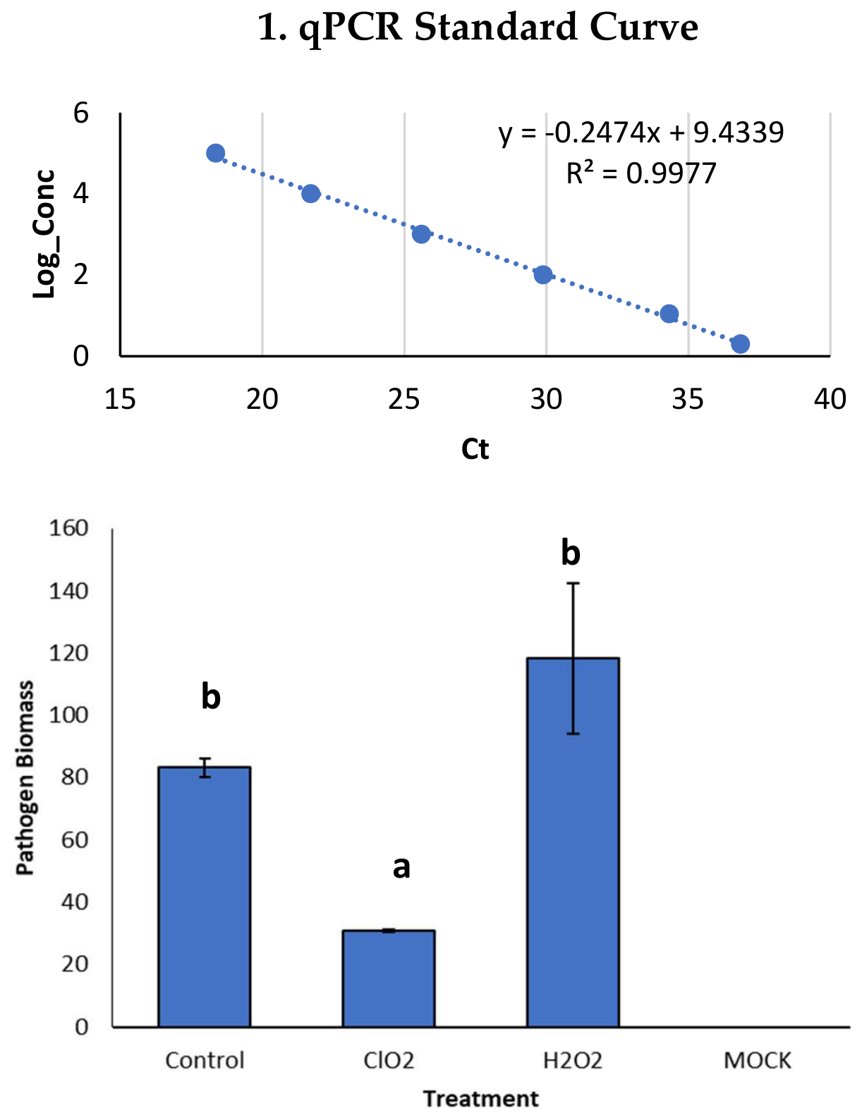
Figure 2.
Macronutrient concentrations in dry pooled plant tissues of pepper plant grown in a closed hydroponic system and supplied with a nutrient solution of the same mineral composition at cropping stages. The 1st treatment was the 3 rotated lines of the control treatment supplied only by the nutrient solution, the 3 rotated lines of the 2nd treatment supplied by the nutrient solution and 2ppm H2O2 and the 3 rotated lines of the 3rd treatment supplied by the nutrient solution and 2ppm CLO2 disinfectant. The cropping stages that measured were the seedling stage (0 DAT), the plant stage at the start of disinfectants use (40 DAT), Fruit stage (71 DAT), at full reproductive stage (86 DAT) and at the end of the experiment (122 DAT). At each cropping stage, different letters indicate significant differences between the three different treatments according to Duncan’s multiple range test (p = 0.05).
Figure 2.
Macronutrient concentrations in dry pooled plant tissues of pepper plant grown in a closed hydroponic system and supplied with a nutrient solution of the same mineral composition at cropping stages. The 1st treatment was the 3 rotated lines of the control treatment supplied only by the nutrient solution, the 3 rotated lines of the 2nd treatment supplied by the nutrient solution and 2ppm H2O2 and the 3 rotated lines of the 3rd treatment supplied by the nutrient solution and 2ppm CLO2 disinfectant. The cropping stages that measured were the seedling stage (0 DAT), the plant stage at the start of disinfectants use (40 DAT), Fruit stage (71 DAT), at full reproductive stage (86 DAT) and at the end of the experiment (122 DAT). At each cropping stage, different letters indicate significant differences between the three different treatments according to Duncan’s multiple range test (p = 0.05).
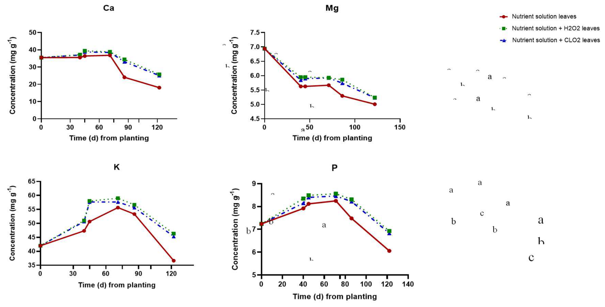
Table 1.
Concentrations of macro- and micronutrients in the root solution, i.e., the nutrient solution contained in the Slabs (EC: 2.80, pH 5.30) and in the supplied nutrient solution (EC: 2.20, pH 5. 50).
Table 1.
Concentrations of macro- and micronutrients in the root solution, i.e., the nutrient solution contained in the Slabs (EC: 2.80, pH 5.30) and in the supplied nutrient solution (EC: 2.20, pH 5. 50).
| Macronutrient | K | Ca | Mg | NH4-N | SO4-S | NO3-N | P |
| Slabs (mmol L-1) | 4.78 | 7.12 | 2.72 | 1.09 | 3.73 | 15.36 | 1.2 |
| NS (mmol L-1) | 6.4 | 4.7 | 1.45 | 0.85 | 1.75 | 14.61 | 1.25 |
| Micronutrient | Fe | Mn | Zn | Cu | B | Mo | |
| Slabs (μmol L-1) | 20 | 12 | 7 | 0.8 | 50 | 0.7 | |
| NS (μmol L-1) | 15 | 10 | 5 | 0.8 | 30 | 0.7 | |
Disclaimer/Publisher’s Note: The statements, opinions and data contained in all publications are solely those of the individual author(s) and contributor(s) and not of MDPI and/or the editor(s). MDPI and/or the editor(s) disclaim responsibility for any injury to people or property resulting from any ideas, methods, instructions or products referred to in the content. |
© 2026 by the authors. Licensee MDPI, Basel, Switzerland. This article is an open access article distributed under the terms and conditions of the Creative Commons Attribution (CC BY) license (http://creativecommons.org/licenses/by/4.0/).
Copyright: This open access article is published under a Creative Commons CC BY 4.0 license, which permit the free download, distribution, and reuse, provided that the author and preprint are cited in any reuse.