

Submitted:
01 September 2026
Posted:
01 September 2026
You are already at the latest version
Abstract
Diatoms—unicellular microalgae essential for global ecosystems—synthesize nanostructured siliceous frustules composed of valves and girdle bands. In recent years, these frustule components have attracted increasing attention as sustainable, eco-friendly, and cost-effective alternatives to selected artificially fabricated micro/nanostructures used in micro- and nanosystems, particularly in fields such as photonics, sensing, and microfluidics, leading to the rise of diatom nanotechnology. Yet, the fabrication of commercially viable diatom-based devices remains hindered by several bottlenecks, including challenges associated with the precise assembly and integration of frustule-derived components within sophisticated micro- and nanosystems. In this perspective, we focus on these challenges and propose automated micromanipulation as a potentially transformative approach for addressing them.
Keywords:
diatom frustule
; automated micromanipulation
; microsystems
; microfluidics
; photonics
Diatoms: A Sustainable Source of Nanostructured Siliceous Frustules
Diatoms are a major group of microalgae that play crucial roles in the global ecosystem, as they generate at least 20% of the global atmospheric oxygen and about 40% of organic carbon in the oceans [1,2,3,4,5]. They are a very ecologically successful group, as evidenced by their abundance in many aquatic habitats, their wide global distribution, and their remarkable diversity [6,7]. Through elegant biomineralization processes, diatoms synthesize a unique nanostructured cell wall—known as frustule—composed of amorphous hydrated silica [3,8]. The geometry and ultrastructure of frustules vary greatly among species and genera [3,9], as illustrated in Figure 1. Each frustule—regardless of its geometry—consists of two overlapping valves and one or more girdle bands [10]. These siliceous parts are often highly ornamented and porous, with pore sizes ranging from a few nm to a few μm [10].
Several diatom species have been successfully isolated and cultivated on laboratory and pilot scales for various purposes, whereas large-scale commercial cultivation remains limited [11,12]. Biomass obtained from cultivation can be used to extract high-value metabolites, including pigments and lipids [13,14,15], while the valves/girdle bands can be retrieved from the biomass by oxidation of organic materials to be utilized in applications [16]. It should be noted that diatom siliceous components can also be extracted from natural sedimentary rocks called diatomite, which contain fossil diatom frustules [17,18]. For several decades, diatomite has been widely used in various applications, such as filtration, insulation, building materials, abrasives, and as functional additives and fillers, owing to their unique physical and chemical properties [19,20,21]. Although fossil frustules are inexpensive and readily available in large quantities in the market, sourcing frustules from cultivation offers sustainability and enables better control over their structural and functional characteristics, which is highly required for advanced technological applications [19].
The Rise of Diatom Nanotechnology
At the end of the 20th century, the idea of utilizing diatom frustules in nanotechnology applications was proposed, taking advantage of their intrinsic nanoscale structural features [22]. This approach is promising as diatoms typically build their frustules with high reproducibility and nanoscale precision that challenges artificial nanofabrication techniques, while relying only on basic nutrients (including silicic acid), sunlight, and carbon dioxide under ambient conditions (see, e.g., [23]). These ideas gained attention among several researchers from various science and engineering disciplines. In 2003, the term ‘Diatom Nanotechnology’ was introduced [8]. In the same year, several researchers interested in this emerging field met at the Diatom Nanotechnology Workshop, held as part of the 17th North American Diatom Society meeting, to foster collaborations and exchange ideas [24]. Since then, the number of relevant publications has increased dramatically, associated with increasing applications in fields such as drug delivery, microfluidics, sensing, catalysis, photonics, and energy harvesting [25]. However, several key bottlenecks still hinder the transfer of these advanced applications from the laboratory to the market, such as scaling up cultivation and silica extraction processes for species intended for nanotechnology applications. Additionally, for fabricating diatom-based photonic and microfluidic devices, the precise assembly and integration of frustule-derived components within these sophisticated micro/nanosystems remains challenging, especially to obtain commercially functioning devices.
Establishing a Monolayer of Diatom Valves
Achieving a uniform monolayer of diatom valves over large substrate areas is considered among the key challenges for integrating these valves into certain optoelectronic devices, such as solar cells, where valves act as photonic crystal-like structures that improve the device efficiency [26,27,28]. Conventional techniques, such as drop-casting and spin-coating, have been investigated and can produce relatively uniform films when optimized [26,28]; however, achieving consistent coverage across large substrate areas remains challenging. Other techniques, including floating, droplet-based, and bubble-assisted assembly, have been reported in the literature to enable better control over monolayer formation and to influence valve orientation [29,30,31].
Beyond uniformity, achieving strong adhesion of the diatom monolayer on substrates is essential for device stability. Various strategies have been reported in the literature to enhance adhesion, including thermal annealing [32], promoting chemical bonding between the diatom valves and the substrate (e.g., HF-assisted bonding method [33]), and surface modification of the substrate using polyelectrolyte multilayers [34].
Microscale Assembly and Immobilization of Diatom Valves
In some applications, such as the fabrication of diatom-based highly sensitive and selective biosensing platforms [35], a continuous monolayer does not provide the required functionality. Instead, a controlled spatial distribution of diatom valves at the microscale is needed. Wang et al. [36] demonstrated a patterning strategy based on inkjet printing combined with multilayer polyelectrolyte deposition (prepared layer-by-layer). In this approach, a negatively charged top layer was established over the substrate, followed by selective deposition of a positively charged polyelectrolyte via inkjet printing, enabling electrostatic binding of negatively charged diatom valves onto predefined patterned regions (with feature sizes of approximately 300–350 µm). Another patterning strategy was reported by Wang et al. [37], where photolithography was used to define microscale patterns on a substrate, followed by ultraviolet-assisted formation of covalent Si–O–Si bonds between diatom silica and a PDMS substrate. This enabled selective and stable immobilization of diatom valves within predefined regions.
Single-Valve Manipulation and Positioning
In applications requiring the use of individual diatom valves, such as their integration as functional microelements into microfluidic systems and photonic devices, precise control over their positioning, spacing, and orientation is critical. Well-established micro- and nanoscale manipulation techniques, such as optical tweezers, are widely applied to a broad range of microparticles [38]. These techniques generally rely on predictable particle responses to externally applied fields, which in turn depend on relatively uniform geometry, density, surface charge, and optical or mechanical properties. Their direct application to diatom valves, however, remains challenging due to the inherent heterogeneity of valves, which often exhibit complex three-dimensional geometries and considerable variability in size and shape among species.
Manual Micromanipulation
In previous studies, manual micromanipulation has emerged as a practical approach for achieving precise control over both the position and orientation of individual diatom valves (see, for example, Losic et al. [39] and Wang et al. [40]). This approach has also been adopted by diatom hobbyists and artists to arrange diatom valves into intricate patterns and designs [41], as shown in Figure 1. This technique typically relies on microneedles mounted on micromanipulators, in combination with optical microscopy, to enable micrometer-scale positioning accuracy (e.g., Figure 2). Using such a system, individual valves can be picked up and precisely transferred to specific locations on substrates, although the process may be influenced by surface forces and environmental conditions. Despite offering exceptional control over position and orientation, this method remains inherently low-throughput, operator-dependent, and difficult to scale. These limitations highlight the need for automation.
Automated Micromanipulation
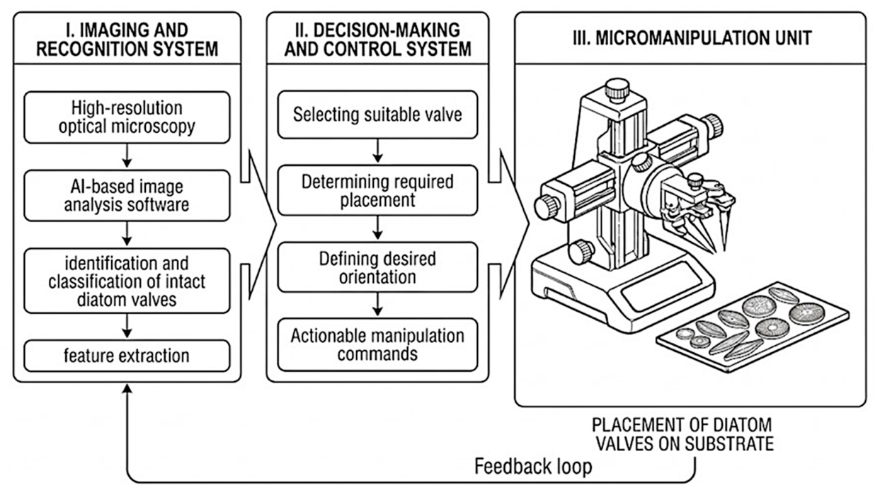
To overcome the limitations of manual micromanipulation, the development of an automated system for handling individual diatom valves is proposed, as outlined in Figure 3. A feasible automated micromanipulation platform would integrate at least three main components: (i) an imaging and recognition system, (ii) a decision-making and control system, and (iii) a micromanipulation unit.
The imaging and recognition system would combine high-resolution optical microscopy with deep learning-based image analysis, particularly convolutional neural networks (CNNs) trained on carefully selected image datasets [42,43], to enable accurate identification and classification of diatom valves within complex samples containing debris and broken fragments. Several studies have investigated CNN-based approaches for diatom valve identification and classification in the context of taxonomy and forensic applications (e.g., [44,45,46,47]), providing a strong foundation for developing the proposed system. Beyond identification, the system may also be adapted to support the extraction of key structural features such as symmetry, pore structure, and assess valve orientation, thereby providing information to support downstream decision-making processes.
Moreover, the decision-making and control system would translate visual information into actionable manipulation commands. This includes identifying and selecting suitable intact valves for pick-up, determining optimal placement locations, and defining the desired orientation based on device-specific requirements. This could be implemented using a combination of rule-based algorithms and adaptive learning strategies [48], enabling real-time feedback and dynamic adjustment during manipulation.
Furthermore, the physical manipulation could be performed using microrobotic tools such as microgrippers, microneedles, or vacuum-based probes. These tools must be carefully designed to handle the fragile and geometrically complex diatom valves, while minimizing damage and ensuring precise placement. Integration with high-precision positioning stages and closed-loop feedback systems would be essential to achieve reliable operation at the microscale.
However, it should be noted that the realization of a fully automated platform for diatom valve manipulation remains technically challenging despite recent advances in micromanipulation and automation. The variability in diatom valve geometry, size, and surface properties complicates both recognition and manipulation processes. Additionally, microscale forces—such as van der Waals interactions and capillary effects—can significantly influence pick-up and release behavior. The fragility of siliceous structures of some species further demands precise control of applied forces and environmental conditions. These challenges, however, are not prohibitive and could be addressed through interdisciplinary approaches integrating microrobotics, materials science, and artificial intelligence.
Conclusion and Outlook
Diatom frustules are a remarkable class of naturally engineered micro- and nanostructures, offering a sustainable and highly versatile platform for a broad range of applications. Over recent years, diatom nanotechnology has demonstrated successful implementation in many fields at laboratory scale. Yet, several obstacles limit the translation of diatom-based technologies from lab to market. In this perspective, we propose an integrated automated micromanipulation system for single-valve handling, positioning, and assembly as a central enabling strategy for diatom-based device fabrication. Such automated platform could significantly advance the field by enabling the transition from proof-of-concept demonstrations toward scalable and reproducible fabrication of diatom-based devices. This approach could unlock the potential of diatom frustules as building blocks for next-generation micro- and nanosystems.
Acknowledgments
The authors sincerely thank Professor Richard Gordon (Retired from University of Manitoba) for proofreading the manuscript.
References
- Armbrust, E.V. The Life of Diatoms in the World’s Oceans. Nature 2009, 459, 185–192. [Google Scholar] [CrossRef]
- Smetacek, V. Making Sense of Ocean Biota: How Evolution and Biodiversity of Land Organisms Differ from That of the Plankton. J. Biosci. 2012, 37, 589–607. [Google Scholar] [CrossRef]
- Round, F.E.; Crawford, R.M.; Mann, D.G. Diatoms: Biology and Morphology of the Genera; Cambridge university press, 1990; ISBN 0521363187. [Google Scholar]
- Leblanc, K.; Quéguiner, B.; Diaz, F.; Cornet, V.; Michel-Rodriguez, M.; Durrieu de Madron, X.; Bowler, C.; Malviya, S.; Thyssen, M.; Grégori, G.; et al. Nanoplanktonic Diatoms Are Globally Overlooked but Play a Role in Spring Blooms and Carbon Export. Nat. Commun. 2018, 9, 953. [Google Scholar] [CrossRef]
- Smol, J.P.; Stoermer, E.F. The Diatoms: Applications for the Environmental and Earth Sciences, 2nd ed.; Cambridge University Press, 2010; ISBN 9781107564961. [Google Scholar]
- Malviya, S.; Scalco, E.; Audic, S.; Vincent, F.; Veluchamy, A.; Poulain, J.; Wincker, P.; Iudicone, D.; de Vargas, C.; Bittner, L.; et al. Insights into Global Diatom Distribution and Diversity in the World’s Ocean. Proc. Natl. Acad. Sci. 2016, 113. [Google Scholar] [CrossRef]
- Guiry, M.D. How Many Species of Algae Are There? A Reprise. Four Kingdoms, 14 Phyla, 63 Classes and Still Growing. J. Phycol. 2024, 60, 214–228. [Google Scholar] [CrossRef]
- Drum, R.W.; Gordon, R. Star Trek Replicators and Diatom Nanotechnology. Trends Biotechnol. 2003, 21, 325–328. [Google Scholar] [CrossRef]
- Spaulding, S.A.; Potapova, M.G.; Bishop, I.W.; Lee, S.S.; Gasperak, T.S.; Jovanoska, E.; Furey, P.C.; Edlund, M.B. Diatoms.Org : Supporting Taxonomists, Connecting Communities. Diatom Res. 2021, 36, 291–304. [Google Scholar] [CrossRef]
- Ghobara, M.M.; Mazumder, N.; Vinayak, V.; Reissig, L.; Gebeshuber, I.C.; Tiffany, M.A.; Gordon, R. On Light and Diatoms: A Photonics and Photobiology Review. In Diatoms: Fundamentals and Applications; Seckbach, J., Gordon, R., Eds.; Wiley-Scrivener, 2019; pp. 129–189. [Google Scholar]
- Wang, J.K.; Seibert, M. Prospects for Commercial Production of Diatoms. Biotechnol. Biofuels 2017, 10. [Google Scholar] [CrossRef]
- Quelhas, P.M.; Trovão, M.; Silva, J.T.; Machado, A.; Santos, T.; Pereira, H.; Varela, J.; Simões, M.; Silva, J.L. Industrial Production of Phaeodactylum Tricornutum for CO2 Mitigation: Biomass Productivity and Photosynthetic Efficiency Using Photobioreactors of Different Volumes. J. Appl. Phycol. 2019, 31, 2187–2196. [Google Scholar] [CrossRef]
- Stonik, V.S.; Stonik, I. Low-Molecular-Weight Metabolites from Diatoms: Structures, Biological Roles and Biosynthesis. Mar. Drugs 2015, 13, 3672–3709. [Google Scholar] [CrossRef]
- Mishra, M.; Arukha, A.P.; Bashir, T.; Yadav, D.; Prasad, G.B.K.S. All New Faces of Diatoms: Potential Source of Nanomaterials and Beyond. Front. Microbiol. 2017, 8. [Google Scholar] [CrossRef]
- Mal, N.; Srivastava, K.; Sharma, Y.; Singh, M.; Kummara; Rao, M.; Manoj; Enamala, K.; Chandrasekhar, K.; Chavali, M. Facets of Diatom Biology and Their Potential Applications. Biomass Convers. Biorefin. 2022. [Google Scholar] [CrossRef]
- Wang, Y.; Cai, J.; Jiang, Y.; Jiang, X.; Zhang, D. Preparation of Biosilica Structures from Frustules of Diatoms and Their Applications: Current State and Perspectives. Appl. Microbiol. Biotechnol. 2013, 97, 453–460. [Google Scholar] [CrossRef]
- Ghobara, M.M.; Mohamed, A. Diatomite in Use: Nature, Modifications, Commercial Applications and Prospective Trends. In Diatoms: Fundamentals and Applications; Seckbach, J., Gordon, R., Eds.; Wiley-Scrivener, 2019; pp. 471–509. [Google Scholar]
- Harwood, D.M. Diatomite. In The Diatoms: Applications for the Environmental and Earth Sciences; Smol, J.P., Stoermer, E.F., Eds.; Cambridge University Press, 2010; pp. 570–574. [Google Scholar]
- Ghobara, M.; El-Sheekh, M.; Hamed, A.F.; Abdelhamid, M.A.A.; Pack, S.P. Diatom Nanostructured Biosilica. In Value-added Products from Algae; Springer International Publishing: Cham, 2024; pp. 461–492. [Google Scholar]
- Galal Mors, H.E. Diatomite: Its Characterization, Modifications and Applications. Asian J. Mater. Sci. 2010, 2, 121–136. [Google Scholar] [CrossRef]
- Crangle, R.D. Diatomite. In 2018 Minerals yearbook; U.S. Geological Survey, 2018; Vol. 22, pp. 1–6. [Google Scholar]
- Parkinson, J.; Gordon, R. Beyond Micromachining: The Potential of Diatoms. Trends Biotechnol. 1999, 17, 190–196. [Google Scholar] [CrossRef]
- Goessling, J.W.; Santiago González, A.A.; Paul Raj, V.S.; Ashworth, M.P.; Manning, S.R.; Lopez-Garcia, M. Biosilica Slab Photonic Crystals as an Alternative to Cleanroom Nanofabrication? Faraday Discuss. 2020, 223, 261–277. [Google Scholar] [CrossRef]
- Gordon, R.; Sterrenburg, F.A.S.; Sandhage, K.H. A Special Issue on Diatom Nanotechnology. J. Nanosci. Nanotechnol. 2005, 5, 1–4. [Google Scholar] [CrossRef]
- Losic, D. Diatom Nanotechnology: Progress and Emerging Applications; Royal Society of Chemistry: Cambridge, 2017; ISBN 978-1-78262-932-0. [Google Scholar]
- Jeffryes, C.; Campbell, J.; Li, H.; Jiao, J.; Rorrer, G. The Potential of Diatom Nanobiotechnology for Applications in Solar Cells, Batteries, and Electroluminescent Devices. Energy Environ. Sci. 2011, 4, 3930. [Google Scholar] [CrossRef]
- Zhang, D.Y.; Wang, Y.; Cai, J.; Pan, J.F.; Jiang, X.G.; Jiang, Y.G. Bio-Manufacturing Technology Based on Diatom Micro- and Nanostructure. Chin. Sci. Bull. 2012, 57, 3836–3849. [Google Scholar] [CrossRef]
- Ghobara, M. Optics of Diatom Frustules Toward Applications and Photobiology; Freie Universität Berlin: Berlin, 2024. [Google Scholar]
- Wang, Y.; Pan, J.; Cai, J.; Zhang, D. Floating Assembly of Diatom Coscinodiscus Sp. Microshells. Biochem. Biophys. Res. Commun. 2012, 420, 1–5. [Google Scholar] [CrossRef]
- Wang, Z.; Cai, J.; Jiang, Y.; Zhang, D. Harnessing a Droplet to Orient and Assemble Diatom Frustules. J. Micromechanics Microengineering 2019, 29, 095007. [Google Scholar] [CrossRef]
- Li, A.; Zhang, W.; Ghaffarivardavagh, R.; Wang, X.; Anderson, S.W.; Zhang, X. Towards Uniformly Oriented Diatom Frustule Monolayers: Experimental and Theoretical Analyses. Microsyst. Nanoeng. 2016, 2, 16064. [Google Scholar] [CrossRef]
- Ren, F.; Campbell, J.; Wang, X.; Rorrer, G.L.; Wang, A.X. Enhancing Surface Plasmon Resonances of Metallic Nanoparticles by Diatom Biosilica. Opt. Express 2013, 21, 15308. [Google Scholar] [CrossRef]
- Pan, J.; Wang, Y.; Cai, J.; Li, A.; Zhang, H.; Jiang, Y.; Zhang, D. Bonding of Diatom Frustules and Si Substrates Assisted by Hydrofluoric Acid. New J. Chem. 2014, 38, 206–212. [Google Scholar] [CrossRef]
- Kraai, J.A.; Wang, A.X.; Rorrer, G.L. Photonic Crystal Enhanced SERS Detection of Analytes Separated by Ultrathin Layer Chromatography Using a Diatom Frustule Monolayer. Adv. Mater. Interfaces 2020, 7, 2000191. [Google Scholar] [CrossRef]
- Li, A.; Cai, J.; Pan, J.; Wang, Y.; Yue, Y.; Zhang, D. Multi-Layer Hierarchical Array Fabricated with Diatom Frustules for Highly Sensitive Bio-Detection Applications. J. Micromechanics Microengineering 2014, 24, 025014. [Google Scholar] [CrossRef]
- Wang, W.; Gutu, T.; Gale, D.K.; Jiao, J.; Rorrer, G.L.; Chang, C. Self-Assembly of Nanostructured Diatom Microshells into Patterned Arrays Assisted by Polyelectrolyte Multilayer Deposition and Inkjet Printing. J. Am. Chem. Soc. 2009, 131, 4178–4179. [Google Scholar] [CrossRef]
- Wang, Y.; Pan, J.; Cai, J.; Li, A.; Chen, M.; Zhang, D. Assembling and Patterning of Diatom Frustules onto PDMS Substrates Using Photoassisted Chemical Bonding. Chem. Lett. 2011, 40, 1354–1356. [Google Scholar] [CrossRef]
- Cheng, K.; Guo, J.; Fu, Y.; Guo, J. Active Microparticle Manipulation: Recent Advances. Sens. Actuators A Phys. 2021, 322, 112616. [Google Scholar] [CrossRef]
- Losic, D.; Rosengarten, G.; Mitchell, J.G.; Voelcker, N.H. Pore Architecture of Diatom Frustules: Potential Nanostructured Membranes for Molecular and Particle Separations. J. Nanosci. Nanotechnol. 2006, 6, 982–989. [Google Scholar] [CrossRef]
- Wang, Z.; Gong, D.; Cai, J. Diatom Frustule Array for Flow-Through Enhancement of Fluorescent Signal in a Microfluidic Chip. Micromachines 2021, 12, 1017. [Google Scholar] [CrossRef]
- Tiffany, M.A.; Nagy, S.S. The Beauty of Diatoms. In Diatoms: Fundamentals and Applications; Wiley, 2019; pp. 33–42. [Google Scholar]
- Yu, H.; Yang, L.T.; Zhang, Q.; Armstrong, D.; Deen, M.J. Convolutional Neural Networks for Medical Image Analysis: State-of-the-Art, Comparisons, Improvement and Perspectives. Neurocomputing 2021, 444, 92–110. [Google Scholar] [CrossRef]
- Chen, L.; Li, S.; Bai, Q.; Yang, J.; Jiang, S.; Miao, Y. Review of Image Classification Algorithms Based on Convolutional Neural Networks. Remote Sens. . 2021, 13, 4712. [Google Scholar] [CrossRef]
- Pedraza, A.; Bueno, G.; Deniz, O.; Cristóbal, G.; Blanco, S.; Borrego-Ramos, M. Automated Diatom Classification (Part B): A Deep Learning Approach. Appl. Sci. 2017, 7, 460. [Google Scholar] [CrossRef]
- van der Kuil, R.; Nooren, K.; Nussbaum, M.; Bennink, E.; Sangiorgi, F.; Donders, T. Generation and Validation of a High-Resolution Fossil Diatom Record from Saliña Bartol, Bonaire, Based on Convolutional Neural Networks. Appl. Comput. Geosci. 2026, 29, 100326. [Google Scholar] [CrossRef]
- Abdelmguid, D.; Kloster, M.; Beszteri, B. Retrieving Fine-Grained Diatom Classifications from Deep Learning Models Trained on Coarse-Grained Data by over-Clustering. Biodivers. Conserv. 2026, 35, 26. [Google Scholar] [CrossRef]
- Li, J.; Jiang, C.; Yao, L.; Zhang, S. SC-DiatomNet: An Efficient and Accurate Algorithm for Diatom Classification. J. Mar. Sci. Eng. 2024, 12, 1862. [Google Scholar] [CrossRef]
- Zhang, X.; Li, G.; Shokouhi, S.; Thein, M.-W.L. Modern Control Meets Machine Learning: A Review and Taxonomy of Synergistic Approaches for Robotics Applications. Actuators 2026, 15, 235. [Google Scholar] [CrossRef]
Figure 1.
An LM micrograph showing cleaned diatom valves arranged and fixed on a glass substrate, prepared and imaged by Emiliano Bellotti. This micrograph reflects precise control over position, spacing, and orientation of diatom valves, achieved through manual micromanipulation.
Figure 1.
An LM micrograph showing cleaned diatom valves arranged and fixed on a glass substrate, prepared and imaged by Emiliano Bellotti. This micrograph reflects precise control over position, spacing, and orientation of diatom valves, achieved through manual micromanipulation.
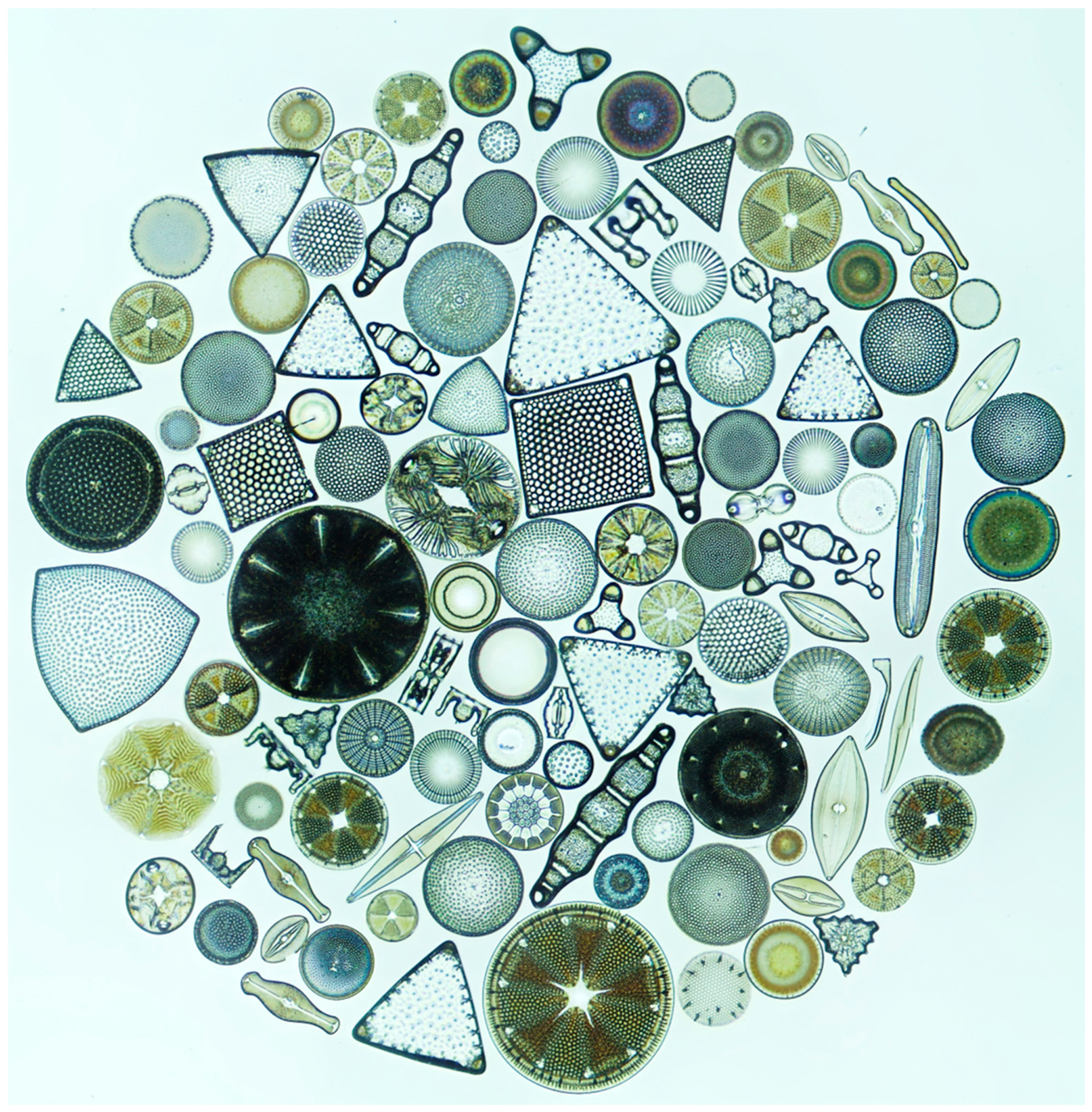
Figure 2.
Example for manual micromanipulation from Wang et al. [40]. (a) Schematic diagram illustrating the micromanipulation steps followed in this study. (b) Images of the micromanipulation setup. (c) SEM image of the fabricated microarray of Coscinodiscus sp. These images were modified and combined from the supplementary materials of Wang et al. [40] and are reproduced here under the Creative Commons Attribution 4.0 License.
Figure 2.
Example for manual micromanipulation from Wang et al. [40]. (a) Schematic diagram illustrating the micromanipulation steps followed in this study. (b) Images of the micromanipulation setup. (c) SEM image of the fabricated microarray of Coscinodiscus sp. These images were modified and combined from the supplementary materials of Wang et al. [40] and are reproduced here under the Creative Commons Attribution 4.0 License.
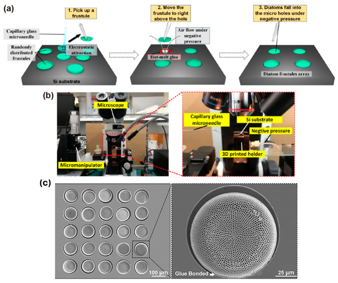
Figure 3.
An outline of the proposed automated micromanipulation system.
Disclaimer/Publisher’s Note: The statements, opinions and data contained in all publications are solely those of the individual author(s) and contributor(s) and not of MDPI and/or the editor(s). MDPI and/or the editor(s) disclaim responsibility for any injury to people or property resulting from any ideas, methods, instructions or products referred to in the content. |
© 2026 by the authors. Licensee MDPI, Basel, Switzerland. This article is an open access article distributed under the terms and conditions of the Creative Commons Attribution (CC BY) license.
Copyright: This open access article is published under a Creative Commons CC BY 4.0 license, which permit the free download, distribution, and reuse, provided that the author and preprint are cited in any reuse.