

Submitted:
31 August 2026
Posted:
01 September 2026
You are already at the latest version
Abstract
Idiopathic normal pressure hydrocephalus (iNPH) is a potentially reversible cause of gait impairment, cognitive decline, and urinary dysfunction in older adults, yet identifying patients most likely to benefit from cerebrospinal fluid (CSF) diversion remains a major diagnostic challenge. For more than five decades, the lumbar infusion test , introduced by Katzman and Hussey in 1970, has served as the principal method for assessing CSF absorptive capacity through measurement of resistance to CSF outflow (Rout). This narrative review examines the historical development, physiological basis, and contemporary clinical application of infusion testing in iNPH, with particular emphasis on the diagnostic and prognostic value of Rout. We critically review the evidence supporting the Rout thresholds recommended by current international, Japanese, and American Academy of Neurology guidelines and reassess their performance using a Bayesian framework. Recalculation of likelihood ratios from published meta-analyses demonstrates that none of the commonly proposed Rout thresholds (10-18 mmHg·min/mL) achieves the prognostic performance typically associated with a strong rule-in or rule-out test. We argue that this limited performance reflects not only methodological heterogeneity in infusion-testing protocols but also a more fundamental problem: the absence of a universally accepted reference standard for iNPH. We further examine the limitations of using postoperative clinical improvement as a surrogate reference standard, including variability in outcome definitions, delayed treatment effects, and the potential for circular reasoning. Finally, we review emerging CSF proteomic biomarkers of neuroinflammation, axonal injury, and concomitant Alzheimer-type pathology that may complement hydrodynamic assessment by capturing the balance between reversible hydrocephalus-related dysfunction and irreversible neurodegenerative burden. We conclude that Rout should be regarded as a valuable but incomplete biomarker and that future progress will likely depend on multimodal, probabilistically interpreted models integrating CSF hydrodynamics, neuroimaging, and molecular biomarkers to improve prognostic stratification and establish more robust diagnostic frameworks for iNPH.
Keywords:
idiopathic normal pressure hydrocephalus
; lumbar infusion test
; resistance to CSF outflow
; diagnostic biomarker
; Bayesian analysis
; likelihood ratio
; reference standard
; shunt responsiveness
; CSF proteomics
"Trainees are too often taught what to think and do, rather than how to think and why to do." David L. Sackett, 1991 [1].
1. Introduction
In 1964, Salomón Hakim described a 16-year-old patient who, following surgical evacuation of an acute subdural hematoma, remained in what would now be considered a “minimally conscious state” [2]. Bilateral carotid arteriography demonstrated hydrocephalus, and lumbar puncture revealed a cerebrospinal fluid (CSF) opening pressure of 150 mmH₂O [2]. Remarkably, the patient showed clear clinical improvement within 24 hours of the removal of 15 mL of CSF, including recovery of the ability to follow commands. This improvement was followed by a dramatic clinical response after ventriculoatrial shunt (VAS) placement [2]. In the same 1964 report, Hakim described two additional patients. The second case involved a 52-year-old man who was admitted for evaluation of progressive cognitive impairment, gait instability, and urinary incontinence, with an initial clinical suspicion of a neurodegenerative disorder. Neuroimaging revealed ventriculomegaly, and treatment with a VAS resulted in marked clinical improvement, with recovery to an almost normal functional state [2]. A third case involved a patient with post-traumatic hydrocephalus who also demonstrated partial clinical recovery following surgery. The paradoxical and unexpected observation that patients with symptomatic hydrocephalus may exhibit normal intracranial pressure (ICP), yet experience substantial clinical improvement following shunt surgery, prompted early efforts to elucidate the pathophysiology of what is now known as normal pressure hydrocephalus (NPH). Hakim further proposed that ventriculomegaly associated with a dementia-like syndrome was not necessarily irreversible and that selected patients could achieve meaningful clinical improvement after CSF diversion [2].
Building on Hakim’s initial report, Hakim and Adams published a second paper in 1965 that revisited two of the cases originally described in 1964 and summarized their experience with a “larger series of cases observed over the previous 10 years.” [3]. In this work, the authors also introduced the concept of the “hydraulic press” as a mechanistic explanation for hydrocephalus despite normal ICP. They proposed that ventricular enlargement increases the surface area over which CSF pressure acts, allowing relatively normal ICP to generate abnormal distending forces on the ventricular walls and adjacent brain tissue [3]. This conceptual framework was later expanded in the influential paper by Adams et al. published in the New England Journal of Medicine, which firmly established and broadened the clinical and pathophysiological concept of NPH [4]. Together, these three papers, coupled with the concluding statement by Adams et al.—that “recognition and treatment could effectively ‘cure’ a condition resembling presenile or senile dementia” [4]—signaled a paradigm shift in neurosurgery. Nevertheless, as with many transformative concepts in medicine, the emergence of NPH was accompanied by considerable controversy, and despite decades of research, several key questions regarding its pathophysiology, diagnosis, and management remain unresolved and continue to be debated.
Idiopathic normal-pressure hydrocephalus (iNPH) is characterized by a complete or incomplete clinical triad of gait impairment, cognitive impairment, and urinary urgency or incontinence, together with ventriculomegaly on neuroimaging, after exclusion of secondary causes of hydrocephalus [5]. Despite continued reluctance and skepticism among some neurologists [6,7], iNPH remains a potentially reversible neurological syndrome when patients are appropriately selected for ventricular shunt implantation and treated with modern shunt systems that incorporate technical advances introduced over the past two decades, particularly gravitational control mechanisms [8]. In some patients, CSF shunting results in only partial improvement of gait and urinary symptoms, whereas others demonstrate marked functional recovery. Still others fail to improve despite apparently appropriate patient selection and technically successful shunt placement. At the same time, growing evidence suggests that iNPH commonly coexists with other neurodegenerative disorders, including Alzheimer’s disease, Parkinson’s disease, vascular dementia, and progressive supranuclear palsy. Such comorbidities may alter the clinical presentation, complicate diagnostic assessment, and contribute substantially to the variability in postoperative outcomes observed after shunt surgery [9].
The exact prevalence of iNPH remains difficult to establish because reported estimates vary substantially according to the diagnostic criteria applied and the populations studied. Despite this uncertainty, iNPH is considered the most common form of hydrocephalus in individuals aged 65 years and older [10]. In a population-based Swedish study, Andersson et al. (2019) reported a prevalence of probable iNPH of 3.7% among individuals aged ≥ 65 years, rising to 8.9% among those aged 80 years and older [11]. According to the Global Burden of Disease Study 2021, life expectancy increased by 22.7 years worldwide between 1950 and 2021 [12]. As populations age and the prevalence of both neurodegenerative disorders and ventriculomegaly increases, distinguishing iNPH from hydrocephalus ex vacuo and incidental ventricular enlargement becomes increasingly important. The 2021 Japanese guidelines, now widely adopted internationally, classify patients as having possible, probable, or unlikely iNPH. However, even a diagnosis of probable iNPH does not reliably predict a favorable response to shunt surgery [5]. A central challenge is to identify patients in whom ventricular enlargement primarily reflects reversible CSF-dynamic disturbances rather than underlying neurodegenerative pathology, which is generally associated with a lower likelihood of responding to CSF diversion. Failure to make this distinction may expose a vulnerable elderly population to unnecessary surgical risks while limiting the potential benefits of treatment.
Reported improvement rates following CSF diversion in patients with probable iNPH vary considerably across studies, ranging from approximately 40% in earlier, more heterogeneous cohorts [13] to more than 75% in recent series using rigorous patient-selection criteria [14]. This variability likely reflects differences in patient selection, outcome measures, follow-up duration, and the inherent biological and clinical heterogeneity of the iNPH population. Importantly, it should not be interpreted as evidence of limited efficacy of shunt surgery. Rather, treatment response exists along a continuum and is influenced by multiple factors, including baseline clinical status, comorbidities, delays in diagnosis and treatment, and the frequent coexistence of neurodegenerative pathology. These factors affect both the magnitude and durability of postoperative improvement. Consequently, the central challenge in patients with suspected iNPH is not simply to establish the diagnosis, but to identify those most likely to benefit from CSF diversion. Ancillary investigations therefore play a crucial role in bridging the gap between clinical suspicion and therapeutic decision-making.
Several ancillary investigations are recommended by current clinical practice guidelines to support the diagnosis of iNPH, including the CSF tap test, external lumbar drainage, ICP monitoring, and the lumbar infusion test (LIT). Although each provides valuable information, all are imperfect and differ in their invasiveness, technical requirements, diagnostic performance, and prognostic value. Among these techniques, the constant-rate LIT—first described by Katzman and Hussey in 1970 and subsequently refined over five decades—occupies a particularly prominent position in the evaluation of suspected iNPH [15]. The LIT is typically performed as an outpatient or short-stay hospital procedure and enables quantification of Rout, a parameter closely linked to the pathophysiology of iNPH. Its use has been endorsed by most clinical practice guidelines, and its diagnostic and prognostic performance has been extensively evaluated in large single-center and multicenter cohorts. Nevertheless, considerable uncertainty remains regarding the optimal Rout threshold for predicting shunt responsiveness across different patient populations [5,16,17,18].
This review critically appraises the LIT, tracing its evolution from the pioneering infusion studies of the 1970s to contemporary computerized assessments of CSF dynamics. We examine the physiological foundations of the test and the methodological challenges that continue to complicate its interpretation. Particular attention is devoted to its diagnostic performance, its limitations in predicting treatment response, and its role in selecting patients for shunt surgery. Special emphasis is placed on Rout, the principal parameter derived from the LIT. We critically evaluate Rout as both a diagnostic and prognostic biomarker, highlighting persistent uncertainty regarding optimal threshold values, substantial variability in predictive performance, and the continuing challenge of identifying patients most likely to benefit from CSF diversion. We further examine a fundamental methodological problem that underlies all diagnostic studies in iNPH—the absence of a universally accepted reference standard. Using a Bayesian framework, we explore how this limitation influences the interpretation of infusion-test results and constrains assessment of their true diagnostic and prognostic value. Finally, to place infusion testing within its physiological context, we review the mechanisms governing CSF production, circulation, and absorption and discuss how emerging molecular biomarkers, machine-learning approaches, and multimodal predictive models may complement hydrodynamic measurements in future diagnostic strategies.
2. Methods and Sources of Evidence
This article was conceived as a narrative review of the historical development, physiological foundations, and clinical application of the LIT in iNPH. Relevant publications were identified through searches of PubMed, Scopus, and Web of Science using combinations of keywords related to idiopathic normal-pressure hydrocephalus, CSF dynamics, lumbar infusion testing, resistance to CSF outflow, CSF tap testing, external lumbar drainage, ICP monitoring, neuroimaging, biomarkers, and shunt responsiveness. The search strategy was supplemented by backward and forward citation tracking of key articles, review of major clinical practice guidelines for iNPH, and literature mapping using Research Rabbit and Connected Papers to identify influential studies and citation networks [19,20]. Particular emphasis was placed on studies addressing CSF dynamics, infusion testing, Rout diagnostic accuracy, shunt responsiveness, and emerging biomarkers in iNPH.
2.1. Selection of Studies Evaluating the Diagnostic Performance of the LIT
For the assessment of Rout as a diagnostic and prognostic biomarker, priority was given to studies incorporated into the International Guidelines, the Japanese Guidelines for iNPH, and the American Academy of Neurology practice guideline, as these constitute the principal evidence base underpinning contemporary clinical practice [5,21,22]. Additional evidence was obtained from large prospective cohort studies and published meta-analyses identified through database searches and citation-network analysis. Studies reporting sensitivity, specificity, predictive values, likelihood ratios, or other measures of diagnostic performance for predefined Rout thresholds were independently reviewed by the two senior authors (M.-A.P. and J.S.). When overlapping cohorts were identified, priority was given to studies with the largest sample size, longest follow-up, or most complete reporting of diagnostic accuracy outcomes. Given the narrative nature of this review, no formal risk-of-bias assessment or quantitative synthesis of primary-study results was undertaken.
2.2. Re-Examining Rout Thresholds Using Likelihood Ratios
To facilitate Bayesian interpretation of published diagnostic-performance data, positive and negative likelihood ratios (LR+ and LR−) were recalculated from reported sensitivity and specificity values using standard formulas [23,24,25]. Sensitivity and specificity estimates were extracted from the major clinical practice guidelines, the primary studies cited therein, and published meta-analyses when available. No pooling of individual studies was performed, and no formal meta-analysis was undertaken. Consequently, the resulting LRs represent secondary calculations based on published data and should not be interpreted as pooled estimates of diagnostic accuracy.
3. CSF Outflow: From Classical Models to Emerging Concepts
Current evidence supports the view that CSF is produced predominantly by energy-dependent secretion within the choroid plexus, replacing earlier theories that regarded passive filtration as the principal mechanism of CSF formation [26]. However, despite decades of investigation, the mechanisms governing CSF drainage from the subarachnoid space remain incompletely understood. Multiple CSF outflow pathways have been described, but their relative importance in maintaining CSF homeostasis remains uncertain and continues to be actively debated. Moreover, much of our current understanding of CSF absorption is based on experimental animal models, and the applicability of these observations to human physiology remains a matter of ongoing controversy [27]. In parallel, growing evidence supporting the existence of the so-called glymphatic system—first characterized in rodent models—has challenged traditional views of CSF–interstitial fluid exchange by emphasizing the role of perivascular pathways in the clearance of metabolic waste from the brain [21,22]. Illiff et al. coined the term “glymphatic” to emphasize the dual nature of this pathway: its reliance on glial-mediated fluid transport and its lymphatic-like function in waste clearance [28]. The glymphatic pathway has been proposed as a brain-wide waste clearance system that functionally compensates for the lack of classical lymphatic vessels within the brain. Nevertheless, its quantitative contribution to CSF clearance, its relationship with conventional absorptive pathways, and its modulation by physiological factors such as sleep, arterial pulsatility, and aging remain incompletely characterized. Consequently, its role in human CSF physiology remains uncertain and continues to be the subject of considerable debate.
Traditionally, CSF absorption has been attributed primarily to drainage through the arachnoid villi and Pacchionian granulations into the dural venous sinuses, particularly the superior sagittal sinus [30]. More recently, evidence has emerged supporting additional CSF outflow pathways, including meningeal lymphatic and perineural routes. Nevertheless, their quantitative contribution to overall CSF clearance remains uncertain. In their landmark studies published in 1876, Key and Retzius proposed that CSF is absorbed mainly through the arachnoid villi and Pacchionian granulations into the dural venous sinuses [31]. This concept subsequently became the dominant model of CSF absorption for many decades. Using gelatin stained with Prussian blue injected into the subarachnoid space of cadavers at pressures of approximately 60 mmHg, they demonstrated continuity between the spinal and cranial subarachnoid compartments. The tracer spread throughout the craniospinal subarachnoid space and extended into the cores of the Pacchionian granulations, where it was interpreted as passing across their cellular lining into the dural venous sinuses (Figure 1). These observations provided some of the earliest anatomical evidence supporting a central role for arachnoid villi and granulations in CSF absorption.
Key and Retzius' proposal that arachnoid villi and Pacchionian granulations represent the primary mechanism of CSF absorption was subsequently challenged by observations that these structures are absent or only partially developed in newborns, infants, and several higher mammalian species. Such findings raised doubts about the universality of the classical absorption model and stimulated the search for alternative pathways involved in CSF drainage and homeostasis [26].
A major body of evidence supporting the model proposed by Key and Retzius was generated by Lewis H. Weed, who, under Harvey Cushing's mentorship, performed a series of seminal studies in 1914 that helped establish the arachnoid villi and Pacchionian granulations as the principal route of CSF absorption [32]. Although Weed's contributions to modern concepts of CSF production, absorption, ICP dynamics, and intracranial compliance are sometimes overlooked, they formed a cornerstone of twentieth-century CSF physiology. Indeed, Cushing relied heavily on Weed's work in developing the concept of the “third circulation,” which became the dominant paradigm of CSF dynamics for decades [26].
The concept that arachnoid villi and Pacchionian granulations represent the primary pathway for CSF absorption was subsequently challenged by Dandy and Blackfan [33,34]. Based on experimental studies in dogs, they proposed that CSF absorption occurs diffusely throughout the subarachnoid space and directly into the vascular circulation, questioning the exclusive role of specialized absorptive structures. Dandy further argued that support for Pacchionian granulations relied largely on postmortem injection experiments performed under artificial pressure conditions [33,34]. He also noted that these structures are poorly developed or absent during early life and in several animal species, suggesting the existence of alternative CSF drainage pathways [33,34]. Stimulated by these competing theories, Weed undertook pioneering studies of CSF physiology, examining CSF production, circulation, absorption, and their relationship to ICP [26]. Using potassium ferrocyanide as a tracer in anesthetized cats, he obtained evidence supporting the hypothesis of Key and Retzius that arachnoid villi and Pacchionian granulations play a major role in CSF absorption [35]. Weed reported that potassium ferrocyanide “was found to have passed directly into the venous sinuses by way of the arachnoid villi” [26], providing experimental support for the classical view that CSF is reabsorbed into the dural venous sinuses through specialized arachnoid structures [35]. He later reaffirmed this interpretation and rejected Dandy and Blackfan’s theory, stating that “Dandy and Blackfan’s conception of diffuse absorption from the subarachnoid space was likewise found untenable” [26].
Further support for the classical model came from Welch and Friedman, who demonstrated in vitro in 1960 that monkey arachnoid villi function as unidirectional valves [36]. Using dura mater as an interface between two chambers, they showed that water flowed only from the arachnoid surface toward the dural venous sinuses, with no reverse flow [36]. Together, these studies reinforced the classical model of CSF circulation that dominated much of the twentieth century and provided the physiological foundation for later infusion tests designed to assess CSF absorptive capacity through measurements of Rout.
4. Historical Development of the Katzman Lumbar Infusion Test
Although Katzman and Hussey are generally credited with introducing the LIT into clinical practice, the origins of infusion testing can be traced to earlier experimental work. To our knowledge, the first description of a ventriculocisternal infusion technique was published by the Belgian physiologist Isidoor Leusen in 1948. Using canine models, Leusen applied this approach to investigate the influence of CSF ionic composition on vasomotor regulation [37].
The technique was subsequently advanced by Pappenheimer et al. in 1962 through the introduction of inulin—a polysaccharide composed mainly of fructose units—as a tracer, allowing the first quantitative measurements of CSF formation and CSF–blood exchange in goats [38]. In the same year, Rall et al. used radiolabeled inulin in dogs to investigate the size of the brain extracellular space [39]. Pappenheimer’s ventriculocisternal infusion technique subsequently became the standard experimental method for studying CSF physiology and was widely adopted in animal studies of CSF formation, circulation, absorption, and solute exchange. Extending this approach to clinical research, Rubin et al. (1966) used the inulin infusion technique to quantify CSF production and investigate the effects of acetazolamide in adults with brain tumors [40], while Cutler et al. conducted a similar study in children with panencephalitis or pontine gliomas [41]. Despite differences in patient populations, both studies demonstrated that, within physiological ranges, CSF absorptive capacity exhibits a linear relationship with ICP [40,41] (Figure 2).
In a landmark study, Lorenzo et al. (1974) applied the Pappenheimer ventriculolumbar perfusion technique to investigate CSF dynamics in five patients with suspected iNPH [42]. They demonstrated that impaired CSF absorption was the predominant physiological abnormality in all cases and proposed that measurement of CSF absorptive capacity could help distinguish iNPH from cerebral atrophy. This study provided some of the earliest clinical evidence linking impaired CSF absorption to the pathophysiology of iNPH and helped establish CSF hydrodynamic testing as a potential diagnostic tool. However, it also highlighted an important limitation: correction of the underlying CSF abnormality does not invariably lead to clinical improvement, particularly in patients with coexisting parenchymal brain pathology [42]. Although the Pappenheimer technique was subsequently adopted by a limited number of investigators in both adult and pediatric populations and provided precise measurements of CSF formation and absorptive capacity, its technical complexity restricted its clinical applicability. The requirement for ventricular cannulation, tracer-based analysis, and prolonged perfusion procedures hindered widespread adoption and ultimately stimulated the search for simpler and more practical methods of assessing CSF dynamics.
4.1. Origins and Rationale
The first systematic attempt to investigate CSF dynamics in humans was undertaken by Jules Masserman, a psychiatrist and neurologist [43]. In his pioneering 1934 studies, Masserman performed prolonged CSF pressure recordings in patients maintained in the lateral decubitus position for at least two hours, reporting a mean ICP of 147.7 mmH₂O (10.8 mmHg) [44]. He further proposed a simple method for estimating CSF production from the volume of CSF removed and the time required for ICP to return to baseline. Applying this approach, he calculated a mean CSF formation rate of approximately 0.30 mL/min [44]. Rubin et al. (1966) proposed that impaired bulk CSF absorption through the outflow pathways is the principal pathophysiological mechanism underlying communicating hydrocephalus [40]. Here, bulk flow denotes the movement of CSF as a fluid mass driven by pressure gradients, in contrast to diffusion, which describes the passive movement of individual molecules according to their concentration gradients. The development of the LIT marked a major step in the clinical application of CSF physiology, providing a practical bedside approach to estimating CSF absorptive capacity without the complexity and invasiveness of the earlier ventriculolumbar perfusion techniques [15].
4.2. Foundations of Infusion Testing: The Foldes–Arrowood Studies
The conceptual origins of the Katzman–Hussey test can be traced to the studies of Foldes and Arrowood (1948), who investigated the effects of continuous subarachnoid infusion while developing a technique for spinal analgesia with 0.5% intrathecal procaine [45]. In fifteen neurosurgical patients undergoing lumbar puncture, they observed that infusion of normal saline at rates of 0.2–0.8 mL/min produced a progressive rise in CSF pressure that eventually reached a steady-state plateau. At this point, the rate of infusion was balanced by CSF absorption, providing early evidence of the pressure–flow relationship underlying CSF dynamics [45]. Infusion rates between 0.30 and 0.65 mL/min typically produced a stable plateau after approximately 40 minutes (Foldes and Arrowood, 1948; Figure 2), reflecting equilibrium between CSF inflow and absorption [45]. The observation that CSF absorption increased nonlinearly with pressure foreshadowed the nonlinear models of intracranial hydrodynamics later developed by Marmarou and colleagues. Foldes and Arrowood also noted that stopping the infusion produced a characteristic biphasic pressure decay, consisting of an initial rapid fall followed by a slower decline. Although the physiological basis of this pattern was not understood at the time, it later became important in the development of models of intracranial compliance and CSF hydrodynamics. Based on these findings, they concluded that the method could serve as “a neurological diagnostic procedure that would give quantitative information as to the changes in CSF absorption in various pathological conditions” [45]. Today, CSF absorptive capacity is commonly quantified by resistance and conductance to CSF outflow (Rou and Cout), the principal hydrodynamic measures derived from the LIT.
4.3. The Classical Katzman–Hussey Infusion Test
Robert Katzman (1925–2008) was a neurologist whose work profoundly influenced the understanding of Alzheimer’s disease (AD) [46]. In collaboration with Francis Hussey, he developed the constant-infusion manometric test—later termed the lumbar infusion test (LIT)—as a clinically applicable bedside method for quantifying CSF absorptive capacity. Their seminal 1970 report in Neurology marked a pivotal transition from experimental CSF hydrodynamics to routine clinical evaluation, providing the foundation for infusion-based assessment of communicating hydrocephalus [15,47]. In the original test, sterile saline was infused into the lumbar subarachnoid space at 0.76 mL/min, approximately twice the normal rate of CSF formation, while CSF pressure was monitored until a stable plateau was reached, usually within 40–60 minutes [15]. Katzman and Hussey proposed that the normal CSF absorptive system possesses considerable reserve capacity, allowing absorption rates several times greater than physiological CSF production before saturation occurs [15].
In a subsequent publication, they reported findings in 40 patients (9 children and 31 adults), further validating the clinical utility of the technique [47]. Three characteristic pressure-response patterns were identified: a normal pattern, with a plateau pressure below 300 mmH₂O (22 mmHg); a severely abnormal pattern, in which pressure rose above 500 mmH₂O (36.7 mmHg) within 10 minutes of infusion; and an intermediate pattern characterized by a more gradual rise beyond 300 mmH₂O, suggesting partial impairment of CSF absorption [47]. The authors also demonstrated the value of the test in distinguishing arrested from progressive hydrocephalus in an infant with ventriculomegaly and developmental delay, illustrating its potential role in assessing disturbances of CSF circulation [47].
Among 14 patients with dementia, most individuals with clinically or pathologically confirmed AD had normal infusion-test results [47]. However, one patient with AD showed both abnormal CSF dynamics and radiological features of communicating hydrocephalus, an observation that anticipated the modern recognition that AD and iNPH frequently coexist and that concomitant neurodegenerative disease may influence the response to shunt surgery [47].
4.4. From Absorptive Capacity to Resistance to CSF Outflow ()
Although Katzman and Hussey pioneered the lumbar infusion test (LIT), the concept of resistance to CSF outflow () was not explicitly described in their original reports. Instead, they interpreted the test as a measure of CSF absorptive reserve, emphasizing the ability of the absorptive pathways to accommodate additional fluid loads rather than a hydraulic resistance parameter [15,47]. A major methodological advance occurred in 1971 when Nelson and Goodman enhanced the LIT by incorporating strain-gauge pressure transducers and continuous recording, improving the accuracy of ICP measurements and simplifying the original procedure [48]. They also identified an infusion rate of 1.5 mL/min as optimal for distinguishing normal from impaired CSF absorption [48].
Like Katzman and Hussey, Nelson and Goodman did not use Rout to distinguish patient groups. Instead, they evaluated the ICP response to infusion in patients with suspected defects of CSF absorption, pseudotumor cerebri, and healthy volunteers. Patients were classified according to the rate of CSF pressure increase during infusion. Normal subjects consistently demonstrated pressure rises of less than 20 mmH₂O/min at an infusion rate of 1.5 mL/min, whereas patients with impaired CSF absorption showed substantially greater pressure elevations [48]. These findings suggested that abnormal CSF absorption could be identified from the ICP response to infusion even before the formal introduction of as a quantitative hydrodynamic parameter. With the development of Marmarou’s pressure–flow model, the concept of absorptive capacity was reformulated as a measurable hydraulic parameter equal to the reciprocal of conductance to CSF outflow (Cout).
4.5. From Davson’s Equation to Marmarou’s Model
Davson's work established that CSF absorption is a pressure-dependent process driven by the pressure gradient between ICP and the venous system, analogous to the flow of fluid through a resistive pathway as described by Poiseuille's law [30]. In the literature, Equation 1—the principal mathematical model describing steady-state ICP—has traditionally been referred to as Davson's equation [49]. However, our review of the original writings of Davson and Marmarou suggests that the equation, in the form now used in infusion studies, may have been first explicitly formulated by Marmarou in his 1973 PhD thesis [50,51]. While its physiological basis clearly originates from Davson's work, the mathematical derivation and subsequent application to CSF hydrodynamics appear to have been developed by Marmarou.
where, ICP = intracranial pressure, If = CSF formation rate (mL/min), = resistance to CSF outflow (mmHg·min/mL) and Pss = superior sagittal sinus pressure (mmHg). A key assumption underlying this equation is that pressure within the superior sagittal sinus (Pss) remains relatively constant. As Davson argued, “The fact that the dural sinuses are enclosed within the tough dura suggests that they are unlikely to be seriously affected by changes in intracranial pressure.” [30]. Although Davson acknowledged that CSF is continuously produced, the equations he used to estimate resistance did not incorporate endogenous CSF formation as an independent variable. Instead, resistance was derived primarily from the relationship between infusion-induced changes in ICP and CSF outflow, under the assumption that the rate of CSF production remained relatively constant throughout the measurement period [30]. Although the physiological concepts underlying Equation 1 derive largely from Davson's work, Marmarou is most closely associated with the notation and formulation commonly used in contemporary CSF hydrodynamic studies. In this framework, If represents the rate of CSF formation, Rout the resistance to CSF outflow, and Pss the superior sagittal sinus pressure [50,51]. This model predicts that increases in Rout lead to higher ICP and provided the physiological foundation for infusion studies aimed at quantifying CSF absorptive capacity [50,51].
In most clinical situations, If is assumed to remain relatively constant, and it is widely accepted that clinically significant increases in CSF production are uncommon, occurring primarily in rare conditions such as choroid plexus papilloma. Consequently, abnormalities in CSF dynamics are generally attributed to alterations in CSF absorption rather than to changes in CSF production. In Marmarou's equation (Equation 1), Pss denotes the pressure within the superior sagittal sinus and represents the downstream pressure opposing CSF absorption through the arachnoid villi and granulations. As defined by this model, ICP is determined not only by the rate of CSF formation (If) and the Rout but also by the pressure within the venous sinuses, represented by the superior sagittal sinus pressure (Pss), as originally proposed by Davson [30].The clinical appeal of Equation 1 lies in its simplicity. By assuming that If and Pss remain constant across individuals and throughout the infusion study, ICP becomes largely a function of Rout, effectively reducing a complex physiological system to a single measurable parameter. This simplification made infusion testing practical for Routine clinical use and established Rout as the principal biomarker derived from the LIT.
Building on Davson’s seminal work, Marmarou et al. pioneered the application of mathematical and simulation-based approaches to the study of CSF dynamics, integrating experimental physiological observations with quantitative modeling of the intracranial system. They developed a conceptual representation of CSF formation and absorption, translated it into mathematical equations, employed electrical circuit analogies for analytical purposes, and implemented these models computationally to simulate CSF dynamics, explicitly incorporating intracranial compliance as a key parameter [50,51]. In his seminal PhD work published in 1973, Anthony Marmarou introduced the bolus injection test and performed a series of constant-rate infusion experiments in cats. His studies demonstrated that the rate of ICP increase is determined by both intracranial compliance and Rout whereas the final steady-state pressure depends solely on the absorptive properties of the CSF system. Building on the pioneering studies of Pappenheimer et al. [38] and Davson’s seminal work on CSF physiology [30], Marmarou developed a mathematical model of CSF dynamics that incorporated intracranial compliance. The resistance to CSF absorption, later termed the resistance to CSF outflow, and denoted as Rout in Equation 1, can be derived and expressed as shown in Equation 2.
where Pp is the plateau pressure reached during infusion, Po is the opening pressure before infusion, and I is the infusion rate expressed in mL/min. Thus, Rout represents the pressure increase required to sustain a unit increase in CSF flow [50]. As Marmarou stated, “According to the system equations, for constant pressure in the dural sinus, the change in steady-state level divided by the change in infusion represents the effective resistance to absorption.” [50].
4.6. Defining Normal Rout: Reference Ranges and Clinical Implications
Rout has traditionally been regarded as both a diagnostic and prognostic biomarker in iNPH. It has been used to identify patients most likely to benefit from CSF shunting and, in some studies, to assist in distinguishing iNPH from cerebral atrophy and other causes of ex vacuo ventriculomegaly. However, more than five decades after the introduction of the LIT, considerable uncertainty remains regarding both the normal reference range of Rout and the threshold most appropriate for predicting postoperative improvement in iNPH patients. This uncertainty reflects not only methodological heterogeneity among studies but also the limited availability of robust normative data, particularly in elderly individuals, in whom age-related increases in Rout may overlap substantially with values reported in patients with iNPH.
In a study of eight healthy volunteers, Albeck et al. (1991) established the first normative values for CSF hydrodynamics using a lumbar constant-pressure technique. They reported a mean ICP of 11 ± 2 mmHg and a mean CSF outflow conductance (Cout) of 0.11 mL/min/mmHg, equivalent to an Rout of approximately 9.1 mmHg·min/mL [52]. These results confirmed the existence of a linear relationship between ICP and CSF absorption and provided independent validation of earlier observations derived from clinical populations [52]. In a second study (1998), Albeck et al. used the LIT in 52 individuals aged 20–88 years without known CSF disorders who were undergoing elective orthopedic or abdominal surgery under spinal anesthesia thereby providing a unique opportunity to establish age-related normative data [53]. This influential study confirmed that the mean Rout in participants younger than 30 years was 10.8 mmHg·min/mL, consistent with earlier findings from the same group, and demonstrated that while mean ICP remained largely unchanged with age, Rout increased linearly according to the equation Rout = 9.88 + 0.075 × age [52]. These findings indicate that Rout increases progressively with age, reaching values approximately 5 mmHg·min/mL higher in octogenarians than in young adults. The authors proposed that this age-related increase in Rout may be attributable to degenerative changes or subclinical leptomeningeal obstruction and warned that the application of a fixed diagnostic threshold—such as 12 mmHg·min/mL—may misclassify healthy elderly individuals as having impaired CSF absorption, thereby increasing the risk of unnecessary shunting [53]. These observations were subsequently extended by Malm et al. (2011), who evaluated CSF hydrodynamics in 40 healthy individuals aged 60–82 years, all of whom underwent Magnetic resonance imaging (MRI) within 24 hours of testing [54]. They found a median ICP of 11.6 mmHg (reference interval 7.8–14.3 mmHg), comparable to values reported in younger populations. The mean Rout was 11.1 mmHg·min/mL; however, the upper reference limit reached 17.4 mmHg·min/mL (90th percentile), substantially exceeding previous estimates for this age group [54]. Neither ventricular size, cerebral atrophy, nor white matter lesions correlated with ICP or Rout; notably, 10% of these healthy elderly individuals exhibited Rout values meeting a commonly used threshold for shunt surgery (≥18 mmHg·min/mL) [54]. Taken together, these findings indicate that an elevated Rout is not specific to iNPH and should be interpreted as a marker of altered CSF dynamics rather than as a definitive indicator of disease. More broadly, they underscore the importance of interpreting Rout within the context of age, clinical presentation, neuroimaging findings, and other complementary investigations, rather than relying on fixed diagnostic thresholds alone.
5. Alternative Infusion-Based Methods for Assessing CSF Dynamics
The pioneering work of Hakim et al. sparked considerable interest in identifying patients with iNPH who might benefit from shunt surgery. However, the diagnostic tools available during the latter half of the twentieth century were limited, making patient selection a major clinical challenge. In particular, radioisotope cisternography proved insufficiently specific, as ventricular reflux—a common finding in communicating hydrocephalus—could also be observed in patients with cerebral atrophy. This diagnostic limitation stimulated the search for more objective methods of assessing CSF hydrodynamics and contributed to the widespread adoption of the LIT as a clinical tool for evaluating CSF absorptive function. Around the same time that Katzman and Hussey introduced their infusion test, several alternative techniques were developed to assess CSF dynamics in iNPH, including ventriculocisternal (or ventriculolumbar) perfusion, Ekstedt's constant-pressure infusion method, and Marmarou's bolus test. Despite the physiological insights generated by these techniques, the LIT became the dominant method for assessing CSF absorptive capacity in iNPH and achieved widespread clinical acceptance. Alternative approaches remained largely restricted to specialized centers and were not widely adopted in Routine practice or incorporated into contemporary diagnostic guidelines. Nevertheless, they warrant brief review because of their historical importance and the critical role they played in shaping modern understanding of CSF hydrodynamics.
5.1. Assessment of CSF Compliance and Absorption: The Sokolowski Technique
Working independently of Marmarou, Sokolowski developed a repeated-bolus injection method that foreshadowed several aspects of contemporary CSF hydrodynamic testing. In a 1974 report, he described a bedside technique intended to provide quantitative estimates of “the rate of CSF formation, rate of absorption, resistance to absorption, and compliance factor” [55]. Unlike Marmarou, Sokolowski employed a repeated-bolus technique and analyzed successive pressure–time segments to derive estimates of Rout and intracranial compliance (C), a parameter describing the capacity of the intracranial compartment to accommodate volume changes with minimal alterations in ICP. Although innovative and ahead of its time, Sokolowski's method was eventually replaced by more standardized techniques, including the constant-rate lumbar infusion test and Marmarou's bolus injection test, which offered greater reproducibility and a more comprehensive characterization of CSF hydrodynamics.
5.2. Ekstedt’s Constant Pressure Infusion Method
Introduced by Ekstedt in 1977, the constant-pressure infusion technique offered an alternative to the Katzman–Hussey test. Instead of infusing fluid at a fixed rate, CSF pressure was maintained at a predetermined level while the flow required to sustain that pressure was measured [56]. Using artificial CSF and a two-needle system for independent pressure monitoring, the method enabled estimation of Cout, CSF formation rate (If), and superior sagittal sinus pressure (Pss), while providing further evidence of a linear pressure–flow relationship within the CSF system [56]. Ekstedt confirmed the longstanding assumption that ICP and CSF flow were linearly related within physiological limits [56]. The principal limitation of Ekstedt's method was its technical complexity. The requirement for a servo-controlled infusion system, continuous flow monitoring, and a two-needle setup for independent pressure measurement made it substantially more demanding than the LIT, limiting its use largely to specialized research centers despite the rich physiological information it provided. Despite its technical complexity, Ekstedt's technique showed excellent agreement with simpler infusion methods. In a comparative study of 16 patients, Børgesen et al. found that values obtained with the constant-pressure method were only slightly lower than those derived from a computerized LIT, with differences remaining within the 95% confidence limits [57]. This finding suggested that the simpler LIT could provide clinically equivalent information while being considerably easier to perform.
5.3. Marmarou’s Bolus Test
Particular mention should be made of Marmarou's bolus test, which we have employed for many years alongside the LIT in the study of iNPH. In this method, the peak ICP response following a rapid volume injection is used to assess intracranial compliance, while the subsequent pressure decay (transient phase) is analyzed to characterize absorptive capacity [21,50,51]. In brief, the Marmarou bolus test is a quantitative method for assessing CSF hydrodynamics through the measurement of intracranial compliance and Rout following the rapid injection of a known fluid volume (usually 8 mL in adults) [21] (Figure 3). Building on his nonlinear mathematical model of the CSF system, Marmarou demonstrated that the relationship between ICP and intracranial volume is exponential, indicating that compliance decreases as pressure increases. To simplify this relationship, he introduced the Pressure–Volume Index (PVI), defined as the volume required to increase resting ICP tenfold on a semilogarithmic pressure–volume curve [51].
During the test, the injected bolus volume, baseline ICP, and peak ICP are used to calculate the PVI, providing a quantitative measure of intracranial compliance (Figure 3). The subsequent pressure decay, as the added fluid is absorbed and ICP returns toward equilibrium, is then analyzed to estimate Rout [51]. For reasons that remain incompletely understood, Rout values derived from Marmarou's bolus technique are consistently lower than those obtained with the LIT [58]. The discrepancy likely reflects differences in the underlying mathematical models, the influence of intracranial compliance, and the distinct physiological conditions under which the two techniques assess CSF dynamics. In a study of 30 adults without intracranial pathology, Sahuquillo et al. established normative values for CSF hydrodynamic parameters using Marmarou’s bolus test [58]. The mean baseline ICP was 8.3 mmHg, the mean pressure-volume index (PVI) was 20.3 mL, and the mean Rout was 3.4 mmHg·min/mL. Based on these findings, the authors proposed an Rout threshold > 5 mmHg·min/mL as indicative of impaired CSF absorption.
6. Current Controversies of the LIT in iNPH
The theoretical basis of the LIT rests on the assumption that impaired CSF absorption is a fundamental physiological abnormality in iNPH. This disturbance can be quantified by Rout or its reciprocal, Cout, which serve as surrogate measures of CSF absorptive capacity. Although these parameters provide objective and reproducible markers of CSF hydrodynamics, their interpretation remains controversial. Five decades after the introduction of the LIT, uncertainty persists regarding the physiological significance of elevated Rout values, the optimal threshold for predicting shunt responsiveness, and the extent to which infusion-test results should influence clinical decision-making.
During the LIT, fluid infusion increases ICP and thereby augments the pressure gradient driving CSF absorption. A new steady state is reached when the combined inflow from endogenous CSF production and external infusion is balanced by CSF outflow, resulting in a plateau pressure (Figure 4). Although the intracranial compartment is traditionally regarded as non-distensible and its contents largely incompressible under the Monro–Kellie doctrine [59,60], modest increases in intracranial volume can be accommodated through displacement of CSF into the spinal compartment, expansion of the spinal dural sac, and redistribution of venous blood. Once this steady state has been achieved, Equation 2 can be used to calculate Rout or its reciprocal, Cout (= 1/Rout) [57,61]. Although infusion testing allows estimation of several physiological parameters, Rout remains the principal biomarker of clinical interest and forms the basis of the conventional use of the LIT in patients with suspected iNPH [61]. In one of the earliest studies investigating the role of infusion testing in suspected NPH, Wolinsky et al. (1973) evaluated the diagnostic and prognostic utility of pneumoencephalography, radioisotope cisternography, and the lumbar infusion test (LIT) in 22 patients presenting with dementia and ventriculomegaly [62]. Using the Katzman–Hussey criterion, they considered a plateau pressure above 300 mmH₂O (approximately 22 mmHg) indicative of impaired CSF absorption and supportive of a diagnosis of NPH [62]. However, the LIT showed limited predictive value: several patients with normal test results improved after VA shunting, whereas some with abnormal results did not benefit from surgery. Consequently, the authors regarded the test as “not useful” for clinical decision-making in their series [62]. In 1980, Vastola retrospectively analyzed a heterogeneous cohort of patients with suspected iNPH and proposed a lower normal limit for Cout of 0.007 mL/min/mmH₂O, corresponding to an Rout of approximately 10.5 mmHg·min/mL in contemporary units [63].This represented one of the earliest attempts to define a quantitative hydrodynamic criterion for selecting patients for shunt surgery.
Although infusion testing allows estimation of several physiological parameters, Rout remains the principal biomarker of clinical interest and forms the basis of the conventional use of the LIT in patients with suspected iNPH [61]. In one of the earliest studies investigating the role of infusion testing in suspected NPH, Wolinsky et al. (1973) evaluated the diagnostic and prognostic utility of pneumoencephalography, radioisotope cisternography, and the lumbar infusion test (LIT) in 22 patients presenting with dementia and ventriculomegaly [62]. Using the Katzman–Hussey criterion, they considered a plateau pressure above 300 mmH₂O (approximately 22 mmHg) indicative of impaired CSF absorption and supportive of a diagnosis of NPH [62]. However, the LIT showed limited predictive value: several patients with normal test results improved after VA shunting, whereas some with abnormal results did not benefit from surgery. Consequently, the authors regarded the test as “not useful” for clinical decision-making in their series [62]. In 1980, Vastola retrospectively analyzed a heterogeneous cohort of patients with suspected iNPH and proposed a lower normal limit for Cout of 0.007 mL/min/mmH₂O, corresponding to an Rout of approximately 10.5 mmHg·min/mL in contemporary units [63].This represented one of the earliest attempts to define a quantitative hydrodynamic criterion for selecting patients for shunt surgery.
In a subsequent study, Børgesen et al. (1993) highlighted important limitations of the LIT, noting that “a plain lumbar infusion at constant rate (so-called Katzman test) is easy to perform but the interpretation of the results is difficult and the obtained values often unreliable” [57]. Despite these concerns, available evidence indicates that the LIT is highly reproducible. Czepko and Cieślicki evaluated the reproducibility of Rout in 27 patients with normal-pressure hydrocephalus by repeating the test after a mean interval of 5.6 months [64]. The close agreement between repeated Rout measurements obtained at an infusion rate of 2.0 mL/min demonstrated excellent test–retest reliability, supporting the stability of this parameter over time. Nevertheless, despite its reproducibility, the search for a clinically meaningful Rout threshold remains unresolved. The central challenge has been to identify a threshold that reliably distinguishes iNPH from ventriculomegaly associated with cerebral atrophy (hydrocephalus ex vacuo) while accurately predicting postoperative improvement after shunt surgery.
Integration of the LIT into Clinical Practice Guidelines for iNPH
During the 1990s, the emergence of Evidence-Based Medicine (EBM) profoundly influenced clinical decision-making and stimulated the development of evidence-based clinical practice guidelines (CPGs) [1]. By integrating the best available evidence with clinical expertise, CPGs sought to standardize care and promote more objective and reproducible approaches to diagnosis and treatment [65]. In iNPH, the increasing use of ancillary investigations—including the LIT, CSF tap test, ICP monitoring, and external lumbar drainage—prompted efforts to clarify their diagnostic and prognostic value and establish evidence-based criteria for patient selection.
The first international CPGs for iNPH published in English were developed by Marmarou et al. in 2005 and became known as the International, or US, Guidelines [66]. Together with the first Japanese guidelines published in 2004 [67], they marked the beginning of evidence-based approaches to iNPH management. At present, three major guideline documents inform clinical practice: (1) the International (US) Guidelines (2005), (2) the Japanese Guidelines, currently in their third English-language edition (2021), and (3) the American Academy of Neurology (AAN) guidelines published in 2015 [5,16,22]. Although the role assigned to the LIT varies among these documents, all recognize it as an important ancillary investigation in patients with suspected iNPH.
In 2013, Wikkelsø et al. reported the European Multicentre Study on iNPH (Eu-iNPH), one of the largest prospective investigations of the prognostic value of Rout in suspected iNPH [18]. Conducted across 13 centers in nine European countries, the study demonstrated that neither elevated Rout nor a positive CSF tap test reliably predicted the magnitude of clinical improvement 12 months after surgery [18]. Although both tests were useful for identifying patients likely to benefit from CSF diversion, a negative result did not reliably exclude a favorable postoperative outcome and therefore could not be used as a basis for withholding treatment [18]. Interpreting the significance of these findings requires an understanding of the metrics used to evaluate diagnostic test performance. Before examining the available evidence on Rout thresholds in greater detail, it is therefore useful to review the principles underlying diagnostic accuracy and their interpretation in clinical practice.
7. Assessing Diagnostic Test Performance
As emphasized by Sackett in his classic discussion of diagnostic testing, ancillary investigations in patients with suspected iNPH serve purposes that extend beyond simply supporting or refuting a diagnosis [1]. They may contribute to disease staging and prognostic stratification, particularly with respect to responsiveness to CSF shunting. Ultimately, however, their clinical utility depends on how accurately they distinguish patients likely to benefit from surgery from those unlikely to improve. As Deeks et al. observed, the value of a diagnostic test lies not solely in its accuracy, but in its ability to improve patient outcomes. As they noted, “more accurate tests will improve patient outcomes if the reductions in false positive or false negative results lead to more people receiving appropriate diagnoses and appropriate treatment” [25,68,69].
The diagnostic performance of a test is typically evaluated by comparing its ability to correctly classify individuals as having or not having a disease against a reference method (the reference standard or gold standard) that establishes the true disease status [68,70,71]. Diagnostic accuracy is conventionally assessed using a 2 × 2 contingency table, which cross-classifies patients according to the results of the index test and the reference standard (Table 1). From this table, sensitivity (S), specificity (Sp), positive predictive value (PPV), and negative predictive value (NPV) can be calculated, each describing a distinct aspect of diagnostic performance [23,69]. The values contained in the four cells of the table (a, b, c, and d) form the basis of all these calculations and therefore determine the estimated diagnostic accuracy of the test [23,69]. Despite their widespread use, these metrics are frequently misunderstood and inconsistently reported in the clinical literature [72,73]. To address these shortcomings, the STARD 2015 (Standards for Reporting Diagnostic Accuracy Studies) statement was developed to improve the completeness, transparency, and reproducibility of diagnostic accuracy research [74]. In this review, the identification, appraisal, and reporting of diagnostic accuracy data were guided by the recommendations of the STARD 2015 statement.
Applied to Rout in iNPH, S and Sp quantify the ability of a given threshold to discriminate between patients classified as responders and non-responders according to the surrogate reference standard of postoperative improvement following shunt surgery [23]. S is defined as the proportion of responders correctly identified by a positive test result and is calculated as S = a/(a + c) [23]. In the example shown in Table 1, the sensitivity is 92.8%, indicating that 90 of the 97 patients who ultimately improved after shunt surgery were correctly classified as test-positive. Sp, in contrast, is defined as the proportion of non-responders correctly identified by a negative test result and is calculated as Sp = d/(b + d) [23]. In the example shown in Table 1, an Rout threshold of 12 mmHg·min/mL correctly classified 23 of the 33 patients who failed to improve after surgery. The specificity is therefore 23/33 = 69.7%. Consequently, approximately 70% of non-responders were correctly classified as test-negative (Rout ≤ 12 mmHg·min/mL), corresponding to true-negative results.
In clinical practice, the value of the LIT lies in its ability to modify the estimated probability that a patient with suspected iNPH will benefit from shunt surgery. From a Bayesian perspective, the test result updates the pre-test probability of shunt responsiveness and generates a post-test probability that can guide therapeutic decision-making [75]. S and Sp, however, do not directly answer this question because they describe test performance within a population rather than the probability of a particular outcome in an individual patient [23]. Traditionally, this information has been conveyed through the positive predictive value (PPV) and negative predictive value (NPV), which estimate the probability of a favorable or unfavorable outcome given a specific test result [68,70]. PPV is defined as the proportion of individuals with a positive test result who truly have the target condition and is calculated as PPV = a/(a + b) [23]. In the example shown in Table 1, the PPV is 90%, indicating that a patient with a positive LIT result (Rout > 12 mmHg·min/mL) has a 90% probability of improving after surgery. Conversely, NPV is defined as the proportion of individuals with a negative test result who truly do not have the target condition and is calculated as NPV = d/(c + d) [23]. In the same example, the NPV is 76.7%, indicating that a negative LIT result (Rout ≤ 12 mmHg·min/mL) is associated with a 76.7% probability of failing to improve after surgery. Thus, PPV and NPV address the clinically relevant question of how likely an individual patient is to benefit from shunt surgery, whereas S and Sp primarily characterize the test performance within a population [23,24,75].
Although PPV and NPV are intuitive and clinically relevant, both are strongly influenced by disease prevalence and may therefore vary substantially across populations [23]. As Guyatt et al. emphasized, different clinical settings inevitably evaluate patients with different pre-test probabilities of disease [76]. In iNPH, these probabilities vary considerably across the referral pathway, from primary care patients with isolated gait disturbance, urinary symptoms, or mild cognitive impairment to highly selected patients referred to specialist dementia clinics and tertiary neurosurgical centers for LIT or ICP monitoring. Consequently, the predictive significance of a positive or negative LIT result depends not only on the intrinsic performance of the test but also on the prevalence of shunt-responsive iNPH in the population under consideration. This dependence on prevalence limits the generalizability of predictive values and has motivated the adoption of Bayesian approaches, which explicitly incorporate pre-test probability into the interpretation of diagnostic test results. Addressing this limitation requires methods that combine pre-test probabilities with test-specific likelihood ratios (LRs) to estimate how diagnostic findings modify the probability of treatment response [25,68].
8. Bayesian Interpretation of Diagnostic Tests
LRs provide the quantitative link between pre-test and post-test probability within a Bayesian framework. Derived from S and Sp, they quantify how much a positive or negative test result changes the probability that a patient has the target condition [24,69,75]. Unlike predictive values, LRs are largely independent of disease prevalence and can therefore be applied across different clinical settings. By combining an estimated pre-test probability with the appropriate LR, clinicians can calculate the corresponding post-test probability, making LRs particularly useful for individualized diagnostic decision-making [24,25,77]. The positive likelihood ratio (LR+) is defined as the probability of a positive test result in individuals with the target condition divided by the probability of a positive test result in those without the condition and is calculated as:
Conversely, the negative likelihood ratio (LR−) is defined as the probability of a negative test result in individuals with the target condition divided by the probability of a negative test result in those without the condition [20,73] and is calculated as:
An LR greater than 1 increases the probability of the target condition, whereas an LR less than 1 decreases it. The further an LR departs from 1, the greater its effect on post-test probability and, consequently, its clinical utility [24,78]. Conventionally, LR+ values greater than 10 and LR− values less than 0.1 provide strong evidence for ruling in and ruling out a condition, respectively, whereas LR+ values between 2 and 5 and LR− values between 0.5 and 0.2 produce only small to moderate changes in probability and rarely alter clinical decisions when considered in isolation [24,25,69].
The principal advantage of LRs is that they translate a patient's pre-test probability into a post-test probability using Bayes' theorem. In clinical practice, the physician begins with an estimate of the probability that a patient has the target condition based on the clinical presentation, neuroimaging findings, and other available information. This pre-test probability is then updated according to the LR associated with the test result. Thus, LRs do not establish a diagnosis directly; rather, they quantify the extent to which a test result should increase or decrease the clinician's confidence in a diagnostic or prognostic hypothesis [24,25].
To illustrate the interpretation of diagnostic accuracy measures and the clinical application of likelihood ratios, Table 1 presents a hypothetical cohort of 130 patients who underwent LIT followed by CSF shunting. Using an Rout threshold of 12 mmHg·min/mL, test results were classified as positive or negative and compared with the clinical outcome 6 months after surgery. Ninety-seven patients (74.6%) demonstrated significant postoperative improvement and were therefore classified as responders according to the surrogate reference standard. In this simulated cohort, the LIT achieved a sensitivity of 92.8% and a specificity of 69.7%, corresponding to an LR+ of 3.1. This indicates that a positive test result (Rout > 12 mmHg·min/mL) is approximately three times more likely in patients who subsequently improve after shunt surgery than in those who do not. Although this increases the probability of shunt responsiveness, the magnitude of the change is modest and falls well below the threshold generally considered to provide strong confirmatory evidence. Consequently, this Rout threshold alone has limited discriminatory ability and should be interpreted alongside other clinical, radiological, and physiological findings.
Within the Bayesian framework, post-test odds are obtained by multiplying the pre-test odds by the likelihood ratio (LR) corresponding to the observed test result. This approach enables clinicians to estimate how a given Rout threshold modifies the probability of shunt responsiveness in an individual patient [77]. Because LRs are largely independent of disease prevalence, they can be applied across populations with different baseline risks, provided that S and Sp remain stable. Consequently, the clinical usefulness of a particular Rout threshold depends not only on its S and Sp but also on the LRs it generates and the resulting change from pre-test to post-test probability. An in-depth discussion of LRs, including their derivation, interpretation, and application in clinical decision-making, is beyond the scope of this review. Readers seeking a comprehensive treatment of these concepts are referred to the authoritative review by Deeks and Altman [25,69].
9. The Persistent Search for an Optimal Rout Threshold
Despite decades of investigation, no consensus has been reached regarding the optimal Rout threshold for predicting improvement after CSF shunting. This lack of agreement reflects the inherent trade-off between S and Sp associated with different cutoff values and has generated considerable uncertainty regarding the clinical utility of the LIT and the extent to which Rout can serve as a reliable predictor of shunt responsiveness. Reported thresholds range from approximately 10 to 18 mmHg·min/mL, reflecting substantial heterogeneity in patient selection, infusion methodology, reference standards, and outcome definitions across studies [79].
The Dutch Normal-Pressure Hydrocephalus Study (1997) was a landmark prospective multicenter trial involving 101 patients, all of whom underwent shunt surgery regardless of their measured Rout [80]. This study enabled a robust evaluation of the prognostic value of LIT across the full spectrum of Rout values. Although Rout did not correlate with the magnitude of postoperative improvement, elevated values remained useful for identifying patients more likely to benefit from shunt surgery. An Rout threshold of 18 mmHg·min/mL yielded a PPV of 92% and an LR+ of 3.5, whereas lower thresholds were associated with poor NPVs because many patients with lower Rout values nevertheless improved after shunt surgery. Approximately 60% of patients experienced worthwhile improvement at one year. These findings suggested that elevated Rout values may support patient selection, but that Rout alone cannot be used to exclude potentially shunt-responsive patients [80].
The first meta-analysis specifically evaluating the prognostic value of Rout in iNPH was conducted by Kim et al. in 2015 [79]. To address the long-standing uncertainty surrounding the optimal Rout threshold for predicting clinical improvement after shunt surgery, the authors analyzed data from nine studies published between 1986 and 2013 [79]. Both Rout thresholds of 12 and 18 mmHg·min/mL were associated with high positive predictive values for postoperative improvement. Among the thresholds evaluated, 12 mmHg·min/mL demonstrated the best overall diagnostic performance, with an accuracy of 72.9% and a sensitivity of 80.3%, although Sp remained only moderate (46.8%) [79]. These findings suggested that lower Rout thresholds are more effective for identifying patients likely to benefit from shunt surgery but do so at the expense of an increased false-positive rate, thereby limiting their overall discriminatory performance [79].
A second meta-analysis, published by van Bilsen et al. in 2025, adopted a Bayesian perspective to examine whether the LIT could function as a reliable rule-out test by identifying patients with iNPH who were unlikely to benefit from shunt surgery [81]. The authors pooled data from 10 studies and evaluated the negative predictive values associated with the most commonly used Rout thresholds [56]. However, differences in study selection, inclusion criteria, and analytical methodology between the two meta-analyses precluded direct comparison or formal pooling of their results [79,81]. Although both meta-analyses reported pooled estimates of S and Sp for the investigated Rout thresholds, neither presented the corresponding LRs. Therefore, we extracted the pooled S and Sp estimates from both meta-analyses and calculated the corresponding LRsfor each proposed Rout threshold. The resulting values, summarized in Table 2, facilitate direct comparison of the diagnostic performance of the different thresholds within a clinically relevant Bayesian framework. To our knowledge, this is the first study to derive and compare the LRs associated with the Rout thresholds evaluated in published meta-analyses, thereby providing a unified Bayesian interpretation of their diagnostic and prognostic performance.
Table 2 demonstrates that none of the Rout thresholds evaluated in either meta-analysis generated LRs indicative of clinically useful diagnostic performance. LR+ ranged from only 1.15 to 1.72, while negative likelihood ratios ranged from 0.41 to 0.81. According to conventional interpretation, these values produce only small changes in post-test probability and are insufficient to confidently rule in or rule out shunt responsiveness [24,25,69]. Although lower Rout thresholds generally achieved higher S and higher thresholds improved Sp, these trade-offs did not translate into clinically meaningful likelihood ratios. Consequently, the available evidence does not support the use of any single Rout threshold as a stand-alone predictor of postoperative improvement. Accordingly, the available evidence indicates that Rout should not be used as a stand-alone predictor but should always be interpreted in conjunction with the patient's clinical presentation, neuroimaging findings, and other diagnostic investigations.
10. The Reference Standard Dilemma in iNPH
The consistently low LRs observed across all proposed Rout thresholds raise an important question: why has a physiological parameter with excellent test–retest reliability failed to emerge as a robust predictor of postoperative outcome? A major contributor to this apparent discrepancy is the reference standard against which Rout has traditionally been evaluated. Unlike many areas of medicine, iNPH lacks a universally accepted diagnostic gold standard. Consequently, most studies assessing the diagnostic or prognostic performance of ancillary investigations, including the LIT, CSF tap test, external lumbar drainage, and ICP monitoring, have adopted postoperative clinical improvement following shunt surgery as a surrogate reference standard. As discussed previously and emphasized by others [6,7], this approach introduces important methodological limitations because postoperative outcome is influenced by numerous factors beyond CSF hydrodynamics, including patient selection, concomitant neurodegenerative disorders, surgical technique, shunt type and valve settings, shunt-related complications, outcome definitions, and duration of follow-up. Consequently, postoperative improvement represents an imperfect reference standard not only for evaluating the prognostic value of Rout but also for confirming the diagnosis of iNPH, a practice that has nevertheless been endorsed by the latest edition (2021) of the Japanese Clinical Practice Guidelines despite its recognized limitations [5].
A fundamental challenge in iNPH is that no universally accepted reference standard exists. Neither clinical features, neuroimaging findings, physiological tests, nor neuropathological observations have been universally accepted as a definitive diagnostic gold standard. Consequently, ancillary investigations such as the LIT are evaluated primarily according to their ability to predict shunt responsiveness rather than to establish the diagnosis itself. Although this approach in iNPH is imperfect and has attracted criticism, it is consistent with the principles of Evidence-Based Medicine, which hold that the ultimate value of a diagnostic test lies in its ability to improve patient outcomes rather than in its diagnostic accuracy alone [1,74]. Nevertheless, this approach creates a methodological paradox: the diagnosis is effectively validated by the patient's response to a treatment that was undertaken because the diagnosis was presumed to be correct in the first place [6,7]. Thus, postoperative improvement functions simultaneously as both the outcome being predicted and the surrogate reference standard against which predictive tests are evaluated. This dual role introduces an inherent risk of circular reasoning and complicates interpretation of the reported diagnostic performance of Rout. Because of this reliance on an imperfect reference standard, the calculated sensitivity, specificity, and likelihood ratios are influenced not only by the intrinsic performance of the LIT but also by the manner in which postoperative outcome is defined, assessed, and measured.
The Challenge of Defining Non-Response
A further consequence of relying on postoperative improvement as a surrogate reference standard is the absence of standardized outcome definitions, particularly the considerable variability in the criteria used to define shunt responsiveness across studies. Studies that define shunt responsiveness as any subjectively perceived clinical improvement typically report higher response rates and, consequently, higher positive predictive values for a given threshold than studies requiring objective, quantitative outcomes assessed at predefined follow-up intervals by independent evaluators. Moreover, the scales used to define postoperative improvement vary substantially. The NPH scale, the modified Rankin Scale used in the Dutch NPH Study, and the three-category clinical improvement scale used in the Cambridge cohorts measure different constructs and apply different thresholds for defining response. This lack of standardization complicates comparisons across studies and is likely a major contributor to the variability reported in the predictive performance of Rout.
A related problem concerns the definition of non-response. Patients classified as non-responders do not necessarily constitute a homogeneous group. Some may have been incorrectly diagnosed, others may harbor substantial concomitant neurodegenerative pathology that limits postoperative improvement, and still others may have potentially shunt-responsive iNPH but experience an unfavorable outcome because of suboptimal valve settings, underdrainage, shunt-related complications, or occult shunt malfunction. Consequently, failure to demonstrate clinical improvement after surgery cannot be assumed to indicate the absence of iNPH or the absence of a physiologically relevant disturbance in CSF dynamics. The duration of follow-up and the independence of outcome assessment introduce further methodological challenges. Response to shunt surgery in iNPH may be transient and may differ across clinical domains, with cognitive improvement often lagging behind gait or functional recovery. As a result, outcome assessments performed at different time points may yield substantially different response rates. Early assessments may fail to identify delayed responders, whereas prolonged follow-up may confound treatment-related improvement with the natural progression of underlying neurodegenerative disease. Consequently, variability in follow-up duration is likely an important contributor to the heterogeneity observed across studies. Moreover, outcome assessments performed by unblinded investigators may be vulnerable to expectation and observer bias, further complicating comparisons across studies.
A patient classified as a non-responder may, in fact, have genuine iNPH and an adequately functioning shunt but may not yet have demonstrated measurable clinical improvement at the time of assessment. Conversely, the absence of improvement may reflect inadequate CSF diversion due to suboptimal valve settings, underdrainage, shunt malfunction, or other shunt-related problems rather than a true lack of responsiveness to treatment. For this reason, patients initially classified as non-responders should ideally undergo systematic re-evaluation several months after surgery, including assessment of shunt function when appropriate. However, such reassessment has rarely been incorporated into published studies, where failure to improve at a predefined follow-up interval is often accepted as definitive evidence of non-response. Pooling genuinely non-responsive patients with those who have inadequate shunt drainage or occult shunt malfunction inflates the apparent non-response rate and lowers the apparent NPVof the diagnostic tests used to select them for surgery.
11. Alternative Diagnostic Approaches to iNPH
As discussed in the 2021 Japanese Clinical Practice Guidelines and other contemporary recommendations, several ancillary investigations are available to support the diagnosis of iNPH and to improve selection of patients for surgery [5,22]. These include the CSF tap test, external lumbar drainage, ICP monitoring, and a variety of neuroimaging-based approaches. Although each of these techniques provides valuable diagnostic or prognostic information, none has achieved sufficient accuracy to predict shunt responsiveness when used in isolation. Consequently, current diagnostic strategies rely on the integration of clinical, radiological, and physiological information rather than on the results of any single test. The following sections briefly review the strengths and limitations of the principal alternative approaches and place the role of the LIT within this broader diagnostic framework.
11.1. Structural Neuroimaging and MRI-Based Markers
Structural neuroimaging plays a central role in the evaluation of iNPH and contributes to patient selection through several well-established markers, including Evans' index as the standard radiological criterion for ventriculomegaly, measurement of the callosal angle on coronal MRI, and the imaging pattern known as disproportionately enlarged subarachnoid-space hydrocephalus (DESH), characterized by ventriculomegaly, enlarged Sylvian fissures, and relative narrowing of the high-convexity and medial subarachnoid spaces [5,82]. A distinctive feature of the Japanese diagnostic algorithm is that patients with the characteristic DESH pattern and typical gait impairment may be classified as having probable iNPH even in the absence of a positive CSF tap test or drainage test [5]. The diagnostic importance of DESH has been further formalized through the development of composite imaging scores, and more recently, artificial intelligence (AI) and machine-learning approaches have been applied to automate the detection of DESH-related features and quantify ventricular morphology [82]. Several studies have associated DESH with a greater likelihood of improvement after shunt surgery and a higher probability of a positive response to the CSF tap test. However, the absence of DESH does not reliably predict treatment failure, and its negative predictive value remains limited. Consequently, although DESH is an important imaging marker that supports both the diagnosis and management of iNPH, it should not be used in isolation to exclude patients from consideration for shunt surgery.
More recently, AI-based brain volumetry has been proposed as a quantitative approach to overcome some of the limitations of traditional two-dimensional imaging markers. Sahuquillo et al. argued that ventricular enlargement in older adults should be viewed as a continuum in which disturbances of CSF dynamics and neurodegenerative atrophy frequently coexist, limiting the diagnostic usefulness of binary morphometric criteria [83]. Using automated volumetric pipelines such as vol2Brain, AI-based brain volumetry enables reproducible three-dimensional quantification of brain structures and CSF compartments. In addition, quantitative volumetric analysis may improve the interpretation of CSF biomarkers by accounting for dilutional effects related to enlarged CSF spaces. Although these techniques remain investigational, they may provide a more objective and biologically grounded framework for patient selection and outcome prediction in iNPH.
Phase-contrast MRI measurement of aqueductal CSF flow has been investigated as a non-invasive biomarker of altered CSF dynamics in iNPH. However, the available evidence remains inconsistent. In a study of 49 patients with iNPH undergoing ventriculoperitoneal shunting, Dixon et al. found that aqueductal CSF flow rates were not significantly associated with postoperative improvement in gait, cognition, or urinary symptoms, and many patients with normal preoperative flow measurements nevertheless experienced meaningful clinical benefit [84]. In the first meta-analysis of phase-contrast MRI studies in iNPH, Whitley et al. reported that aqueductal stroke volume and peak CSF flow velocity were significantly higher in patients with iNPH than in healthy controls, supporting the concept of hyperdynamic CSF flow in iNPH [85]. However, the prognostic value of these parameters remained inconsistent across studies, and no flow metric demonstrated sufficient reliability for predicting response to shunt surgery. The authors concluded that phase-contrast MRI should not be used as a stand-alone predictor of surgical outcome, highlighting the need for standardized acquisition protocols and flow measurements to improve reproducibility across centers [85].
11.2. The CSF Tap Test
The CSF tap test is the simplest, most widely available, and most commonly used ancillary investigation in the diagnostic workup of iNPH. Its practical advantages, including the absence of specialized equipment, a procedure time of less than 30 minutes, direct assessment of clinical response following CSF removal, and a complication profile largely limited to the risks of lumbar puncture, make it an attractive first-line test. A systematic review by Mihalj et al. (2016) reported a pooled sensitivity of 58% and specificity of 75%, corresponding to an LR+ of 2.32 and an LR− of 0.56 [86]. These findings are consistent with the position adopted by the 2021 Japanese Clinical Practice Guidelines, which state that improvement of the clinical triad following the CSF tap test strongly supports the diagnosis of iNPH and predicts a favorable response to shunt surgery [5]. Conversely, the absence of improvement does not exclude iNPH, and clinicians should remain aware of the possibility of false-negative results and subsequent improvement after shunt intervention. Taken together, the pooled S, Sp and LRs indicate that the CSF tap test functions primarily as a rule-in rather than a rule-out investigation. Thus, a positive response increases diagnostic confidence and supports proceeding to shunt surgery, whereas a negative result should not be used in isolation to exclude patients from potentially beneficial treatment.
11.3. External Lumbar Drainage
External lumbar drainage (ELD) is generally regarded as one of the most sensitive supplementary tests for identifying patients with iNPH who are likely to benefit from shunt surgery. The procedure involves continuous lumbar CSF drainage through an intrathecal catheter, typically over several days, allowing removal of substantially larger CSF volumes than can be achieved with a single tap test. The 2021 Japanese CPGs discuss the CSF tap test and ELD within the same section but do not provide a detailed evaluation of ELD as a separate diagnostic modality [5].
Relatively few studies have specifically investigated the role of ELD in iNPH. In a prospective study of 68 patients with suspected iNPH, Mahr et al. assessed the predictive value of clinical evaluation, 72-hour ELD, overnight ICP monitoring, and the LIT [87]. Patient selection for shunt surgery was largely based on a positive ELD response together with favorable subjective assessments by patients and their relatives (n = 33, 48.5%). This introduces a potential source of selection bias, as the test under evaluation also contributed to the decision to proceed with surgery. Within this cohort, ELD demonstrated the highest predictive value among the investigated modalities, with a positive response correctly predicting postoperative clinical improvement in 87.9% of patients [87]. Although ELD appeared more sensitive than the CSF tap test in this study, no published evidence has conclusively demonstrated that it provides superior overall predictive accuracy for identifying patients who will benefit from shunt surgery. The principal disadvantages of ELD include its invasive nature, the requirement for hospitalization and prolonged monitoring, and the risk of complications such as infection, meningitis, CSF leakage, overdrainage, and patient discomfort. Although reported rates vary across studies, meningitis has been described in approximately 4-12% of patients undergoing prolonged lumbar drainage [88,89].
11.4. Intracranial Pressure Monitoring
Continuous ICP monitoring has long been proposed as an ancillary investigation in patients with suspected iNPH [58,90,91,92]. Among the diagnostic tests currently used in iNPH, it is arguably the most invasive, typically requiring hospital admission, prolonged recording, specialized equipment, and experienced personnel [93]. As a result, it is resource-intensive and associated with procedural risks, including complications related to catheter placement and infection. However, the risk of infectious complications is generally lower than that associated with ELD [88,89]. Unlike the CSF tap test or ELD, which primarily provide a binary assessment of clinical response, ICP monitoring permits quantitative characterization of multiple physiological parameters, including mean ICP, pulse amplitude, slow vasogenic waves (B waves), compensatory reserve, and indices of intracranial compliance. From a physiological perspective, it is therefore the most comprehensive method currently available for assessing disturbances of CSF dynamics. Despite these advantages, published studies have yielded inconsistent results regarding the predictive value of individual ICP-derived parameters for shunt responsiveness. Although ICP monitoring is discussed in the major contemporary clinical practice guidelines for iNPH, it occupies a relatively limited role within current diagnostic algorithms, and the available evidence has not been reviewed as extensively as that supporting the CSF tap test, ELD, or the LIT [5,16,22]. As a result, ICP monitoring has not achieved the same degree of acceptance as the CSF tap test, ELD, or lumbar infusion testing and remains confined largely to specialized centers with expertise in ICP monitoring and CSF dynamics. Nevertheless, its limited adoption should not be interpreted as evidence of limited physiological or prognostic value. On the contrary, ICP monitoring provides access to a broad range of dynamic variables that cannot be obtained from other ancillary investigations and may offer unique insights into the pathophysiology of iNPH with a relatively low and generally acceptable procedural risk. Contemporary advances in signal processing, waveform analysis, and multimodal predictive modeling may help clarify the role of ICP monitoring in the diagnostic evaluation of patients with suspected iNPH.
In a systematic review and meta-analysis of 35 studies, Thavarajasingam et al. (2021) found that ICP monitoring demonstrated the highest predictive accuracy for shunt responsiveness, followed by ELD, the LIT, and the CSF tap test [94]. Based on these findings, the authors proposed a two-tiered diagnostic strategy in which readily available first-line investigations, such as the CSF tap test and LIT, are followed by ICP monitoring or ELD in patients who remain strongly suspected of having iNPH despite negative initial test results [94].
12. Beyond CSF Hydrodynamics: Proteomics and Emerging Biomarkers
The frequent overlap between iNPH and neurodegenerative disease, particularly AD, means that a substantial proportion of patients meeting the clinical and radiological criteria for probable iNPH may harbor concomitant brain pathology that limits their response to shunt surgery, irrespective of their Rout value. For example, a patient with iNPH and coexisting AD pathology may exhibit ventriculomegaly and an elevated Rout, reflecting impaired CSF dynamics, while cognitive symptoms are driven predominantly by the underlying neurodegenerative process and therefore remain largely unresponsive to shunt surgery.
Neuropathological studies have shown that concomitant AD and cerebrovascular pathology are common among patients meeting clinical criteria for iNPH [95]. Of particular relevance, the presence of such comorbid pathology has been associated with poorer outcomes after shunt surgery, particularly with respect to cognitive improvement. In a prospective study of 37 patients with iNPH, Hamilton et al. demonstrated that concomitant AD pathology substantially influences the response to shunt surgery. Cortical biopsies obtained at the time of shunt insertion revealed AD-related pathology in 67.6% of patients [95]. While patients with absent or mild pathology showed significant postoperative improvement, those with moderate-to-severe AD pathology exhibited worse baseline cognitive performance and derived little or no clinical benefit from shunting [95]. These findings underscore the need for biomarkers capable of identifying coexisting neurodegenerative pathology before surgery and improving prognostic stratification.
Much of the interest in CSF biomarkers in iNPH has focused on the amyloid cascade, largely as a consequence of the intense research effort directed toward AD [96]. Because AD is characterized by the accumulation of misfolded amyloid-β and tau proteins within amyloid plaques and neurofibrillary tangles, respectively, CSF biomarkers reflecting these pathological processes have become widely used for the detection of concomitant AD pathology in patients with iNPH. Several studies have identified a characteristic CSF biomarker profile in iNPH, consisting of reduced concentrations of amyloid-β peptides and soluble amyloid precursor proteins in the presence of normal or only mildly reduced tau levels. In contrast, AD is typically characterized by elevated tau concentrations and a disproportionate reduction in amyloid-β42, providing a potentially useful biochemical distinction between the two conditions [96]. Recent advances in mass spectrometry (MS)-based proteomics have enabled detailed characterization of the CSF proteome in iNPH, expanding biomarker discovery beyond the amyloid cascade and providing new insights into the underlying pathophysiology of the disease [96,97].
In a landmark proteomic study, Ying et al. (2026) applied Gene Ontology (GO) enrichment analysis to characterize the biological pathways associated with proteins altered in iNPH [97]. The authors identified 1,775 proteins in ventricular CSF samples from shunted patients. Their analysis revealed enrichment of pathways related to neuroinflammation, extracellular matrix remodeling, and neurodegeneration, underscoring the complex and multifactorial pathophysiology of the disorder [97].
Emerging evidence suggests that disturbances of CSF dynamics in iNPH are accompanied by chronic neuroinflammation involving blood-brain barrier dysfunction, complement activation, and altered cytokine signaling [98]. Consistent with this hypothesis, Ying et al. found increased concentrations of several inflammatory proteins, including CCL2 (MCP-1), CXCL12, and CCL14, in patients with iNPH compared with elderly controls [97]. MCP-1 is a chemokine that recruits immune cells to sites of inflammation and is widely regarded as a marker of neuroinflammatory activity. In addition to inflammatory pathways, proteomic analyses identified alterations in proteins involved in extracellular matrix remodeling, suggesting changes in tissue architecture, cell adhesion, and perivascular homeostasis, as well as proteins associated with axonal injury, synaptic dysfunction, and reactive gliosis, indicating that neurodegenerative processes may contribute to symptom development and variability in response to shunt surgery [97]. The neuroinflammatory response observed in iNPH is considerably more complex than can be discussed within the scope of this review, and interested readers are referred to the recent systematic review by Kwiecień et al. [98]. In addition to evidence of inflammation, the proteomic profile of iNPH was characterized by a marked reduction in proteins involved in synaptic organization, neuronal connectivity, and axonal development, including L1CAM, APOE, and NRCAM. These changes suggest impaired neuronal integrity and synaptic dysfunction, further supporting the concept that neurodegenerative processes contribute to the clinical manifestations of iNPH [97,98]. While an exhaustive discussion of all potential biomarkers in iNPH is beyond the scope of this review, emerging evidence suggests that the future of iNPH diagnostics will depend on the integration of proteomic profiling with neuroimaging, CSF hydrodynamics, physiological monitoring, and clinical assessment within multimodal predictive frameworks.
13. From Rout to Multimodal Biomarkers: Future Directions in iNPH
More than sixty years after Hakim's original description, iNPH remains a syndrome in search of a diagnostic gold standard. What has changed is our understanding of why that standard has proven so elusive: not because infusion testing is fundamentally flawed, but because the disorder it seeks to characterize is far more biologically heterogeneous and more deeply intertwined with age-related neurodegenerative pathology than the original hydraulic model ever anticipated. Despite decades of research, no single physiological, imaging, or molecular marker has demonstrated sufficient accuracy to reliably identify all patients with probable iNPH who will benefit from shunt surgery. The available evidence suggests that this limitation reflects not only the shortcomings of individual tests but also the marked biological heterogeneity of the disorder.
More than two decades ago, these methodological challenges were explicitly recognized. In the 2005 International Guidelines, Marmarou et al. outlined the characteristics of an ideal diagnostic study, including enrollment of both possible and probable iNPH patients, administration of all ancillary tests irrespective of previous results, shunt surgery for all patients with acceptable surgical risk regardless of test findings, and the use of standardized outcome measures with high interobserver reliability [16]. Such a study has yet to be conducted, largely because of substantial ethical and practical constraints. As a result, estimates of diagnostic performance continue to rely on evidence generated under study designs that are vulnerable to bias. Under these circumstances, it is perhaps unsurprising that no single biomarker has emerged as a definitive predictor of shunt responsiveness.
13.1. iNPH as a Spectrum Disorder
Available evidence suggests that iNPH is best viewed as a spectrum disorder rather than a single disease entity. At one end of the spectrum are patients with predominantly reversible hydrocephalus-related dysfunction and mechanical axonal stress who are more likely to improve after shunt surgery; at the other are patients with a greater burden of neurodegenerative pathology, including amyloid and tau abnormalities, who generally exhibit a less favorable response to treatment. An additional factor that is often overlooked is the delay between symptom onset, diagnosis, and treatment. Prolonged exposure to abnormal CSF dynamics may result in progressive neuronal and white-matter injury that becomes only partially reversible, thereby reducing the potential benefit of shunt surgery even in patients with genuine iNPH. Shunt responsiveness therefore reflects the balance between reversible CSF-dynamic disturbances and irreversible neurodegenerative or structural brain changes. This biological heterogeneity provides a plausible explanation for why no single biomarker, including Rout, has demonstrated sufficient diagnostic or prognostic accuracy when used in isolation. Within this framework, the future role of Rout may need to be reconsidered.
Recent research increasingly supports the view that pre-existing neurodegenerative burden is among the strongest predictors of poor response to shunt surgery in iNPH [95,97,98]. These studies suggest that neither the clinical triad nor Rout derived from the LIT is sufficient, in isolation, for accurate prognostic stratification. Instead, molecular and biological biomarkers appear necessary to identify patients most likely to benefit from treatment. Collectively, these findings reflect a broader shift from viewing iNPH as a purely hydrodynamic disorder toward a more integrated model that incorporates molecular and cellular mechanisms, as well as the balance between reversible hydrocephalus-related dysfunction and irreversible neurodegenerative pathology.
13.2. The Future Role of Rout
The question of why a central metric of CSF dynamics has struggled to reliably predict shunt outcomes has challenged clinicians and researchers for decades. Several factors may account for this persistent paradox. First, as discussed previously, Rout increases physiologically with age and, although elevated resistance to CSF outflow appears to be a prerequisite for the development of iNPH, it is probably not sufficient on its own to cause the syndrome. Many older adults exhibit elevated Rout values without displaying the clinical or radiological features of iNPH, suggesting that Rout reflects an important but not exclusive component of the underlying pathophysiology.
Second, technological advances in computerized data acquisition and signal analysis now allow extraction of additional CSF dynamic parameters, particularly craniospinal compliance, that could be incorporated into the routine interpretation of the LIT. Compliance plays a critical role in determining how the intracranial system accommodates volume changes, and its impairment may be a more sensitive indicator of exhausted compensatory reserve than Rout alone. Although several investigators have proposed and implemented compliance-related metrics, their use has largely remained restricted to a limited number of specialized research centers [93,99].
13.3. The Reference Standard Dilemma
One potential solution to the reference standard dilemma in iNPH is the use of a composite reference standard. Rather than classifying patients solely according to postoperative improvement following shunt surgery, a composite standard integrates multiple sources of information, including clinical assessment, quantitative gait measures, cognitive testing, functional outcome scales, neuroimaging findings, and potentially CSF or blood biomarkers. Patients are then classified according to the combined weight of evidence provided by these complementary measures. Such an approach may better reflect the multifactorial nature of iNPH, reduce the influence of errors associated with any single outcome measure, and provide a more accurate representation of the underlying biological and clinical reality of the disorder.
Although composite reference standards offer a potential solution to the absence of a definitive gold standard in iNPH, their implementation is not without challenges. Fundamental questions remain regarding which variables should be included, how individual components should be weighted, and how the resulting standard should be validated. Furthermore, the marked clinical and biological heterogeneity of iNPH raises concerns about the generalizability of any single composite definition across different patient populations. Consequently, while composite reference standards may reduce some of the limitations associated with outcome-based definitions of shunt responsiveness, they also introduce new methodological complexities that require careful consideration.
13.4. Toward Multimodal Predictive Models
Recent machine-learning analyses of CSF proteomic profiles further support this direction by demonstrating that combinations of molecular biomarkers may improve prognostic stratification beyond the performance achievable with any single marker alone [97]. More broadly, future predictive models are likely to integrate molecular biomarkers with neuroimaging, CSF hydrodynamics, ICP-derived measures, and clinical variables. Such multimodal approaches may better capture the biological heterogeneity of iNPH and provide more accurate individualized estimates of shunt responsiveness than any single biomarker considered in isolation.
The path forward lies not in the search for a single Rout threshold, but in embedding infusion testing within a standardized, multimodal, and probabilistically interpreted diagnostic framework. We hypothesize that the future of outcome prediction in iNPH will depend less on identifying a single superior biomarker and more on integrating complementary sources of information. By combining Rout, quantitative neuroimaging measures, cognitive profiles, and CSF proteomic signatures within a unified predictive framework, it may become possible to estimate shunt responsiveness with substantially greater accuracy than can be achieved by any individual marker alone. Such multimodal models would better reflect the multifactorial nature of iNPH and could ultimately support more personalized patient selection and shared decision-making for shunt surgery.
13.5. Beyond the Responder/Non-Responder Paradigm
Clinical improvement after surgery may vary substantially across symptom domains, with gait, cognition, and urinary dysfunction often showing different magnitudes and time courses of recovery. Reducing such complex outcomes to a binary classification inevitably results in loss of information and may contribute to the modest predictive performance observed for individual biomarkers. A Bayesian framework provides a useful and contemporary alternative to traditional dichotomous classifications. Rather than categorizing patients as responders or non-responders, Bayesian approaches estimate the probability of treatment benefit by combining pre-test clinical information with LRs derived from ancillary investigations. Such approaches would be better suited to capturing the biological heterogeneity of iNPH and could generate individualized estimates of treatment response based on the combined contribution of physiological, imaging, molecular, and clinical variables. From this perspective, the more clinically relevant question is not whether a patient is a responder or non-responder, but the probability and magnitude of improvement that can reasonably be expected after shunt surgery.
To fully realize the promise of precision medicine and shared decision-making in iNPH, clinicians require accurate estimates of both pre-test and post-test probability tailored to individual patients. Advances in machine learning and AI offer the possibility of generating such estimates by integrating clinical features, neuroimaging findings, CSF hydrodynamics, cognitive profiles, and molecular biomarkers. Rather than relying on a single threshold-based test result, AI-assisted multimodal models may ultimately provide personalized estimates of shunt responsiveness, allowing diagnostic evidence to be interpreted within the biological and clinical context of each patient.
A parallel priority is the standardization of outcome definitions. Future studies should adopt predefined criteria for shunt responsiveness, standardized outcome scales, fixed follow-up intervals, and independent blinded outcome assessment. Standardization should extend beyond outcome scales to include follow-up duration and timing of assessment. Evaluations performed at predefined intervals (e.g., 6, 12, and 24 months) would help distinguish early responders, delayed responders, transient responders, and true non-responders, thereby providing a more nuanced characterization of treatment response. Greater methodological uniformity would reduce between-study heterogeneity, improve the comparability of diagnostic accuracy estimates, and facilitate more reliable evaluation of existing and emerging biomarkers.
13.6. Conclusions and Future Perspectives
The future of iNPH research is likely to depend less on the search for a single diagnostic test and more on the development of integrated physiological, imaging, and molecular models capable of capturing the heterogeneity of the disorder. Taken together, the evidence reviewed here suggests that reliance on Rout alone oversimplifies the complexity of CSF dynamics and the biological processes underlying iNPH. A multimodal approach incorporating compliance-related metrics, pulsatility measures, neuroimaging markers, and emerging molecular biomarkers may provide a more robust and clinically meaningful assessment of shunt responsiveness. Rather than being viewed as a stand-alone predictor, Rout may ultimately find its greatest value as one component of a broader diagnostic framework that integrates complementary physiological and biological information. Such an integrated framework is likely to improve patient selection, refine prognostic stratification, and support a more personalized management of iNPH.
Author Contributions
We have followed the Contributor Role Taxonomy (CRediT) to describe the author contributions [100]. Conceptualization: M.-A.P., J.S. and T.D.; Literature search: All authors; Data curation: C.F, M.A.-N., M.-A.P., J.S.; Writing—Original Draft: J.S., M.-A.P.; Writing—Review & Editing: all authors; Funding Acquisition: M.-A.P. and J.S.; Role of the Funder/Sponsor: funders had no role in the design and conduct of the study, nor in the review or approval of the manuscript. All authors have read and agreed to the published version of the manuscript.
Funding
The authors report no conflicts of interest concerning the materials or methods used in this study or the findings presented in this paper. The funding bodies had no role in the design of the study; the collection, analysis, or interpretation of the data; the writing of the manuscript; or the decision to submit the paper for publication. This study was partially funded by the Instituto de Salud Carlos III (ISCIII) through project PI25/01408, co-funded by the European Union and by the grant 2021SGR/00810 from the Agència de Gestió d’Ajuts Universitaris i de Recerca (AGAUR), Departament de Recerca i Universitats de la Generalitat de Catalunya, Spain. This project also received support from the European Union’s Horizon Europe research and innovation programme under the Marie Sklodowska-Curie Grant Agreement No. 101081441, Agencia Estatal de Investigación (AEI) (SCOSWEAR PID2023-147553OB-I00, SAFEICP PLEC2022-009290), Fundació CELLEX Barcelona, Fundació Mir-Puig, Generalitat de Catalunya (CERCA, AGAUR, RIS3CAT, SGR-01457), European Commission (VASCOVID, TINYBRAINS, fastMOT, Prometeus), LitMuscleDTS22/00023 from Institute de Salud Carlos III and NIH R01NS090874.
Institutional Review Board Statement
Institutional Review Board Statement: Ethical review and approval were waived for this study because it is a narrative review based exclusively on previously published literature. Any patient information presented in illustrative figures was fully anonymized in accordance with applicable European Union data protection regulations.
Informed Consent Statement
Patient consent was waived because no identifiable personal information was included in this review and all patient data presented were fully anonymized.
Acknowledgments
The authors gratefully acknowledge the members of the Neurotraumatology Research Unit and collaborators from the ICFO group for their intellectual contributions through years of discussions, journal clubs, research meetings, and critical scientific debate. Many of the ideas explored in this review were refined through collective efforts to address challenging questions related to CSF dynamics, intracranial pressure, cerebral blood flow, brain perfusion, and hydrocephalus. The authors recognize that scientific progress often emerges from a collaborative intellectual environment and believe that several of the concepts presented in this review would not have developed without these sustained interactions and exchanges of ideas. The authors gratefully acknowledge the nursing staff of the Neurosurgery Ward and the Neurosurgery Operating Theatres for their dedication and invaluable collaboration in the assessment of cerebrospinal fluid (CSF) dynamics in patients with suspected normal-pressure hydrocephalus. Their commitment has been essential to the clinical and research activities underlying this work.
Conflicts of Interest
The authors declare that they have no conflicts of interest. All authors certify that they have no affiliations with, or involvement in, any organization or entity with any financial interest (including honoraria, educational grants, participation in speakers’ bureaus, employment, consultancies, stock ownership, or other equity interests, as well as expert testimony or patent-licensing arrangements), or non-financial interests (including personal or professional relationships, affiliations, knowledge, or beliefs) related to the subject matter or materials discussed in this manuscript.
Use of Artificial Intelligence Tools
In the preparation of this manuscript, the authors used the following artificial intelligence (AI) and AI-assisted tools: 1. Claude (claude.ai, Anthropic PBC, San Francisco, CA, USA; model: Claude Sonnet 4.6; accessed April 2025) to assist with prose drafting, narrative synthesis, and structured revision of the manuscript against primary source papers. 2. Microsoft Copilot (Microsoft Corporation, Redmond, WA, USA) to assist with text editing and source summarization. 3. ResearchRabbit (ResearchRabbit Inc.; researchrabbit.ai; accessed May 2026) as a literature discovery and citation-mapping tool. (4) Gemini (Google LLC, Mountain View, CA, USA; gemini.google.com; accessed May 2026) to generate the conceptual illustration presented in Figure 1. The authors developed the conceptual framework, selected and interpreted the cited literature, and were responsible for all scientific conclusions presented in the manuscript. All AI-generated content was critically reviewed, edited, and verified against primary sources by the authors. The authors take full responsibility for the accuracy, integrity, and originality of the published work. None of the AI tools were used for statistical analyses or interpretation of the clinical evidence presented in this review.
Abbreviations
The following abbreviations are used in this manuscript:
| Abbreviation | Definition |
| AAN | American Academy of Neurology |
| AD | Alzheimer's disease |
| AI APOE |
Artificial intelligence Apolipoproteína E |
| CCL2 | C-C motif chemokine ligand 2 |
| CSF | Cerebrospinal fluid |
| Cout CXCL12 |
Conductance to cerebrospinal fluid outflow C-X-C motif chemokine ligand 12 |
| DESH | Disproportionately enlarged subarachnoid-space hydrocephalus |
| EBM | Evidence-based medicine |
| ELD | External lumbar drainage |
| GO | Gene Ontology |
| ICP | Intracranial pressure |
| If | Rate of cerebrospinal fluid formation |
| iNPH | Idiopathic normal-pressure hydrocephalus |
| LIT | Lumbar infusion test |
| LR | Likelihood ratio |
| LR+ | Positive likelihood ratio |
| LR− | Negative likelihood ratio |
| MCP-1 | Monocyte chemoattractant protein-1 |
| MRI | Magnetic resonance imaging |
| MS NRCAM |
Mass spectrometry Neuronal cell adhesion molecule |
| NPH | Normal-pressure hydrocephalus |
| NPV | Negative predictive value |
| PC-MRI | Phase-contrast magnetic resonance imaging |
| PEG | Pneumoencephalography |
| PPV | Positive predictive value |
| Pss | Superior sagittal sinus pressure |
| PVI | Pressure–Volume Index |
| Rout | Resistance to cerebrospinal fluid outflow |
| SSS | Superior sagittal sinus |
| STARD | Standards for Reporting Diagnostic Accuracy Studies |
| STARD-AI | STARD for Artificial Intelligence |
| VA | Ventriculoatrial |
| VAS | Ventriculoatrial shunt |
References
- Sackett, D.L.; Haynes, R.B.; Tugwell, P.; Guyatt, G.H. Clinical Epidemiology; Little, Brown: Boston, 1991; Vol. 2, ISBN 0-316-76599-6. [Google Scholar]
- Hakim, S. Algunas Observaciones Sobre La Presión Del LCR. Síndrome Hidrocefálico En El Adulto Con “Presión Normal” Del LCR, Facultad de Medicina, Universidad Javierana; Colombia: Bogota, 1964. [Google Scholar]
- Hakim, S.; Adams, R.D. The Special Clinical Problem of Symptomatic Hydrocephalus with Normal Cerebrospinal Fluid Pressure. Observations on Cerebrospinal Fluid Hydrodynamics. J. Neurol. Sci. 1965, 2, 307–327. [Google Scholar] [CrossRef] [PubMed]
- Adams, R.D.; Fisher, C.M.; Hakim, S.; Ojemann, R.G.; Sweet, W.H. Symptomatic Occult Hydrocephalus with “Normal” Cerebrospinal-Fluid Pressure.A Treatable Syndrome. N Engl. J. Med. 1965, 273, 117–126. [Google Scholar] [CrossRef] [PubMed]
- Nakajima, M.; Yamada, S.; Miyajima, M.; Ishii, K.; Kuriyama, N.; Kazui, H.; Kanemoto, H.; Suehiro, T.; Yoshiyama, K.; Kameda, M.; et al. Guidelines for Management of Idiopathic Normal Pressure Hydrocephalus (Third Edition): Endorsed by the Japanese Society of Normal Pressure Hydrocephalus. Neurol. Med. Chir. Tokyo 2021, 61, 63–97. [Google Scholar] [CrossRef] [PubMed]
- Saper, C.B. The Emperor Has No Clothes. Ann. Neurol. 2016, 79, 165–166. [Google Scholar] [CrossRef] [PubMed]
- Saper, C.B. Time to Reconsider the Value of Shunting Procedures for “Idiopathic Normal Pressure Hydrocephalus.”. Ann. Neurol. 2026, 99, 838–843. [Google Scholar] [CrossRef] [PubMed]
- Sahuquillo, J.; Rosas, K.; Calvo, H.; Alcina, A.; Gándara, D.; López-Bermeo, D.; Poca, M.-A. How to Choose a Shunt for Patients with Normal Pressure Hydrocephalus: A Short Guide to Selecting the Best Shunt Assembly. J. Clin. Med. 2021, 10, 1210. [Google Scholar] [CrossRef] [PubMed]
- Shimada, T.; Sakurai, A.; Niiyama, S.; Kawamura, K.; Nakajima, M.; Okuzumi, A.; Hatano, T.; Miyajima, M.; Hattori, N.; Tsunemi, T. Idiopathic Normal Pressure Hydrocephalus Concomitant with Progressive Supranuclear Palsy. Park. Relat. Disord. 2025, 132, 107273. [Google Scholar] [CrossRef] [PubMed]
- Hakim, F.; Jaramillo-Velásquez, D.; González, M.; Gómez, D.F.; Ramón, J.F.; Serrano-Pinzón, M.; Hakim, F.; Jaramillo-Velásquez, D.; González, M.; Gómez, D.F.; et al. Normal Pressure Hydrocephalus: Revisiting the Hydrodynamics of the Brain. In Cerebrospinal Fluid; IntechOpen, 2021; ISBN 978-1-83969-696-1. [Google Scholar]
- Andersson, J.; Rosell, M.; Kockum, K.; Lilja-Lund, O.; Söderström, L.; Laurell, K. Prevalence of Idiopathic Normal Pressure Hydrocephalus: A Prospective, Population-Based Study. PLoS ONE 2019, 14, e0217705. [Google Scholar] [CrossRef] [PubMed]
- Liu, W.; Deng, W.; Gong, X.; Ou, J.; Yu, S.; Chen, S. Global Burden of Alzheimer’s Disease and Other Dementias in Adults Aged 65 Years and over, and Health Inequality Related to SDI, 1990–2021: Analysis of Data from GBD 2021. BMC Public Health 2025, 25, 1256. [Google Scholar] [CrossRef] [PubMed]
- Hebb, A.O.; Cusimano, M.D. Idiopathic Normal, Pressure Hydrocephalus: A Systematic Review of Diagnosis and Outcome. Neurosurgery 2001, 49, 1166–1184. [Google Scholar] [CrossRef]
- Salih, A.; Arif, A.; Varadpande, M.; Fernandes, R.T.; Jankovic, D.; Kalasauskas, D.; Ottenhausen, M.; Kramer, A.; Ringel, F.; Thavarajasingam, S.G. The Effectiveness of Various CSF Diversion Surgeries in Idiopathic Normal Pressure Hydrocephalus: A Systematic Review and Meta-Analysis. eClinicalMedicine 2024, 77. [Google Scholar] [CrossRef] [PubMed]
- Katzman, R.; Hussey, F. A Simple Constant-infusion Manometric Test for Measurement of CSF Absorption: I. Rationale and Method. Neurology 1970, 20, 534–534. [Google Scholar] [CrossRef] [PubMed]
- Marmarou, A.; Bergsneider, M.; Klinge, P.; Relkin, N.; Black, P.McL. The Value of Supplemental Prognostic Tests for the Preoperative Assessment of Idiopathic Normal-Pressure Hydrocephalus. Neurosurgery 2005, 57, S2-17-S2-28. [Google Scholar] [CrossRef] [PubMed]
- Marmarou, A.; Bergsneider, M.; Relkin, N.; Klinge, P.; Black, P.M. Development of Guidelines for Idiopathic Normal-Pressure Hydrocephalus: Introduction. Neurosurgery 2005, 57, S1–S3. [Google Scholar] [CrossRef] [PubMed]
- Wikkelso, C.; Hellstrom, P.; Klinge, P.M.; Tans, J.T.J. The European iNPH Multicentre Study on the Predictive Values of Resistance to CSF Outflow and the CSF Tap Test in Patients with Idiopathic Normal Pressure Hydrocephalus. J. Neurol. Neurosurg. Psychiatry 2013, 84, 562–568. [Google Scholar] [CrossRef] [PubMed]
- Research Rabbit. Available online: https://www.researchrabbit.ai/ (accessed on 26 July 2026).
- Connected Papers. Available online: https://www.connectedpapers.com/ (accessed on 30 July 2026).
- Marmarou, A.; Young, H.F.; Aygok, G.A.; Sawauchi, S.; Tsuji, O.; Yamamoto, T.; Dunbar, J. Diagnosis and Management of Idiopathic Normal-Pressure Hydrocephalus: A Prospective Study in 151 Patients. J. Neurosurg. 2005, 102, 987–997. [Google Scholar] [CrossRef] [PubMed]
- Halperin, J.J.; Kurlan, R.; Schwalb, J.M.; Cusimano, M.D.; Gronseth, G.; Gloss, D. Practice Guideline: Idiopathic Normal Pressure Hydrocephalus: Response to Shunting and Predictors of Response: Report of the Guideline Development, Dissemination, and Implementation Subcommittee of the American Academy of Neurology. Neurology 2015, 85, 2063–2071. [Google Scholar] [CrossRef] [PubMed]
- Akobeng, A.K. Understanding Diagnostic Tests 1: Sensitivity, Specificity and Predictive Values. Acta Paediatr. 2007, 96, 338–341. [Google Scholar] [CrossRef] [PubMed]
- Akobeng, A.K. Understanding Diagnostic Tests 2: Likelihood Ratios, Pre- and Post-test Probabilities and Their Use in Clinical Practice. Acta Paediatr. 2007, 96, 487–491. [Google Scholar] [CrossRef] [PubMed]
- Deeks, J.J.; Altman, D.G. Diagnostic Tests 4: Likelihood Ratios. BMJ 2004, 329, 168–169. [Google Scholar] [CrossRef] [PubMed]
- Weed, L.H. The Cerebrospinal Fluid. Physiol. Rev. 1922, 2, 171–203. [Google Scholar] [CrossRef]
- Kapoor, K.G.; Katz, S.E.; Grzybowski, D.M.; Lubow, M. Cerebrospinal Fluid Outflow: An Evolving Perspective. Brain Res. Bull. 2008, 77, 327–334. [Google Scholar] [CrossRef] [PubMed]
- Corbali, O.; Levey, A.I. Glymphatic System in Neurological Disorders and Implications for Brain Health. Front. Neurol. 2025, 16, 1543725. [Google Scholar] [CrossRef] [PubMed]
- Iliff, J.J.; Wang, M.; Liao, Y.; Plogg, B.A.; Peng, W.; Gundersen, G.A.; Benveniste, H.; Vates, G.E.; Deane, R.; Goldman, S.A.; et al. A Paravascular Pathway Facilitates CSF Flow through the Brain Parenchyma and the Clearance of Interstitial Solutes, Including Amyloid β. Sci. Transl. Med. 2012, 4, 147ra111. [Google Scholar] [CrossRef] [PubMed]
- Davson, H.; Hollingsworth, G.; Segal, M.B. The Mechanism of Drainage of the Cerebrospinal Fluid. Brain 1970, 93, 665–678. [Google Scholar] [CrossRef] [PubMed]
- Key, A.; Retzius, G. Studien in Der Anatomie Des Nervensystems Und Des Bindegewebes; Norstedt, P.A., Söner, Eds.; Stockholm: Stockholm, 1876. [Google Scholar]
- Carmel, P.W. Lewis Hill Weed. Neurosurgery 1985, 17, 114–117. [Google Scholar] [CrossRef] [PubMed]
- Dandy, W.E. Where Is Cerebrospinal Fluid Absorbed ? J. Am. Med. Assoc. 1929, 92, 2012–2014. [Google Scholar] [CrossRef]
- Dandy, W.E.; Blackfan, K.D. Internal Hydrocephalus. An Experimental, Clinical and Pathological Study. Am. J. Child 1914, 8, 406–482. [Google Scholar]
- Weed, L.H. Studies on Cerebro-Spinal Fluid. No. III: The Pathways of Escape from the Subarachnoid Spaces with Particular Reference to the Arachnoid Villi. J. Med. Res. 1914, 31, 51–91. [Google Scholar] [PubMed]
- Welch, K.; Friedman, V. The Cerebrospinal Fluid Valves. Brain 1960, 83, 454–469. [Google Scholar] [CrossRef] [PubMed]
- Leusen, I. Influences des modifications simultanées de la concentration en ions K et Ca du liquide céphalorachidien sur le système vasomoteur. Experientia 1948, 4, 154–155. [Google Scholar] [CrossRef] [PubMed]
- Pappenheimer, J.R.; Heisey, S.R.; Jordan, E.F.; Downer, J. deC. Perfusion of the Cerebral Ventricular System in Unanesthetized Goats. Am. J. Physiol.-Leg. Content 1962, 203, 763–774. [Google Scholar] [CrossRef]
- Rall, D.P.; Oppelt, W.W.; Patlak, C.S. Extracellular Space of Brain as Determined by Diffusion of Inulin from the Ventricular System. Life Sci. 1962, 1, 43–48. [Google Scholar] [CrossRef]
- Rubin, R.C.; Henderson, E.S.; Ommaya, A.K.; Walker, M.D.; Rall, D.P. The Production of Cerebrospinal Fluid in Man and Its Modification by Acetazolamide. J. Neurosurg. 1966, 25, 430–436. [Google Scholar] [CrossRef] [PubMed]
- Cutler, R.W.P.; Page, L.; Galicich, J.H.; Watters, G.V. Formation and Absorption of Cerebrospinal Fluid in Man. Brain 1968, 91, 707–720. [Google Scholar] [CrossRef] [PubMed]
- Lorenzo, A.V.; Bresnan, M.J.; Barlow, C.F. Cerebrospinal Fluid Absorption Deficit in Normal Pressure Hydrocephalus. Arch. Neurol. 1974, 30, 387–393. [Google Scholar] [CrossRef] [PubMed]
- Visotsky, H. Jules H.; Masserman, M.D. One Hundred and Seventh President, 1978-1979. Am. J. Psychiatry 1979, 136, 1023–1025. [Google Scholar] [CrossRef] [PubMed]
- Masserman, J.H. Cerebrospinal Hydrodynamics. IV. Clinical and Experimental Studies. Arch. Neurol. Psychiatry 1934, 32, 523–553. [Google Scholar] [CrossRef]
- Foldes, F.F.; Arrowood, J.G. Changes in Cerebrospinal Fluid Pressure under the Influence of Continuous Subarachnoidal Infusion of Normal Saline. J. Clin. Invest. 1948, 27, 346–351. [Google Scholar] [CrossRef] [PubMed]
- Thal, L.J. Reminiscences: Robert Katzman and Robert Terry. Alzheimers Dement. 2005, 1, 73–73. [Google Scholar] [CrossRef] [PubMed]
- Hussey, F.; Schanzer, B.; Katzman, R. A Simple Constant-Infusion Manometric Test for Measurement of CSF Absorption. II. Clinical Studies. Neurology 1970, 20, 665–680. [Google Scholar] [CrossRef] [PubMed]
- Nelson, J.R.; Goodman, S.J. An Evaluation of the Cerebrospinal Fluid Infusion Test for Hydrocephalus. Neurology 1971, 21, 1037–1037. [Google Scholar] [CrossRef] [PubMed]
- Lalou, A.D.; Levrini, V.; Garnett, M.; Nabbanja, E.; Kim, D.J.; Gergele, L.; Bjornson, A.; Czosnyka, Z.; Czosnyka, M. Validation of Davson’s Equation in Patients Suffering from Idiopathic Normal Pressure Hydrocephalus. Acta Neurochir. Wien. 2018, 160, 1097–1103. [Google Scholar] [CrossRef] [PubMed]
- Marmarou, A. A Theoretical Model and Experimental Evaluation of the Cerebrospinal Fluid System; Drexel University: Philadelphia, 1973. [Google Scholar]
- Marmarou, A.; Shulman, K.; LaMorgese, J. Compartmental Analysis of Compliance and Outflow Resistance of the Cerebrospinal Fluid System. J. Neurosurg. 1975, 43, 523–534. [Google Scholar] [CrossRef] [PubMed]
- Albeck, M.J.; Børgesen, S.E.; Gjerris, F.; Schmidt, J.F.; Sørensen, P.S. Intracranial Pressure and Cerebrospinal Fluid Outflow Conductance in Healthy Subjects. J. Neurosurg. 1991, 74, 597–600. [Google Scholar] [CrossRef] [PubMed]
- Albeck, M.J.; Skak, C.; Nielsen, P.R.; Olsen, K.S.; Børgesen, S.E.; Gjerris, F. Age Dependency of Resistance to Cerebrospinal Fluid Outflow. J. Neurosurg. 1998, 89, 275–278. [Google Scholar] [CrossRef] [PubMed]
- Malm, J.; Jacobsson, J.; Birgander, R.; Eklund, A. Reference Values for CSF Outflow Resistance and Intracranial Pressure in Healthy Elderly. Neurology 2011, 76, 903–909. [Google Scholar] [CrossRef] [PubMed]
- Sokolowski, S.J. A New Quantitative Technique for the Assessement of Cerebrospinal Fluid Absorption in Man. J. Neurol. Sci. 1974, 23, 37–47. [Google Scholar] [CrossRef] [PubMed]
- Ekstedt, J. CSF Hydrodynamic Studies in Man. 1. Method of Constant Pressure CSF Infusion. J. Neurol. Neurosurg. Psychiatry 1977, 40, 105–119. [Google Scholar] [CrossRef] [PubMed]
- Børgesen, S.E.; Albeck, M.J.; Gjerris, F.; Czosnyka, M.; Laniewski, P. Computerized Infusion Test Compared to Steady Pressure Constant Infusion Test in Measurement of Resistance to CSF Outflow. Acta Neurochir. (Wien) 1992, 119, 12–16. [Google Scholar] [CrossRef] [PubMed]
- Sahuquillo, J.; Poca, M.A.; Chasampi, A.; Muxi, T.; Campos, E.; Rubio, E. Normal CSF Dynamics in Man. Estimation of Pressure-Volume Index and Resistance to Outflow of CSF in Adults without Intracranial Pathology. In Intracranial Pressure VIII; Springer-Verlag: Rotterdam, 1992. [Google Scholar]
- Picard, N.A. The Monro-Kellie Doctrine in Its Own Context. J. Neurosurg. 2025, 142, 12–18. [Google Scholar] [CrossRef] [PubMed]
- Brasil, S.; Patriota, G.C.; Godoy, D.A.; Paranhos, J.L.; Rubiano, A.M.; Paiva, W.S. Monro-Kellie 4.0: Moving from Intracranial Pressure to Intracranial Dynamics. Crit. Care 2025, 29, 229. [Google Scholar] [CrossRef] [PubMed]
- Lalou, A.D.; Czosnyka, M.; Placek, M.M.; Smielewski, P.; Nabbanja, E.; Czosnyka, Z. CSF Dynamics for Shunt Prognostication and Revision in Normal Pressure Hydrocephalus. J. Clin. Med. 2021, 10, 1711. [Google Scholar] [CrossRef] [PubMed]
- Wolinsky, J.S.; Barnes, B.D.; Margolis, M.T. Diagnostic Tests in Normal Pressure Hydrocephalus. Neurology 1973, 23, 706–713. [Google Scholar] [CrossRef] [PubMed]
- Vastola, E.F. CSF Formation and Absorption Estimates by Constant Flow Infusion Method. Arch. Neurol. 1980, 37, 150–154. [Google Scholar] [CrossRef] [PubMed]
- Czepko, R.; Cieslicki, K. Repeated Assessment of Suspected Normal Pressure Hydrocephalus in Non-Shunted Cases. A Prospective Study Based on the Constant Rate Lumbar Infusion Test. Acta Neurochir. (Wien) 2016, 158, 855–863. [Google Scholar] [CrossRef] [PubMed]
- Institute of Medicine (U.S.). Committee on Standards for Developing Trustworthy Clinical Practice Guidelines Clinical Practice Guidelines We Can Trust; National Academies Press: Washington, DC, 2011; p. 13058. ISBN 978-0-309-16422-1. [Google Scholar]
- Marmarou, A.; Bergsneider, M.; Relkin, N.; Klinge, P.; Black, P.McL. Development of Guidelines for Idiopathic Normal-Pressure Hydrocephalus: Introduction. Neurosurgery 2005, 57, S2-1-S2-3. [Google Scholar] [CrossRef] [PubMed]
- Ishikawa, M. Clinical Guidelines for Idiopathic Normal Pressure Hydrocephalus. Neurol. Med. Chir. 2004, 44, 222–223. [Google Scholar] [CrossRef] [PubMed]
- Deeks, J.J.; Bossuyt, P.; Leeflang, M.M.; Takwoingi, Y. (Eds.) Cochrane Handbook for Systematic Reviews of Diagnostic Test Accuracy; John Wiley & Sons, Inc: Hoboken, NJ, 2023; ISBN 978-1-119-75618-7. [Google Scholar]
- Deeks, J.J.; Bossuyt, P.; Leeflang, M.M.; Takwoingi, Y. (Eds.) Chapter 4. Understanding Test Accuracy Measures. In Cochrane handbook for systematic reviews of diagnostic test accuracy; John Wiley & Sons, Inc: Hoboken, NJ, 2023; ISBN 978-1-119-75618-7. [Google Scholar]
- Haynes, R.B. Evaluating Diagnostic Tests. In Clinical epidemiology: how to do clinical practice research; Lippincott Williams & Wilkins: Philadelphia, 2006; pp. 273–322. ISBN 0-7817-4524-1. [Google Scholar]
- Sounderajah, V.; Guni, A.; Liu, X.; Collins, G.S.; Karthikesalingam, A.; Markar, S.R.; Golub, R.M.; Denniston, A.K.; Shetty, S.; Moher, D.; et al. The STARD-AI Reporting Guideline for Diagnostic Accuracy Studies Using Artificial Intelligence. Nat. Med. 2025, 31, 3283–3289. [Google Scholar] [CrossRef] [PubMed]
- Morgan, D.J.; Pineles, L.; Owczarzak, J.; Magder, L.; Scherer, L.; Brown, J.P.; Pfeiffer, C.; Terndrup, C.; Leykum, L.; Feldstein, D.; et al. Accuracy of Practitioner Estimates of Probability of Diagnosis before and after Testing. JAMA Intern. Med. 2021, 181, 747. [Google Scholar] [CrossRef] [PubMed]
- Morgan, D.J.; Scherer, L.D.; Korenstein, D. Improving Physician Communication about Treatment Decisions: Reconsideration of “Risks vs Benefits.”. JAMA 2020, 324, 937. [Google Scholar] [CrossRef] [PubMed]
- Bossuyt, P.M.; Reitsma, J.B.; Bruns, D.E.; Gatsonis, C.A.; Glasziou, P.P.; Irwig, L.; Lijmer, J.G.; Moher, D.; Rennie, D.; de Vet, H.C.W.; et al. STARD 2015: An Updated List of Essential Items for Reporting Diagnostic Accuracy Studies. BMJ 2015, 351, h5527. [Google Scholar] [CrossRef] [PubMed]
- Guyatt, G.; Rennie, D.; Meade, M.; Cook, D. (Eds.) Chapter 18. Diagnostic Tests. In Users’ guides to the medical literature: a manual for evidence-based clinical practice; McGraw-Hill Education: New York, 2015; pp. 345–357. ISBN 978-0-07-179071-0. [Google Scholar]
- Guyatt, G.; Rennie, D.; Meade, M.; Cook, D. (Eds.) Users’ Guides to the Medical Literature: A Manual for Evidence-Based Clinical Practice, 3rd ed.; McGraw-Hill Education: New York, 2015; ISBN 978-0-07-179071-0. [Google Scholar]
- Dujardin, B.; Van Den Ende, J.; Van Gompel, A.; Unger, J.-P.; Van Der Stuyft, P. Likelihood Ratios: A Real Improvement for Clinical Decision Making? Eur. J. Epidemiol. 1994, 10, 29–36. [Google Scholar] [CrossRef] [PubMed]
- Halkin, A. Likelihood Ratios: Getting Diagnostic Testing into Perspective. QJM 1998, 91, 247–258. [Google Scholar] [CrossRef] [PubMed]
- Kim, D.-J.; Kim, H.; Kim, Y.-T.; Yoon, B.C.; Czosnyka, Z.; Park, K.-W.; Czosnyka, M. Thresholds of Resistance to CSF Outflow in Predicting Shunt Responsiveness. Neurol. Res. 2015, 37, 332–340. [Google Scholar] [CrossRef] [PubMed]
- Boon, A.J.W.; Tans, J.T.J.; Delwel, E.J.; Egeler-Peerdeman, S.M.; Hanlo, P.W.; Wurzer, H.A.L.; Avezaat, C.J.J.; de Jong, D.A.; Gooskens, R.H.J.M.; Hermans, J. Dutch Normal-Pressure Hydrocephalus Study: Prediction of Outcome after Shunting by Resistance to Outflow of Cerebrospinal Fluid. J. Neurosurg. 1997, 87, 687–693. [Google Scholar] [CrossRef] [PubMed]
- van Bilsen, M.W.T.; Volovici, V.; Arts, S.; Van Den Abbeele, L.; Boogaarts, H.D.; Bartels, R.H.M.A.; Van Lindert, E.J. The Diagnostic Value of the Lumbar Infusion Test to Predict Symptomatic Improvement after Shunting for Normal Pressure Hydrocephalus. A Meta-Analysis. Acta Neurochir. (Wien) 2025, 167, 180. [Google Scholar] [CrossRef] [PubMed]
- Pyrgelis, E.-S.; Velonakis, G.; Papageorgiou, S.G.; Stefanis, L.; Kapaki, E.; Constantinides, V.C. Imaging Markers for Normal Pressure Hydrocephalus: An Overview. Biomedicines 2023, 11, 1265. [Google Scholar] [CrossRef] [PubMed]
- Sahuquillo, J.; Al-Nusaif, M.; Sahuquillo-Muxi, A.; Duch, P.; Poca, M.-A. Redefining Idiopathic Normal Pressure Hydrocephalus Using AI-Driven Brain Volumetry. Biomedicines 2026, 14, 677–694. [Google Scholar] [CrossRef] [PubMed]
- Dixon, G.R.; Friedman, J.A.; Luetmer, P.H.; Quast, L.M.; McClelland, R.L.; Petersen, R.C.; Maher, C.O.; Ebersold, M.J. Use of Cerebrospinal Fluid Flow Rates Measured by Phase-Contrast MR to Predict Outcome of Ventriculoperitoneal Shunting for Idiopathic Normal-Pressure Hydrocephalus. Mayo Clin. Proc. 2002, 77, 509–514. [Google Scholar] [CrossRef] [PubMed]
- Whitley, H.; Skalický, P.; Zazay, A.; Bubeníková, A.; Bradáč, O. Volumes and Velocities: Meta-Analysis of PC-MRI Studies in Normal Pressure Hydrocephalus. Acta Neurochir. (Wien) 2024, 166, 463. [Google Scholar] [CrossRef] [PubMed]
- Mihalj, M.; Dolic, K.; Kolic, K.; Ledenko, V. CSF Tap Test - Obsolete or Appropriate Test for Predicting Shunt Responsiveness? A Systemic Review. J. Neurol. Sci. 2016, 362, 78–84. [Google Scholar] [CrossRef] [PubMed]
- Mahr, C.V.; Dengl, M.; Nestler, U.; Reiss-Zimmermann, M.; Eichner, G.; Preuss, M.; Meixensberger, J. Idiopathic Normal Pressure Hydrocephalus: Diagnostic and Predictive Value of Clinical Testing, Lumbar Drainage, and CSF Dynamics. J. Neurosurg. 2016. [Google Scholar] [CrossRef] [PubMed]
- Coplin, W.M.; Avellino, A.M.; Kim, D.K.; Winn, H.R.; Grady, M.S. Bacterial Meningitis Associated with Lumbar Drains: A Retrospective Cohort Study. J. Neurol. Neurosurg. Psychiatry 1999, 67, 468–473. [Google Scholar] [CrossRef] [PubMed]
- Baig Mirza, A.; Velicu, M.A.; Vastani, A.; Fayez, F.; Georgiannakis, A.; Rashed, S.; Sharma, C.; El Sheikh, M.; MacCormac, O.; Lam, P.Y.; et al. Lumbar Drain Infection Rates: A Comprehensive Risk Factor Analysis from a Multicenter Retrospective Study of 1000+ Cases. Neurosurgery 2026, 98, 552–560. [Google Scholar] [CrossRef] [PubMed]
- Sahuquillo, J.; Poca, M.A.; Chasampi, A.; Ramirez, F.; Gabas, E.; Rubio, E. Intracranial Pressure and Cerebrospinal Fluid Dynamics in the Adult Chronic Hydrocephalus Syndrome. In Intracranial Pressure VIII; Springer-Verlag: Rotterdam, 1992. [Google Scholar]
- Raftopoulos, C.; Chaskis, C.; Delecluse, F.; Cantrainet, F.; Bidauti, L.; Brotchi, J. Morphological Quantitative Analysis of Intracranial Pressure Waves in Normal Pressure Hydrocephalus. Neurol. Res. 1992, 14, 389–396. [Google Scholar] [CrossRef] [PubMed]
- Poca, M.A.; Mataro, M.; del Mar, M.M.; Arikan, F.; Junque, C.; Sahuquillo, J. Is the Placement of Shunts in Patients with Idiopathic Normal-Pressure Hydrocephalus Worth the Risk? Results of a Study Based on Continuous Monitoring of Intracranial Pressure. J.Neurosurgery 2004, 100, 855–866. [Google Scholar] [CrossRef] [PubMed]
- Otero Rodríguez, Á.; Arandia Guzmán, D.Á.; García Martín, A.; Torres Carretero, L.; Garrido Ruiz, A.; Sousa Casasnovas, P.; Roa Montes de Oca, J.C. Prognostic Value of the Pulse Pressure Amplitudes, Time to Reach the Plateau and the Slope Obtained in the Lumbar Infusion Test for the Study of Idiophatic Normal Pressure Hydrocephalus. Neurocir. Engl. Ed. 2022, 33, 120–129. [Google Scholar] [CrossRef] [PubMed]
- Thavarajasingam, S.G.; El-Khatib, M.; Rea, M.; Russo, S.; Lemcke, J.; Al-Nusair, L.; Vajkoczy, P. Clinical Predictors of Shunt Response in the Diagnosis and Treatment of Idiopathic Normal Pressure Hydrocephalus: A Systematic Review and Meta-Analysis. Acta Neurochir. (Wien) 2021, 163, 2641–2672. [Google Scholar] [CrossRef] [PubMed]
- Hamilton, R.; Patel, S.; Lee, E.B.; Jackson, E.M.; Lopinto, J.; Arnold, S.E.; Clark, C.M.; Basil, A.; Shaw, L.M.; Xie, S.X.; et al. Lack of Shunt Response in Suspected Idiopathic Normal Pressure Hydrocephalus with Alzheimer Disease Pathology. Ann. Neurol. 2010, 68, 535–540. [Google Scholar] [CrossRef] [PubMed]
- Schirinzi, T.; Sancesario, G.M.; Di Lazzaro, G.; D’Elia, A.; Imbriani, P.; Scalise, S.; Pisani, A. Cerebrospinal Fluid Biomarkers Profile of Idiopathic Normal Pressure Hydrocephalus. J. Neural Transm. 2018, 125, 673–679. [Google Scholar] [CrossRef] [PubMed]
- Ying, Y.; Lin, J.; Gao, W.; Yue, L.; Zeng, Q.; Bartas, K.; Cheong, D.; Jiang, H.; Zheng, Z.; Shi, L.; et al. Proteomic Profiling in Cerebrospinal Fluid Reveal Biomarkers for Shunt Outcome in Idiopathic Normal-Pressure Hydrocephalus. J. Adv. Res. 2026, 80, 671–682. [Google Scholar] [CrossRef] [PubMed]
- Kwiecień, A.; Dudzic, M.; Lemański, A.; Kalka, J.M.; Drużdż, A.; Hojan, K.; Palandri, G.; Sokół, B. Decoding the CSF Proteomic Signature of Idiopathic Normal Pressure Hydrocephalus: A Systematic Review. Molecules 2026, 31, 2319. [Google Scholar] [CrossRef] [PubMed]
- Otero-Rodriguez, A.; Arandia-Guzman, D.A.; Pascual-Argente, D.; Ruiz-Martin, L.; De Oca, J.-C.R.-M.; Garcia-Martin, A.; Torres-Carretero, L.; Uriel-Lavin, R.; Garrido-Ruiz, P.-A.; Rodriguez-Cedeño, D.; et al. Slope until Reaching the Plateau: A New Predictor of Valve Response Obtained by Lumbar Infusion Test for Idiopathic Normal Pressure Hydrocephalus. Acta Neurochir. (Wien) 2023, 165, 2533–2539. [Google Scholar] [CrossRef] [PubMed]
- Allen, L.; O’Connell, A.; Kiermer, V. How Can We Ensure Visibility and Diversity in Research Contributions? How the Contributor Role Taxonomy (CRediT) Is Helping the Shift from Authorship to Contributorship. Learn. Publ. 2019, 32, 71–74. [Google Scholar] [CrossRef]
Figure 1.
Structural morphology of arachnoid villi and arachnoid (Pacchionian) granulations. Contemporary scientific illustration inspired by the classical observations of Key, Retzius, and Weed, depicting, in coronal section, the anatomical relationships among the cerebral cortex, subarachnoid space, dura mater, arachnoid villi, arachnoid granulations, and the superior sagittal sinus (SSS). The main panel illustrates multiple macroscopic arachnoid granulations projecting through the dura into the lumen of the SSS, consistent with the classical model of cerebrospinal fluid (CSF) absorption. Inset (bottom right): Enlarged schematic illustrating the transition from a simple microscopic arachnoid villus (left) to a larger and more complex arachnoid granulation (right). Both structures are depicted as specialized protrusions of the arachnoid membrane containing a connective-tissue core and a cellular covering through which CSF has traditionally been thought to pass into the venous circulation. Abbreviation: SSS, superior sagittal sinus. Conceptual illustration generated with Gemini (Google LLC) and reviewed and edited by the authors.
Figure 1.
Structural morphology of arachnoid villi and arachnoid (Pacchionian) granulations. Contemporary scientific illustration inspired by the classical observations of Key, Retzius, and Weed, depicting, in coronal section, the anatomical relationships among the cerebral cortex, subarachnoid space, dura mater, arachnoid villi, arachnoid granulations, and the superior sagittal sinus (SSS). The main panel illustrates multiple macroscopic arachnoid granulations projecting through the dura into the lumen of the SSS, consistent with the classical model of cerebrospinal fluid (CSF) absorption. Inset (bottom right): Enlarged schematic illustrating the transition from a simple microscopic arachnoid villus (left) to a larger and more complex arachnoid granulation (right). Both structures are depicted as specialized protrusions of the arachnoid membrane containing a connective-tissue core and a cellular covering through which CSF has traditionally been thought to pass into the venous circulation. Abbreviation: SSS, superior sagittal sinus. Conceptual illustration generated with Gemini (Google LLC) and reviewed and edited by the authors.
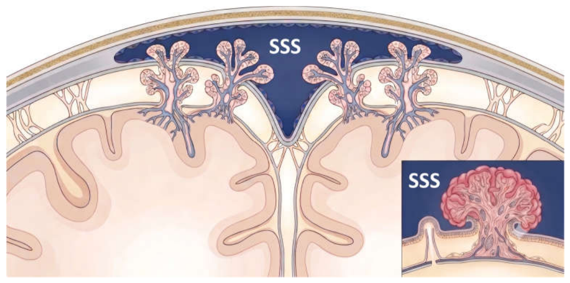
Figure 2.
The relationship between cerebrospinal fluid (CSF) flow and outflow pressure, as originally illustrated by Cutler et al. (Figure 4), is schematically reproduced here using the ggplot2 package in R to emphasize key physiological concepts. The figure demonstrates that CSF formation (in blue) remains relatively constant ( ) and is largely independent of intracranial pressure (ICP), whereas CSF absorption (in red) increases linearly once a threshold pressure is exceeded (). At low pressures below this threshold, absorption is zero; beyond it (marked by the red intersection point at ), absorption rises proportionally with increasing pressure. The intersection of the formation and absorption curves defines the equilibrium ICP, at which CSF production equals absorption. This linear pressure–flow relationship provides the physiological basis for the concept of resistance to CSF outflow and underpins modern infusion-based techniques for assessing CSF dynamics.
Figure 2.
The relationship between cerebrospinal fluid (CSF) flow and outflow pressure, as originally illustrated by Cutler et al. (Figure 4), is schematically reproduced here using the ggplot2 package in R to emphasize key physiological concepts. The figure demonstrates that CSF formation (in blue) remains relatively constant ( ) and is largely independent of intracranial pressure (ICP), whereas CSF absorption (in red) increases linearly once a threshold pressure is exceeded (). At low pressures below this threshold, absorption is zero; beyond it (marked by the red intersection point at ), absorption rises proportionally with increasing pressure. The intersection of the formation and absorption curves defines the equilibrium ICP, at which CSF production equals absorption. This linear pressure–flow relationship provides the physiological basis for the concept of resistance to CSF outflow and underpins modern infusion-based techniques for assessing CSF dynamics.
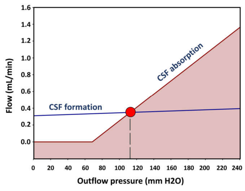
Figure 3.
Marmarou’s bolus test. A total volume of 8 mL of normal saline was injected at an approximate rate of 1 mL/s. The graph illustrates the baseline intracranial pressure (P₀), the peak pressure immediately after bolus injection (Pp), and the pressure measured 2 minutes later (Pt). This test provides a rapid assessment of intracranial compliance (C) and resistance to cerebrospinal fluid outflow (Rout). In this example, the calculated values were a pressure-volume index (PVI) of 16.2 mL, a compliance of 0.9 mL/mmHg, an Rout of 9.1 mmHg·min/mL, and a CSF outflow conductance (Cout) of 0.110 mL/min/mmHg.
Figure 3.
Marmarou’s bolus test. A total volume of 8 mL of normal saline was injected at an approximate rate of 1 mL/s. The graph illustrates the baseline intracranial pressure (P₀), the peak pressure immediately after bolus injection (Pp), and the pressure measured 2 minutes later (Pt). This test provides a rapid assessment of intracranial compliance (C) and resistance to cerebrospinal fluid outflow (Rout). In this example, the calculated values were a pressure-volume index (PVI) of 16.2 mL, a compliance of 0.9 mL/mmHg, an Rout of 9.1 mmHg·min/mL, and a CSF outflow conductance (Cout) of 0.110 mL/min/mmHg.
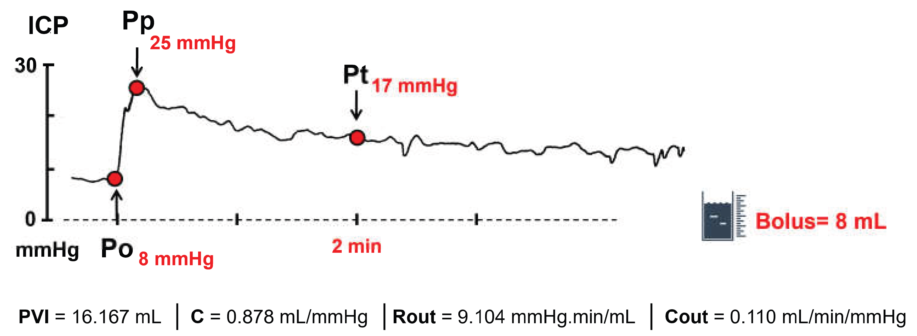
Figure 4.
Katzman infusion test performed using a continuous infusion of Ringer’s lactate at 1.65 mL/min. Baseline intracranial pressure (ICP) was 18 mmHg (Po), and the mean plateau pressure was 52 mmHg (Pp). The total infusion period extended from 13:30 to 14:10. Segment A shows the phase of progressive increase in ICP, whereas segment B corresponds to the plateau phase. The resistance to cerebrospinal fluid outflow (Rout) was 20.2 mmHg·min/mL, and the corresponding conductance to outflow (Cout = 1/ Rout ) was 0.049 mL/min/mmHg.
Figure 4.
Katzman infusion test performed using a continuous infusion of Ringer’s lactate at 1.65 mL/min. Baseline intracranial pressure (ICP) was 18 mmHg (Po), and the mean plateau pressure was 52 mmHg (Pp). The total infusion period extended from 13:30 to 14:10. Segment A shows the phase of progressive increase in ICP, whereas segment B corresponds to the plateau phase. The resistance to cerebrospinal fluid outflow (Rout) was 20.2 mmHg·min/mL, and the corresponding conductance to outflow (Cout = 1/ Rout ) was 0.049 mL/min/mmHg.
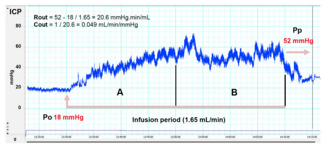

Table 1.
Simulated 2 × 2 contingency table illustrating the calculation of diagnostic accuracy metrics for an Rout threshold of 12 mmHg·min/mL. In this hypothetical cohort of 130 patients undergoing shunt surgery, 97 patients experienced postoperative improvement and 33 did not. The selected threshold yielded a sensitivity of 92.8% (90/97) and a specificity of 69.7% (23/33). Based on these values, the positive likelihood ratio (LR+) was calculated as 0.93/(1 − 0.70) = 3.1. An LR+ of 3.1 indicates that a positive test result (Rout > 12 mmHg·min/mL) is approximately three times more likely to occur in a patient who will improve after shunt surgery than in a patient who will not. This represents only a modest increase in the probability of postoperative improvement and falls well below the LR+ values generally considered strong evidence for ruling in a diagnosis or predicting treatment response.
Table 1.
Simulated 2 × 2 contingency table illustrating the calculation of diagnostic accuracy metrics for an Rout threshold of 12 mmHg·min/mL. In this hypothetical cohort of 130 patients undergoing shunt surgery, 97 patients experienced postoperative improvement and 33 did not. The selected threshold yielded a sensitivity of 92.8% (90/97) and a specificity of 69.7% (23/33). Based on these values, the positive likelihood ratio (LR+) was calculated as 0.93/(1 − 0.70) = 3.1. An LR+ of 3.1 indicates that a positive test result (Rout > 12 mmHg·min/mL) is approximately three times more likely to occur in a patient who will improve after shunt surgery than in a patient who will not. This represents only a modest increase in the probability of postoperative improvement and falls well below the LR+ values generally considered strong evidence for ruling in a diagnosis or predicting treatment response.
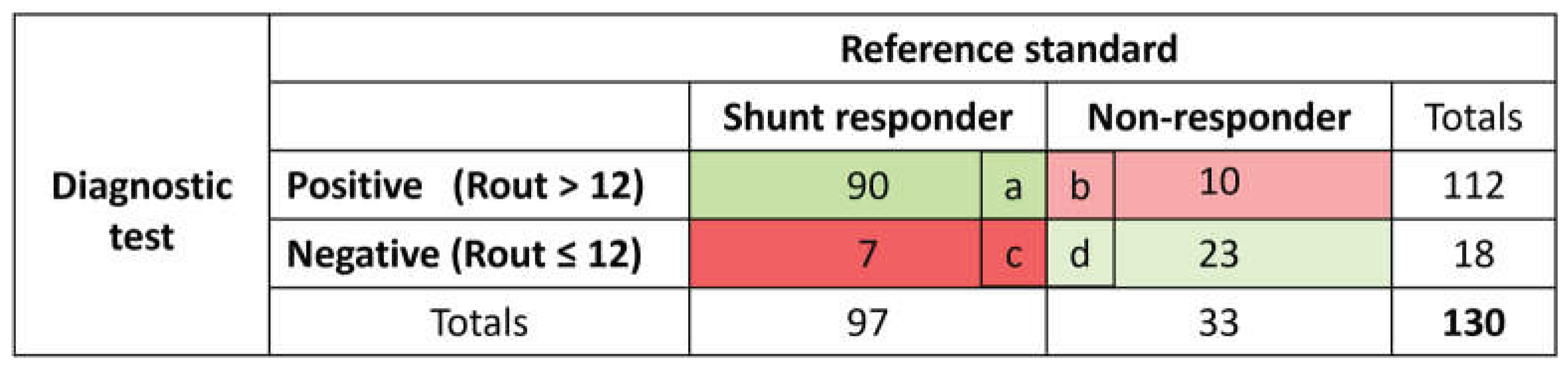 |
Table 2.
Diagnostic performance of the most commonly used Rout thresholds reported in the two published meta-analyses of lumbar infusion testing (LIT) in idiopathic normal-pressure hydrocephalus (iNPH). Sensitivity (S), specificity (Sp), and the corresponding positive (LR+) and negative (LR−) likelihood ratios are presented. Because neither meta-analysis reported likelihood ratios, LR+ and LR− were calculated from the published sensitivity and specificity estimates. These values are included to facilitate Bayesian interpretation of the proposed Rout thresholds and to illustrate the extent to which LIT results modify the probability of shunt responsiveness. Sensitivity and specificity values are reproduced from the original meta-analyses; only the corresponding likelihood ratios were derived.
Table 2.
Diagnostic performance of the most commonly used Rout thresholds reported in the two published meta-analyses of lumbar infusion testing (LIT) in idiopathic normal-pressure hydrocephalus (iNPH). Sensitivity (S), specificity (Sp), and the corresponding positive (LR+) and negative (LR−) likelihood ratios are presented. Because neither meta-analysis reported likelihood ratios, LR+ and LR− were calculated from the published sensitivity and specificity estimates. These values are included to facilitate Bayesian interpretation of the proposed Rout thresholds and to illustrate the extent to which LIT results modify the probability of shunt responsiveness. Sensitivity and specificity values are reproduced from the original meta-analyses; only the corresponding likelihood ratios were derived.
| Author | Rout | S | Sp | LR(+) | LR(-) |
|---|---|---|---|---|---|
| Kim 2015 | 10 | 91.6 | 20.7 | 1.15 | 0.41 |
| 12 | 80.3 | 46.8 | 1.51 | 0.42 | |
| 15 | 64.3 | 62.0 | 1.69 | 0.58 | |
| 18 | 42.7 | 75.2 | 1.72 | 0.76 | |
| van Bilsen 2025 | 12 | 76.9 | 34.0 | 1.17 | 0.68 |
| 14 | 81.6 | 37.2 | 1.30 | 0.49 | |
| 18 | 36.6 | 78.0 | 1.66 | 0.81 |
Disclaimer/Publisher’s Note: The statements, opinions and data contained in all publications are solely those of the individual author(s) and contributor(s) and not of MDPI and/or the editor(s). MDPI and/or the editor(s) disclaim responsibility for any injury to people or property resulting from any ideas, methods, instructions or products referred to in the content. |
© 2026 by the authors. Licensee MDPI, Basel, Switzerland. This article is an open access article distributed under the terms and conditions of the Creative Commons Attribution (CC BY) license (http://creativecommons.org/licenses/by/4.0/).
Copyright: This open access article is published under a Creative Commons CC BY 4.0 license, which permit the free download, distribution, and reuse, provided that the author and preprint are cited in any reuse.